giant perimedullary arteriovenous fistulas of the spine ...giant perimedullary arteriovenous...
TRANSCRIPT

Giant Perimedullary Arteriovenous Fistulas of the Spine: Clinical andRadiologic Features and Endovascular Treatment
F. Ricolfi, P. Y. Gobin, A. Aymard, F. Brunelle, A. Gaston, and J. J. Merland
PURPOSE: To present the clinical and radiologic features of giant perimedullary arteriovenousfistulas (GAVFs) in 12 consecutive cases and to evaluate the results of endovascular treatment.METHODS: We retrospectively reviewed the clinical and radiologic data as well as the results ofballoon endovascular treatment obtained from 1980 to 1989. RESULTS: GAVFs, defined as largeintradural perimedullary direct arteriovenous high-flow shunts, are revealed mainly in childhoodeither by subarachnoid hemorrhage or by progressive neurologic disorders. MR imaging andmyelography show major vascular dilatations. The angioarchitecture of GAVFs can only beassessed by selective spinal angiography. Ten patients were treated by balloon occlusion resultingin eight anatomic cures and six good clinical results. One balloon migrated to the venous side,leading to clinical deterioration. CONCLUSION: GAVF is a special subgroup of intradural peri-medullary arteriovenous fistula. The endovascular approach should be the first line of treatment,with surgery reserved for special circumstances. Nondetachable balloon occlusion is a safe andefficient method for treating GAVFs.
Index terms: Fistula, arteriovenous; Fistula, therapeutic blockade; Spinal cord
AJNR Am J Neuroradiol 18:677–687, April 1997
Giant perimedullary arteriovenous fistula(GAVF) is a rare intraspinal vascular malforma-tion that consists of a large direct arteriovenousshunt between anterior and/or posterior spinalarteries and a spinal vein. The shunt is intra-dural and lies on the surface of the spinal cord(1, 2). Selective spinal cord angiography en-ables this lesion to be distinguished from other,more common dural or intradural vascular mal-formations. These GAVFs are usually revealedin childhood by subarachnoid hemorrhage or bya progressive neurologic deficit. We report theclinical and radiologic findings of 12 consecu-tive cases of GAVFs and present the outcome ofendovascular treatment. GAVF, with its specific
Received April 4, 1995; accepted after revision October 7, 1996.From the Service de Neuroradiologie, Hopital Henri Mondor, Creteil
(F.R., A.G.), the Service de Neuroradiologie Interventionnelle, Hopital Lari-boisiere, Paris (P.Y.G., A.A., J.J.M.), and the Service de Radiologie, HopitalNecker Enfants Malades, Paris (F.B.), France.
Address reprint requests to Frederic Ricolfi, MD, Service de Neuroradi-ologie, Hopital Henri Mondor, 51, avenue du Marechal delattre de Tassigny,94010, Creteil, France.
AJNR 18:677–687, Apr 1997 0195-6108/97/1804–0677
© American Society of Neuroradiology
67
single-shunt pattern, is suitable for balloon oc-clusion.
Material and Methods
Patients
From 1980 to 1989, 12 patients with GAVFs were ad-mitted for examination and treatment. Two patients fromthis series (cases 3 and 4) have been the subjects ofprevious reports (3, 4). The data obtained from clinicalrecords, radiologic studies, and follow-up examinationswere available in 11 of the 12 cases.
Clinical data are summarized in Table 1. The first symp-toms appeared before the age of 10 years in eight patientsand before the age of 40 years in all cases. Clinical pre-sentation consisted of subarachnoid hemorrhage in fivecases (three isolated and two associated with regressiveparaplegia), sudden and regressive hemiplegia in onecase, progressive paraparesis or tetraparesis in threecases, sciatic pain in two cases, and isolated sphincterdisturbances in one case. The interval between initialsymptoms and treatment ranged from 1 week to 23 years(mean, 5 years). The clinical course consisted of progres-sive deterioration in nine cases and acute neurologic de-terioration in two cases. Three patients had an initial sub-arachnoid hemorrhage with normal findings at cerebralangiography followed by progressive paraparesis a few
7

678 RICOLFI AJNR: 18, April 1997
TABLE 1: Clinical findings in 12 patients with giant arteriovenous fistula (GAVF)
CaseAge, y, at Diagnosisand Treatment/Sex
Site ofGAVF
Initial Symptoms Clinical Course Symptoms at Treatment
1 2, 5/F C-5 Hemiplegia Progressive deterioration over 3 y Tetraparesis2 0.3, 5/F L-1 Regressive paraplegia Two episodes of regressive access Sphincter disturbance3 24, 43/M L-1 SAH Progressive paraparesis over 2 y Paraparesis4 19, 22/M T-11 SAH Progressive deterioration over 1 y Brown-Sequard paralysis
at L T-105 9, 10/M T-9 Paraparesis Progressive deterioration over 1 y Paraparesis6 5, 28/M T-8, L1 SAH with regressive
paraplegiaProgressive deterioration over 10 y Sciatic pain and
sphincter disturbance7 4, 5/M C-6 Paraparesis Progressive deterioration over 1 y Tetraparesis8 2, 4/M T-12 SAH Progressive deterioration over 1 y
and second SAHParaplegia
9 4, 6/M T-12 Sciatic pain Stepwise progression Cauda equina syndrome10 39, 39/F L-2 Sphincter disturbance Progressive over 6 mo Sphincter disturbance11 28, 33/F T-12 Sciatic pain Stable Sciatic pain12 5, 5/M L-1 SAH with paraplegia Regressive access Normal
Note.—SAH indicates subarachnoid hemorrhage.
years later. Two patients experienced deterioration of theirsymptoms during exertion. The upper limit of clinicalsymptoms (spinal and nerve root) corresponded to thelevel of the GAVF. The AVF was cervical in location in twocases, thoracic in one case, and at the level of the conusmedullaris in eight cases. One patient (case 6) had boththoracic and lumbar GAVFs. One patient (case 1) sufferedfrom metameric angiomatosis (Cobb syndrome) with adiffuse cutaneous high-flow arteriovenous malformation(AVM) of the upper limb and thorax associated with acervical GAVF. Another patient (case 6) had a familyhistory of Weber-Osler-Rendu disease. A spinal bruit waspresent in one patient (case 1).
Plain radiographs of the spine were obtained for allpatients. Myelography was performed in nine cases. Fivepatients had computed tomography (CT) and five patientshad magnetic resonance (MR) imaging. Since most of theCT and MR studies were performed in other hospitals, theexamination protocols were not standardized. Spinal an-giography was performed by a transfemoral approach, viaan arterial sheath, using local anesthesia and neurolept-analgesia in adults and general anesthesia in children.Thoracoabdominal aortography was initially performed infive patients. Selective spinal angiography was performedwith 5F catheters in adults and 3.6F catheters in children,with a distal end custom shaped with steam. Either ionic ornonionic contrast medium was injected. Selective injec-tions were done by hand. Angiograms were obtained eitheron conventional film in the earliest cases or with the use ofdigital equipment at an exposure rate of one frame persecond (prolonged for 25 seconds when abnormal vascu-larity was identified) for the more recent cases. Supineanteroposterior views were routinely obtained with addi-tional lateral views for radiculomedullary arteries.
Radiologic findings are summarized in Table 2. Plainradiographs showed enlargement of the spinal canal in fivecases (Fig 1). Large serpentine filling defects suggestive oflarge vessels were obvious at myelography. A partial
blockage of contrast material was observed in one case. Inanother case, the myelogram suggested a tumor, but anaccurate diagnosis (vascular enhancement) was obtainedwith CT and confirmed by spinal angiography. CT wasperformed in five patients (in three, after intravenous ad-ministration of contrast material and in two after intrathe-cal administration) and showed large serpentine intraduralvascular structures. MR imaging was performed in fivepatients, and the vascular abnormalities were always seenas large serpentine signal voids (Fig 2A and Fig 3A). Inthree cases a venous aneurysm compressed the cord (Fig2A and Fig 3A). The spinal cord signal was normal in threeof the five patients examined with MR imaging. In one ofthe remaining cases, MR images revealed a hemorrhagicarea surrounding an intramedullary venous aneurysm(case 9) (Fig 2A). In the remaining case (case 12), anintramedullary low-intensity signal on a T1-weighted im-age above the level of the GAVF was associated with aslight enlargement of the spinal cord (Fig 3A).
Myelography, CT, and MR imaging are all able to showspinal vascular lesions, but only spinal angiography canaccurately depict a GAVF by showing the high-flow shuntand the absence of interposed nidus between the arterialand venous sides of the vascular lesion (Fig 1A and B; Fig2; and Fig 3B and C). Initial thoracoabdominal aortogra-phy identified the multiple arterial feeders of the GAVF infive cases, and in one patient (case 1) a metameric lesionwas injected. Superselective angiography was required toidentify the various arterial feeders, their size and tortuos-ity, the flow, the exact location of the shunt and its mor-phology, and the venous drainage. The selective angio-grams guided the choice of the best approach forembolization of the malformation. Owing to the large shuntand dilution of contrast material by neighboring spinalarteries and collaterals, the side of the shunt could notalways be clearly demonstrated until balloon test occlu-sion was performed (Fig 2B and C). The main arterialsupply was derived from the anterior spinal artery in eight

AJNR: 18, April 1997 ARTERIOVENOUS FISTULAS 679
TABLE 2: Radiologic findings in 12 patients with giant arteriovenous fistula
Case Radiography Myelography CT MR Imaging Angiography: Arteries‡ Angiography: Veins
1 Cervical spinal canalwidening
. . . Whole cervical spinalcanalenhancement*
. . . L C4-C6 ASA (L vertebralartery)
Huge fistula venousaneurysm
2 Normal Large vessels . . . . . . L L-2 ASA; L T-9 ASA; RT-12 ASA
Fistula venousaneurysm
3 Normal Large vessels . . . . . . R T-10 ASA; L L-1 ASA;R and L L-2 PSA
. . .
4 Normal Large vessels . . . . . . L T-11 PSA; R T-11 PSA;R T-12 ASA
. . .
5 Normal Large vessels . . . . . . R T-9 PSA; L T-6 ASA; RT-12 PSA
. . .
6 L1-2 spinal canalwidening
Large vessels Intradural largevesselenhancement*
Large vessels; T-8compressivevenousaneurysm
L T-10 ASA; R T-9 PSA;R T-12 PSA
Huge distant venousaneurysm
7 Cervical spinal canalwidening
Partial block Whole cervical spinalcanalenhancement*
. . . L C-5 ASA; L C-1 ASA; RT-3 ASA
Huge fistula venousaneurysm
8 T11-12 spinal canalwidening
Large vessels Dilated perimedullaryvessels†
. . . L T-9 ASA Fistula venousaneurysm
9 T11-L2 spinal canalwidening
. . . . . . Large vessels; T-12 venousaneurysm withlocalhemorrhage
R T-11 PSA; L T-9 and RL-1 ASA; L T-12 and RL-3 PSA
Distant T-12 hugevenous aneurysm
10 Normal . . . . . . Large vessels L T-9 ASA; L T-10; R T-11; L T-12; L L-2; L L-3PSA
. . .
11 Normal Large vessels Dilated perimedullaryvessels†
Large vessels L L-1 ASA; L T-9 ASA; LT-11 PSA
. . .
12 Normal Large vessels . . . Large vessels; T-5compressivevenousaneurysm;spinal cord T2hypersignal
L L-1 PSA; R T-12 and RT-4 ASA; R L-2; L T-12;R and L T-7, L T-9 PSA
Huge distant T-5venous aneurysmposterior fossavenous drainage
* Intravenous contrast-enhanced CT scan.† Postmyelography CT scan.‡ ASA indicates anterior spinal artery; PSA, posterior spinal artery; boldface indicates main arterial feeder.
cases (Fig 4A) and from the posterior spinal artery in fivecases (Fig 2B). Venous drainage was regional (less thanfive metameric levels above or beneath the shunt) in 11cases (ascending, descending, or mixed). In case 12 (co-nus GAVF draining to the posterior fossa via an anteriorspinal vein), the venous drainage resembled that of lowerflow perimedullary AVFs and of dural AVFs (Fig 3). Thedraining veins were severely dilated (“giant” AVF). A ve-nous aneurysm with the radiologic features of an intraspi-nal space-occupying lesion was present in seven cases.The venous aneurysm was located at the AVF venous sitein four cases (Fig 1) and distant from the shunt in threecases (Fig 3).
Endovascular Treatment
All adult embolization procedures were performed us-ing local anesthesia and neuroleptanalgesia with frequent
testing of motor and sensory functions. Children youngerthan 12 years were treated while under general anesthesia.In case 12, somatosensory evoked cortical potentials weremonitored during treatment. Except for two patients (cas-es 1 and 2), who were treated at the beginning of our serieswith gelatin sponge particles, all patients were treated byballoon occlusion.
A double femoral artery puncture was performed in allpatients except one (case 7), in whom an axillary puncturewas performed. One vascular approach was used to intro-duce the balloon and the other was used to control theother arterial feeders. Latex balloons, inflated with contrastagent, were used in all cases (Figs 1C, 2D, 3D, and 4B). Incases 3 and 4, a detachable balloon was used with aninternal valve attached to the tip of a polytef catheter(outer diameter, 0.5 mm; inner diameter, 0.3 mm) andwas positioned through a 7F guiding catheter. In all thefollowing cases, the balloon delivery system consisted of a

680 RICOLFI AJNR: 18, April 1997
Fig 1. Case 7: 5-year-old boy with progressive tetraparesis.A, Anteroposterior view of right vertebral artery angiogram (head turned to left) shows fenestrated anterior spinal artery displaced to
the right side of the spinal canal. The shunt (asterisk) fills directly a huge venous aneurysm.B, Lateral view of right deep cervical artery angiogram before treatment.C, One-month follow-up control angiogram of right deep cervical artery shows that the shunt is occluded by a nondetachable balloon
(via a deep cervical artery) and the anterior spinal artery is reduced in size. Note the enlarged spinal canal.
Fig 2. Case 9: 6-year-old boy with a stepwise progression of a cauda equina syndrome.A, Sagittal T1-weighted MR image shows a vascular aneurysm (star) surrounded by a rim of hyperintense signal, attributed to
subacute hemorrhage (black arrow), which is compressing the conus medullaris. Note the intradural large vascular signal voids (whitearrows).
B and C, Selective angiograms of right T-11 (B) posterior spinal artery and right L-1 anterior spinal artery (C). The shunt is markedby an enlargement of the vessel (arrowheads). All the afferent arteries join this shunt by the perimedullary arterial network. Note thedilution of contrast material and/or an enlargement of the artery when two arterial feeders join in a common trunk before the shunt (blackarrow).
D, Occlusion of the GAVF by a nondetachable balloon (star) floated through the main arterial feeder (right T-11 posterior spinalartery) with a good clinical result. Good stability of the balloon is obtained on the venous side of the shunt (arrowheads).
E, Sagittal T1-weighted MR image 3 months after treatment shows that the venous aneurysm and large vessels have disappeared. Asmall hypointense signal at the posterior surface of the conus remains.

AJNR: 18, April 1997 ARTERIOVENOUS FISTULAS 681
Fig 3. Case 12: 5-year-old boy withsubarachnoid hemorrhage and progres-sive paraplegia.
A, Sagittal T1-weighted MR imageshows large intradural vascular signalvoids with an anterior intramedullary vas-cular aneurysm at T4-5 level. Note thecentral hypointense signal of the thoracicspinal cord.
B and C, Selective angiograms of leftT-12 (B) posterior spinal artery and rightT-12 anterior spinal artery (C). The shuntsitu (long arrow) is marked by focal vas-cular ectasia (arrowheads). Two large ar-teries meet at this point: left L-1 posteriorspinal artery (not shown) and a commontrunk fed by right T-12 anterior spinal ar-tery and left T-12 posterior spinal artery(star). Note the anterior ascending venousdrainage (short arrow), which reaches theposterior fossa (not shown).
D, Balloon floated into the shuntthrough left L-1 posterior spinal artery and inflated. Note stagnation of con-trast material caused by occlusion of the AVF.
E, Control sagittal T1-weighted MR image 1 week after treatment showshyperintense signal of the thrombosed T4-5 venous aneurysm and metallicartifact related to the balloon. The spinal cord is reduced in size comparedwith that on the pretreatment MR image, with persistent hypointense signal ofthe central cord.
coaxial 1F polyethylene catheter inside a 3F polyethylenecatheter (5, 6). The balloon and flexible 1F catheter werecarried by blood flow to the fistula. Once the balloon wasinflated, complete occlusion of the shunt was confirmed byangiograms obtained via the control catheter (Fig 3D).Balloons were either detachable (cases 5 and 6) or non-detachable (cases 7 through 12). In the latter case, the endof the polyethylene 1F catheter was enlarged by steamand introduced into the lumen of the balloon. Once theneck of the balloon was fixed well around the catheter, ashort piece of supple tube, forming a cuff over the neck,secured the balloon. After the nondetachable balloon waspositioned, the proximal end of the catheter was tied intoseveral tight knots, the introducer system was withdrawnover the knots, and the knotted catheter was implanted inthe subcutaneous tissues near the arterial puncture siteduring the same session. (At the beginning of our experi-ence, in case 7, we left the catheter exposed at the axillafor 48 hours for the purpose of deflating the balloon in caseof clinical worsening due to thrombosis. Infection at the
site of the puncture was complicated by septicemia lead-ing to removal of the balloon system. This patient wassuccessfully treated in a later session). The space betweenthe two coaxial catheters is small enough to prevent bloodreflux, thereby making perfusion unnecessary. No antico-agulation therapy was used during or after the procedure.
Clinical follow-up was performed 1 week, 1 month, andevery year after treatment. Position and inflation of theballoon was monitored by spinal plain films until its disap-pearance and deflation. Angiographic follow-up was per-formed at 1 week and 1 year after treatment. The fistulawas considered to be occluded when both shunt and ve-nous drainage were no longer opacified by the arterialfeeders and collaterals and when the spinal vessels hadreturned to their normal size (Fig 1C).
The clinical status of the patients before and after treat-ment was classified according to the following criteria: goodmeant the patient was independently ambulatory, with norestricted activity; fair indicated the need for crutches or acane to walk, with occasional urinary incontinence or reten-
682 RICOLFI AJNR: 18, April 1997
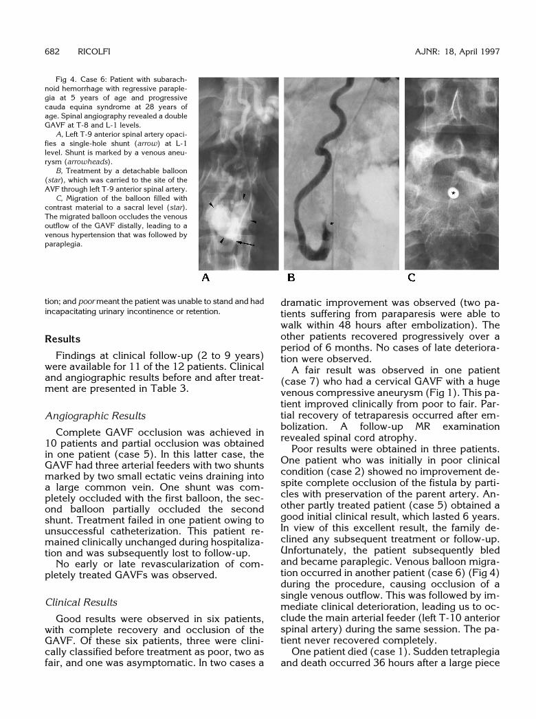
Fig 4. Case 6: Patient with subarach-noid hemorrhage with regressive paraple-gia at 5 years of age and progressivecauda equina syndrome at 28 years ofage. Spinal angiography revealed a doubleGAVF at T-8 and L-1 levels.
A, Left T-9 anterior spinal artery opaci-fies a single-hole shunt (arrow) at L-1level. Shunt is marked by a venous aneu-rysm (arrowheads).
B, Treatment by a detachable balloon(star), which was carried to the site of theAVF through left T-9 anterior spinal artery.
C, Migration of the balloon filled withcontrast material to a sacral level (star).The migrated balloon occludes the venousoutflow of the GAVF distally, leading to avenous hypertension that was followed byparaplegia.
tion; and poor meant the patient was unable to stand and hadincapacitating urinary incontinence or retention.
Results
Findings at clinical follow-up (2 to 9 years)were available for 11 of the 12 patients. Clinicaland angiographic results before and after treat-ment are presented in Table 3.
Angiographic Results
Complete GAVF occlusion was achieved in10 patients and partial occlusion was obtainedin one patient (case 5). In this latter case, theGAVF had three arterial feeders with two shuntsmarked by two small ectatic veins draining intoa large common vein. One shunt was com-pletely occluded with the first balloon, the sec-ond balloon partially occluded the secondshunt. Treatment failed in one patient owing tounsuccessful catheterization. This patient re-mained clinically unchanged during hospitaliza-tion and was subsequently lost to follow-up.
No early or late revascularization of com-pletely treated GAVFs was observed.
Clinical Results
Good results were observed in six patients,with complete recovery and occlusion of theGAVF. Of these six patients, three were clini-cally classified before treatment as poor, two asfair, and one was asymptomatic. In two cases a
dramatic improvement was observed (two pa-tients suffering from paraparesis were able towalk within 48 hours after embolization). Theother patients recovered progressively over aperiod of 6 months. No cases of late deteriora-tion were observed.
A fair result was observed in one patient(case 7) who had a cervical GAVF with a hugevenous compressive aneurysm (Fig 1). This pa-tient improved clinically from poor to fair. Par-tial recovery of tetraparesis occurred after em-bolization. A follow-up MR examinationrevealed spinal cord atrophy.
Poor results were obtained in three patients.One patient who was initially in poor clinicalcondition (case 2) showed no improvement de-spite complete occlusion of the fistula by parti-cles with preservation of the parent artery. An-other partly treated patient (case 5) obtained agood initial clinical result, which lasted 6 years.In view of this excellent result, the family de-clined any subsequent treatment or follow-up.Unfortunately, the patient subsequently bledand became paraplegic. Venous balloon migra-tion occurred in another patient (case 6) (Fig 4)during the procedure, causing occlusion of asingle venous outflow. This was followed by im-mediate clinical deterioration, leading us to oc-clude the main arterial feeder (left T-10 anteriorspinal artery) during the same session. The pa-tient never recovered completely.
One patient died (case 1). Sudden tetraplegiaand death occurred 36 hours after a large piece

AJNR: 18, April 1997 ARTERIOVENOUS FISTULAS 683
TABLE 3: Results of endovascular treatment in 12 patients with giant arteriovenous fistula
CaseClinical Score
beforeTreatment*
Endovascular Procedure† AngiographicResult
Clinical Score afterTreatment*
Follow-up,y‡
1 Poor Particles (L vertebral ASA) Cured Death (2 d later) . . .
. . .2 Poor Particles (L L-2 ASA) Cured Poor; progressive
deteriorationA:2
C:43 Poor Detachable balloon (L T-10 ASA) Cured Good A:0.5
C:24 Fair Detachable balloon (L T-11 PSA) Cured Good A:1
C:95 Poor Detachable balloon (R T-9 PSA; R
T-12 PSA)Partial Poor; initially good: SAH
and paraplegia at 6 yA:0.4
C:86 Fair First procedure: detachable balloon
(L T-10 ASA); second procedure:detachable balloon (R T-9 PSA)
Cured Poor (balloon migration) A:1
C:27 Poor First procedure: nondetachable
balloon (L C-5 ASA); secondprocedure: nondetachable balloon
Cured Fair (sepsis, spinal cordatrophy)
A:0.3
C:88 Poor Nondetachable balloon (L T-9 PSA) Cured Good A:0.1
C:49 Poor Nondetachable balloon (L T-9 PSA) Cured Good A 0.1
C:510 Fair Nondetachable balloon (L T-9 ASA) Cured Good A:1
C:411 Good Nondetachable balloon (L L-1 ASA) Failure (spasm) Unchanged . . .
. . .
12 Good Nondetachable balloon (PSA) Cured Good A:1C:4
* Good indicates independent ambulant patient with no restriction of activity; Fair means the patient requires crutches or one stick to walk;Poor indicates the patient is unable to stand and has incapacitating urinary incontinence or retention.
† ASA indicates anterior spinal artery; PSA, posterior spinal artery.‡ A indicates control angiogram; C, clinical follow-up.
of gelatin sponge was used to occlude com-pletely the three radicular feeders of a cervicalGAVF with a huge venous pouch.
Discussion
Spinal AVMs are classified according to theiranatomic location and angioarchitecture(7–11). The three types of vascular malforma-tions of the spine may be distinguished by theirangiographic anatomy (12): AVFs character-ized by the direct communication betweenfewer than three arteries and a single vein, AVFscharacterized by several arteries feeding shuntsin the wall of a single vein, and AVFs charac-terized by multiple shunts (nidi) between arte-rioles and venules (AVMs). This distinction is ofmajor importance, since the endovasculartreatments are radically different. AVFs and
AVMs may be part of a more complex vascularmalformative syndrome, such as metamericCobb syndrome or disseminated Weber-Osler-Rendu disease.
Depending on the site of the arteriovenousanomalies (AVFs or AVMs), several anatomicgroups may be described. Dural AVFs with peri-medullary venous drainage are situated withinthe dura and are supplied by meningeal arteries.These slow-flow malformations were initiallydescribed by Kendall and Logue (13) and Mer-land et al (14). They are typically seen in maleadults and manifested by progressive myelop-athy with exacerbation of symptoms at physicalactivity. The myelopathy is due to venous hy-pertension of the spinal cord.
Epidural AVFs with or without perimedullaryvenous drainage may clinically resemble duralAVFs if there is slow flow with perimedullary

684 RICOLFI AJNR: 18, April 1997
venous drainage (15). They may present withhemorrhage in cases of high-flow reflux into theintradural veins.
Intradural AVMs (true AVMs) are supplied byradiculomedullary arteries and may be situatedpartially or totally within the spinal cord (subpi-al) or at the surface of the spinal cord (pial),where they may be either arteriolovenulous fis-tulas or AVFs. Intradural spinal AVMs are usu-ally diagnosed in young patients who are lessthan 30 years old and present with an acuteonset of symptoms and the presence of sub-arachnoid hemorrhage.
Perimedullary direct AVFs, initially describedby Djindjian et al (16), are defined by a singleshunt and have been classified into three typesaccording to flow (4, 7–9). Type I correspondsto a single, slow-flow direct AVF between a non-dilated anterior spinal artery and a spinal vein.This type can be easily misdiagnosed. It occursin young adults with progressive myelopathy.Type II is also a single direct AVF, with greaterflow than in type I and an ampullary dilatation ofthe venous side of the shunt. In both types I andII, the venous drainage is extensive, as in thedural AVF type. Type III, or giant, AVFs, whichare the subject of this article, are high-flowshunts with one or several large or giant feeders.Veins are severely dilated and ectasia or truevenous aneurysms are encountered either nearthe shunt or more distally. This group differsfrom the two other types by its high flow and itslocal venous drainage to the epidural space. Wefound 25 cases in the literature that satisfy thetype III perimedullary AVF criteria (17–27).
Pathogenesis
The congenital origin of GAVFs is suggestedby the following arguments. GAVFs are usuallydiscovered during childhood (mean age of di-agnosis in our cases, 11.5 years; in the litera-ture, 13.5 years) (Table 1). They may be asso-ciated with or be part of complex vascularmalformation syndromes as described in theliterature and as was seen in two of our patients(cases 1 and 6) (1, 18, 20–22, 26, 27). Theseangiodysplasias (Klippel-Trenaunay, Parkes-Weber, Cobb syndrome, Weber-Osler-Rendudisease) are known to be caused by early (3 to5 weeks) developmental abnormalities of theprimary vascular network (28, 29).
GAVFs may develop at all levels of the spinalcord with a higher prevalence at the conus med-
ullaris (see Table 1). This latter location has aphysiologically rich blood supply of the poste-rior neuropore during embryonic life corre-sponding to high mitotic and/or metabolic ac-tivity (30, 31). It is conceivable that this area ofrich blood supply may frequently be the site ofdysplasia, whether it be of genetic or teratoge-netic origin (32). This hypothesis may be gen-eralized to other sites of GAVFs (cervical andthoracic) in that the primary vascular network issituated on the anterior surface of the spinalcord and the GAVFs are usually located at theanterior surface of the spinal cord (33).
Pathophysiology of Clinical Signs
GAVFs are manifested by two major types ofsymptoms: subarachnoid hemorrhage and pro-gressive neurologic deficits. Subarachnoidhemorrhage was present in five (42%) of ourpatients and in 10 patients reported in the liter-ature) (1, 17, 18, 20, 22, 27). Only two of ourpatients had associated paraplegia without evi-dence of hematomyelia on MR images. Patho-logic or surgical data concerning the mecha-nism of bleeding are lacking, but the role ofvenous impairment due to high flow may besuggested. In case 9, MR images revealed ve-nous ectasia surrounded by a clot (Fig 2A),suggesting the site of venous rupture. Venousectasia may reflect not only the high flow of theshunt but also the relative insufficiency of ve-nous drainage. Moreover, arterial aneurysms(constituting possible hemorrhagic lesions)have not been reported in GAVFs. The role offlow in the development of hemorrhage in peri-medullary AVFs may be suggested by the factthat types I and II do not bleed (4, 9).
Progressive neurologic deficits (includingmyelopathy, radiculomyelopathy, and radicu-lopathy) were encountered in 57% of cases.Three mechanisms are commonly proposed toexplain these symptoms: steal syndrome, ve-nous ischemia, and nerve compression.
A steal syndrome is conceivable, sinceGAVFs are supplied by the anterior spinal ar-tery. However, because no method is availablefor measuring spinal cord blood flow, a stealsyndrome remains a matter of speculation. Inthe cases reported in the literature, acute occlu-sion of the GAVF was never followed by so-called normal arterial breakthrough, whichwould reflect vascular dysregulation due tochronic ischemia (34).

AJNR: 18, April 1997 ARTERIOVENOUS FISTULAS 685
Venous ischemia due to increased venouspressure may be a possibility, since venousdrainage may not always adapt to the amountof arterial inflow. No mention of cord edema orswelling is made in the single reported case withpathologic work-up (1). Only five of our pa-tients were studied by MR imaging. In four caseswe were unable to see any sign of spinal cordedema. Nevertheless, in one patient (case 12)who had a conus GAVF and an ascending ve-nous drainage into the posterior fossa, swellingof the thoracic spinal cord associated with acentral low signal intensity on T1-weighted im-ages was observed (Fig 3A). These findingsmay correspond to medullary edema as ob-served in dural fistulas with perimedullary ve-nous drainage. This hypothesis is further sup-ported by the fact that after treatment the spinalcord returned to its normal size (Fig 3E).
A direct cord or nerve root compression byhypertrophied vascular structures was clearlyevident in five of our cases and in four casesreported in the literature (18–20, 24). Regard-less of the mechanism proposed to explain theclinical symptoms, they are clearly related toflow and are dramatically improved after cor-rection of the shunt.
Treatment
Balloon occlusion of an AVF is suitable inthese lesions by virtue of the angioarchitectureof GAVFs with their single arteriovenous shunt.Balloon procedures seem to be safe and effi-cient regardless of AVF location. In our experi-ence, these balloons (filled with contrast mate-rial only) deflated between 1 month and 1 yearafter the procedure. No recurrence of the fistulaor pulmonary migration was observed, probablybecause of the thrombosis of the feeder and theinitial vein after AVF occlusion; thus, ballooninflation with a polymerizing material, which ismore complex and less safe, appears to be un-necessary. The advantages of balloon occlusionare that balloons are easily carried by the flow tothe fistula, even in very tortuous vessels, andthat they can be repositioned. Balloon occlusionwas achieved in 10 cases, effecting a technicalcure in eight cases.
We observed two complications, as follows.Venous occlusion due to (detachable) balloonmigration occurred in one patient. When theballoon was inflated, just before complete oc-clusion, the AVF flow caused the balloon to
detach. The balloon migrated into the venousdrainage and blocked the single venous outletat a sacral level (Fig 4). The patient sufferedacute back pain with paraplegia a few minuteslater. To reduce pressure in the AVF, we per-formed a more proximal occlusion of the arterialfeeders in the same session, with occlusion ofthe anterior spinal artery. This complication ledus to use a nondetachable balloon in subse-quent procedures. In another patient, septice-mia occurred following infection from the cath-eter, which was left in place too long. Novascular trauma occurred as a result of ballooninflation; nevertheless, such a risk cannot becompletely excluded, as Halbach et al (27) de-scribed in a case of anterior spinal artery rup-ture due to inflation of a silicone balloon.
Neurologic impairment was not observed inany of the nine patients in whom acute and totalocclusion was performed. Acute occlusion of anAVF has been considered to be responsible foredematous and/or hemorrhagic complicationsassociated with dysautoregulation of the neuralarteries (normal blood pressure breakthrough)(34). Our impression is that successful treat-ment requires correct positioning of the balloonon the arterial side of the fistula; but in one ofour patients (case 9), stability of the ballooncould only be obtained on the venous side of theshunt.
In the days following balloon occlusion, threepatients with good results experienced moder-ate back pain and slight fever, which spontane-ously resolved after 3 days. These symptomswere attributed to thrombosis of the GAVF.
Particulate embolization was used in the firsttwo patients (before embolization balloons wereroutinely available) who had neurologic deteri-oration, suggesting extensive arterial obstruc-tion or venous thrombosis. In the autopsy casereported by Benhaiem-Sigaux et al (1), a mas-sive gelatin sponge pulmonary embolism wasthought to be the cause of death. Particulateembolic material is not adapted for use in largedirect fistulas because particles may passthrough the arteriovenous shunt.
In our experience, embolization using a flow-guided nondetachable balloon was safe and ef-fective, and no recurrence was observed. Theadvantage of a nondetachable balloon is that itassists in localizing the shunt and causes stag-nation of all the afferent arteries of the fistula.The balloon may be repositioned. Its nonde-tachability assures safety, and experience has

686 RICOLFI AJNR: 18, April 1997
shown that it has a good long-term tolerance. Ifocclusion of an AVF is performed with a detach-able balloon instead of our device, it is our opin-ion that a more proximal nondetachable balloonshould be used temporarily to stop or decreaseflow in order to prevent unwanted migration ofthe detached balloon and subsequent, poten-tially catastrophic, complications.
Alternative endovascular procedures havebeen developed, including more supple micro-catheters and microcoils. These methods arenot flow-dependent, but allow for progressivedeposit of embolic agents. Furthermore, thesemethods can be used in a venous approach.Halbach et al (27) reported a series of 10GAVFs (types II and III) in which the use ofmicrocoils in combination with particles (poly-vinyl alcohol or silk suture) was favored. Onepatient reported by Nakstad et al (26), in whomthe location of the shunt was clearly defined,was cured by microcoil embolization.
Nine patients were operated on before theintroduction of endovascular treatment (18–21,23–25). Surgery was considered to be difficultand hemorrhage occurred in two cases. Owingto the improvement of embolization techniques,we think that surgery should be proposed onlyafter an unsuccessful endovascular attempt, asreported by Heros et al (2) and Halbach et al(27). If a surgical approach is chosen, superse-lective identification of the shunt is required toallow safe and precise placement of the clip.Intraoperative spinal angiography enables veri-fication of the correct position of the clip.
In conclusion, GAVFs are rare conditions thatare more common in children than in adults.Spinal MR imaging facilitates diagnosis of theseintradural vascular malformations and visual-ization of medullary compression caused by avenous aneurysm. On the basis of our experi-ence and because of the angioarchitecture ofAVFs, we believe that endovascular intraarterialtreatment by nondetachable latex balloons is asafe and reliable method for occluding this typeof AVF. The transvenous approach, AVF coil-ing, or surgery may be indicated in instances ofunusual anatomic conditions or unsuccessfulballoon occlusion.
References1. Benhaiem-Sigaux N, Zerah M, Gherardi R, Bellot J, Hurth M,
Poirier J. A retromedullary arteriovenous fistula associated withthe Klippel-Trenaunay-Weber syndrome. Acta Neuropathol 1985;66:318–324
2. Heros RC, Debrun GM, Ojemann RG, Lasjaunias PL, Naessens PJ.Direct spinal arteriovenous fistula: a new type of spinal AVM.J Neurosurg 1986;64:134–139
3. Riche MC, Scialfa G, Gueguen B, Merland JJ. Giant extramedul-lary arteriovenous fistula supplied by the anterior spinal artery:treatment by detachable balloons. AJNR Am J Neuroradiol 1983;4:391–394
4. Gueguen B, Merland JJ, Riche MC, Rey A. Vascular malforma-tions of the spinal cord: intrathecal perimedullary arteriovenousfistulas fed by medullary arteries. Neurology 1987;37:969–979
5. Merland JJ, Rufenacht D, Guimaraens L, Riche MC. A polyethyl-ene microcatheter with a latex balloon as an implant for perma-nent vascular occlusions: a way for the treatment of distal intra-cranial or intraspinal large A. V. fistulas or aneurysms. In: Valk J,ed. Neuroradiology 1985/1986. International Congress series 698.New York, NY: Elsevier; 1986:299–300
6. Rufenacht DA, Merland JJ, Guimaraens L. A nondetachable latexballoon for temporary diagnostic or permanent therapeutic occlu-sions of cerebral vessels and assistance for intravascular manip-ulations during hyperselective catheterizations. In: Valk J, ed.Neuroradiology 1985/1986. International Congress series 698.New York, NY: Elsevier; 1986:295–298
7. Merland JJ, Reizine D. Malformations vasculaires vertebromedul-laires. In: Encycl Med Chir: Radiodiagnostic II. 5 ed. Paris, France:Techniques; 1987:31671 G10:1–21
8. Riche MC, Reizine D, Melki JP, Merland JJ. Classification of spinalcord vascular malformations. Radiat Med 1985;3:17–24
9. Casasco AE, Houdart E, Gobin YP, Aymard A, Guichard JP,Rufenacht DA. Embolization of spinal vascular malformations.Neuroimaging Clin N Am 1992;2:337–358
10. Symon L, Kuyama H, Kendall B. Dural arteriovenous malforma-tions of the spine: clinical features and surgical results in 55 cases.J Neurosurg 1984;60:238–247
11. Rosenblum B, Oldfield EH, Doppman JL, Di Chiro G. Spinalarteriovenous malformations: a comparison of dural arterio-venous fistulas and intradural AVM’s in 81 patients. J Neurosurg1987;67:795–802
12. Houdart E, Gobin YP, Casasco A, Aymard A, Herbreteau D, Mer-land JJ. A proposed angiographic classification of intracranialarteriovenous fistulae and malformations. Neuroradiology 1993;35:381–385
13. Kendall BE, Logue V. Spinal epidural angiomatous malformationsdraining into intrathecal veins. Neuroradiology 1977;13:181–189
14. Merland JJ, Riche MC, Chiras J. Intraspinal extramedullary artrio-venous fistulae draining into medullary veins. J Neuroradiol 1980;7:271–320
15. Cahan LD, Higashida RT, Halbach VV, Hieshima GB. Variants ofradiculomeningeal vascular malformations of the spine. J Neuro-surg 1987;66:333–337
16. Djindjian M, Djindjian R, Rey A, Hurth M, Houdart R. Intraduralextramedullary spinal arterio-venous malformations fed by theanterior spinal artery. Surg Neurol 1977;8:85–93
17. Picard L, Vert P, Renard M, Hepner H, Lepoire J. Aspects radio-anatomiques des angiomes medullaires. Neurochirurgie 1969;18:519–528
18. Djindjian R, Hurth M, Rey A, Houdart R. Spinal angiomas inRendu-Osler disease. J Neuroradiol 1974;1:289–350
19. Sutton T, Murray PJ, Alexander WJ, Blundell JE. Arteriovenousmalformations of the spinal cord in childhood. Radiology 1973;109:621–622
20. Kaplan P, Hollenberg RD, Frazer FC. A spinal arteriovenous mal-formation with hereditary cutaneous hemangiomas. Am J DisChild 1976;130:1329–1331

AJNR: 18, April 1997 ARTERIOVENOUS FISTULAS 687
21. Merry GS, Appleton DB. Spinal arterial malformation in a childwith hereditary hemorrhagic telengiectasia. J Neurosurg 1976;44:613–616
22. Djindjian M, Djindjian R, Hurth M, Rey A, Houdart E. Spinal cordarteriovenous malformations and Klipple-Trenaunay-Weber syn-drome. Surg Neurol 1977;8:229–237
23. Scarff TB, Reigel DH. Arteriovenous malformation of the spinalcord in children. Childs Brain 1979;5:341–351
24. Binder B, Eng GD, Milhorat TH, Galioto F. Spinal arteriovenousmalformations in an infant: unusual symptomatology an pathol-ogy. Dev Med Child Neurol 1982;24:380–385
25. Doppman JL, Di Chiro G, Dwyer AJ, Frank JL, Oldfield EH.Magnetic resonance imaging of spinal arteriovenous malforma-tions. J Neurosurg 1987;66:830–834
26. Nakstad PH, Hald JK, Bakke SJ. Multiple spinal arteriovenousfistulas in Klipple-Trenaunay-Weber syndrome treated with pla-tinium fibre coils. Neuroradiology 1993;35:163–165
27. Halbach VV, Higashida RT, Dowd CF, Fraser KW, Edwards MS,Barnwell SL. A treatment of giant intradural (perimedullary) arte-riovenous fistulas. Neurosurgery 1993;33:972–980
28. Andre JM. Les Dysplasies Vasculaires Systematisees. Paris,France: L’Expansion Scientifique Francaise; 1973:31–43
29. Dobbelaere P, Dhellemmes P, Bousquet C, Urbain JP, Michaux F.Metameric angiomatosis: a report of two cases. J Neuroradiol1978;5:225–235
30. Lemire RJ, Loeser JD, Leech RW, Alword EC. Secondary caudalneural tube formation. In: Normal and Abnormal Development ofthe Human Nervous System. Hagerstown, NJ: Harper & Row;1975:71–83
31. Moore KL. The nervous system. In: The Developing Human. 4thed. Philadelphia, Pa: Saunders; 1988:52–54, 365–390
32. Barkovich AJ, Raghavan N, Chuang S, Peck WW. The wedge-shaped cord terminus: a radiographic sign of caudal regression.AJNR Am J Neuroradiol 1989;10:1223–1231
33. Torr JBD. The embryological development of the anterior spinalartery in man. J Anat 1957;91:587
34. Halbach VV, Higashida RT, Hieshima GB, Norman D. Normalperfusion pressure breackthrough occurring during treatment ofcarotid and vertebral fistulas. AJNR Am J Neuroradiol 1987;8:751–756