treatment of vertebral arteriovenous fistulas
TRANSCRIPT
Van V. Halbach1 Randall T. Higashida
Grant B. Hieshima
This article appears in the November/December 1987 issue of AJNR and the February 1988 issue of AJR.
Received February 18, 1987; accepted after revision June 17, 1987.
, All authors: Departments of Radiology and Neurological Surgery, University of California, San Francisco Medical Center, 505 Parnassus Ave ., San Francisco, CA 94143. Address reprint requests to V. V. Halbach , Neuroradiology, C-309.
AJNR 8:1121-1128, November/December 1987 0195-6108/87/0806-1121 © American Society of Neuroradiology
Treatment of Vertebral Arteriovenous Fistulas
1121
Twenty patients with vertebral arteriovenous fistulas (eight spontaneous, six traumatic without vertebral artery transection, and six traumatic with vertebral artery transection) were treated by transvascular embolization techniques, resulting in complete fistula closure in all patients. The fistulas were located at C1-C2 in 45%, C2-C3 in 25%, C4-C5 in 15%, C5-C6 in 10%, and C6-C7 in 5%. Trauma was the most common cause: 30% followed knife wounds, 20% followed gunshot injuries, and 10% followed blunt trauma. Eight patients had spontaneous fistulas, two associated with fibromuscular dysplasia. Three patients-all with large, long-standing fistulas-developed neurologic deficits coincident with the abrupt closure of the fistula, which resolved with reestablishment of fistula flow. Two of these patients were treated by staged closure; the other one by gradual closure. In all three cases the result was complete fistula closure without neurologic sequelae. The remaining spontaneous fistulas were all closed by balloon embolization with preservation of the vertebral artery and without deficits. The six patients with traumatic fistulas without transection were cured by balloon embolization, without deficits; in four there was also preservation of vertebral flow. The other six patients had traumatic fistulas with transection and were all cured by balloon embolization with preservation of flow in two. Four patients required bilateral approaches to the fistula to achieve complete fistula closure. The only complication was a mild residual Wallenberg syndrome after occlusion of the posterior inferior cerebellar artery in the treatment of a transection located at C1.
In our opinion, transvascular techniques are the treatment of choice for vertebral arteriovenous fistulas.
Vertebral arteriovenous fistulas are abnormal connections between the extracranial vertebral artery or its branches and neighboring veins. They are uncommon lesions, with only 81 cases reported prior to 1977 [1]. The most common cause of these fistulas is penetrating neck injuries, usually knife wounds or gunshot injuries . Iatrogenic causes include the sequelae of direct puncture of the carotid or vertebral artery for diagnostic angiography [2, 3], inadvertent vertebral damage during the insertion of venous catheters [4 , 5] , or complications of anterior interbody fusion [6]. Diseases associated with vertebral arteriovenous fistulas include fibromuscular dysplasia [7-9] and neurofibromatosis [10-12]. Blunt trauma in association with cervical spine fracture has been reported as a cause [13, 14]. Many cases are spontaneous [15] and often thought to be congenital. Symptoms often related to the fistula are bruit and neck pain , but brain and spinal cord dysfunction related to steal , venous hypertension, or mechanical compression have also been reported [1, 16] . Traumatic fistulas can present with massive hemorrhage, neurologic deficits, expanding hematomas, pseudoaneurysms, and airway obstruction. Treatments have included proximal ligation , trapping procedures , direct surgical exposure and closure, combined balloon embolization and surgical ligation , and , more recently, embolization with balloons or steel coils [16-20] . Over the past 7 years we have treated 20 vertebral arteriovenous fistulas by transvascular embolization techniques, and we report the results of those treatments.
1122 HALBACH ET AL. AJNR:8, November/December 1987
Materials and Methods
Twenty patients with vertebral fistulas were treated by using transarterial embolization. All procedures were performed with local anesthesia and IV sedation to allow continuous neurologic monitoring. The femoral arterial access was used for all diagnostic and therapeutic procedures. A diagnostic angiogram with catheter tip located near the fistula was obtained via the involved vertebral artery, and rapid filming was used to delineate the site of the fistula, presence of arterial narrowing or damage, and draining venous structures. If antegrade flow up the involved vertebral artery above the fistula was not observed, then gentle advancement of a soft-tipped guidewire was attempted to place the diagnostic catheter above the level of the fistula. If successful , an angiogram was obtained with the catheter tip located just above the fistula site. Angiograms of the contralateral vertebral, ipsilateral carotid, thyrocervical , and costocervical arteries were obtained as indicated. If continuity of the artery was maintained (no transection) then a 7.3/5-French coaxial catheter system was placed in the artery with the safest access to the fistula, usually the involved vertebral artery. SystemiC anticoagulation with heparin, usually 5000 units bolus and 2500 units/hr in a 70-kg patient, was given unless contraindicated . If the patient had a pseudoaneurysm or expanding hematoma, then anticoagulation was not used. The inner 5-French catheter was removed and a 4/2-French coaxial polyethylene catheter system with a silicone detachable balloon attached to the 2-French catheter was placed through the 7.3-French catheter. The space between the 4- and 7.3-French catheters was perfused with heparinized saline through a sidearm attachment. Injection of contrast material can also be made through this space during balloon placement. The balloon was advanced under fluoroscopic guidance into the fistula and inflated. If in satisfactory position , the balloon was detached by traction or coaxial technique. Attempts were made to preserve vertebral arterial flow if possible.
If vertebral transection was present, an attempt was made to place the catheter system into the distal transected segment via the ipsilateral vertebral artery.
If a vertebral transection was present and the diagnostic catheter could not be advanced into the distal vertebral artery, then a contralateral approach was initially used. The balloon was flow-directed up the contralateral vertebral artery across the junction of the vertebral arteries, down the distal ipsilateral vertebral artery, and to a point above the level of the fistula. If in satisfactory position, the balloon was then detached by gentle traction. The 7.3-French catheter was then placed in the ipsilateral vertebral artery and balloons were positioned in the fistula site and ipsilateral vertebral artery near the site of the fistula. After final detachment, a control angiogram was obtained from all potential collateral sources that could supply the fistula. If the fistula was occluded, the heparin anticoagulation was reversed with IV protamine sulfate (1 mg of protamine sulfate counteracts 100 units of circulating heparin) given slowly over 5 min. Follow-up angiograms were obtained if the patient had symptoms related to the fistula or if the control angiogram demonstrated subtotal occlusion. The patients were followed by the authors at 1-month, 6-month, and yearly intervals after treatment. The follow-up period was 5-72 months (average, 35 months).
The age and gender of each patient, location and cause of the fistula, symptoms, presence of vertebral transection , treatment, and complications are summarized in Table 1.
Results
Nineteen of 20 patients were treated by transarterial balloon embolization. In the other patient (case 12) the orifice of the fistula was too small to permit the balloon to enter the fistula; therefore, a balloon was transiently positioned in the vertebral artery above the fistula and silicone spheres were embolized behind (proximal to) the balloon into the fistula. All 20 patients had complete closure of their fistulas by transvascular treatments.
TABLE 1: Summary of Vertebral Fistulas in Patients Treated with Transvascular Embolization
Case Age Gender Site Cause Symptoms/Findings
Vertebral Artery Treatment No. Transection/Occlusion
1 61 F R C1 Spontaneous Bruit Occlusion Three balloons 2 23 F L C1 Spontaneous Weakness No Three balloons 3 29 F R C4-C5 Spontaneous Neck pain, radiculopathy No One balloon 4 27 M L C2-C3 Gunshot wound Hemorrhage Occlusion Three balloons 5 24 M R C1 Gunshot wound External hemorrhage Transection/occlusion Six balloons 6 20 M L C1 Blunt trauma Bruit, pain Transection Seven balloons 7 20 M R C5- C6 Spontaneous Subarachnoid hemorrhage No One balloon 8 64 F L C2-C3 Fibromuscular Bruit, pain No One balloon
dysplasia 9 35 M L C1 Spontaneous Bruit, pain No Two balloons
10 20 M R C1 Blunt trauma Bruit Transection/occlusion Ten balloons 11 30 M L C1 Spontaneous Exercise intolerance No Four balloons 12 26 F L C2-C3 Fibromuscular Pain , bruit, quadriparesis No Silas tic spheres
dysplasia 13 60 M R C6-C7 Gunshot wound Hematoma No One balloon 14 61 M R C4-C5 Stab wound Bruit No One balloon 15 25 M R C1 Gunshot wound Bruit Transection Six balloons 16 28 M LC2 Stab wound External hemorrhage Transection/occlusion Five balloons 17 21 M L C1 Stab wound External hemorrhage Transection/occlusion Three balloons 18 28 M L C2-C3 Stab wound External hemorrhage No Three balloons 19 26 M L C4-C5 Stab wound Pain , hematoma Occlusion Two balloons 20 29 M L C5- C6 Stab wound Radiculopathy, pain No Four balloons
Note.-Treatment resulted in fistula closure in all cases. There was only one complication, occlusion of the posterior inferior cerebellar artery in case 6. R = right ; L = left .
AJNR:8, November/December 1987 TREATMENT OF VERTEBRAL FISTULAS 1123
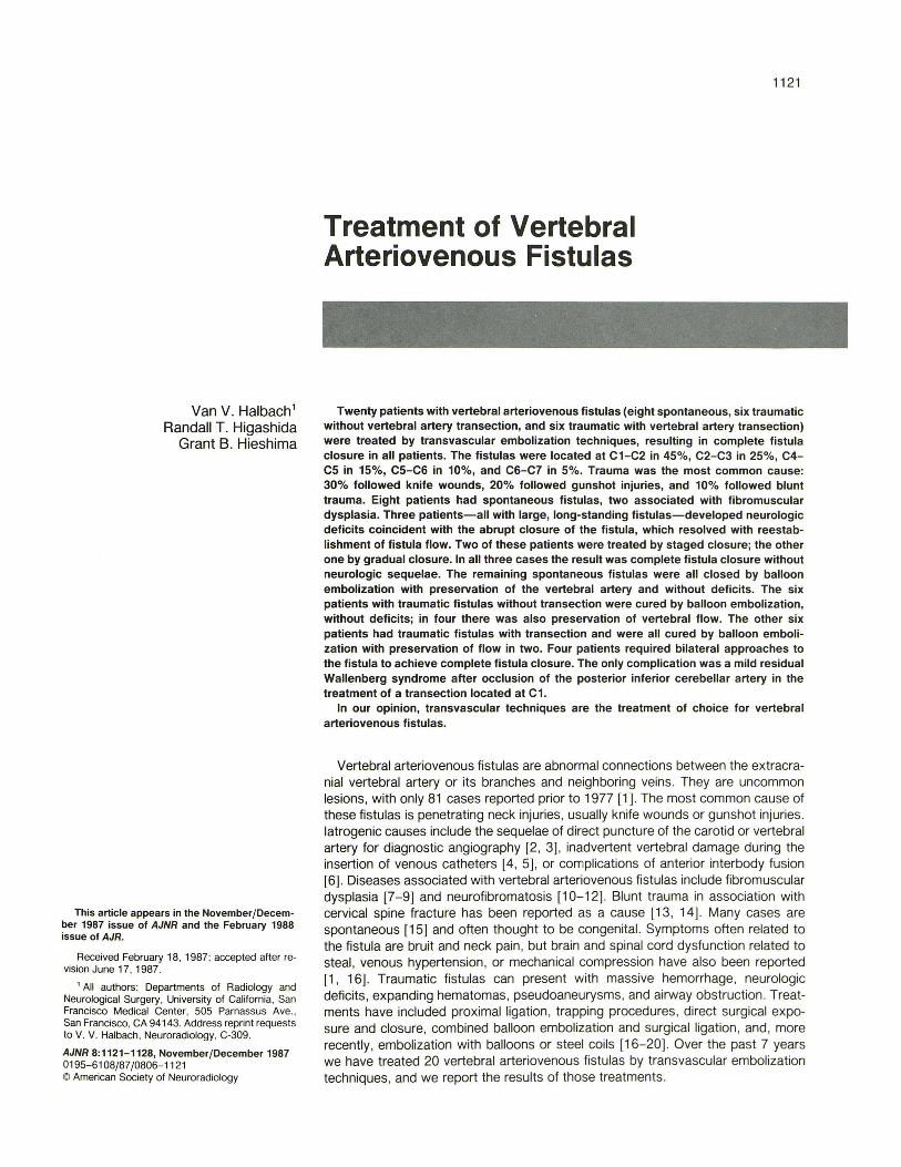
A B c Fig. 1.-Case 7: 20-year-old man with spontaneous vertebral fistula at C5-C6 who presented with massive subarachnoid hemorrhage. A, Anteroposterior view of right vertebral artery injection shows vertebral fistula with venous drainage to epidural venous plexus and medullary veins
(arrows). B, Selective radicular artery injection, oblique view, shows site of fistula with venous drainage to epidural venous plexus (sfraight arrows) and medullary
veins (curved arrow). C, Selective right vertebral artery injection, anteroposterior view after embolization, shows balloon in fistula with preservation of vertebral flow.
Spontaneous Fistulas
In spontaneous fistulas the diameter of the involved feeding arteries is often enlarged, presumedly secondary to longstanding fistula flow. Of the eight patients with spontaneous fistulas, three developed reversible neurologic deficits coincident with the abrupt closure of the fistula. With reestablishment of fistula flow the neurologic deficits promptly resolved. Fistula closure was staged in two patients, and in the third the occlusion was gradual over a 2-hr period . None sustained permanent neurologic sequelae.
In all eight patients, transarterial occlusive procedures resulted in complete closure of the fistula and preservation of flow in the involved vertebral artery. Case 7 (Fig. 1) was a 20-year-old man who had subarachnoid hemorrhage secondary to a fistula supplied by a radicular branch of the right vertebral artery with venous drainage to the epidural plexus and medullary veins. A single detachable balloon was positioned at the fistula with complete closure and no further episodes of hemorrhage. Case 1, with a large, high-flow fistula, developed delayed thrombosis in the large vertebral artery weeks after balloon embolization. This event was associated with mild pain, but without neurologic deficit, and was presumed to be secondary to the slow flow in the dilated vessel after closure.
In the two patients with fibromuscular dysplasia, complete closure was achieved without deficits.
Traumatic Fistulas Without Transection
In the six patients with traumatic fistulas without transection, the involved vertebral artery was preserved in four. In the other two patients narrowing and intimal damage in the involved artery prevented preservation of flow. All procedures were performed from the involved vertebral artery and none resulted in neurologic deficits.
Case 14 (Fig. 2) was a 51-year-old man who developed a right vertebral fistula after a penetrating trauma. A single balloon was positioned within the fistula and detached, resulting in complete closure of the fistula .
Traumatic Vertebral Fistulas with Transection
Six patients had complete transection of the vertebral artery. In four patients penetrating injuries were responsible : two knife wounds and two gunshot injuries. In the other two patients , blunt trauma was responsible with severe whiplash in one and fracture of C1 in the other. With complete transection there is retraction of proximal and distal ends of the severed artery away from the site of the injury. There is usually a pseudoaneurysm associated with the fistula. The walls of the pseudoaneurysm are made of clot and offer little structural support unless surrounded by firm structures such as bone or ligaments. Our earliest experience with balloon

1124 HALBACH ET AL. AJNR :8, November/December 1987
A B
placement within the pseudoaneurysms was that the balloons dissected into the walls of the pseudoaneurysm causing expansion and sometimes mass effect. In case 5 (Fig. 3) there was a vertebral transection with pseudoaneurysm at the level of C1 secondary to a gunshot injury. Several balloons were positioned within the pseudoaneurysm with enlargement of this structure and persistent fistula flow . To achieve closure of the fistula , balloons were positioned in both the proximal and distal severed vertebral artery segments and detached. Because the flow in the distal vertebral is retrograde into the fistula, yertebral artery and fistula occlusion were tolerated in all six patients who required vertebral artery occlusion.
Case 17 (Fig. 4) was a 21-year-old man who developed a left vertebral transection after a knife wound. Injection of the involved vertebral artery showed filling of a pseudoaneurysm at the level of C2 . Injection of the contralateral vertebral artery showed retrograde flow down the involved vertebral artery and entry into the same pseudoaneurysm at a higher level. Because of the discontinuity of the severed ends and retrograde flow down the distal left vertebral artery, treatment could not be rendered entirely from the involved side. Therefore , a balloon was flow-directed up the right vertebral artery and then down the distal left vertebral artery. It was detached above the fistula with preservation of flow in the posterior inferior cerebellar artery (PICA). Several more balloons were then detached in the distal left vertebral artery and in the site of the fistula , with complete closure of the fistula without deficits.
Case 10 (Fig. 5) was a 20-year-old man with transection at C1 with pseudoaneurysm formation. The distal vertebral artery could not be reached from an ipsilateral approach. There-
Fig, 2.-Case 14: 61-year-old man with neck injury.
A, Right vertebral artery injection, early arterial phase, shows fistula at C4-C5 with drainage to paravertebral veins.
8, Same vessel after embolization. A single balloon has been placed at fistula site with preservation of vertebral artery flow.
fore, a trapping procedure conSisting of contralateral followed by ipsilateral occlusion resulted in a complete cure. Note the acute angle between the two vertebral arteries, which required a sharp curve in the 2-French catheter to negotiate this bend. Despite the proximity of the PICA to the fistula site, flow in this vessel was preserved.
The only complication in our series occurred during treatment in case 6, when the PICA origin was occluded. In one of our first cases, previously reported [21 j, the balloon could not be pOSitioned below the PICA origin because of the proximity to the fistula. Although the patient tolerated test occlusion of the PICA for 20 min , he developed a delayed mild Wallenberg syndrome.
Djscussion
The long intraforaminal course and posterior location of the vertebral artery accounts for its rare involvement in penetrating injuries of the neck. Injuries to the short first segment, from its origin to its entry into the foramen transversarium (usually at C6) usually involve other vital structures and have a high morbidity. Most traumatic vertebral fistulas involve the long second portion (intraforaminal), whereas spontaneous fistulas usually involve the third portion (where the artery leaves the foramen of the atlas to where it enters the foramen magnum) [15]. Surgical approaches to these regions have been described but are not without morbidity [22-25]. The earliest attempts at surgical control have involved ligation of the involved vertebral artery [17]. This often has resulted in decreased flow to the fistula but it also could potentiate steal down to the distal vertebral artery and result in cerebral steal

AJNR :8. November/December 1987 TREATMENT OF VERTEBRAL FISTULAS 1125
Fig. 3.-Case 5: 24-year-old man. A, Right vertebral artery angiogram, oblique
view, shows fistula at C1 with pseudoaneurysm. B, Same vessel, lateral view, after partial em
bolization with persistent fistula filling and diversion of venous flow to paravertebral veins.
C, Left vertebral artery injection, Towne projection, with steal down distal right vertebral artery into fistula.
D, Left vertebral artery injection after embolization. Occlusion of fistula with preservation of posterior inferior cerebellar artery.
symptoms [26, 27]. Trapping procedures with ligation of the vertebral artery on either side of the fistula may be effective; however, collaterals may maintain fistula patency [28]. Surgical exposure of the fistula involving the second portion of the vertebral artery (intraforaminal) can be difficult. The surrounding arterialized Batson plexus can cause extensive hemorrhage during surgical exposure, and damage can occur to surrounding structures such as the phrenic nerve, brachial plexus, and cervical spinal cord [29, 30]. In an attempt to minimize the hemorrhage during surgical exposure, several techniques have been employed, including temporary occlusion of the fistula site with angioplasty catheters [31] , intraoperative placement of balloon catheters at the fistula site, and hypotensive anesthesia [30]. Combined surgical and balloon embolization techniques have been used to complete trapping procedures [32-34]. More recently , trapping procedures with detachable balloons and balloon embolization with preservation of vertebral flow have been described [35-37] . Occlusion by spring coils has also been reported [38 , 39] .
Of the eight spontaneous fistulas in our series , three de-
B
veloped reversible neurologic deficits coincident with the abrupt closure of the fistula. We postulated that the chronically ischemic cerebral vasculature was unable to regulate cerebral blood flow or,ce normal perfusion was reestablished. This has been documented experimentally in animals as well as after surgical resection of large cerebral arteriovenous malformations and is described as normal perfusion-pressure breakthrough [20]. One of our three patients presented with progressive cerebral steal symptoms before treatment, which can signify the cerebral vasculature 's loss of normal autoregulation. All three patients had large, long-standing fistulas. In these three patients slow, gradual occlusion in one and staged occlusion in the other two allowed complete fistula closure without the development of permanent neurologic deficits. In addition to the three patients with vertebral fistulas we have also reported the development of normal perfusion-pressure breakthrough during the closure of carotid cavernous fistulas [40] . In all eight patients with spontaneous fistulas in our series, vertebral flow was preserved and complete fistula closure was achieved with transvascular embolization .

83M
A c Fig. 4.-Case 17: 21 -year-old man with penetrating neck injury. A, Left vertebral artery injection, lateral view, shows vertebral artery fistula (arrow) draining into pseudoaneurysm. B, Right vertebral artery injection, anteroposterior view, shows retrograde steal down distal left vertebral artery into same pseudoaneurysm (arrow).
Higher level of entry into pseudoaneurysm is consistent with vertebral transection. C, Right vertebral artery injection, anteroposterior view, after balloon embolization. Both contralateral and ipsilateral embolization approaches have
been used to effect complete fistula closure.
Fig. S.-Case 10: 20-year-old man with blunt trauma. A, Right vertebral artery injection, anteroposterior view, shows site of fistula (arrow) at C1. B, Left vertebral artery injection, anteroposterior projection, shows transection (arrow) and drainage into pseudoaneurysm. C, Same vessel, after embolization. By using both contralateral and ipsilateral approaches, balloons (arrowheads) have been positioned in both proximal
and distal vertebral segments as well as within fistula, resulting in complete cure and preservation of posterior inferior cerebellar artery.
AJNR:8, November/December 1987 TREATMENT OF VERTEBRAL FISTULAS 1127
Patients in our series with traumatic fistulas without transection were all treated with transvascular embolization with preservation of flow in four of six involved vertebral arteries.
Vertebral artery transection often presents a diagnostic and therapeutic challenge. Because the cause is often trauma (100% in our series), presentation is generally acute, with external hemorrhage or expanding pseudoaneurysms. The retracted severed ends of the vertebral artery make preservation of flow difficult or impossible. In the six patients in our series with transection , vertebral patency could be preserved only in two. The retracted ends of the vertebral artery and associated pseudoaneurysm often require trapping procedures to achieve fistula closure; these were performed in four patients. Great care must be taken to ensure that the fistula is closed before or coincident with the closure of the vertebral artery. If the proximal vertebral artery is closed before the fistula is obliterated, then increased steal from the distal vertebral artery into the fistula could compromise cerebral circulation. Therefore, in complete vertebral transection where preservation of the artery is not technically possible, the distal vertebral artery should be occluded first. Because of the retraction of the severed ends of the vertebral artery, retrograde flow in the distal vertebral segment, and turbulent flow within the fistula , it is often technically difficult to reach the distal vertebral artery across the fistula site from the ipsilateral approach. It is sometimes necessary to approach the distal vertebral segment from the contralateral vertebral artery to perform a trapping procedure involving the fistula. This maneuver must be undertaken with extreme caution and carries several potential hazards. Occlusion of the contralateral vertebral artery increases the steal from the posterior fossa circulation and may cause neurologic deficits if prolonged. The angle between the two distal vertebral arteries is often acute and negotiation may be difficult. Often , a sharp curve in the distal 2-French catheter is necessary to execute this maneuver. Because of the tortuosity of the distal vertebral arteries, coaxial detachment is impossible. Traction detachment is necessary when the balloon is positioned in the distal vertebral segment above the fistula , and it is important that the final balloon position be below the origin of the PICA. The only complication in our series (in one of our earliest cases) followed occlusion of the PICA, which resulted in the development of a delayed mild Wallenberg syndrome with left face and body hypalgesia and residual clumsiness in the ipsilateral upper extremity.
In addition to the trapping of both the proximal and distal ends of the involved vertebral artery, balloons are placed within the fistula itself to prevent fistula patency from potential collaterals. Both silicone and latex balloons are adequate for occlusion; however, only silicone balloons were used in our series.
In conclusion , transvascular balloon embolization is currently the method of choice in the treatment of vertebral fistulas . In patients with spontaneous or traumatic fistulas without transections, it is often possible to preserve vertebral arterial flow. Patients with long-standing fistulas may be at risk to develop neurologic deficits if abruptly occluded; these can be averted with gradual or staged fistula closure. In patients with transection, a trapping procedure is often necessary to achieve complete fistula closure and can be performed from a transvascular route.
REFERENCES
1. Nagashima C, Iwasaki T, Kawanuma S, Sakaguchi A, Kamisasa A, Suzuki K. Traumatic arteriovenous fistula of the vertebral artery with spinal cord symptoms: case report . J Neurosurg 1977 ;46 :681-687
2. Olson RW, Hillier LB Jr, Svien HJ . Arteriovenous fistula : a complication of vertebral angiography; report of a case, J Neurosurg 1963;20 : 73
3. Philipsson J, Karnell J. Arteriovenos fistel efter punktion av a. vertebralis for cerebral angiografi. Opusc Med (Stock h) 1956;1 :49
4. Colley DP. Vertebral arteriovenous fistula : an unusual complication of Swan-Ganz catheter insertion. AJNR 1985;6(1): 1 03-1 04
5. Robinson PN , Jewkes DA, Kendall B. Vertebrovertebral arteriovenous fistula: a complication of internal jugular catheterization . Anaesthesia 1984;39(1): 46-47
6. Weinberg PE, Flom RA. Traumatic vertebral arteriovenous fistula. Surg Neuro/ 1973;1 : 162- 167
7. Hieshima GB, Cahan LD, Mehringer CM , Bentson JR. Spontaneous arteriovenous fistulas of cerebral vessels in association with fibromuscular dysplasia. Neurosurgery 1986 ;18(4) :454-458
8. Bahar S, Chiras J, Carpena JP, Meder JF, Bories J. Spontaneous vertebrovertebral arteriovenous fis tula associated with fibro-muscular dysplasia: report of two cases. Neuroradio/ogy 1984 ;26(1) :45-49
9. Reddy sv, Karnes WE, Earnest F IV, Sundt TM Jr. Spontaneous arteriovenous fistulas of cerebral vessels in association with fibromuscular dysplasia. J Neurosurg 1981 ;54(3):399-402
10. Deans WR , Block S, Leibrock L, Berman BM, Skultety FM. Arteriovenous fis tula in patients with neurofibromatosis. Radiology 1982;144(1): 1 03- 1 07
11. Parkinson D, Hay R. Neurofibromatosis. Surg Neuro/ 1986 ;25(1) : 1 09- 113 12. Kamiyama K, Endo S, Horie Y, Koshu K, Takaku A. Neurofibromatosis
associated with intra- and extracranial aneurysms and extracranial vertebral arteriovenous fistula. No Shinkei Geka 1985;13(8) :875-880
13. Hayes P, Gerlock AJ Jr, Cobb CA. Cervical spine trauma: a cause of vertebral artery injury. J Trauma 1980;20(10) :904-905
14. Avellanosa AM, Glawauer FE, Oh YS. Traumatic vertebral arteriovenous fistula associated with cervical spine fracture. J Trauma 1977 ;17 :885-888
15. DeBray JM, Bertrand P, Bertrand R, Jeanvoine H. Les fistules arterioveineuses spontanees de I'artere vertebra Ie: a propos d'un cas" revue doe la li tterature. Rev Med Interne 1986;7(2) : 133-139
16. Reizine D, Laouiti M, Guimaraens L, Riche MC, Merland JJ . Vertebral arteriovenous fistulas: clinical presentation, angiographical appearance and endovascular treatment-a review of 25 cases. Ann Radiol (Paris) 1985;28(6): 425-438
17. Matas R. Traumatisms and traumatic aneurisms of the vertebral artery and their surgical treatment with the report of a cured case. Ann Surg 1893;18:477-521
18. Goodman SJ, Hasso A, Kirkpatrick D. Treatment of vertebrojugular fistula by balloon occlusion: case report . J Neurosurg 1975;43 :362- 367
19. Debrun G, Legre J, Karbarian M, Tapias PL, Caron JP. Endovascular occlusion of vertebral fistulae by detachable balloon occlusion with conservation of the vertebral blood flow. Radiology 1979;13: 141 - 147
20. Spetzler RF, Wilson CB, Weinstein P, et al. Normal perfusion pressure breakthrough theory. Clin Neurosurg 1978;25 :65 1
21. Miller RE, Hieshima GB, Giannotta SL, Grinnell VS, Mehringer CM , Kerin DS. Acute traumatic vertebral arteriovenous fistula: balloon occlusion with the use of a contralateral approach. Neurosurgery 1984 ;14(2) :225- 229
22. Elkin DC, Harris MH. Arteriovenous aneurysm of the vertebral vessels: report of ten cases. Ann Surg 1948 ;124 :934-951
23. Stapleford RG, Gruenberg JC, Wolford DG, Kerchner JB. Vertebral artery injury: case report and review of operative approaches. Henry Ford Hosp Moo J 1981 ;29 : 148- 152
24 . Berthelot JL, Andreassian B, Hureau J. Surgical approach to the 3rd segment of the vertebral artery by a paramedian posterior route. Nouv Presse Med 1983;12(22): 1423-1425
25 . Benhamou AC , George B, Nerland JJ , Bories J, Natali H. Treatment by a direct surgical approach of a false arteriovenous traumatic aneurysm between the vertebral artery and the internal jugular vein at the level of the atlas. J Chir (Paris) 1979;116(11 ):659- 662
26. Chou SN , French LA. Arteriovenous fistula of vertebral vessels in the neck . J Neurosurg 1964 ;22 :77-80
27. Goodman SJ , Hasso A, Kirkpatrick D. Treatment of vertebrojugular fistula by balloon occlusion: case report . J Neurosurg 1975;43 :362- 367
28. Stecken J, Jan M, Lapierre F, Mbouyou D. Postarteriographic vertebral arteriovenous fistula . Neurochirurgie 1983;29(2): 161 - 165
29 . Fairman RM , Grossman RL, Goldberg HL, Kivuls J, PerloH U . A new
1128 HALBACH ET AL. AJNR:8, November/December 1987
approach to the treatment of vertebral arteriovenous fistulas . Surgery 1984;95(1) : 112- 115
30. Kornmesser TW, Bergan JJ . Anatomic control of vertebral arteriovenous fistula. Surgery 1974;75 : 80-86
31 . Schumacher M, Arnolds B, Wimmer B, Kauffmann GW, Gilsbach J. Temporary preoperative balloon catheter blockage of a traumatic giant aneurysm and of an a.v. fistula of the vertebral artery. ROFO 1985;143(3) : 326- 330
32. Berguer R, Feldman AJ, Wilner HI , Laze A. Arteriovenous vertebral fistulae: cure by combination of operation and detachable intravascular balloon. Ann Surg 1982;196(1) :65- 68
33. Picard L, Marchal JC, Georges B, et al. Radical surgery for a multipedunculated vertebral fistula by a combined sequential endo- and exovascular approach. Rev Otoneuroophtha/mol 1981 ;53(2): 167 -175
34. SClafani SJ, Panetta T, Goldstein AS, et al. The management of arterial injuries caused by penetration of zone III of the neck. J Trauma 1985;25(9): 871 - 881
35. Debrun G, Lacour P, Caron JP, Hurth M, Comoy J, Yeravel Y. Detachable balloon and calibrated-leak balloon techniques in the treatment of cerebral vascular lesions. J Neurosurg 1978;49 :635-649
36. Hieshima GB, Mehringer CM, Grinnell VS, Hasso AN , Siegel NH, Pribram HF. Emergency occlusive techniques. Surg Neuro/1978 ;9:293-302
37. Hungerford GD, Perto PL. Detachable balloon treatment of carotid-cavernous and vertebra-vertebral fistulas . J SC Med Assoc 1982;78(9) :479-483
38. Roper PR, Guinto FC Jr, Wolma FJ. Posttraumatic vertebral artery aneurysm and arteriovenous fistula: a case report . Surgery 1984;96(3): 556-559
39. Kawano Y, Takizawa S, Shibuya H, et al. A successful transcatheter embolization with occluding spring emboli for vertebral arteriovenous fistulas following insertion of Swan-Ganz catheter. Nippon Ganka Gakkai Zasshi 1985;86(3) :325-329
40. Halbach VV, Higashida RT, Hieshima GB, Norman D. Normal perfusion breakthrough occurring during treatment of carotid and vertebral fistulas. AJNR 1987;8 :751-756