recurrence of “cured” dural arteriovenous fistulas after
TRANSCRIPT

Neurosurg Focus / Volume 32 / May 2012
Neurosurg Focus 32 (5):E12, 2012
1
Dural arteriovenous fistulas represent a group of arteriovenous shunts that account for 10%–15% of all intracranial AVMs and represent the most
commonly encountered spinal vascular malformations.26,28 These lesions may demonstrate variable clinical features ranging from an asymptomatic presentation to neurologi-cal impairment and hemorrhage. The aggressiveness of DAVFs is primarily determined by the pattern of venous drainage with a higher hemorrhagic risk corresponding to retrograde leptomeningeal venous drainage. Borden et al.4 and Cognard et al.7 have offered 2 commonly used classifi-cation schemes to categorize fistula behavior largely based on the angiographic pattern of venous drainage.
Treatment options for DAVFs have included con-servative management, radiosurgery, open surgery, and endovascular embolization, either alone or in combina-tion.19,34,41 Advancements in endovascular techniques have led to an increased use of this method to safely treat DAVFs using cyanoacrylate, ethyl alcohol, coils, and par-ticles. Successful treatment requires occlusion of the ve-
nous recipient, as occlusion of the feeding arteries alone can lead to recruitment of other inputs and persistence of the fistula. For this reason, transvenous embolization has generally been favored in the past for intracranial DAVFs. However, in many cases, direct endovascular ac-cess to the draining vein is not possible, and transarte-rial embolization is necessary instead. In those circum-stances, liquid embolic agents are preferred, and several endovascular techniques have been devised to enhance penetration into the draining vein.2
More recently, there has been an increasing number of endovascular embolizations performed using Onyx (ev3 Neurovascular), a novel ethylene vinyl alcohol copolymer preparation with low viscosity and delayed precipitation.30 While many authors have reported high curative rates, the longterm durability of such treat-ments remains uncertain as DAVF recurrence has been observed in patients after successful treatment.9,13,27 We report 3 cases of apparent cure of intracranial and spinal DAVFs that developed subsequent recanalization despite the documented presence of Onyx in the draining vein, and we review the literature for DAVF recurrence.
Recurrence of “cured” dural arteriovenous fistulas after Onyx embolization
Peter AdAmczyk, m.d., Arun PAul AmAr, m.d., WilliAm J. mAck, m.d., And donAld W. lArsen, m.d., m.B.A.Department of Neurological Surgery, Keck School of Medicine, University of Southern California, Los Angeles, California
Endovascular embolization with Onyx has been increasingly used to treat intracranial and spinal dural arteriove-nous fistulas (DAVFs). Several case series have been published in recent years reporting high DAVF cure rates with this technique. Although it is seldom reported, DAVF recurrence may occur despite initial “cure.” The authors present 3 separate cases of a recurrent DAVF after successful transarterial Onyx embolization. Despite adequate Onyx penetration into the fistula and draining vein, these cases demonstrate that DAVF recanalization may reappear with filling from previous or newly recruited arterial feeders. Other published reports of DAVF recurrence are examined, and potential contributory factors are discussed. These cases highlight the need for awareness of this possible phe-nomenon and suggest that followup angiography should be considered in patients treated with catheter embolization.(http://thejns.org/doi/abs/10.3171/2012.2.FOCUS1224)
key Words • dural arteriovenous fistula • embolization • Onyx • recanalization • recurrence
1
Abbreviations used in this paper: AVM = arteriovenous malfor-mation; DAVF = dural arteriovenous fistula.
Unauthenticated | Downloaded 03/18/22 05:55 PM UTC
P. Adamczyk et al.
2 Neurosurg Focus / Volume 32 / May 2012
Case ReportsCase 1
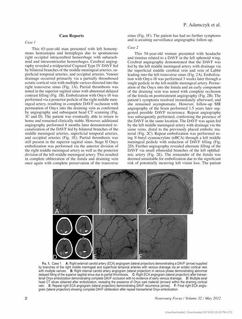
This 63yearold man presented with left homony-mous hemianopia and hemiplegia due to spontaneous right occipital intracerebral hemorrhage with subarach-noid and intraventricular hemorrhages. Cerebral angiog-raphy revealed a midparietal Cognard Type IV DAVF fed by bilateral branches of the middle meningeal arteries, su-perficial temporal arteries, and occipital arteries. Venous drainage occurred primarily via a partially thrombosed ectatic cortical vein with multiple varices directed into the right transverse sinus (Fig. 1A). Partial thrombosis was noted in the superior sagittal sinus with abnormal delayed contrast filling (Fig. 1B). Embolization with Onyx18 was performed via a posterior pedicle of the right middle men-ingeal artery, resulting in complete DAVF occlusion with permeation of Onyx into the draining vein as confirmed by angiography and subsequent head CT scanning (Fig. 1C and D). The patient was eventually able to return to home and remained clinically stable. However, additional angiography performed 8 months later demonstrated re-canalization of the DAVF fed by bilateral branches of the middle meningeal arteries, superficial temporal arteries, and occipital arteries (Fig. 1E). Partial thrombosis was still present in the superior sagittal sinus. Stage II Onyx embolization was performed via the anterior division of the right middle meningeal artery as well as the posterior division of the left middle meningeal artery. This resulted in complete obliteration of the fistula and draining vein once again with complete preservation of the transverse
sinus (Fig. 1F). The patient has had no further symptoms and is awaiting surveillance angiographic followup.
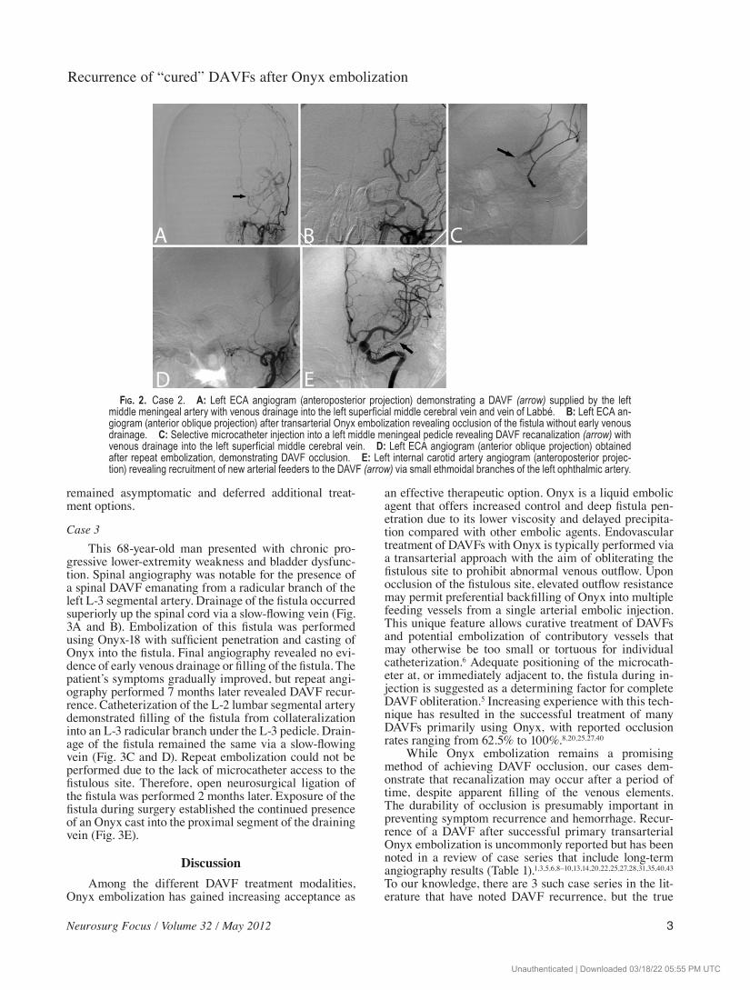
Case 2This 54yearold woman presented with headache
and tinnitus related to a DAVF in the left sphenoid wing. Cerebral angiography demonstrated that the DAVF was fed by the left middle meningeal artery with drainage via the superficial middle cerebral vein and vein of Labbé leading into the left transverse sinus (Fig. 2A). Emboliza-tion with Onyx18 was performed 3 weeks later through a single pedicle in the left middle meningeal artery. Perme-ation of the Onyx into the fistula and an early component of the draining vein was noted with complete occlusion of the fistula on posttreatment angiography (Fig. 2B). The patient’s symptoms resolved immediately afterward, and she remained asymptomatic. However, followup MR angiography of the brain performed 1.5 years later sug-gested possible DAVF recurrence. Repeat angiography was subsequently performed, confirming the presence of the DAVF in the same location. The DAVF was again fed by the left middle meningeal artery with drainage via the same veins, distal to the previously placed embolic ma-terial (Fig. 2C). Repeat embolization was performed us-ing Nbutyl cyanoacrylate (nBCA) through a left middle meningeal pedicle with reduction of DAVF filling (Fig. 2D). Further angiography revealed alternate filling of the DAVF via small ethmoidal branches of the left ophthal-mic artery (Fig. 2E). The remainder of the fistula was deemed unsuitable for embolization due to the significant risk of potentially incurring left vision loss. The patient
Fig. 1. Case 1. A: Right external carotid artery (ECA) angiogram (lateral projection) demonstrating a DAVF (arrow) supplied by branches of the right middle meningeal and superficial temporal arteries with venous drainage via an ectatic cortical vein with multiple varices. B: Right internal carotid artery angiogram (lateral projection) in venous phase demonstrating abnormal delayed filling of the superior sagittal sinus due to partial thrombosis. C: Right ECA angiogram (lateral projection) after transar-terial Onyx embolization demonstrating complete DAVF occlusion with no evidence of early venous drainage. D: Multiple axial head CT slices obtained after embolization, revealing the presence of Onyx cast material (arrows) within the draining cortical vein. E: Repeat right ECA angiogram (lateral projection) demonstrating DAVF recurrence (arrow). F: Final right ECA angio-gram (lateral projection) showing complete DAVF obliteration after repeat transarterial Onyx embolization.
Unauthenticated | Downloaded 03/18/22 05:55 PM UTC
Neurosurg Focus / Volume 32 / May 2012
Recurrence of “cured” DAVFs after Onyx embolization
3
remained asymptomatic and deferred additional treat-ment options.
Case 3This 68yearold man presented with chronic pro-
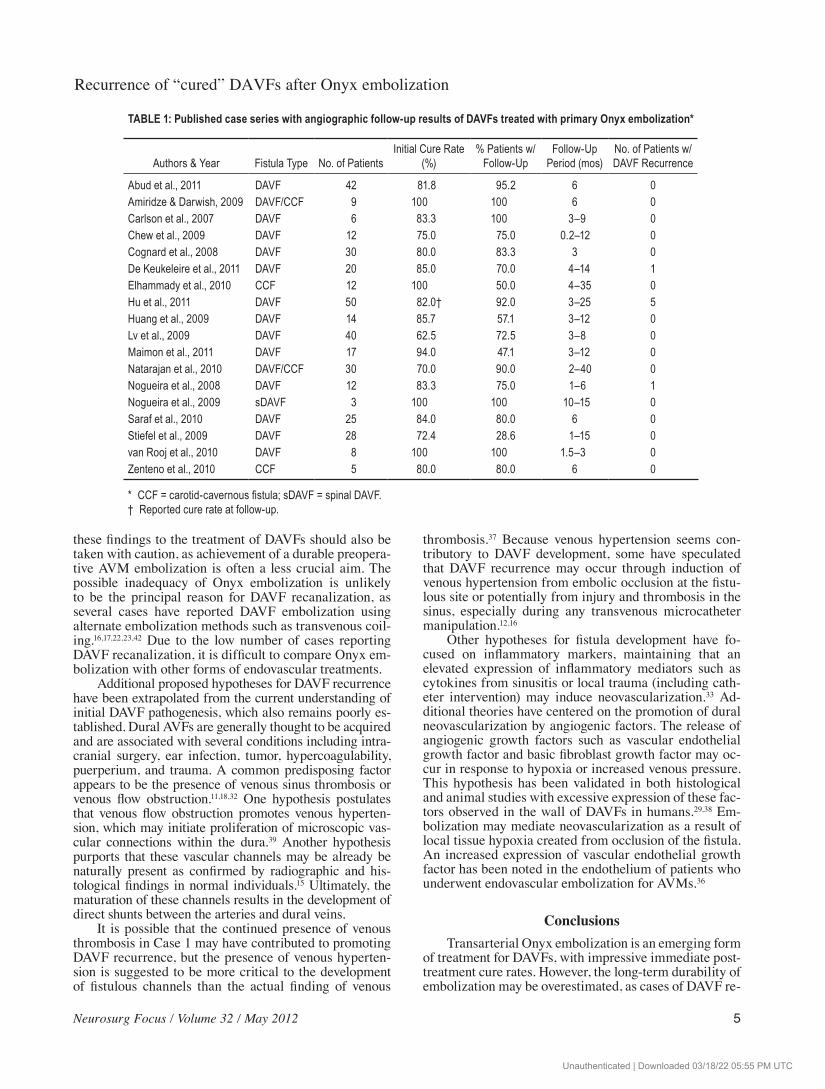
gressive lowerextremity weakness and bladder dysfunc-tion. Spinal angiography was notable for the presence of a spinal DAVF emanating from a radicular branch of the left L3 segmental artery. Drainage of the fistula occurred superiorly up the spinal cord via a slowflowing vein (Fig. 3A and B). Embolization of this fistula was performed using Onyx18 with sufficient penetration and casting of Onyx into the fistula. Final angiography revealed no evi-dence of early venous drainage or filling of the fistula. The patient’s symptoms gradually improved, but repeat angi-ography performed 7 months later revealed DAVF recur-rence. Catheterization of the L2 lumbar segmental artery demonstrated filling of the fistula from collateralization into an L3 radicular branch under the L3 pedicle. Drain-age of the fistula remained the same via a slowflowing vein (Fig. 3C and D). Repeat embolization could not be performed due to the lack of microcatheter access to the fistulous site. Therefore, open neurosurgical ligation of the fistula was performed 2 months later. Exposure of the fistula during surgery established the continued presence of an Onyx cast into the proximal segment of the draining vein (Fig. 3E).
DiscussionAmong the different DAVF treatment modalities,
Onyx embolization has gained increasing acceptance as
an effective therapeutic option. Onyx is a liquid embolic agent that offers increased control and deep fistula pen-etration due to its lower viscosity and delayed precipita-tion compared with other embolic agents. Endovascular treatment of DAVFs with Onyx is typically performed via a transarterial approach with the aim of obliterating the fistulous site to prohibit abnormal venous outflow. Upon occlusion of the fistulous site, elevated outflow resistance may permit preferential backfilling of Onyx into multiple feeding vessels from a single arterial embolic injection. This unique feature allows curative treatment of DAVFs and potential embolization of contributory vessels that may otherwise be too small or tortuous for individual catheterization.6 Adequate positioning of the microcath-eter at, or immediately adjacent to, the fistula during in-jection is suggested as a determining factor for complete DAVF obliteration.5 Increasing experience with this tech-nique has resulted in the successful treatment of many DAVFs primarily using Onyx, with reported occlusion rates ranging from 62.5% to 100%.8,20,25,27,40
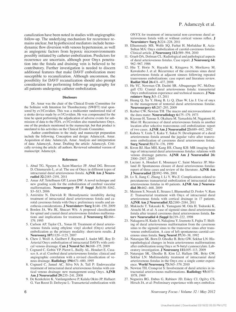
While Onyx embolization remains a promising method of achieving DAVF occlusion, our cases dem-onstrate that recanalization may occur after a period of time, despite apparent filling of the venous elements. The durability of occlusion is presumably important in preventing symptom recurrence and hemorrhage. Recur-rence of a DAVF after successful primary transarterial Onyx embolization is uncommonly reported but has been noted in a review of case series that include longterm angiography results (Table 1).1,3,5,6,8–10,13,14,20,22,25,27,28,31,35,40,43 To our knowledge, there are 3 such case series in the lit-erature that have noted DAVF recurrence, but the true
Fig. 2. Case 2. A: Left ECA angiogram (anteroposterior projection) demonstrating a DAVF (arrow) supplied by the left middle meningeal artery with venous drainage into the left superficial middle cerebral vein and vein of Labbé. B: Left ECA an-giogram (anterior oblique projection) after transarterial Onyx embolization revealing occlusion of the fistula without early venous drainage. C: Selective microcatheter injection into a left middle meningeal pedicle revealing DAVF recanalization (arrow) with venous drainage into the left superficial middle cerebral vein. D: Left ECA angiogram (anterior oblique projection) obtained after repeat embolization, demonstrating DAVF occlusion. E: Left internal carotid artery angiogram (anteroposterior projec-tion) revealing recruitment of new arterial feeders to the DAVF (arrow) via small ethmoidal branches of the left ophthalmic artery.
Unauthenticated | Downloaded 03/18/22 05:55 PM UTC

P. Adamczyk et al.
4 Neurosurg Focus / Volume 32 / May 2012
prevalence of this phenomenon remains uncertain as re-peat angiographic results are not uniformly reported in the literature.9,13,27 Additionally, the time period for recur-rence is unclear as the angiographic followup period in this review varies significantly from less than 1 month to 40 months.
De Keukeleire et al.9 described a patient in their se-ries who presented with a Cognard Type II DAVF and experienced fistula recurrence upon 6month followup angiography despite symptomatic improvement. In all of our cases, the patients also demonstrated improvement despite DAVF recanalization, suggesting that clinical symptoms cannot be reliably used to discern for poten-tial recurrence. Nogueira et al.27 described a patient with a Cognard Type IV DAVF who underwent successful transarterial Onyx embolization; however, the DAVF re-curred 4.6 months later with interval recruitment of alter-nate feeders. This case is similar to our Case 2 in which the presence of new arterial feeders was observed via the ophthalmic artery subsequent to embolization. In the cases published in the literature, it is not clear whether oc-clusion of the venous recipient pouch was achieved dur-ing the initial embolization. In a large case series of 50 patients, Hu et al.13 noted that several patients developed new fistulous connections at other sinuses upon followup despite initial angiographic success. One of these patients initially underwent treatment of a DAVF draining into the left transversesigmoid sinus, but the patient was found to have a new DAVF draining into the confluence of sinuses with subsequent drainage into the right sigmoid sinus and superior sagittal sinus.
The appearance of a new DAVF distal to the site of the initial fistula was not found in our cases, and it is unknown whether this relates to the same underlying pathophysiology. The principal mechanism of DAVF re-canalization remains elusive, and it is uncertain whether a DAVF recurs from development of a new fistula or from maturation of an angiographically occult fistula that al-ready existed in the vasculature. One hypothesis purports that small arterial feeders are not initially opacified due to smallcaliber flow limitation or from restricted outflow during the initial curative stage. During the time that elapses after treatment, these arterial feeders may enlarge with or without stimulation from endovascular treat-ment.12,13 Furthermore, poor penetration of Onyx into the fistulous component and draining vein may slowly allow for recruitment of nonvisualized arterial supply into the lowpressure system.27
Empirical evidence suggests that the presence of a dense Onyx cast at the fistulous site is a determining factor in ensuring adequate penetration by angiography. How-ever, no definitive angiographic methods are available to establish sufficient Onyx penetration beyond subjective visualizations of cast density with contrast stasis proxi-mal to the occlusion. In all 3 reported cases, a permanent cast was noted at followup evaluation both radiographi-cally, as well as upon visual inspection in Case 3 during surgical ligation. Despite sufficient Onyx injections, it has been suggested that DAVF recanalization may occur due to incomplete durability of the Onyx agent.13 In cere-bral AVMs, recanalization was noted in 18% of resected specimens previously embolized with Onyx.24 Relating
Fig. 3. Case 3. A and B: Left L-3 segmental artery injection on spinal angiography (anteroposterior projection) in early arte-rial (A) and late arterial (B) phases revealing a DAVF fed by a radicular branch at this level (single arrow) with drainage into a slow flowing vein (double arrows). C and D: Left L-2 segmental artery injection on spinal angiography (anteroposterior projection) in early arterial (C) and late arterial (D) phases, demonstrating DAVF recurrence from a collateralized radicular branch under the L-3 pedicle with similar drainage into a slow flowing vein (double arrows). E: Visual inspection during surgical ligation revealing the presence of Onyx cast material within the draining vein (arrow) of the fistula.
Unauthenticated | Downloaded 03/18/22 05:55 PM UTC
Neurosurg Focus / Volume 32 / May 2012
Recurrence of “cured” DAVFs after Onyx embolization
5
these findings to the treatment of DAVFs should also be taken with caution, as achievement of a durable preopera-tive AVM embolization is often a less crucial aim. The possible inadequacy of Onyx embolization is unlikely to be the principal reason for DAVF recanalization, as several cases have reported DAVF embolization using alternate embolization methods such as transvenous coil-ing.16,17,22,23,42 Due to the low number of cases reporting DAVF recanalization, it is difficult to compare Onyx em-bolization with other forms of endovascular treatments.
Additional proposed hypotheses for DAVF recurrence have been extrapolated from the current understanding of initial DAVF pathogenesis, which also remains poorly es-tablished. Dural AVFs are generally thought to be acquired and are associated with several conditions including intra-cranial surgery, ear infection, tumor, hypercoagulability, puerperium, and trauma. A common predisposing factor appears to be the presence of venous sinus thrombosis or venous flow obstruction.11,18,32 One hypothesis postulates that venous flow obstruction promotes venous hyperten-sion, which may initiate proliferation of microscopic vas-cular connections within the dura.39 Another hypothesis purports that these vascular channels may be already be naturally present as confirmed by radiographic and his-tological findings in normal individuals.15 Ultimately, the maturation of these channels results in the development of direct shunts between the arteries and dural veins.
It is possible that the continued presence of venous thrombosis in Case 1 may have contributed to promoting DAVF recurrence, but the presence of venous hyperten-sion is suggested to be more critical to the development of fistulous channels than the actual finding of venous
thrombosis.37 Because venous hypertension seems con-tributory to DAVF development, some have speculated that DAVF recurrence may occur through induction of venous hypertension from embolic occlusion at the fistu-lous site or potentially from injury and thrombosis in the sinus, especially during any transvenous microcatheter manipulation.12,16
Other hypotheses for fistula development have fo-cused on inflammatory markers, maintaining that an elevated expression of inflammatory mediators such as cytokines from sinusitis or local trauma (including cath-eter intervention) may induce neovascularization.33 Ad-ditional theories have centered on the promotion of dural neovascularization by angiogenic factors. The release of angiogenic growth factors such as vascular endothelial growth factor and basic fibroblast growth factor may oc-cur in response to hypoxia or increased venous pressure. This hypothesis has been validated in both histological and animal studies with excessive expression of these fac-tors observed in the wall of DAVFs in humans.29,38 Em-bolization may mediate neovascularization as a result of local tissue hypoxia created from occlusion of the fistula. An increased expression of vascular endothelial growth factor has been noted in the endothelium of patients who underwent endovascular embolization for AVMs.36
ConclusionsTransarterial Onyx embolization is an emerging form
of treatment for DAVFs, with impressive immediate post-treatment cure rates. However, the longterm durability of embolization may be overestimated, as cases of DAVF re-
TABLE 1: Published case series with angiographic follow-up results of DAVFs treated with primary Onyx embolization*
Authors & Year Fistula Type No. of PatientsInitial Cure Rate
(%)% Patients w/
Follow-UpFollow-Up
Period (mos)No. of Patients w/ DAVF Recurrence
Abud et al., 2011 DAVF 42 81.8 95.2 6 0Amiridze & Darwish, 2009 DAVF/CCF 9 100 100 6 0Carlson et al., 2007 DAVF 6 83.3 100 3–9 0Chew et al., 2009 DAVF 12 75.0 75.0 0.2–12 0Cognard et al., 2008 DAVF 30 80.0 83.3 3 0De Keukeleire et al., 2011 DAVF 20 85.0 70.0 4–14 1Elhammady et al., 2010 CCF 12 100 50.0 4–35 0Hu et al., 2011 DAVF 50 82.0† 92.0 3–25 5Huang et al., 2009 DAVF 14 85.7 57.1 3–12 0Lv et al., 2009 DAVF 40 62.5 72.5 3–8 0Maimon et al., 2011 DAVF 17 94.0 47.1 3–12 0Natarajan et al., 2010 DAVF/CCF 30 70.0 90.0 2–40 0Nogueira et al., 2008 DAVF 12 83.3 75.0 1–6 1Nogueira et al., 2009 sDAVF 3 100 100 10–15 0Saraf et al., 2010 DAVF 25 84.0 80.0 6 0Stiefel et al., 2009 DAVF 28 72.4 28.6 1–15 0van Rooj et al., 2010 DAVF 8 100 100 1.5–3 0Zenteno et al., 2010 CCF 5 80.0 80.0 6 0
* CCF = carotid-cavernous fistula; sDAVF = spinal DAVF.† Reported cure rate at follow-up.
Unauthenticated | Downloaded 03/18/22 05:55 PM UTC
P. Adamczyk et al.
6 Neurosurg Focus / Volume 32 / May 2012
canalization have been noted in studies with angiographic followup. The underlying mechanism for recurrence re-mains unclear, but hypothesized mediators include hemo-dynamic flow diversion with venous hypertension, as well as angiogenic factors from hypoxic microenvironments possibly initiated by catheter embolization. Predictors for recurrence are uncertain, although poor Onyx penetra-tion into the fistula and draining vein is believed to be contributory. Further investigation is needed to discern additional features that make DAVF embolization more susceptible to recanalization. Although uncommon, the possibility for DAVF recanalization should also prompt consideration for performing followup angiography for all patients undergoing catheter embolization.
Disclosure
Dr. Amar was the chair of the Clinical Events Committee for the Solitaire with Intention for Thombectomy (SWIFT) trial spon-sored by ev3/Covidien. This was a multicenter randomized trial of a stroke device made by ev3/Covidien. He was compensated for the time he spent performing the adjudication of adverse events for sub-mission of data to the FDA. ev3/Covidien also manufactures Onyx, one of the products mentioned in the manuscript, but that product is unrelated to his activities on the Clinical Events Committee.
Author contributions to the study and manuscript preparation include the following. Conception and design: Adamczyk, Amar. Acquisition of data: Adamczyk, Amar. Analysis and interpretation of data: Adamczyk, Amar. Drafting the article: Adamczyk. Criti-cally revising the article: all authors. Reviewed submitted version of manuscript: Adamczyk.
References
1. Abud TG, Nguyen A, SaintMaurice JP, Abud DG, Bresson D, Chiumarulo L, et al: The use of Onyx in different types of intracranial dural arteriovenous fistula. AJNR Am J Neuro-radiol 32:2185–2191, 2011
2. Amar AP, Teitelbaum GP, Larsen DW: A novel technique and new grading scale for the embolization of cerebral vascular malformations. Neurosurgery 59 (5 Suppl 3):S158–S162, S3–S13, 2006
3. Amiridze N, Darwish R: Hemodynamic instability during treatment of intracranial dural arteriovenous fistula and ca-rotid cavernous fistula with Onyx: preliminary results and an-esthesia considerations. J Neurointerv Surg 1:146–150, 2009
4. Borden JA, Wu JK, Shucart WA: A proposed classification for spinal and cranial dural arteriovenous fistulous malforma-tions and implications for treatment. J Neurosurg 82:166–179, 1995
5. Carlson AP, Taylor CL, Yonas H: Treatment of dural arterio-venous fistula using ethylene vinyl alcohol (Onyx) arterial embolization as the primary modality: shortterm results. J Neurosurg 107:1120–1125, 2007
6. Chew J, Weill A, Guilbert F, Raymond J, Audet ME, Roy D: Arterial Onyx embolisation of intracranial DAVFs with corti-cal venous drainage. Can J Neurol Sci 36:168–175, 2009
7. Cognard C, Gobin YP, Pierot L, Bailly AL, Houdart E, Casa-sco A, et al: Cerebral dural arteriovenous fistulas: clinical and angiographic correlation with a revised classification of ve-nous drainage. Radiology 194:671–680, 1995
8. Cognard C, Januel AC, Silva NA Jr, Tall P: Endovascular treatment of intracranial dural arteriovenous fistulas with cor-tical venous drainage: new management using Onyx. AJNR Am J Neuroradiol 29:235–241, 2008
9. De Keukeleire K, Vanlangenhove P, Kalala Okito JP, Hallaert G, Van Roost D, Defreyne L: Transarterial embolization with
ONYX for treatment of intracranial noncavernous dural ar-teriovenous fistula with or without cortical venous reflux. J Neurointerv Surg 3:224–228, 2011
10. Elhammady MS, Wolfe SQ, Farhat H, Moftakhar R, AzizSultan MA: Onyx embolization of carotidcavernous fistulas. Clinical article. J Neurosurg 112:589–594, 2010
11. Graeb DA, Dolman CL: Radiological and pathological aspects of dural arteriovenous fistulas. Case report. J Neurosurg 64: 962–967, 1986
12. Hiu T, Horie N, Hayashi K, Kitagawa N, Morikawa M, Kawakubo J, et al: Recurrence of the cavernous sinus dural arteriovenous fistula at adjacent sinuses following repeated transvenous embolizations: case report and literature review. Radiat Med 26:431–437, 2008
13. Hu YC, Newman CB, Dashti SR, Albuquerque FC, McDou-gall CG: Cranial dural arteriovenous fistula: transarterial Onyx embolization experience and technical nuances. J Neu-rointerv Surg 3:5–13, 2011
14. Huang Q, Xu Y, Hong B, Li Q, Zhao W, Liu J: Use of onyx in the management of tentorial dural arteriovenous fistulae. Neurosurgery 65:287–293, 2009
15. Kerber CW, Newton TH: The macro and microvasculature of the dura mater. Neuroradiology 6:175–179, 1973
16. Kiyosue H, Tanoue S, Okahara M, Yamashita M, Nagatomi H, Mori H: Recurrence of dural arteriovenous fistula in another location after selective transvenous coil embolization: report of two cases. AJNR Am J Neuroradiol 23:689–692, 2002
17. Kubota Y, Ueda T, Kaku Y, Sakai N: Development of a dural arteriovenous fistula around the jugular valve after transve-nous embolization of cavernous dural arteriovenous fistula. Surg Neurol 51:174–176, 1999
18. Kwon BJ, Han MH, Kang HS, Chang KH: MR imaging find-ings of intracranial dural arteriovenous fistulas: relations with venous drainage patterns. AJNR Am J Neuroradiol 26: 2500–2507, 2005
19. Luciani A, Houdart E, Mounayer C, Saint Maurice JP, Mer-land JJ: Spontaneous closure of dural arteriovenous fistulas: report of three cases and review of the literature. AJNR Am J Neuroradiol 22:992–996, 2001
20. Lv X, Jiang C, Zhang J, Li Y, Wu Z: Complications related to percutaneous transarterial embolization of intracranial dural arteriovenous fistulas in 40 patients. AJNR Am J Neurora-diol 30:462–468, 2009
21. Maimon S, Nossek E, Strauss I, Blumenthal D, Frolov V, Ram Z: Transarterial treatment with Onyx of intracranial dural arteriovenous fistula with cortical drainage in 17 patients. AJNR Am J Neuroradiol 32:2180–2184, 2011
22. Makiuchi T, Takasaki K, Yamagami M, Oda H, Todoroki K, Atsuchi M, et al: A case of sigmoid sinus dural arteriovenous fistula after treated cavernous dural arteriovenous fistula. In-terv Neuroradiol 4 (Suppl 1):219–222, 1998
23. Nakagawa H, Kudo S, Nakajima Y, Izumoto S, Fujita T: Shift-ing of dural arteriovenous malformation from the cavernous sinus to the sigmoid sinus to the transverse sinus after trans-venous embolization. A case of left spontaneous carotidcav-ernous sinus fistula. Surg Neurol 37:30–38, 1992
24. Natarajan SK, Born D, Ghodke B, Britz GW, Sekhar LN: His-topathological changes in brain arteriovenous malformations after embolization using Onyx or Nbutyl cyanoacrylate. Lab-oratory investigation. J Neurosurg 111:105–113, 2009
25. Natarajan SK, Ghodke B, Kim LJ, Hallam DK, Britz GW, Sekhar LN: Multimodality treatment of intracranial dural arteriovenous fistulas in the Onyx era: a single center experi-ence. World Neurosurg 73:365–379, 2010
26. Newton TH, Cronqvist S: Involvement of dural arteries in in-tracranial arteriovenous malformations. Radiology 93:1071–1078, 1969
27. Nogueira RG, Dabus G, Rabinov JD, Eskey CJ, Ogilvy CS, Hirsch JA, et al: Preliminary experience with onyx emboliza-
Unauthenticated | Downloaded 03/18/22 05:55 PM UTC

Neurosurg Focus / Volume 32 / May 2012
Recurrence of “cured” DAVFs after Onyx embolization
7
tion for the treatment of intracranial dural arteriovenous fistu-las. AJNR Am J Neuroradiol 29:91–97, 2008
28. Nogueira RG, Dabus G, Rabinov JD, Ogilvy CS, Hirsch JA, Pryor JC: Onyx embolization for the treatment of spinal dural arteriovenous fistulae: initial experience with longterm fol-lowup. Technical case report. Neurosurgery 64:E197–E198, 2009
29. Rothbart D, Awad IA, Lee J, Kim J, Harbaugh R, Criscuolo GR: Expression of angiogenic factors and structural proteins in central nervous system vascular malformations. Neurosur-gery 38:915–925, 1996
30. Roy D, Raymond J: The role of transvenous embolization in the treatment of intracranial dural arteriovenous fistulas. Neu-rosurgery 40:1133–1144, 1997
31. Saraf R, Shrivastava M, Kumar N, Limaye U: Embolization of cranial dural arteriovenous fistulae with ONYX: indica-tions, techniques, and outcomes. Indian J Radiol Imaging 20:26–33, 2010
32. Sarma D, ter Brugge K: Management of intracranial dural ar-teriovenous shunts in adults. Eur J Radiol 46:206–220, 2003
33. S Miyachi E, Izumi T, Matsubara N, Naito T, Haraguchi K, Wakabayashi T: Mechanism of the formation of dural arterio-venous fistula: the role of the emissary vein. Interv Neurora-diol 17:195–202, 2011
34. Söderman M, Edner G, Ericson K, Karlsson B, Rähn T, Ulfars-son E, et al: Gamma knife surgery for dural arteriovenous shunts: 25 years of experience. J Neurosurg 104:867–875, 2006
35. Stiefel MF, Albuquerque FC, Park MS, Dashti SR, McDougall CG: Endovascular treatment of intracranial dural arteriove-nous fistulae using Onyx: a case series. Neurosurgery 65 (6 Suppl):132–140, 2009
36. Sure U, Butz N, Schlegel J, Siegel AM, Wakat JP, Mennel HD, et al: Endothelial proliferation, neoangiogenesis, and potential de novo generation of cerebrovascular malformations. J Neu-rosurg 94:972–977, 2001
37. Terada T, Higashida RT, Halbach VV, Dowd CF, Tsuura M, Komai N, et al: Development of acquired arteriovenous fistu-las in rats due to venous hypertension. J Neurosurg 80:884–889, 1994
38. Uranishi R, Nakase H, Sakaki T: Expression of angiogenic growth factors in dural arteriovenous fistula. J Neurosurg 91:781–786, 1999
39. van Dijk JM, Willinsky RA: Venous congestive encephalopa-thy related to cranial dural arteriovenous fistulas. Neuroim-aging Clin N Am 13:55–72, 2003
40. van Rooij WJ, Sluzewski M: Curative embolization with Onyx of dural arteriovenous fistulas with cortical venous drainage. AJNR Am J Neuroradiol 31:1516–1520, 2010
41. van Rooij WJ, Sluzewski M, Beute GN: Dural arteriovenous fistulas with cortical venous drainage: incidence, clinical pre-sentation, and treatment. AJNR Am J Neuroradiol 28:651–655, 2007
42. Yamashita K, Taki W, Nakahara I, Nishi S, Sadato A, Kikuchi H: Development of sigmoid dural arteriovenous fistulas after transvenous embolization of cavernous dural arteriovenous fistulas. AJNR Am J Neuroradiol 14:1106–1108, 1993
43. Zenteno M, SantosFranco J, RodríguezParra V, Balderrama J, AburtoMurrieta Y, VegaMontesinos S, et al: Management of direct carotidcavernous sinus fistulas with the use of eth-ylenevinyl alcohol (Onyx) only: preliminary results. Clinical article. J Neurosurg 112:595–602, 2010
Manuscript submitted January 15, 2012.Accepted February 6, 2012.Please include this information when citing this paper: DOI:
10.3171/2012.2.FOCUS1224. Address correspondence to: Peter Adamczyk, M.D., 1520 San
Pablo Street, Suite 3800, Los Angeles, California 90033. email: [email protected].
Unauthenticated | Downloaded 03/18/22 05:55 PM UTC