final combined-pesch-ocular emergencies aafp a&u …corneal edema from acute glaucoma*, cl...
TRANSCRIPT

Opthalmology
Opthalmology
Theodor Pesch, MD, PhD
Disclosure Statement:
• Neither I nor any immediate family member (parent, sibling, spouse or child) has a financial relationship with or interest in any commercial entity that may have a direct interest in the subject matter of this article.
It is the policy of the AAFP that all individuals in a position to control content disclose any relationships with commercial interests upon nomination/invitation of participation. Disclosure documents are reviewed for potential conflicts of interest. If conflicts are identified, they are resolved prior to confirmation of participation. Only participants who have no conflict of interest or who agree to an identified resolution process prior to their participation were involved in this CME activity.
Learning Objectives
1. Utilize several different types of eye examination (e.g., slit lamp examination, and ophthalmoscope) methods and how to interpret the results accurately.
2. Recognize globe rupture, avoid complications.
3. Distinguish the signs and symptoms of hyphema and how to treat it in an urgent care setting.
4. Identify possible etiologies of acute red eye.
5. Diagnose complications associated with diabetes and diabetic eye disease.
6. Identify and treat retinal detachment.
Ocular Emergencies in ED
Most Common
• Trauma
• Retinal detachment– flashes, floaters, visual field defect
• Central retinal artery occlusion– CRAO
True ocular emergencies• Glaucoma
• Acute angle closure
• Infection• Orbital cellulitis
• Ischemia• Central retinal artery occlusion
• Visual field defect• Acute retinal detachment
• Trauma: chemical, blunt
AUDIENCE RESPONSE SYSTEM
Which clinical diagnosis does notrequire immediate eye referral?
a. Acute angle closure glaucoma
b. Orbital cellulitis
c. Central retinal artery occlusion
d. Acute retinal detachment
e. Trauma: chemical, blunt

Opthalmology
GENERAL EYE EXAM / TOOLS
1. Utilize several different types of eye examination methods (e.g., slit lamp examination, and ophthalmoscope) and how to interpret the results accurately.
Eight-part Eye Exam1. Visual Acuity / Color Vision2. External Examination3. Ocular Motility4. Pupils5. Visual Fields6. Tonometry7. Anterior Segment Examination
- Slit-lamp
8. Fundus Examination- Ophthalmoscopes
Preparation
• History
• Focused physical exam– Eye exam bills at level 3
• Comfort of patient– Anesthesia
– Clean instruments
Pain Management
PROPARACAINE• Onset 15 seconds
• Duration 20 minutes
TETRACAINE• Onset delayed
• Duration > 1 hour
----------------------------------
(COCAINE)• Softens corneal epithelium
Eye Drops
• Have patient look up
– Keep tip off eye
• Treat both eyes
– avoids blepharospasm
Eye Drops
• Alternative Method– Create small lake
– Allow to warm up
• Topical anesthesia if– Blepharospasm
– Tonometry
– Prior to fluorescein
Photo courtesy of Theodor Pesch, MD, PhD

Opthalmology
Eye Drops
• Open eyes:– Automatic inoculation
• Keep tip off eye
• Single use containers– avoid P. aeruginosa
• Fluorescein
Photo courtesy of Theodor Pesch, MD, PhD
Medical Mydriasis
Caveats:
• Vision blurs
• Accommodation impaired
• Threatened Acute Glaucoma
• Must not operate machines, drive– Bring sun shades, driver
Medical Mydriasis
Avoid dilating pupils if:
• flat anterior chamber
• narrow anterior chamber angle
• Can precipitate acute glaucoma (NAG)
Medical Mydriasis
Quick tests to exclude Narrow A/C Angle
• Slit Lamp Exam
• Penlight Test
– Illuminate cornea tangentially
– If the light extends over more than 1/3 ofthe iris, refer to an ophthalmologist
Abnormal Penlight Test
© Microsoft INITIAL DATA COLLECTIONVisual Acuity, Color Vision, Visual Fields, Ocular Motility, IOP

Opthalmology
VISUAL ACUITY& ACCOMMODATION
Visual Acuity
• Should be 20/20 for all
– Enter written order for billing– Obtain pre- and post procedure
• documents pre-existing condition
• PIN-HOLE test• blocks aberration
– If VA improves refract
Pinhole Visual Acuity
Stenopaeic filters: only most central beams pass through the pinhole aperture, blocking aberration from the lens periphery: better acuity.
Far sighted:hyperopic
Normal sighted:emmetropic
Near sighted:myopic
Causes of Traumatic Visual Loss
1. Refractive Error - pinhole test!
2. Media Opacities -• subluxed lens, corneal scar, vitreous
hemorrhage, hyphema
3. Macular Disease -• choroidal rupture, retinal detachment,
commotio (retinal concussion)
4. Optic Nerve Disease -• traumatic optic neuropathy, CN2 avulsion
Myopia
© Microsoft
Corrected Refraction
© Microsoft

Opthalmology
PERRLA?
• Test accommodation
• Headaches
• Arms too short to read
– If not done, it is PERRL!
VISUAL FIELD SCREENING
Eyesight with Glaucoma Kestenbaum Test
Kestenbaum Test Kestenbaum Test

Opthalmology
Kestenbaum Test Kestenbaum Test
Test 360°
Retest randomly
Near Visual FieldsAmsler Grid Tests Macula
• Focus on the central dot with one eye covered:
• Am I able to see the corners and sides of the square?– Do I see any wavy lines?– Are there any holes or
missing areas?
• If the lines of grid do not look straight or areas appear to be missing and/or distorted, you should inform a retinal specialist.
Near Visual Fields
Peripheral Visual Field > 30˚0 5.5
[deg]
≈1.5mm across
≈ 1500 microns
≈ 5.5 degrees
≈ 1 disc-diameter (1DD)
The smallest aperture in the direct ophthalmoscope projects a spot of light ≈ surface area of a normal optic nerve head L Optic Disk R
180˚180˚
Landmark: Optic Disc SizeLandmark: Optic Disc Size
COLOR VISION & IOP
Opthalmology
Color Vision
• Ishihara Test for screening– Only test red/green opponent colors
• Velhagen Panel D-50 Test– Also tests blue/yellow system
– Saturated
– Desaturated
Color Vision
• Simple office test– Present saturated red stimulus
• Red cap bottle, Sharpie®
– Swing to other eye
– Ask if any differences noted
• Monocular desaturation:
– subtle sign for glaucoma
Acute Angle Closure Rx
IMMEDIATE home remedies
• Bright lights
• Hot soaks
• Double shot Booze
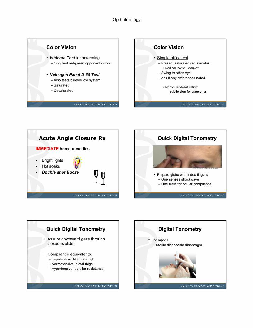
Quick Digital Tonometry
• Palpate globe with index fingers:– One senses shockwave– One feels for ocular compliance
Photo courtesy of Theodor Pesch, MD, PhD
Quick Digital Tonometry
• Assure downward gaze through closed eyelids
• Compliance equivalents:– Hypotensive: like mid-thigh– Normotensive: distal thigh– Hypertensive: patellar resistance
Digital Tonometry
• Tonopen– Sterile disposable diaphragm

Opthalmology
Schiøtz Tonometry - avoid
• Plunger indents anesthetized cornea
• Use average read outs of 3 weights
• To calibrate scale, adjust plunger on reference cornea
© 2008 International Centre for Eye Health, London
Goldmann Applanation Tonometer
• Gold standard tonometer• Slit lamp mounted
• Sterilizable tip,
• Prions take exception
Goldmann Applanation Tonometer
Goldmann Mires aka Semicircles
SLIT LAMP
Slit Lamp
• A slit lamps serves to view the anteriorsegment of the eye in high magnification
Portable Slit Lamp (Heine)
Ocular / Objective
@ 45° to beam
Lightsource
Rheostat
Battery
Helpful in determining anterior chamber depth

Opthalmology
Reversal of acute NAG
• Lost anterior chamber in narrow angle glaucoma. – Rx: serial Pilocarpine eye drops, IV Diamox, IV Mannitol
Glaucoma - NAG
Acute Narrow Angle Glaucoma– 10% of all glaucoma - 2nd commonest
• Apposition of the anterior chamber angle– a.k.a. synechial closure– aqueous drainage site– anterior displacement of lens-iris diaphragm
• Resultant high IOP rapidly damages CN2
“The sun must not set over an acute NAG”
http://office.microsoft.com/en-us/images/results.aspx?qu=sunset&ex=1#ai:MP900178497|
GLOBE RUPTURE2. Recognize globe rupture, avoid complications.
Facial Soft Tissue Injuries
• Ca. 2.4 million traumatic eyeinjuries each year in the US
• up to 15% of these occur in theworkplace
On SUSPICION of Rupture
• DO NOT – instill eye drops or ointments
– evert the eyelid
– put any pressure on the globe
• DO:– Boost tetanus / diphtheria immunity
– Place orbital shield
Opthalmology
Lac: Punctum, Canaliculus
http://webeye.ophth.uiowa.edu/eyeforum/atlas/pages/canalicular-laceration-lid.html
Asymmetric Red Reflex
http://webeye.ophth.uiowa.edu/eyeforum/atlas/pages/asteroid-hyalosis.html
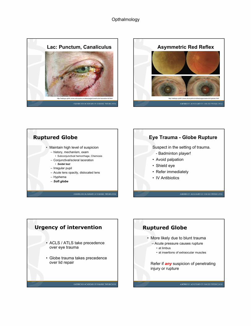
Ruptured Globe
• Maintain high level of suspicion– history, mechanism, exam
• Subconjunctival hemorrhage, Chemosis
– Conjunctival/scleral laceration • Seidel test
– Irregular pupil
– Acute lens opacity, dislocated lens
– Hyphema
– Soft globe
Eye Trauma - Globe Rupture
Suspect in the setting of trauma.
- Badminton player!
• Avoid palpation
• Shield eye
• Refer immediately
• IV Antibiotics
Urgency of intervention
• ACLS / ATLS take precedence over eye trauma
• Globe trauma takes precedence over lid repair
Ruptured Globe
• More likely due to blunt trauma – Acute pressure causes rupture
• at limbus
• at insertions of extraocular muscles
Refer if any suspicion of penetrating injury or rupture
Opthalmology
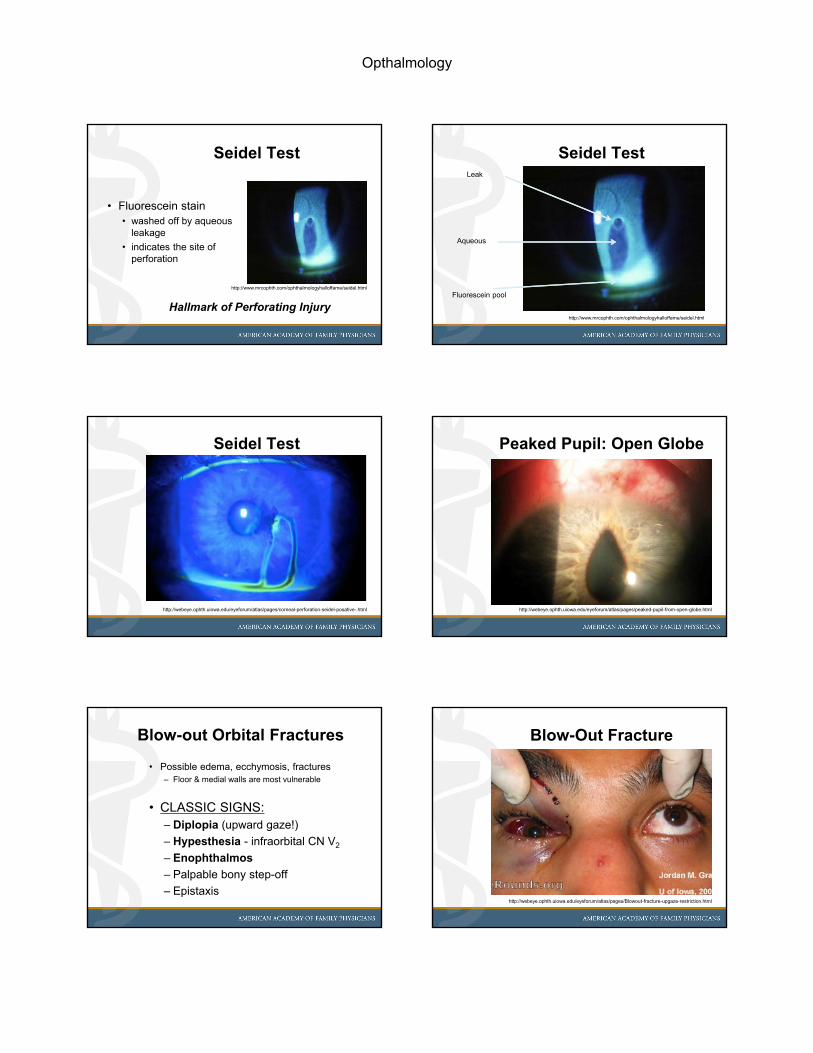
Seidel Test
• Fluorescein stain• washed off by aqueous
leakage
• indicates the site of perforation
Hallmark of Perforating Injury
http://www.mrcophth.com/ophthalmologyhalloffame/seidel.html
Seidel TestLeak
Aqueous
Fluorescein pool
http://www.mrcophth.com/ophthalmologyhalloffame/seidel.html
Seidel Test
http://webeye.ophth.uiowa.edu/eyeforum/atlas/pages/corneal-perforation-seidel-posative-.html
Peaked Pupil: Open Globe
http://webeye.ophth.uiowa.edu/eyeforum/atlas/pages/peaked-pupil-from-open-globe.html
Blow-out Orbital Fractures
• Possible edema, ecchymosis, fractures– Floor & medial walls are most vulnerable
• CLASSIC SIGNS: – Diplopia (upward gaze!)
– Hypesthesia - infraorbital CN V2
– Enophthalmos
– Palpable bony step-off
– Epistaxis
Blow-Out Fracture
http://webeye.ophth.uiowa.edu/eyeforum/atlas/pages/Blowout-fracture-upgaze-restriction.html

Opthalmology
HYPHEMA
3. Distinguish the signs and symptoms of hyphema and how to treat it in an urgent care setting.
Eye Emergencies - Trauma
Hyphema
• Highly suspicious for – ruptured globe
– other deeper trauma
• Place protective shield and refer
Acute Eye Trauma - Hyphema
• Blood in the anterior chamber
– Look for air-fluid level
• Consider possible complications– globe rupture
– retinal detachment
Hyphema
Hyphema scale
Grade Size• 0 No layered blood,
circulating RBCs• I < ⅓• II ⅓ to ½• III ½ to subtotal• IV total a/c filled
Hyphema?
• No: subconjunctival hemorrhage– a.k.a. hyposphagma

Opthalmology
Eye Trauma Care: Watch TV!Restrict activity!
45° head elevation
– avoid rebleed
• Eye shield
• Avoid NSAIDS – try APAP
Acute Red EyeSymptom Differential Diagnoses
Itching Allergy
Scratching, burning Dry eye, foreign body,
trichiasis, conjunctivitis
Focal lid tenderness Chalazion, hordeolum
Deep, intense pain Corneal abrasion, sinusitis,
(epi-) scleritis, glaucoma*
Photophobia Corneal abrasion, iritis,
acute glaucoma*
Halos around light Corneal edema from acute glaucoma*, CL overwear
Ocular Foreign Bodies
• Diagnosis
– a. Always flip both lids
– b. Fluorescein with blue light to reveal subtle corneal abrasions
– c. Document Seidel status
– d. H/o driving nail, grinding or drilling metal: dilated fundoscopy
Ocular Foreign Bodies
• Removal of Corneal FB
• Visual acuity pre and post
• Anesthetize both eyes
– a. Try quick pass with Q-tip
– b. Burr or 30 gauge needle helpful
– c. No success: call ophthalmologist
Lid eversion technique
• Relax M. levatorpalpebrae in down ward gaze
• Place lever at proximal rim of tarsal plate
• Pull lashes down then towards you and up.
http://www.med.uottawa.ca/procedures/slamp/eversion.htm
Corneal rust FB narrow beam
http://webeye.ophth.uiowa.edu/eyeforum/atlas/pages/Ferrous-foreign-body-Rust-ring.html

Opthalmology
Corneal rust FB broad beam
http://webeye.ophth.uiowa.edu/eyeforum/atlas/pages/Ferrous-foreign-body-Rust-ring.html
Pingueculitis
• Small yellow conjunctival patches
• Symptoms: Redness/irritation
– Inflammation vs. infection
• Etiology mixed– UV exposure, wind, dust, dry climate
– Asthenopia - visual strain• “Over-minused” distance refraction
• Undercorrected near addition
Pincuecula (“fatty tumor”)
http://www.eyeatlas.com/Eyeatlas/Conjunctiva.html#16
Pingueculitis - Treatment
Eye protection
– Artificial tears
– NSAID ophthalmic solutions
– Check eyeglass Rx, UVA/B filter
– Pterygium:• excise if threatens central cornea
• Optional:
– topical steroids
Viral Conjunctivitis
• Signs & Symptoms– Watery discharge
– Palpable pre-auricular nodes
– Often associated with URI
• Rx:– Artificial tears, cool compresses
Keratitis (Adenovirus)
http://www.eyeatlas.com/Eyeatlas/Cornea.html#16

Opthalmology
Keratitis (Adenovirus)
http://www.eyeatlas.com/Eyeatlas/Cornea.html#19
Keratitis (Aspergillus)
http://www.eyeatlas.com/Eyeatlas/Cornea.html#18
Corneal abrasion
http://www.eyeatlas.com/Eyeatlas/Cornea.html#5
Corneal abrasion
http://www.eyeatlas.com/Eyeatlas/Cornea.html#4
Corneal Pannus (CL wear)
http://www.eyeatlas.com/Eyeatlas/Cornea.html#9
Bullous Episcleritis
http://www.eyeatlas.com/Eyeatlas/Sclera.html#2

Opthalmology
Episcleritis
• Subconjunctival inflammation (Tenon’s)
• Idiopathic
– rarely systemic disease association
• Episcleritis vs. scleritis:
– Topical vasoconstrictors blanch injection
– Topical anesthetics relieve pain
• Rx: Topical steroids
– PredForte 1%• shake well
Iritis / anterior Uveitis
• Conjunctivitis:– all conjunctiva is
red
• Iritis:– Pericorneal ciliary
injection
Preseptal cellulitis
• Orbital septum: continuation of periosteum of orbital bones
– Chemosis, erythema, min. proptosis– Vision & ocular motility intact
• Treatment: systemic antibiotics• Complication:
– orbital cellulitis if left untreated
Orbital cellulitis
Orbital cellulitis
• Signs & Symptoms– Impaired ocular motility ±pain
– Chemosis, erythema, ± ↓ vision, proptosis
– Decreased acuity and color vision
– Afferent pupillary defect (APD)• Swinging Flashlight Test
• Treatment: IV antibiotics, admit– Blood Cultures; orbital CT: sinusitis? Abscess?
• Complications:– Meningitis, cavernous sinus thrombosis
OPHTHALMOSCOPY
Abnormal screening tests suggest location of pathology:
Consider Slit Lamp exam and Ophthalmoscopy

Opthalmology
Direct ophthalmoscopy
• Real, upright retinal image – 15X magnified– Limited to central 30˚of retina
Useful for screening of
• the posterior pole
• the red reflex and its symmetry
• focal anterior segment exam
Direct Ophthalmoscopes
What’s wrong here?What’s wrong here?
Photo courtesy from Theodor Pesch, MD, PhD
Ophthalmoscopy ExamOphthalmoscopy Exam
Photo courtesy from Theodor Pesch, MD, PhD
Direct Ophthalmoscopes
Welch Allyn® PanOptic™ hand-held direct illuminating direct opthalmoscope
• incorporates Axial PointSource™ Optics
• easier to enter small pupils
• wider, panoramic view of the fundus– 5X larger than achieved with a standard
opthalmoscope in an undilated eye
Direct Ophthalmoscopes
Modern ophthalmoscope design
• light, a prism and a mirror, and lenses mounted in the head of the instrument– Prism and mirror illuminate the retina
– Lenses can be selected to focus image
– Filters: neutral-gray, cobalt blue, red free, polarized; various apertures
• Handle contains the battery

Opthalmology
Systematic Fundus Survey
x
Pars plana
Ora serrata
Macula
temporalnasal
long ciliary nerve
short ciliary nerve
Equator
Optic nerve
Vortex vein ampulla
Systematic Fundus Survey
x
Systematic Fundus SurveyCentral 30°
x
Hollenhorst Plaque BRAO
http://webeye.ophth.uiowa.edu/eyeforum/atlas/pages/Hollenhorst-plaque-BRAO-cherry-red-spot.htm
Systematic Fundus Exam
• Use a technique for thorough overlapping examination
• Have the patient look in all different directions of gaze
• Follow excursions of eye with ophthalmoscope
Systematic Fundus Survey
Secondary gaze
Primary gaze

Opthalmology
Systematic Fundus Survey
x1
Systematic Fundus Survey
x
2
Systematic Fundus Survey
x
3
Systematic Fundus Survey
x
4
Systematic Fundus Survey
x
5
Systematic Fundus Survey
x
6

Opthalmology
Systematic Fundus Survey
x
7
Systematic Fundus Survey
x 8
Systematic Fundus Survey
x
Pars plana
Ora serrata
Macula
temporalnasal
long ciliary nerve
short ciliary nerve
Equator
Optic nerve
Vortex vein ampulla
Systematic Fundus Survey
x
O. D.O. S.
Mapping paper
5 Key Items of CN2 Exam
1. Scleral ring: delineates the optic disk2. Neural retinal rim3. Retinal nerve fiber layer4. Region of parapapillary atrophy5. Retinal and optic disk hemorrhage?
Cup/Disc Ratio
0.1-0.2 0.4-0.5 0.7-0.8
Cup
Disc
0.0
1.0
‘Doughnut Rule: more dough is better.’
Normal cup/disc ratio ≤ 0.4

Opthalmology
Normal Cup/Disc Ratio Cup/Disc Ratio Samples
1
2
3
1. Normal C/D 0.1
2. Borderline C/D 0.4 - 0.5
3. Glaucoma C/D 0.8- Vessels undermine optic rim- Temporal myopic conus
Abnormal C/D ratio: Glaucoma Cup/Disc Ratio
0.4-0.5
Oblique Optic Nerve Head
… can be a normal variant
DDx: Tilted Disc
… can be a normal variant
Arteriosclerotic ChangesWidth• Grade I: 3/4 normal caliber
• Grade II: 1/2 normal caliber
• Grade III: 1/3 normal caliber
• Grade IV: thread-like or invisible
• "AV nicking”– Arterio-venous crossing changes
– with venous constriction and banking

Opthalmology
Ophthalmoscopy Filters
The ‘red-free’ filter shines ‘green’ light.
• The retinal blood vessel walls and the retinal pigment epithelium (RPE) act like a red filter
• Red-free light - (i.e., green light) – blocks out the choroid
• red and ‘green’ cancel each other out• enhances details of retinal blood columns
Arteriosclerotic Changes
Grade I 3/4 normal caliber
Grade II 1/2 normal caliber
Grade III 1/3 normal caliber
Grade IV thread-like or invisible
A/V Nicking• Arterio-venous crossing changes
• Venous constriction and banking
Arteriolo / Venular Width Ratio
Arteriosclerotic Changes
Width of arteriole
Light reflex
Arteriosclerotic ChangesColor of Light Reflex• Copper wire arterioles
– the central light reflex occupies most of the width
• Silver wire arterioles– the central light reflex occupies all of
the width of the arteriole
• Sclerotic vessels
Arteriosclerotic Changes
Copper wirearterioles
Light reflex occupies most of the width
Silver wirearterioles
Light reflex occupies all of the width
Sclerotic vessels Light reflex narrowed, rarefied vascular bed
Arteriosclerotic Changes
• Ischemia, e.g. "cotton wool spots”
• Hemorrhages: pre-, intra-, subretinal
• Central Edema: Ring of exudates around the fovea (aka macular star)
• Papilledema or optic disc prominence– HTN, elevated intracranial pressure
• Visual acuity loss– typically due to macular involvement
Opthalmology
HTN Retinopathy
http://webeye.ophth.uiowa.edu/eyeforum/atlas/pages/hypertensive-retinopathy.html
Malignant Hypertension
http://webeye.ophth.uiowa.edu/eyeforum/atlas/pages/malignant-hypertension.htm
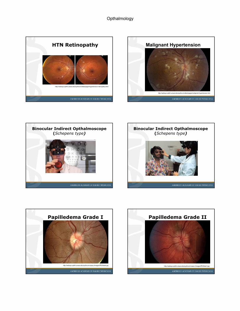
Binocular Indirect Opthalmoscope(Schepens type)
Binocular Indirect Opthalmoscope(Schepens type)
Papilledema Grade I
http://webeye.ophth.uiowa.edu/eyeforum/cases-i/images/PE/Slide6.jpg
Papilledema Grade II
http://webeye.ophth.uiowa.edu/eyeforum/cases-i/images/PE/Slide7.jpg

Opthalmology
Papilledema Grade III
http://webeye.ophth.uiowa.edu/eyeforum/cases-i/images/PE/Slide8.jpg
Papilledema Grade IV
http://webeye.ophth.uiowa.edu/eyeforum/cases-i/images/PE/Slide9.jpg
Papilledema Grade V
http://webeye.ophth.uiowa.edu/eyeforum/cases-i/images/PE/Slide10.jpg
BRVO
http://webeye.ophth.uiowa.edu/eyeforum/atlas/pages/Branched-Retinal-Vein-Occlusion.html
CRVO
http://webeye.ophth.uiowa.edu/eyeforum/atlas/pages/Central-Retinal-Vein-Occlusion-CRVO-2.html
Branch Artery Occlusion
http://webeye.ophth.uiowa.edu/eyeforum/atlas/pages/BRAO-Branched-Retinal-Artery-Occlusion.html

Opthalmology
CRAO w/ cilioretinal vessel
http://webeye.ophth.uiowa.edu/eyeforum/atlas/pages/CRAO.html
Commotio Retinae
http://webeye.ophth.uiowa.edu/eyeforum/atlas/pages/commotio-retinae.html
DIABETES
5. Diagnose complications associated with diabetes and diabetic eye disease.
Sorry Folks: No Slides
• The listed references can be utilized to review and understand the tenets of dilated eye examination for patients with a history of Diabetes mellitus.
• Mastery of the previously reviewed techniques is required to perform a meaningful exam
• Refer to ophthalmologist
Diabetes & Eye
Vascular changes
• Iris– Neovascularization glaucoma
• Retina– Diabetic retinopathy (DR)
– Retinal detachment (RD)
• Complication: Blindness
Diabetic Retinopathy DR
• 4 years post initial DM diagnosis– 20-25% incidence of DR
• 15 years post initial DM diagnosis– ~98% DR incidence in DM-1
– ~60-80% DR incidence in DM-2
• Screen with dilated eye exam– DM-1 within 3 years
– DM-2 immediately (unknown onset!)

Opthalmology
DR Screening**
• No retinopathy: q 3 years
• Mild NPDR*: q 1 year
• Mod/severe NPDR*: q 2-4 months
• PDR: as indicated by progress
*more frequently if macular edema
**Images: see http://cgeye.org
DR Therapy
• Control of underlying disease
• DM, HTN, other
• PDR:
• Photocoagulation
• Vitrectomy
• Secondary glaucoma management
DR Stages
• Non-Proliferative DR - mild– Microaneurysms: 1st sign of disease
– Intraretinal hemorrhage
– Following nerve fiber layer
– Hard exudates
– Macular Edema
DR Stages
• Non-Proliferative DR – mod/severe– 4:2:1 rule
– Hemorrhages in 4 quadrants
– Venous bleeding in 2 quadrants
– IRMA (intraretinal microvascular abnormalities) in 1 quadrant
• Severe NPDR progresses to PDR– 50% in 1 year
DR Stages
• Proliferative DR – PDR– Neovascularization of disk NVD
– Neovascularization elsewhere NVE
– Neovascularization of iris NVI• Risk of neovascularization glaucoma
RETINAL DETACHMENT - RD6. Identify and treat retinal detachment.

Opthalmology
Sorry Folks: No Slides
• The listed references can be utilized to review and understand the tenets of dilated eye examination for patients with a history of Diabetes mellitus.
• Mastery of the previously reviewed techniques is required to perform a meaningful exam
• Refer to ophthalmologist
Retinal Detachment
• Rhegmatogenous (intrinsic)– Retinal breaks
– Myopia
• Non-Rhegmatogenous (extrinsic)– Exudative (serous)
– Traction
• Retinopathy of Prematurity (ROP)
RD - Signs & Symptoms
• Floaters
• Photopsia (light flashes)– Due to tugging on retina
• Visual field loss– Shadow or veil
• Vision loss– Suggests macular involvement
RD - Differential Diagnosis
• Retinal Detachment RD– Retina separates from retinal pigment
epithelium RPE
• Posterior Vitreous Detachment PVD– Vitreous detaches from retina
• Consult ophthalmology– Needs dilated eye exam
– Urgent therapy if confirmed
Ocular Emergencies References
• Larkin GL. Retinal detachment. Accessed March 23, 2007, at: http://www.emedicine.com/emerg/topic504.htm.
• Prabhat Pokhrel, Sanaz Loftus. Ocular Emergencies. Am FamPhysician.2007;76(6):829-836.
• Cain W Jr, Sinskey RM. Detection of anterior chamber leakage with Seidel’s test. Arch Ophthalmol. 1981;99:2013.
• Syed Azhar. Acute Red Eye. Am Fam Physician. 2007;76(6):857-858.
• Taylor HR, West SK, Rosenthal FS, Munoz B, Newland HS, Emmett EA. Corneal changes associated with chronic UV irradiation. Arch Ophthalmol. 1989;107:1481–4.
• Frucht-Pery J, Siganos CS, Solomon A, Shvartzenberg T, Richard C, Trinquand C. Topical indomethacin solution versus dexamethasone solution for treatment of inflamed pterygium and pinguecula: a prospective randomized clinical study. Am J Ophthalmol. 1999;127:148–52.
• Keeler, R Br J Ophthalmol 2002;86:602-603; Figure 4 Galezowskiophthalmoscope 1882.
Online Resources• Online Atlas of Ophthalmology
– www.eyeatlas.com– www.eyerounds.org– http://webeye.ophth.uiowa.edu/eyeforum/picosearc
h.htm• Wisconsin Retinopathy Standards
– www.eyephoto.ophth.wisc.edu/ResearchAreas/Diabetes/DiabStds.htm
• Glaucoma images– www.eyesearch.com/glaucoma.images.htm
• Sickle cell disease– sickle.bwh.harvard.edu/outpatient.html
Opthalmology
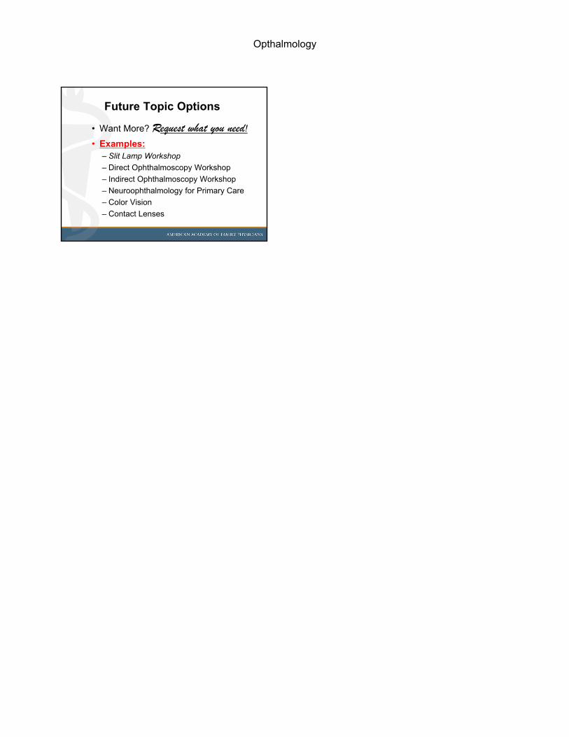
Future Topic Options
• Want More? Request what you need!
• Examples:– Slit Lamp Workshop
– Direct Ophthalmoscopy Workshop
– Indirect Ophthalmoscopy Workshop
– Neuroophthalmology for Primary Care
– Color Vision
– Contact Lenses