anatomical studies on the tarsal joint of buffalo calves (bubalus ….pdf · 2014-09-28 ·...
TRANSCRIPT

International Journal of Science and Research (IJSR) ISSN (Online): 2319-7064
Impact Factor (2012): 3.358
Volume 3 Issue 9, September 2014 www.ijsr.net
Licensed Under Creative Commons Attribution CC BY
Anatomical Studies on the Tarsal Joint of Buffalo Calves (Bubalus bubalis)
Supriya .B*1, T. S. Chandrasekhara Rao2
1Assistant Professor, Department of Veterinary Anatomy,
College of Veterinary Science, Sri Venkateswara Veterinary University, Tirupati- 517502, A.P, India
2Dean, Faculty of Veterinary Science,
Sri Venkateswara Veterinary University, Tirupati- 517502, A.P, India Abstract: The study was conducted to through light on special morphological features of buffalo tarsus compared to that of cattle. It was conducted on 15 apparently healthy buffalo calf limbs of 1- 1½year age buffaloes collected from the local slaughter house in the Department of Veterinary Anatomy, College Of Veterinary Science, Tirupati. The tarsal joint of buffalo comprised of the bones and ligaments nearly similar to those of ox. The individual tarsal bones had well developed articular facets. The present paper describes the detailed anatomical description of ligaments of tarsus of buffalo calf and how they differ from that of white cattle. Keywords: Tarsal articulation, Buffalo calf, ligament, tarsal joint, synovial sac 1. Introduction The disease conditions of the joints of the limbs drastically reduce the productive and reproductive capabilities of animals. Extensive information was available on the anatomy of the joints of horse, dogs, pig and the cattle in standard text books dealing with anatomy and clinical anatomy [1], [3], [4], [6], and [7]. Several anatomical aspects of joints which were specific to buffaloes and vary with those of ox were not well documented. Hence, the present study was undertaken to through light on special morphological features of buffalo tarsus. Nomina Anatomica Veterinaria [5] followed for nomenclature. 2. Materials and Methods The study was conducted on fifteen apparently healthy 1-1½ year old buffalo calf limbs procured from the local slaughter house irrespective of breed, sex and nutritional status. The limbs of the animals were preserved in 10% formalin and the gross morphology of joints and relations were studied by careful dissection. The dissection was also carried out in some unpreserved fresh specimens to study the movements of the joints. The ligaments of the joints were painted with acrylic paints for better visualization. 3. Results and discussion The tarsal joint of buffalo calf was a composite joint comprised of tarsocrural joint, proximal intertarsal joint, distal intertarsal joint and the tarsometatarsal joint. The capsula articularis was attached around the tibial articular surface above and fused third and fourth large metatarsal below. The joint showed tibiotarsal, proximal tarsal, distal tarsal and tarsometatarsal synovial sacs. The tibiotarsal sac was larger. The plantar annular ligament completely covered the tarsal groove. The lateral collateral ligament showed two parts, the long lateral collateral ligament and the short lateral collateral
ligament. The former extended from lateral aspect of the lateral malleolus to proximal end of fused third and fourth large metatarsal and also attached to the lateral aspect of bones in its course. The latter was deep to former and extended from the anterior aspect of lateral malleolus and ended on lateral aspect of body of fibular tarsal just below articular surface of lateral malleolus. The calcaneometatarseal ligament originated proximally from distal end of fibular tarsal and ended on the fused third and fourth large metatarsal dorsal to long lateral collateral ligament (fig. 1). The long medial collateral ligament extended from distal end of tibia to the proximal end of fused third and fourth large metatarsal attached to the medial aspect of tarsal along its course. The short medial collateral ligament was divided into three parts out of which two parts, pars tibiotalaris were attached to the tibial tarsal and one part pars tibiocalcanea was attached to fibular tarsal (fig.2) the latter in white cattle divided into onlyvtwo parts [4]. The lig. talocentrodisto- metatarseum was a flat ligament extending from the medial surface of tibial tarsal and end on fused third and fourth large metatarsal attaching to medial aspect of the tarsals along its course (fig.2).
Paper ID: SEP14678 2101
International Journal of Science and Research (IJSR) ISSN (Online): 2319-7064
Impact Factor (2012): 3.358
Volume 3 Issue 9, September 2014 www.ijsr.net
Licensed Under Creative Commons Attribution CC BY
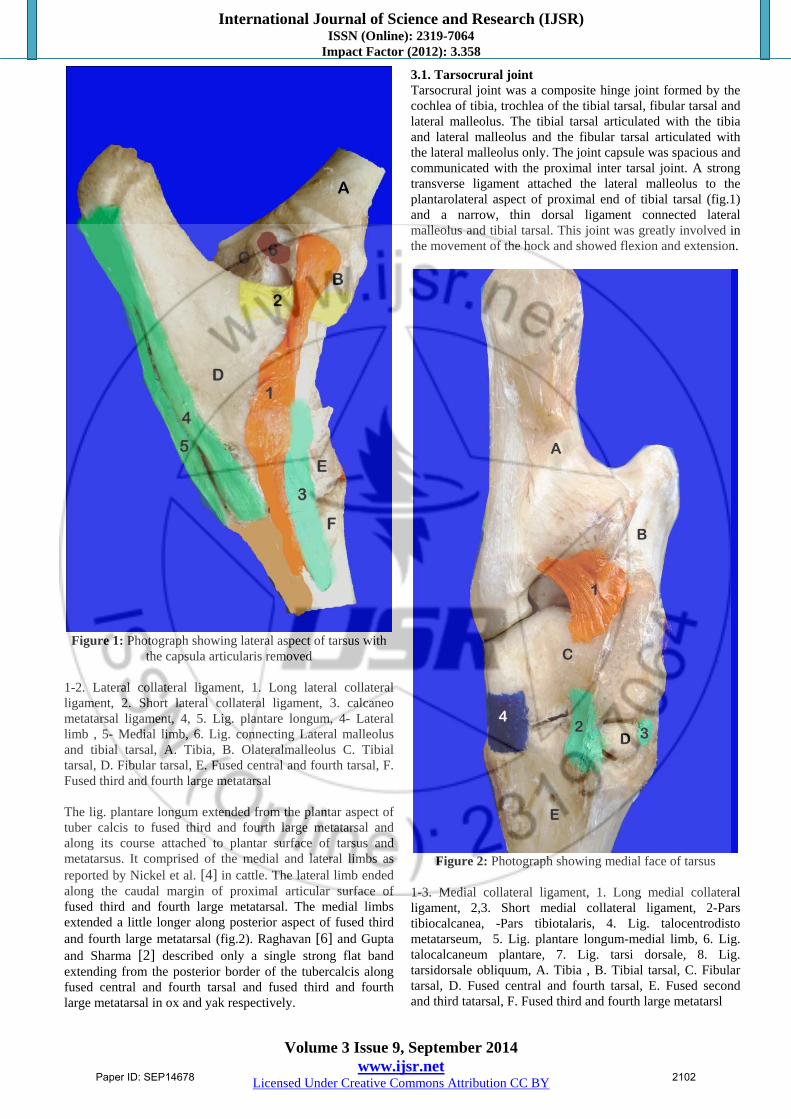
Figure 1: Photograph showing lateral aspect of tarsus with
the capsula articularis removed 1-2. Lateral collateral ligament, 1. Long lateral collateral ligament, 2. Short lateral collateral ligament, 3. calcaneo metatarsal ligament, 4, 5. Lig. plantare longum, 4- Lateral limb , 5- Medial limb, 6. Lig. connecting Lateral malleolus and tibial tarsal, A. Tibia, B. Olateralmalleolus C. Tibial tarsal, D. Fibular tarsal, E. Fused central and fourth tarsal, F. Fused third and fourth large metatarsal The lig. plantare longum extended from the plantar aspect of tuber calcis to fused third and fourth large metatarsal and along its course attached to plantar surface of tarsus and metatarsus. It comprised of the medial and lateral limbs as reported by Nickel et al. [4] in cattle. The lateral limb ended along the caudal margin of proximal articular surface of fused third and fourth large metatarsal. The medial limbs extended a little longer along posterior aspect of fused third and fourth large metatarsal (fig.2). Raghavan [6] and Gupta and Sharma [2] described only a single strong flat band extending from the posterior border of the tubercalcis along fused central and fourth tarsal and fused third and fourth large metatarsal in ox and yak respectively.
3.1. Tarsocrural joint Tarsocrural joint was a composite hinge joint formed by the cochlea of tibia, trochlea of the tibial tarsal, fibular tarsal and lateral malleolus. The tibial tarsal articulated with the tibia and lateral malleolus and the fibular tarsal articulated with the lateral malleolus only. The joint capsule was spacious and communicated with the proximal inter tarsal joint. A strong transverse ligament attached the lateral malleolus to the plantarolateral aspect of proximal end of tibial tarsal (fig.1) and a narrow, thin dorsal ligament connected lateral malleolus and tibial tarsal. This joint was greatly involved in the movement of the hock and showed flexion and extension.
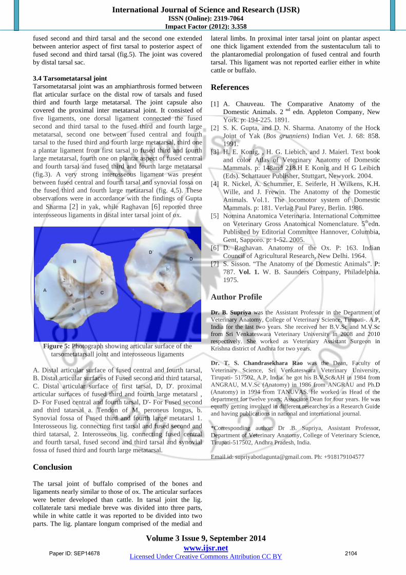
Figure 2: Photograph showing medial face of tarsus
1-3. Medial collateral ligament, 1. Long medial collateral ligament, 2,3. Short medial collateral ligament, 2-Pars tibiocalcanea, -Pars tibiotalaris, 4. Lig. talocentrodisto metatarseum, 5. Lig. plantare longum-medial limb, 6. Lig. talocalcaneum plantare, 7. Lig. tarsi dorsale, 8. Lig. tarsidorsale obliquum, A. Tibia , B. Tibial tarsal, C. Fibular tarsal, D. Fused central and fourth tarsal, E. Fused second and third tatarsal, F. Fused third and fourth large metatarsl
Paper ID: SEP14678 2102

International Journal of Science and Research (IJSR) ISSN (Online): 2319-7064
Impact Factor (2012): 3.358
Volume 3 Issue 9, September 2014 www.ijsr.net
Licensed Under Creative Commons Attribution CC BY
3.2 Proximal inter tarsal joint Proximal inter tarsal joint was an amphiarthrosis formed between the tibial tarsal and fibular tarsal proximally and fused central and fourth tarsal distally. It included artc. talocalcaneocentralis and artc. calcaneoquartalis. The articular sac was roomy. On the dorsal aspect flat oblique ligament extend between the two trochlear ridges connected the tibial tarsal and fused central and fourth tarsal (fig.2). Two collateral ligaments were present. On plantar aspect one thick ligament extended from the sustentaculum tali to the plantaromedial prolongation of fused central and fourth tarsal (fig 3).
Figure 3: Photograph showing posterior aspect of tarsal joint
(Lig. plantare longum removed) A. Fibular tarsal, B. Tibial tarsal, C. Fused central and fourth tarsal, D. first tarsal, E. Fused third and fourth large metatarsl 1. lig. connecting sustentaculum tali and fused central and fourth tarsal, 2. plantar ligament between first tarsal and fused third and fourth large metatarsl, 3. Lig. connecting first tarsal and fused second and third tatarsal, 4. plantar ligament connecting fused central and fourth tarsal and fused third and fourth large metatarsal. The tibial tarsal and fibular tarsal were connected by one lateral ligament and two interosseous ligaments. The lateral ligament extended between anterior margin of body of
fibular tarsal and craniolateral margin of tibial tarsal just above the lateral condyle. One very thick interosseous ligament extended from lateral aspect of tibial tarsal to the medial aspect of the fibular tarsal (fig.4) while the second one extended from the depression on the posterior surface of tibial tarsal to fibular tarsal. No movements were observed in this joint.
Figure 4: Photograph showing interosseous ligaments on a cut surface of tarsal articulation (arrow indicates tendon of
M. peroneus longus) A. Tibia, B. Fibular tarsal, C. Tibial tarsal, D. Fused central and fourth tarsal, E. Fused second and third tarsal, F. Fused third and fourth large metatarsal 1. Interosseous lig. between tibial tarsal and fibular tarsal, 2. Interosseous lig. connecting the fibular tarsal and fused central and fourth tarsal, 3. Interosseous lig. connecting fused central and fourth tarsal, fused second and third tarsal and synovial fossa of fused third and fourth large metatarsal. 3.3 Distal inter tarsal joint Distal inter tarsal joint was an amphiarthrosis formed by fused central and fourth tarsal proximally and fused second and third tarsal and the first tarsal distally. On the dorsal aspect a ligament connected the fused central and fourth tarsal and fused second and third tarsal (lig. tarsi dorsale) (fig.2). The joint had two interosseous ligaments. The first one extended between the fused central and fourth tarsal to
Paper ID: SEP14678 2103
International Journal of Science and Research (IJSR) ISSN (Online): 2319-7064
Impact Factor (2012): 3.358
Volume 3 Issue 9, September 2014 www.ijsr.net
Licensed Under Creative Commons Attribution CC BY
fused second and third tarsal and the second one extended between anterior aspect of first tarsal to posterior aspect of fused second and third tarsal (fig.5). The joint was covered by distal tarsal sac. 3.4 Tarsometatarsal joint Tarsometatarsal joint was an amphiarthrosis formed between flat articular surface on the distal row of tarsals and fused third and fourth large metatarsal. The joint capsule also covered the proximal inter metatarsal joint. It consisted of five ligaments, one dorsal ligament connected the fused second and third tarsal to the fused third and fourth large metatarsal, second one between fused central and fourth tarsal to the fused third and fourth large metatarsal, third one a plantar ligament from first tarsal to fused third and fourth large metatarsal, fourth one on plantar aspect of fused central and fourth tarsal and fused third and fourth large metatarsal (fig.3). A very strong interosseous ligament was present between fused central and fourth tarsal and synovial fossa on the fused third and fourth large metatarsal (fig. 4,5). These observations were in accordance with the findings of Gupta and Sharma [2] in yak, while Raghavan [6] reported three interosseous ligaments in distal inter tarsal joint of ox.
Figure 5: Photograph showing articular surface of the
tarsometatarsall joint and interosseous ligaments A. Distal articular surface of fused central and fourth tarsal, B. Distal articular surfaces of Fused second and third tatarsal, C. Distal articular surface of first tarsal, D, D'. proximal articular surfaces of fused third and fourth large metatarsl , D- For Fused central and fourth tarsal, D'- For Fused second and third tatarsal a. Tendon of M. peroneus longus, b. Synovial fossa of Fused third and fourth large metatarsl 1. Interosseous lig. connecting first tarsal and fused second and third tatarsal, 2. Interosseous lig. connecting fused central and fourth tarsal, fused second and third tarsal and synovial fossa of fused third and fourth large metatarsal. Conclusion The tarsal joint of buffalo comprised of the bones and ligaments nearly similar to those of ox. The articular surfaces were better developed than cattle. In tarsal joint the lig. collaterale tarsi mediale breve was divided into three parts, while in white cattle it was reported to be divided into two parts. The lig. plantare longum comprised of the medial and
lateral limbs. In proximal inter tarsal joint on plantar aspect one thick ligament extended from the sustentaculum tali to the plantaromedial prolongation of fused central and fourth tarsal. This ligament was not reported earlier either in white cattle or buffalo. References [1] A. Chauveau. The Comparative Anatomy of the
Domestic Animals. 2 nd edn. Appleton Company, New York. p: 194-225. 1891.
[2] S. K. Gupta, and D. N. Sharma. Anatomy of the Hock Joint of Yak (Bos grunniens) Indian Vet. J. 68: 858. 1991.
[3] H. E. Konig, , H. G. Liebich, and J. Maierl. Text book and color Atlas of Veterinary Anatomy of Domestic Mammals. p: 148and 218.H E Konig and H G Leibich (Eds). Schattauer Publisher, Stuttgart, Newyork. 2004.
[4] R. Nickel, A. Schummer, E. Seiferle, H .Wilkens, K.H. Wille, and J. Frewin. The Anatomy of the Domestic Animals. Vol.1. The locomotor system of Domestic Mammals. p: 181. Verlag Paul Parey, Berlin. 1986.
[5] Nomina Anatomica Veterinaria. International Committee on Veterinary Gross Anatomical Nomenclature. 5thedn. Published by Editorial Committee Hannover, Columbia, Gent, Sapporo. p: 1-52. 2005.
[6] D. Raghavan. Anatomy of the Ox. P: 163. Indian Council of Agricultural Research, New Delhi. 1964.
[7] S. Sisson. “The Anatomy of the Domestic Animals”. P: 787. Vol. 1. W. B. Saunders Company, Philadelphia. 1975.
Author Profile Dr. B. Supriya was the Assistant Professor in the Department of Veterinary Anatomy, College of Veterinary Science, Tirupati-. A.P, India for the last two years. She received her B.V.Sc and M.V.Sc from Sri Venkateswara Veterinary University in 2008 and 2010 respectively. She worked as Veterinary Assistant Surgeon in Krishna district of Andhra for two years. Dr. T. S. Chandrasekhara Rao was the Dean, Faculty of Veterinary Science, Sri Venkateswara Veterinary University, Tirupati- 517502. A.P, India. he got his B.V.Sc&AH in 1984 from ANGRAU, M.V.Sc (Anatomy) in 1986 from ANGRAU and Ph.D (Anatomy) in 1994 from TANUVAS. He worked as Head of the department for twelve years; Associate Dean for four years. He was equally getting involved in different researches as a Research Guide and having publications in national and international journal. *Corresponding author: Dr .B. Supriya, Assistant Professor, Department of Veterinary Anatomy, College of Veterinary Science, Tirupati-517502, Andhra Pradesh, India. Email.id: [email protected]. Ph: +918179104577
Paper ID: SEP14678 2104