epigenetic modification of the renin-angiotensin...
TRANSCRIPT
Epigenetic Modification of the Renin-Angiotensin System inthe Fetal Programming of Hypertension
Irina Bogdarina, Simon Welham, Peter J. King, Shamus P. Burns, Adrian J.L. Clark
Abstract—Hypertension is a major risk factor for cardiovascular and cerebrovascular disease. Lifelong environmentalfactors (eg, salt intake, obesity, alcohol) and genetic factors clearly contribute to the development of hypertension, butit has also been established that stress in utero may program the later development of the disease. This phenomenon,known as fetal programming can be modeled in a range of experimental animal models. In maternal low protein dietrat models of programming, administration of angiotensin converting enzyme inhibitors or angiotensin receptorantagonists in early life can prevent development of hypertension, thus implicating the renin-angiotensin system in thisprocess. Here we show that in this model, expression of the AT1b angiotensin receptor gene in the adrenal gland isupregulated by the first week of life resulting in increased receptor protein expression consistent with the increasedadrenal angiotensin responsiveness observed by others. Furthermore, we show that the proximal promoter of the AT1b
gene in the adrenal is significantly undermethylated, and that in vitro, AT1b gene expression is highly dependent onpromoter methylation. These data suggest a link between fetal insults to epigenetic modification of genes and theresultant alteration of gene expression in adult life leading ultimately to the development of hypertension. It seemshighly probable that similar influences may be involved in the development of human hypertension. (Circ Res.2007;100:520-526.)
Key Words: hypertension � angiotensin receptors � fetal programming � DNA methylation
Although hypertension is recognized as 1 of the majorcontributing factors to cerebrovascular and cardiovascu-
lar disease,1 its pathogenesis remains incompletely under-stood. Genetic and environmental factors clearly contribute tothis,2 but on current models fail to account entirely for thedisease. There is now substantial epidemiological evidencethat intrauterine stress may program the later development ofthe disease.3 The phenomenon of fetal programming can bemodeled effectively in several mammalian species.4 Mostinvestigators have used models in which mothers are sub-jected to relative undernutrition during pregnancy. Offspringfrom these pregnancies exhibit later development of hyper-tension, insulin resistance, glucose intolerance and frankdiabetes, the extent of each of these depending on the speciesand experimental model.4
Administration of a low protein diet (8% protein in place of18% protein in normal rat chow with the calorific content ofprotein made up in the form of carbohydrate) to pregnant ratseither until term or weaning has been widely used in theinvestigation of the pathogenesis of hypertension. This modelproduces offspring of reduced birth weight in which elevatedsystolic and diastolic blood pressures, as measured by tailcuff methods or by indwelling carotid artery catheters, can beidentified as early as 4 weeks of age.5 Hypertension can be
prevented in this model by administration of angiotensinconverting enzyme inhibitors or angiotensin receptor antag-onists, but not by nifedipene, between 2 and 4 weeks of age.6,7
This is highly suggestive of a role of the renin-angiotensinsystem (RAS) in disease pathogenesis.
The consequence of the fetal insult in this and otherprogramming models is that the phenotype is altered in astable, but subtle manner. Such a phenotypic alterationmay be achieved either by a change in the number ordistribution of differentiated cells, or by changes in geneexpression by individual cells, and there is evidence tosupport both occurrences.8 Following embryo implanta-tion, the majority of the genome is demethylated.9 Theprocess of differentiation and development is accompaniedby the selective methylation of genes that are not neededfor function of the differentiated cell. As this process ofDNA methylation takes place in utero and in early post-natal development, it is a good candidate for disturbanceby environmental interference, and thus provides a poten-tial mechanism for fetal programming. Whereas methyl-ation patterns are generally considered to be established inearly postnatal life and persist thereafter, there is evidencefrom human monozygotic twin studies that methylationpatterns can change with ageing.10
Original received September 5, 2006; revision received January 3, 2007; accepted January 17, 2007.From the Centre for Endocrinology (I.B., S.W., P.J.K., A.J.L.C.), Barts & the London, Queen Mary University of London, UK; Department of
Chemical and Biological Sciences (S.P.B.), University of Huddersfield, UK.Correspondence to Professor A.J.L. Clark, Centre for Endocrinology, John Vane Science Centre, William Harvey Research Institute, Charterhouse
Square, London EC1M 6BQ, UK. E-mail [email protected]© 2007 American Heart Association, Inc.
Circulation Research is available at http://circres.ahajournals.org DOI: 10.1161/01.RES.0000258855.60637.58
520
by guest on June 28, 2018http://circres.ahajournals.org/
Dow
nloaded from
by guest on June 28, 2018http://circres.ahajournals.org/
Dow
nloaded from
by guest on June 28, 2018http://circres.ahajournals.org/
Dow
nloaded from
by guest on June 28, 2018http://circres.ahajournals.org/
Dow
nloaded from
by guest on June 28, 2018http://circres.ahajournals.org/
Dow
nloaded from
by guest on June 28, 2018http://circres.ahajournals.org/
Dow
nloaded from
by guest on June 28, 2018http://circres.ahajournals.org/
Dow
nloaded from
by guest on June 28, 2018http://circres.ahajournals.org/
Dow
nloaded from
by guest on June 28, 2018http://circres.ahajournals.org/
Dow
nloaded from
by guest on June 28, 2018http://circres.ahajournals.org/
Dow
nloaded from

It has been shown that a maternal low protein diet isassociated with reduced global methylation, and it may bethat it is deficiency of specific amino acids, eg, glycine,required to generate methyl donors, that underlies suchchanges.11 Supplementation of low protein diets with glycineor folate reverses the programming effect of those diets.12,13
However such a mechanism may lack gene specificity,whereas other data argues that it is specific genes that aresusceptible to this effect.14–17
Thus in this study we set out to test the hypothesis thatalteration of DNA methylation of 1 or more RAS componentgenes might underlie the alteration of gene expression thatculminated in the development of hypertension.
Materials and MethodsAnimalsAll animal procedures were conducted in an approved facility inaccordance with the Scientific Procedures (Animals) Act 1986, UK,and were approved by the Institutional Animal Use Ethics Commit-tee. The model of fetal programming and the phenotypic character-istics of adult MLP rats have been described in detail.4 Briefly ratdams (Wistar, Charles River, UK) were placed on either the normal20% protein (control) rat chow or 8% protein (MLP, protein replacedwith carbohydrate) at conception. Offspring were randomly culled to8 in each litter at birth (4 male, 4 female) and then weaned on toidentical (20% protein) rat chow at 3 weeks of age. Tissues (liver,lung, kidney, whole brain, adrenal, heart) were harvested from allmembers of a litter at 1, 4, or 12 weeks of age, and were immediatelydeep-frozen in liquid nitrogen and stored at �80°C until furtheranalysis. Up to 3 litters were studied in each set of analyses.
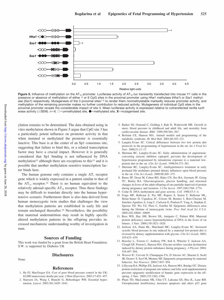
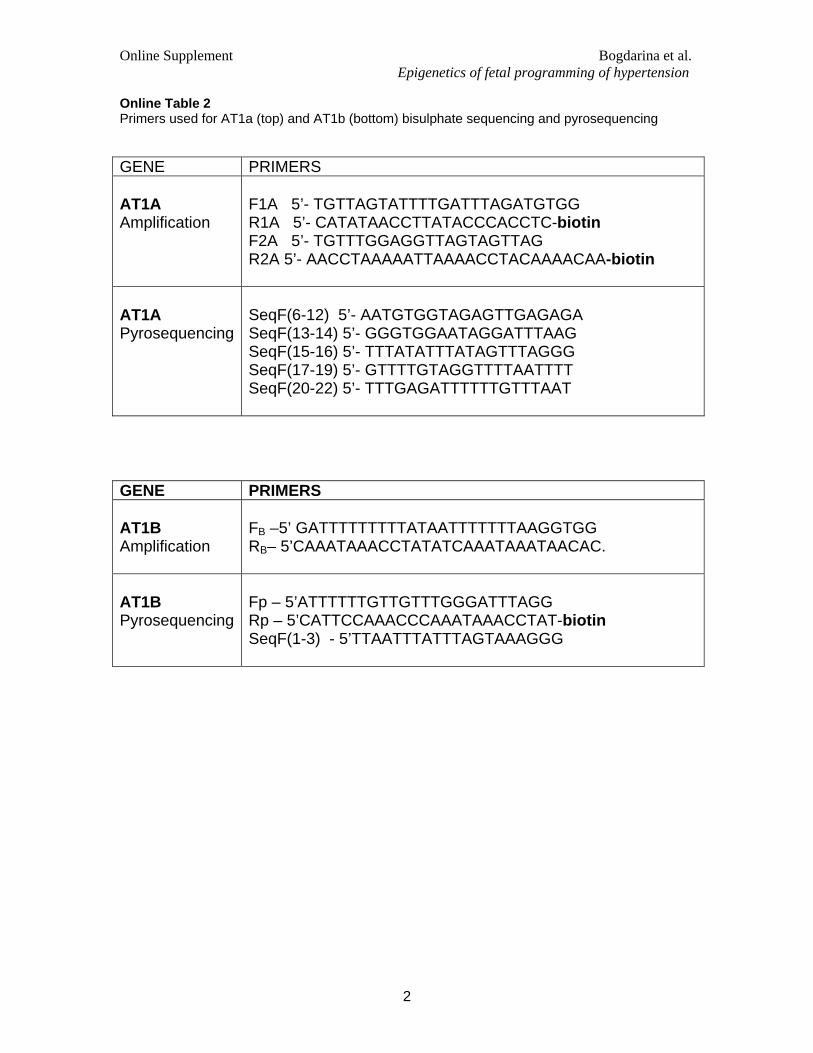
RNA Isolation and QuantitationTotal RNA was isolated (QIAgen, Crawley, UK), quantitated, andreverse-transcribed. Real-time QPCR was performed using a 2-stepcycling protocol: 95°C�10 minutes, then 40 cycles of 95°C�30sand 59°C�1 minute. Q-PCR analysis was conducted in optical96-well plates with optical caps, using the MX4000 (Stratagene).Primers and probes were designed to be intron-spanning whereverpossible. All probes were 5� labeled with 6-FAM as a reporter dyeand TAMRA as the 3� quencher. The 18S ribosomal RNA was usedas an internal reference control. Amplification plots were analyzedusing MX4000 software version 3.0 (Stratagene). RNA expressiondata were given as copy number of gene of interest/�g of RNA.Standards used were PCR fragments purified from polyacrylamidegel electrophoresis. All PCR reactions were performed in triplicate.Gene specific primer and probe sequences are shown in Table I ofthe online data supplement available at http://circres.ahajournals.org.
ImmunoblottingRat adrenals were homogenized in RIPA buffer. Laemmli bufferwithout mercaptoethanol was added to equal amounts of superna-tants (50 �g of protein). Proteins were incubated for 30 minutes at55°C before being separated on SDS-PAGE and blotted onto PVDFmembranes. Membranes were incubated with a monoclonal AT1receptor antibody (TONI-1, 1:400, Abcam, Cambridge, UK) orrabbit anti-MAPK (1:1000, NEB, Hitchin, UK) as a loading controland immune complexes were developed using enhanced chemilumi-nescence reagents (Amersham Pharmacia Biotech, Chalfont StGiles, UK).
Genomic DNA and Bisulphite ModificationGenomic DNA was isolated from whole adrenal or adrenal cortexusing DNAsol reagent (Invitrogen, Paisley, UK) or QIAamp minikit(QIAgen). DNA was then digested with restriction enzymes EcoRVand Bgl II (NEB), deproteinised with phenol/chloroform and ethanolprecipitated. DNA was treated with sodium bisulphite according tomodifications of the original protocol17 and as described.18 DNA was
ethanol precipitated, dissolved in 50 �L water and used immediatelyfor PCR or stored at �20°C. PCR conditions were: 94°C�12 min-utes, then 40 cycles of 94°C�30s, 52°C�1 minute, 72°C�30s andfinally 1 cycle of 7 minutes�72°C. The reaction mixture contained1�Ampli Gold PCR buffer, 0.2 mmol/L dNTPs, 2 mmol/L MgCl2,1 �mol/L primers, 1.25 U of AmpliTaq Gold DNA polymerase(Applied Biosystems, Warrington, UK) and 2 to 5 �L DNAtemplate. Primer sequences are shown in supplemental Table II of theonline data supplement available at http://circres.ahajournals.org
PyrosequencingPCR and sequencing primers for pyrosequencing were designedusing PSQ Assay design software (Biotage AB, Uppsala). One of thePCR primers was biotinylated, and the biotinylated strands werepurified and sequenced using the PSQTM 96MA 2.1 instrument(Biotage AB). The primer sequences are listed in supplemental TableII. Calibration curves were recorded using five mixtures of PCRproducts (0, 25, 50, 75 and 100% methylation) prepared from clonedfully methylated and unmethylated gene promoter region of the ratAT1b receptor.
TOPO-TA Cloning and SequencingGel sliced PCR products were passed through SNAP columns(Invitrogen) and used immediately for TOPO- cloning according tothe manufacturers instruction (Invitrogen). Transformed TOP10 E.coli cells were selected on LB-ampicillin (100 �g/�L) agar andsubjected to bacterial colony PCR. 15 independent clones containingthe appropriate sized insert for each amplified fragment weresequenced on an ABI 3700 automated DNA sequencer (AppliedBiosystems) in accordance with the manufacturer’s instructions.
AT1b Promoter AnalysisA 1.2kb fragment (positions 277 to 1611 from Genbank U01033)containing the rat AT1b receptor promoter was cloned into pGL3basic (Promega). The promoter was methylated in vitro with 10 U ofSssI or Hha1 (NEB) according to the manufacturers instructions.Controls included a mock methylated construct. 150 ng of eachplasmid were then cotransfected with 20 ng of the pRL-CMV Renillacontrol vector (Promega) into mouse Y1 cells. After 24 hours celllysates were prepared and luciferase activity was measured using theDual-Luciferase reporter assay (Promega) and a Wallac Victor2 1420Multilabel counter (Perkin Elmer, Finland). Reporter activity wascalculated by normalizing the reporter luciferase value to that of theRenilla control vector. Site-directed mutagenesis of C residues atCpG sites 1 to 3 in the modified AT1b promoter (positions 277 to1454 from Genbank U01033) was performed using the QuickChangeSite-directed mutagenesis protocol (Stratagene) according to to themanufacturer’s instructions. Mutations were confirmed by DNAsequencing.
Statistical AnalysisExpression levels were compared using student’s 2-tailed t test andmethylation density in control and MLP adrenals was comparedusing �2.
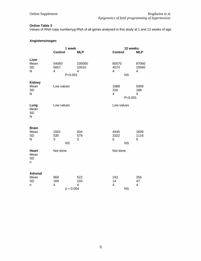
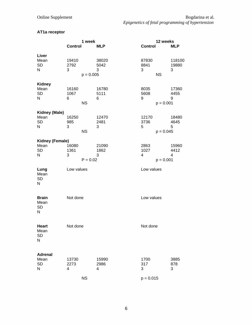
ResultsProgrammed and control animals from 10 control (5 male, 5female) and 12 MLP (7 male, 5 female) offspring from 3litters in each group were obtained as described and wereharvested for liver, lung, kidney, brain, heart, and adrenaleither at 1 or 12 weeks after birth. Real-time RT-PCR assaysfor angiotensinogen, angiotensin converting enzyme (ACE),renin, and the AT1a, AT1b, and AT2 angiotensin receptors wereperformed. Absolute values for RNA copy number of angio-tensinogen in the liver, renin in the kidney, ACE in the lungand the 3 angiotensin receptors in the adrenal are shown inFigure 1. Detailed results of all analyses are shown in
Bogdarina et al Epigenetics of Fetal Programming of Hypertension 521
by guest on June 28, 2018http://circres.ahajournals.org/
Dow
nloaded from

supplemental Table III. The only genes which showed asignificant increase in expression in maternal low proteinoffspring (MLP) at both 1 and 12 weeks were the AT1a
angiotensin receptor in the female, but not the male kidney,and the AT1b angiotensin receptor in the adrenal gland. RNAcopy number for this gene is considerably greater than that forthe other angiotensin receptors in this tissue. In contrast, theAT2 receptor showed significant reduction of expression inthe adrenal at both 1 and 12 weeks in MLP offspring.
Confirmation that these changes resulted in changes inreceptor protein was provided by immunoblotting of adrenallysates using an antibody that recognizes both AT1a and AT1b
receptors which reveals significant differences on densitom-etry (Figure 2). The histological appearance of the adrenalwas also investigated and shows no significant change instructure or zonal distribution (Figure 3).
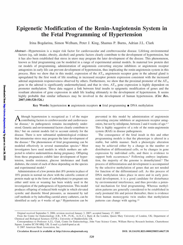
As the principal site of AT1b expression in the rat is theadrenal zona glomerulosa (ZG)20,21 we prepared adrenal
capsules (which contain the majority of the ZG cell layer)from 4 week old programmed and control animals and usedpooled tissue from one male and one female for methylationanalysis to focus on the 3 CpG sites in the proximal AT1b
promoter. Bisulphite converted DNA was subcloned and 30individual clones from each group were sequenced Thisshowed significantly less methylation at the three potentiallymethylatable sites in the proximal AT1b promoter (�2�7.37;P�0.01) (Figure 4a and b). The alteration of AT1b geneexpression was confirmed in these same samples usingreal-time RT-PCR (Figure 4c).
In view of the possibility that pooling might obscure thevariation between samples these studies were repeated in 4week old animals from three independent litters in whicheach animal’s whole adrenals were treated independently.Bisulphite conversion, PCR and subcloning was performed asbefore and revealed methylation at 21.8% of CpG sites incontrol animals against 7.4% of sites in programmed animals.
Angiotensinogen - liver Renin - kidney ACE - lung
AT1a - adrenal AT1b - adrenal AT2 - adrenal
1 wk 12 wk 1 wk 12 wk 1 wk 12 wk
1 wk 12 wk 1 wk 12 wk 1 wk 12 wk
RNA copy no./g of total RNA
(x10 )3
RNA copy no./g of total RNA
(x10 )3
b c
d e f
a
Figure 1. Altered expression of RAS genes in MLPoffspring. Real-time RT-PCR was used to quanti-tate angiotensinogen, renin, ACE, and the AT1a,AT1b, and AT2 angiotensin receptor mRNAs in vari-ous control and MLP offspring rat tissues. Notableshifts in expression at either 1 or 12 weeks of age,or both, were demonstrated (a) for angiotensino-gen in liver, (b) renin in kidney, and (d, e, and f) the3 angiotensin receptors in the adrenal. Note thedifferent range of the y axis scale. No changes inACE expression were apparent (c). Data areexpressed as RNA copy number per �g of totalRNA. White columns, control animals; black col-umns, MLP animals. *P�0.05; **P�0. 01;***P�0.001.
MAPK
AT1
a
Control MLP
1 2 3 1 2 3 4
RL
U
0
0.2
0.4
0.6
0.8
1.0
1.2
1.4
1.6
b
*
Figure 2. a, Immunoblot of AT1a and b receptor in whole adrenal from control and MLP rats at 12 weeks age. Total MAPK is used as aloading control. b, Densitometric quantitation of (a), corrected for protein loading and mean values�SD for 3 control (white bar) and 4MLP adrenals (shaded bar) are shown. *P�0.05
522 Circulation Research March 2, 2007
by guest on June 28, 2018http://circres.ahajournals.org/
Dow
nloaded from

These results are shown in Figure 4d and are significantlydifferent when assessed as a group (all 3 sites considered,�2�7.73, P�0.05) or in the case of sites 1 and 3 whenconsidered individually (site 1, �2�11.88, P�0.01; site 3,�2�7.65, P�0.05). Pyrosequencing analysis of this sameregion confirmed a similar significant reduction in methyl-ation of each of the three sites (data not shown).
Analysis of 17 CpG sites in the AT1a promoter and firstexon using Pyrosequencing showed only occasional methyl-ation of site 13 (position 3278 in rat AT1a sequence; accessionnumber S66402) and approximately 20% methylation of site14 (position 3264), both located in exon 1. There was nodifference in methylation frequency at this site between 5MLP and 4 control offspring. There was no methylation ofany other CpG site in this promoter in the adrenal.
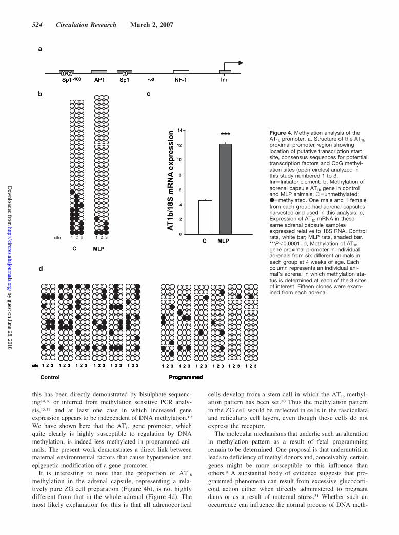
The influence of methylation of the AT1b promoter on geneexpression was demonstrated by transfection of 1.2 Kbp ofthe AT1b promoter coupled to a luciferase reporter gene intomouse adrenocortical Y1 cells. Treatment of this promoterwith the site-specific Hha1 methylase or the nonspecific CpGmethylase Sss1 before transfection results in methylation ofeither 1 or all 6 CpG sites respectively in this fragment, andin the latter case is associated with markedly reduced pro-moter activity. When the 3 proximal sites analyzed formethylation were mutated so that they could no longer bemodified by Sss1, significant loss of promoter activity wasobserved. Methylation of the 3 remaining upstream CpG siteswith Sss1 had no further influence. The contribution of eachof the three proximal sites, and notably site 3 is revealed byanalysis of the effect of their individual mutation withoutmethylation (Figure 5).
DiscussionWe show here that offspring of mothers fed a low protein dietdevelop increased expression of the AT1b receptor mRNA andprotein in the adrenal. The antibody used in immunoblotting willdetect the AT1a receptor equally well, but as the relativeexpression of AT1b at the mRNA level in these adrenal glands ismuch greater we believe that the majority of receptor proteinidentified on immunoblotting is the AT1b form. Similar findingsof increased AT1 receptor expression have been reported insheep following maternal protein deprivation.22 Moreover it hasalso been shown that the MLP rat adrenal is more responsive toangiotensin II (Ang II).23 Importantly we show that thesechanges are apparent very early in the life of programmedoffspring and persist until at least 12 weeks of age. Certain genes
(angiotensinogen, renin and AT1a receptor in kidney and AT1a
and AT2 receptor in the adrenal) show significant changes inexpression at 12 weeks of age which may relate to the earlyconsequences of developing hypertension. Other genes such asangiotensinogen and the AT1a receptor in the liver show in-creased expression at 1 week of age which has normalized by 12weeks. We speculate that this may reflect recovery from theperiod of relative undernutrition. These studies were not set up toinvestigate sex differences in gene expression, but it is interest-ing to note that female rats show significantly increased AT1a
expression in the MLP kidney at both 1 and 12 weeks of age andthis observation may be worthy of further investigation.
Interestingly, expression of the AT1b receptor is similar orreduced in the spontaneously hypertensive rat when comparedwith control Wistar Kyoto rats, probably as an adaptive re-sponse.24 Because this receptor mediates the action of thepeptide Ang II to stimulate adrenal aldosterone production, it islikely to contribute to the subsequent development of hyperten-sion. Furthermore, it has been shown that blockade of Ang IIproduction with ACE inhibitors, or interaction with the AT1
receptor using a receptor antagonist will prevent development ofhypertension in MLP offspring in contrast to nifedipene.6,7
These findings confirm an important, if not unique, role for thisreceptor in this model. Investigation of the role of the AT1b
receptor in mice in which 1 or the other, or both AT1 receptorshave been deleted suggests the AT1b receptor contributes toblood pressure maintenance and in the absence of AT1a receptorsmediates a pressor response to angiotensin 25–27
Two possible explanations for increased receptor expres-sion are conceivable. Expression of the AT1b receptors in therat adrenal is almost entirely restricted to the ZG, and fetalhyperproliferation of this cell type might be consistent withthe increased quantity of AT1b mRNA expressed in theadrenal. Differentiation of the rat ZG cells is first apparentjust before birth28,29 and although this might be influenced bythe continuation of the low protein diet during suckling, thehypertensive phenotype can equally be observed in animalsthat receive the low protein diet in pregnancy alone. Howeverhistological examination of the adrenal (Figure 3) shows noapparent ZG hyperplasia and MLP animals cannot be distin-guished from controls.
A second explanation for increased AT1b receptor geneexpression is that the fetal insult has induced a persistentmodification of AT1b gene expression. Altered DNA methyl-ation has long been proposed as a candidate mechanism forfetal programming, although there are few examples in which
Figure 3. Adrenal histology from control(a) and MLP rat (b) at 10X magnification.ZG�zona glomerulosa; ZFR�zona fas-ciculata and reticularis. Scale bar repre-sents 100 �m.
Bogdarina et al Epigenetics of Fetal Programming of Hypertension 523
by guest on June 28, 2018http://circres.ahajournals.org/
Dow
nloaded from
this has been directly demonstrated by bisulphate sequenc-ing14,16 or inferred from methylation sensitive PCR analy-sis,15,17 and at least one case in which increased geneexpression appears to be independent of DNA methylation.19
We have shown here that the AT1b gene promoter, whichquite clearly is highly susceptible to regulation by DNAmethylation, is indeed less methylated in programmed ani-mals. The present work demonstrates a direct link betweenmaternal environmental factors that cause hypertension andepigenetic modification of a gene promoter.
It is interesting to note that the proportion of AT1b
methylation in the adrenal capsule, representing a rela-tively pure ZG cell preparation (Figure 4b), is not highlydifferent from that in the whole adrenal (Figure 4d). Themost likely explanation for this is that all adrenocortical
cells develop from a stem cell in which the AT1b methyl-ation pattern has been set.30 Thus the methylation patternin the ZG cell would be reflected in cells in the fasciculataand reticularis cell layers, even though these cells do notexpress the receptor.
The molecular mechanisms that underlie such an alterationin methylation pattern as a result of fetal programmingremain to be determined. One proposal is that undernutritionleads to deficiency of methyl donors and, conceivably, certaingenes might be more susceptible to this influence thanothers.8 A substantial body of evidence suggests that pro-grammed phenomena can result from excessive glucocorti-coid action either when directly administered to pregnantdams or as a result of maternal stress.31 Whether such anoccurrence can influence the normal process of DNA meth-
Programmed
site 1 2 3 1 2 3 1 2 3 1 2 3 1 2 3 1 2 3 1 2 3 1 2 3 1 2 3 1 2 3 1 2 3 1 2 3
-100Sp1 Sp1AP1 InrNF-1-501 2 3
Programmed
site 1 2 3 1 2 3 1 2 3 1 2 3 1 2 3 1 2 3 1 2 3 1 2 3 1 2 3 1 2 3 1 2 3 1 2 3
Control Programmed
site 1 2 3 1 2 3 1 2 3 1 2 3 1 2 3 1 2 3 1 2 3 1 2 3 1 2 3 1 2 3 1 2 3 1 2 3
Programmed
site 1 2 3 1 2 3 1 2 3 1 2 3 1 2 3 1 2 3 1 2 3 1 2 3 1 2 3 1 2 3 1 2 3 1 2 3
b
-100Sp1 Sp1AP1 InrNF-1-501 2 3
-100Sp1 Sp1AP1 InrNF-1-501 2 3
MLPCC MLP
0
2
4
6
8
10
12
14
AT
1b/1
8S m
RN
A e
xpre
ssio
n
c
1 2 3 1 2 3
a
site
d
*** Figure 4. Methylation analysis of theAT1b promoter. a, Structure of the AT1b
proximal promoter region showinglocation of putative transcription startsite, consensus sequences for potentialtranscription factors and CpG methyl-ation sites (open circles) analyzed inthis study numbered 1 to 3.Inr�Initiator element. b, Methylation ofadrenal capsule AT1b gene in controland MLP animals. E�unmethylated;●�methylated. One male and 1 femalefrom each group had adrenal capsulesharvested and used in this analysis. c,Expression of AT1b mRNA in thesesame adrenal capsule samplesexpressed relative to 18S RNA. Controlrats, white bar; MLP rats, shaded bar.***P�0.0001. d, Methylation of AT1b
gene proximal promoter in individualadrenals from six different animals ineach group at 4 weeks of age. Eachcolumn represents an individual ani-mal’s adrenal in which methylation sta-tus is determined at each of the 3 sitesof interest. Fifteen clones were exam-ined from each adrenal.
524 Circulation Research March 2, 2007
by guest on June 28, 2018http://circres.ahajournals.org/
Dow
nloaded from
ylation remains to be determined. The data obtained using invitro methylation shown in Figure 5 argue that CpG site 3 hasa particularly potent influence on promoter activity in thatwhen mutated or methylated the promoter is essentiallyinactive. This base is at the center of an Sp1 consensus site,suggesting that failure to bind this, or a related transcriptionfactor may have a crucial impact. However it is generallyconsidered that Sp1 binding is not influenced by DNAmethylation32 although there are exceptions to this33 and it ispossible that another methylation sensitive transcription fac-tor binds here.
The human genome only contains a single AT1 receptorgene which is widely expressed in a pattern similar to that ofthe AT1a receptor.34 There is no human equivalent to therelatively adrenal-specific AT1b receptor. Thus these findingsmay be difficult to translate directly into the human hyper-tensive scenario. Furthermore there is also data derived fromhuman monozygotic twin studies that challenges the viewthat methylation patterns are established in early life andremain unchanged thereafter.10 Nevertheless, the possibilitythat maternal undernutrition may result in highly specificaltered methylation patterns in the offspring provides in-creased mechanistic understanding worthy of investigation inman.
Sources of FundingThis work was funded by a grant from the British Heart Foundation.S.W. is supported by Diabetes UK
DisclosuresNone.
References1. He FJ, MacGregor GA. Cost of poor blood pressure control in the UK:
62,000 unnecessary deaths per year. J Hum Hypertens. 2003;17:455–457.2. Staessen JA, Wang J, Bianchi G, Birkenhager WH. Essential hyper-
tension. Lancet. 2003;361:1629–1641.
3. Barker DJ, Osmond C, Golding J, Kuh D, Wadsworth ME. Growth inutero, blood pressure in childhood and adult life, and mortality fromcardiovascular disease. BMJ. 1989;298:564–567.
4. Bertram CE, Hanson MA. Animal models and programming of themetabolic syndrome. Br Med Bull. 2001;60:103–121.
5. Langley-Evans SC. Critical differences between two low protein dietprotocols in the programming of hypertension in the rat. Int J Food SciNutr. 2000;51:11–17.
6. Sherman RC, Langley-Evans SC. Early administration of angiotensin-converting enzyme inhibitor captopril, prevents the development ofhypertension programmed by intrauterine exposure to a maternal low-protein diet in the rat. Clin Sci (Lond). 1998;94:373–381.
7. Sherman RC, Langley-Evans SC. Antihypertensive treatment in earlypostnatal life modulates prenatal dietary influences upon blood pressurein the rat. Clin Sci (Lond). 2000;98:269–275.
8. Burns SP, Desai M, Cohen RD, Hales CN, Iles RA, Germain JP, GoingTC, Bailey RA. Gluconeogenesis, glucose handling, and structuralchanges in livers of the adult offspring of rats partially deprived of proteinduring pregnancy and lactation. J Clin Invest. 1997;100:1768–1774.
9. Cedar H. DNA methylation and gene activity. Cell. 1988;53:3–4.10. Fraga MF, Ballestar E, Paz MF, Ropero S, Setien F, Ballestar ML,
Heine-Suner D, Cigudosa JC, Urioste M, Benitez J, Boix-Chornet M,Sanchez-Aguilera A, Ling C, Carlsson E, Poulsen P, Vaag A, Stephan Z,Spector TD, Wu YZ, Plass C, Esteller M. Epigenetic differences ariseduring the lifetime of monozygotic twins. Proc Natl Acad Sci U S A.2005;102:10604–10609.
11. Rees WD, Hay SM, Brown DS, Antipatis C, Palmer RM. Maternalprotein deficiency causes hypermethylation of DNA in the livers of ratfetuses. J Nutr. 2000;130:1821–1826.
12. Jackson AA, Dunn RL, Marchand MC, Langley-Evans SC. Increasedsystolic blood pressure in rats induced by a maternal low-protein diet isreversed by dietary supplementation with glycine. Clin Sci (Lond). 2002;103:633–639.
13. Brawley L, Torrens C, Anthony FW, Itoh S, Wheeler T, Jackson AA,Clough GF, Poston L, Hanson MA. Glycine rectifies vascular dysfunctioninduced by dietary protein imbalance during pregnancy. J Physiol. 2004;554:497–504.
14. Weaver IC, Cervoni N, Champagne FA, D’Alessio AC, Sharma S, SecklJR, Dymov S, Szyf M, Meaney MJ. Epigenetic programming by maternalbehavior. Nat Neurosci. 2004;7:847–854.
15. Lillycrop KA, Phillips ES, Jackson AA, Hanson MA, Burdge GC. Dietaryprotein restriction of pregnant rats induces and folic acid supplementationprevents epigenetic modification of hepatic gene expression in the off-spring. J Nutr. 2005;135:1382–1386.
16. Pham TD, MacLennan NK, Chiu CT, Laksana GS, Hsu JL, Lane RH.Uteroplacental insufficiency increases apoptosis and alters p53 gene
0.0 0.5 1.0 1.5 2.0 2.5 3.0 3.5 4.0 4.5
Relative light units
1 2 3
1 32
2
2
1
1
3
3
Hha1
Sss1
Sss1
Figure 5. Influence of methylation on the AT1b promoter. Luciferase activity of AT1b-luc transiently transfected into mouse Y1 cells in thepresence or absence of methylation of either 1 or 6 CpG sites in the proximal promoter using Hha1 methylase (Hha1) or Sss1 methyl-ase (Sss1) respectively. Mutagenesis of the 3 proximal sites1–3 to render them nonmethylatable markedly reduces promoter activity, andmethylation of the remaining promoter makes no further contribution to reduced activity. Mutagenesis of individual CpG sites in theproximal promoter reveals the considerable impact of site 3. Mean luciferase activity is expressed relative to cotransfected renilla lucif-erase activity (�SEM). n�6. E�unmethylated site, ●�methylated site, X�mutagenised site.
Bogdarina et al Epigenetics of Fetal Programming of Hypertension 525
by guest on June 28, 2018http://circres.ahajournals.org/
Dow
nloaded from

methylation in the full-term IUGR rat kidney. Am J Physiol Regul IntegrComp Physiol. 2003;285:R962–R970.
17. Dolinoy DC, Weidman JR, Waterland RA, Jirtle RL. Maternal genisteinalters coat color and protects Avy mouse offspring from obesity bymodifying the fetal epigenome. Environ Health Perspect. 2006;114:567–572.
18. Frommer M, McDonald LE, Millar DS, Collis CM, Watt F, Grigg GW,Molloy PL, Paul CL. A genomic sequencing protocol that yields apositive display of 5-methylcytosine residues in individual DNA strands.Proc Natl AcadSci USA. 1992;89:1827–1831.
19. Bogdarina I, Murphy HC, Burns SP, Clark AJ. Investigation of the role ofepigenetic modification of the rat glucokinase gene in fetal programming.Life Sci. 2004;74:1407–1415.
20. Sandberg K, Ji H, Clark AJ, Shapira H, Catt KJ. Cloning and expressionof a novel angiotensin II receptor subtype. J Biol Chem. 1992;267:9455–9458.
21. Gasc JM, Shanmugam S, Sibony M, Corvol P. Tissue-specific expressionof type 1 angiotensin II receptor subtypes. An in situ hybridization study.Hypertension. 1994;24:531–537.
22. Whorwood CB, Firth KM, Budge H, Symonds ME. Maternal undernu-trition during early to midgestation programs tissue-specific alterations inthe expression of the glucocorticoid receptor, 11beta-hydroxysteroiddehydrogenase isoforms, and type 1 angiotensin ii receptor in neonatalsheep. Endocrinol. 2001;142:2854–2864.
23. McMullen S, Gardner DS, Langley-Evans SC. Prenatal programming ofangiotensin II type 2 receptor expression in the rat. Br J Nutr. 2004;91:133–140.
24. Johren O, Golsch C, Dendorfer A, Qadri F, Hauser W, Dominiak P.Differential expression of AT1 receptors in the pituitary and adrenalgland of SHR and WKY. Hypertension. 2003;41:984–990.
25. Chen X, Li W, Yoshida H, Tsuchida S, Nishimura H, Takemoto F, OkuboS, Fogo A, Matsusaka T, Ichikawa I. Targeting deletion of angiotensintype 1B receptor gene in the mouse. Am J Physiol. 1997;272:F299–F304.
26. Oliverio MI, Best CF, Kim HS, Arendshorst WJ, Smithies O, CoffmanTM. Angiotensin II responses in AT1A receptor-deficient mice: a role forAT1B receptors in blood pressure regulation. Am J Physiol. 1997;272:F515–F520.
27. Audoly LP, Oliverio MI, Coffman TM. Insights into the functions of type1 (AT1) angiotensin receptors provided by gene targeting. Trends Endo-crinol Metab. 2000;11:263–269.
28. Mitani F, Mukai K, Ogawa T, Miyamoto H, Ishimura Y. Expression ofcytochromes P450aldo and P45011 beta in rat adrenal gland during lategestational and neonatal stages. Steroids. 1997;62:57–61.
29. Wotus C, Levay-Young BK, Rogers LM, Gomez-Sanchez CE, EngelandWC. Development of adrenal zonation in fetal rats defined by expressionof aldosterone synthase and 11beta-hydroxylase. Endocrinol. 1998;139:4397–4403.
30. Hammer GD, Parker KL, Schimmer BP. Minireview: transcriptionalregulation of adrenocortical development. Endocrinol. 2005;146:1018–1024.
31. Seckl JR, Meaney MJ. Glucocorticoid programming. Ann N Y Acad Sci.2004;1032:63–84.
32. Tate PH, Bird AP. Effects of DNA methylation on DNA-binding proteinsand gene expression. Curr Opin Genet Dev. 1993;3:226–231.
33. Alikhani-Koopaei R, Fouladkou F, Frey FJ, Frey BM. Epigenetic regu-lation of 11 beta-hydroxysteroid dehydrogenase type 2 expression. J ClinInvest. 2004;114:1146–1157.
34. Inagami T. Recent progress in molecular and cell biological studies ofangiotensin receptors. Curr Opin Nephrol Hypertens. 1995;4:47–54.
526 Circulation Research March 2, 2007
by guest on June 28, 2018http://circres.ahajournals.org/
Dow
nloaded from

Irina Bogdarina, Simon Welham, Peter J. King, Shamus P. Burns and Adrian J.L. ClarkHypertension
Epigenetic Modification of the Renin-Angiotensin System in the Fetal Programming of
Print ISSN: 0009-7330. Online ISSN: 1524-4571 Copyright © 2007 American Heart Association, Inc. All rights reserved.is published by the American Heart Association, 7272 Greenville Avenue, Dallas, TX 75231Circulation Research
doi: 10.1161/01.RES.0000258855.60637.582007;100:520-526; originally published online January 25, 2007;Circ Res.
http://circres.ahajournals.org/content/100/4/520World Wide Web at:
The online version of this article, along with updated information and services, is located on the
http://circres.ahajournals.org/content/suppl/2007/01/25/01.RES.0000258855.60637.58.DC1Data Supplement (unedited) at:
http://circres.ahajournals.org//subscriptions/
is online at: Circulation Research Information about subscribing to Subscriptions:
http://www.lww.com/reprints Information about reprints can be found online at: Reprints:
document. Permissions and Rights Question and Answer about this process is available in the
located, click Request Permissions in the middle column of the Web page under Services. Further informationEditorial Office. Once the online version of the published article for which permission is being requested is
can be obtained via RightsLink, a service of the Copyright Clearance Center, not theCirculation Researchin Requests for permissions to reproduce figures, tables, or portions of articles originally publishedPermissions:
by guest on June 28, 2018http://circres.ahajournals.org/
Dow
nloaded from
Online Supplement Bogdarina et al. Epigenetics of fetal programming of hypertension
1
Online Table 1 Sequence of DNA oligonucleotide primers and probes used in RNA quantitation experiments
GENE PRIMERS and PROBES
RENIN NM_012642
5’ (F) GTAACTGTGGGTGGAATCATTGTG 5’ (R) TGGGAGAGAATGTGGTCGAAGA probe TTGGAGAGGTCACCGAGCTGCCCC
AT1A M86912
5’ (F) GGAGAGGATTCGTGGCTTGAG 5’ (R) CTTTCTGGGAGGGTTGTGTGAT probe TTCCACCCGATCACCGATCACCGG
AT1B M90065
5’ (F) TTGTCCACCCAATGAAGTCTCG 5’ (R) CGCAAACTGTGATATTGGTGTTCT probe CCGCCGCACGATGCTGGTAGCC
AT2 D16840
5’ (F) CATCACCAGCAGTCTTCCTTTTG 5’ (R) AAAACAGTGAGACCACAACAATGT probe CGCAACTGGCACCAATGAGTCCGC
ACE NM_12544
5’ (F) CGGGTCGCAGAGGAATTCTT 5’ (R) CCTGAAGTCCTTCCTGTTGTAGA probe CACGCAGAGGCATGGCACACCACC
Angiotensinogen NM_134432
5’ (F) AGAACCCCAGTGTGGAGACG 5’ (R) AGCCAACCTTTGAGCCTGTGCCCA probe AGCCAACCTTTGAGCCTGTGCCCA
Online Supplement Bogdarina et al. Epigenetics of fetal programming of hypertension
2
Online Table 2 Primers used for AT1a (top) and AT1b (bottom) bisulphate sequencing and pyrosequencing GENE PRIMERS AT1A Amplification
F1A 5’- TGTTAGTATTTTGATTTAGATGTGG R1A 5’- CATATAACCTTATACCCACCTC-biotin F2A 5’- TGTTTGGAGGTTAGTAGTTAG R2A 5’- AACCTAAAAATTAAAACCTACAAAACAA-biotin
AT1A Pyrosequencing
SeqF(6-12) 5’- AATGTGGTAGAGTTGAGAGA SeqF(13-14) 5’- GGGTGGAATAGGATTTAAG SeqF(15-16) 5’- TTTATATTTATAGTTTAGGG SeqF(17-19) 5’- GTTTTGTAGGTTTTAATTTT SeqF(20-22) 5’- TTTGAGATTTTTTGTTTAAT
GENE PRIMERS AT1B Amplification
FB –5’ GATTTTTTTTTATAATTTTTTTAAGGTGG RB– 5’CAAATAAACCTATATCAAATAAATAACAC.
AT1B Pyrosequencing
Fp – 5’ATTTTTTGTTGTTTGGGATTTAGG Rp – 5’CATTCCAAACCCAAATAAACCTAT-biotin SeqF(1-3) - 5’TTAATTTATTTAGTAAAGGG
Online Supplement Bogdarina et al. Epigenetics of fetal programming of hypertension
3
Online Table 3 Values of RNA copy number/µg RNA of all genes analysed in this study at 1 and 12 weeks of age Angiotensinogen 1 week 12 weeks Control MLP Control MLP Liver Mean 54060 105000 80570 87060 SD 5657 10010 4574 10940 N 4 4 4 4 P<0.001 NS Kidney Mean Low values 1688 5058 SD 316 188 N 4 4 P<0.001 Lung Low values Low values Mean SD N Brain Mean 1501 934 4445 1839 SD 535 579 3322 1116 N 3 3 6 6 NS NS Heart Not done Not done Mean SD n Adrenal Mean 969 522 242 256 SD 169 103 14 47 n 4 4 4 4 p = 0.004 NS

Online Supplement Bogdarina et al. Epigenetics of fetal programming of hypertension
4
Renin 1 week 12 weeks Control MLP Control MLP Liver Not done Not done Mean SD N Kidney Mean 27170 25640 55770 244800 SD 6250 7469 28440 76450 N 8 8 8 8 NS p<0.001 Kidney (Male) Mean 26820 24980 80640 305100 SD 2529 6525 13300 45990 N 4 4 4 4 NS p<0.001 Kidney (Female) Mean 27510 26300 30900 184400 SD 9189 9297 7850 42650 N 4 4 4 4 NS p<0.001 Lung Not done Not done Mean SD N Brain Low values Low values Mean SD N Heart Not done Not done Mean SD N Adrenal Low values Low values Mean SD N

Online Supplement Bogdarina et al. Epigenetics of fetal programming of hypertension
5
ACE 1 week 12 weeks Control MLP Control MLP Liver Not done Not done Mean SD N Kidney Mean 349300 328500 340500 333000 SD 81700 44300 78700 37500 N 4 4 4 4 NS NS Lung Mean 2708000 2464000 2832000 2664000 SD 747700 519300 834800 553700 N 5 5 5 5 NS NS Brain Not done Not done Mean SD N Heart Not done Not done Mean SD N Adrenal Not done Not done Mean SD N
Online Supplement Bogdarina et al. Epigenetics of fetal programming of hypertension
6
AT1a receptor 1 week 12 weeks Control MLP Control MLP Liver Mean 19410 38020 87830 118100 SD 2792 5042 8841 19880 N 3 3 3 3 p = 0.005 NS Kidney Mean 16160 16780 8035 17360 SD 1067 5111 5608 4455 N 6 6 9 9 NS p = 0.001 Kidney (Male) Mean 16250 12470 12170 18480 SD 985 2481 3736 4645 N 3 3 5 5 NS p = 0.045 Kidney (Female) Mean 16080 21090 2863 15960 SD 1361 1862 1027 4412 N 3 3 4 4 P = 0.02 p = 0.001 Lung Low values Low values Mean SD N Brain Not done Low values Mean SD N Heart Not done Not done Mean SD N Adrenal Mean 13730 15990 1700 3885 SD 2273 2986 317 878 N 4 4 3 3 NS p = 0.015

Online Supplement Bogdarina et al. Epigenetics of fetal programming of hypertension
7
AT1b receptor 1 week 12 weeks Control MLP Control MLP Liver Low values Low values Mean SD N Kidney Low values Mean 18790 16480 SD 19410 11880 N 6 6 Lung Low values Low values Mean SD N Brain Low values Low values Mean SD N Heart Low values Low values Mean SD N Adrenal Mean 99430 182000 186000 678800 SD 4410 4593 12800 224000 N 4 4 4 4 p = 0.04 p = 0.004

Online Supplement Bogdarina et al. Epigenetics of fetal programming of hypertension
8
AT2 receptor 1 week 12 weeks Control MLP Control MLP Liver Low values Low values Mean SD N Kidney Not done Mean 13810 17710 SD 5888 1079 N 6 6 NS Lung Not done Not done Mean SD N Brain Not done Not done Mean SD N Heart Low values Low values Mean SD N Adrenal Mean 71730 25350 54380 36100 SD 10560 2700 15980 2934 N 6 6 6 6 p<0.001 p = 0.02