effect of melatonin on the renin-angiotensin-aldosterone
TRANSCRIPT
molecules
Article
Effect of Melatonin on theRenin-Angiotensin-Aldosterone System inL-NAME-Induced Hypertension
Fedor Simko 1,2,3,*, Tomas Baka 1, Kristina Krajcirovicova 1, Kristina Repova 1 ID ,Silvia Aziriova 1, Stefan Zorad 3, Marko Poglitsch 4, Michaela Adamcova 5, Russel J. Reiter 6,†
and Ludovit Paulis 1,7,† ID
1 Institute of Pathophysiology, Faculty of Medicine, Comenius University, Sasinkova 4,81108 Bratislava, Slovakia; [email protected] (T.B.); [email protected] (K.K.);[email protected] (K.R.); [email protected] (S.A.); [email protected] (L.P.)
2 3rd Department of Internal Medicine, Faculty of Medicine, Comenius University, 83305 Bratislava, Slovakia3 Institute of Experimental Endocrinology, Biomedical Research Center, Slovak Academy of Sciences,
84505 Bratislava, Slovakia; [email protected] Attoquant Diagnostics, 1030 Vienna, Austria; [email protected] Department of Physiology, School of Medicine, Charles University, 50003 Hradec Kralove, Czech Republic;
[email protected] Department of Cellular and Structural Biology, UT Health Science Center, San Antonio, TX 78229, USA;
[email protected] Institute of Normal and Pathological Physiology, Center for Experimental Medicine,
Slovak Academy of Sciences, 81371 Bratislava, Slovakia* Correspondence: [email protected]† These authors contributed equally to this work.
Received: 20 December 2017; Accepted: 25 January 2018; Published: 29 January 2018
Abstract: The renin-angiotensin-aldosterone system (RAAS) is a dominant player in severalcardiovascular pathologies. This study investigated whether alterations induced by L-NAME,(NLG)-nitro-L-arginine methyl ester, a nitric oxide synthase inhibitor, and the protective effectof melatonin are associated with changes in the RAAS. Four groups of 3-month-old male Wistarrats (n = 10) were treated as follows for four weeks: untreated controls, rats treated with melatonin(10 mg/kg/day), rats treated with L-NAME (40 mg/kg/day), and rats treated with L-NAME +melatonin. L-NAME administration led to hypertension and left ventricular (LV) fibrosis in termsof enhancement of soluble, insoluble and total collagen concentration and content. Melatoninreduced systolic blood pressure enhancement and lowered the concentration and content of insolubleand total collagen in the LV. The serum concentration of angiotensin (Ang) 1–8 (Ang II) and itsdownstream metabolites were reduced in the L-NAME group and remained unaltered by melatonin.The serum aldosterone level and its ratio to Ang II (AA2-ratio) were increased in the L-NAME groupwithout being modified by melatonin. We conclude that L-NAME-hypertension is associated withreduced level of Ang II and its downstream metabolites and increased aldosterone concentrationand AA2-ratio. Melatonin exerts its protective effect in L-NAME-induced hypertension withoutaffecting RAAS.
Keywords: L-NAME; fibrosis; melatonin; angiotensin II; angiotensin 1–7; aldosterone
1. Introduction
Nitric oxide (NO) provides cardiovascular protection by reducing blood pressure and via itsantiproliferative and antifibrotic actions [1,2]. Chronic L-NAME ((NLG)-nitro-L-arginine methyl ester)
Molecules 2018, 23, 265; doi:10.3390/molecules23020265 www.mdpi.com/journal/molecules

Molecules 2018, 23, 265 2 of 15
administration causes hypertension and target organ damage via the inhibition of NO synthaseactivity reducing NO production [3–5]. Moreover, L-NAME-induced deterioration of the vasodilatoryfunction of the renal artery may trigger renin release and renin-angiotensin-aldosterone system (RAAS)activation [6,7].
Melatonin (N-acetyl-5-methoxytryptamine), the secretory product of the vertebrate pineal gland,influences biological rhythms [8,9], it also has a number of beneficial effects on the cardiovascularsystem including blood pressure reduction and attenuation of target organ damage [10–12]. Conversely,melatonin deficiency induced by experimental pinealectomy or continuous light exposure in rats resultsin the development of hypertension and myocardial fibrosis [13,14]. The pathomechanism of melatoninprotection seems to be highly complex. Besides the activation of specific membrane and nuclearreceptors [15,16], melatonin provides cardiovascular protection on several levels including antioxidantand scavenging actions, endothelial protection or sympatholytic effects [13,17–20], and additionalpleiotropic effects are emerging [21]. However, there is a shortage of data on the ability of melatoninto interfere with neurohumoral activation, particularly with the RAAS in variable cardiovascularpathologies. The aim of the current study was to test whether hemodynamic and structural changesinduced by L-NAME are associated with the modification in the RAAS and whether melatonininterferes with RAAS alterations.
2. Results
2.1. Cardiovascular Parameters
After four weeks of treatment, systolic blood pressure (SBP) was 124.68 ± 1.52 mmHg in thecontrol and 185 ± 3.05 mmHg in L-NAME groups. SBP was significantly lower (p < 0.05) dueto melatonin treatment (15%) compared to the L-NAME group (Figure 1A). The left ventricularweight/body weight (LVW/BW) ratio after four weeks of treatment was 1.229 ± 0.040 mg/g and1.450 ± 0.043 mg/g, in the control and L-NAME-groups, respectively; this was not influenced bymelatonin treatment (Figure 1B). The body weight (BW), absolute left ventricular weight (LVW) andright ventricular weight (RVW) and RVW/BW ratio are described in Table 1.
Molecules 2018, 23, x 2 of 15
vasodilatory function of the renal artery may trigger renin release and renin-angiotensin-aldosterone system (RAAS) activation [6,7].
Melatonin (N-acetyl-5-methoxytryptamine), the secretory product of the vertebrate pineal gland, influences biological rhythms [8,9], it also has a number of beneficial effects on the cardiovascular system including blood pressure reduction and attenuation of target organ damage [10–12]. Conversely, melatonin deficiency induced by experimental pinealectomy or continuous light exposure in rats results in the development of hypertension and myocardial fibrosis [13,14]. The pathomechanism of melatonin protection seems to be highly complex. Besides the activation of specific membrane and nuclear receptors [15,16], melatonin provides cardiovascular protection on several levels including antioxidant and scavenging actions, endothelial protection or sympatholytic effects [13,17–20], and additional pleiotropic effects are emerging [21]. However, there is a shortage of data on the ability of melatonin to interfere with neurohumoral activation, particularly with the RAAS in variable cardiovascular pathologies. The aim of the current study was to test whether hemodynamic and structural changes induced by L-NAME are associated with the modification in the RAAS and whether melatonin interferes with RAAS alterations.
2. Results
2.1. Cardiovascular Parameters
After four weeks of treatment, systolic blood pressure (SBP) was 124.68 ± 1.52 mmHg in the control and 185 ± 3.05 mmHg in L-NAME groups. SBP was significantly lower (p < 0.05) due to melatonin treatment (15%) compared to the L-NAME group (Figure 1A). The left ventricular weight/body weight (LVW/BW) ratio after four weeks of treatment was 1.229 ± 0.040 mg/g and 1.450 ± 0.043 mg/g, in the control and L-NAME-groups, respectively; this was not influenced by melatonin treatment (Figure 1B). The body weight (BW), absolute left ventricular weight (LVW) and right ventricular weight (RVW) and RVW/BW ratio are described in Table 1.
Figure 1. The influence of melatonin (LN + Mel) on blood pressure (A) and relative left ventricular weight (LVW/BW); (B) in L-NAME-treated rats. Wistar controls (c); L-NAME (LN); melatonin (Mel); n = 10 per group; * p < 0.05 vs. c; # p < 0.05 vs. LN; + p < 0.05 vs. Mel.
Figure 1. The influence of melatonin (LN + Mel) on blood pressure (A) and relative left ventricularweight (LVW/BW); (B) in L-NAME-treated rats. Wistar controls (c); L-NAME (LN); melatonin (Mel);n = 10 per group; * p < 0.05 vs. c; # p < 0.05 vs. LN; + p < 0.05 vs. Mel.
Molecules 2018, 23, 265 3 of 15
Table 1. The body weight (BW), right ventricular weight (RVW), relative RVW (RVW/BW) and leftventricular weight (LVW) in control rats (c), rats treated with melatonin (Mel), with L-NAME (LN),with L-NAME and melatonin (LN + Mel) (n = 10 per group).
BW (g) LVW (mg) RVW (mg) RVW/BW (mg/g)
c 346.50 ± 6.19 425.30 ± 14.05 164.50 ± 6.08 0.48 ± 0.03Mel 318.00 ± 7.50 427.00 ± 17.08 160.00 ± 8.40 0.50 ± 0.03LN 318.50 ± 7.64 461.90 ± 17.98 155.00 ± 7.04 0.49 ± 0.03
LN + Mel 304.00 ± 11.57 * 457.50 ± 22.53 158.60 ± 9.50 0.52 ± 0.03
Values are mean ± S.E.M., * p < 0.05 vs. c.
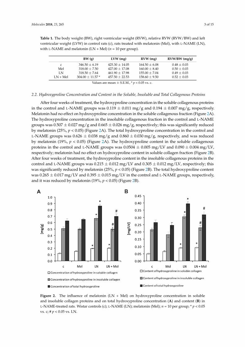
2.2. Hydroxyproline Concentration and Content in the Soluble, Insoluble and Total Collagenous Proteins
After four weeks of treatment, the hydroxyproline concentration in the soluble collagenous proteinsin the control and L-NAME groups was 0.119 ± 0.011 mg/g and 0.194 ± 0.007 mg/g, respectively.Melatonin had no effect on hydroxyproline concentration in the soluble collagenous fraction (Figure 2A).The hydroxyproline concentration in the insoluble collagenous fraction in the control and L-NAMEgroups was 0.507 ± 0.027 mg/g and 0.665 ± 0.026 mg/g, respectively; this was significantly reducedby melatonin (25%, p < 0.05) (Figure 2A). The total hydroxyproline concentration in the control andL-NAME groups was 0.626 ± 0.038 mg/g and 0.860 ± 0.030 mg/g, respectively, and was reducedby melatonin (19%, p < 0.05) (Figure 2A). The hydroxyproline content in the soluble collagenousproteins in the control and L-NAME groups was 0.0504 ± 0.005 mg/LV and 0.090 ± 0.004 mg/LV,respectively; melatonin had no effect on hydroxyproline content in soluble collagen fraction (Figure 2B).After four weeks of treatment, the hydroxyproline content in the insoluble collagenous proteins in thecontrol and L-NAME groups was 0.215 ± 0.012 mg/LV and 0.305 ± 0.012 mg/LV, respectively; thiswas significantly reduced by melatonin (25%, p < 0.05) (Figure 2B). The total hydroxyproline contentwas 0.265 ± 0.017 mg/LV and 0.395 ± 0.015 mg/LV in the control and L-NAME groups, respectively,and it was reduced by melatonin (19%, p < 0.05) (Figure 2B).
Molecules 2018, 23, x 3 of 15
Table 1. The body weight (BW), right ventricular weight (RVW), relative RVW (RVW/BW) and left ventricular weight (LVW) in control rats (c), rats treated with melatonin (Mel), with L-NAME (LN), with L-NAME and melatonin (LN + Mel) (n = 10 per group).
BW (g) LVW (mg) RVW (mg) RVW/BW (mg/g) c 346.50 ± 6.19 425.30 ± 14.05 164.50 ± 6.08 0.48 ± 0.03
Mel 318.00 ± 7.50 427.00 ± 17.08 160.00 ± 8.40 0.50 ± 0.03 LN 318.50 ± 7.64 461.90 ± 17.98 155.00 ± 7.04 0.49 ± 0.03
LN + Mel 304.00 ± 11.57 * 457.50 ± 22.53 158.60 ± 9.50 0.52 ± 0.03 Values are mean ± S.E.M., * p < 0.05 vs. c.
2.2. Hydroxyproline Concentration and Content in the Soluble, Insoluble and Total Collagenous Proteins
After four weeks of treatment, the hydroxyproline concentration in the soluble collagenous proteins in the control and L-NAME groups was 0.119 ± 0.011 mg/g and 0.194 ± 0.007 mg/g, respectively. Melatonin had no effect on hydroxyproline concentration in the soluble collagenous fraction (Figure 2A). The hydroxyproline concentration in the insoluble collagenous fraction in the control and L-NAME groups was 0.507 ± 0.027 mg/g and 0.665 ± 0.026 mg/g, respectively; this was significantly reduced by melatonin (25%, p < 0.05) (Figure 2A). The total hydroxyproline concentration in the control and L-NAME groups was 0.626 ± 0.038 mg/g and 0.860 ± 0.030 mg/g, respectively, and was reduced by melatonin (19%, p < 0.05) (Figure 2A). The hydroxyproline content in the soluble collagenous proteins in the control and L-NAME groups was 0.0504 ± 0.005 mg/LV and 0.090 ± 0.004 mg/LV, respectively; melatonin had no effect on hydroxyproline content in soluble collagen fraction (Figure 2B). After four weeks of treatment, the hydroxyproline content in the insoluble collagenous proteins in the control and L-NAME groups was 0.215 ± 0.012 mg/LV and 0.305 ± 0.012 mg/LV, respectively; this was significantly reduced by melatonin (25%, p < 0.05) (Figure 2B). The total hydroxyproline content was 0.265 ± 0.017 mg/LV and 0.395 ± 0.015 mg/LV in the control and L-NAME groups, respectively, and it was reduced by melatonin (19%, p < 0.05) (Figure 2B).
Figure 2. The influence of melatonin (LN + Mel) on hydroxyproline concentration in soluble and insoluble collagen proteins and on total hydroxyproline concentration (A) and content (B) in L-NAME-treated rats. Wistar controls (c); L-NAME (LN); melatonin (Mel); n = 10 per group; * p < 0.05 vs. c; # p < 0.05 vs. LN.
Figure 2. The influence of melatonin (LN + Mel) on hydroxyproline concentration in solubleand insoluble collagen proteins and on total hydroxyproline concentration (A) and content (B) inL-NAME-treated rats. Wistar controls (c); L-NAME (LN); melatonin (Mel); n = 10 per group; * p < 0.05vs. c; # p < 0.05 vs. LN.
Molecules 2018, 23, 265 4 of 15
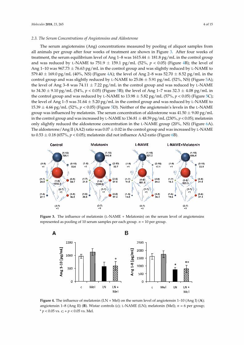
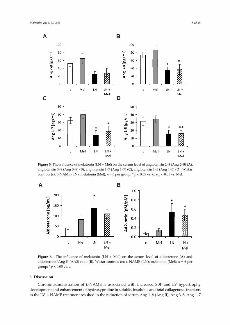
2.3. The Serum Concentrations of Angiotensins and Aldosterone
The serum angiotensins (Ang) concentrations measured by pooling of aliquot samples fromall animals per group after four weeks of treatment are shown in Figure 3. After four weeks oftreatment, the serum equilibrium level of Ang 1–8 was 1615.44 ± 181.8 pg/mL in the control groupand was reduced by L-NAME to 751.9 ± 159.1 pg/mL (52%, p < 0.05) (Figure 4B); the level ofAng 1–10 was 967.73 ± 76.63 pg/mL in the control group and was slightly reduced by L-NAME to579.40 ± 169.0 pg/mL (40%, NS) (Figure 4A); the level of Ang 2–8 was 52.70 ± 8.52 pg/mL in thecontrol group and was slightly reduced by L-NAME to 25.06 ± 5.91 pg/mL (52%, NS) (Figure 5A);the level of Ang 3–8 was 74.11 ± 7.22 pg/mL in the control group and was reduced by L-NAMEto 34.30 ± 9.10 pg/mL (54%, p < 0.05) (Figure 5B); the level of Ang 1–7 was 32.3 ± 4.08 pg/mL inthe control group and was reduced by L-NAME to 13.98 ± 5.82 pg/mL (57%, p < 0.05) (Figure 5C);the level of Ang 1–5 was 31.64 ± 5.20 pg/mL in the control group and was reduced by L-NAME to15.39 ± 4.66 pg/mL (52%, p < 0.05) (Figure 5D). Neither of the angiotensin’s levels in the L-NAMEgroup was influenced by melatonin. The serum concentration of aldosterone was 41.50 ± 9.00 pg/mLin the control group and was increased by L-NAME to 136.81 ± 48.59 pg/mL (230%, p < 0.05); melatoninonly slightly reduced the aldosterone concentration in the L-NAME group (20%, NS) (Figure 6A).The aldosterone/Ang II (AA2) ratio was 0.07 ± 0.02 in the control group and was increased by L-NAMEto 0.53 ± 0.18 (657%, p < 0.05); melatonin did not influence AA2-ratio (Figure 6B).
Molecules 2018, 23, x 4 of 15
2.3. The Serum Concentrations of Angiotensins and Aldosterone
The serum angiotensins (Ang) concentrations measured by pooling of aliquot samples from all animals per group after four weeks of treatment are shown in Figure 3. After four weeks of treatment, the serum equilibrium level of Ang 1–8 was 1615.44 ± 181.8 pg/mL in the control group and was reduced by L-NAME to 751.9 ± 159.1 pg/mL (52%, p < 0.05) (Figure 4B); the level of Ang 1–10 was 967.73 ± 76.63 pg/mL in the control group and was slightly reduced by L-NAME to 579.40 ± 169.0 pg/mL (40%, NS) (Figure 4A); the level of Ang 2–8 was 52.70 ± 8.52 pg/mL in the control group and was slightly reduced by L-NAME to 25.06 ± 5.91 pg/mL (52%, NS) (Figure 5A); the level of Ang 3–8 was 74.11 ± 7.22 pg/mL in the control group and was reduced by L-NAME to 34.30 ± 9.10 pg/mL (54%, p < 0.05) (Figure 5B); the level of Ang 1–7 was 32.3 ± 4.08 pg/mL in the control group and was reduced by L-NAME to 13.98 ± 5.82 pg/mL (57%, p < 0.05) (Figure 5C); the level of Ang 1–5 was 31.64 ± 5.20 pg/mL in the control group and was reduced by L-NAME to 15.39 ± 4.66 pg/mL (52%, p < 0.05) (Figure 5D). Neither of the angiotensin’s levels in the L-NAME group was influenced by melatonin. The serum concentration of aldosterone was 41.50 ± 9.00 pg/mL in the control group and was increased by L-NAME to 136.81 ± 48.59 pg/mL (230%, p < 0.05); melatonin only slightly reduced the aldosterone concentration in the L-NAME group (20%, NS) (Figure 6A). The aldosterone/Ang II (AA2) ratio was 0.07 ± 0.02 in the control group and was increased by L-NAME to 0.53 ± 0.18 (657%, p < 0.05); melatonin did not influence AA2-ratio (Figure 6B).
Figure 3. The influence of melatonin (L-NAME + Melatonin) on the serum level of angiotensins represented as pooling of 10 serum samples per each group. n = 10 per group.
Figure 4. The influence of melatonin (LN + Mel) on the serum level of angiotensin 1–10 (Ang I) (A); angiotensin 1–8 (Ang II) (B). Wistar controls (c); L-NAME (LN); melatonin (Mel); n = 6 per group; * p < 0.05 vs. c; + p < 0.05 vs. Mel.
Figure 3. The influence of melatonin (L-NAME + Melatonin) on the serum level of angiotensinsrepresented as pooling of 10 serum samples per each group. n = 10 per group.
Molecules 2018, 23, x 4 of 15
2.3. The Serum Concentrations of Angiotensins and Aldosterone
The serum angiotensins (Ang) concentrations measured by pooling of aliquot samples from all animals per group after four weeks of treatment are shown in Figure 3. After four weeks of treatment, the serum equilibrium level of Ang 1–8 was 1615.44 ± 181.8 pg/mL in the control group and was reduced by L-NAME to 751.9 ± 159.1 pg/mL (52%, p < 0.05) (Figure 4B); the level of Ang 1–10 was 967.73 ± 76.63 pg/mL in the control group and was slightly reduced by L-NAME to 579.40 ± 169.0 pg/mL (40%, NS) (Figure 4A); the level of Ang 2–8 was 52.70 ± 8.52 pg/mL in the control group and was slightly reduced by L-NAME to 25.06 ± 5.91 pg/mL (52%, NS) (Figure 5A); the level of Ang 3–8 was 74.11 ± 7.22 pg/mL in the control group and was reduced by L-NAME to 34.30 ± 9.10 pg/mL (54%, p < 0.05) (Figure 5B); the level of Ang 1–7 was 32.3 ± 4.08 pg/mL in the control group and was reduced by L-NAME to 13.98 ± 5.82 pg/mL (57%, p < 0.05) (Figure 5C); the level of Ang 1–5 was 31.64 ± 5.20 pg/mL in the control group and was reduced by L-NAME to 15.39 ± 4.66 pg/mL (52%, p < 0.05) (Figure 5D). Neither of the angiotensin’s levels in the L-NAME group was influenced by melatonin. The serum concentration of aldosterone was 41.50 ± 9.00 pg/mL in the control group and was increased by L-NAME to 136.81 ± 48.59 pg/mL (230%, p < 0.05); melatonin only slightly reduced the aldosterone concentration in the L-NAME group (20%, NS) (Figure 6A). The aldosterone/Ang II (AA2) ratio was 0.07 ± 0.02 in the control group and was increased by L-NAME to 0.53 ± 0.18 (657%, p < 0.05); melatonin did not influence AA2-ratio (Figure 6B).
Figure 3. The influence of melatonin (L-NAME + Melatonin) on the serum level of angiotensins represented as pooling of 10 serum samples per each group. n = 10 per group.
Figure 4. The influence of melatonin (LN + Mel) on the serum level of angiotensin 1–10 (Ang I) (A); angiotensin 1–8 (Ang II) (B). Wistar controls (c); L-NAME (LN); melatonin (Mel); n = 6 per group; * p < 0.05 vs. c; + p < 0.05 vs. Mel.
Figure 4. The influence of melatonin (LN + Mel) on the serum level of angiotensin 1–10 (Ang I) (A);angiotensin 1–8 (Ang II) (B). Wistar controls (c); L-NAME (LN); melatonin (Mel); n = 6 per group;* p < 0.05 vs. c; + p < 0.05 vs. Mel.
Molecules 2018, 23, 265 5 of 15
Molecules 2018, 23, x 5 of 15
Figure 5. The influence of melatonin (LN + Mel) on the serum level of angiotensin 2–8 (Ang 2–8) (A); angiotensin 3–8 (Ang 3–8) (B); angiotensin 1–7 (Ang 1–7) (C); angiotensin 1–5 (Ang 1–5) (D). Wistar controls (c); L-NAME (LN); melatonin (Mel); n = 6 per group; * p < 0.05 vs. c; + p < 0.05 vs. Mel.
Figure 6. The influence of melatonin (LN + Mel) on the serum level of aldosterone (A) and aldosterone/Ang II (AA2) ratio (B). Wistar controls (c); L-NAME (LN); melatonin (Mel); n = 6 per group; * p < 0.05 vs. c.
3. Discussion
Chronic administration of L-NAME is associated with increased SBP and LV hypertrophy development and enhancement of hydroxyproline in soluble, insoluble and total collagenous fractions in the LV. L-NAME treatment resulted in the reduction of serum Ang 1–8 (Ang II), Ang 3–8, Ang 1–7 and Ang 1–5 levels and also an enhancement of aldosterone level and aldosterone/angiotensin II ratio. Melatonin reduced SBP and the concentration and content of LV hydroxyproline in insoluble and
Figure 5. The influence of melatonin (LN + Mel) on the serum level of angiotensin 2–8 (Ang 2–8) (A);angiotensin 3–8 (Ang 3–8) (B); angiotensin 1–7 (Ang 1–7) (C); angiotensin 1–5 (Ang 1–5) (D). Wistarcontrols (c); L-NAME (LN); melatonin (Mel); n = 6 per group; * p < 0.05 vs. c; + p < 0.05 vs. Mel.
Molecules 2018, 23, x 5 of 15
Figure 5. The influence of melatonin (LN + Mel) on the serum level of angiotensin 2–8 (Ang 2–8) (A); angiotensin 3–8 (Ang 3–8) (B); angiotensin 1–7 (Ang 1–7) (C); angiotensin 1–5 (Ang 1–5) (D). Wistar controls (c); L-NAME (LN); melatonin (Mel); n = 6 per group; * p < 0.05 vs. c; + p < 0.05 vs. Mel.
Figure 6. The influence of melatonin (LN + Mel) on the serum level of aldosterone (A) and aldosterone/Ang II (AA2) ratio (B). Wistar controls (c); L-NAME (LN); melatonin (Mel); n = 6 per group; * p < 0.05 vs. c.
3. Discussion
Chronic administration of L-NAME is associated with increased SBP and LV hypertrophy development and enhancement of hydroxyproline in soluble, insoluble and total collagenous fractions in the LV. L-NAME treatment resulted in the reduction of serum Ang 1–8 (Ang II), Ang 3–8, Ang 1–7 and Ang 1–5 levels and also an enhancement of aldosterone level and aldosterone/angiotensin II ratio. Melatonin reduced SBP and the concentration and content of LV hydroxyproline in insoluble and
Figure 6. The influence of melatonin (LN + Mel) on the serum level of aldosterone (A) andaldosterone/Ang II (AA2) ratio (B). Wistar controls (c); L-NAME (LN); melatonin (Mel); n = 6 pergroup; * p < 0.05 vs. c.
3. Discussion
Chronic administration of L-NAME is associated with increased SBP and LV hypertrophydevelopment and enhancement of hydroxyproline in soluble, insoluble and total collagenous fractionsin the LV. L-NAME treatment resulted in the reduction of serum Ang 1–8 (Ang II), Ang 3–8, Ang 1–7
Molecules 2018, 23, 265 6 of 15
and Ang 1–5 levels and also an enhancement of aldosterone level and aldosterone/angiotensin II ratio.Melatonin reduced SBP and the concentration and content of LV hydroxyproline in insoluble andtotal collagen fractions in L-NAME group. These hemodynamic and structural changes induced bymelatonin were associated with the maintained reduction of the Ang II level.
The important issue is the dose of melatonin that should be used to counteract the pathologicalalterations induced by L-NAME. The dose 40 mg/kg/day of L-NAME in this experiment was chosenbased on our previous studies [3,4,22–25] and it yielded expected hemodynamic and structuralalterations. For melatonin, the dose 10 mg/kg/day used in our previous experiments was shown tolower increased blood pressure and protect the cardiovascular system against the deleterious effectsof increased hemodynamic load. On the other hand, in our previous pilot studies, a higher dose ofmelatonin (30 mg/kg/day) demonstrated obvious sedative effect. Thus, the dose 10 mg/kg/day ofmelatonin is effective, tolerated and the effects observed in this study can be well compared with thedata of previous experiments.
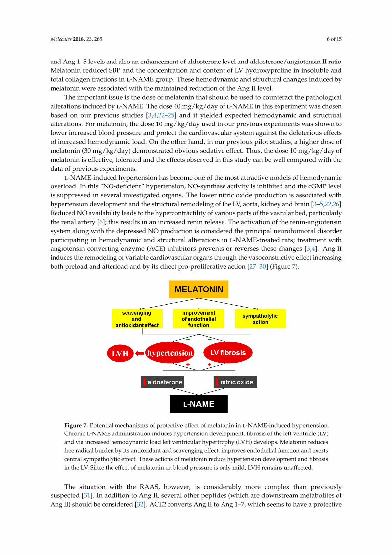
L-NAME-induced hypertension has become one of the most attractive models of hemodynamicoverload. In this “NO-deficient” hypertension, NO-synthase activity is inhibited and the cGMP levelis suppressed in several investigated organs. The lower nitric oxide production is associated withhypertension development and the structural remodeling of the LV, aorta, kidney and brain [3–5,22,26].Reduced NO availability leads to the hypercontractility of various parts of the vascular bed, particularlythe renal artery [6]; this results in an increased renin release. The activation of the renin-angiotensinsystem along with the depressed NO production is considered the principal neurohumoral disorderparticipating in hemodynamic and structural alterations in L-NAME-treated rats; treatment withangiotensin converting enzyme (ACE)-inhibitors prevents or reverses these changes [3,4]. Ang IIinduces the remodeling of variable cardiovascular organs through the vasoconstrictive effect increasingboth preload and afterload and by its direct pro-proliferative action [27–30] (Figure 7).
Molecules 2018, 23, x 6 of 15
total collagen fractions in L-NAME group. These hemodynamic and structural changes induced by melatonin were associated with the maintained reduction of the Ang II level.
The important issue is the dose of melatonin that should be used to counteract the pathological alterations induced by L-NAME. The dose 40 mg/kg/day of L-NAME in this experiment was chosen based on our previous studies [3,4,22–25] and it yielded expected hemodynamic and structural alterations. For melatonin, the dose 10 mg/kg/day used in our previous experiments was shown to lower increased blood pressure and protect the cardiovascular system against the deleterious effects of increased hemodynamic load. On the other hand, in our previous pilot studies, a higher dose of melatonin (30 mg/kg/day) demonstrated obvious sedative effect. Thus, the dose 10 mg/kg/day of melatonin is effective, tolerated and the effects observed in this study can be well compared with the data of previous experiments.
L-NAME-induced hypertension has become one of the most attractive models of hemodynamic overload. In this “NO-deficient” hypertension, NO-synthase activity is inhibited and the cGMP level is suppressed in several investigated organs. The lower nitric oxide production is associated with hypertension development and the structural remodeling of the LV, aorta, kidney and brain [3–5,22,26]. Reduced NO availability leads to the hypercontractility of various parts of the vascular bed, particularly the renal artery [6]; this results in an increased renin release. The activation of the renin-angiotensin system along with the depressed NO production is considered the principal neurohumoral disorder participating in hemodynamic and structural alterations in L-NAME-treated rats; treatment with angiotensin converting enzyme (ACE)-inhibitors prevents or reverses these changes [3,4]. Ang II induces the remodeling of variable cardiovascular organs through the vasoconstrictive effect increasing both preload and afterload and by its direct pro-proliferative action [27–30] (Figure 7).
Figure 7. Potential mechanisms of protective effect of melatonin in L-NAME-induced hypertension. Chronic L-NAME administration induces hypertension development, fibrosis of the left ventricle (LV) and via increased hemodynamic load left ventricular hypertrophy (LVH) develops. Melatonin reduces free radical burden by its antioxidant and scavenging effect, improves endothelial function and exerts central sympatholytic effect. These actions of melatonin reduce hypertension development and fibrosis in the LV. Since the effect of melatonin on blood pressure is only mild, LVH remains unaffected.
The situation with the RAAS, however, is considerably more complex than previously suspected [31]. In addition to Ang II, several other peptides (which are downstream metabolites of Ang II) should be considered [32]. ACE2 converts Ang II to Ang 1–7, which seems to have a protective feature in the cardiovascular system. Ang 1–7 has a counteracting effect against Ang II-induced vasoconstriction, inflammation, and cellular growth signaling at the level of the heart and
Figure 7. Potential mechanisms of protective effect of melatonin in L-NAME-induced hypertension.Chronic L-NAME administration induces hypertension development, fibrosis of the left ventricle (LV)and via increased hemodynamic load left ventricular hypertrophy (LVH) develops. Melatonin reducesfree radical burden by its antioxidant and scavenging effect, improves endothelial function and exertscentral sympatholytic effect. These actions of melatonin reduce hypertension development and fibrosisin the LV. Since the effect of melatonin on blood pressure is only mild, LVH remains unaffected.
The situation with the RAAS, however, is considerably more complex than previouslysuspected [31]. In addition to Ang II, several other peptides (which are downstream metabolites ofAng II) should be considered [32]. ACE2 converts Ang II to Ang 1–7, which seems to have a protective
Molecules 2018, 23, 265 7 of 15
feature in the cardiovascular system. Ang 1–7 has a counteracting effect against Ang II-inducedvasoconstriction, inflammation, and cellular growth signaling at the level of the heart and blood vesselsunder conditions of hypertension, myocardial remodeling and heart failure or stroke [33–36]. It alsoseems that Ang 1–5, the scission product of Ang 1–7, could provide cardiovascular protection throughthe stimulation of the atrial natriuretic peptide release via MAS receptors [37]. These substances maybe a therapeutic target or direct cardiovascular protectives. The administration of Ang 1–7 to mice withtype 2 diabetes reduced cardiomyocyte hypertrophy, inflammatory cell infiltration and fibrosis andincreased blood vessel number in the heart tissue [34] and prevented the development of hypertensionand end-organ damage in L-NAME-treated spontaneously hypertensive rats [38]. Thus, these peptidesmight be of therapeutic value for individuals with reactive heart hypertrophy and fibrotic rebuilding.Herein, L-NAME induced hypertension and fibrosis of the left ventricle. Although Ang II is generallyconsidered to be a principle player in the development of undesirable alterations in the cardiovascularsystem during the hemodynamic overload [3,30], in our experiment the serum equilibrium Ang IIlevel was reduced in the L-NAME group. Thus, the activation of systemic Ang II production seemsnot to be consistent with the development of pathologic alterations in a state with NO-deficiency.Moreover, Ang 1–7 and Ang 1–5, which are potentially protective, were also decreased in the systemiccirculation and their deficit may have participated in the development of alterations during L-NAMEadministration. However, these downstream metabolites are produced in much lower amounts thanAng II.
The serum level of aldosterone was, in contrast, significantly enhanced in the L-NAME groupin the current study. Based on previous findings, aldosterone produced in the adrenal cortex viathe stimulation of AT1 receptors plays one of the principle roles in the fibrotic remodeling of the LVduring hemodynamic overload [39,40] or in heart failure [41,42]. Although the original data regardingan aldosterone dominant action in the development of cardiovascular alterations were achieved inaldosterone-infused rats fed with high salt diet [39,40], data has emerged that mineralocorticoidreceptor activation is a key player in the development and maintenance of cardiac and vascularremodeling in a broad spectrum of cardiovascular pathologies [43,44]. Indeed, the addition ofthe aldosterone receptor inhibitor to the standard treatment remarkably reduced mortality inthe population with heart failure in the RALES [41] and EPHESUS [42] trials. We suggest thatL-NAME-induced NO-deficient hypertension is another condition, where aldosterone could be theprinciple pathophysiological alteration resulting in the development of hypertension, LV hypertrophyand collagen remodeling.
Our findings in L-NAME hypertension are partially in concert with previously publishedfindings. While increased plasma renin activity was reported by several laboratories [7,45], andlocal ACE activity was stimulated in the LV and aorta, serum ACE activity was not increased inL-NAME-induced hypertension [7]. On the other hand, an increase in aldosterone concentrationwas observed in this model and myocardial fibrosis induced via NOS blockade was supposedlyinduced by the elevated aldosterone level via the increased AT1 receptor number in the adrenalgland [46]; this is supported by our observed increase in the AA2-ratio. Moreover, administration ofspironolactone improved NO production and reduced hypertension and left ventricular remodelingin L-NAME-hypertensive rats [47]. Aldosterone production is mainly regulated by Ang II and it isreasonable to expect its decrease along with the reduction of Ang II levels. However, there are severalpathways which could explain the fact that aldosterone may be produced relatively independently ofAng II synthesis. First, besides Ang II, aldosterone secretion is modified by a variety of other factorssuch as adrenocorticotropin, atrial natriuretic peptide or K+ and Mg2+ levels [48]. Recent findings evenindicate that leptin, the adipocyte-derived hormone, may increase the aldosterone production in obeseindividuals contributing to the development of hypertension [49]. Second, the number of AT1 receptorsin the zona glomerulosa of the suprarenal cortex may be of importance. Usui et al. [46] suggested thatobserved increased aldosterone production in the L-NAME-model may be determined by increasedAT1 receptor expression in the suprarenal cortex. Supposedly, increased number of AT1 receptor in the
Molecules 2018, 23, 265 8 of 15
suprarenal gland may have counterbalanced the decreased level of Ang II regarding the aldosteroneproduction in our experiment. Third, a number of papers indicate that NO inhibits aldosteronesecretion in glomerulosa cells through a cGMP-independent mechanism [50,51], potentially via directreduction of steroidogenesis in zona glomerulosa or by downregulation of AT1 receptors [52]. Indeed,L-NAME administration increased the level of aldosterone even 50-fold [53] independently on reninor Ang II level [54]. Thus, not Ang II but rather the enhancement of the systemic aldosterone levelshould be considered to play a pivotal role in the end-organ damage in NO-deficient rats consideringprevious findings [55] and also the current results.
Besides angiotensin and aldosterone, sympathetic nervous system and baroreflex-inducedalterations of blood pressure (BP) should be considered. Acute inhibition of the NO-synthesisby i.v. injection of L-NAME induced significant rise in BP and baroreflex sensitivity. Althoughin sympathectomised rats the BP response after L-NAME was observed, baroreflex sensitivityaugmentation did not occur. The authors suggested that the reversal of cardiac autonomic controlattenuation in sympathectomised L-NAME-treated rats reflected an important role of sympatheticinnervations in the acute L-NAME-induced hypertension [56]. However, according to our results itmay be hypothesized that under chronic L-NAME-treatment the situation may be different. IncreasedSBP, achieved potentially via reduced NO production by L-NAME and increased aldosterone secretion,might inhibit the sympathetic activity via the preserved baroreflex mechanism, thus reducing the toneof the renal artery and renin release with attenuation of angiotensin production.
Melatonin is produced in the pineal gland and a variety of other organs [57]. One function ofmelatonin is to modulate circadian rhythmicity of a number of physiological functions [8,58], melatoninexerts complex antioxidant effects both intra- and extracellularly [18,59,60] and improves endothelialvasodilatory function via the enhancement of NO bioavailability both in peripheral tissues and inthe brain [11,61]. As a result, melatonin and its metabolites protect the cardiovascular system interms of antihypertensive and antiremodeling effects [62–67]. Indeed, melatonin reduced fibrosis inspontaneously hypertensive rats [68], L-NAME-induced hypertension [69], continuous-light- [70] andcontinuous light + L-NAME-hypertension [71] and in isoproterenol-induced heart failure [72]. It wasrecently proposed that melatonin is likely an important antifibrotic agent in all organs [66]. It wasrepeatedly revealed that the antifibrotic effects of several substances were related to the modificationof the neurohumoral balance, yet the data on melatonin interference with the RAAS are missing. In thecurrent study, melatonin did not have a significant effect on angiotensin II, aldosterone or on potentiallyprotective substances Ang 1–7, Ang 1–5 or Ang 3–8 and it does not seem that the cardio-protectiveeffect of melatonin was achieved by the interference with the RAAS. However, melatonin may reduceblood pressure via several potential ways. This indolamine was shown to reduce free radical burdenin variable models of experimental hypertension in our laboratory [68,72,73], including L-NAME-and continuous light + L-NAME-induced hypertension [25,26,69]. In our previous experiments,the reduction of oxidative stress by melatonin was associated with enhanced NOS activity [26,73],attenuation of endothelium-derived constricting factor concentration and reduction of vascular walltension [26]. Thus, although we did not measure the parameters characterizing NO-production inthe current study, our previous experiments indicate that blood pressure reduction by melatoninand its antiremodeling effect may be associated with the improvement of endothelial function inL-NAME-hypertension (Figure 7).
Besides its effect on vascular system, melatonin may exert part of its antihypertensive action viainteraction with the central nervous system. The physiology of melatonin is tightly bound withthe sympathetic system. On the one hand, the melatonin release is controlled by sympatheticafferentation to the pineal gland, involving the mutual interaction of light/darkness with theretina, suprachiasmatic nucleus (SCN), paraventricular nucleus and stimulation of pineal β1- andα1-adrenergic receptors [74,75]. On the other hand, the GABA-ergic signaling in neurons from SCNto variable parts of the brain and ventrolateral medulla may be modulated by melatonin activityproviding a protective mechanism against excessive sympathetic excitation [76]. Indeed, chronic
Molecules 2018, 23, 265 9 of 15
administration of melatonin reduced BP, heart rate, improved β-adrenergic receptor function andbaroreflex in spontaneously hypertensive rats [77]. In healthy young men, melatonin reduced SBP,pulse wave velocity along with the reduction of noradrenalin levels [78]. It seems reasonable to supposethat the inhibition of the sympathetic tone on the central or vascular level might participate in theantihypertensive and organ-protective action of melatonin also in the L-NAME-model of hypertension.
The preserved low level of Ang II despite the SBP reduction by melatonin is an interesting finding.We hypothesize that the effect of melatonin on arterial blood pressure need not necessarily result inthe reduced renal perfusion, potentially by virtue of improved NO-availability in the renal arterywhich might counterbalance the systemic blood pressure reduction and maintain kidney perfusion.Moreover, renin release is controlled by a number of neurohumoral stimuli including sympatheticnervous system or oxidative load, both being suppressed by melatonin [11,28].
We conclude that L-NAME-induced hypertension is associated with a reduced level of Ang II andits downstream metabolites and with an increased serum concentration of aldosterone and AA2-ratio.Melatonin does not change the level of angiotensins and aldosterone in NO-deficient hypertension.It is suggested that melatonin exerts its protective effects without affecting the RAAS.
4. Materials and Methods
4.1. Animals and Treatment
Male adult (three-month-old) Wistar rats (Department of Toxicology and Laboratory AnimalsBreeding, Dobra Voda, Slovakia) were randomly divided into four groups (10 per group): untreatedcontrols, rats treated with melatonin (10 mg/kg/day), rats treated with L-NAME (40 mg/kg/day), andrats treated with L-NAME + melatonin. L-NAME and melatonin were dissolved in drinking water andtheir concentration was adjusted to daily water consumption to ensure the correct dosage. The naturalwater consumption was 12–13 mL/100 g of body weight. To ensure that all of water with dissolvedmelatonin was actually consumed by a particular rat, only 10 mL/100 mg of water-melatonin solutionwas offered. The solution was prepared as follows: 10 mg of melatonin was dissolved in 100 mLof water, while no additional substance was added to dissolve the substance. Rats were housed inindividual cages at 22–24 ◦C and fed a regular pellet diet ad libitum. All experimental procedures werecarried out in accordance with the Guide for the Care and Use of Laboratory Animals published bythe US National Institutes of Health (NIH Publication No. 8523, revised 1996). The experiment wasapproved by the ethical committee of the Institute of Pathophysiology, Faculty of Medicine, ComeniusUniversity, Bratislava, Slovakia (approval number: 1306/14-221).
Systolic blood pressure was measured each week by noninvasive tail-cuff plethysmography(Hugo-Sachs Elektronic, Freiburg, Germany) during five days of the week (from Monday to Friday),always from 7:00 to 9:00 a.m. After four weeks, rats were euthanized and their body weight (BW),heart weight, and left ventricular and right ventricular weights (LVW and RVW) were determinedand their relative weights (LVW/BW and RVW/BW ratio) were calculated. Left ventricular sampleswere frozen at −80 ◦C and later used for the determination of hydroxyproline concentrations. Unlessotherwise stated, all chemicals were purchased from Sigma Chemical Co. (Deisenhofen, Germany).
4.2. Determination of Hydroxyproline
The samples from the left ventricle were treated stepwise with different buffers as describedpreviously [79]. The soluble collagenous proteins were extracted with 0.5 mol/L CH3COOH-pepsinbuffer (at 4 ◦C) and the remaining insoluble collagenous proteins with 1.25 mol/L NaOH (20 minat 105 ◦C). Hydroxyproline concentration (a marker of fibrosis) was estimated in both collagenousfractions using spectrophotometry at 550 nm [80]. The hydroxyproline content was expressed in mgper total weight of the LV. Ten cardiac tissue samples were used for analysis of hydroxyproline.
Molecules 2018, 23, 265 10 of 15
4.3. Angiotensins and Aldosterone Analyses
The qualitative equivalence of circulating and equilibrium angiotensin levels in rats has beendocumented and established with the equilibrium levels providing higher sensitivity compared to“snap shot” measurements in plasma treated with various protease inhibitors. Therefore equilibriumangiotensin peptide concentrations and aldosterone levels were determined by mass spectrometry inthe serum samples as described previously [32,81,82]. Briefly, the conditioned serum was equilibratedat 37 ◦C for 30 min followed by the stabilization of equilibrium peptide levels. The stabilized sampleswere further spiked with stable isotope-labeled internal standards for each angiotensin metabolites(Ang I, Ang II, Ang 1–7, Ang 1–5, Ang 2–8 and Ang 3–8) and aldosterone at concentrations of200 pg/mL and 500 pg/mL, respectively. Following C18-based solid-phase extraction, the sampleswere subjected to LC-MS/MS analysis using a reversed-phase analytical column (Acquity UPLC®
C18, Waters Corp., Milford, MA, USA) operating in line with a XEVO TQ-S triple quadrupole massspectrometer (Waters Corp.) in MRM mode. Internal standards were used to correct for peptiderecovery of the sample preparation procedure for each angiotensin metabolite in each individualsample. Ang peptide concentrations were calculated considering the corresponding response factorsdetermined in appropriate calibration curves in the original sample matrix, on the condition thatintegrated signals exceeded a signal-to-noise ratio of 10. Sample of 6 animals were used for analysis ofangiotensin and aldosterone.
4.4. Statistical Analyses
Results are expressed as mean ± S.E.M. A one-way, two-tailed analysis of variance (ANOVA) andthe Bonferroni test were used for statistical analysis of SBP, BW, ventricular weights and hydroxyprolineconcentration and content in the LV and LSD (Fisher’s least significant difference) test was used forstatistical analysis of RAAS data. The differences were considered significant if the p-value was <0.05.
5. Limitations
The most reliable information regarding the efficiency of absorption of the administeredsubstances is to measure their plasmatic concentrations. The investigation of L-NAME or melatoninplasmatic concentrations was beyond our technical possibilities. However, based on this and severalprevious experiments with L-NAME and/or melatonin [3,22,25,26,71,83], it may be supposed that bothsubstances were absorbed reliably, since hemodynamic and target organ effects of both substanceswere clearly demonstrated: gradual blood pressure increase along with fibrotic remodeling of the LVin L-NAME group and attenuation of SBP increase and fibrotic remodeling of the LV when melatoninwas applied simultaneously with L-NAME.
The measurement of angiotensins and aldosterone in samples from all ten investigated animals isdesirable. Indeed, we have measured and reported angiotensins/aldosterone levels in the pools of allinvestigated animals in each particular treatment group (obtaining thus a physical average of the levelsin each group). To assess the intragroup variability (needed for statistical analysis), we have performedmeasurements in 6 randomly selected (for cost effectiveness) individual animals from each group.Nevertheless, the average values obtained from 6 and 10 animals were comparable and statisticallysignificant differences were obtained from the analyses of 6 animals already documenting sufficientstatistical power.
Acknowledgments: This work was supported by following grants for scientific research: VEGA 1/0071/15,VEGA 2/0195/15, VEGA 1/0127/17, UK/96/2015 and by Programme PROGRES Q 40/5.
Author Contributions: Fedor Simko wrote the paper; Fedor Simko and Tomas Baka conceived and designed theexperiments; Tomas Baka, Kristina Krajcirovicova, Kristina Repova, Silvia Aziriova, Marko Poglitsch and MichaelaAdamcova performed the experiments and acquired data; Fedor Simko, Tomas Baka, Kristina Krajcirovicova,Kristina Repova, Silvia Aziriova, Stefan Zorad, Marko Poglitsch, Michaela Adamcova, Russel J. Reiter and LudovitPaulis analyzed the data; all authors revised the manuscript and approved the final version to be submitted.
Conflicts of Interest: The authors declare no conflict of interest.
Molecules 2018, 23, 265 11 of 15
References
1. Simko, F.; Simko, J. The potential role of nitric oxide in the hypertrophic growth of the left ventricle.Physiol. Res. 2000, 49, 37–46. [PubMed]
2. Simko, F. Is NO the king? Pathophysiological benefit with uncertain clinical impact. Physiol. Res. 2007, 56,S1–S6. [PubMed]
3. Pechanova, O.; Bernatova, I.; Pelouch, V.; Simko, F. Protein remodelling of the heart in NO-deficienthypertension: The effect of captopril. J. Mol. Cell. Cardiol. 1997, 29, 3365–3374. [CrossRef] [PubMed]
4. Bernatova, I.; Pechanova, O.; Pelouch, V.; Simko, F. Regression of chronic L-NAME-treatment-induced leftventricular hypertrophy: Effect of captopril. J. Mol. Cell. Cardiol. 2000, 32, 177–185. [CrossRef] [PubMed]
5. Bernatova, I.; Pechanova, O.; Simko, F. Captopril prevents NO-deficient hypertension and left ventricularhypertrophy without affecting nitric oxide synthase activity in rats. Physiol. Res. 1996, 45, 311–316. [PubMed]
6. Holecyova, A.; Torok, J.; Bernatova, I.; Pechanova, O. Restriction of nitric oxide rather than elevated bloodpressure is responsible for alterations of vascular responses in nitric oxide-deficient hypertension. Physiol. Res.1996, 45, 317–321. [PubMed]
7. Takemoto, M.; Egashira, K.; Usui, M.; Numaguchi, K.; Tomita, H.; Tsutsui, H.; Shimokawa, H.; Sueishi, K.;Takeshita, A. Important role of tissue angiotensin-converting enzyme activity in the pathogenesis of coronaryvascular and myocardial structural changes induced by long-term blockade of nitric oxide synthesis in rats.J. Clin. Investig. 1997, 15, 278–287. [CrossRef] [PubMed]
8. Matsumura, R.; Node, K.; Akashi, M. Estimation methods for human circadian phase by use of peripheraltissues. Hypertens. Res. 2016, 39, 623–627. [CrossRef] [PubMed]
9. Simko, F. Chronobiology of blood pressure: Emerging implications of melatonin. Eur. J. Clin. Investig. 2012,42, 1252–1254. [CrossRef] [PubMed]
10. Simko, F.; Paulis, L. Melatonin as a potential antihypertensive treatment. J. Pineal Res. 2007, 42, 319–322.[CrossRef] [PubMed]
11. Paulis, L.; Simko, F. Blood pressure modulation and cardiovascular protection by melatonin: Potentialmechanisms behind. Physiol. Res. 2007, 56, 671–684. [PubMed]
12. Reiter, R.J.; Manchester, L.C.; Fuentes-Broto, L.; Tan, D.X. Cardiac hypertrophy and remodelling:Pathophysiological consequences and protective effects of melatonin. J. Hypertens. 2010, 28, S7–S12.[CrossRef] [PubMed]
13. Simko, F.; Pechanova, O. Remodelling of the heart and vessels in experimental hypertension: Advances inprotection. J. Hypertens. 2010, 28, S1–S6. [CrossRef] [PubMed]
14. Simko, F.; Reiter, R.J.; Pechanova, O.; Paulis, L. Experimental models of melatonin-deficient hypertension.Front. Biosci. 2013, 18, 616–625. [CrossRef]
15. He, B.; Zhao, Y.; Xu, L.; Gao, L.; Su, Y.; Lin, N.; Pu, J. The nuclear melatonin receptor RORα is a novelendogenous defender against myocardial ischemia/reperfusion injury. J. Pineal Res. 2016, 60, 313–326.[CrossRef] [PubMed]
16. Nduhiraband, I.F.; Lamont, K.; Albertyn, Z.; Opie, L.H.; Lecour, S. Role of toll-like receptor 4 inmelatonin-induced cardioprotection. J. Pineal Res. 2016, 60, 39–47. [CrossRef] [PubMed]
17. Simko, F.; Pechanova, O. Recent trends in hypertension treatment: Perspectives from animal studies.J. Hypertens. 2009, 27, S1–S4. [CrossRef] [PubMed]
18. Reiter, R.J.; Mayo, J.C.; Tan, D.X.; Sainz, R.M.; Alatorre-Jimenez, M.; Qin, L. Melatonin as an antioxidant:Under promises but over delivers. J. Pineal Res. 2016, 61, 253–278. [CrossRef] [PubMed]
19. Opie, L.H.; Lecour, S. Melatonin has multiorgan effects. Eur. Heart J. Cardiovasc. Pharmacother. 2016, 2,258–265. [CrossRef] [PubMed]
20. Simko, F.; Baka, T.; Paulis, L.; Reiter, R.J. Elevated heart rate and nondipping heart rate as potential targetsfor melatonin: A review. J. Pineal Res. 2016, 61, 127–137. [CrossRef] [PubMed]
21. Reiter, R.J.; Tan, D.X.; Galano, A. Melatonin: Exceeding expectations. Physiology 2014, 29, 325–333. [CrossRef][PubMed]
22. Bernatova, I.; Pechanova, O.; Simko, F. Effect of captopril in L-NAME-induced hypertension on the ratmyocardium, aorta, brain and kidney. Exp. Physiol. 1999, 84, 1095–1105. [PubMed]
Molecules 2018, 23, 265 12 of 15
23. Simko, F.; Matuskova, J.; Luptak, I.; Krajcirovicova, K.; Kucharska, J.; Gvozdjakova, A.; Babal, P.;Pechanova, O. Effect of simvastatin on remodeling of the left ventricle and aorta in L-NAME-inducedhypertension. Life Sci. 2004, 23, 1211–1224. [CrossRef]
24. Simko, F.; Luptak, I.; Matuskova, J.; Krajcirovicova, K.; Sumbalova, Z.; Kucharska, J.; Gvozdjakova, A.;Simko, J.; Babal, P.; Pechanova, O.; et al. L-arginine fails to protect against myocardial remodelling inL-NAME-induced hypertension. Eur. J. Clin. Investig. 2005, 35, 362–368. [CrossRef] [PubMed]
25. Paulis, L.; Pechanova, O.; Zicha, J.; Liskova, S.; Celec, P.; Mullerova, M.; Kollar, J.; Behuliak, M.; Kunes, J.;Adamcova, M.; et al. Melatonin improves the restoration of endothelium-derived constricting factorsignalling and inner diameter in the rat femoral artery after cessation of L-NAME treatment. J. Hypertens.2010, 28 (Suppl. 1), S19–S24. [CrossRef] [PubMed]
26. Paulis, L.; Pechanova, O.; Zicha, J.; Krajcirovicova, K.; Barta, A.; Pelouch, V.; Adamcova, M.;Simko, F. Melatonin prevents fibrosis but not hypertrophy development in the left ventricle ofNG-nitro-L-arginine-methyl ester hypertensive rats. J. Hypertens. 2009, 27, S11–S16. [CrossRef]
27. Simko, F.; Simko, J. Heart failure and angiotensin converting enzyme inhibition: Problems and perspectives.Physiol. Res. 1999, 48, 1–8. [PubMed]
28. Simko, F.; Simko, J.; Fabryova, M. ACE-inhibition and angiotensin II receptor blockers in chronic heartfailure: Pathophysiological consideration of the unresolved battle. Cardiovasc. Drugs Ther. 2003, 17, 287–290.[CrossRef] [PubMed]
29. Paulis, L.; Unger, T. Novel therapeutic targets for hypertension. Nat. Rev. Cardiol. 2010, 7, 431–441. [CrossRef][PubMed]
30. Nehme, A.; Zibara, K. Efficiency and specificity of RAAS inhibitors in cardiovascular diseases: How toachieve better end-organ protection? Hypertens. Res. 2017. [CrossRef] [PubMed]
31. Hrenak, J.; Paulis, L.; Simko, F. Angiotensin A/Alamandine/MrgD Axis: Another Clue to UnderstandingCardiovascular Pathophysiology. Int. J. Mol. Sci. 2016, 17, 1098. [CrossRef] [PubMed]
32. Basu, R.; Poglitsch, M.; Yogasundaram, H.; Thomas, J.; Rowe, B.H.; Oudit, G.Y. Roles of Angiotensin Peptidesand Recombinant Human ACE2 in Heart Failure. J. Am. Coll. Cardiol. 2017, 69, 805–819. [CrossRef] [PubMed]
33. Jiang, F.; Yang, J.; Zhang, Y.; Dong, M.; Wang, S.; Zhang, Q.; Liu, F.F.; Zhang, K.; Zhang, C. Angiotensin-converting enzyme 2 and angiotensin 1–7: Novel therapeutic targets. Nat. Rev. Cardiol. 2014, 11, 413–426.[CrossRef] [PubMed]
34. Papinska, A.M.; Mordwinkin, N.M.; Meeks, C.J.; Jadhav, S.S.; Rodgers, K.E. Angiotensin-(1–7) administrationbenefits cardiac, renal and progenitor cell function in db/db mice. Br. J. Pharmacol. 2015. [CrossRef] [PubMed]
35. Joviano-Santos, J.V.; Santos-Miranda, A.; Joca, H.C.; Cruz, J.S.; Ferreira, A.J. New insights into the elucidationof angiotensin-(1–7) in vivo antiarrhythmic effects and its related cellular mechanisms. Exp. Physiol. 2016,101, 1506–1516. [CrossRef] [PubMed]
36. Zhang, X.; Cheng, H.J.; Zhou, P.; Kitzman, D.W.; Ferrario, C.M.; Li, W.M.; Cheng, C.P. Cellular basis ofangiotensin-(1–7)-induced augmentation of left ventricular functional performance in heart failure. Int. J.Cardiol. 2017, 236, 405–412. [CrossRef] [PubMed]
37. Yu, L.; Yuan, K.; Phuong, H.T.; Park, B.M.; Kim, S.H. Angiotensin-(1-5), an active mediator ofrenin-angiotensin system, stimulates ANP secretion via Mas receptor. Peptides 2017, 86, 33–41. [CrossRef][PubMed]
38. Benter, I.F.; Yousif, M.H.; Anim, J.T.; Cojocel, C.; Diz, D.I. Angiotensin-(1–7) prevents development of severehypertension and end-organ damage in spontaneously hypertensive rats treated with L-NAME. Am. J.Physiol. Heart Circ. Physiol. 2006, 290, H684–H691. [CrossRef] [PubMed]
39. Brilla, C.G.; Weber, K.T. Mineralocorticoid excess, dietary sodium, and myocardial fibrosis. J. Lab. Clin. Med.1992, 120, 893–901. [PubMed]
40. Young, M.J.; Rickard, A.J. Mineralocorticoid receptors in the heart: Lessons from cell-selective transgenicanimals. J. Endocrinol. 2015, 224, R1–R13. [CrossRef] [PubMed]
41. Pitt, B.; Zannad, F.; Remme, W.J.; Cody, R.; Castaigne, A.; Perez, A.; Palensky, J.; Wittes, J. The effect ofspironolactone on morbidity and mortality in patients with severe heart failure. Randomized AldactoneEvaluation Study Investigators. N. Engl. J. Med. 1999, 341, 709–717. [CrossRef] [PubMed]
Molecules 2018, 23, 265 13 of 15
42. Pitt, B.; Remme, W.; Zannad, F.; Neaton, J.; Martinez, F.; Roniker, B.; Bittman, R.; Hurley, S.; Kleiman, J.;Gatlin, M.; Eplerenone Post-Acute Myocardial Infarction Heart Failure Efficacy and Survival StudyInvestigators. Eplerenone, a selective aldosterone blocker, in patients with left ventricular dysfunctionafter myocardial infarction. N. Engl. J. Med. 2003, 348, 1309–1321. [CrossRef] [PubMed]
43. Fuller, P.J.; Young, M.J. Mechanisms of mineralocorticoid action. Hypertension 2005, 46, 1227–1235. [CrossRef][PubMed]
44. Young, M.J. Mechanisms of mineralocorticoid receptor-mediated cardiac fibrosis and vascular inflammation.Curr. Opin. Nephrol. Hypertens. 2008, 17, 174–180. [CrossRef] [PubMed]
45. Arnal, J.F.; Warin, L.; Michel, J.B. Determinants of aortic cyclic guanosine monophosphate in hypertensioninduced by chronic inhibition of nitric oxide synthase. J. Clin. Investig. 1992, 90, 647–652. [CrossRef][PubMed]
46. Usui, M.; Ichiki, T.; Katoh, M.; Egashira, K.; Takeshita, A. Regulation of angiotensin II receptor expression bynitric oxide in rat adrenal gland. Hypertension 1998, 32, 527–533. [CrossRef] [PubMed]
47. Simko, F.; Matuskova, J.; Luptak, I.; Pincikova, T.; Krajcirovicova, K.; Stvrtina, S.; Pomsar, J.; Pelouch, V.;Paulis, L.; Pechanova, O. Spironolactone differently influences remodeling of the left ventricle and aorta inL-NAME-induced hypertension. Physiol. Res. 2007, 56, S25–S32. [PubMed]
48. Mulrow, P.J. Angiotensin II and aldosterone regulation. Regul. Pept. 1999, 17, 27–32. [CrossRef]49. Faulkner, J.L.; Bruder-Nascimento, T.; Belin de Chantemèle, E.J. The regulation of aldosterone secretion
by leptin: Implications in obesity-related cardiovascular disease. Curr. Opin. Nephrol. Hypertens. 2017, 10.[CrossRef] [PubMed]
50. Rebuffat, P.; Malendowicz, L.K.; Nussdorfer, G.G.; Mazzocchi, G. Stimulation of endogenous nitric oxideproduction is involved in the inhibitory effect of adrenomedullin on aldosterone secretion in the rat. Peptides2001, 22, 923–936. [CrossRef]
51. Sainz, J.M.; Reche, C.; Rabano, M.A.; Mondillo, C.; Patrignani, Z.J.; Macarulla, J.M.; Pignataro, O.P.; Trueba, M.Effects of nitric oxide on aldosterone synthesis and nitric oxide synthase activity in glomerulosa cells frombovine adrenal gland. Endocrine 2004, 24, 61–71. [CrossRef]
52. Nithipatikom, K.; Holmes, B.B.; McCoy, M.J.; Hillard, C.J.; Campbell, W.B. Chronic administration of nitricoxide reduces angiotensin II receptor type 1 expression and aldosterone synthesis in Zona glomerulosa cells.Am. J. Physiol. Endocrinol. Metab. 2004, 287, E820–E827. [CrossRef] [PubMed]
53. Ikeda, H.; Tsuruya, K.; Toyonaga, J.; Masutani, K.; Hayashida, H.; Hirakata, H.; Iida, M. Spironolactonesuppresses inflammation and prevents L-NAME-induced renal injury in rats. Kidney Int. 2009, 75, 147–155.[CrossRef] [PubMed]
54. Muldowney, J.A.; Davis, S.N.; Vaughan, D.E.; Brown, N.J. NO synthase inhibition increases aldosterone inhumans. Hypertension 2004, 44, 739–745. [CrossRef] [PubMed]
55. Suehiro, T.; Tsuruya, K.; Ikeda, H.; Toyonaga, J.; Yamada, S.; Noguchi, H.; Tokumoto, M.; Kitazono, T.Systemic aldosterone, but not angiotensin II, plays a pivotal role in the pathogenesis of renal injury in chronicnitric oxide-deficient male rats. Endocrinology 2015, 156, 2657–2666. [CrossRef] [PubMed]
56. Chaswal, M.; Das, S.; Prasad, J.; Katyal, A.; Fahim, M. Cardiac autonomic function in acutely nitric oxidedeficient hypertensive rats: Role of the sympathetic nervous system and oxidative stress. Can. J. Physiol.Pharmacol. 2011, 89, 865–874. [CrossRef] [PubMed]
57. Acuna-Castroviejo, D.; Escames, G.; Venegas, C.; Diaz-Casado, M.E.; LimaCabello, E.; Lopez, L.C.;Rosales-Corall, S.; Tan, D.X.; Reiter, R.J. Extrapineal melatonin: Sources, regulation, and potential functions.Cell. Mol. Life Sci. 2014, 71, 2997–3025. [CrossRef] [PubMed]
58. Tan, D.X.; Manchester, L.C.; Reiter, R.J. CSF generation by pineal gland results in a robust melatonin circadianrhythm in the third ventricle as an unique light/dark signal. Med. Hypotheses 2016, 86, 3–9. [CrossRef][PubMed]
59. Tan, D.X.; Manchester, L.C.; Esteban-Zubero, E.; Zhou, Z.; Reiter, R.J. Melatonin as a Potent and InducibleEndogenous Antioxidant: Synthesis and Metabolism. Molecules 2015, 20, 18886–18906. [CrossRef] [PubMed]
60. Mukherjee, D.; Ghosh, A.K.; Dutta, M.; Mitra, E.; Mallick, S.; Saha, B.; Reiter, R.J.; Bandyopadhyay, D.Mechanisms of isoproterenol-induced cardiac mitochondrial damage: Protective actions of melatonin.J. Pineal Res. 2015, 58, 275–290. [CrossRef] [PubMed]
Molecules 2018, 23, 265 14 of 15
61. Agabiti-Rosei, C.; Favero, G.; De Ciuceis, C.; Rossini, C.; Porteri, E.; Rodella, L.F.; Franceschetti, L.; MariaSarkar, A.; Agabiti-Rosei, E.; Rizzoni, D.; et al. Effect of long-term treatment with melatonin on vascularmarkers of oxidative stress/inflammation and on the anticontractile activity of perivascular fat in agingmice. Hypertens. Res. 2017, 40, 41–50. [CrossRef] [PubMed]
62. Simko, F.; Pechanova, O. Potential roles of melatonin and chronotherapy among the new trends inhypertension treatment. J. Pineal Res. 2009, 47, 127–133. [CrossRef] [PubMed]
63. Simko, F.; Paulis, L. Antifibrotic effect of melatonin—Perspective protection in hypertensive heart disease.Int. J. Cardiol. 2013, 168, 2876–2877. [CrossRef] [PubMed]
64. Dominguez-Rodriguez, A.; Abreu-Gonzalez, P.; Piccolo, R.; Galasso, G.; Reiter, R.J. Melatonin is associatedwith reverse remodeling after cardiac resynchronization therapy in patients with heart failure and ventriculardyssynchrony. Int. J. Cardiol. 2016, 221, 359–363. [CrossRef] [PubMed]
65. Dominguez-Rodriguez, A.; Abreu-Gonzalez, P.; de la Torre-Hernandez, J.M.; Gonzalez-Gonzalez, J.;Garcia-Camarero, T.; Consuegra-Sanchez, L.; Garcia-Saiz, M.D.; Aldea-Perona, A.; Virgos-Aller, T.;Azpeitia, A.; et al. MARIA Investigators. Effect of intravenous and intracoronary melatonin as an adjunct toprimary percutaneous coronary intervention for acute ST-elevation myocardial infarction: Results of theMelatonin Adjunct in the acute myocaRdial Infarction treated with Angioplasty trial. J. Pineal Res. 2017, 62.[CrossRef]
66. Hu, W.; Ma, Z.; Jiang, S.; Fan, C.; Deng, C.; Yan, X.; Di, S.; Lv, J.; Reiter, R.J.; Yang, Y. Melatonin: The dawningof a treatment for fibrosis? J. Pineal Res. 2016, 60, 121–131. [CrossRef] [PubMed]
67. Hu, J.; Zhang, L.; Yang, Y.; Guo, Y.; Fan, Y.; Zhang, M.; Man, W.; Gao, E.; Hu, W.; Reiter, R.J.; et al. Melatoninalleviates postinfarction cardiac remodeling and dysfunction by inhibiting Mst1. J. Pineal Res. 2017, 62.[CrossRef]
68. Simko, F.; Pechanova, O.; Pelouch, V.; Krajcirovicova, K.; Mullerova, M.; Bednarova, K.; Adamcova, M.;Paulis, L. Effect of melatonin, captopril, spironolactone and simvastatin on blood pressure and left ventricularremodelling in spontaneously hypertensive rats. J. Hypertens. 2009, 27, S5–S10. [CrossRef] [PubMed]
69. Paulis, L.; Pechanova, O.; Zicha, J.; Barta, A.; Gardlik, R.; Celec, P.; Kunes, J.; Simko, F. Melatonin interactionswith blood pressure and vascular function during L-NAME-induced hypertension. J. Pineal Res. 2010, 48,102–108. [CrossRef] [PubMed]
70. Simko, F.; Pechanova, O.; Repova-Bednarova, K.; Krajcirovicova, K.; Celec, P.; Kamodyova, N.; Zorad, S.;Kucharska, J.; Gvozdjakova, A.; Adamcova, M.; et al. Hypertension and cardiovascular remodelling in ratsexposed to continuous light: Protection by ACE-inhibition and melatonin. Mediat. Inflamm. 2014, 2014,703175. [CrossRef] [PubMed]
71. Simko, F.; Pechanova, O.; Pelouch, V.; Krajcirovicova, K.; Celec, P.; Palffy, R.; Bednarova, K.; Vrankova, S.;Adamcova, M.; Paulis, L. Continuous light and L-NAME-induced left ventricular remodelling: Differentprotection with melatonin and captopril. J. Hypertens. 2010, 28, S13–S18. [CrossRef] [PubMed]
72. Simko, F.; Bednarova-Repova, K.; Krajcirovicova, K.; Hrenak, J.; Celec, P.; Kamodyova, N.; Gajdosechova, L.;Zorad, S.; Adamcova, M. Melatonin reduces cardiac remodeling and improves survival in rats withisoproterenol-induced heart failure. J. Pineal Res. 2014, 57, 177–184. [CrossRef] [PubMed]
73. Simko, F.; Pechanova, O.; Repova, K.; Aziriova, S.; Krajcirovicova, K.; Celec, P.; Tothova, L.; Vrankova, S.;Balazova, L.; Zorad, S.; et al. Lactacystin-Induced Model of Hypertension in Rats: Effects of Melatonin andCaptopril. Int. J. Mol. Sci. 2017, 25, 1612. [CrossRef] [PubMed]
74. Moore, R.Y. Neural control of the pineal gland. Behav. Brain Res. 1996, 73, 125–130. [CrossRef]75. Reiter, R.J. Pineal melatonin: Cell biology of its synthesis and of its physiological interactions. Endocr. Rev.
1991, 12, 151–180. [CrossRef] [PubMed]76. Pechanova, O.; Paulis, L.; Simko, F. Peripheral and central effects of melatonin on blood pressure regulation.
Int. J. Mol. Sci. 2014, 15, 17920–17937. [CrossRef] [PubMed]77. Girouard, H.; Denault, C.; Chulak, C.; de Champlain, J. Treatment by N-acetylcysteine and melatonin
increases cardiac baroreflex and improves antioxidant reserve. Am. J. Hypertens. 2004, 17, 947–954. [CrossRef][PubMed]
78. Arangino, S.; Cagnacci, A.; Angiolucci, M.; Vacca, A.M.; Longu, G.; Volpe, A.; Melis, G.B. Effects of melatoninon vascular reactivity, catecholamine levels, and blood pressure in healthy men. Am. J. Cardiol. 1999, 83,1417–1419. [CrossRef]
Molecules 2018, 23, 265 15 of 15
79. Pelouch, V.; Milerova, M.; Ostadal, B.; Samanek, M.; Hucan, B. Protein profiling of human atrial andventricular musculature: The effect of normoxaemia and hypoxaemia in congenital heart diseases.Physiol. Res. 1993, 42, 235–242. [PubMed]
80. Reddy, K.; Enwemeka, C.S. A simplified method for the analysis of hydroxyproline in biological tissues.Clin. Biochem. 1996, 29, 225–229. [CrossRef]
81. Sharp, S.; Poglitsch, M.; Zilla, P.; Davies, N.H.; Sturrock, E.D. Pharmacodynamic effects of C-domain-specificACE inhibitors on the renin-angiotensin system in myocardial infarcted rats. J. Renin Angiotensin AldosteroneSyst. 2015, 16, 1149–1158. [CrossRef] [PubMed]
82. Pavo, N.; Goliasch, G.; Wurm, R.; Novak, J.; Strunk, G.; Gyöngyösi, M.; Poglitsch, M.; Säemann, M.D.;Hülsmann, M. Low- and High-renin Heart Failure Phenotypes with Clinical Implications. Clin Chem. 2017.[CrossRef] [PubMed]
83. Pechanova, O.; Zicha, J.; Paulis, L.; Zenebe, W.; Dobesova, Z.; Kojsova, S.; Jendekova, L.; Sladkova, M.;Dovinova, I.; Simko, F.; et al. The effect of N-acetylcysteine and melatonin in adult spontaneouslyhypertensive rats with established hypertension. Eur. J. Pharmacol. 2007, 30, 129–136. [CrossRef] [PubMed]
Sample Availability: Samples of the compounds are available from the authors.
© 2018 by the authors. Licensee MDPI, Basel, Switzerland. This article is an open accessarticle distributed under the terms and conditions of the Creative Commons Attribution(CC BY) license (http://creativecommons.org/licenses/by/4.0/).