energy-dependent stability of shewanella oneidensis mr-1 ...stability, we investigated whether...
TRANSCRIPT

JOURNAL OF BACTERIOLOGY, July 2011, p. 3257–3264 Vol. 193, No. 130021-9193/11/$12.00 doi:10.1128/JB.00251-11Copyright © 2011, American Society for Microbiology. All Rights Reserved.
Energy-Dependent Stability of Shewanella oneidensis MR-1 Biofilms�
Renee M. Saville,1 Shauna Rakshe,2 Janus A. J. Haagensen,1Soni Shukla,1 and Alfred M. Spormann1,2,3*
Departments of Civil and Environmental Engineering,1 Chemical Engineering,2 and Biological Sciences,3
Stanford University, Stanford, California
Received 21 February 2011/Accepted 30 April 2011
Stability and resistance to dissolution are key features of microbial biofilms. How these macroscopicproperties are determined by the physiological state of individual biofilm cells in their local physical-chemicaland cellular environment is largely unknown. In order to obtain molecular and energetic insight into biofilmstability, we investigated whether maintenance of biofilm stability is an energy-dependent process and whethertranscription and/or translation is required for biofilm dissolution. We found that in 12-hour-old Shewanellaoneidensis MR-1 biofilms, a reduction in cellular ATP concentration, induced either by oxygen deprivation orby addition of the inhibitor of oxidative phosphorylation carbonyl cyanide m-chlorophenylhydrazone (CCCP),dinitrophenol (DNP), or CN�, resulted in massive dissolution. In 60-hour-old biofilms, the extent of uncoupler-induced cell loss was strongly attenuated, indicating that the integrity of older biofilms is maintained by meansother than those operating in younger biofilms. In experiments with 12-hour-old biofilms, the transcriptionaland translational inhibitors rifampin, tetracycline, and erythromycin were found to be ineffective in preventingenergy starvation-induced detachment, suggesting that neither transcription nor translation is required forthis process. Biofilms of Vibrio cholerae were also induced to dissolve upon CCCP addition to an extent similarto that in S. oneidensis. However, Pseudomonas aeruginosa and P. putida biofilms remained insensitive to CCCPaddition. Collectively, our data show that metabolic energy is directly or indirectly required for maintainingcell attachment, and this may represent a common but not ubiquitous mechanism for stability of microbialbiofilms.
Microbial biofilms are surface-associated, dynamic commu-nities that respond to changing external and internal environ-ments, enabling continued acclimation of the biofilm popula-tion (8). In biofilms, individual cells are associated with eitherother biofilm cells or a matrix, which generally consists ofexopolysaccharides, proteins, and DNA, and are thereby re-tained in biofilms that form under hydrodynamic conditions(5). Under such conditions, two opposite processes determinethe stability of microbial biofilms: cellular attachment and de-tachment. Both processes are mechanistically linked in thatthose factors that mediate attachment of cells need to be in-activated when cells detach.
Cell detachment in “young” biofilms occurs in response toenvironmental or cellular changes and often involves exter-nal or population-induced nutrient limitation, includingstarvation for carbon, nitrogen, or oxygen (2, 7, 9, 15, 25, 27,33). For example, rapid dispersal of Pseudomonas aerugi-nosa and P. putida biofilms were reported in response tolimitation in carbon substrate and oxygen (9, 25). In thesecases, most of the biofilm dissolution occurred within a fewminutes after onset of nutrient deprivation. In the formercase, there was also a correlation between protein dephos-phorylation and extent of detachment (25). Also, Vibriocholerae biofilms have been found to naturally dissolve to alarge extent in response to metabolic cues (22). Shewanellaoneidensis MR-1 is a Gram-negative facultative gammapro-
teobacterium capable of unprecedented respiratory diver-sity under anoxic conditions, including reduction of insolu-ble Fe(III) and Mn(IV) oxides (23). We previously showedthat the molecular mechanisms that control S. oneidensisMR-1 biofilm formation are primarily enabled by the man-nose-sensitive hemagglutinin (MSHA) and mxd gene sets(26). The msh genes encode a type IV pilus apparatus whilethe mxd genes putatively encode a carbohydrate-containingcell-associated component. Moreover, oxygen depletion in-duced rapid dissolution of aerobically growing biofilms (31,33). We also showed that intracellular cyclic di-GMP (c-di-GMP) influences biofilm formation in this microorganism byincreasing biofilm biomass and by rendering biofilms resis-tant to detachment (31). Overexpression of a cyclic-di-GMP-specific phosphodiesterase in S. oneidensis reducedintracellular cyclic di-GMP, inhibited biofilm formation byreducing adhesion of single cells, and induced detachmentin established biofilms. However, the molecular basis of theassociation between a biofilm cell and other cells or a bio-film cell and the biofilm matrix is still insufficiently under-stood.
In this work, we investigated the energetic and gene ex-pression requirements for biofilm stability primarily in S.oneidensis but also extended our key findings to Vibrio chol-erae and Pseudomonas sp., which are two of the most studiedbiofilm “model” systems. The data presented here suggestthat maintenance of the stability of young biofilms requiresmetabolic energy and that control of detachment is primar-ily posttranslational. The latter finding is also particularlyimportant, as subpopulations of biofilm microbes are gen-
* Corresponding author. Mailing address: James H. Clark Center,E250, Stanford University, Stanford, CA 94305-5429. Phone: (650)723-3668. Fax: (650) 724-4927. E-mail: [email protected].
� Published ahead of print on 13 May 2011.
3257
on February 28, 2020 by guest
http://jb.asm.org/
Dow
nloaded from
erally considered to be resistant to conventional antibiotictreatment (1, 6, 21).
MATERIALS AND METHODS
Growth conditions and media. The strains used in this study are summarizedin Table 1. Escherichia coli strains were grown in Luria-Bertani (LB) medium at37°C, and Shewanella oneidensis MR-1 strains were grown at 30°C in LB, lactatemedium (LM) (32), or minimal medium (MM) (31). If required, the medium wassolidified with 1.5% (wt/vol) agar or 0.3% (wt/vol) agar for motility plates andsupplemented with 10 �g/ml gentamicin and/or 25 �g/ml kanamycin. Geneinduction from the lacIq1-PLac-gfp construct, which expresses green fluorescentprotein (GFP) upon chemical induction, was achieved by addition of 10 mMisopropyl-�-D-thiogalactopyranoside (IPTG).
Strain construction in S. oneidensis MR-1. All genetic work was carried outaccording to standard protocols or by following the manufacturer’s instructions.Kits for the isolation and/or purification of plasmid DNA were obtained fromQiagen (Valencia, CA), and enzymes were purchased from New England Bio-Labs (Beverly, MA). In order to construct a system for controlled gene inductionin S. oneidensis MR-1, chromosomal insertion of a lacIq1-PLac-gfp fusion wascarried out using a Tn7 delivery system by four-parental mating using pSM2360as a delivery plasmid as described for S. oneidensis MR-1 (32).
Biofilm cultivation and image acquisition. Biofilms were cultivated at 30°C inLM in three-channel flow cells with individual channel dimensions of 1 by 4 by40 mm. Microscope coverslips (Fisher Scientific, Pittsburgh, PA) were used as acolonization surface, glued with silicone (GE Sealants & Adhesives, Hunters-ville, NC) onto the flow cells, and left to dry for 24 h at room temperature priorto use. Assembly, sterilization, and inoculation of the flow system were carriedout essentially as described previously (32).
For switching medium, the flow was arrested briefly, and the medium wasexchanged in the bubble trap and the upstream tubing. This process took nolonger than 1 min, and control channels, where the medium flow was stopped inparallel channels without changing the medium, ensured that the observed ef-fects were not due to that short arrest of flow. Confocal laser scanning micros-copy (CLSM) was performed at defined locations in the flow chamber close tothe inflow before and after the treatment.
Stop-of-flow-induced detachment was carried out essentially as described ear-lier (33). Briefly, the medium flow was arrested for 15 min and subsequentlyresumed for the same amount of time. CLSM images were taken immediatelybefore the stop of flow and after 15 min of flow.
Induction of gfp expression was executed by switching the irrigating LM to LMcontaining 10 mM IPTG (Sigma). Inhibition of gene expression was executed byswitching the irrigating LM to LM containing rifampin, erythromycin, or tetra-cycline (all from Sigma Chemical Company). The concentrations used were 100,50, or 25 �g/ml, respectively. Metabolic inhibitor experiments were executed byswitching the irrigating medium to LM containing carbonyl cyanide m-chloro-phenylhydrazone (CCCP) (Sigma), sodium cyanide (Baker), or dinitrophenol(DNP) (Sigma). The concentration used was 20 �M, 150 mM, or 2 mM, respec-tively, for S. oneidensis. For the remaining organisms, 50 �M CCCP was used.These were determined to be the lowest concentrations of each uncoupler at
which detection of fluorescence was prevented under biofilm growth conditions.For stock solutions, solid CCCP (80 mM), DNP (1 M), and sodium cyanide (0.5M) were dissolved in dimethyl sulfoxide (DMSO), ethanol, and water, respec-tively, prior to dilution into LM. Control channels were irrigated with LMcontaining the same added volume of each stock solvent only as used in exper-imental channels to test for solvent-dependent effects.
Microscopic visualization and image acquisition of biofilms were conducted atthe Stanford Biofilm Research Center using an upright Leica TCS SP2 confocallaser scanning microscope (Leica Microsystems, Heidelberg, Germany)equipped with a 63�/1.2-W objective. For displaying biofilm images, CLSMimages were processed using the IMARIS software package (Bitplane AG,Zurich, Switzerland) and Adobe Photoshop. Biofilm parameters, such as biomassand average biofilm thickness, were quantified with the software programCOMSTAT (10).
Nucleotide extraction from planktonic cultures. For CCCP and oxygen star-vation experiments, planktonic cultures were grown in LM to an optical densityat 600 nm (OD600) of approximately 0.3, and either the metabolic activity wasinhibited with 40 �M CCCP or the culture was deprived of oxygen. Control flaskswere grown in parallel and were either untreated or treated with equivalentvolumes of DMSO. CCCP was dissolved in DMSO to 80 mM, and this was addedto planktonic cultures to give a final CCCP concentration of 40 �M. Thisconcentration of CCCP was determined to be the lowest at which growth andfluorescence detection were blocked under planktonic conditions. Oxygen star-vation was induced by removing the flask from the shaker, flushing the headspacewith nitrogen, and placing a rubber stopper in the flask neck. In a controlexperiment, oxygen concentration was measured in similarly treated cultureswith an oxygen-sensitive electrode. The concentration decreased below the levelof detection within 12 min. Four samples (525 �l) from each culture were takenat designated time points and flash frozen on liquid nitrogen for subsequentnucleotide assays.
Cyclic-di-GMP and ATP determination. For ATP determination, culturesamples were thawed on ice and immediately assayed. Intracellular ATP wasquantified using the BacTiterGlo reagent (Promega PR-G8230) according tothe manufacturer’s protocol, with a lysis time of 2.5 min. Luminescence wasmeasured with a Mithras LB940 luminometer (Berthold Technologies). ATPsolutions used to establish the standard curve were made in the culturemedium.
Extraction of cyclic di-GMP was accomplished as described previously by theheat and ethanol addition method (31). After extraction, supernatants weredried on a Thermo Savant SpeedVac SPD (Thermo Fisher Scientific, Waltham,MA) and then stored at �20°C for subsequent liquid chromatography-massspectrometry (LC-MS) quantitation of the cyclic di-GMP (see below).
Cyclic di-GMP was quantified by LC-MS as previously described (31). Allsamples were analyzed on a Shimadzu high-performance liquid chromatography(HPLC) system (Columbia, MD) coupled to a Sciex API 3000 triple quadrupolemass spectrometer (MDS Sciex, Ontario) operating in positive electrospray ion-ization multiple reaction monitoring (MRM) mode. The high-performance liq-uid chromatography system was equipped with an autosampler and degasser,which were used for solvent delivery and sample introduction. Triplicate sampleswere injected twice.
TABLE 1. Strains used in this study
Strain Relevant genotype and phenotype or characteristics Source orreference
Shewanella oneidensisAS93 S. oneidensis MR-1 tagged with egfp (constitutively expressed) in a mini-Tn7
construct; Gmr Cmr32
AS130 S. oneidensis MR-1 tagged with egfp under inducible control of the Lacpromoter in a mini-Tn7 construct; Gmr Cmr
AS141 AS93 �mxdB (SO4179); Gmr Cmr 31AS645 AS93 �pilD (SO0414); Gmr Cmr 26AS647 AS93 �pilT (SO3351); Gmr Cmr 26AS648 AS93 �mshA (SO4105); Gmr Cmr 26
Vibrio cholerae A1552 Vibrio cholerae El Tor, Inaba, wild type, smooth, tagged with GFPmut3b*using a mini-Tn7 construct; Rifr Smr Gmr
22
Pseudomonas aeruginosa Pseudomonas aeruginosa PAO1 tagged with a mini-Tn7 GFPmut3b*cassette inserted into the chromosome; Gmr
20
Pseudomonas putida P. putida R1 with a mini-Tn5–Km–rrnBP1–gfp�mut3b*�–T0–T1 cassetteinserted into the chromosome; Nalr Kmr
28
3258 SAVILLE ET AL. J. BACTERIOL.
on February 28, 2020 by guest
http://jb.asm.org/
Dow
nloaded from

RESULTS
Metabolic energy is required for maintenance of biofilmstability. We previously showed that oxygen depletion or a stopin medium flow, which imposes conditions of oxygen depletionin a hydrodynamic, aerobic biofilm, leads to massive detach-ment of single cells (33). We hypothesized that this oxygenlimitation likely impacts the cellular ATP concentration andthat this could affect the availability of cellular energy of indi-vidual cells and thus the stability of biofilms. In order to de-termine whether or not metabolic energy is required for bio-film stability, and particularly for biofilm detachment, wesubjected Shewanella oneidensis MR-1 (AS93) to threemetabolic inhibitors that specifically affect ATP synthesis in re-spiring microorganisms. Carbonyl cyanide m-chlorophenylhy-drazone (CCCP) and dinitrophenol (DNP) are membrane-permeable, weak acids and act with high specificity asprotonophores to collapse the electrochemical proton poten-tial across the cytoplasmic membrane (11, 13). As a conse-quence, no ATP synthesis occurs via a chemiosmotic mecha-nism when a bacterial culture is amended with theseprotonophores. Sodium cyanide (NaCN) inhibits cellular res-piration by binding to cytochrome oxidase of the electrontransport chain, thus preventing electron flux and consequentlythe generation of an electrochemical proton potential andATP synthesis via electron transport phosphorylation. Whenthese metabolic inhibitors were tested in 12-hour-old S. one-idensis MR-1 biofilms, massive biofilm dissolution occurredimmediately after their addition to the irrigating medium (Fig.1A). This uncoupler-induced detachment resulted in loss ofindividual cells from the biofilm rather than of large cell clus-ters, which is similar to the detachment observed in response toa stop-of-flow treatment (33). To test whether the residualbiofilm was still sensitive to a stop-of-flow-induced detach-ment, after 60 min of inhibitor treatment, biofilms were sub-jected to a stop-of-flow assay as described previously (33), and30 min later, images were again recorded by CLSM. Quanti-fication of these images revealed that no further biomass waslost from the uncoupler-treated biofilms while untreated con-trol biofilms were fully detachable (Fig. 1B).
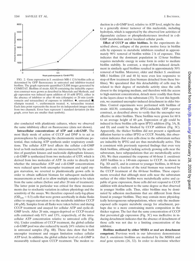
In order to show that these inhibitors act as such in ourhydrodynamic biofilm system, we conducted control experi-ments to test whether the inhibitors are active in situ in biofilmcells. We used S. oneidensis MR-1 strain AS130 containing achromosomally inserted reporter gene construct consisting ofgfp under the control of Ptac, which is repressed by the alsochromosomally encoded LacIq1 protein. For biofilm cells toexpress gfp and to become fluorescent, transcription in re-sponse to IPTG addition as well as translation is necessary.Thus, monitoring expression of gfp enables us to observe theseprocesses directly and in vivo. As both processes are stronglyATP dependent, gfp expression also served here as a qualita-tive surrogate for the availability of cellular ATP.
Biofilms of strain AS130 were grown under hydrodynamicconditions as described above. In the absence of IPTG, nodetectable GFP fluorescence was observed, whereas upon ad-dition of 10 mM IPTG, GFP fluorescence in cells throughoutthe biofilm was readily observed via CLSM (Fig. 2 and 3). After1 h, nearly 20% of the maximum inducible GFP fluorescencewas observed. In contrast, biofilms treated prior to induction
with specific inhibitors of transcription (100 �g/ml rifampin) ortranslation (25 �g/ml tetracycline or 50 �g/ml erythromycin;see below) did not yield any detectable fluorescence after 1 h(Fig. 2). In separate experiments, we found that addition of theprotonophores CCCP and DNP and of NaCN to AS130 bio-films prior to addition of IPTG resulted in no observable flu-orescence (data not shown). These experiments showed thatsmall molecules such as IPTG, rifampin, tetracycline, erythro-mycin, and the metabolic inhibitors CCCP, DNP, and NaCNcould access the majority of cells in the biofilm and that dif-fusional barriers did not restrict mass transport of these com-pounds into the biofilm. Moreover, we found that these insitu-grown biofilm cells were competent for induction of geneexpression as well as sensitive to inhibitors of transcription,translation, and energy conservation. These experiments were
FIG. 1. Stability of 12-hour S. oneidensis MR-1 biofilms treatedwith metabolic inhibitors. (A) The graph represents quantified CLSMimages generated by COMSTAT. Biofilms were exposed to mediaamended with indicated metabolic inhibitors for 90 min, and imageswere obtained at the designated time points. �, untreated; �, 2 mMDNP; F, 20 �M CCCP; �, 1 mM sodium cyanide. Each data pointrepresents the mean value for four independent images monitored intwo separate channels. Error bars represent 1 standard deviation.(B) The graph represents quantified CLSM images generated byCOMSTAT. The degree of biomass detachment as a result of a 90-minexposure to medium amended with metabolic inhibitor alone (leftpanel) or followed by a stop of flow (right panel) is shown. White,untreated; black, 20 �M CCCP; dark gray, 2 mM DNP; light gray, 1mM sodium cyanide. Each data point represents the mean value forfour independent images monitored in two separate channels. Errorbars represent 1 standard deviation.
VOL. 193, 2011 STABILITY OF S. ONEIDENSIS MR-1 BIOFILMS 3259
on February 28, 2020 by guest
http://jb.asm.org/
Dow
nloaded from
also conducted with planktonic cultures, where we observedthe same inhibitory effect on fluorescence (data not shown).
Intracellular concentrations of ATP and c-di-GMP. Themost likely mode of action of CCCP and DNP is to act asprotonophores by collapsing the chemiosmotic membrane po-tential, thus reducing ATP synthesis under respiratory condi-tions. The cellular ATP level affects the cellular c-di-GMPlevel as both nucleotide pools are interconnected by the activ-ities of guanylate kinases and nucleoside diphosphate kinases;c-di-GMP is synthesized from two molecules of GTP, which isderived from two molecules of ATP. In order to directly testwhether the intracellular ATP and c-di-GMP concentrationswere reduced upon both uncoupler treatment and rapid oxy-gen starvation, we reverted to planktonically grown cells inorder to obtain sufficient biomass for subsequent nucleotidemeasurements as well as to allow multiple samples to be takenfrom the same culture (before and after 20 min of treatment).The latter point in particular was critical for these measure-ments due to stochastic variation in culture physiology and thesensitivity of the assays. We therefore exposed early-log-phase(OD600 0.3) cells that were grown aerobically in shake flaskseither to oxygen starvation or to the metabolic inhibitor CCCP(40 �M). Samples from all flasks were taken before and duringCCCP treatment and assayed for cellular ATP and cyclic-di-GMP levels. After 20 min, oxygen-deprived or CCCP-treatedcells contained only 81% and 13%, respectively, of the intra-cellular ATP concentration relative to untreated cells (Fig.4A). Under conditions of CCCP treatment, the global cellularc-di-GMP pool was also reduced by nearly 24% relative to thatin untreated samples (Fig. 4B). These data show that bothuncoupler treatment and oxygen limitation reduce cellularATP level. In addition, the global cellular level of c-di-GMP ismeasurably reduced upon CCCP treatment. The modest re-
duction in c-di-GMP level, relative to ATP level, might be dueto a generally slower turnover of this nucleotide, includinghydrolysis, which is supported by the observed low activities ofdiguanylate cyclases or phosphodiesterases involved in c-di-GMP metabolism and/or localized cellular pools.
Effect of CCCP on older biofilms. In the experiments de-scribed above, collapse of the proton motive force in biofilmcells by exposure to metabolic inhibitors resulted in approxi-mately 90% removal of biofilm within 2 h of exposure. Thisindicates that the dominant population in 12-hour biofilmsrequires metabolic energy in some form in order to mediatebiofilm stability. In contrast, a stop-of-flow-induced detach-ment in similarly aged biofilms resulted in approximately 50%removal of biofilm. Moreover, older and thicker S. oneidensisMR-1 biofilms (18 and 48 h) were even less responsive tostop-of-flow treatment (less biomass detached from these bio-films). We speculated that this detachability of cells may berelated to their degree of metabolic activity since the cellsclosest to the irrigating medium, and therefore with the accessto high-nutrient concentration, detach more readily than thosein the layers closer to the substratum (33). To test this hypoth-esis, we examined uncoupler-induced detachment in older bio-films. Control experiments were performed with biofilms ofstrain AS130, containing the IPTG-inducible GFP reporterconstruct, as described above to verify that the uncoupler waseffective in older biofilms. These biofilms were grown for 60 hto an average height of 60 �m. Expression of gfp could beinduced in these biofilm cells upon IPTG addition (Fig. 3A, B,and D) and could be blocked by CCCP addition (Fig. 3C).Apparently, the thicker biofilms did not present a significantdiffusion barrier to either IPTG or CCCP. Notably, this obser-vation indicates that even cells close to the substratum surfaceare inducible by IPTG and are thus metabolically active. Thisis consistent with previously reported findings that even verythick biofilms, although lacking actively growing cells near thesubstratum, have subpopulations that are metabolically active(30). We then examined the response of 60-hour S. oneidensisAS93 biofilms to a 140-min exposure to CCCP. As shown inFig. 3D and E, and in contrast to younger biofilms, in 60-hourbiofilms only a fraction of the total biomass was removed bythe CCCP treatment of the 60-hour biofilms. These experi-ments revealed that although most cells near the substratumsurface of the older biofilm were metabolically active and ca-pable of gene expression, those cells did not respond to CCCPaddition with detachment to the same degree as that observedin younger biofilm cells. Thus, older biofilms may be domi-nated by adhesion mechanisms that are different from thosefound in younger biofilms. They may contain more physiolog-ically heterogeneous subpopulations, where only the medium-exposed cells require metabolic energy for attachment, per-haps due to a more constraining matrix composition in thethicker regions. The fact that the same concentration of CCCPthat prevented gfp expression (Fig. 3C) was ineffective in in-ducing detachment indicates that the absence of detachment ofthose cells was not due to a limiting concentration of theinhibitor.
Biofilms mediated by either MSHA or mxd are detachmentcompetent. Previous work in our laboratory demonstratesthat S. oneidensis biofilms are mediated by the MSHA andmxd gene systems (26, 32). In order to determine whether
FIG. 2. Gene expression in S. oneidensis MR-1 12-h biofilm cells asdetermined by GFP fluorescence in untreated and inhibitor-treatedbiofilms. The graph represents quantified CLSM images generated byCOMSTAT. Biofilms of strain AS130 containing the inducible expres-sion construct were grown as described in Materials and Methods, andgfp expression was induced upon addition of 10 mM IPTG, either inthe absence of inhibitor or after 40 min (rifampin) or 20 min (eryth-romycin and tetracycline) of exposure to inhibitor. �, untreated; f,rifampin treated; �, erythromycin treated; �, tetracycline treated.Each data point represents the mean for six independent images takenfrom two channels. Error bars represent 1 standard deviation (in thisgraph, error bars are smaller than symbols).
3260 SAVILLE ET AL. J. BACTERIOL.
on February 28, 2020 by guest
http://jb.asm.org/
Dow
nloaded from

the MSHA or the mxd biofilm system mediates the energy-dependent biofilm attachment, we applied the stop-of-flowassay to 12-h biofilms of relevant mutants. Figure 5 illus-trates that biofilms formed by the mutant lacking the mxdBgene (�mxdB) and the MSHA pilus (�mshA �pilD �pilT)are capable of stop-of-flow-induced detachment. These datacollectively show that both the MSHA and the mxd genesystems require metabolic energy for maintenance of attach-ment.
Detachment of S. oneidensis MR-1 cells from biofilms iscontrolled posttranslationally. As we had demonstrated thatboth uncoupler treatment and oxygen limitation rapidly af-fected cellular ATP and partially c-di-GMP concentration(Fig. 4), and that CCCP also inhibited transcription (Fig. 2),we wanted to test whether the role of cellular energy inbiofilm dissolution was indirect, e.g., acting at the transcrip-tional level, or direct, acting posttranslationally (e.g., onsome type of cellular adhesion machinery). Control experi-ments similar to those described above, testing the effec-tiveness of these inhibitors, revealed that rifampin, tetracy-
cline, and erythromycin, which are inhibitors of tran-scription or translation, respectively, functioned as such un-der the biofilm conditions applied here in that they inhibitedthe expression of gfp as quantified by CLSM in 12-hourbiofilms of strain AS130 (Fig. 2). We then treated 12-hourwild-type biofilms with rifampin, which caused no loss ofbiomass within the time frame of this experiment (data notshown), and subsequently examined whether these biofilmswere competent to undergo detachment upon a stop of flow.Biofilms treated with the translational inhibitor erythromy-cin or tetracycline (again, no loss of biomass occurred fromthis treatment) also detached upon a stop of flow (Fig. 6).Alternatively, if rifampin-treated cells were exposed toCCCP rather than a stop of flow for 90 min, this exposurecaused detachment of 75% of the biomass (data not shown).These experiments indicate that transcription and proteinsynthesis are not required for the rapid oxygen starvation-dependent detachment response or for the uncoupler-me-diated detachment. This suggests that the requirement forcellular energy in biofilm stability is direct.
FIG. 3. Gene expression and stability of 60-h S. oneidensis MR-1 biofilms. Biofilms of strain AS130 containing IPTG-inducible gfp were grownin a flow chamber as described in Materials and Methods, and gfp expression was induced upon addition of 10 mM IPTG, either in the absenceof (A) of or after exposure to (C) CCCP. (A) Cross-section of 60-hour biofilm exposed to IPTG for final 2 h of growth; (B) same cross section,stained with SYTO63 to visualize total biofilm; (C) 60-hour biofilm treated with the metabolic inhibitor CCCP (20 �M) for 10 min prior to andduring a 2-h IPTG exposure; (D) cross section of 60-hour AS93 (constitutive gfp expression) biofilms prior to CCCP exposure; (E) same crosssection after 140 min of CCCP exposure (20 �M). Scale bars represent 50 �m.
VOL. 193, 2011 STABILITY OF S. ONEIDENSIS MR-1 BIOFILMS 3261
on February 28, 2020 by guest
http://jb.asm.org/
Dow
nloaded from

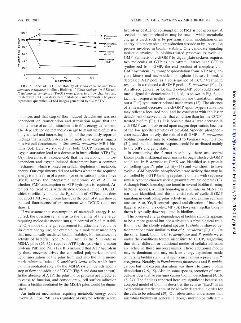
Effect of metabolic inhibitors on Vibrio cholerae and Pseu-domonas aeruginosa biofilms. In order to test whether the ob-served sensitivity to uncouplers and metabolic inhibitors in S.oneidensis is particular to this microorganism or a more gen-eral phenomenon, we tested Vibrio cholerae and Pseudomonasaeruginosa biofilms for uncoupler-mediated detachment. Bio-films of V. cholerae (A1552) and P. aeruginosa PAO1 weregrown in glycerol and glucose minimal medium in flow cells,respectively, and treated with 50 �M CCCP as describedabove. As indicated in Fig. 7, biofilms of V. cholerae readilydetached upon addition of CCCP in a response similar to thatobserved in S. oneidensis, while P. aeruginosa biofilms did notdecrease in biomass. We also tested P. aeruginosa �pel as wellas P. putida and again did not observe detachment (data notshown). Collectively, these observations showed that the un-coupler-mediated detachment is not unique to S. oneidensisMR-1 but is a microorganism-specific rather than a generalbiofilm characteristic.
DISCUSSION
The data shown here provide some mechanistic insights intothe stability and dissolution of biofilms. Since biofilms havebeen defined as consisting of cells that are generally fixedwithin a “sticky” matrix, one of the noteworthy findings re-ported in this study is that biofilm stability actually requiresmetabolic energy (Fig. 1 and 7) (29). Treatment of 12-hour S.oneidensis MR-1 biofilms with metabolic inhibitors CCCP,DNP, and NaCN resulted in rapid detachment of individualcells. Notably, the extent of this dissolution in young biofilmswas even greater than the stop-of-flow-induced detachment(10% versus 50% biomass retention). The facts that detach-ment was observed within 15 min after the addition of the
FIG. 4. Intracellular ATP and c-di-GMP concentrations inplanktonic S. oneidensis MR-1 cells. Cultures were grown aerobi-cally in a shaking flask to an optical density of 0.3 (600 nm), andeither CCCP was added or oxygenation was prevented by stoppingthe shaking as described in Materials and Methods. At the indicatedtime points after initiation of treatment, samples were removed andATP (A) and cyclic di-GMP (B) were assayed. Values expressed arepercentages of initial values (�M/OD600). White, untreated control;black, 40 �M CCCP; gray, oxygen deprived. Each data point rep-resents the mean value for six samples measured twice from twoseparate experiments. Error bars represent 1 standard deviation.
FIG. 5. Detachment of S. oneidensis MR-1 biofilm mutant cells in12-h biofilms. The graph represents quantified CLSM images gener-ated by COMSTAT. Biofilms of the respective in-frame deletion mu-tants were grown in flow chambers, and stop of flow (SOF) was appliedto induce detachment. Each data point is the mean value for at leastfour independent images taken from two channels. Error bars repre-sent 1 standard deviation.
FIG. 6. Transcription/translation-independent detachment of S.oneidensis. Stop of flow induced detachment in inhibitor-treated bio-films. The graph represents quantified CLSM images generated byCOMSTAT. Biofilms were incubated in the flowthrough mediumamended with inhibitors for 40 (rifampin) or 20 (erythromycin andtetracycline) minutes prior to application of a stop-of-flow detachmentassay as described in Materials and Methods. Each data point repre-sents the mean for four independent images taken from two separatechannels. Error bars represent 1 standard deviation.
3262 SAVILLE ET AL. J. BACTERIOL.
on February 28, 2020 by guest
http://jb.asm.org/
Dow
nloaded from
inhibitors and that stop-of-flow-induced detachment was notdependent on transcription and translation argue that themaintenance of cellular attachment itself is energy dependent.The dependence on metabolic energy to maintain biofilm sta-bility is novel and interesting in light of the previously reportedfindings that a sudden decrease in molecular oxygen triggersmassive cell detachment in Shewanella oneidensis MR-1 bio-films (33). Here, we showed that both CCCP treatment andoxygen starvation lead to a decrease in intracellular ATP (Fig.4A). Therefore, it is conceivable that the metabolic inhibitor-dependent and oxygen-induced detachment have a commonmechanism, which is based on cellular depletion of metabolicenergy. Our experiments did not address whether the requiredenergy is in the form of a proton (or other cation) motive force(PMF) across the cytoplasmic membrane or of ATP orwhether PMF consumption or ATP hydrolysis is required. At-tempts to treat cells with dicylcocarbonyldiimide (DCCD),which specifically inhibits the Fo subunit of ATPase but doesnot affect PMF, were inconclusive, as the control strain showedinduced fluorescence after treatment with DCCD (data notshown).
If we assume that consumption of metabolic energy is re-quired, the question remains as to the identity of the energy-requiring molecular mechanism(s) in control of biofilm stabil-ity. One mode of energy requirement for attachment could bevia direct energy use, for example, by a molecular machinerythat mechanically mediates biofilm stability. For instance, theactivity of bacterial type IV pili, such as the S. oneidensisMSHA pilus (26, 32), requires ATP hydrolysis via the motorproteins PilB and PilT (17). It is assumed that ATP hydrolysisby these enzymes drives the controlled polymerization anddepolymerization of the pilus from and into the pilin mono-meric subunits. Indeed, S. oneidensis �mxd cells, which formbiofilms mediated solely by the MSHA system, detach upon astop of flow and addition of CCCP (Fig. 5 and data not shown).In the absence of ATP, the pilus motor proteins are predictedto cease to function, and consequently, cell surface adhesionwithin a biofilm mediated by the MSHA pilus would be dimin-ished.
An indirect mechanism requiring metabolic energy couldinvolve ATP or PMF as a regulator of enzyme activity, where
hydrolysis of ATP or consumption of PMF is not necessary. Asecond indirect mechanism may be one in which metabolicenergy is used, such as by posttranslational modulation of anenergy-dependent signal transduction cascade or by a secretionprocess involved in biofilm stability. One candidate signalingmolecule involved in biofilm-related processes is cyclic di-GMP. Synthesis of c-di-GMP by diguanylate cyclases requirestwo molecules of GTP as a substrate. Intracellular GTP issynthesized from GMP, the end product of complete c-di-GMP hydrolysis, by transphosphorylation from ATP by guan-ylate kinase and nucleoside diphosphate kinases. Indeed, adecreased ATP pool, as a consequence of CCCP treatment,resulted in a reduced c-di-GMP pool in S. oneidensis (Fig. 4).An altered general or localized c-di-GMP pool could consti-tute a signal for detachment. Indeed, as shown in Fig. 6, de-tachment requires neither transcription nor translation, rulingout a FleQ-type transcriptional mechanism (12). The absenceof a measured decrease in c-di-GMP upon oxygen starvationmay reflect a localized pool and be consistent with the lesserdetachment observed under that condition than for the CCCP-treated biofilm (Fig. 1). It is possible that a large decrease inc-di-GMP was not observed upon oxygen deprivation, becauseof the low specific activities of c-di-GMP-specific phosphodi-esterases. Alternatively, the role of c-di-GMP in S. oneidensisbiofilm formation may be confined to biomass accumulation(31), and the detachment response could be attributed mainlyto the cell’s energetic state.
In considering the former possibility, there are severalknown posttranslational mechanisms through which c-di-GMPcould act. In P. aeruginosa, FimX was identified as a proteincontrolling type IV pilus activity (14) and was shown to havecyclic-di-GMP-specific phosphodiesterase activity that may becontrolled by a GTP-binding regulatory domain with sequencesimilarity to the characteristic diguanylate cyclase domain (19).Although FimX homologs are found in several biofilm-formingbacterial species, a FimX homolog in S. oneidensis MR-1 hasnot been identified, and the potential role of cyclic-di-GMPsignaling in controlling pilus activity in this organism remainsunclear. Also, YcgR controls speed and direction of bacterialflagellar rotation via c-di-GMP (3). However, flagellar biosyn-thesis is typically downregulated in biofilms.
The observed energy dependence of biofilm stability appearsto be a more general but not a ubiquitous physiological trait.Biofilms of the closely related species V. cholerae showed de-tachment behavior similar to that of S. oneidensis (Fig. 6). Onthe other hand, biofilms of P. aeruginosa and P. putida were,under the conditions tested, insensitive to CCCP, suggestingthat either different or additional modes of cellular adhesionare active in those microorganisms. These additional modesmay be dominant and may mask an energy-dependent modeconferring biofilm stability, if such a mechanism is present in P.aeruginosa. Notably, in Pseudomonas fluorescens and P. putida,carbon but not oxygen starvation was shown to cause biofilmdissolution (7, 9, 15). Also, in some species, secretion of extra-cellular degradative enzymes causes biofilm detachment (4, 16,18, 24). The findings reported here are significant because anaccepted model of biofilms describes the cells as “fixed” in anextracellular matrix that must be actively degraded in order forthe cells to be released (29). Our observation underscores thatmicrobial biofilms in general, although morphologically simi-
FIG. 7. Effect of CCCP on stability of Vibrio cholerae and Pseu-domonas aeruginosa biofilms. Biofilms of Vibrio cholerae (A1552) andPseudomonas aeruginosa (PAO1) were grown in a flow chamber andtreated with CCCP as described in Materials and Methods. The graphrepresents quantified CLSM images generated by COMSTAT.
VOL. 193, 2011 STABILITY OF S. ONEIDENSIS MR-1 BIOFILMS 3263
on February 28, 2020 by guest
http://jb.asm.org/
Dow
nloaded from
lar, can be maintained by different mechanisms or combina-tions thereof.
ACKNOWLEDGMENTS
This work was supported by NSF grant MCB-0617952 to A.M.S. anda grant from the Carlsberg foundation and the Lundbeck foundation toJ.A.J.H.
REFERENCES
1. Anderson, G. G., and G. A. O’Toole. 2008. Innate and induced resistancemechanisms of bacterial biofilms. Curr. Top. Microbiol. Immunol. 322:85–105.
2. Applegate, D. H., and J. D. Bryers. 1991. Effects of carbon and oxygenlimitations and calcium concentrations on biofilm removal processes. Bio-technol. Bioeng. 37:17–25.
3. Boehm, A., 2010. Second messenger-mediated adjustment of bacterial swim-ming velocity. Cell 141:107–116.
4. Boyd, A., and A. M. Chakrabarty. 1994. Role of alginate lyase in cell de-tachment of Pseudomonas aeruginosa. Appl. Environ. Microbiol. 60:2355–2359.
5. Branda, S. S., S. Vik, L. Friedman, and R. Kolter. 2005. Biofilms: the matrixrevisited. Trends Microbiol. 13:20–26.
6. Costerton, J. W., P. S. Stewart, and E. P. Greenberg. 1999. Bacterial biofilms:a common cause of persistent infections. Science 284:1318–1322.
7. Delaquis, P. J., D. E. Caldwell, J. R. Lawrence, and A. R. McCurdy. 1989.Detachment of Pseudomonas fluorescens from biofilms on glass surfaces inresponse to nutrient stress. Microb. Ecol. 18:199–210.
8. Donlan, R. M. 2002. Biofilms: microbial life on surfaces. Emerg. Infect. Dis.8:881–890.
9. Gjermansen, M., P. Ragas, C. Sternberg, S. Molin, and T. Tolker-Nielsen.2005. Characterization of starvation-induced dispersion in Pseudomonasputida biofilms. Environ. Microbiol. 7:894–906.
10. Heydorn, A., et al. 2000. Quantification of biofilm structures by the novelcomputer program COMSTAT. Microbiology 146(Pt. 10):2395–2407.
11. Heytler, P. G., and W. W. Prichard. 1962. A new class of uncoupling agents—carbonyl cyanide phenylhydrazones. Biochem. Biophys. Res. Commun.7:272–275.
12. Hickman, J. W., and C. S. Harwood. 2008. Identification of FleQ fromPseudomonas aeruginosa as a c-di-GMP-responsive transcription factor.Mol. Microbiol. 69:376–389.
13. Hopfer, U., A. L. Lehninger, and T. E. Thompson. 1968. Protonic conduc-tance across phospholipid bilayer membranes induced by uncoupling agentsfor oxidative phosphorylation. Proc. Natl. Acad. Sci. U. S. A. 59:484–490.
14. Huang, B., C. B. Whitchurch, and J. S. Mattick. 2003. FimX, a multidomainprotein connecting environmental signals to twitching motility in Pseudomo-nas aeruginosa. J. Bacteriol. 185:7068–7076.
15. Hunt, S. M., E. M. Werner, B. Huang, M. A. Hamilton, and P. S. Stewart.
2004. Hypothesis for the role of nutrient starvation in biofilm detachment.Appl. Environ. Microbiol. 70:7418–7425.
16. Itoh, Y., X. Wang, B. J. Hinnebusch, J. F. Preston III, and T. Romeo. 2005.Depolymerization of beta-1,6-N-acetyl-D-glucosamine disrupts the integrityof diverse bacterial biofilms. J. Bacteriol. 187:382–387.
17. Jakovljevic, V., S. Leonardy, M. Hoppert, and L. Sogaard-Andersen. 2008.PilB and PilT are ATPases acting antagonistically in type IV pilus functionin Myxococcus xanthus. J. Bacteriol. 190:2411–2421.
18. Kaplan, J. B., C. Ragunath, N. Ramasubbu, and D. H. Fine. 2003. Detach-ment of Actinobacillus actinomycetemcomitans biofilm cells by an endoge-nous beta-hexosaminidase activity. J. Bacteriol. 185:4693–4698.
19. Kazmierczak, B. I., M. B. Lebron, and T. S. Murray. 2006. Analysis of FimX,a phosphodiesterase that governs twitching motility in Pseudomonas aerugi-nosa. Mol. Microbiol. 60:1026–1043.
20. Klausen, M., et al. 2003. Biofilm formation by Pseudomonas aeruginosa wildtype, flagella and type IV pili mutants. Mol. Microbiol. 48:1511–1524.
21. Lewis, K. 2008. Multidrug tolerance of biofilms and persister cells. Curr.Top. Microbiol. Immunol. 322:107–131.
22. Muller, J., M. C. Miller, A. T. Nielsen, G. K. Schoolnik, and A. M. Spor-mann. 2007. vpsA- and luxO-independent biofilms of Vibrio cholerae. FEMSMicrobiol. Lett. 275:199–206.
23. Myers, C. R., and K. H. Nealson. 1988. Bacterial manganese reduction andgrowth with manganese oxide as the sole electron acceptor. Science 240:1319–1321.
24. Pecharki, D., F. C. Petersen, and A. A. Scheie. 2008. Role of hyaluronidasein Streptococcus intermedius biofilm. Microbiology 154:932–938.
25. Sauer, K., et al. 2004. Characterization of nutrient-induced dispersion inPseudomonas aeruginosa PAO1 biofilm. J. Bacteriol. 186:7312–7326.
26. Saville, R. M., N. Dieckmann, and A. M. Spormann. 2010. Spatiotemporalactivity of the mshA gene system in Shewanella oneidensis MR-1 biofilms.FEMS Microbiol. Lett. 308:76–83.
27. Sawyer, L. K., and S. W. Hermanowicz. 2000. Detachment of Aeromonashydrophila and Pseudomonas aeruginosa due to variations in nutrient supply.Water Sci. Technol. 41:139–145.
28. Sternberg, C., et al. 1999. Distribution of bacterial growth activity in flow-chamber biofilms. Appl. Environ. Microbiol. 65:4108–4117.
29. Sutherland, I. 2001. Biofilm exopolysaccharides: a strong and sticky frame-work. Microbiology 147:3–9.
30. Teal, T. K., D. P. Lies, B. J. Wold, and D. K. Newman. 2006. Spatiometabolicstratification of Shewanella oneidensis biofilms. Appl. Environ. Microbiol.72:7324–7330.
31. Thormann, K. M., et al. 2006. Control of formation and cellular detachmentfrom Shewanella oneidensis MR-1 biofilms by cyclic di-GMP. J. Bacteriol.188:2681–2691.
32. Thormann, K. M., R. M. Saville, S. Shukla, D. A. Pelletier, and A. M.Spormann. 2004. Initial phases of biofilm formation in Shewanella oneidensisMR-1. J. Bacteriol. 186:8096–8104.
33. Thormann, K. M., R. M. Saville, S. Shukla, and A. M. Spormann. 2005.Induction of rapid detachment in Shewanella oneidensis MR-1 biofilms. J.Bacteriol. 187:1014–1021.
3264 SAVILLE ET AL. J. BACTERIOL.
on February 28, 2020 by guest
http://jb.asm.org/
Dow
nloaded from