edta chelation therapy - dr. crantondrcranton.com/chelation/edtastudies.pdfedta chelation therapy a...
TRANSCRIPT

EDTA Chelation Therapy
A Non-surgical Treatment for
Heart Disease
and
Atherosclerosis
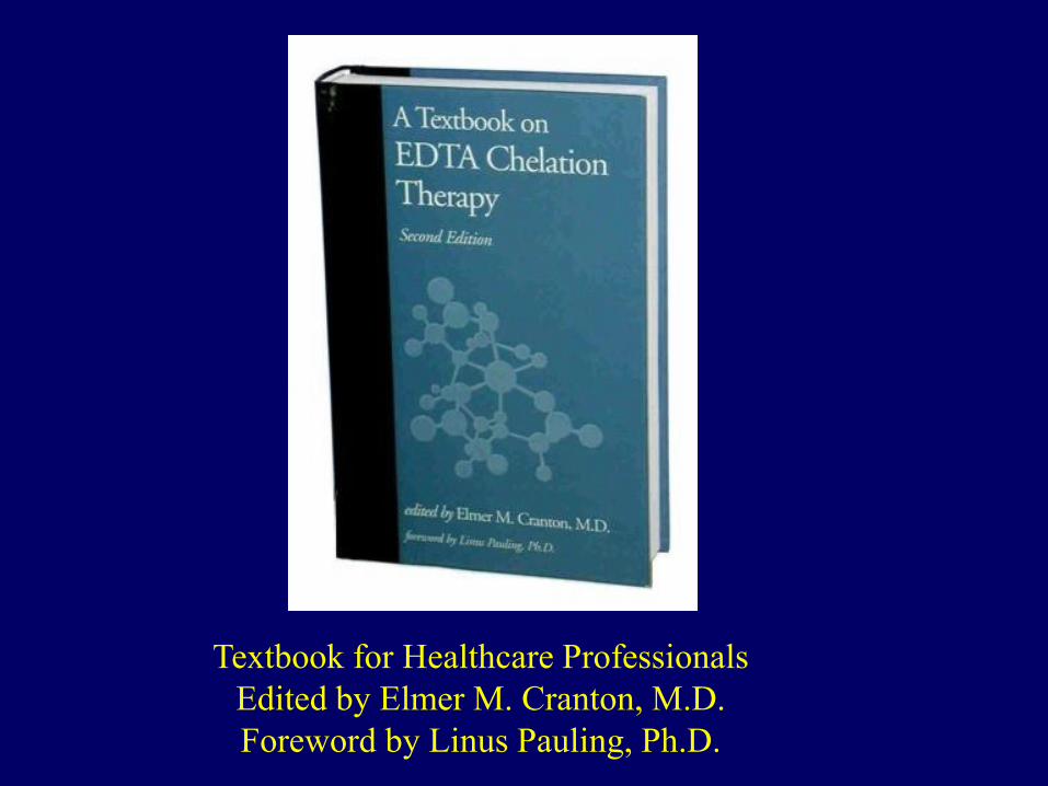
Textbook for Healthcare Professionals
Edited by Elmer M. Cranton, M.D.
Foreword by Linus Pauling, Ph.D.
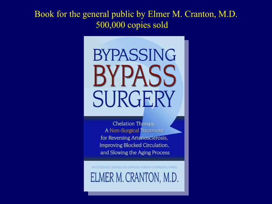
Book for the general public by Elmer M. Cranton, M.D.
500,000 copies sold

A Simple 3-hour Office Procedure
• No need for a hospital
• Patients walk, drive or ride safely
to and from treatments
• 3 hour intravenous infusions of
EDTA for 20 to 30 treatments
• Administered safely by a nurse

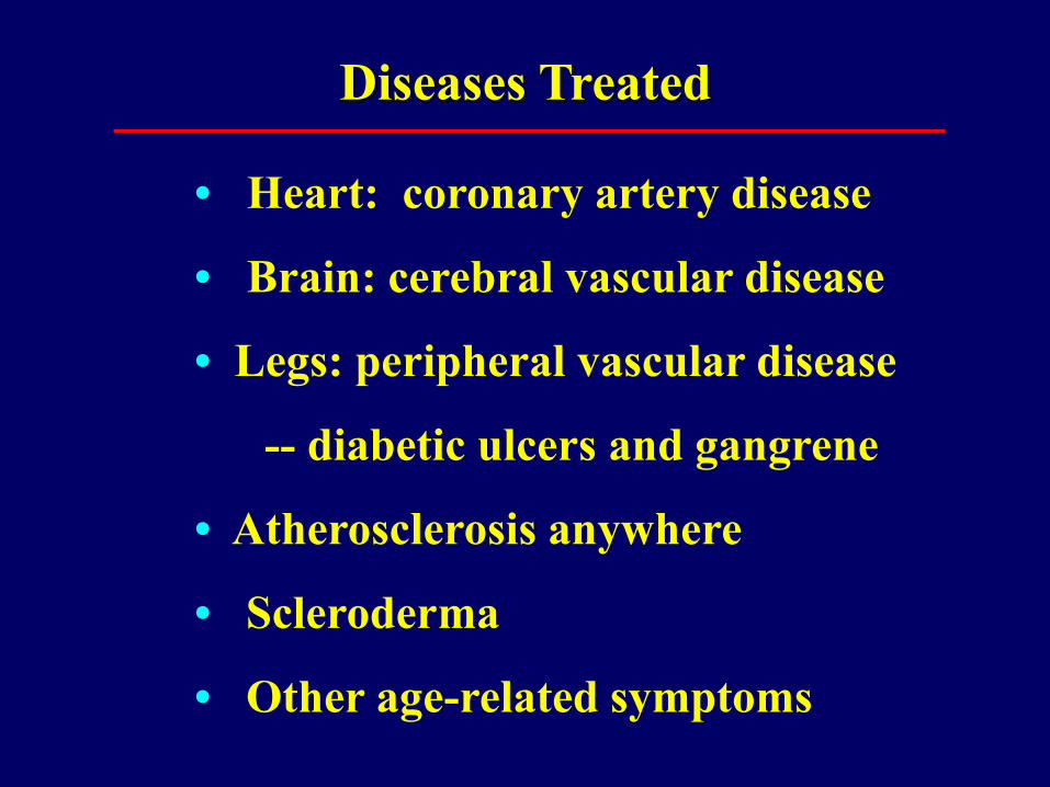
Diseases Treated
• Heart: coronary artery disease
• Brain: cerebral vascular disease
• Legs: peripheral vascular disease
-- diabetic ulcers and gangrene
• Atherosclerosis anywhere
• Scleroderma
• Other age-related symptoms
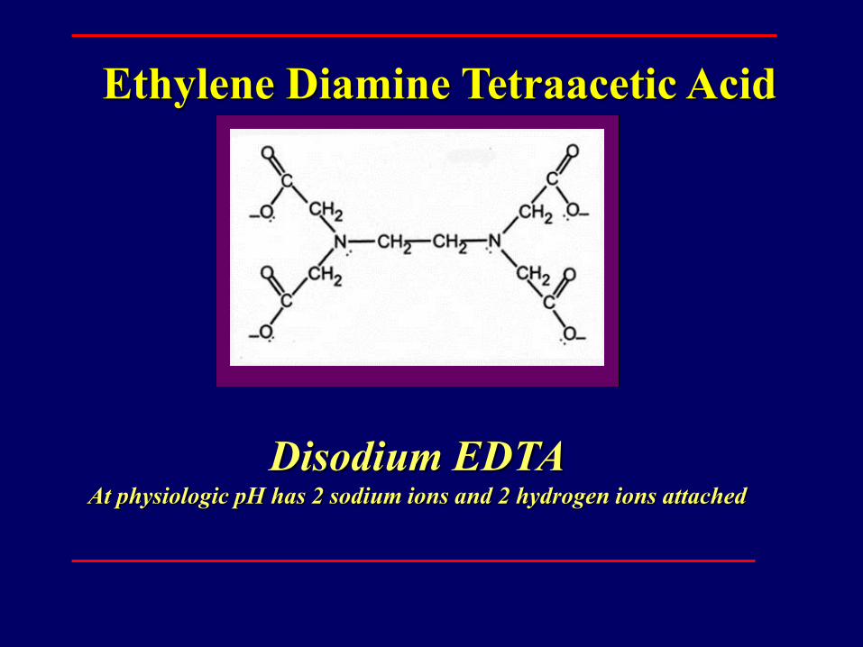
Ethylene Diamine Tetraacetic Acid
Disodium EDTA At physiologic pH has 2 sodium ions and 2 hydrogen ions attached
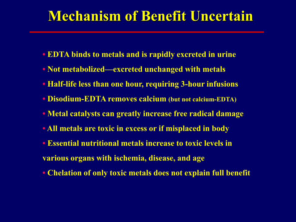
Mechanism of Benefit Uncertain
• EDTA binds to metals and is rapidly excreted in urine
• Not metabolized—excreted unchanged with metals
• Half-life less than one hour, requiring 3-hour infusions
• Disodium-EDTA removes calcium (but not calcium-EDTA)
• Metal catalysts can greatly increase free radical damage
• All metals are toxic in excess or if misplaced in body
• Essential nutritional metals increase to toxic levels in
various organs with ischemia, disease, and age
• Chelation of only toxic metals does not explain full benefit

Increase in urinary excretion of metals
following I.V. disodium EDTA
Nutritional Elements Manganese 132 times baseline excretion
Zinc 62 times baseline
Iron 56 times baseline
Cobalt 12 times baseline
Calcium 10 times baseline
Toxic Elements Lead 8 times baseline excretion
Cadmium 5 times baseline
Nickel 5 times baseline
Aluminum 3 times baseline
Arsenic 2 times baseline
Mercury 2 times baseline
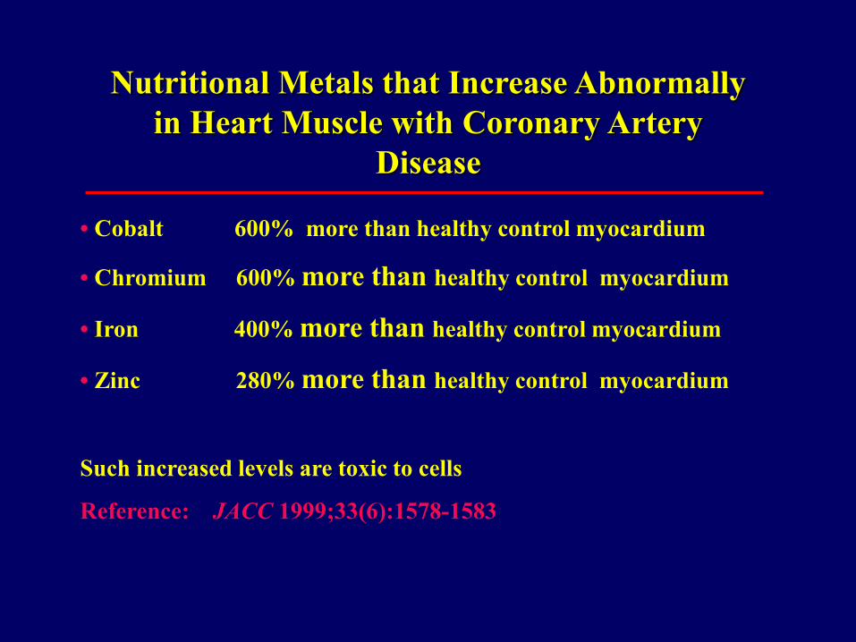
Nutritional Metals that Increase Abnormally
in Heart Muscle with Coronary Artery
Disease
• Cobalt 600% more than healthy control myocardium
• Chromium 600% more than healthy control myocardium
• Iron 400% more than healthy control myocardium
• Zinc 280% more than healthy control myocardium
Such increased levels are toxic to cells
Reference: JACC 1999;33(6):1578-1583
All Published Studies
Show Benefit
For detailed scientific references with analysis :
www.drcranton.com

EDTA Chelation
Coronary Heart Disease Clark, et al 1956
• 20 patients — 16 unstable angina
• Complete relief of angina in 17
• 16 asymptomatic at 21 months
• ECG normalized in 33%
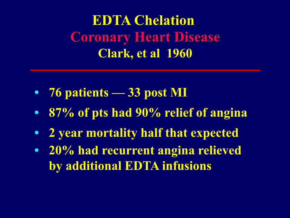
EDTA Chelation
Coronary Heart Disease Clark, et al 1960
• 76 patients — 33 post MI
• 87% of pts had 90% relief of angina
• 2 year mortality half that expected
• 20% had recurrent angina relieved
by additional EDTA infusions
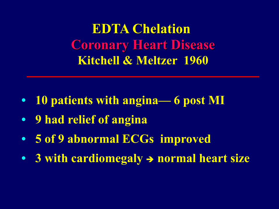
EDTA Chelation
Coronary Heart Disease Kitchell & Meltzer 1960
• 10 patients with angina— 6 post MI
• 9 had relief of angina
• 5 of 9 abnormal ECGs improved
• 3 with cardiomegaly normal heart size
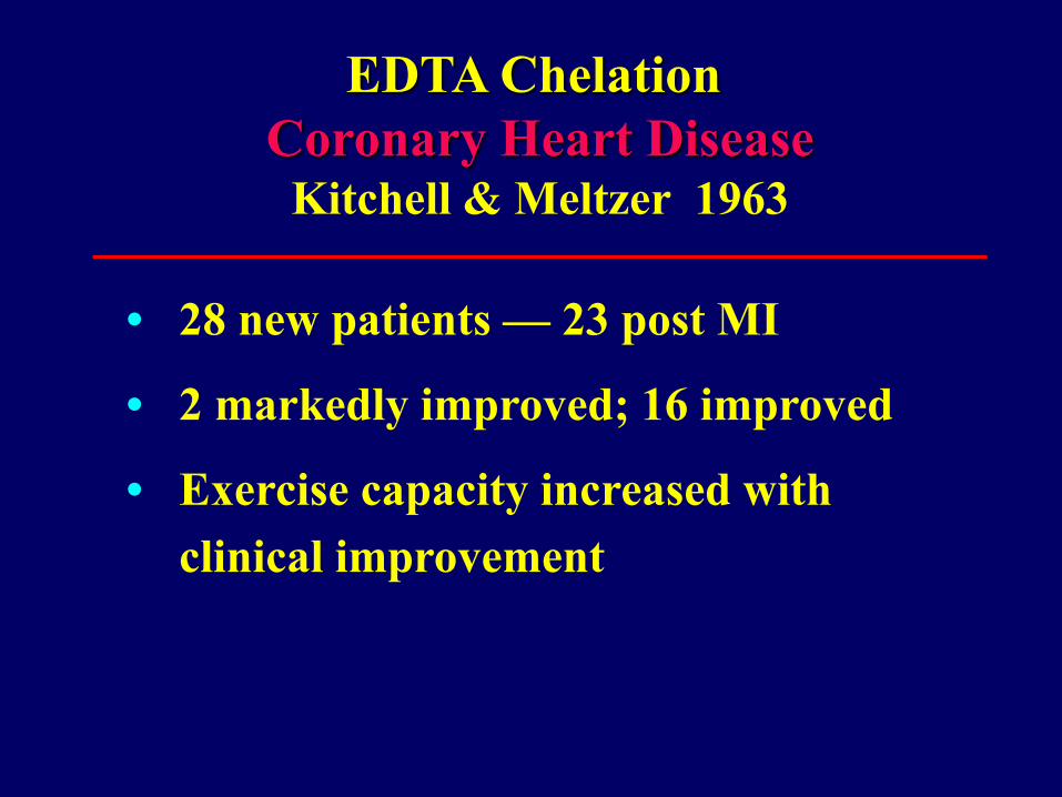
EDTA Chelation
Coronary Heart Disease Kitchell & Meltzer 1963
• 28 new patients — 23 post MI
• 2 markedly improved; 16 improved
• Exercise capacity increased with
clinical improvement
EDTA Chelation
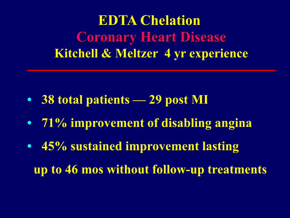
Coronary Heart Disease Kitchell & Meltzer 4 yr experience
• 38 total patients — 29 post MI
• 71% improvement of disabling angina
• 45% sustained improvement lasting
up to 46 mos without follow-up treatments

EDTA Chelation
Coronary Heart Disease
Other Case-series
• Olszewer & Carter 844 patients
77% marked improvement
3% no improvement
• Deucher 215 patients
70% symptomatic improvement
• Hancke & Flytie 265 patients
91% symptomatic improvement
8% unchanged; 1% worse

EDTA Chelation
Peripheral Vascular Disease Clarke et al. 1960
• 31 pts — 22 rest pain, 1 dry gangrene
• 74% relief of rest pain & claudication
• 1 pt no change, 2 pts worse
• 2 amputated,
• 3/4 pts recurrent sx responded to rx

EDTA Chelation
Peripheral Vascular Disease Lamar 1964, 1966
• 18 with diabetic ulcers, gangrene
• 100% had measurable improvement
• Healed ulcerations, improved pulses

EDTA Chelation
Peripheral Vascular Disease Olszewer & Carter 1989
• 1130 patients
• 91% complete recovery
• 8% good recovery
• 7 of 10 pts with dry gangrene had satisfactory
recovery
EDTA Chelation
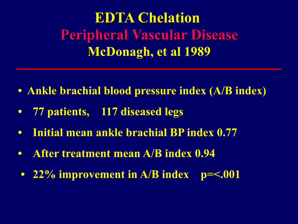
Peripheral Vascular Disease McDonagh, et al 1989
• Ankle brachial blood pressure index (A/B index)
• 77 patients, 117 diseased legs
• Initial mean ankle brachial BP index 0.77
• After treatment mean A/B index 0.94
• 22% improvement in A/B index p=<.001
EDTA Chelation
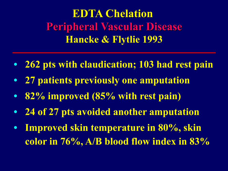
Peripheral Vascular Disease Hancke & Flytlie 1993
• 262 pts with claudication; 103 had rest pain
• 27 patients previously one amputation
• 82% improved (85% with rest pain)
• 24 of 27 pts avoided another amputation
• Improved skin temperature in 80%, skin
color in 76%, A/B blood flow index in 83%
EDTA Chelation
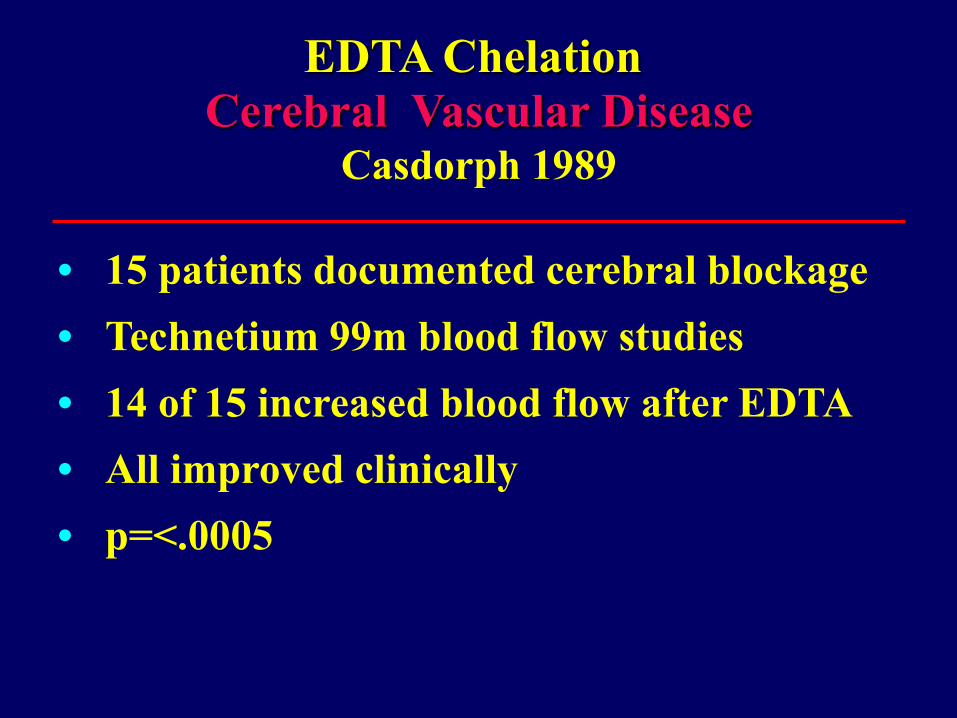
Cerebral Vascular Disease Casdorph 1989
• 15 patients documented cerebral blockage
• Technetium 99m blood flow studies
• 14 of 15 increased blood flow after EDTA
• All improved clinically
• p=<.0005

Measuring Brain Blood Flow Increase
Technetium 99m Before and after Chelation Therapy
Injecting Radioactive Isotope
Head positioned over scintillation Camera
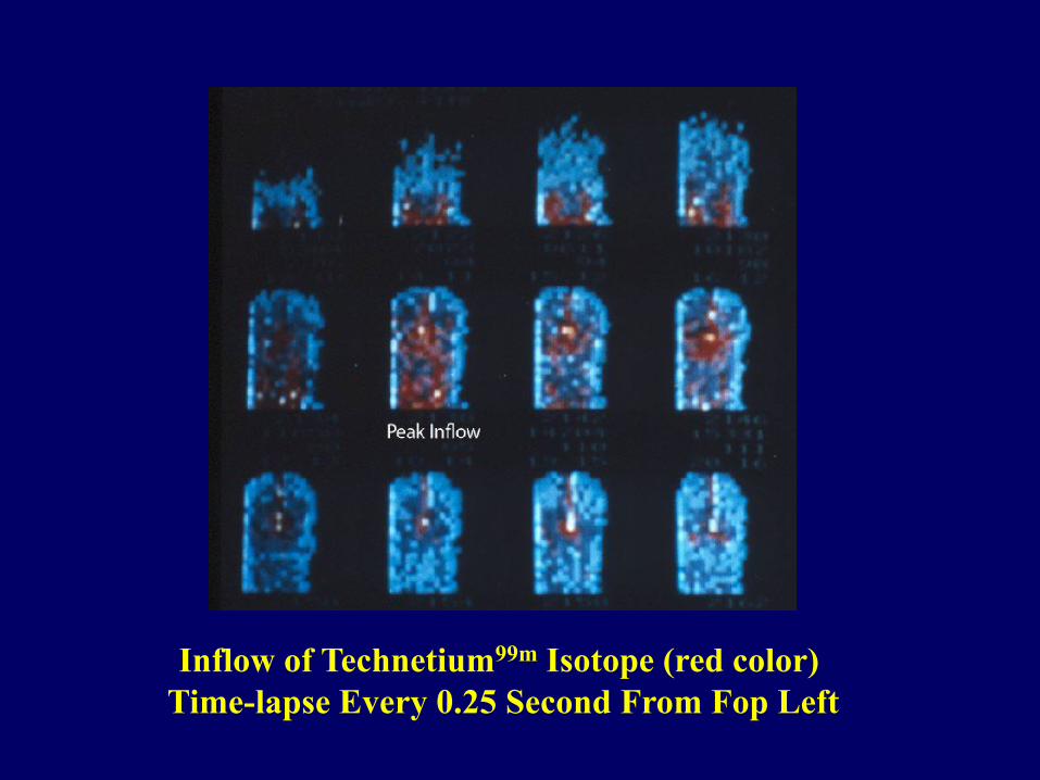
Inflow of Technetium99m Isotope (red color)
Time-lapse Every 0.25 Second From Fop Left
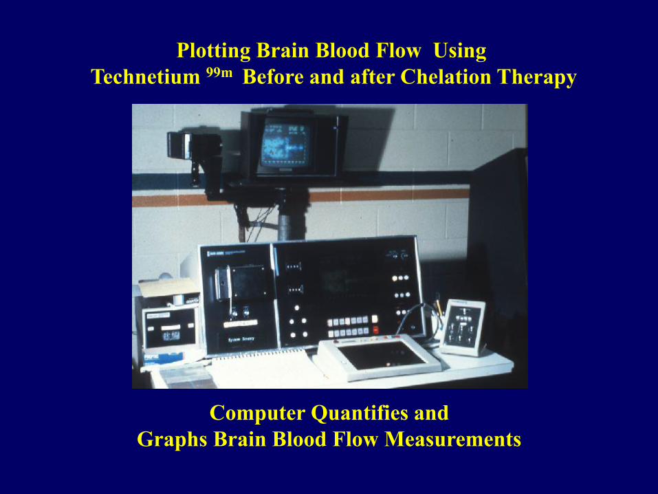
Plotting Brain Blood Flow Using
Technetium 99m Before and after Chelation Therapy
Computer Quantifies and
Graphs Brain Blood Flow Measurements

Casdorph, 1989
Computer Generated Graph of Blood Flowing Into Brain
Showing changes with blockage to flow
A = peak inflow over time A - delayed inflow
b = maximum outflow b - outflow delayed and less
complete with blockage
c = second pass
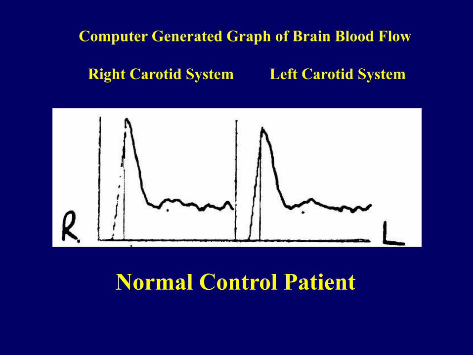
Computer Generated Graph of Brain Blood Flow
Right Carotid System Left Carotid System
Normal Control Patient
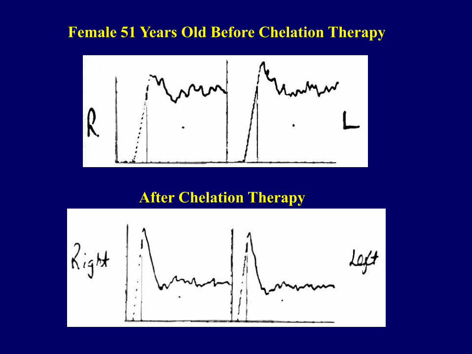
Female 51 Years Old Before Chelation Therapy
After Chelation Therapy
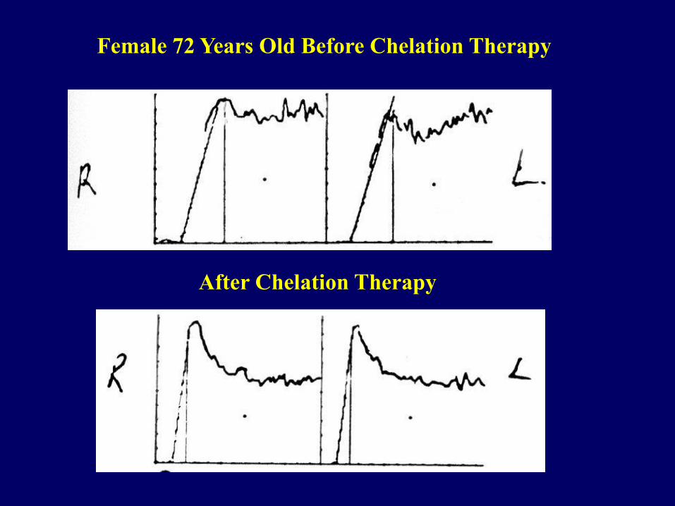
Female 72 Years Old Before Chelation Therapy
After Chelation Therapy
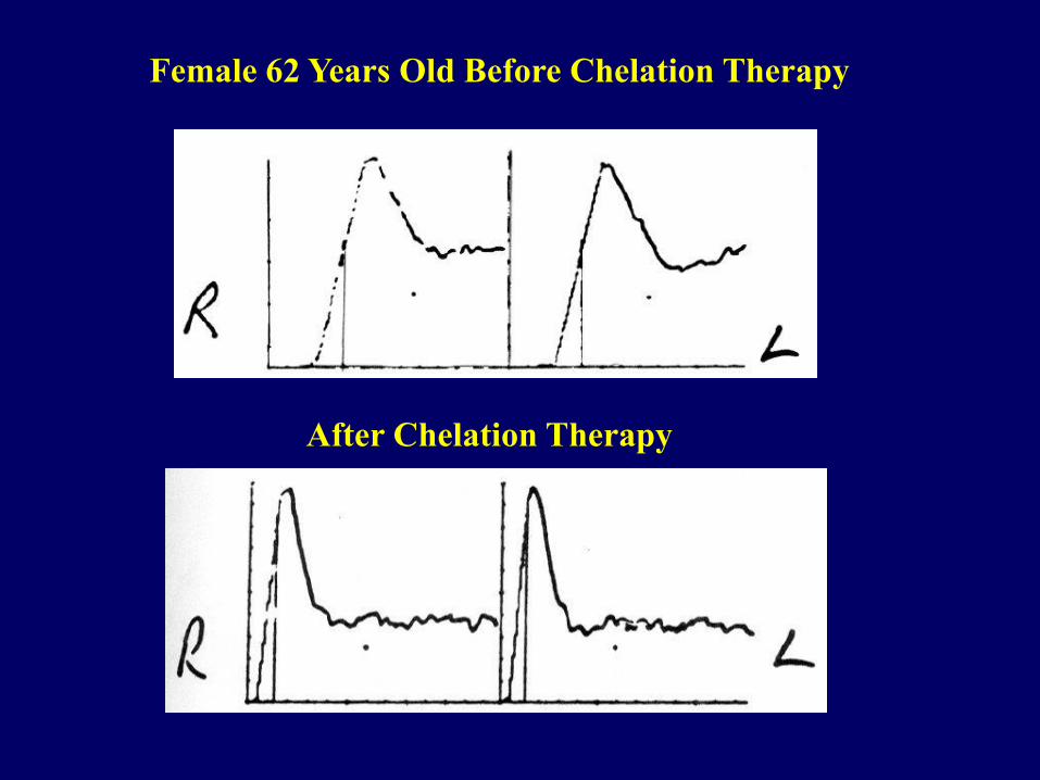
Female 62 Years Old Before Chelation Therapy
After Chelation Therapy
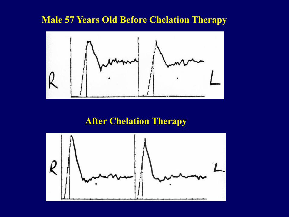
Male 57 Years Old Before Chelation Therapy
After Chelation Therapy
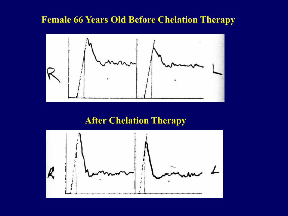
Female 66 Years Old Before Chelation Therapy
After Chelation Therapy
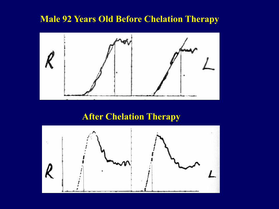
Male 92 Years Old Before Chelation Therapy
After Chelation Therapy
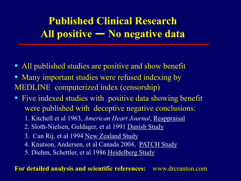
Published Clinical Research
All positive — No negative data
• All published studies are positive and show benefit
• Many important studies were refused indexing by
MEDLINE computerized index (censorship)
• Five indexed studies with positive data showing benefit
were published with deceptive negative conclusions:
1. Kitchell et al 1963, American Heart Journal, Reappraisal
2. Sloth-Nielsen, Guldager, et al 1991 Danish Study
3. Can Rij, et al 1994 New Zealand Study 4. Knutson, Andersen, et al Canada 2004, PATCH Study
5. Diehm, Schettler, et al 1986 Heidelberg Study
For detailed analysis and scientific references: www.drcranton.com
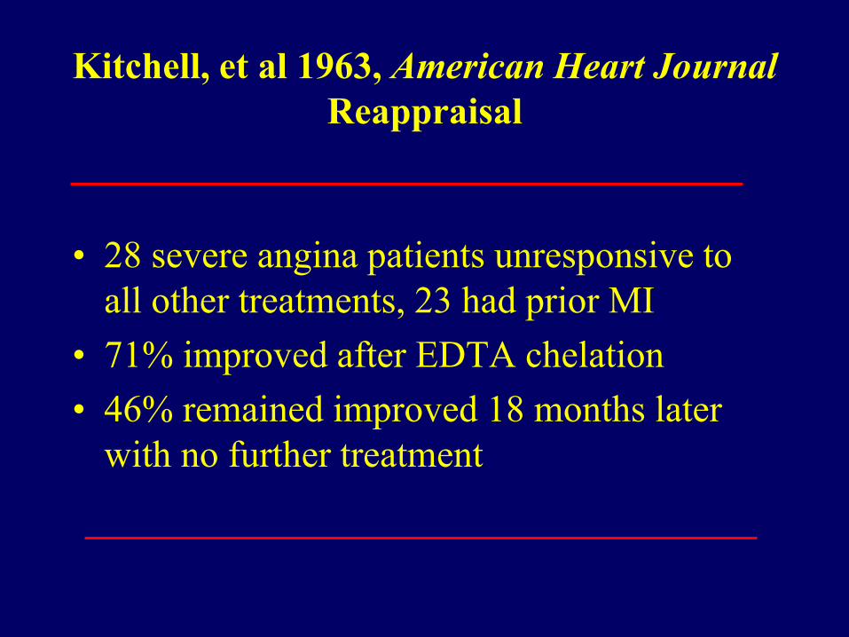
Kitchell, et al 1963, American Heart Journal
Reappraisal
• 28 severe angina patients unresponsive to
all other treatments, 23 had prior MI
• 71% improved after EDTA chelation
• 46% remained improved 18 months later
with no further treatment
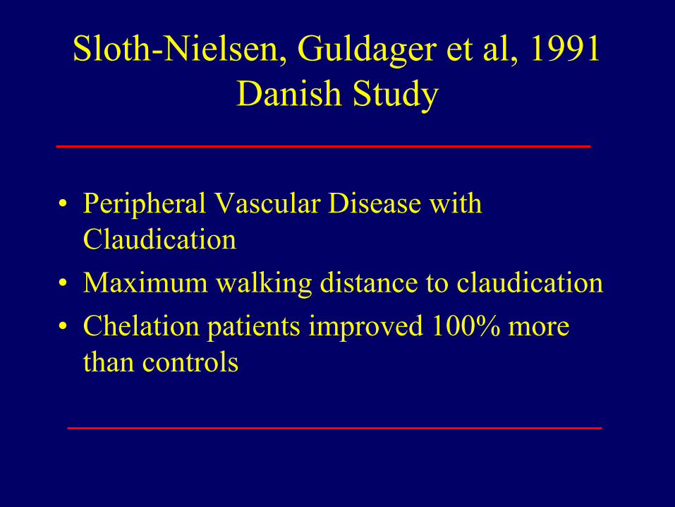
Sloth-Nielsen, Guldager et al, 1991
Danish Study
• Peripheral Vascular Disease with
Claudication
• Maximum walking distance to claudication
• Chelation patients improved 100% more
than controls
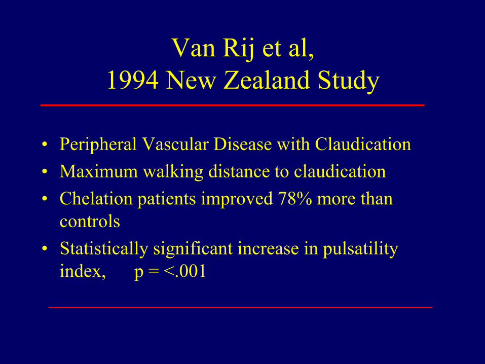
Van Rij et al,
1994 New Zealand Study
• Peripheral Vascular Disease with Claudication
• Maximum walking distance to claudication
• Chelation patients improved 78% more than
controls
• Statistically significant increase in pulsatility
index, p = <.001
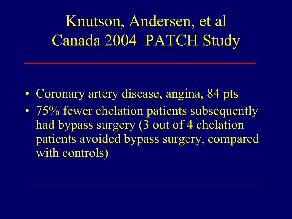
Knutson, Andersen, et al
Canada 2004 PATCH Study
• Coronary artery disease, angina, 84 pts
• 75% fewer chelation patients subsequently had bypass surgery (3 out of 4 chelation patients avoided bypass surgery, compared with controls)
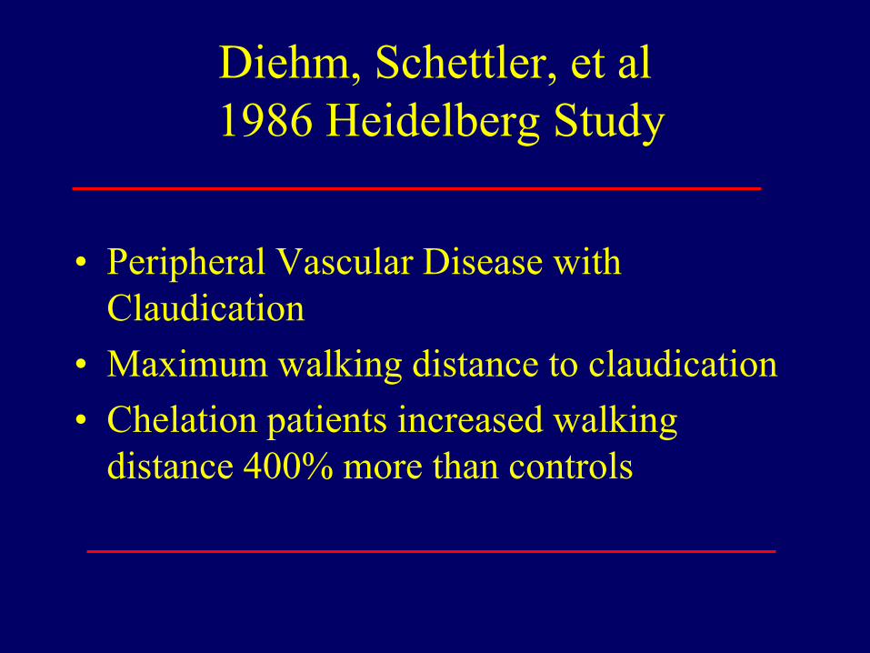
Diehm, Schettler, et al
1986 Heidelberg Study
• Peripheral Vascular Disease with
Claudication
• Maximum walking distance to claudication
• Chelation patients increased walking
distance 400% more than controls
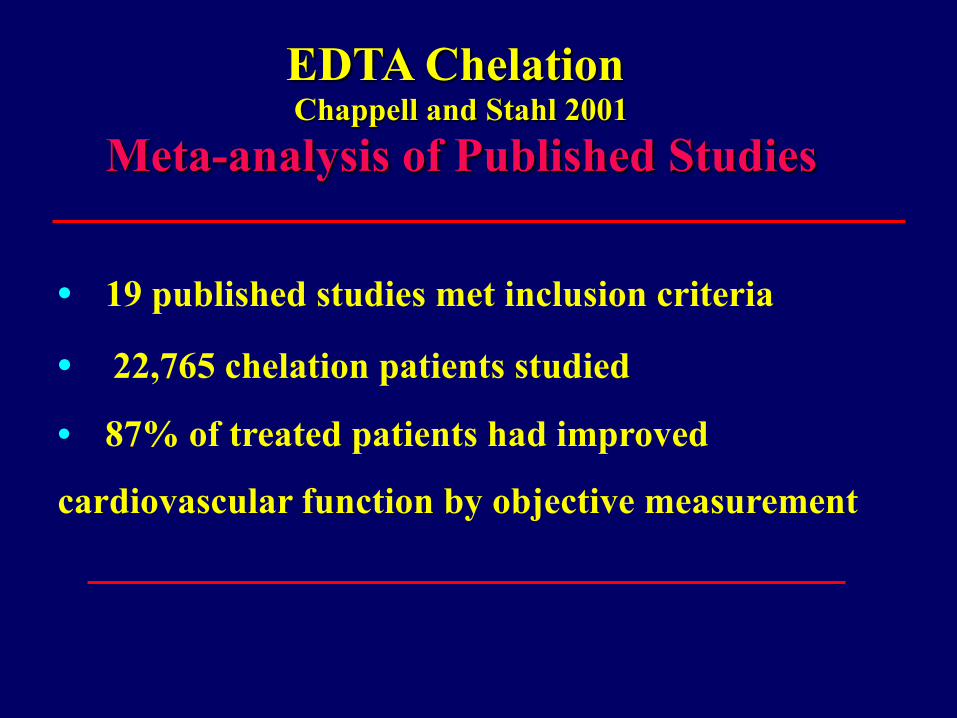
EDTA Chelation Chappell and Stahl 2001
Meta-analysis of Published Studies
• 19 published studies met inclusion criteria
• 22,765 chelation patients studied
• 87% of treated patients had improved
cardiovascular function by objective measurement

A Chelation Patient Previously Told
by Two Surgeons that Amputation Was The Only
Possible Treatment
51 Year Old Male Diabetic Patient
Before Chelation Therapy
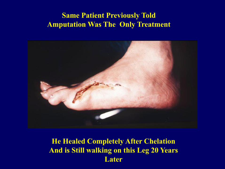
Same Patient Previously Told
Amputation Was The Only Treatment
He Healed Completely After Chelation
And is Still walking on this Leg 20 Years
Later

Chelation Patient Previously Told
Amputation Was The Only Treatment
60 Year Old Male Diabetic Before
Chelation Therapy
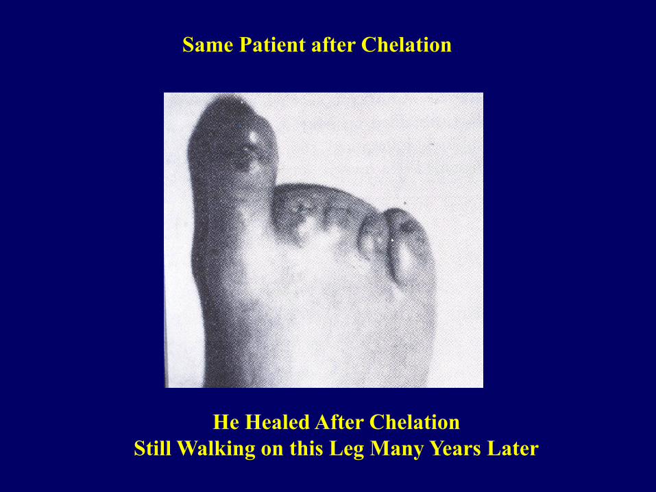
Same Patient after Chelation
He Healed After Chelation
Still Walking on this Leg Many Years Later
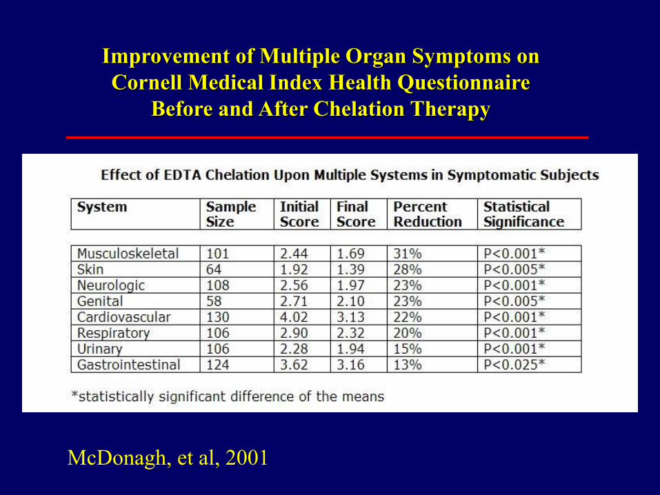
Improvement of Multiple Organ Symptoms on
Cornell Medical Index Health Questionnaire
Before and After Chelation Therapy
McDonagh, et al, 2001
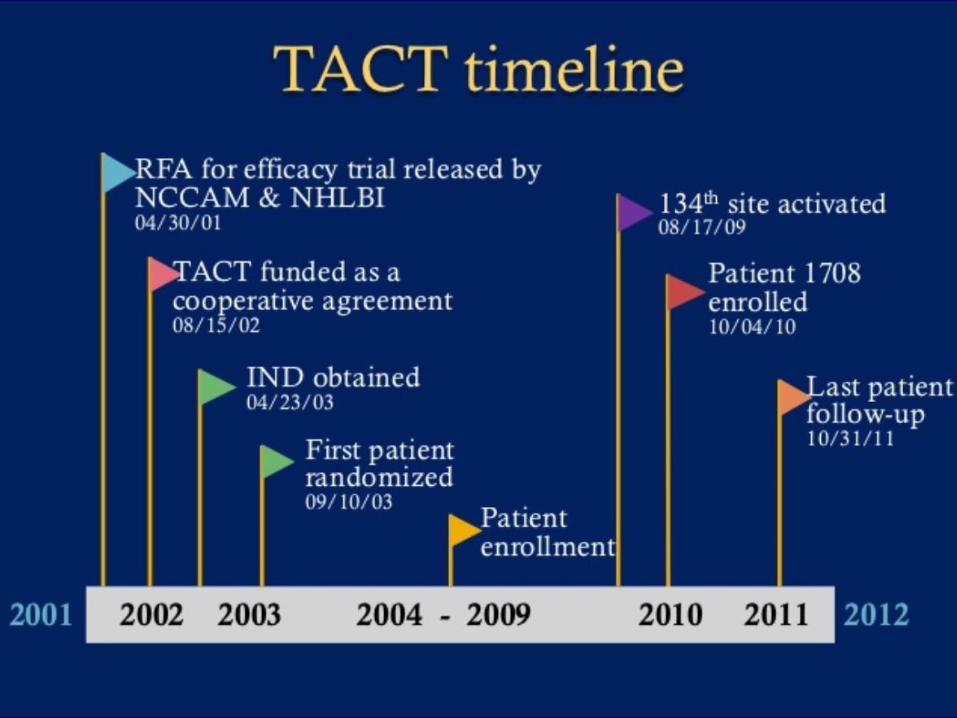
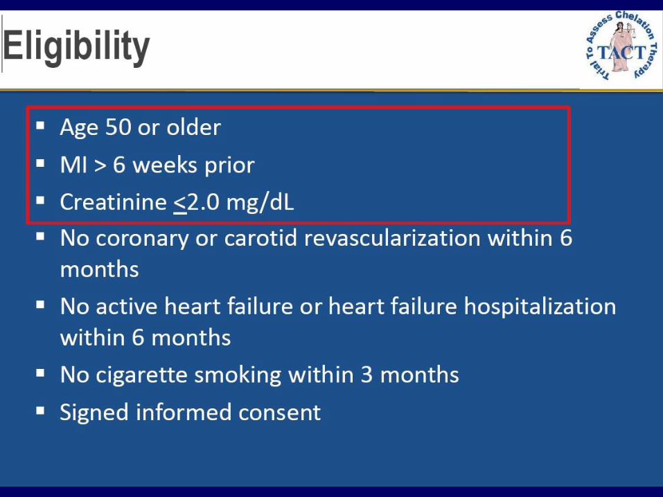


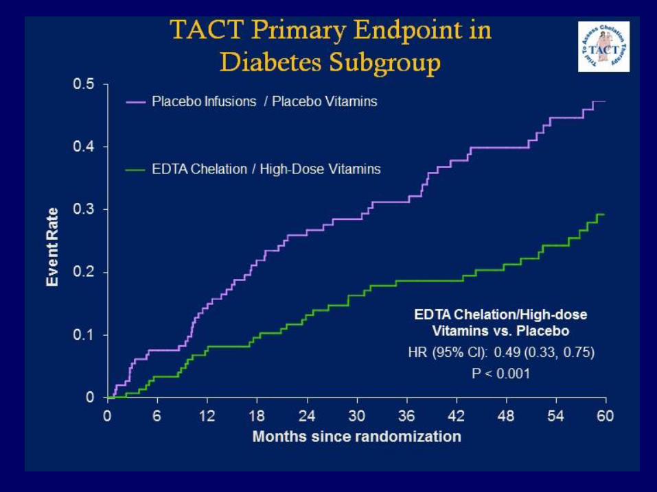
TACT STUDY RESULTS
A statistically significant benefit overall, but greater benefit
for the third of all patients with diabetes.
For Patients with diabetes
40% reduction in risk of death from a cardiovascular event
52% reduced risk of recurrent MI
43% reduction in death from all causes