chelation therapy for intermittent claudication and ... · • chelation therapy (ct) is a...
TRANSCRIPT

Chelation Therapy for Intermittent Claudication and Coronary Heart Disease
i
Chelation therapy for Intermittent Claudication and
Coronary Heart Disease
A Birmingham Technology Assessment Group Report
Authors: Martin Connock, Jayne Wilson, Fujian Song, Chris Hyde, and Catherine Meads
Department of Public Health & Epidemiology University of Birmingham Edgbaston
Birmingham B15 2TT UK
Acknowledgements: Janine Dretzke (BTAG) for invaluable help with studies reported in German, especially the thesis by M. Gleuβner describing the Hopf trial. Richard Wilson for providing information about numbers of surgical procedures performed. Wayne Perry for making available the review of chelation therapy by Olmstead. Chris Leonard and Rebecca Mason for administrative support. Prof Gregory YH Lip and Dr Wayne Perry for peer review of the report. ISBN NO: 07044 2343X © Copyright, West Midlands Health Technology Assessment Collaboration Department of Public Health and Epidemiology University of Birmingham 2002

Chelation Therapy for Intermittent Claudication and Coronary Heart Disease
ii
West Midlands Health Technology Assessment Collaboration The West Midlands Health Technology Assessment Collaboration (WMHTAC) produce rapid systematic reviews about the effectiveness of healthcare interventions and technologies, in response to requests from West Midlands Health Authorities or the HTA programme. Reviews usually take 3-6 months and aim to give a timely and accurate analysis of the quality, strength and direction of the available evidence, generating an economic analysis (where possible a cost-utility analysis) of the intervention. About InterTASC West Midlands HTAC is a member of InterTASC which is a national collaboration with three other units who do rapid reviews: the Trent Working Group on Acute Purchasing; the Wessex Institute for Health Research and Development; York Centre for Reviews and Dissemination. The aim of InterTASC is to share the work on reviewing the effectiveness and cost-effectiveness of health care interventions in order to avoid unnecessary duplication and improve the peer reviewing and quality control of reports. Contributions of Authors Martin Connock was the main author. He was responsible for the day-to-day management of the report; undertook all searches; designed the protocol, designed and piloted study inclusion and data extraction; undertook assessment of study eligibility, validity and extracted and collated data; liased with external experts and wrote and collated the report. Jayne Wilson undertook assessment of study eligibility; extracted data; read and edited the manuscript. Chris Hyde and Fujian Song resolved problems relating statistical analyses, direction of the report and read and edited parts of the manuscript. Catherine Meads was the project manager and took overall responsibility for the report. She advised on protocol development and all aspects of the report; assisted and advised on writing of the report and provided general statistical advice. Conflicts of Interest None.

Chelation Therapy for Intermittent Claudication and Coronary Heart Disease
iii
West Midlands Development and Evaluation Committee Recommendation:
The recommendation for the use of chelation therapy for intermittent
claudication and coronary heart disease chelation is:
Not supported
Randomised controlled trials have failed to generate convincing evidence of effectiveness. Only Level III evidence exists to indicate any
effectiveness of chelation therapy
Anticipated Expiry Date
Valid until further notice


Chelation Therapy for Intermittent Claudication and Coronary Heart Disease
1
CONTENTS ABBREVIATIONS AND ACRONYMS.............................................................................................................. 4
1. SUMMARY....................................................................................................................................................... 5
2. INTRODUCTION ............................................................................................................................................ 6
3. BACKGROUND AND UNDERLYING HEALTH PROBLEM .................................................................. 6 3.1 NATURAL HISTORY ....................................................................................................................................... 6
3.1.1 Arterial disease...................................................................................................................................... 6 3.1.2 Atherosclerosis ...................................................................................................................................... 7 3.1.3 Peripheral arterial disease and Coronary heart disease...................................................................... 7 3.1.4 Medical examination of arterial tree .................................................................................................... 8
3.1.4.1 Physical examination (pulse and blood pressure) .......................................................................... 8 3.1.4.2 Ultrasonic methods ......................................................................................................................... 9 3.1.4.3 Magnetic Resonance Imaging (MRI)............................................................................................ 10 3.1.4.4 Angiography ................................................................................................................................. 10 3.1.4.5 Computed Tomography ................................................................................................................ 10 3.1.4.6 Exercise methods .......................................................................................................................... 10
3.1.5 Relevant outcome measures................................................................................................................. 11 3.1.5.1 Exercise test outcomes.................................................................................................................. 11 3.1.5.2 Ankle / brachial blood pressure index (ABI) ................................................................................ 12 3.1.5.3 Angiographic measures................................................................................................................. 12
3.2 PREVALENCE............................................................................................................................................... 12 3.2.1 Cardiovascular disease ....................................................................................................................... 12
3.2.1.1 Mortality ....................................................................................................................................... 12 3.2.1.2 Morbidity ...................................................................................................................................... 13
3.2.2 PAD affecting the leg........................................................................................................................... 13 3.2.2.1 Mortality ....................................................................................................................................... 13 3.2.2.2 Morbidity ...................................................................................................................................... 13
3.2.3 Coronary heart disease........................................................................................................................ 13 3.2.3.1 Mortality ....................................................................................................................................... 13 3.2.3.2 Morbidity ...................................................................................................................................... 14
3.2.4 West Midlands burden of angina and PAD affecting the leg............................................................... 14 3.3 CURRENT SERVICE PROVISION ..................................................................................................................... 15
3.3.1 PAD affecting the leg........................................................................................................................... 15 3.3.2 Angina.................................................................................................................................................. 15 3.3.3 Implications of service provision......................................................................................................... 16
3.4 DESCRIPTION OF INTERVENTION:- CHELATION THERAPY AND ITS USES ...................................................... 16 3.4.1 Introduction ......................................................................................................................................... 16 3.4.2 Chelating agents .................................................................................................................................. 16 3.4.3 Concept of Chelation Therapy............................................................................................................. 16 3.4.4 Ethylene diamine tetra-acetic acid (EDTA)......................................................................................... 17 3.4.5 EDTA chelation therapy for atherosclerosis ....................................................................................... 19
4. QUESTIONS ADDRESSED BY THIS REVIEW ....................................................................................... 20
5. METHODS...................................................................................................................................................... 20 5.1 CLINICAL EFFECTIVENESS........................................................................................................................... 20
5.1.1 Search strategy .................................................................................................................................... 20 5.1.2 Inclusion criteria ................................................................................................................................. 21
5.1.2.1 Primary studies of clinical effectiveness....................................................................................... 21 5.1.2.2 Reviews of clinical effectiveness + meta-analyses ....................................................................... 21
5.1.3 Exclusion criteria ................................................................................................................................ 21 5.1.4 Quality assessment strategy................................................................................................................. 21 5.1.5 Data extraction strategy .................................................................................................................... 22
5.2 ECONOMIC ANALYSIS .................................................................................................................................. 22

Chelation Therapy for Intermittent Claudication and Coronary Heart Disease
2
5.2.1 Search strategy .................................................................................................................................... 22 5.2.2 Inclusion and exclusion criteria .......................................................................................................... 22 5.2.3 Economic evaluation ........................................................................................................................... 22
6. QUALITY, DIRECTION AND STRENGTH OF THE EVIDENCE (CLINICAL EFFECTIVENESS + REVIEWS)........................................................................................................................................................... 23
6.1 NUMBER AND TYPES OF STUDIES ................................................................................................................. 23 6.2 RANDOMISED CONTROLLED TRIALS ............................................................................................................ 23
6.2.1 Studies found........................................................................................................................................ 23 6.2.2 Randomised controlled trials of CT for PAD patients with IC ............................................................ 24
6.2.2.1 Studies found and trial design....................................................................................................... 24 6.2.2.2 Trial quality assessment................................................................................................................ 25 6.2.2.3 Trial size and study power ............................................................................................................ 26 6.2.2.4 Summary of trial quality ............................................................................................................... 26 6.2.2.5 Results of randomised control trials of CT for patients with IC ................................................... 27 6.2.2.6 Summary of results of CT for IC .................................................................................................. 31
6.2.3 Randomised controlled trials of CT for coronary heart disease patients ............................................ 34 6.2.3.1 Studies found and trial design....................................................................................................... 34 6.2.3.2 Quality assessment........................................................................................................................ 35 6.2.3.3 Results of RCTs of chelation therapy for patients with CHD....................................................... 36 6.2.3.4 Summary of results of RCTs of chelation therapy for CHD......................................................... 38
6.3 REVIEWS OF CHELATION THERAPY FOR PAD AND CHD ............................................................................. 39 6.3.1 Reviews found and characteristics of included reviews....................................................................... 39 6.3.2 Quality assessment of reviews according to QUORUM check list ...................................................... 40 6.3.3 Results of Reviews and analysis of Review Quality by additional criteria .......................................... 41
6.3.3.1 General considerations.................................................................................................................. 41 6.3.3.2 Grier and Meyers (1993) review72 ................................................................................................ 42 6.3.3.3 Chappell and Janson (1996) review69 ........................................................................................... 43 6.3.3.4 Ernst (1997) review71.................................................................................................................... 44 6.3.3.5 Elihu et al. (1998) review70 ........................................................................................................... 47 6.3.3.6 Olmstead (1998) review36 ............................................................................................................. 48 6.3.3.7 Ernst (2000) review77.................................................................................................................... 49 6.3.3.8 Summary of quality of reviews..................................................................................................... 51
6.3.4 Summary of results of reviews ............................................................................................................. 51 7. ECONOMIC ANALYSIS ........................................................................................................................... 51
8. CONCLUSIONS.............................................................................................................................................. 53
9. REFERENCES ............................................................................................................................................... 68
Chelation Therapy for Intermittent Claudication and Coronary Heart Disease
3
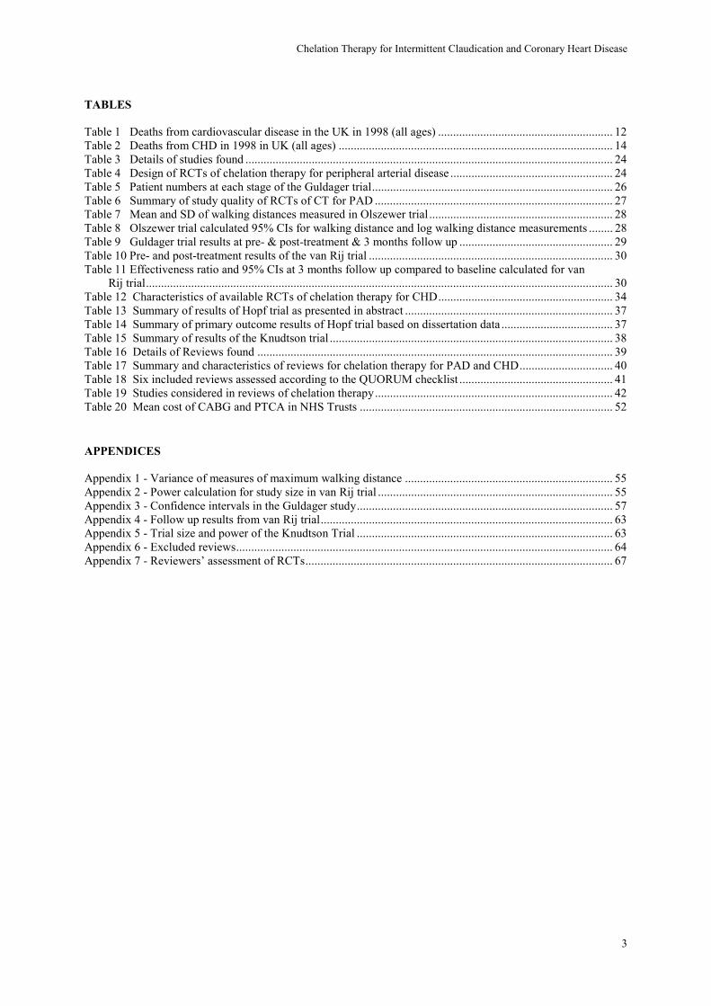
TABLES Table 1 Deaths from cardiovascular disease in the UK in 1998 (all ages) .......................................................... 12 Table 2 Deaths from CHD in 1998 in UK (all ages) ........................................................................................... 14 Table 3 Details of studies found .......................................................................................................................... 24 Table 4 Design of RCTs of chelation therapy for peripheral arterial disease ...................................................... 24 Table 5 Patient numbers at each stage of the Guldager trial................................................................................ 26 Table 6 Summary of study quality of RCTs of CT for PAD ............................................................................... 27 Table 7 Mean and SD of walking distances measured in Olszewer trial ............................................................. 28 Table 8 Olszewer trial calculated 95% CIs for walking distance and log walking distance measurements ........ 28 Table 9 Guldager trial results at pre- & post-treatment & 3 months follow up ................................................... 29 Table 10 Pre- and post-treatment results of the van Rij trial ................................................................................. 30 Table 11 Effectiveness ratio and 95% CIs at 3 months follow up compared to baseline calculated for van
Rij trial........................................................................................................................................................... 30 Table 12 Characteristics of available RCTs of chelation therapy for CHD.......................................................... 34 Table 13 Summary of results of Hopf trial as presented in abstract ..................................................................... 37 Table 14 Summary of primary outcome results of Hopf trial based on dissertation data ..................................... 37 Table 15 Summary of results of the Knudtson trial .............................................................................................. 38 Table 16 Details of Reviews found ...................................................................................................................... 39 Table 17 Summary and characteristics of reviews for chelation therapy for PAD and CHD............................... 40 Table 18 Six included reviews assessed according to the QUORUM checklist ................................................... 41 Table 19 Studies considered in reviews of chelation therapy............................................................................... 42 Table 20 Mean cost of CABG and PTCA in NHS Trusts .................................................................................... 52 APPENDICES Appendix 1 - Variance of measures of maximum walking distance ..................................................................... 55 Appendix 2 - Power calculation for study size in van Rij trial .............................................................................. 55 Appendix 3 - Confidence intervals in the Guldager study..................................................................................... 57 Appendix 4 - Follow up results from van Rij trial................................................................................................. 63 Appendix 5 - Trial size and power of the Knudtson Trial ..................................................................................... 63 Appendix 6 - Excluded reviews............................................................................................................................. 64 Appendix 7 - Reviewers� assessment of RCTs...................................................................................................... 67

Chelation Therapy for Intermittent Claudication and Coronary Heart Disease
4
Abbreviations and Acronyms ABI Ankle/brachial blood pressure index. ACD Absolute claudication distance. ACAM American College for Advancement in Medicine. BMI Body mass index. CABG Coronary artery bypass graft. CHD Coronary heart disease. CLI Critical limb ischaemia. CRD Centre for Research and Dissemination. CT Chelation therapy. CVD Cardiovascular disease. EBCT Electron beam computed tomography. ECG Electrocardiograph. EDTA Ethylenediamine tetraacetic acid. GP General practitioner. IC Intermittent claudication. ICD Intermittent claudication distance. IVUS Intra-vascular ultrasound. MRI Magnetic resonance imaging. MWD Maximum walking distance. NO Nitric oxide. PAD Peripheral arterial disease. PFWD Pain free walking distance. PTCA Percutaneous transluminal coronary angioplasty. QUOROM Quality of reporting of meta-analyses. RCT Randomised controlled trial. SPECT Single photon emission computer assisted tomography. WD Walking distance.

Chelation Therapy for Intermittent Claudication and Coronary Heart Disease
5
1. Summary
• Chelation therapy (CT) is a treatment proposed for coronary heart disease (CHD) and intermittent claudication (IC). A course of treatment consists of a series of intravenous infusions of a solution containing the chelating agent EDTA. Each infusion takes about 3 hours and infusions are repeated about twice a week, typically until 20 or 40 infusions have been administered.
• CHD and IC are manifestations of reduced local blood supply due to narrowing of
arteries caused by atherosclerosis. CHD is responsible for about 25% of all deaths in the UK. CHD causes severe angina, heart attacks and heart failure. About 5.3% of men and 3.2% of women in the UK suffer from angina. People with IC may experience severe leg pain. Their condition is debilitating and associated with increased risk of death and of non-fatal heart attack and stroke. About 5% of people aged 55 to 74 years suffer from IC but prevalence data is limited. In the West Midlands approximately 200,000 people per year might be candidates for treatment with CT.
• This report examines whether CT is more clinically effective and cost effective than
placebo for CHD and IC. • Electronic data-bases, the Internet and current literature were searched to identify
reviews, controlled studies of effectiveness, and economic studies on CT. Predefined eligibility criteria were applied to the recovered literature. Two RCTs for CHD, three RCTs for IC and six reviews of CT were included for analysis. One economic analysis was found but was unobtainable from the British library. Two reviewers independently extracted data.
• For CHD, neither of the two included RCTs showed a statistically significant
difference in the primary outcome measures. For IC, all three RCTs were underpowered and one was very small (n=10). This RCT showed a statistically significant difference in favour of CT for the primary outcome measure whereas the other two showed no significant difference. Small effect sizes in favour of CT were observed in some secondary outcome measures.
• The quality of reviews was very variable and their conclusions in some cases
extremely polarised. Deriving a consensus opinion on effectiveness from the included reviews could not be justified!.
• The cost of CT ranges from £1330 to £4775 per patient depending on the number of
infusions administered. As there was no evidence of clinical effectiveness, a cost utility analysis was not performed.
• Currently there is little objective evidence that CT is effective for CHD or IC.
Conversely there is little evidence that CT does harm. In order to establish the true level of effectiveness of CT, large numbers of patients would need to be enrolled in an RCT. This is very unlikely to be carried out.
! This part of the report (pp 37-50) does not assess the effectiveness of CT but examines the validity of other reviews; readers may wish to omit this section if their primary interest is in the effectiveness of the intervention.

Chelation Therapy for Intermittent Claudication and Coronary Heart Disease
6
2. Introduction Peripheral arterial disease (PAD) and coronary heart disease (CHD) are arterial diseases with high prevalence in the UK. They are associated with considerable mortality and morbidity, high cost to the NHS and the erosion of quality of life for patients. Treatment is based on drug therapies that address symptoms or reduce risk factors for vascular disease and on invasive surgical treatments that aim to replace or improve the function of affected arteries. Surgical procedures are costly, technically demanding, not always successful, and often require repetition. A non-invasive or less invasive therapy that achieved the same outcomes of symptom relief could have wide potential application. There are claims that EDTA-chelation therapy (CT) may represent such a treatment or an alternative to drug therapy. It involves the intravenous infusion over several hours of a solution of ethylenediamine tetraacetic acid (EDTA). A course of treatment consists of 20 or more infusions delivered at a rate of about 2 per week. This report examines whether CT is effective and cost effective for the treatment of CHD and intermittent claudication caused by PAD. 3. Background and underlying health problem 3.1 Natural History 3.1.1 Arterial disease Disease of the arterial wall is the major cause of cardiovascular disease (CVD) which includes diseases of the heart and circulatory system and represents the single most important medical condition in the UK. The artery wall is a complicated structure. Outer layers contain muscle cells and fibres made of collagen. These make the artery elastic so it can resist the pressure of blood and can respond to signals that control diameter and the vessel�s capacity to carry blood. The innermost lining is the endothelium that separates the rest of the arterial tissue from the blood. It is supplied with nerve endings. In response to nerve signals the endothelium releases nitric oxide (NO) that causes muscle cells in the artery wall to relax and the diameter of the artery to enlarge and a greater quantity of blood to pass along. This is one way in which blood supply can be tailored to meet demand, for example during physical exercise. An unhealthy artery with a damaged wall may be less able to respond in an appropriate way. Breach of the endothelium allows blood to contact collagen with the likelihood of blood clot (thrombus) formation. Arteries become diseased by local changes in the thickness or strength of the artery wall. When locally weakened the channel may widen and the artery balloon to form an aneurysm. More commonly the wall thickens locally and the artery channel narrows (stenosis). By far the commonest cause of stenosis is atherosclerosis. Complete blockage (occlusion) is most often caused by atherosclerosis plus a thrombus. Narrowing of the channel reduces the supply of oxygenated blood to downstream tissues (ischaemia). Complete arterial blockage with severe ischaemia results in the death of that tissue (infarction).

Chelation Therapy for Intermittent Claudication and Coronary Heart Disease
7
3.1.2 Atherosclerosis Atherosclerosis causes more deaths in the UK than any other medical condition. It is a common and progressive deterioration of arterial structure and function characterized by fatty deposits, termed atheromas or plaques, which build up in the walls of major arteries. Atheroma progression has been classified into five phases on the basis of morphological characteristics.1 Atheroma starts as a simple fatty streak or series of blobs that barely raise the inner surface of the artery lining. Streaks and blobs consist of living fat-laden cells (foam cells) that accumulate locally just below the endothelium. In westernized populations fatty streaks typically tend to progress insidiously over decades. They gradually form larger plaque structures of more complex composition that represent a serious medical condition. Plaques are characterized by:-
• their accumulation of extra-cellular fatty deposits (predominantly cholesterol); • fat-laden dead and dying foam cells; • the production of collagen fibres; • the multiplication of smooth muscle cells that may partially or wholly cap the plaque
and separate it from the blood flow within the artery; • their accumulation of calcium deposits (calcium hydroxyapatite).
Plaques and their cellular caps are not stable. Post mortem studies on persons dying from causes other than atherosclerosis indicate that plaques frequently fracture resulting in thrombus formation. This may cause occlusion and consequent infarction. Calcium ions play an important role in calcification of plaques and also in thrombogenesis. Without bound calcium several proteins of the clotting cascade fail to function. Calcium ions can be removed from these proteins in freshly taken blood samples by use of strong chelating agents (e.g. citrate or EDTA); this will halt the clotting process and keep the blood liquid. The anticoagulant drug Warfarin reduces the calcium binding capacity of clotting proteins and so reduces the blood�s clotting ability in an individual. Several important risk factors for atherosclerosis have been identified. These include:-
• lack of physical activity (20 minute of vigorous activity on <12 occasions in last 4 weeks).
• obesity (BMI >30). • raised blood pressure (>140/90 mmHg). • smoking. • raised blood cholesterol (> 5.2 mmol/l).
It is probable that only about half of the risk factors for atherosclerosis are known and many have yet to be discovered.2 3.1.3 Peripheral arterial disease and Coronary heart disease The vast majority of peripheral arterial disease (PAD) and coronary heart disease (CHD) is caused by atherosclerosis.
Chelation Therapy for Intermittent Claudication and Coronary Heart Disease
8
PAD affecting the leg involves reduced arterial blood flow to the legs and can be associated with leg pain, compromised walking ability, anxiety and curtailment of normal life activities.3,4 Three categories of atherosclerosis-dependent PAD affecting the leg have been distinguished:- • Asymptomatic PAD. • Intermittent claudication (IC), the most common symptomatic PAD. Patients experience
cramping leg pain induced by walking and relieved by rest. The pain may be in the buttock, thigh, calf or foot or in a combination of these sites. People with IC are only able to walk short distances pain-free.
• Critical limb ischaemia (CLI) is a rare and more severe symptomatic category characterised by rest pain accompanied by ulceration or gangrene. CLI may require limb amputation.
CHD involves reduction or complete obstruction of blood flow through the coronary arteries by narrowing due to atherosclerosis and/or a blood clot (thrombus). CHD causes:-
• angina; [constricting chest pain occurring on exercise (stable angina); or recurring at rest or with increasing frequency and severity on exertion (unstable angina)].
• heart attack (myocardial infarction). • irregular heart beat (arrhythmia). • heart failure.
The impact of angina on a patient�s quality of life can range from very mild pain on exertion to severe disabling pain at rest. On the basis of symptom severity, stable and unstable angina have been classified into 4 and 3 subcategories respectively.5 Untreated CHD is progressive and leads to death from heart attack (acute myocardial infarction) or heart failure. Evidence indicates that when properly managed, progression of CHD can be slowed down and possibly reversed in some people. 3.1.4 Medical examination of arterial tree 3.1.4.1 Physical examination (pulse and blood pressure) The pulse in an artery can be detected by placing a finger on the artery and feeling the throb beneath (palpation). A weak or missing pulse in an artery indicates the possibility of poor blood flow (arterial insufficiency). Pulse strength measured by palpation has been graded on arbitrary scales (e.g. 0 to 4).6 Bilateral examination allows comparisons to be made but all such assessments are subjective, and prone to observer variation.6 During each pulse of blood flow the pressure exerted on the artery wall rises and then falls. The highest point of this pressure build up (systolic pressure) and the lowest (diastolic pressure) can be measured using a sphygmomanometer. Abnormally high values for systolic and diastolic blood pressure are indicative of someone at risk of arterial disease. Most frequently a superficial artery in the arm (brachial) is examined because it is accessible and the pressure there is similar to that in the aorta. Measuring systolic pressure in peripheral arteries (e.g. the leg) can provide information on arterial health in various parts of the arterial tree. Low systolic pressure is indicative of

Chelation Therapy for Intermittent Claudication and Coronary Heart Disease
9
narrowing somewhere above the detection point. Under standard physiological conditions a pressure drop beyond an arterial narrowing is not expected until about 80% of the cross sectional area of the channel has become blocked (occluded). Segmental (upper thigh, above the knee, below the knee, above ankle and toe) blood pressure measurement in the arterial tree of the leg allows detection of narrowing. A ratio of ankle to brachial pressure of ≤ 0.9 is more than 95% -sensitive in detecting angiogram-positive arterial disease in the leg. Lower ankle/brachial blood pressure index (ABI) values are indicative of greater severity of disease. Arterial disease can result in disturbances in the flow of blood near an abnormality; for example the flow may become turbulent rather than smooth. Turbulence can sometimes be detected as a murmur using a stethoscope. 3.1.4.2 Ultrasonic methods Because some arteries of interest are small or deep their blood pressure and pulse may be difficult to investigate by sphygmomanometry or palpation. However, these parameters can be investigated using ultrasonic technology. When used to investigate arteries some of the high frequency ultrasound bursts rebound from moving red blood cells in an artery. By using a Doppler ultrasound instrument the flow of blood in an artery can be detected and its velocity estimated. The values obtained can be compared with �normal� values and to values measured for the corresponding artery on the other side of the body. The Doppler effect is especially useful for a small artery with a non-palpable pulse. Using an inflatable cuff connected to a pressure gauge together with a Doppler instrument to detect the return of systolic flow it is possible to measure systolic pressure in small peripheral arteries and obtain a value for ABI (see above). Alternatively the return of blood flow can be detected via a strain gauge transducer linked to a pulse volume recorder (plethysmograph). In the �reactive hyperaemia test� the systolic pressure in an artery. Then the artery is closed with a pressure cuff at a standard pressure (e.g. 50 mm Hg above systolic) for a standard period of time (e.g. 5 minutes). After this the artery is opened again and bood pressure measured after a standard time has elapsed (e.g. 15 secs) after reopening. If the pressure estimated in this second measure is less than the pre-closure value then a �hyperaemic drop� has been observed. This test provides a measure that depends on the elasticity of the vessel wall. The size of the pressure drop is represented as a percentage fall from pre-test pressure. Details of blood flow and turbulence can be detected using sophisticated ultrasound imaging that also locates and quantifies abnormalities. These �Duplex� instruments may incorporate multiple generator/detector assemblies. They combine Doppler and amplitude information. More recently phase and harmonic details in the echoes have also been incorporated into the analyses. By means of computer technology this information is integrated into real time two-dimensional colour images. These reveal details of the shape and dimensions of the arterial channel (e.g. any narrowing or widening), how the blood flows in the channel (e.g. speed, direction and turbulence characteristics through time), and information about the thickness of the wall. In some investigations the contrast in the image and its definition are enhanced by injecting agents which increase the harmonic content of the echoes. Using duplex ultrasound the severity and location of stenoses in arteries can be estimated. Information helps in monitoring the progress of disease through time, the evaluation of

Chelation Therapy for Intermittent Claudication and Coronary Heart Disease
10
applied therapies and is a guide for future angiography used to pinpoint lesions prior to invasive therapies such as arterial bypass surgical interventions. In the last decade intra-vascular ultrasound techniques (IVUS) have been developed. Instrumentation mounted on the tip of a catheter introduced into the coronary arteries allows sophisticated cross sectional imaging. Information on the lumen, thickness and tissue characteristics of an individual lesion in the artery wall can be obtained. 3.1.4.3 Magnetic Resonance Imaging (MRI) MRI is a non-invasive method of mapping internal structures. It employs radio-frequency radiation and controlled magnetic fields to produce high quality images. The images depend on the spatial distribution of protons in tissue and on parameters relating to their motion.7 Imaging of arteries employs procedures that discriminate between stationary tissue and flowing blood. A bolus of contrast agent is administered prior to imaging. MRI is more sensitive than ultrasound but more time consuming to perform and requires sophisticated apparatus of considerable capital cost. 3.1.4.4 Angiography An even more precise but invasive method for detecting arterial disease involves injecting a chemical that can absorb X-rays (radio-opaque dye). The dye passes along the artery of interest while a beam of X-rays is directed at the site. The resulting X-ray picture (angiogram) illustrates the shape of the blood space within the artery. Abnormal narrowing or widening of the artery channel can be located and quantified and thickening of the artery wall identified and quantified. 3.1.4.5 Computed Tomography Recent advances in computed tomography instrumentation have allowed ultra-fast imaging (e.g. 10 images / sec) that minimises the motion-interference that is a particular problem in investigating coronary arteries that change position with heart beat. In electron beam computed tomography (EBCT) the X-rays are generated from an electron beam impacting a tungsten target. Multiple images can be combined. Fat deposits and calcium deposits in the artery wall contrast strongly with other arterial tissue and it is possible to locate and quantify calcification. The precision and non-invasiveness of these methods recommend them, however EBCT scanners are more expensive than conventional scanners and are available at relatively few sites. 3.1.4.6 Exercise methods Poor oxygen supply compromises physiological function. Muscle activity (i.e. exercise) is associated with greatly increased demand for oxygen and so it is especially susceptible to arterial insufficiency. Muscle subjected to arterial insufficiency is less capable of doing work and muscle pain may be experienced. Isotonic exercise tests have been developed in which an individual performs measurable dynamic exercise on a treadmill or other similar machines. The exercise is designed to stress the muscle system under investigation. The performance of an individual in such tests can

Chelation Therapy for Intermittent Claudication and Coronary Heart Disease
11
help to diagnose the presence and extent of arterial and cardiac disease and its development through time with or without intervention. Exercise testing can incorporate measures of oxygen consumption and/or imaging methods such as thallium single photon emission computer assisted tomography (SPECT). For example stress exercise on a treadmill is performed and at some time, usually near the end of the exercise stress, a radioisotope (e.g.82Rhubidium or 201Thallium) is injected into an artery. Scintillation detection of emitted gamma rays allows passage of blood through the myocardium (or other scanned region) to be imaged and monitored. This allows abnormalities in cardiac function to be detected.8,9 Arterial insufficiency in heart muscle results in altered contraction characteristics. These can be monitored, recorded and measured using ECG (electrocardiogram) apparatus coupled with heart rate and blood pressure measurements during specified exercise tasks. ST segment depression of the ECG is the most common manifestation of exercise-induced myocardial ischaemia. Vigilance is required during testing so as to minimise untoward events and complications secondary to testing. 3.1.5 Relevant outcome measures Objective outcome measures frequently used in trials of therapies for PAD and CHD include the following:- 3.1.5.1 Exercise test outcomes The most commonly employed exercise test involves walking on a treadmill. Usually the treadmill is set at constant speed (typically 1.5 to 2 mph or ~3.6 km/h). In constant-load tests the slope of the treadmill is kept constant during the test (typically 10o or 12o). In graded treadmill testing the slope of the treadmill is gradually increased (typically in increments of 3.5% or 2%) according to a set temporal programme. It has been claimed that constant-load tests are inferior to graded tests in several respects.10 Graded tests have greater dynamic range so that few if any patients need to be excluded from a study; they show little evidence of improved performance with repeated testing and have a satisfactory within subject coefficient of variance. It is contended that graded tests relate closely to patient walking ability in everyday life and therefore small changes in performance in graded tests are likely to be clinically significant. Various end points or measures are employed with treadmill tests. Usually the speed of the treadmill and the time spent walking on it are used to calculate the distance walked. For patients with intermittent claudication two outcome measures are made. These are:
• walking distance to the patient�s first experience of claudication pain [Intermittent Claudication Distance (ICD); or Pain-free Walking Distance (PFWD)].
• walking distance to maximum level of bearable claudication pain [Absolute Claudication Distance (ACD) or Maximum Walking Distance (MWD)].
It is claimed that MWD and PFWD measured using treadmill exercise tests provide an objective assessment of severity of PAD.

Chelation Therapy for Intermittent Claudication and Coronary Heart Disease
12
For patients with angina a typical end point is the exercise time or work output to a detected change in the ECG signal (e.g. ST segment depression). A treadmill or bicycle ergometer (which can measure work and/or power output) is typically employed. The Master�s two step test11-13 uses an apparatus of prescribed dimensions that consists of two ascending steps in tandem with two descending steps. The patient ascends two steps and descends two steps to complete one trip. The patient then turns through 180o and repeats the process to complete a second trip. This activity is repeated until a predetermined number of trips have been completed in the prescribed period of time (usually 3 min). The number of trips to be completed by an individual patient (and therefore the rate of stepping) varies according to age and sex and is determined from a table of standards. The ECG signal is monitored before, during, immediately after, and at 2 and 6 min post-exercise. Used mainly for diagnosis, the test is adaptable as an outcome measure (e.g. Number of trips completed to onset of claudication). 3.1.5.2 Ankle / brachial blood pressure index (ABI) Change in ABI (ankle/brachial blood pressure index) after treatment is a frequently used outcome measure in investigations of therapeutic interventions for PAD. 3.1.5.3 Angiographic measures A more rarely employed outcome measure is change in angiograms obtained using X-ray contrast media. These are best coupled with systems for objective scoring of the artery lumen to determine degree of stenosis of identified vessels. The low throughput of samples coupled with the invasiveness of this methodology mean it has been used on a limited scale in trials. 3.2 Prevalence 3.2.1 Cardiovascular disease 3.2.1.1 Mortality Diseases of the heart and vascular system (cardiovascular disease) are the main cause of death in the UK accounting for over a quarter of a million deaths in 1998 (Table 1) (Table 1.2 in Coronary Heart disease Statistics14).
Table 1 Deaths from cardiovascular disease in the UK in 1998 (all ages) CAUSE OF DEATH MEN WOMEN TOTAL
ALL CAUSES 298,767 327,384 626,151 CARDIOVASCULAR DISEASE 122,218 (40.9%) 134,492 (41.1%) 256,710 (40.0%) Although the cardiovascular disease death rate has been falling since the 1970s it is still one of the main causes of premature death (death before age of 75) accounting for 38% of premature deaths for men and 30% for women. CHD is the major contributor to cardiovascular disease mortality.

Chelation Therapy for Intermittent Claudication and Coronary Heart Disease
13
3.2.1.2 Morbidity According to the Health Survey for England 1998 15 27.9% of men and 27.8% of women self-reported cardiovascular disease conditions and 10% of adults reported long-standing cardiovascular disease. 3.2.2 PAD affecting the leg 3.2.2.1 Mortality Peripheral arterial disease patients occasionally die as direct result gangrene in their leg, but this is an extremely rare event. People with critical limb ischaemia have widespread atherosclerosis and poor prognosis with 20% dead within 1 year of presentation and only 53% alive with both legs.16,17 People with IC likewise have systemic atherosclerosis and have a high risk of mortality; between 30% and 50% of those referred to hospital being dead within 5 years (a rate ~3 times that of people without IC).18,19 3.2.2.2 Morbidity About 30% of the UK population over 55 years old have PAD affecting the leg, but most of this is asymptomatic.6 Population surveys indicate that intermittent claudication (IC) is rare in people under 55 years old (less than 1%) but then increases with age. Overall prevalence for men and women between 55 and 74 years old is approximately 5%.6,20,21 For pre-menopausal women prevalence is about half that of men of similar age.22 After the menopause this sex difference disappears. Limited information suggests that the annual incidence in the UK is about 1.8% for 55 to 74 year olds. In the general population ~40% of people with IC have angina (2 to 4 times the rate in people without IC). Of people with IC referred to hospital 90% were found to have coronary heart disease and they were at about two fold greater risk of non-fatal myocardial infarction or stroke than people without IC.18,23 Regional variations in prevalence of IC in the UK are uncertain, but as with coronary heart disease there is likely to be a North South divide and a progressively increasing prevalence with lower social class.17 Critical limb ischaemia (CLI) is rare; an incidence in the range 500-1000 per million people (0.05 to 0.1%) per year has been calculated.24 3.2.3 Coronary heart disease 3.2.3.1 Mortality CHD accounts for about 25% of all deaths in the UK. The mortality rate increases rapidly with age and at any age is greater for men than for women. The rate is higher in men in manual occupations compared with those in non-manual and is higher than average for ethnic South Asians living in the UK.

Chelation Therapy for Intermittent Claudication and Coronary Heart Disease
14
Table 2 Deaths from CHD in 1998 in UK (all ages) NUMBER OF DEATHS FROM CHD
MEN WOMEN TOTAL 74,542 62,611 137,153
Mortality from CHD peaked in the 1970s and has steadily declined by about a third since then. This decline has been greater in the 16-64 age group than 65 to 74 age group.14 3.2.3.2 Morbidity Information regarding the prevalence of coronary heart disease in UK is fragmentary. Existing monitoring systems have focussed on acute episodes and death rather than on disease burden. The Health survey for England 98 reported on prevalence estimated in two ways:- a) on the basis of doctor-confirmed diagnosis; b) on the basis of self reported symptoms obtained via questionnaire. The two methods yielded different results. Doctor-confirmed angina had an estimated prevalence of 5.3% in men and 3.2% in women. Prevalence increased up to 75 years of age in both sexes and at all ages was higher in men than women. The current prevalence (i.e. that reported in the last 12 months) was estimated as 3.2% and 2.5% for men and women respectively. 3.2.4 West Midlands burden of angina and PAD affecting the leg Using the estimated prevalence of doctor-confirmed angina in England (above) and the projected estimate of the West Midlands population for 2001, we estimate there are approximately 140,340 men and 86,240 women (total 226,580) with angina in the West Midlands region. Using data from the same source we estimate that about 152,100 (67%) of these people would suffer angina in a 12-month period and would consult their doctor. This corresponds reasonably with the 167,000 coronary heart disease consultations for the region in 1996 reported in �Key Health Statistics from General Practice�. Based on this estimate about 150,000 people per year in the region might be suitable for chelation therapy for treatment of angina. In the year 1999-2000 there were 28,400 admissions to West Midlands regional hospitals for which the primary or secondary diagnosis was PAD caused by atherosclerosis. If we assume that in the majority of these patients the disease affected the leg(s) then they account for about half of the 52,000 patients with intermittent claudication calculated from estimated prevalence (above). Many of these patients might be suitable for chelation therapy. Adding angina and IC patients together a total approaching 200,000 possible candidates for chelation therapy per year is obtained. Lack of information prevents an estimate of how many of these possible candidates might seek or request chelation therapy. In the UK chelation therapy is administered almost exclusively outside of the NHS. No private clinics in the region offer this service. The nearest clinics are in Atherton (Lancashire) and London. The only clinic in the West Midlands region (Haseley Clinic in Warwick) closed recently and does not appear to have reopened.

Chelation Therapy for Intermittent Claudication and Coronary Heart Disease
15
3.3 Current service provision 3.3.1 PAD affecting the leg The aims of primary care are to provide diagnosis, to control risk factors, to alleviate symptoms and to make appropriate referral. Treatment modes to modify risk factors of cardiovascular disease in PAD patients include:- • Life style changes emphasising cessation of smoking and uptake of exercise. • Antiplatelet therapy; drug therapies for lowering blood lipid levels, for control of diabetes,
and for the control of hypertension. Modes of treatment aimed at relieving symptoms of IC include:- • exercise programmes. • Vasodilator and anticoagulant drug therapies, drugs that alter the flow properties of blood
by modifying deformability of red blood cell membranes, other drugs with various proposed mechanisms of action.
Systematic reviews of the efficacy of the various available interventions for relief of IC indicate weak evidence and/or marginal effectiveness.15,25-28 Secondary care procedures for people with intermittent claudication and people with critical limb ischaemia include:- • Transluminal operations performed mainly on femoral and iliac arteries. • Iliac and femoral bypass operations. • Limb amputation. In England approximately 22,000 femoral artery and 4,000 iliac artery transluminal operations are performed annually.29 Corresponding numbers for bypass operations are 5,500 (femoral) and 1000 (iliac).29 Provision of this secondary care in the UK is diverse with the majority of the vascular surgery performed by general surgery units in district general hospitals rather than in tertiary centres.17 3.3.2 Angina For most patients treatment remains with the GP. A small proportion are referred to secondary and tertiary centres. Treatment is aimed at relief of symptoms, at halting or slowing the progression of disease, and at reduction in risk factors. Treatment for angina includes:- • Advice on life style:- cessation of smoking; adoption of exercise; avoidance of obesity. • Administration of drugs:- anticoagulants and antiplatelet agents to reduce thromboses;
agents to lower blood lipid levels; vasodilators; agents to lower blood pressure. • Surgery:- coronary artery bypass graft (CABG); percutaneous transluminal coronary
angioplasty (PTCA) with or without stents. • Rehabilitation programmes for survivors of myocardial infarction. • Alternative therapies:- including over-the-counter remedies such as garlic powder tablets,
garlic oil capsules, fish oil capsules. Prescription records show that during the �90s there was a steady rise in the proportion of patients given lipid lowering statins and aspirin. A similar increase in surgical interventions
Chelation Therapy for Intermittent Claudication and Coronary Heart Disease
16
occurred. The number of CABGs performed in the UK doubled over the 10 years 1987 to 1997 reaching 28,000 per year. The number of PTCAs increased at an even faster rate (about 7 fold) reaching 25,000 per year 1997-98. 3.3.3 Implications of service provision In relation to disease burden consultation rates for peripheral vascular disease in the UK are extremely low (estimated at 82 per 10,000 people per year) and the disease probably under diagnosed. It is possible therefore that current service provision of surgery for IC in the UK is inadequate. Under-provision of surgery for CHD in the UK is implicit in the National Service Framework�s goal of doubling CABGs and PTCAs over the 5 years 1998 to 2003. Chelation therapy has been particularly canvassed as an alternative to surgical intervention for CHD and IC patients. If shown to be effective it might help address the shortfalls described above. 3.4 Description of intervention:- Chelation therapy and its uses 3.4.1 Introduction EDTA chelation therapy is one approach suggested for the treatment of atherosclerosis. It involves the intra-venous infusion of a solution of EDTA. It was introduced in the 1950s in the USA and was advocated for PAD, CHD and stroke patients. Early use of the therapy was haphazard with regard to dosage of EDTA, rates of infusion, and the frequency of repeat infusions. After an early flush of enthusiasm following its inception the value of chelation therapy for atherosclerosis became a topic of dispute. In many countries chelation therapy is now generally administered outside of mainstream state-supported or insurance scheme-supported medicine and has become classified under the umbrella terms of �complementary�, �holistic� or �alternative medicine�. 3.4.2 Chelating agents Chelating agents react with metal ions to form a particular class of metal complexes called metal chelates. In this reaction at least two reactive groups (�chela� or �claws�) of the chelating agent become fastened to the metal ion so as to form a heterocyclic ring (defined as a cyclic structure in which the participating atoms represent at least two different elements). Many chelating agents have been developed and some have found uses in medicine. 3.4.3 Concept of Chelation Therapy Chelation therapy is the administration of a chelating agent as a form of treatment. In theory the chelating agent extracts unwanted metal ions from various cellular or extra-cellular sites, circulates in the blood stream with its bound metal ion, and reaches the kidney where it is voided in the urine. In this way unwanted metal may be eliminated from the body. Except for reaction with a stronger chelating agent, or replacement by a preferred metal, a metal ion fully bound to a chelating agent is unavailable for other chemical reaction; it effectively disappears from solution.

Chelation Therapy for Intermittent Claudication and Coronary Heart Disease
17
The most obvious circumstances in which chelation therapy might be useful are:
• when a toxic metal has inadvertently accumulated in the body (e.g. lead poisoning, accident with radioactive isotope, arsenical chemical warfare agent).
• when a pathological condition has resulted in an abnormal build up of a metal (e.g. copper build up in Wilson�s disease, or iron accumulation after repeated blood transfusion for thalassemia).
In the 1950s it was widely recognised that calcium deposits accumulate in atherosclerotic arteries and attempts to remove these deposits by chelation therapy were started. The rationale for the therapy was that arteries would become less blocked by removal of accumulated calcium. After the 50s dominant theories minimised the importance of calcium in atherogenesis and viewed its accumulation as a late secondary event. Modern imaging techniques (e.g. Electron beam computed tomography; EBCT) and new methods of investigation have recently refocused attention on calcium. It is now realised that the extent of calcification in the coronary arteries is a good prognostic indicator for acute adverse events.30 Calcification of atheromas is no longer viewed as a passive precipitation. Instead the same proteins and regulatory mechanisms that control bone deposition and resorption are thought to be involved. The relationship between plaque calcification and plaque stability and potential thrombosis is unknown. Whether decalcification of a plaque would stabilise or labilise its structure is uncertain. Other mechanisms of action for chelation therapy have been suggested. These include the proposed lowering of blood cholesterol levels, and the removal of transition metal ions (copper and iron) thereby minimising the oxidative changes to blood lipoprotein particles that are currently thought to play a pivotal role in the development of atheromas. 3.4.4 Ethylene diamine tetra-acetic acid (EDTA) EDTA is a chelating agent that is able to bind most metals. It has various applications in biology and medicine, the most familiar as an anti-coagulant for blood collection. The binding affinity of EDTA for different metals has been estimated. Equilibrium constants for EDTA-metal chelates vary from lower values (lower affinity) of 107 to 1011 (for group IIA metals e.g. Calcium, Magnesium, Barium) to higher values (1019 to 1025) for other biologically significant metals such as Iron and Copper. The most important factor determining the affinities of different metals is the pH of the medium. In biological materials most metals (other than Na and K) are bound to physiological chelating agents (especially proteins); EDTA will only remove these if it is a stronger chelator of the particular metal. Which metals form chelates with Na2-EDTA in a biological environment depends on their relative concentrations, their chemical state (freely ionised, inorganic precipitate, or bound to biological chelating agents), and their affinity for Na2-EDTA in the prevailing conditions (e.g. of pH). The most abundant chelate formed on introduction of 3 g of Na2-EDTA into the circulation is calcium chelate.31 Na2-EDTA chelates calcium to form five pentacyclic heterocycle rings that completely enclose the metal and create a complex with no net charge. This structure is represented in the diagram below (Fig. 1).

Chelation Therapy for Intermittent Claudication and Coronary Heart Disease
18
. Fig. 1 Administration The most common route of administration of EDTA is by intravenous infusion. This mode is an approved treatment for lead poisoning. Recently EDTA tablets have been marketed, but significant intestinal absorption is unlikely. Free acid EDTA is poorly soluble and exerts an acid pH. The disodium salt of EDTA is more soluble and can be administered near to physiological pH. The American College for Advancement in Medicine32 has recommended a protocol for EDTA therapy (Rozema 1997 33: �The protocol for the safe and effective administration of EDTA and other chelating agents for vascular disease and metal toxicity therapy�). A description of the protocol is available on the internet.34 It involves the intra-venous infusion of 500 ml of solution containing 3 g disodium EDTA together with various additional substances including vitamins, magnesium chloride and sodium bicarbonate. The protocol does not specify the pH or osmolarity of the infusion mixture. Infusion lasts for 1.5 to 3 hours and typically 20 to 40 infusions are administered at a rate of about 2 per week. Minor variations on this protocol have been commonly used but have mainly concerned a larger volume of infusate (e.g. 1 litre for 500 ml) that quantitatively delivers the standard amount of chelator (about 3g EDTA). When magnesium chloride is included in the infusion virtually all the Na2-EDTA will be administered as its magnesium chelate. Depleted blood calcium levels are rapidly replenished from soft tissue stores, however very rapid infusions of Na2-EDTA can precipitously lower serum calcium levels with a risk of tetany and death. Slow infusions have marginal effects while magnesium Na2-EDTA infusions only mildly decrease serum calcium with rapid return to normal. Loss of ionised blood calcium stimulates release of parathyroid hormone which increases calcium reabsorption in the kidney, intestinal calcium uptake, and release of calcium from bone stores.
O
Ca
CH2
OC
NCH2
NCH2
OC
NaO
NaO
OC
C
CH2
CH2
O
O
CH2
The structure of the metal chelate formed between disodium EDTA and calcium. The calcium ion is held in five pentacyclic rings and it�s positive charge is neutralised. Na O
Na O
Chelation Therapy for Intermittent Claudication and Coronary Heart Disease
19
Theoretically 1g Na2-EDTA can chelate a maximum of about 120 mg calcium. The quantity of ionised blood calcium removable as a result of the standard infusion of 3 g of EDTA is very small (maximum about 360 mg) relative to bone stores (typically about 1 Kg). Changes in parathyroid hormone induced by repeated infusions of EDTA could conceivably be relevant for persons at risk of osteoporosis (e.g. post-menopause women). EDTA and EDTA-metal chelates are not metabolised. After venous infusion of Na2-EDTA, calcium-EDTA metal chelate is lost to the urine. Other metal chelates (e.g. zinc, copper, iron, cadmium, manganese, vanadium, and lead) are also voided in the urine as is unaltered Na2-EDTA. All EDTA is voided in one form or another within ~24 hrs of administration.35 3.4.5 EDTA chelation therapy for atherosclerosis EDTA chelation therapy has been claimed an effective therapy for many conditions. In recent reviews Olmstead36 lists 39 separate conditions and Meyer37 lists 28. In the UK its use for atherosclerosis started in 1985. Since then approximately 10,000 individuals have been treated. According to the Arterial Health Foundation this service is currently provided by eight doctors administering at 5 clinics (federated as �The Arterial Disease Clinic�). According to the Arterial Disease Clinic�s web site38 and promotional material the therapy may be suitable for patients with angina, claudication, memory loss due to PAD, and for stroke patients. It is not an approved therapy for these conditions and has been administered outside of the NHS with costs met by patients rather than health insurance schemes. The Arterial Disease Clinic lists three categories of patient that may be suitable for chelation therapy as follows:- • Preventative cases; symptom-free individuals with a family history of cardiovascular
disease and with risk factors (e.g. raised blood cholesterol). • Moderate cases; people with clinical conditions that may lead to eventual surgery. • Severe cases; usually people who have had surgery that has failed them or persons
wishing to avoid surgery such as amputation. According to Arterial Disease Clinic information a course of chelation therapy encompasses the following elements:- • An initial examination that includes:- Doppler ultrasound examination of 24 arterial sites
supplying the brain and legs; blood tests for kidney and liver function; urine analysis with creatinine clearance measurement to establish kidney function; physical examination; red cell magnesium; resting ECG test; atherosclerosis risk factor analysis (cholesterol, ferritin, fibrinogen, lipoprotein a, apo-lipoprotein E, and homocysteine).
• Venous infusion of EDTA solution according to the protocol of The American College for Advancement in Medicine (see above). An infusion lasts about 3.5 hours during which time the patient reclines in a chair and is free to eat, drink and chat. The procedure is closely monitored for cardiac function and blood pressure (avoidance of hypotensive episodes). Infusions are repeated at a rate of one a week to a total that depends on clinical judgement (typically 20 to 40 infusions).
• Oral vitamin supplements that form an integral part of the therapy. • Interim and end of treatment tests including Doppler examination, and urine creatinine
measurement. Potential advantages of chelation therapy include:- • Its low degree of invasiveness compared with CABG or PTCA. • Its out-patient mode of administration contrasting with CABG.

Chelation Therapy for Intermittent Claudication and Coronary Heart Disease
20
• Its possible avoidance of surgery. • Its lesser requirement for operator training. • Its lack of requirement for cardiac surgery facilities in the vicinity of the clinic. • Its relatively low requirement for high-grade technology and staff back-up. Compared with other conservative management regimes such as oral drug therapy it suffers the disadvantage of being time consuming and involving travel by the patient. Almost from its first use for cardiovascular diseases, considerable controversy has surrounded the question of whether EDTA chelation therapy is effective. Strong opinions have been expressed both for and against its use with the result that it has become a highly contentious topic. Both RCTs and reviews have been published about chelation therapy. There has been debate on whether systematic reviews or randomised controlled trials represent the best evidence that can be used when considering efficacy of an intervention39-42. Reviews, especially systematic reviews, have considerable opinion-forming influence. A review of reviews on chelation therapy has been included in the present report so as to summarise the direction of their conclusions and to assess their quality. 4. Questions addressed by this review The question addressed in this review is what is the effectiveness and cost of EDTA-chelation therapy for the treatment of patients with intermittent claudication or coronary heart disease. 5. Methods 5.1 Clinical effectiveness 5.1.1 Search strategy A scoping search was done in March 2001. For clinical effectiveness the detailed search strategy involved looking for randomised control trials (RCTs), case control studies, systematic and other reviews. Both index terms and text words were used. Embase (1980 to April 2001) and Medline (1966 to April 2001) were searched on Ovid. No language restrictions were applied. Further searches of Medline and Embase were done in July 2001 to check for any recent papers. Searches were: - 1. for RCTs using the NHS CRD43 search strategy for RCTs and the search terms exp.
chelation therapy, EDTA, ethylenediamine tetraacetic acid, exp. Chelating agents, exp. Edetic acid.
2. for case control studies, using the search terms exp. Cohort studies, exp. Case control studies, exp. chelation therapy, exp. Chelating agents, exp. EDTA, exp. Edetic acid.
The following additional sources were searched during April 2001; • Cinahl. • Grateful Med. • Cochrane Library. • CHID online (The Combined Health Information Data Base). • NCCAM website (National center for complementary and alternative medicine).

Chelation Therapy for Intermittent Claudication and Coronary Heart Disease
21
• Internet Search Engine (Google, Dogpile). • Web of Science (MIMAS). • Biomednet. • Reference lists in review articles, meta-analyses and RCTs. • Hand search of current literature available online at Birmingham University and held in
the University library; this included specialised and general journals publishing papers on atherosclerosis:- JAMA, NEJM, BMJ, Lancet, Circulation, Atherosclerosis, Annals of Internal Medicine. The issues searched covered from Sept 2000 up to July 2001; earlier publications were assumed to have been entered onto Medline and Embase data bases.
• Practitioners of chelation therapy and colleagues were consulted for references and web sites.
5.1.2 Inclusion criteria 5.1.2.1 Primary studies of clinical effectiveness • Study design:- RCTs. Other controlled studies were accepted if >100 people were
included. • Population:- Atherosclerosis causing PAD with IC, or CHD. • Intervention:- Chelation therapy involving repeated infusion of EDTA solutions
containing at least 1 g EDTA per infusion with a total of at least 10 infusions. • Comparator:- RCTs:- Placebo or other interventions that were not chelation therapy. Case
control studies:- matched untreated controls. • Outcomes:- A measure of effectiveness determined using an exercise test. 5.1.2.2 Reviews of clinical effectiveness + meta-analyses Systematic reviews, critical reviews and other reviews were included if they attempted a critical analysis of quantitative primary data on efficacy of EDTA chelation therapy for atherosclerosis causing peripheral arterial disease with intermittent claudication, or coronary heart disease. Meta-analyses were included if they reported a summary estimate of effectiveness of chelation therapy for IC or angina. 5.1.3 Exclusion criteria All studies: Atherosclerotic cerebral disease. Studies involving chelation therapy with chelating agents other than EDTA. Reviews: uncritical reiteration of conclusions from papers reporting primary data. 5.1.4 Quality assessment strategy The following factors were considered when evaluating RCTs:- • The method of randomisation used, concealment of allocation and how this might effect
outcomes. • Whether baseline characteristics were similar between groups. • Whether groups were treated similarly except for the randomised intervention. • The extent of treatment crossover. • The nature and extent of loss to follow up. • The extent of blinding of assessment.

Chelation Therapy for Intermittent Claudication and Coronary Heart Disease
22
• Whether the analysis was carried out on intention to treat basis. • Whether the conclusions match the results. In addition RCTs were scored on a scale based on that proposed by Jadad.44 The following factors were considered in evaluating reviews:- • The degree to which QUOROM guidelines45 were fulfilled. • Whether statements were matched by the references given in their support. • Whether statements were errors, misrepresentations or unsubstantiated by evidence. Meta-analyses were evaluated in terms of the following factors46:- • Study protocol in advance. • Complete literature search. • Selection of studies objective and reproducible. • Analysis of individual patient data when results found in different settings are combined. • The need for future studies defined. 5.1.5 Data extraction strategy Two reviewers independently extracted the data from all the included studies into predefined tables. One discrepancy was resolved by discussion. 5.2 Economic analysis 5.2.1 Search strategy For economic evaluation the NHS CRD (Centre for Reviews and Dissemination) search strategy [�All Databases� (DARE, NHS Economic Evaluation Database, HTA)] was used.47 Medline and EMBASE were also searched on Ovid in July 2001. Search terms included: exp economics; exp economics, hospital; medical/or exp economics; nursing/ or economics; pharmaceutical; exp costs and cost analysis; exp cost of illness; exp economic value of life; exp health care costs; exp economics, medical/; exp �fees and charges�/; (costs or costs or costed or costing).mp.; No language restrictions were applied. 5.2.2 Inclusion and exclusion criteria The inclusion and exclusion criteria were applied as for the clinical effectiveness section (5.1.2.1 and 5.1.3). In addition, included studies must include either assessment of resource implications and or costs. There were no language restrictions. Inclusion and exclusion criteria were applied by two reviewers. 5.2.3 Economic evaluation The economic evaluation was a cost study of chelation therapy. The analysis was done from the perspective of the NHS. This was so as to gauge the cost impact of the therapy should it be adopted within the NHS.

Chelation Therapy for Intermittent Claudication and Coronary Heart Disease
23
6. Quality, direction and strength of the evidence (clinical effectiveness + reviews) 6.1 Number and types of studies The outcome log of the studies identified from literature searches is shown in the diagram below: 6.2 Randomised controlled trials 6.2.1 Studies found Six RCTs were found.48-53 No other controlled trials or case control studies were found. One RCT (Kitchell et al 196348) was excluded because no objective measure of outcome was reported. In this RCT four intervention and five placebo patients received 12 weeks of initial treatment in a cross-over design study. At 6 and 12 weeks post initial treatment they were evaluated in terms of �benefit� or �no benefit�. The patients then returned to a further 12 weeks of treatment in cross over mode. At least three of the nine patients failed to complete this second phase of treatment. The authors remarked that no valid conclusions could be drawn from their study.
880 citations identified from
searches
208 potentiallyrelevant citations
672 irrelevant citations
182 abstracts of possible relevance
26 abstracts irrelevant
10 potentially relevant articles
30 Reviews of interest
123 articles of interest
19 articles of no relevance
5 RCTs included in
effectiveness
4 duplicates of RCTs
1 RCT excluded from
effectiveness
24 reviews excluded
6 reviews included

Chelation Therapy for Intermittent Claudication and Coronary Heart Disease
24
Table 3 Details of studies found Type of study Number
of studies Source
R C T 3 Embase,Medline or Biomednet R C T 1 Internet [Alberta Heritage Foundation] R C T 2 Referenced in Journal
6.2.2 Randomised controlled trials of CT for PAD patients with IC 6.2.2.1 Studies found and trial design Three randomised controlled trials were found.51-53 One of these trials was reported several times.53-57 All three trials compared EDTA chelation therapy with placebo. They employed an exercise test (walking distance) as the primary outcome. Details of trial designs are shown in the Table 4.
Table 4. Design of RCTs of chelation therapy for peripheral arterial disease TRIAL
Olszewer et al.,1990 Guldager et al.,1992† van Rij et al.,1994 Patient Group
Men with PAD 41-53 years old (mean 47). Stable intermittent claudication. Pain-free walking distance 100 to 300 metres. Ankle/brachial BPI 0.75 - 0.4.
>40 years old (mean 65), 65% men. Stable intermittent claudication for ≥12 months. Pain-free walking * 50-200m. Ankle/brachial BPI <0.8
>45 years old (mean 67), 82% men. Intermittant claudication. Arteriographically-confirmed PAD. ≤20% variation in pain-free walking distance.
Interv-ention
Infusion 10 ml solution containing 1.5 g Na2 EDTA + 1 g MgSO4 + vitamins & heparin in Ringer�s lactate [Ringer�s lactate not defined]. 10 infusions. Time unspecified.
3-4 h infusion of 1000 ml isotonic solution containing 3g Na2 EDTA + 8.4 g NaCl. 20 infusions over period of 46 days (31-69 days).
3-4 h infusion of 500 ml isotonic solution containing 3g Na2 EDTA + 0.76 g MgCl2 + 0.84g NaHCO3, in �normal� saline + vitamins. 20 infusions at 2 / week for 10 weeks
Comp-arator
As intervention but minus Na2 EDTA. 10 infusions
Isotonic NaCl (minus Na2 EDTA). 20 infusions over period of 46 days (range 27-63 days).
500 ml �normal� saline + vitamins. 20 infusions at 2 / week over period of 10 weeks.
Primary outcome
�Walking distance� test [+ Master�s two step test��, + bicycle exercise test]. Comparison between baseline and after 10 infusions.
Pain free and maximal walking distances on a treadmill. Differences between baseline and various time points calculated. Comparison in differences CT v. placebo.
Pain free and maximal walking distances on a treadmill. Differences between baseline and various time points calculated. Comparison in differences CT v. placebo.
Second-ary outcomes
Blood pressure measurements.
Subjective evaluation by patients. Change in ankle/brachial BPI before v after treatment. Side effects during treatment period.
Many measures (n≥14) covering arterial & cardiac function, quality of life & mood state, & patient assessment of treatment.
No. at start 10; CT= 5 & PL=5 159; CT=80 & PL=79 32; CT=15 & PL=17 No. at end 10 ; CT= 5 & PL=5 153; CT=75 & PL=78 32; CT=15 & PL=17
Follow up time Not reported
3 mo. n=149; CT=66 & PL=67 6 mo. n=123; CT=51 & PL=56 3 mo n=32; CT=15 & PL=17
CT = chelation therapy. PL = placebo. PAD = peripheral arterial disease. BPI = blood pressure index. � this was a multicentre trial. �� Master�s two step exercise test described by Master and Oppenheimer 1929.11 The Olszewer trial did not describe the method of measurement of walking distance and it is assumed that maximum walking distance only was estimated. In Guldager and van Rij trials a fixed incline-constant speed treadmill was employed to measure walking distance. Two

Chelation Therapy for Intermittent Claudication and Coronary Heart Disease
25
measures were made:- 1] the pain-free walking distance (PFWD), which is the distance walked up to the first onset of claudication; 2] the maximum walking distance (MWD), which is the distance walked up to the point when the patient cannot continue despite a severe whipping, various inducements and other encouragement. 6.2.2.2 Trial quality assessment The quality of trials was assessed according to criteria described in Methods and set out by Jüni et al (2001).58 i] The Olszewer trial52 The method of randomisation was not described. Patients and investigators were effectively blinded to treatment allocation. Treatment crossover was planned after the tenth infusion but because of dramatic improvements in all intervention but no placebo patients cross over and blinding were abandoned and the study continued as an open label trial of the intervention for all patients. Baseline characteristics were similar between groups but demographic details were meagre. Patients were treated similarly except for the randomised intervention. There were no losses to follow up. ii] The Guldager trial53 Block randomisation was used but no further details of the randomisation procedure were supplied. Baseline characteristics were similar between groups except that the placebo group maximum walking distance standard deviation was very large (169% of the mean) compared with that for the intervention group (32% of the mean). Such large variance precludes a significant difference between means but might indicate some difference between groups. The placebo group standard deviation value is large compared with values reported for this outcome measure in a representative selection of other trials (see Appendix 1). Groups were treated similarly except for the randomly allocated intervention. Patient numbers completing to each stage of the study were stated but their distribution between groups is not clear and not all withdrawals are accounted for. Numbers of patients stated to have completed to each stage do not correspond with the numbers contributing outcome data. Observations on some patients were said to be �censored� and their data excluded from analysis. Clear reasons for censoring were not provided. Numbers of censored data were stated to be small but in fact exceed 10% as shown in Table 5.

Chelation Therapy for Intermittent Claudication and Coronary Heart Disease
26
Table 5 Patient numbers at each stage of the Guldager trial
STAGE
Stated no. of
patients
Losses from previous
stage�
Losses fully accounted for
Number that contributed
data�
% of data
�censored� � Baseline 159 0 153 4% 10�th infusion ? ? 0 143 ?
Post treatment 153 6 6 135 12%
3 month follow up 149 10 0 133 11%
6 month follow up 123 26 8 101 18% � Calculated from data tables of Guldager trial. Blinding was broken at the end of treatment so that investigators were aware of patient allocation during outcome measurements at 3 month and 6 month follow up. Of 123 patients that completed the study only 36% were still blinded. How and when blinding was broken for the other 64% was not stated. No reason was provided for why blinding was abandoned. iii] Van Rij trial51 Block randomisation was used but no further details of the randomisation procedure were supplied. Baseline characteristics were similar for both groups. Blinding procedure was described and both investigators and patients were blinded to treatment allocation throughout the study. There were no losses to follow up and all patient data was used for analysis. 6.2.2.3 Trial size and study power Walking distance of claudicants is variable. With repetitive testing it tends to increase.10 Better designed trials carry out pre-trial tests to recruit patients with reproducible walking of known variance. A calculation should then be done to estimate suitable trial size. Only van Rij described pre-trial tests and performed a calculation for trial size. However the calculation was flawed and the trial underpowered by a factor of ~10 (see Appendix 2). The Vascular Clinical Trialists10 have recommended trial sizes for trials of pharmacological agents for intermittent claudication. They estimate that, using a walking performance end point, for a 25% difference between control and intervention to be detectable at P < 0.05 the trial would require ~200 patients. According to this all three of the trials under consideration here were underpowered except for detection of >25% differences between treatments. 6.2.2.4 Summary of trial quality A summary of quality aspects58,59 of the three RCTs is shown in Table 6.

Chelation Therapy for Intermittent Claudication and Coronary Heart Disease
27
Table 6. Summary of study quality of RCTs of CT for PAD
TRIAL
QUALITY ASPECT Olszewer et al., 1990
Guldager et al.,1992
van Rij et al., 1994
Qualitative criticisms
Duration time of treatment not
specified. Timing of outcome
measure unclear
Placebo and intervention
groups may have differed at start of
the trial§
Power calculation for trial size either
flawed or ill-described.
Trial size Very small (10) Moderate (159) Small (32) Pre-trial estimate of likely outcome variance and calculation of trial size required
No No Yes
Population inclusion & exclusion criteria adequately described Yes Yes Yes Demographic details given Inadequate Yes Yes Groups similar at start Probably Probably Yes Blinding method described No No No Blinding maintained Yes, up to the
10�th infusion� Not during follow
up� � Yes Full description of Treatment No Yes Yes Description of outcome methods Incomplete Complete Complete Full account of losses to follow up Yes No Yes
Non-complier results carried forward NA No NA
Data presented allow calculation of authors� end point conclusion
Uncertain
No
No
Jadad-based summary score. [0 (worse) -5(best)]
3 2 4
NA: Not Applicable (no withdrawals occurred in these trials). � Original trial planned cross over after 10 infusions but this design abandoned then together with blinding. �� Blinding of investigators abandoned at end of treatment, that of patients (74%) at indeterminate times after end of treatment. § Very large SD for maximum walking distance at baseline for placebo group. 6.2.2.5 Results of randomised control trials of CT for patients with IC In the three RCTs of EDTA-chelation therapy for patients with intermittent claudication the primary outcome was based on walking distance at one or more time points during or after treatment and on comparison of these distances with those observed at baseline. i] The Olszewer trial Olszewer measured WD at baseline and after 10 infusions (end of treatment) for a total of 10 patients randomised to CT or placebo. Olszewer rounded walking distances to the nearest 10 metres (all distances quoted are a multiple of 10). Mean and SD of measures were not reported. We have calculated these and they are shown in Table 7.

Chelation Therapy for Intermittent Claudication and Coronary Heart Disease
28
Table 7 Mean and SD‡ of walking distances measured in Olszewer trial Intervention Placebo
OUTCOME
follow up no.
Distance (m) at baseline mean (SD)
Distance (m) at post-treatment mean (SD)
Change
Mean (SD)
follow up no.
Distance (m) at baseline
mean (SD)
Distance (m) at post-treatment mean (SD)
Change
Mean (SD)
Walking distance
5 160 (59) 424 (110) � 264 (51.8) 5 192 (29)� 212 (49) � 20 (20.5)
� All means and SD values were calculated from published data of Olszewer. Olszewer reported in the text a 2.2 and 1.04 fold increase in walking distance for the intervention and placebo groups respectively. The correct value (shown in table 2 of Olszewer) should be 2.65 (i.e. 424/160). This increase is substantial. Olszewer reported a significant difference (P<0.05) between intervention and placebo groups. Their statistical procedure is not clearly described and not easily identified. We used Olszewer�s data for individuals to calculate 95% confidence interval for mean walking distance for each group. We did this for walking distances and also log transformed walking distances. The results are shown in the Table 8.
Table 8 Olszewer trial calculated 95% CIs for walking distance and log walking distance measurements
95% CONFIDENCE
INTERVAL�
95% CONFIDENCE
INTERVAL�
GROUP
Time of outcome measure-ment
WD in metres
Inter-group significant difference*
Log WD��
Inter-group significant difference*
INTERVENTION Pre-T 87-233 4.58 to 5.47 PLACEBO Pre-T 155-229 NO 5.05 to 5.44 NO
INTERVENTION Post-T 287-561 5.73 to 6.33 PLACEBO Post-T 152-272 YES 5.02 to 5.64 YES
WD = walking distance. Pre-T = pre-treatment (i.e. baseline). Post-T = post treatment. = no overlap in 95% CIs between intervention and placebo groups. � All values calculated from appropriate data in Olszewer. * P<0.05 intervention v placebo. �� nb. These values are not merely ln (95% CI of WD in metres) but 95% CIs of ln WD. Using these methods of analysis the intervention is significantly more effective than placebo. We also used log WD to calculate the ratio of change (intervention/placebo) from pre- to post-treatment (effectiveness ratio) and it�s 95% CI. The result was: 2.5 (95% confidence interval: 2.1 to 2.97). Again according to this result the intervention is significantly more effective (2.5 fold) than placebo. The reliability of this result is called into question because of the small size and low quality of the trial. ii] Guldager trial Guldager measured pain-free and maximum walking distance. Measures were made at baseline, mid-treatment (10th infusion), end of-treatment, and at 3 and 6 months follow up. Means and standard deviations were tabulated. Guldager also tabulated the effectiveness ratio with 95% CI (ratio of intervention/placebo for % change in WD from baseline) calculated from log transformed data for individuals. Should the lower 95% CI value for effectiveness

Chelation Therapy for Intermittent Claudication and Coronary Heart Disease
29
ratio be greater than one then the intervention is significantly more effective than placebo (at P= 0.05 level). At no time was there a significant difference between placebo and intervention. Table 9 shows the results for baseline, post-treatment and 3 months follow up.
Table 9. Guldager trial results at pre- & post-treatment & 3 months follow up INTERVENT-
ION PLACEBO
OUTCOME TIME n mean (SD) n mean (SD)
Effectiveness Ratio and (95% CI)†
Pain-free WD Pre-T 75 74 (25) 78 82 (36) NA Pain-free WD Post-T 68 93 (40) 67 109 (56) 0.91 (0.79 to 1.04) Pain-free WD 3 months follow up 66 95 (48) 67 102 (42) 0.98 (0.85 to 1.13) Maximum WD Pre-T 75 119 (38) 78 157 (266) NA Maximum WD Post-T 68 159 (99) 67 206 (239) 0.92 (0.81 to 1.05) Maximum WD 3 months follow up 66 162 (101) 67 204 (248) 0.94 (0.82 to 1.08) � According to Guldager calculated from antilog of ratio of means of ln (% change in individual walking distances)60 NA = not applicable. PreT = pre-teatment. Post-T = post-treatment. Guldager stated that the values they log transformed were the % change from baseline of each individual�s walking distance. A complex variance model was used to take into account the effect of several covariates in estimating differences between groups. Because of this complex treatment and lack of individual patient data the calculation of the effectiveness ratio and its confidence interval cannot be checked directly. However Guldager�s 95% CIs for effectiveness ratios appear much narrower than would be expected (see Appendix 3). The 95% confidence intervals quoted are reasonable only when it is assumed that the effectiveness ratio was calculated from the �change in the log WD� of individuals or from �change in log of % WD� (where 100% is defined as pre-treatment WD) rather than from �log % change in WD� (as stated by the authors). Whatever statistical handling of the available results is employed no significant difference between the two groups is found. There was no significant difference between groups with regard to side effects or adverse events. iii] van Rij trial van Rij measured pain-free and maximum walking distance. Measures were made for 32 randomised patients at baseline, end of-treatment, and at 3 months follow up. Means and standard deviations were tabulated. Non-significant increases over baseline walking distance were observed for intervention and placebo groups. There was no significant difference between groups in walking distance at any time point or between groups in change in walking distance at any time point relative to baseline. van Rij log transformed walking distances. The method used to calculate effect sizes is not clear. The authors state �effect sizes were calculated to standardise comparison of changes in mean values of variables following treatment�. However they did not report effectiveness ratios or effect sizes of any form. We were unable to calculate effectiveness values from the van Rij paper because individual patient data was not presented. We calculated 95% confidence intervals for group walking distances and checked for within group and between group significant differences. The results for pre- and post-treatment are shown in the Table 10. The considerable overlap in 95% confidence intervals for WDs indicate a likely lack of within group or between group significant differences and this is confirmed by the 95% CIs of differences between means (which all span zero).
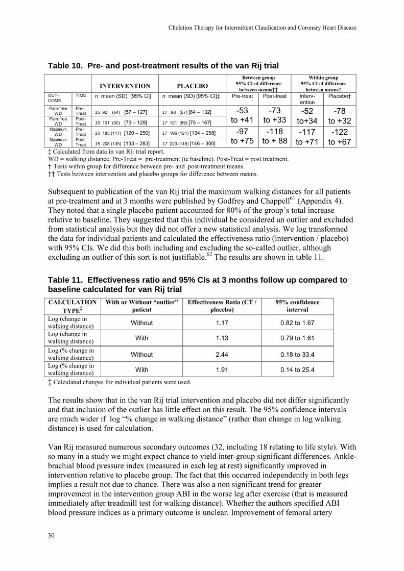
Chelation Therapy for Intermittent Claudication and Coronary Heart Disease
30
Table 10. Pre- and post-treatment results of the van Rij trial
INTERVENTION
PLACEBO Between group
95% CI of difference between means��
Within group 95% CI of difference
between means� OUT-COME
TIME n mean (SD) [95% CI] n mean (SD) [95% CI]� Pre-treat Post-treat Interv-ention
Placebo�
Pain-free WD
Pre-Treat 15 92 (64) [57 � 127] 17 98 (67) [64 � 132]
Pain-free WD
Post-Treat 15 101 (50) [73 � 129] 17 121 (89) [75 � 167]
-53 to +41
-73 to +33
-52 to+34
-78 to +32
Maximum WD
Pre-Treat 15 185 (117) [120 � 250] 17 196 (121) [134 � 258]
Maximum WD
Post-Treat 15 208 (135) [133 � 283] 17 223 (149) [146 � 300]
-97 to +75
-118 to + 88
-117 to +71
-122 to +67
� Calculated from data in van Rij trial report. WD = walking distance. Pre-Treat = pre-treatment (ie baseline). Post-Treat = post treatment. � Tests within group for difference between pre- and post-treatment means. �� Tests between intervention and placebo groups for difference between means. Subsequent to publication of the van Rij trial the maximum walking distances for all patients at pre-treatment and at 3 months were published by Godfrey and Chappell61 (Appendix 4). They noted that a single placebo patient accounted for 80% of the group�s total increase relative to baseline. They suggested that this individual be considered an outlier and excluded from statistical analysis but they did not offer a new statistical analysis. We log transformed the data for individual patients and calculated the effectiveness ratio (intervention / placebo) with 95% CIs. We did this both including and excluding the so-called outlier, although excluding an outlier of this sort is not justifiable.62 The results are shown in table 11. Table 11. Effectiveness ratio and 95% CIs at 3 months follow up compared to baseline calculated for van Rij trial CALCULATION
TYPE� With or Without �outlier�
patient Effectiveness Ratio (CT /
placebo) 95% confidence
interval Log (change in walking distance) Without 1.17 0.82 to 1.67 Log (change in walking distance) With 1.13 0.79 to 1.61
Log (% change in walking distance) Without 2.44 0.18 to 33.4
Log (% change in walking distance) With 1.91 0.14 to 25.4
� Calculated changes for individual patients were used. The results show that in the van Rij trial intervention and placebo did not differ significantly and that inclusion of the outlier has little effect on this result. The 95% confidence intervals are much wider if log �% change in walking distance� (rather than change in log walking distance) is used for calculation. Van Rij measured numerous secondary outcomes (32, including 18 relating to life style). With so many in a study we might expect chance to yield inter-group significant differences. Ankle-brachial blood pressure index (measured in each leg at rest) significantly improved in intervention relative to placebo group. The fact that this occurred independently in both legs implies a result not due to chance. There was also a non significant trend for greater improvement in the intervention group ABI in the worse leg after exercise (that is measured immediately after treadmill test for walking distance). Whether the authors specified ABI blood pressure indices as a primary outcome is unclear. Improvement of femoral artery

Chelation Therapy for Intermittent Claudication and Coronary Heart Disease
31
82
97
109
93102
95
119
97
74
101
405060708090
100110120130140
0 5.3
wal
king
dis
tanc
e (m
etre
s)
98 101
123
104
92
121
405060708090
100110120130140
0 5.3
wal
king
dis
tanc
e (m
etre
s)
pulsatility in the worse leg was significantly greater in the intervention group. Similar but non significant trends were observed for the better leg and for tibial artery pulsatility indices. There was no significant difference between groups with regard to side effects and adverse events. The van Rij trial report mentioned 12 month follow up data to be published elsewhere. When we contacted the author he confirmed that the data had never been published and sent a brief summary of the results. His e mail is reproduced in Appendix 5. 6.2.2.6 Summary of results of CT for IC A graphical summary of the primary outcome results is shown in Figs 2 and 3.
Guldager trial (1992). (placebo n = 78, intervention n= 75) Pre- Mid- Post- 3 month 6 month Treatment Treatment Treatment Follow up Follow up van Rij trial (1994). (placebo n = 17, intervention n= 15) Pre- Post- 3 month Treatment Treatment Follow up
Graphical summary of Pain Free Walking Distance measures reported in RCTs of chelation therapy for intermittent claudication. Error bars represent 95% confidence intervals. ■ = intervention. □ = placebo.
Fig. 2

Chelation Therapy for Intermittent Claudication and Coronary Heart Disease
32
192160
212
424
050
100150200250300350400450500
0 5 .3
wal
king
dis
tanc
e(m
etre
s)
208196 185223 230 233
050
100150200250300350400450500
0 5 .3
wal
king
dis
tanc
e(m
etre
s)
159157119
192164
206 204162
194 180
050
100150200250300350400450500
0 5 .3
wal
king
dis
tanc
e(m
etre
s) Olszewer trial (1990). (placebo n = 5, intervention n= 5) Pre- Post- Treatment Treatment Guldager trial (1992). (placebo n = 78, intervention n= 75) Pre- Mid- Post- 3 month 6 month Treatment Treatment Treatment Follow up Follow up van Rij trial (1994). (placebo n = 17, intervention n= 15) Pre- Post- 3 month . Treatment Treatment Follow up Graphical summary of Maximum Walking Distance measures reported in RCTs of chelation therapy for intermittent claudication. Error bars represent 95% confidence intervals. ■ = intervention. □ = placebo. Fig. 3

Chelation Therapy for Intermittent Claudication and Coronary Heart Disease
33
The very small trial of Olszewer52 (n=10) shows striking benefit of chelation therapy relative to placebo and statistical difference (P<0.05) between treatments. The largest trial (Guldager et al53 (n=153) and the intermediate-sized trial (van Rij et al51 (n=32) indicate no benefit of chelation therapy relative to placebo. Any conclusions drawn from these results will depend on the weight given to each study. This in turn depends on quality assessment of the three trials. All three trials performed reasonably well when tested against the pre-set criteria listed in the Methods section. Individual analysis of each trial has revealed some failures of design, reporting and statistical analysis. These failures are summarised as follows:- a] Olszewer trial.
• Intervention and placebo intravenous infusion solutions not clearly described. • Methods of outcome measurement very poorly described with some features
nonsensical (claudicants operating an exercise bicycle at 50 km/hr for 3 to 6 min). • Duration of study not stated. • Incorrect calculation of improvement factor for intervention group. • Statistical analysis obscure/absent. • Implausible intervention results: all CT patients improve dramatically and all placebo
patients remain essentially unchanged or deteriorate; after cross over to CT the original placebo group all improve dramatically and original CT group crossed over to placebo all improve further (carry over effect).
b] Guldager trial
• Blinding abrogated before end of study. • Results from about 10% patients �censored�; �last result� not brought forward for
analysis. • Non-compliers incompletely accounted for. • 95% confidence intervals for primary outcome (effectiveness ratio) implausibly
narrow and indicative of misreporting of calculation method.
c] van Rij trial
• Power calculation incorrect or at best obscure and trial severely underpowered to test proposed hypothesis.
• No quantitative value given for relative effectiveness of intervention (merely a statement that P > 0.05, but statistical procedure unclear).
In view of the very low quality and small size of the Olszewer trial its findings are overwhelmed by the results of the other two trials. These failed to demonstrate that CT has a beneficial effect with regard to the primary outcome. The direction of evidence indicates that CT may completely lack benefit .

Chelation Therapy for Intermittent Claudication and Coronary Heart Disease
34
That some secondary outcomes in the van Rij trial were in favour of chelation therapy might imply that further study is justified. Such a study might require very large numbers of patients (500 to1000) to demonstrate if such marginal effectiveness was real. The clinical significance for claudicants of small gains is controversial.27,63,64 The proposed funding for an RCT of CT was recently estimated for the NIH by Goertz to be $ 3 million65 The NIH research fund for alternative medicine has recently increased substantially.66 The trials were of relatively short duration. Two of the trials included follow up periods of 3 or 6 months that extended beyond treatment. Trial groups were seriously ill patients with life threatening atherosclerosis. Adverse events and side effects of treatments were observed. Neither study reported a significant difference in these between groups. One patient in the Guldager study died, however there was no indication in the report of the cause or to which group the individual belonged. Consideration of the evidence brought to bear on the question of safety of chelation therapy is included in section 6.3. 6.2.3 Randomised controlled trials of CT for coronary heart disease patients 6.2.3.1 Studies found and trial design Three RCTs48-50 were found one of which48 was excluded because it lacked an objective measure of effectiveness using an exercise test. The two included trials Hopf et al. (1985)49 and Knudtson (2002)50 had only been published in abstract at the time of searching. On request we received a 68 page report67 of the Hopf trial. This account is in German and is a dissertation prepared by one of the trial authors as part of her qualification for �Doktorgrades der Medizin des Fachbereichs Humanmedizin�. The Knudtson trial was submitted for publication early in 2001. Requests for a fuller account than the abstract were unsuccessful because of the trialists� respect for journal policy. The authors informed us of imminent publication in Dec 2001 and publication occurred in the 23 Jan 2002 issue of JAMA (it is not represented in the �potentially relevant papers box� of the outcome log on p 24). Characteristics of the two trials are shown in Table 12.
Table 12. Characteristics of available RCTs of chelation therapy for CHD
TRIAL

Chelation Therapy for Intermittent Claudication and Coronary Heart Disease
35
Characteristic
Hopf et al.1997� Single centre. Knudtson et al. 2002*. Multi-centre
Patient group Men with angina on effort and with angiographically confirmed CHD. Age range 43-64 (mean 59.5).
Men and women >21 years of age with proven ischaemic heart disease & stable angina. Non-candidates for re-vascularisation & no previous CT. Ability to perform exercise test. No abnormal renal or liver function.
Intervention Infusion of solution containing 3 to 4g Na2EDTA in isotonic sterile saline with 10 m eq MgCl2. Delivered every third day for 20 infusions.
Infusion of 500 ml Na2EDTA (40 mg/Kg) solution�� over 3 hr delivered 2 / week for 15 weeks and then once / month for total of 33 treatments��.
Comparator As intervention but minus EDTA. As intervention but isotonic saline substituted for EDTA��.
Primary outcome
Exercise stress test:- [a] Time (min) able to perform work (at maximum individual output, Watt) up to 6 min maximum, while monitored for ST depression of ECG trace; [b] �Ischaemic score��.
Time to ischaemia (ECG detection of ST segment depression) on a graded-stress treadmill test. Differences between baseline and 27 weeks post-randomisation calculated. Comparison in differences CT v. placebo.
Timing of primary outcome measures
Before and immediately after treatment, and at 3 to 6 months after treatment.
At baseline and at 15 and 27 weeks post-randomisation.
Secondary outcome(s)
Patient well-being + side effects; Lab: measures:- Blood Ca, calcitonin, PTH, electrolytes, creatinine, lipids. Angiographic coronary scores. 201Tl scintillography of myocardium; Ventricular ejection faction using 99Tc gamma camera.
Exercise functional reserve���; quality of life assessment (Seattle Angina Questionnaire; Duke Activity Status index; Health Status Survey Short Form-36); clinical events.
No. at start 16 CT = 8 PL = 8
84 CT = 41 PL = 43 No. at end 15 at end of treatment; 7 at follow up. 78 CT = 39 PL = 39���
Follow up time 3 to 6 months 12 months for clinical events. CT = chelation therapy. PL = placebo. ECG = electrocardiogram. PTH = parathyroid hormone.
* Details from full paper . � Details extracted from report by M. Gleuβner.67 �� Solution cotained 5% dextran, 80 mg Lidocaine, 5 g ascorbic acid, 0.7 g magnesium sulphate, and sodium bicarbonate to titrate to physiological pH. ��� calculated from maximum oxygen consumption and time to anaerobic threshold . � Ischaemic score = [(100 x mm ST depression) / (Watt x F)] where F = fraction of 6 min that excerise was performed. �� Both groups received oral vitamins on non-treatment days. ��� Intention to treat analysis. 6.2.3.2 Quality assessment i] Hopf trial The randomisation method was not described. Assessors were blinded. Treatment of patients was delivered by a �believer� in chelation therapy. It is not clear if this individual was also an assessor. It is likely patients were effectively blinded to treatment. Baseline charactistics were provided including patient history of MI and bypass grafts, grade of angina, and scores for number and extent of main coronary artery branches with stenosis (2.6 and 2.1 for placebo intervention group respectively). It is unlikely the two groups differed significantly at baseline. The method of primary outcome measurement was fully described in the dissertation report as follows:- patients operated an exercise machine at their maximum individual power output capacity while linked to an ECG recorder that monitored ST segment depression (mm).

Chelation Therapy for Intermittent Claudication and Coronary Heart Disease
36
Exercise was continued for up to 6 min or such time that the patient discontinued because of angina, breathlessness or exhaustion. The machine allowed measurement of power output (Watt). The assessor recorded duration of exercise (t; in min), power output (P; in Watt) and maximum ST depression on ECG trace (D; in mm). If ST depression was observed then an ischaemia score was calculated:- Ischaemia score = (100 x D) / (P x [t/6]); [a measure of ST depression per fractional work performed]. The numerous methods used to measure secondary outcomes were also described. Pre- and post-treatment outcome measures were made but the exact timing of follow up measures was not given (between 3 and 6 months after treatment). One placebo group patient did not complete treatment (withdrew for bypass operation) and did not contribute post treatment data. From the variance in the primary outcome measure at pretreatment it is clear the study was underpowered except for detection of large changes in the primary outcome. ii] Knudtson trial The Knudtson trial full paper provides trial details lacking in the abstract. The method of randomisation was partially described (randomisation in blocks of ten) and the fact of allocation concealment from investigators was stated and the method indentified. Extensive baseline characeristics were tabulated and there were no statistically significant differences between groups; however the chelation group had experienced more myocardial infarctions (20/41 v.12/43) and more individuals in the placebo group were receiving nitrate therapy (19/43 v. 10/41). Similarity in baseline characteristics indicated successful randomisation. Therapy was described as �nurse-supervised�. Apart from the substitution of saline for EDTA both groups received the same treatment. Infusion solutions were indistinguishable by colour or labelling. Infusions were slow and contained Lidocaine to minimise the possibility of unblinding (EDTA infusion might be distinguished from saline because of stinging sensation). It is likely therefore that care was the same for both groups. Losses to follow up were low (less than 10%) and were similar between groups. All losses were accounted for. Blinding of patients was safeguarded by slow infusion, use of Lidocaine, blinding of investigators and similar appearance of infusion solutions. Methods of outcome measurement were clearly described and intention to treat analysis (last observation carried forward) was employed. A power calculation was performed prior to the study and the study was adequately powered for the primary outcome measure. This study was judged to be of high quality and scored 5 on a Jadad-based scale (range 0-5). 6.2.3.3 Results of RCTs of chelation therapy for patients with CHD i] Hopf trial The abstract of this trial provided values for mean change of work duration from baseline to end of treatment for the two groups. These results are shown in table 13.

Chelation Therapy for Intermittent Claudication and Coronary Heart Disease
37
Table 13. Summary of results of Hopf trial as presented in abstract TRIAL
Group
n at start
n at finish
OUTCOME� Mean increase in time of work duration (at end of
treatment v baseline) Value (min)
Statistical
significance (Placebo v intervention)
Intervention 8 8 1.2 Hopf et al., 1997 Placebo 8 7� 1.1
No difference between groups
� Mean group power output during work was similar in the two groups (106 and 94 Watt in intervention and placebo groups respectively). � number obtained from report not abstract. It is unclear how these results were derived from the fuller data provided in the dissertation report. We have recalculated the means and 95% confidence intervals using data for individual patients in the dissertation. They are shown in Table 14. No significant difference between groups in the mean change from baseline was observed for any primary outcome. Secondary outcomes were also not significantly different.
Table 14. Summary of primary outcome results of Hopf trial based on dissertation data
INTERVENTION PLACEBO OUTCOME n mean 95% CI n mean 95% CI
before� 8 4.6 3.1 to 6.0 7 4.4 3.4 to 5.5 after�� 8 5.3 4.2 to 6.4 7 5.3 4.4 to 6.2
Duration of exercise test
(min)�. change٭ 8 + 0.75 7 + 0.86 before 7 2.3 1.22 to 3.37 7 2.1 0.97 to 3.23 after 5 1.8 0.89 to 2.65 6 1.9 0.78 to 2.97
Ischaemic score��.
change � 0.53 � 0.22 before 8 80.9 48 to 114 7 74.6 47 to 102 after 8 95.1 65 to 126 7 88.3 69 to 108 Watts x FD٭٭
change 8 + 14.2 7 + 13.7 � Times were measured to the nearest half minute, the means we have calculated are the same as reported in the dissertation. �� The mean we calculated for the intervention group after treatment differs from that in the dissertation because one patient�s data was omitted from the latter. � Before treatment. �� After treatment. ٭ Mean of individual changes (before treatment minus after treatment). ٭٭ FD = fractional duration of maximum 6 minutes. ii] Knudtson trial This trial was sufficiently powered to detect as statistically significant a difference between groups of 60 sec for change in mean time to ischaemia (pre-treatment to end of treatment; see Appendix 6). This represents an increase over pre-treatment time to ischaemia of ~20% for the intervention group. Both groups significantly improved to the same extent their performance in the primary outcome measure (time to ischaemia). Thus the time to ischaemia at post-treatment (27 weeks) was significantly different from baseline (by about 10%) for both placebo and intervention groups. However there was no significant difference between groups for change in outcome (end of treatment relative to baseline). These results are shown in the Table 15.

Chelation Therapy for Intermittent Claudication and Coronary Heart Disease
38
Table 15. Summary of results of the Knudtson trial
Group
n at start*
BASELINE Mean time
(sec) to ischaemia
[SD]
OUTCOME� Mean increase in
time (sec) to ischaemia at 27
weeks v baseline Value [ 95% CI]
Statistical
significance (P values
27 weeks v baseline)
Statistical
significance ( P value
Placebo v. Intervention )
PLACEBO 43 572 [172] 54 [ 23 to 84 ] <.001
EDTA 41 589 [186] 63 [ 29 to 95] <.001
Difference between groups 9 [-36 to 53] .69
Number completed = 39 in each group, last observation carried forward. � Treatment ceased at ٭week 27 at which point primary outcome measure was made. 95% CI = 95% confidence interval. Similarly there were no statistically significant differences between groups in any secondary outcomes. The intervention group did show a significant increase in maximum oxygen consumption (baseline v. 27 weeks P = 0.03) whereas the control group showed a non-significant increase, however again there was no significant difference in change between groups. Clinical events observed over the 12 month follow up period did not differ between groups. 6.2.3.4 Summary of results of RCTs of chelation therapy for CHD A graphical summary of the primary outcome results is shown in Figs 4. Hopf Trial Knudtson Trial Summary of outcome measures reported in RCTs of chelation therapy for CHD. Error bars represent 95% confidence interval. ● = placebo ■ = intervention ▲ = difference between placebo and intervention. Fig. 4
-50
-25
0
25
50
75
100
placebo n = 43
interventionn = 41
Mean change in time to ischaemia Difference between means
-25
-15
-5
5
15
25
35Mean change in work
13.7 14.2
0.63
-25
-15
-5
5
15
25
35
WO
RK
(wat
ts x
frac
tn'l
time)
interventionn = 8
placebo n = 7
Difference between means
54 63
9
-50
-25
0
25
50
75
100
time
(sec
onds
)

Chelation Therapy for Intermittent Claudication and Coronary Heart Disease
39
The Hopf trial was small (only16 patients). The reported difference between groups in mean number of stenosed coronary branches at the start of the trial was unlikely to have invalidated the trial. Variances in the primary outcome measures were large and the trial was under-powered except for detection of large changes. No significant affect of chelation therapy was found. The Knudtson trial was of moderate size (78 patients completed). It was well designed and conducted. It was sufficiently powered to detect an effect size of 20% which by consensus represents a minimally important clinical improvement. No statistically significant effect of chelation therapy was found. The authors concede that generalization of their findings should be restricted to a population of similar characteristics to the study group (i.e. patients with stable angina, non-candidates for revascularization and able to perform on a treadmill). Since both intervention and placebo groups received oral multivitamins daily the authors could not exclude the possibility that these supplements might be partially responsible for the improvements observed in both groups. A Hawthorne effect provides an alternative explanation. 6.3 Reviews of chelation therapy for PAD and CHD 6.3.1 Reviews found and characteristics of included reviews Thirty potentially relevant reviews were found. These dated from 1976 to 2001. They are listed in the Table 16.
Table 16. Details of Reviews found Type of study Number
of studies Source
Systematic & other Reviews 19 Embase,Medline or Biomednet Systematic / Critical Reviews 2 CHID
Systematic / Critical Reviews 5 Referenced in Journal or cited in Web of Science
Systematic / Critical Reviews 2 Cinahl Meta-analyses 2 Referenced in Journal
Of the 30 reviews found 24 were excluded for reasons tabulated in the Appendix 7. The six included reviews36,68-72 all attempted a critical analysis of primary data. They span 1993 to 2000 and therefore were able to evaluate at least two of the published RCTs of chelation therapy. Main features of these reviews are shown in Table 17.

Chelation Therapy for Intermittent Claudication and Coronary Heart Disease
40
Table 17. Summary and characteristics of reviews for chelation therapy for PAD and CHD Authors [study type]�
Objective of Review��
Search strategy and outcome٭ ٭
Inclusion exclusion criteria.
Results
Grier & Meyers 1993 [review article]
To determine safety & efficacy of EDTA chelation for atherosclerosis
Electronic Medline search + citation searches. Cited all RCTs to date of publication; also 14 of the 21 uncontrolled studies to 1993 retrieved and listed in Ernst 2000§.
Criteria not stated; all types of study of CT efficacy included.
Best evidence shows therapy is ineffective. EDTA CT should not be used in clinical practice to treat atherosclerosis.
Chappel & Janson 1996 [historic review]
Review research literature, current evidence of effectiveness, & potential mechanisms of action of EDTA
Strategy not described. Cited all RCTs to date of publication; also 7 of the 22 uncontrolled studies retrieved and listed in Ernst 2000§.
Criteria not stated; all types of study of CT efficacy included.
CT is a valuable therapeutic option for vascular disease.
Ernst E. 1997 [systematic review]
Review evidence from RCTs regarding safety and effectiveness of CT for PAOD
Extensive electronic and other searches. Cited all RCTs to date of publication.
Only RCTs admitted.
CT for PAOD is not superior to placebo, is associated with considerable risks and costs and should be considered obsolete
Elihu et al. 1998 [therapeutic review]٭
Assess if there is sufficient evidence for the clinical use of CT in CVD
Strategy not described. Cited all RCTs to publication date except 1 abstract; also 4 of the 22 uncontrolled studies retrieved and listed in Ernst 2000§.
Criteria not stated; all types of study of CT efficacy included.
More controlled studies are required٭ to determine efficacy in CVD before broad use in clinical practice.
Olmstead 1998 [critical review]
Provide � critical analysis of evidence for and against efficacy of EDTA chelation.
Strategy not described. Cited all RCTs to publication date except 1 abstract; also 19 of the 22 uncontrolled studies retrieved and listed in Ernst 2000§.
Criteria not stated; all types of study of CT efficacy included.
More �balanced� controlled studies are required to determine efficacy.
Ernst 2000 [systematic review]
Summarize all the clinical evidence for or against effectiveness and efficacy of CT for CHD.
Extensive electronic and other searches. Cited all RCTs to publication date. 22 uncontrolled studies found and listed dating from 1955-93.
All clinical studies of intravenous CT for CHD
Given the potential for CT to cause adverse effects, this treatment should now be considered obsolete.
CT = chelation therapy. CVD = cardiovascular disease. CHD = Coronary heart disease. PAOD = peripheral arterial occlusive disease. RCT = randomised controlled trial. � The study type as stated in the text or page header of the publication concerned. �� As stated explicitly by authors. ٭ This review considers several chelating agents in addition to EDTA. .We assume that we have identified all RCTs published in full or abstract form ٭ ٭§The included reviews have considered several types of study whereas our search strategy was aimed at controlled studies; we have therefore used the list of found studies in Ernst 2000 as a basis to compare reviews. 6.3.2 Quality assessment of reviews according to QUORUM check list The included reviews were assessed according to the criteria set out in the Methods section. The QUORUM checklist is a list of the most important elements thought necessary for good reporting of a systematic review or meta-analysis.45 Two of the reviews (Ernst 199771 and Ernst 200068) were formally identified as systematic and one other, Grier & Meyers 199372, was also systematic although not formally identified as such. The remaining reviews were not systematic but since their aim was identical to that of the others they were also assessed against the QUORUM checklist. The results are shown in the Table 18.

Chelation Therapy for Intermittent Claudication and Coronary Heart Disease
41
Table 18. Six included reviews assessed according to the QUORUM checklist . Reported? (Y/N/I)
Heading Subheading Description | REVIEW † G & C & E1 E l et Olm E2 M J al
Title Identify the report as a meta-analysis [or systematic review] of RCTs��� N NA Y NA NA Y Abstract Use a structured format����������������������� Y N NA N N Y
Describe Objectives The clinical question explicitly��������������������. Y N NA Y Y Y Data sources The databases (ie, list) and other information sources���������.. Y N NA N N Y Review methods The selection criteria (ie, population, intervention, outcome, and study design); methods for validity assessment, data abstraction, and study characteristics, and quantitative data synthesis in sufficient detail to permit replication��. Y N NA Y N Y Results Characteristics of the RCTs included and excluded; qualitative and quantitative findings (ie, point estimates and confidence intervals); and subgroup analyses N N NA N I Y Conclusion The main results��������������������������. Y Y NA Y Y Y
_________ Describe Introduction The explicit clinical problem, biological rationale for the intervention, and rationale for review ������������������������� Y Y Y Y Y Y Describe Methods Searching The information sources, in detail (e.g., databases, registers, personal files, expert informants, agencies, hand-searching), and any restrictions (years considered, publication status,l anguage of publication)��������.. Y I Y N N Y Selection The inclusion and exclusion criteria (defining population, intervention, principal outcomes, and study design Validity assessment The criteria and process used (e.g., masked conditions, quality assessment, and their findings )�� N N Y N Y٭ Y٭ Data abstraction The process or processes used (completed independently, in duplicate) N N N N N N Study The type of study design, participants� characteristics, details of intervention, characteristics outcome definitions, &c, and how clinical heterogeneity was assessed���.. Y N Y N Y Y Quantitative The principal measures of effect (e.g., relative risk), method of combining data synthesis results (statistical testing and confidence intervals), handling of missing data; how statistical heterogeneity was assessed; rationale for any a-priori sensitivity and subgroup analyses; and any assessment of publication bias�. NA NA NA NA NA NA Describe Results Trial flow Provide a meta-analysis profile summarising trial flow (see figure) ����� NA N NA NA NA NA Study Present descriptive data for each trial (e.g., age, sample size, intervention, Characteristics dose, duration, follow-up period) ������������������. Y N Y Y Y Y Quantitative Report agreement on the selection and validity assessment; present simple data synthesis summary results (for each treatment group in each trial, for each primary outcome); present data needed to calculate effect sizes and confidence intervals in intention-to-treat analyses (e.g. 2x 2 tables of counts, means and SDs, proportions) �������������������������. I N I N Y Y Discussion Summarise key findings; discuss clinical inferences based on internal and external validity; interpret the results in light of the totality of available evidence; describe potential biases in the review process (e.g., publication bias); and suggest a future research agenda �������������. Y N Y Y Y Y G & M = Grier and Meyers, 1993; C & J = Chappell and Janson, 1996; E1 = Ernst 1997; El et al = Elihu et al, 1998; Olm = Olmstead 1998; E2 = Ernst 2000. � Only in Ernst 1997 and Ernst 2000 was the body of the text formally subdivided into Introduction, Methods, Results and Discussion sections; Elihu et al, 1998 and Olmstead, 1998 lacked an Abstract; for the latter review the summary sections 5.1.4 and 5.2.7 were taken to be equivalent for an Abstract. * These reviews included all types of studies. Some Descriptors in the QUORUM table have multiple elements, with respect to these Y = all or nearly all elements reported in the review; N = none or very few of the elements reported in the review; I = incomplete i.e. some elements only reported in the review. NA = not applicable. 6.3.3 Results of Reviews and analysis of Review Quality by additional criteria 6.3.3.1 General considerations The individual reviews were assessed according to whether statements were matched by references given in their support and whether statements were errors, misrepresentations or
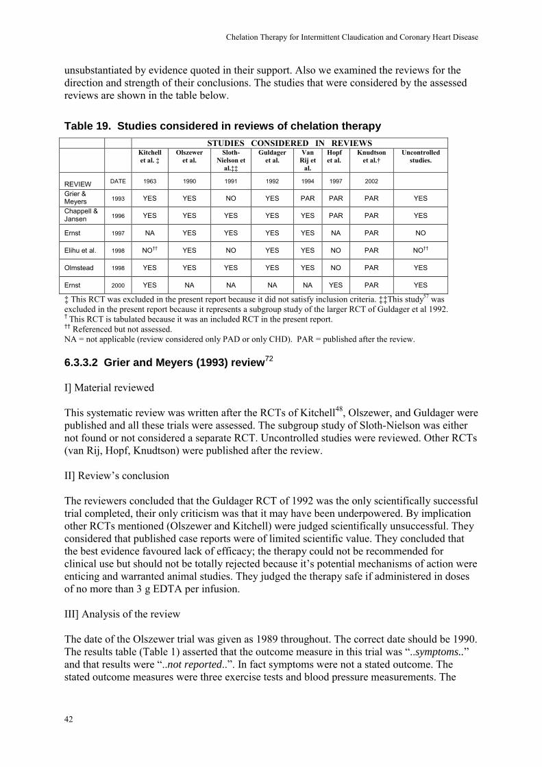
Chelation Therapy for Intermittent Claudication and Coronary Heart Disease
42
unsubstantiated by evidence quoted in their support. Also we examined the reviews for the direction and strength of their conclusions. The studies that were considered by the assessed reviews are shown in the table below.
Table 19. Studies considered in reviews of chelation therapy STUDIES CONSIDERED IN REVIEWS Kitchell
et al. � Olszewer
et al. Sloth-
Nielson et al.��
Guldager et al.
Van Rij et
al.
Hopf et al.
Knudtson et al.�
Uncontrolled studies.
REVIEW DATE 1963 1990 1991 1992 1994 1997 2002
Grier & Meyers 1993 YES YES NO YES PAR PAR PAR YES
Chappell & Jansen 1996 YES YES YES YES YES PAR PAR YES
Ernst 1997 NA YES YES YES YES NA PAR NO
Elihu et al. 1998 NO�� YES NO YES YES NO PAR NO��
Olmstead 1998 YES YES YES YES YES NO PAR YES
Ernst 2000 YES NA NA NA NA YES PAR YES
� This RCT was excluded in the present report because it did not satisfy inclusion criteria. ��This study57 was excluded in the present report because it represents a subgroup study of the larger RCT of Guldager et al 1992. � This RCT is tabulated because it was an included RCT in the present report. �� Referenced but not assessed. NA = not applicable (review considered only PAD or only CHD). PAR = published after the review. 6.3.3.2 Grier and Meyers (1993) review72 I] Material reviewed This systematic review was written after the RCTs of Kitchell48, Olszewer, and Guldager were published and all these trials were assessed. The subgroup study of Sloth-Nielson was either not found or not considered a separate RCT. Uncontrolled studies were reviewed. Other RCTs (van Rij, Hopf, Knudtson) were published after the review. II] Review�s conclusion The reviewers concluded that the Guldager RCT of 1992 was the only scientifically successful trial completed, their only criticism was that it may have been underpowered. By implication other RCTs mentioned (Olszewer and Kitchell) were judged scientifically unsuccessful. They considered that published case reports were of limited scientific value. They concluded that the best evidence favoured lack of efficacy; the therapy could not be recommended for clinical use but should not be totally rejected because it�s potential mechanisms of action were enticing and warranted animal studies. They judged the therapy safe if administered in doses of no more than 3 g EDTA per infusion. III] Analysis of the review The date of the Olszewer trial was given as 1989 throughout. The correct date should be 1990. The results table (Table 1) asserted that the outcome measure in this trial was �..symptoms..� and that results were �..not reported..�. In fact symptoms were not a stated outcome. The stated outcome measures were three exercise tests and blood pressure measurements. The

Chelation Therapy for Intermittent Claudication and Coronary Heart Disease
43
individual patient data for the three exercise tests were reported in full and group results for other outcomes were also reported. The results table misrepresents the Guldager trial by quoting distances walked as though they were gains in distance walked. Although the review mentioned the violation of blinding in the Olszewer trial the abandonment of blinding in the Kitchell trial and during follow-up in the Guldager RCT were not mentioned. 6.3.3.3 Chappell and Janson (1996) review69 I] Material reviewed This review was written after the RCTs of Kitchell, Olszewer, Guldager and van Rij were published. These were all considered together with the subgroup study of Sloth-Nielson57 which was partly reviewed as a separate trial. Uncontrolled studies were also reviewed. II] Review�s conclusion These reviewers strongly endorsed the efficacy of chelation therapy. The findings reported in uncontrolled case studies were judged valid and of scientific value. The negative RCTs of Guldager and van Rij, and the subgroup study of Sloth-Nielson, were criticised but also used as evidence for effectiveness of chelation. The reviewers concluded that chelation therapy was valuable (�..87% of patients demonstrate improvement with objective testing..�) and safe. They further considered it might be reasonable to treat healthy persons who ask for chelation therapy since it removes potentially toxic heavy metals to which most people have been exposed. III] Analysis of the review The reviewers stated �..Olszewer completed a small blinded cross-over study...that demonstrated significant improvement in both walking distance and ankle/brachial index measurements..�. This statement was not qualified in any way. It is misleading and misrepresents the trial because the cross over study design was not completed but abandoned at cross over at which point blinding was also violated. The Guldager trial and the Sloth-Nielson subgroup study were criticised for claiming to use, but not actually using, the ACAM protocol for infusion of EDTA. In fact this claim was not made in the trial reports. Guldager stated that they omitted magnesium sulphate (MgSO4) from the infusion (and therefore did not exactly follow the ACAM protocol). It is also explicit that vitamins that are included in the ACAM infusion protocol were given separately to trial patients as a daily tablet. No mention of the ACAM protocol occurred in the paper of Sloth-Nielson et al. The van Rij trial was criticised for having compared two chelating solutions rather than one chelating solution v. a placebo. The two chelating solutions were described as �..one with EDTA and the other with thiamin and ascorbate..�. This is misleading since both placebo and intervention infusions contained equal amounts of ascorbate and thiamin. The essential difference between intervention and placebo was the presence in the former of 3 g of

Chelation Therapy for Intermittent Claudication and Coronary Heart Disease
44
disodium EDTA, a well known chelating agent. It is true that ascorbate does have some chelating abilities although these are weak compared with EDTA. Of the van Rij trial the reviewers stated “..both groups of patients showed significant overall improvement (EDTA 60% versus thiamin / ascorbate 59%)..”. There are several reasons why this statement is misleading:- (a)�..overall improvement..� was not defined. It implied the combining of outcomes into a single measure of improvement, a procedure not performed in the review itself or in the trial report. (b) The �60� and �59%” cannot refer to degree of improvement. The degree of improvement in walking distances were actually only in the range 6 to 20%; all blood pressure changes were less than 10%; among 18 different life style measures no improvement was greater than 32%. Increases in �subjective walking distances� were > 100%, but these values have no clinical significance whatsoever since they merely represent the patient�s guess as to how far they walked. (c) The van Rij study did report increased (not �improved�) walking distance (whether maximum or pain-free walking distance is unclear) in 60 and 59% of intervention and placebo patients at a single time point. They did not comment on proportions of patients with respect to any other outcome measure or time point. Since the degree of increase in the patients was not specified and because there is random variation in any measure, we would expect that (with null-effect treatments) on average about half of the patients would increase and half would decrease their walking distance. Thus an increase in walking distance in 59 or 60% of patients is about what would be expected by chance. The fact that the degree of improvement is unspecified renders the unqualified statement made in the review at best meaningless and possibly misleading. The reviewers stated that in the trials of Sloth-Nielson, Guldager and van Rij �..50 to 60% of patients improved..�. It is not clear if this refers to one or all three of the trials. It is impossible to justify this statement from the data published in the trials since, except for a single secondary outcome (transcutaneous oxygen tension) in one study57 no individual patient data was presented. Again, since the degree of improvement is not specified, we would expect by chance that on average about half of the patients to change in one direction and half in the other direction. The statement is at best meaningless and at worst misleading. 6.3.3.4 Ernst (1997) review71 I] Material reviewed This systematic review was about RCTs of chelation therapy for peripheral occlusive arterial disease. It was written after the RCTs of Olszewer, Guldager and van Rij were published. All three were considered in the review together with the subgroup study of Sloth-Nielson57 which was treated as a separate study. Uncontrolled studies were not included. II] Review�s conclusion The Sloth-Nielson paper was acknowledged to be a subgroup study of the larger Guldager RCT, but it was nevertheless treated as an independent investigation. The positive trial of Olszewer was judged of such poor quality as to preclude interpretation. The three negative RCTs of Guldager, Sloth-Nielson and van Rij were found to be of �..outstanding methodological rigour..� yielding results that �..clearly and conclusively show that .. chelation therapy does not ameliorate symptoms of the disease, nor .. change objective signs of the disease..to a greater extent than placebo..�. Lack of demonstrable efficacy and the

Chelation Therapy for Intermittent Claudication and Coronary Heart Disease
45
3 monthsfollow up
baseline
157
119
204
162
5075
100125150175200225250275
0 5 .3
wal
king
dis
tanc
e(m
etre
s)Guldager MWD at baseline and at3 months follow up. (open symbolplacebo, filled symbol intervention).Error bars represent 95%confidence intervals of the mean.
baseline3 monthfollow up
0.5320.5020.492 0.490 0.481
0.498 0.4920.492 0.5030.521
0.3
0.4
0.5
0.6
0.7
0 5
Ankle
/Bra
chia
l Inde
x
Beforetreatment
After 7�thinfusion
After 14�thinfusion
Aftertreatment
After 3 monthsfollow up
! = intervention, ? = placebo. We have attempted to reproduce the errorbars of the original figure pro-rata. It is assumed they represent standarddeviation of the mean, but this was not stated. Data calculated from the graphshown in figure 2 of Sloth-Nielson et al.{215}.
◊
possibility of �..severe, life-threatening adverse effects..� led this reviewer to the emphatic conclusion that the therapy should considered obsolete. III] Analysis of the review The reviewer remarked on the subgroup study “..in this sub-population there was a (non-significant) trend for the MWD and the peripheral arterial pressure to increase to a greater extent in the placebo group than in the EDTA group..�. This statement is not supported by the data (see Fig 1 & 2. of Sloth-Nielson et al57) and is unjustified because the full data from the study of 153 patients were available to the reviewer. Sloth-Neilson et al. commented �..in this study the walking distance improved equally in both groups during the first ten infusions …after this the walking distances were similar..�. Maximum walking distance data for the full study is shown in the diagram below (Fig. 5); it fails to demonstrate any such trend. Fig. 5
Sloth-Nielson did not report peripheral arterial pressures but provided ankle/brachial blood pressure measures in a graph. Sloth-Nielson commented �..ankle-brachial indices were constant in both groups during the study period..�, and �..ankle/brachial indices before treatment, after 7 infusions, after 14 infusions, after a total of 20 infusions and after 3 months follow up are shown in Fig2. The curves are horizontal and are not significantly different..�. We have redrawn the data from the trialists� Fig 2 and this is shown below (Fig. 6). It does not support the reviewer�s comment. Fig. 6

Chelation Therapy for Intermittent Claudication and Coronary Heart Disease
46
2.372.38
2.39
2.35
2.32
2.36
2.382.39
2.23
2.28
2.33
2.38
2.43
-1 0 1 2 3 4 5 6 7
time from start of treatment (months)
seru
m c
alci
um (m
mol
/ l)
Van Rij was quoted as stating that 59 and 60% patients �improved� in the two groups. In fact van Rij states that 59% and 60% increased their walking distance (type unspecified) at 3 months follow up.
The reviewer pointed out that authors of the Olszewer trial were �..unclear as to the method of data analysis..” and that they had stated �..there were no intergroup differences at the end of the double blind phase..�. The review�s summary Table lists the result of the Olszewer trial as �..no intergroup difference..�. However, whatever the shortcomings of the trialists� data analysis, or of their statements based on these, reviewers are empowered to conduct their own analysis because individual patient data was reported.73 When this is done the between group significant difference in walking distance after ten infusions (the end of the double blind phase) is obvious (see fig 1 top panel page 31 and Table 8 page 28). Therefore the results of this trial are not reliably represented in the review. The Sloth-Nielson subgroup study was referenced in support of the statement:- “..in one trial, 6 patients of the experimental group showed clinical signs of (potentially lethal) hypocalcemia..�. However this reference did not mention hypocalcaemia and with regard to side effects of treatment the authors commented:- �side effects of treatment were non specific and there was no difference between the two groups�. Among “..side effects during treatment..� the full Guldager trial report does list 6 intervention patients, but also 2 placebo patients (left unmentioned in the review), as exhibiting �hypocalcaemic symptoms�. Hypocalcaemic symptoms were not defined. Neither whole group or individual patient blood calcium measures were provided. According to the text, �..eight patients (5 intervention and 3 placebo) received intravenous calcium gluconate..� . Why 5 intervention and 3 placebo patients should be treated when hypocalcaemic symptoms were observed in 6 and 2 respectively is unclear. Another subgroup study55 (not Sloth-Nielson) did report serum calcium levels in a single-centre subgroup (n=55) of the full Guldager trial (n=153). This paper was not referenced by the reviewer. In this study there was no significant difference between intervention and placebo groups for their change of serum calcium levels in response to treatment. Individual patient data was not provided and there was no mention of the number of patients with hypocalcaemia, �..potentially lethal..� or otherwise. We have calculated 95% confidence intervals for serum calcium concentrations reported in this subgroup study and these are shown in the figure below (Fig. 7). Because of the overlap of 95% confidence intervals for the intervention group data the authors� contention that a significant fall in serum calcium was observed in this group at end of EDTA treatment appears questionable. Fig. 7 Serum calcium concentrations in subgroup of patients from the Guldager trial53. Solid symbols = intervention, hollow symbols = placebo. Error bars are 95% confidence intervals calculated from the data presented in Guldager et al. 1993.55 Treatment period

Chelation Therapy for Intermittent Claudication and Coronary Heart Disease
47
The review asserted that:- �..through it’s chelating action EDTA treatment is associated with severe life threatening events..� This was referenced with Guldager et al.,199253 and Meltzer et al., 196174. The former is an RCT in which the authors described side effects that �..were generally non-specific and showed no preponderance in any of the groups..” . Lack of clarity in the description of drop-outs means we are unable to allocate to which group all the patients suffering the serious adverse events (one death, two strokes) belonged. This reference does not support the assertion. The other reference74 offered in support described uses of EDTA from more than 40 years ago. It reported on 2000 infusions of 3g Na2 EDTA administered on alternating days over a two year period to 81 consecutive patients. No cases of nephrotoxicity were observed. Prothrombin time was prolonged by about 30% but no adverse haemorrhagic events were noted and prothrombin time returned to normal within twelve hours of infusion. Other side effects considered included hypotension, vitamin deficiency syndromes, hypothyroidism, gastrointestinal problems, abdominal pain and local pain during infusion. The incidence of side effects was rare. The authors concluded “..we have found no serious side effects or toxicity with use of EDTA when administered as a 3 g dose and infused as a 0.5% solution over 2.5 to 3 hours. It is therefore our opinion that the drug can be used without danger over prolonged periods..” . This reference does not support the review�s statement above. Proponents of chelation therapy were accused of an.. �outdated understanding of atherogenesis�.. in attempting to ..�deblock arteries�.. by.. �extracting calcium�.. from ��plaques�.. they were adhering to.. �pathophysiological models…in overt discordance with present knowledge�. This accusation is unjustified. Firstly modern theories of plaque pathogenesis focus on metal ion-dependent free radical damage to LDL particles which, in theory at least, might be treatable via chelating agents (as is claimed by proponents). Secondly, calcification of plaques61,75 has recently become detectable by sensitive non-invasive techniques and evidence has been presented that it predicts severity of atherosclerosis as indicated by adverse events.30,75,76 This implies, but does not establish, a role for calcification in pathogenesis of plaques. 6.3.3.5 Elihu et al. (1998) review70 I] Material reviewed This review was about the efficacy of several chelating agents for cardiovascular disease. The Hopf trial was either not found or left unconsidered because it was only available as an abstract. The existence of the Kitchell trial was referenced but it was not reviewed. Uncontrolled studies are mentioned but not assessed. As no completed RCTs of EDTA for CVD were included Elihu reviewed the PAD studies of Olszewer, Guldager and van Rij. The Sloth-Nielson subgroup study was either not found or not considered a separate trial. II] Review�s conclusion There was no explicit comment on the relative quality of the RCTs. It was pointed out that the largely negative van Rij trial had reported �.small improvements...at 3 months post chelation..� in ankle/brachial blood pressure index and femoral artery pulsatility. The reviewers concluded that �..there are conflicting results even within well-controlled trials..�. They considered serious side effects worrisome but commented that few adverse effects had

Chelation Therapy for Intermittent Claudication and Coronary Heart Disease
48
been reported when the AMCAM guidelines were followed. They concluded that too few controlled studies had been performed to warrant clinical use of EDTA for cardiovascular disease. With respect to PAD they were non-committal, commenting merely that 2 of 3 RCTs had revealed no benefit compared to placebo. III] Analysis of the review We did not identify erroneous or unsubstantiated statements in the parts of the review that considered EDTA. 6.3.3.6 Olmstead (1998) review36 I] Material reviewed This review was written after the studies of Kitchell, Olszewer, Sloth-Nielson, Guldager, van Rij and Hopf were published. All except Hopf were considered. Uncontrolled studies were also reviewed. The review was not published in a peer-reviewed journal. It was initiated by a commission from the National Institute of Health�s Office of Alternative Medicine (USA). II] Review�s conclusion Available clinical data on EDTA therapy for CHD was judged scant and of poor quality. In the author�s opinion only future prospective controlled clinical trials could firmly establish if EDTA was effective for symptoms of CHD or could alter the natural history of the disease. With regard to PAD the reviewer assessed the quality and results of the Olszewer, Guldager, van Rij trials and the Sloth-Nielson subgroup study. The Olszewer trial was judged inadequate in many respects and the outcome tests were described as scientifically invalid. The Guldager trial was judged underpowered; in addition this trial was severely criticised for: a) poor retention of patients; b) inability to account for all dropouts; c) violation of blinding; d) inadequate effort to establish reproducibility in outcome measures prior to commencement of the trial; e) failure to employ intention to treat analysis; f) inappropriate statistical procedures. Despite the trialists� conclusion that chelation therapy did not result in improvement of intermittent claudication the reviewer stated �.. the presented data, questionable as they are, indicated the mean MWD increased in the EDTA group..�. and pointed out that at 6 months the intervention group MWD was 51.3% increased from baseline whereas the placebo group was increased by 23.6% (see Fig 1, page 31). The under-powering of the van Rij trial was described as � ..a major failing..�. Otherwise the trial was judged well conducted. The statistically significant superiority of chelation therapy in improving ankle/brachial blood pressure index and femoral artery pulsatility was described as having clinically uncertain but intriguing implications. In the reviewer�s opinion these findings indicated that EDTA chelation may improve non-invasive parameters of leg perfusion in patients with PAD. The Sloth-Nielson subgroup study was criticised for :- a) it�s small numbers (n=30); b) lack of blinding of investigators in measurement of walking distance; c) the use of arteriograms

Chelation Therapy for Intermittent Claudication and Coronary Heart Disease
49
assessed subjectively rather than by a quantitative method; d) lack of information concerning the timing of the follow up arteriograms. The arteriograms were dismissed as representing little more than opinion. The poor quality of RCTs so far published (1998) and the hints of therapeutic effectiveness evident to the reviewer in these studies led him to the opinion that effectiveness of EDTA chelation for vascular occlusive disease was still an open question that awaited the outcome of a well conducted adequately powered RCT. III] Analysis of the review This critical review is by far the most extensive review we included here. It covered all aspects of EDTA chelation therapy. We did not identify erroneous or unsubstantiated statements in those parts of the review that considered efficacy of EDTA therapy for intermittent claudication or CHD. 6.3.3.7 Ernst (2000) review77 I] Material reviewed This systematic review was about EDTA chelation therapy for coronary heart disease. It was written after the RCT report of Kichell (1963) and abstract of Hopf (1997) were published. These, together with 22 uncontrolled studies, were reviewed. The Abstract and full report of the RCT by Knudtson were published after the review. II] Review�s conclusion The RCT reports of Kitchell 1963 and of Hopf 1997 were described as far from satisfactory. It was judged that these and the 22 uncontrolled studies found provided no reliable evidence of benefit. The risks, including the possibility of death, were considered substantial. The safety of the more recent protocols (presumably ACAM, but not specified) was judged not established and unconvincing. The review concluded that chelation therapy for CHD should be discarded and considered obsolete. III] Analysis of the review Data in the review were said to be presented in � .. predetermined, standardized format (Tables I and II)..�. Only one table was published. All data may not have been presented. The reviewer asserted �.. when angiographic verification is sought in case studies of chelation therapy the results are negative…” . This was supported by two references.78,79 Each of these describes a single patient. One was a 57 year old man with PAD who received 30 EDTA infusions and whose angiographic results deteriorated; the other a 65 year old man with chronic angina who received 50 infusions of EDTA and one month later developed total occlusion of the right coronary artery. The case study of McDonagh & Rudolph (1993)80, which reports angiographically determined improvement in a single patient after EDTA infusion, was not mentioned. These appear to be the only published angiographic studies of chelation therapy in which objective measures were made. It is misleading to only mention the negative reports.

Chelation Therapy for Intermittent Claudication and Coronary Heart Disease
50
The reviewer�s statement �.. several deaths have been reported with little doubt about the causal role of chelation therapy..� was supported by one reference.81 This paper provided details of only one patient, an 81 years old woman treated with EDTA for intermittent claudication. During the course of treatment she developed leg pain at rest and early lower leg gangrene (a natural history observed many times for the disease). Arteriography showed occlusion of the distal aorta and iliac arteries. Endarterectomy and an aortobifemoral bypass operation were performed. The patient developed renal failure �..in the post-operative period… and died 5 days later..�. The time that elapsed between the end of chelation and surgery and death was not clear. No information on the dose level of EDTA, the duration or the frequency of dosage was provided. The author stated �..post-mortem examination of the kidneys showed that renal failure was due to acute tubular necrosis�, and �..useless therapy may have been responsible for the fatal complication..�. It is arguable that death could have resulted from complications of surgical intervention rather than chelation therapy. This reference also contained the statement “.it is known that three other deaths have occurred during therapy..”. Details were lacking and such information can only be viewed as anecdote. In support of the statement �..the risks associated with chelation therapy are substantial.. � the reviewer provided 7 references.37,69,71,72,79,82,83 Three of these were other reviews. Two of these made the following statements with regard to safety :- �.. EDTA chelation therapy is a safe and valuable addition to treatment protocols for all types and severity of vascular disease..�69; �.. EDTA does appear safe when used in doses < 3 g per infusion..�72. It must be questionable whether these references support the reviewer�s statement. The third review quoted in support was the author�s own systematic review of chelation therapy for peripheral arterial occlusive disease71 (see earlier). In this other review the references used in support of the statement �..EDTA treatment is associated with severe life threatening adverse effects..� were the Guldager RCT 53and the 1961 report of Meltzer et al.74 The lack of support provided by these has already been discussed above. Three of the remaining four supporting references for risks with chelation therapy were:-
• Wirebrough and Garaets 199079 (Referenced as �Winebrough� and Garaets in most papers) presented one case study and �..reviewed reports that substantiate lack of efficacy in treatment of coronary atherosclerosis..�. The report documents progression of the disease in one patient despite chelation therapy. The focus of the reference is lack of efficacy and information on safety is anecdotal.
• One reference83 listed those medical bodies that considered in 1983 that EDTA therapy was of questionable safety especially for patients with coronary heart disease. This reference is a list of opinions, has no bibliography and provides no primary data or sources of data on safety.
• Peterson (1983)82 speculated that adverse events resulting from EDTA therapy had been under-reported. He described a single patient who, treated with EDTA, developed life-threatening vasculitis. He also alluded to another recipient of EDTA therapy treated by a colleague. These patients were judged to suffer the manifestation of �..autoimmune episodes..� induced by EDTA therapy. Allergic reactions to many medications are widely recognised today, can be life threatening and are generally interpreted as rare individualistic responses that practitioners should be alert to. This reference does little to substantiate the reviewer�s inference.

Chelation Therapy for Intermittent Claudication and Coronary Heart Disease
51
6.3.3.8 Summary of quality of reviews The quality of the included reviews was variable. The three systematic reviews were marred by statements that were not matched by references given in their support, were in error, or were misrepresentations unsubstantiated by evidence quoted in their support. To avoid bias the conclusions drawn by reviewers should flow from the results obtained. In the reviews by Ernst the style in which the results were presented appeared to have been influenced by the conclusion arrived at. This also appeared to be the case in the review of Chappel and Jansen, but here the bias was in favour of chelation therapy rather than against it. The reviews of Olmstead (1998) and Elihu et al (1998) were by comparison free of these markers of poor quality. The latter review was brief and rather superficial. The former review was extensive and, in the parts relevant to the present report, was of higher quality than the other included reviews. 6.3.4 Summary of results of reviews Reviewers did not identify their methods of quality assessment of individual RCTs. The assessments ranged from superficial to very detailed. The outcomes of assessment of RCTs varied greatly. A single RCT was described as having �..outstanding methodological rigour..� by one reviewer while another reviewer found that it was �..characterized by inadequate planning, flawed design, inappropriate execution and improper statistical analysis..”. Several reviews argued that the conclusions expressed by authors of RCTs were unjustified or wrong. All RCTs were severely criticised in at least two reviews. A summary table of the reviewers� assessments of the RCTs they considered when determining the efficacy of chelation therapy for peripheral arterial disease is given in Appendix 8. The reviews were polarised in their opinions on the effectiveness and safety of chelation therapy. At one extreme Chappell and Jansen concluded that CT was valuable, effective and safe. At the other extreme Ernst concluded that it was likely unsafe, certainly ineffective and should be abandoned. Between these extremes the other reviewers judged the therapy as administered according to ACAM protocol probably safe. With regard to effectiveness the earliest review considered that animal studies were warranted, another reviewer considered clinical use of EDTA (for CHD) unjustified but that chelation therapy in general might hold promise, while the remaining reviewer considered effectiveness to be an open question and awaited a quality RCT to settle the issue. 7. Economic analysis The electronic searches yielded 446 hits all of which were excluded. One potentially useful economic study was found by checking citation lists.84 This book entitled �The cost effectiveness of alternative medicines� was not obtained at the time of completion of this report.

Chelation Therapy for Intermittent Claudication and Coronary Heart Disease
52
The cost of a course of chelation therapy depends on the number infusions administered during a course of treatment and the cost of initial and final medical assessments of the patient. The likely number of infusions is 10, 20 or 40 depending on clinical judgement. The infusions are associated with the following obligatory costs:- [i] £102 per intravenous infusion. [ii] £30 medical monitoring after every fifth infusion of the series (other than the last infusion). [iii] £55 follow-up medical assessment at the end of the series of infusions. [iv] £29 vitamin supplements. Thus the obligatory costs associated with infusions are:- For 10 infusions £1105 For 20 infusions £2185 For 40 infusions £4345 Initial Medical Assessment of the patient includes ECG, Blood/Urine tests and doctor�s assessment costing £225. Additionally patients may receive Bi-directional Doppler Ultrasound examination (£75) or Ultrasound heart scan (£130). It is likely IC patients would receive the former option, and angina patients the latter, however it is conceivable that an individual patient might receive both options. The possible costs associated with this initial examination are thus: £225 (no doppler or heart scan), or £300 (with doppler), or £355 (with heart scan), or £430 (with both doppler and heart scan). The possible combined costs of an infusion course + initial medical assessment are as follows:- • For 10 infusions £1330, £1405 or £1535 • For 20 infusions £2410, £2485 or £2615 • For 40 infusions £4570, £4645 or £4775 After a course of treatment some patients later return for further therapy. There is no information about the proportion of patients that receive further courses of therapy or about the time lapse between repeated courses of infusion. For purposes of comparison with the above costs the mean cost to providers (NHS Trusts) of CABG and PTCA are shown below (data taken from the NHS Reference costs 200085).
Table 20. Mean cost of CABG and PTCA in NHS Trusts PROCEDURE HRG�� code Mean cost (£)�
Elective Non-elective Day case CABG E04 4956 5206 572� PTCA E15 2369 2478 1267
� Data relates to period 1998-1999. West Midland NHS Trust average costs are lower than the national mean. �� Health Resource Group. � Very few day-case CABG are performed.

Chelation Therapy for Intermittent Claudication and Coronary Heart Disease
53
8. Conclusions Uncontrolled studies are of no use for judging effectiveness of chelation therapy because:- • Descriptive studies (e.g. case series) are inherently incapable of demonstrating a cause-
effect relationship86 (such as that CT is effective for CHD or IC). • A pronounced placebo effect has been reported in many controlled studies that
investigated various interventions for intermittent claudication and CHD. • The most commonly used outcome measures (walking distance or exercise performance)
tend to improve with repeated testing irrespective of intervention. • Many uncontrolled studies have employed subjective, ill defined, or poorly validated
outcome measures. Three RCTs investigated whether chelation therapy was effective for IC. They did not provide convincing evidence of effectiveness of the therapy. One trial was small and of such poor methodological quality that the declared results carry no weight. The other two trials were underpowered and provided no statistically significant evidence for effectiveness with respect to the primary outcome measure. Trials were so few and so underpowered that subgroup analysis could serve no purpose. The possibility that intermittent claudication patients who have progressed to critical limb ischaemia might benefit from chelation therapy is never likely to be addressed in an RCT since recruitment of appropriate numbers would represent an insurmountable problem. Some of these patients are judged inoperable while others eschew surgery. Uncontrolled studies have claimed that chelation therapy benefits some of these patients. The question arises should the NHS, in the present and future absence of strong evidence of effectiveness, provide chelation therapy for these few extreme and inoperable patients should they request it? Two RCTs investigated the effectiveness of chelation therapy for CHD. Neither study provided evidence for effectiveness. One study was considerably underpowered. The other study was adequately powered and well conducted. It represents the only RCT of chelation therapy that contained sufficient participants to test the hypothesis in question. Details of this study became available in Jan 2002. This trial provided no evidence that EDTA chelation therapy was effective. Like the two better quality trials for IC this trial provided oral multi-vitamins for both intervention and placebo groups. A major ingredient of CT is EDTA. This agent is responsible for nearly all the chelating power of the infusion and its properties are responsible for the title by which the therapy has become known and marketed. It is the only ingredient of the treatment that requires intra-venous infusion; all other components (other than heparin, which was presumably first introduced to avoid clotting problems) can be more simply administered by mouth. Thus if EDTA was shown to be ineffective it would be reasonable to expect that intravenous administration should be abandoned. Proponents of CT reject this position. They view CT as a �holistic therapy� encompassing many active ingredients that might have synergistic interactions (thus any demonstration that EDTA on its own was ineffective would not be persuasive because possible synergisms would

Chelation Therapy for Intermittent Claudication and Coronary Heart Disease
54
have been ignored). They also argue that it has never been tested in a properly conducted RCT. According to this stance either published RCTs have failed to employ the definitive ACAM protocol because the intervention infusion has lacked essential ingredients (e.g.vitamins), or RCTs have made an inappropriate comparison because intervention and placebo groups have both received essential ingredients (e.g. multivitamins that should have only been components of the intervention). The four RCTs that failed to demonstrate effectiveness of EDTA each included oral multi-vitamins in both arms of the trial. Thus if synergisms between EDTA and these ingredients exist they would have contributed to any superiority of intervention versus placebo. Since no superiority was detected in any of these trials such potential synergisms would seem unlikely. Several of the included reviews of EDTA chelation therapy were highly polarised in their conclusions with regard to effectiveness. A consensus conclusion derived from the included reviews cannot be justified. The least biased of the reviews concluded that more research was required. In conclusion there is no convincing objective evidence that chelation therapy is effective for IC or CHD. Published RCTs represent the only available objective evidence on CT and fail to demonstrate effectiveness; however, these trials do not demonstrate that the therapy is ineffective. Most trials were underpowered, or there were hints of small positive effects, or it was desirable that a wider patient group was addressed. The Knudtson trial of CT for CHD was adequately powered but, as pointed out by the authors, the generalizability of its findings limited. The cost of a course of chelation therapy ranges from £1330 for ten infusions to £4775 for forty infusions. Further research : Further adequately powered RCTs are required to determine the ineffectiveness or effectiveness of CT for CHD and IC. From the small effect sizes observed in completed studies it is clear that a further RCT employing the same primary outcome measures would require a very large number of participants to test the hypothesis that chelation is more effective than placebo. A study with these numbers is unlikely to be undertaken because it would require considerable funding; it is questionable whether the small effect sizes that may exist have any clinical significance. A smaller study employing a validated primary outcome measure that was associated with less variance might be feasible, but no such measure appears to exist. To avoid ambiguity of findings it would be necessary for the intervention to contain EDTA plus other ingredients encompassed in the �holistic� therapy advocated by practitioners of CT, while the placebo arm should lack these ingredients. It would avoid confusion if such trials were described as investigating the effectiveness of �holistic chelation therapy�, or if the therapy was renamed to more accurately reflect the philosophy of its practitioners.

Chelation Therapy for Intermittent Claudication and Coronary Heart Disease
55
Appendix 1 - Variance of measures of maximum walking distance Medline and Embase were searched from 1990 to the present for meta-analyses of studies of claudication. Those in which maximum walking distance was a measured outcome were included. Two meta-analyses were found.26,27 Means and standard deviations of maximum walking distances (MWD) summarised in these papers were taken as representative values and tabulated for comparison with those reported in the Guldager and van Rij trials. MWD mean (metres) and standard deviation (SD) in studies of therapies for claudication
Study N mean SD SD as % of mean
Larsen and Lassen.,1966 87 7 233 149 64 Plac� post� Porter and Baur., 1982 88 40 250 172 69 Plac post
Di Perri and Guerrini.,1983 89 12 215 98 70 Plac post Volker 1983 90 26 290 86 30 Plac post
Roekaerts and DeLeers.,1984 91 8 190 85 45 Plac post Trubestein et al., 1984 92 50 314 176 56 Plac post
Reilly et al 1987 93 10 191 159 83 Plac post Gillings et al.,1987 94 61 193 105 54 Plac post
Rudofsky et al., 1989 95 79 287 215 75 Plac post Lindgarde et al., 1989 96 74 200 138 69 Plac post Lundgren et al.,1989 97 21 570 349 61 Plac post Adhoute et al., 1990 98 42 337 171 51 Plac post
Hiatt et al.,1990 99 9 381 156 41 Plac post Mannarino et al.,1991 100 10 115 34 30 Plac post Guldager et al., 1992 53 78 157 266 169 Plac pre�� Guldager et al., 1992 53 67 206 239 116 Plac post
Ernst et al., 1992 101 20 420 229 55 Plac post van Rij et al., 1994 51,102 17 196 121 62 Plac pre
Regensteiner et al., 1996 102 8 392 145 37 Plac post
� Placebo group. � Post treatment value. �� Pre treatment value.
Appendix 2 - Power calculation for study size in van Rij trial Walking distance of individual claudicants is variable. Better designed studies carry out pre-trial measures to estimate this variance and then perform a power calculation to determine suitable trial size. van Rij explicitly described pre-trial tests and performed a power calculation. Unfortunately this power calculation was flawed and the study consequently underpowered. Four pieces of information are required for a power calculation:- • the effect size expected. • the SD of the outcome measure.

Chelation Therapy for Intermittent Claudication and Coronary Heart Disease
56
• the alpha value required; essentially this is the acceptable probability (in the opinion of the trialists) that observations which deviate (by the expected effect size) from the null hypothesis are due to the play of chance. Usually alpha is set at 0.05 (i.e. the trialist rejects the null hypothesis if the results obtained would only occur by chance on 1 in 20 occasions).
• the beta value required; Beta is the tolerable probability (in the opinion of the trialists) of accepting the null hypothesis when it is in fact false (i.e. type 2 error; failure to reject the null hypothesis when it is false). Beta is usually set at 0.2 (the trialist is willing to accept a probablitiy of 0.2 that play of chance will lead to false acceptance of the null hypothesis).
From the SD and the effect size the �Standardised Effect Size� (SES) can be calculated (SES = effect size / SD). Using the value of SES, of alpha and of beta, the number of patients required in each group of the trial can be read off from published tables. The larger the standardised effect size the greater the number of participants required in the trial (alpha and beta remaining unchanged). van Rij fixed alpha and beta at 0.05 and 0.2 respectively. The SD value for the calculation was not stated. The effect size is ambiguously stated once as 0.2 and on another occasion as 10% (i.e. 0.1). It is possible, but unclear, that either or both numbers actually refer to Standardised effect Size. The number of patients for each group was then calculated by van Rij to be 15 and the actual number of patients used in the trial was 32 (15 +17). In the table below we have calculated participant numbers using standardised effect sizes of 0.1 and 0.2 (rows 1 and 2). The results are approximately 3000 and 800 respectively. Alternatively van Rij may mean that effect size is 0.1 or 0.2; with these values we used the standard deviation of the placebo group maximum walking distance at pre-treatment (mean MWD 196 metres [SD = 121 metres]) to make the calculation. Effect sizes of 0.1 and 0.2 then correspond 20 metres and 40 metres respectively giving Standardised Effect Sizes of:- 20/121 and 40/121, or 0.165 and 0.33 respectively. With alpha and beta set at 0.05 and 0.2 these Standardised effect Sizes generate participant numbers of approximately 1000 and 300 respectively (rows 3 and 4 in the table). It is possible to use the actual participant number used by van Rij (i.e. 32) to calculate the Standardised Effect Size detectable at alpha 0.05 and beta 0.2. The result is 0.7. Given that the standard deviation observed for the outcome measure is 121/196 = 0.62 the effect size (ES) detectable (at P 0.05) in the trial is approximately 0.89 [i.e. a 89% increase in walking distance] (bottom row in the table). A further ambiguity in the van Rij paper complicates the issue of the power calculation. It is possible that the effect size the authors actually considered refers to the difference between intervention and placebo in the change in walking distance after treatment. This is not made clear in the report. If this is the case then the trial was more underpowered than calculated above.

Chelation Therapy for Intermittent Claudication and Coronary Heart Disease
57
Mean
Walking distance in metres
SD
Effect Size (ES)
Standardised effect
Size = [effect size] / SD
αααα
ββββ
Number of patients required in each arm of
study
0.1
0.05 0.2 1570
0.2 0.05 0.2 393
196 m 121 m 20 m 20/121 = 0.165 0.05 0.2 (393-698) ~500
196 m 121 m 40 m 40/121 = 0.33 0.05 0.2 (98 � 174) ~150
Calculated effect size [% change in walking distance] for actual numbers used
196 m
121 m
OR
121/196
= 0.62
0.89
= 89%
SD / ES = 0.7
0.62 / ES = 0.7
ES = 0.62 / 0.7
ES = 0.89
0.05
0.2
(17+15)/2 =16
� van Rij assumed 80% power (β = 0.2) to avoid a Type II error and maximum chance of a type I error of 5% (α = 0.05); The calculation assumes a null hypothesis that after chelation therapy the maximum walking distance is the same as after placebo.
Appendix 3 - Confidence intervals in the Guldager study Guldager et al. stated that they calculated effectiveness ratio (ER) using the logged % change in individual walking distances. They quote (ER) and 95% confidence intervals (CIs). For example at 3 months follow up for MWD:- ER and (95% CIs) = 0.94 (0.82 to 1.08) The confidence intervals reported are surprisingly narrow relative to the large standard deviations quoted for group mean walking distances and when compared with those calculable from individual patient walking distances available from the van Rij study (Godfrey and Chappell 199661). It is not possible to check the Guldager CIs directly because individual walking distances of patients were not provided. However it is possible to back calculate a value for the SD of �the % mean change from baseline of the groups�, and to then compare this with the actual standard deviation quoted for the mean of the groups MWD; as follows:- Since Guldager log transformed �% change in individual WDs� we take the natural log of the ER and CIs giving: - (ln ER) = -0.061875; and (ln CIs) = lower CI -0.198451 and upper CI = 0.076961 The difference between (ln ER) and (ln CI) is given by:

Chelation Therapy for Intermittent Claudication and Coronary Heart Disease
58
(ln upper CI) � (ln ER) = 0.13886� (ln lower CI) � (ln ER) = 0.13657� [� these differ slightly because of rounding in the Guldager report] Taking the average difference we have:- (0.13886 + 0.13657) / 2 = 0.1377 The difference between ln ER and ln CI (0.1377) = SE x t Where SE is :- the standard error of the difference between means (intervention v. placebo) of �ln % change in WD of individual patients�. Guldager quote patient numbers as: n1 (placebo group) = 67 n2 (CT group) = 66 Thus the degrees of freedom = 131; giving a t value = 1.978 Since SE x t = 0.377 we have SE = 0.377 / 1.987 = 0.0693 SE is related to �pooled SD of difference between means� (SDp) as follows: SE = SDp x √ ( 1/ n1 + 1/ n2 ) Thus SDp = SE / √ ( 1/ n1 + 1/ n2 ) = 0.06962/0.17342 = 0.401 The pooled SD (SDp) relates to the SD values for mean % individual change in WD for the two groups (intervention and placebo) as follows: SDp = √ [(SD1)2 x (n1 �1) + (SD2)2 x (n2 �1) ] / ( n1 + n2 � 2) Where SD1 and SD2 are the SD values for the two groups. If we assume equal variance in the two experimental groups (SD1= SD2), which is a reasonable assumption if patients were successfully randomised to groups, then
SDp = √ [(SD)2 x (n1 �1) + (SD)2 x (n2 �1)] / ( n1 + n2 � 2) = √SD2 x 131 / 131 = √SD2 = SD THUS:- SD1 and SD2 = SDp = 0.401 This value is the SD of the natural log of the mean % change in the individual patient WD at 3 months follow up compared to baseline. The antilog = 1.5%. This implies that if the individual change in walking distances averaged 33% then the SD of this mean would be approximately 1.5% (indicating that 68% of the individuals changed their walking distance by between 31.5% and 34.5%). This range is extremely narrow and is implausible especially in view of the very large SD of MWD for groups (shown below), and when compared with the SD calculated using individual data provided in the other study (van Rij). Group results from the Guldager trial (MWDs):- Pre-treat MWD 3 month MWD % change MWD ln % change Mean (SD) Mean (SD) 119 (38) 162 (101) 36% 3.58 intervention 157 (266) 204 (248) 30% 3.4 placebo

Chelation Therapy for Intermittent Claudication and Coronary Heart Disease
59
The contrast between confidence intervals stated in the Guldager report and those calculated from individual data of the van Rij study is highlighted when normal distributions are drawn using appropriate means and SDs; as can be seen below (Fig.8) the Guldager distribution is extremely narrow compared to the van Rij distributions. Fig. 8 It is likely Guldager actually log transformed % WDs of individuals (not % change in WD). To do this baseline WD would be designated = 100% (ln 100 = 4.60517). The post-treament walking distance is then calculated as a % of that at baseline and the natural log taken (e.g. log 80% = 4.382 OR log 120% = 4.78749). The change in log % WD for an individual is then given by:{ ln (% WD post-treatment) ― ln 100}; e.g. (4.382 ― 4.60517) or (4.78749 ― 4.60517) ; i.e. ― .22314 OR + 0.18232 . This treatment would generate confidence intervals more commensurate with those quoted in the Guldager report. Analysis of individual patient data from the van Rij trial After the publication of the van Rij trial the maximum walking distances of individual patients at pre-treatment and 3 months follow up became available (Godfrey and Chappell 61). Van Rij did not report an effect size of chelation therapy on walking distance (the primary outcome measure). They stated that �walking distances were log transformed� and that �effect sizes were calculated to standardise comparison of changes in mean values following treatment�. The individual patient data is tabulated below and is used to calculate an effect size as an effectiveness ratio. This has been done by three methods:- 1. Using changes in log of walking distance for each individual. 2. Using log of % changes in walking distance (according to the statement in the report of
the Guldager trial). In this case the sign (+ or -) of the % change in walking distance was ignored for the logarithmic process.
3. Using the chage in the log of the % walking distance having defined the walking distance before treatment as 100%.
normal distributions for ln (% change MWD)
0
0.2
0.4
0.6
0.8
1
-10 -8 -6 -4 -2 0 2 4 6 8 10mean (ln % change MWD)
freq
uenc
y
Guldager mean 3.5; SD 0.4
van Rij (intervention)mean = 1.46; SD =3.8,
van Rij (placebo)mean = 0.81; SD = 3.35,

Chelation Therapy for Intermittent Claudication and Coronary Heart Disease
60
Individual patient data for intervention group of van Rij trial.
Walking distance of individuals
(metres)
Pati-ent no.
Before treatment
3 months post
treatment
Change from
before to post
% change from
before to post
ln (%
Change)
Change in ln
(%WD) [before
treatment = 100%]
ln WD
before treat- ment
ln WD 3
months post
treat- ment
change in ln WD
1 168 449 281 167.3 5.12 0.983 5.12 6.11 0.98 2 384 521 137 35.68 3.575 0.305 5.95 6.26 0.31 3 57 67 10 17.54 2.865 0.162 4.04 4.20 0.16 4 281 133 -148 -52.67 -3.964 -0.748 5.64 4.89 -0.75 5 141 345 204 144.7 4.975 0.895 4.95 5.84 0.89 6 152 308 156 102.6 4.631 0.706 5.02 5.73 0.71 7 54 92 38 70.37 4.254 0.533 3.99 4.52 0.53 8 116 203 87 75 4.317 0.560 4.75 5.31 0.56 9 301 301 0 0 0 0 5.71 5.71 0.00 10 253 174 -79 -31.23 -3.441 -0.374 5.53 5.16 -0.37 11 124 103 -21 -16.94 -2.829 -0.186 4.82 4.63 -0.19 12 97 187 90 92.78 4.53 0.656 4.57 5.23 0.66 13 129 302 173 134.1 4.899 0.851 4.86 5.71 0.85 14 426 255 -171 -40.14 -3.692 -0.513 6.05 5.54 -0.51 15 88 62 -26 -29.55 -3.386 -0.350 4.48 4.13 -0.35
Mean 185 233 48.7 44.6 1.457 0.232 5.0332 5.2652 0.232 SD 117 138 129 73.1 3.81 0.564 0.6389 0.6674 0.564
As can be seen from the table above normalising the before-treatment individual walking distances to 100%, calculating post-treatment walking distance as a %, taking logarithms, and then calculating the change in log % (post minus pre-treatment) gives the same result as merely taking logarithms of the walking distances and then calculating log post-treament minus log pre-treatment values.

Chelation Therapy for Intermittent Claudication and Coronary Heart Disease
61
Individual patient data for placebo group of van Rij trial Walking distance of individuals
(metres)
Pati-ent no.
Before treatment
3 months post
treatment
Change
from before
to 3 months
post
% change from
before to 3 months
post
ln %
Change
Change in ln
(%WD) [before
treatment = 100%]
ln WD before
treat- ment
ln WD 3
months post
treat- ment
change in ln WD
1 346 263 -83 -24 -3.18 -0.274 5.85 5.57 -0.27 2 424 903 479 113 4.727 0.756 6.05 6.81 0.76 3 317 261 -56 -17.7 -2.87 -0.194 5.76 5.56 -0.19 4 292 320 28 9.589 2.261 0.092 5.68 5.77 0.09 5 226 292 66 29.2 3.374 0.256 5.42 5.68 0.26 6 104 156 52 50 3.912 0.405 4.64 5.05 0.41 7 113 218 105 92.92 4.532 0.657 4.73 5.38 0.66 8 141 143 2 1.418 0.35 0.014 4.95 4.96 0.01 9 109 272 163 149.5 5.008 0.914 4.69 5.61 0.91 10 341 285 -56 -16.4 -2.8 -0.179 5.83 5.65 -0.18 11 340 262 -78 -22.9 -3.13 -0.261 5.83 5.57 -0.26 12 87 110 23 26.44 3.275 0.235 4.47 4.70 0.23 13 53 52 -1 -1.89 -0.63 -0.019 3.97 3.95 -0.02 14 80 61 -19 -23.8 -3.17 -0.271 4.38 4.11 -0.27 15 107 117 10 9.346 2.235 0.089 4.67 4.76 0.09 16 174 90 -84 -48.3 -3.88 -0.659 5.16 4.50 -0.66 17 79 112 33 41.77 3.732 0.349 4.37 4.72 0.35
Mean 196 230 34.4 21.7 0.809 0.112 5.0849 5.1973 0.112 SD 121 195 133 41.3 3.35 0.416 0.6539 0.7039 0.416 In order to calculate ER according to the methods described we extracted appropriate means and standard deviations from the results tables above. These are presented in the table below. INTERVENTION PLACEBO MEAN (SD) MEAN (SD) Individual change in (ln of WD) 0.23193 0.56376 0.11234 0.41609 Ln (% individual change in WD) 1.457 3.81 0.809 3.35 Change ln % walking distance 0.23193 0.56376 0.11234 0.41609 To calculate the difference between means and 95% confidence intervals for the difference the software (CIA) accompanying �Statistics with Confidence�60,103 was used. The print outs from these tests are shown below:-

Chelation Therapy for Intermittent Claudication and Coronary Heart Disease
62
1] Using change in (log WD) all patients in the placebo group (including postulated outlier). Unpaired Sample
Sample EDTA Sample PlaceboSample size 15 17Mean of individual log WD .231932 .112338Standard Deviation of above .5637652 .4160887Difference between sample means 0.11959400Standard Error of difference 0.174d.f. 30 t 2.04295% Confidence Interval for the difference between means -0.235 to 0.474EXP of above diff between means and 95% CI = 1.127 95% CI 0.791 to 1.61
Thus the ER and 95% confidence intervals are:- 1.127 (0.79 to 1.6) 2] Using change in (log WD) excluding the postulated outlier in the placebo group (patient 2). Unpaired Samples: VAN RIJ DIFF OF LOG WD AT 3 MO MINUS LOG WD AT BASE LINE
Sample EDTA Sample PLACEBO (minus patient#2)Sample size 15 16Mean .231932 .07211Standard Deviation .5637652 .3941149Difference between sample means 0.15982200Standard Error of difference 0.174d.f. 29 t 2.04595% Confidence Interval for the difference between means -0.196 to 0.515EXP of difference between means and 95% CI = 1.1733 95% CIs 0.822 to 1.674
Thus the ER and 95% confidence intervals are:- 1.173 (0.82 to 1.67) 3] Using log (% change in individual WD) and including the postulated outlier in the placebo group (patient 2). Unpaired Samples
Sample EDTA Sample PlaceboSample size 15 17Mean 1.457 .809Standard Deviation 3.812 3.353Difference between sample means 0.6480Standard Error of difference 1.266d.f. 30 t 2.04295% Confidence Interval for the difference between means -1.938 to 3.234EXP of the difference between means and 95% CI = 1.9117 95% CIs 0.144 to 25.4
Thus ER and 95% CIs are 1.9 (0.14 to 25.4) 4] Using change in � log % individual WD� (relative to pre-treatment WD =100%)Unpaired Samples
EDTA placeboSample size 15 17Mean .231932 .112338Standard Deviation .5637652 .4160887Difference between sample means 0.11959400Standard Error of difference 0.174d.f. 30 t 2.04295% Confidence Interval for the difference between means -0.235 to 0.474EXP of the difference between means and 95% CI = 1.127 95% CIs 0.79 to 1.606 Thus ER and 95% CIs are 1.127 (0.79 to 1.606)

Chelation Therapy for Intermittent Claudication and Coronary Heart Disease
63
Appendix 4 - Follow up results from van Rij trial The e mail from Professor van Rij in response to our query with regard to follow up results in the van Rij trail is given below. Date sent: Tue, 5 Jun 2001 11:01:10 +1200 To: "MJ Connock" <[email protected]> From: Andre van rij <[email protected]> Subject: Re: chelation therapy >Hi Martin thanks for your enquiry. Unfortunately we did not get around to publishing the additional data. There was not much to it. The longer term followup was no different to what we published for the early followup. The lipid work suggested some improvement in the lipid profile with chelation but with the numbers this was borderline and for some not readily apparent reason fasting glucose was marginally better in the chelated group. None of these observations were sufficient to extend the study and were not consequential for our major study aim i.e. the impact on peripheral vascular performance. Sorry that I have not much to contribute further in your assessment report. yours sincerely Andre van Rij Appendix 5 - Trial size and power of the Knudtson Trial Knudtson reported mean (and 95% CIs) for �mean change in time to ischeamia� as:- Placebo = 57.2 sec (23.5 to 90.9 ) Intervention = 65.5 sec (31 to 100). From this one confidence interval can be computed as:- Placebo = 33.7 (n =39) Intervention = 34.5 (n=39) Since CI = t x SE and t at 38 degrees of freedom = 2.024 we calculate SE as: Placebo = 16.65 Intervention = 17.045 Since SE = S/n0.5 (where S = standard deviation of the mean) we calculate the standard deviations as: Placebo = 103.98 Intervention = 106.45

Chelation Therapy for Intermittent Claudication and Coronary Heart Disease
64
From these mean and S values we calculate the difference between the means and the 95% confidence intervals for this difference. We did this using CIA software 104 The answer obtained = �8.3 (95% CIs �55.758 to 39.158). This agrees with the report by Knudtson. The pooled standard deviation for this difference between means can be calculated to be 105.16 as follows:- One confidence interval = (55.758 + 39.158) / 2 = 47.458 = t x SE With 78 �2 degrees of freedom t = 1.992 Thus SE = 23.8243 and = SD x [(1/39 + 1/39)0.5] = SD x [(2/39) 0.5] = SD x 0.2264 Where SD is the pooled standard deviation of the difference between the means. Thus SD = 23.8234 / .2264 = 105.16 (units are sec). Using the above values we calculated the smallest effect size (i.e. difference between group means for increase in time to ischaemia) required to reach significance as follows:- Assuming a two tailed null hypothesis that there is no difference in effect between intervention and placebo. Given group sizes to be 39 for placebo and intervention; Adopting the probability of a type I error (α value) of 0.05 and a power of 80% (β=0.2) not to commit a type II error (i.e. fail to detect a difference between means when one actually exists). Then from tables the standardised effect size required is between 0.6 to 0.7. Since standardised effect size = Effect size / SD , and SD = 105 sec we have an effect size = (0.6 to 0.7) x 105 sec = 63 to 73 sec. Thus the Knudtson trial was capable of detecting a difference of about 1 minute between group mean time increases to ischaemia. Since administration of placebo resulted in an increased time to ischaemia of about 1 minute (from the baseline time of about 10 min) the increase required in the intervention group (to reach significance with respect to placebo) = 1 min + 63 to 73 sec = ~ 2 min. This is approximately double the increase actually observed for the intervention group and corresponds to a 20% increase from the baseline time to ischaemia of ~10 min.
Appendix 6 - Excluded reviews Twenty four reviews were excluded because they lacked a critical analysis of primary data or because they did not consider EDTA for atherosclerosis. They are listed in chronological order in the table below.

Chelation Therapy for Intermittent Claudication and Coronary Heart Disease
65
Table of excluded reviews
First
author(s)
TITLE
Paper type
Reason for exclusion
Comments
Soffer 1976105
Chihuahuas and laetrile, chelation therapy, and honey from Boulder, Colorado.
Editorial No critical analysis of objective primary data.
Casdorph 1983106
Chelation therapy: a reappraisal Letter No critical analysis of objective primary data.
Pentel et al. 1984 107
Chelation therapy for the treatment of atherosclerosis. An appraisal.
Narrative review
No critical analysis of objective primary data.
Rathmann & Golightly
1984108
Chelation therapy of atherosclerosis.
Review No critical analysis of objective primary data.
Cortese 1992109
Chelation therapy. The bypass alternative?
Review No critical analysis of objective primary data.
Chappell & Stahl
1993110
The correlation between EDTA Chelation Therapy and improvement in Cardiovascular Function: A met-analysis.
Meta-analysis & review
Unjustifiable combination of different
disease conditions, different study designs and different outcome
measures.
84% of patients from a single
study with unique outcome
measure.
Chappell, Stahl & Evans 1994111
EDTA Chelation Therapy for vascular disease: a Meta-analysis using unpublished data.
Meta-analysis & review
Unjustifiable combination of different conditions and different outcome measures for purposes of analysis.
80% of patients provided by a single study with unusual
outcome measure.
Hershko 1994112
Control of disease by selective iron depletion: a novel therapeutic strategy utilizing iron chelators.
Review No consideration of EDTA for
atheroscerosis.
Mulcahy et al. 1994113
Lasers, burns, cuts, tingles and pumps: a consideration of alternative treatments for intractable angina.
Editorial No critical analysis of primary data.
Chappell 1995114
EDTA chelation therapy should be more commonly used in the treatment of vascular disease.
Review No critical analysis of primary data.
Tandem review with Margolis.
1995 Margolis 1995115
Chelation therapy is ineffective for the treatment of peripheral vascular disease.
Review No critical analysis of primary data.
Tandem review with Chappell.
1995 Conti
1995116 Chelation therapy for atherosclerosis: one man's view.
Editorial No critical analysis of primary data.
Mikaelsen 1995117
EDTA in the treatment of arteriosclerosis.
Review No critical analysis of primary data.
In Norwegian; exclusion based
on English Abstract.
Vecchio et al. 1995118
Management of cardiac complications in patients with thalassemia major.
Review No consideration of EDTA for
atherosclerosis.
Chappell 1997119
Applications of EDTA chelation therapy.
Review No critical analysis of primary data.
Lewin 1997120
Chelation therapy for cardiovascular disease.
Review and [editorial].
No critical analysis of primary data
Oliver 1997121
MDs remain sceptical as chelation therapy goes mainstream in Saskatchewan.
Review No critical analysis of primary data
Johnson & Eckerly 1998122
Complementary approaches to combating atherosclerosis.
Review No critical analysis of primary data.
Kidd 1998123
Integrative cardiac revitalization: bypass surgery, angioplasty, and chelation. Benefits, risks, and limitations.
Review. No critical analysis of primary data
Gundling & Ernst
1999124
Complementary and alternative medicine in cardiovascular disease: what is the evidence it works?
Review. Duplicates other reviews {Ernst 1997, Ernst
2000}
Lamas & Ackermann
2000125
Clinical evaluation of chelation therapy: is there any wheat amidst the chaff?
Editorial No critical analysis of primary data.

Chelation Therapy for Intermittent Claudication and Coronary Heart Disease
66
Lee 2000126 Ask the doctor. My neighbour has atherosclerosis in his leg arteries and he has been getting chelation therapy. He swears it has made a huge difference and thinks I should try it for my coronary artery disease. Your advice?
Review No critical analysis of primary data.
Long title, brief text ~ 500
words.
Hiatt 2001 A64
Drug therapy - Medical treatment of peripheral arterial disease and claudication.
Review Does not consider EDTA chelation therapy
Hiatt 2001 B127
New treatment options in intermittent claudication: the US experience.
Review Does not consider EDTA chelation therapy
The two meta-analyses110,111 did not meet inclusion criteria because they did not analyse IC and CHD separately; rather, studies of different diseases (IC, other PAD conditions, angina, stroke) were combined to obtain a spuriously precise but misleading estimate of the overall effect of treatment. This estimate was in the form of a correlation coefficient. A single case series128 accounted for more than 80% of patients (19,000) in both analyses; this series employed a unique outcome measure (thermography) which was not used in any of the other studies included in the meta-analyses and which appears not to have been validated. This large case series was available to the meta-analysts as a pre-publication release. It does not appear to have been subsequently published. Faced with the problem of no controls the authors proceeded to �simply consider the existing study data (i.e. case series reports) to be data for the treatment group and compare the improvement in cardiovascular function of the treatment group to a control group defined to have no improvement in cardiovascular function”. They further asserted:- “It needs to be shown that the assumption of a no-treatment with no improvement control is reasonable. This meta-analysis will use the blinded study of Olszewer, Sabbag and Carter52 to show this.” The invention of a null-effect control group in this way is clearly invalid and renders the meta-analysis meaningless. The assumption of a no treatment-control group with no improvement was claimed to be reasonable because a particular RCT showed no improvement in the control group. However this trial only had 5 control subjects (compared with a total of 22,675 subjects considered in the meta-analysis). Other RCTs that have reported a placebo effect in the control group were not taken into account.

Chelation Therapy for Intermittent Claudication and Coronary Heart Disease
67
Appendix 7 - Reviewers’ assessment of RCTs Quality of published RCTs of Chelation therapy according to included reviews
Randomised Control Trial Assessed and RCT conclusion
Kitchell et al.,1963
Olszewer et al., 1990
Guldager et al.,1992 van Rij et al., 1994
Hopf et al.,1997
Knudtson et al.,2001
No valid conclusion can
be drawn (study number too small; drop
out rate too high)
EDTA improved
clinical status of patients at a
significance level of P<0.05
Study failed to demonstrate evidence of any effect of CT in patients with IC. CT
does not have a potential role in the
clinical therapy of PAD.
CT has no significant
beneficial effects over placebo in patients with IC.
Previous encouraging
reports of CT not substantiated.
CT was not found effective in slowing the progression of coronary heart
disease.
No evidence to support a beneficial
effect of CT in patients with IHD.
Review and . review . type Grier & Myers 1993
SR -
NR Unsuccessful
study Unsuccessfull study
Only scientifically valid trial published to date, well designed but may
have insufficient power.
NA NA NA
Ernst 1997 & Ernst 2000
SR
Study extremely small lacking sufficient detail for objective assessment.
Study of poor quality that lacks methodological data essential for interpretation.
Study of outstanding methodological rigor.
Study of
outstanding methodological
rigor.
Study of high methodological standard but not
reported in sufficient detail
to allow fair evaluation.
NA
Lewin., 1997
NR Assessment purely
descriptive
Assessment purely descriptive
Assessment purely
descriptive.
NA
Elihu et al., 1998
NR Not assessed &/or not found.
Assessment purely descriptive
Assessment purely descriptive
Assessment purely
descriptive.
NA &/or NF NA
Olm-stead 1998
CR
Statistical analysis unrecognisable as an accepted biostatistical procedure. A 100% clinical response rate reported which challenges the credulity of clinicians.
Flawed study with respect to inadequate
planning, flawed design, inappropriate
execution and inappropriate statistical
analysis.
A well
conducted but poorly planned study. Lack of power in the
study precluded an effective test
of the hypothesis put
before it.
Not assessed
NA
�Assessment purely descriptive� refers to an account that reiterates or paraphrases the results and conclusions of the RCT without addressing its quality. NA not assessed; NF not found; SR systematic review; CR critical review; NR narrative review.

Chelation Therapy for Intermittent Claudication and Coronary Heart Disease
68
9. REFERENCES
1 Stary,HC, Chandler,AB, Dinsmore,RE, et al. A definition of advanced types of atherosclerotic lesions and
a classification of atherosclerosis: a report from the Committee on Vascular lesions of the Council on Atherosclerosis, American Heart Association. Circulation 92, 1335-1374.1995
2 Castelli,WP. Lipids, risk factors and ischaemic heart disease. Atherosclerosis 124 Suppl, S1-S9.1996
3 Khaira,HS, Hanger,R, Shearman,CP. Quality of life in patients with intermittent claudication. Eur J Vasc Endovasc Surg 11, 65-69.1996
4 McDermott,MM, Mehta,S, Liu,K, et al. Leg symptoms, the ankle-brachial index, and walking ability in patients with peripheral arterial disease. J General internal medicine 14, 173-181.2001
5 Cox J, Naylor CD. The Canadian Cardiovascular Society grading scale for angina pectoris: is it time for refinements? Annals of Internal Medicine 1992; 117(8):677-683.
6 Fowkes FG, Housley E, Cawood EH, Macintyre CC, Ruckley CV, Prescott RJ. Edinburgh Artery Study: prevalence of asymptomatic and symptomatic peripheral arterial disease in the general population. International Journal of Epidemiology 1991; 20:384-392.
7 Dumoulin CL, Hart HJ. Magnetic resonance angiography. Radiology 1986; 161:717-720.
8 Fletcher GF, Balady G, Froelicher VF, Hartley LH, Haskell WL, Pollock ML. Exercise Standards : A Statement for Healthcare Professionals From the American Heart Association. Circulation 2001; 91(2):580-615.
9 Lauer MS, Francis GS, Okin PM, Pashkow FJ, Snader CE, Marwick TH. Impaired Chronotropic Response to Exercise Stress Testing as a Predictor of Mortality. JAMA 1999; 281 (6):524-529.
10 Hiatt WR, Hirsch AT, Regensteiner JG, Brass EP. Clinical-trials for claudication - assessment of exercise performance, functional status, and clinical end-points. Circulation 1995; 92(3):614-621.
11 Master AM, Oppenheimer ET. A simple exercise tolerance test for circulatory efficiency with standard tables for normal individuals. American Journal of Medical Science 1929; 177:223-243.
12 Master AM. The Master two-step test. Am Heart J 1968; 75(6):809-837.
13 Master AM. Master's two-step test. Ann Intern Med 1971; 75(5):804-805.
14 Petersen S, Rayner M, Press V. Coronary heart disease statistics 2000 edition. 1-135. 2000. British Heart Foundation Statistics Database. Coronary Heart Disease Statistics.
15 Joint Health Surveys Unit, Ed.Primatesta P. Health Survey for England. "Cardiovascular Disease 98". http://www.official-documents.co.uk/document/doh/survey98/hse-02.htm
16 Wolfe JHN. Defining the outcome of critical ischaemia. A one year prospective study. Br J Surg 1986; 73:321.
17 Fowkes,FG Peripheral vascular disease. http://hcna.radcliffe-online.com/pvd.htm

Chelation Therapy for Intermittent Claudication and Coronary Heart Disease
69
18 Dormandy J, Mahir M, Ascady G, Balsano F, De L, Blombery P, et al. Fate of the patient with chronic leg ischaemia. A review article. J Cardiovasc Surg (Torino) 30(1):50-57.
19 Leng GC, Fowkes FG. The epidemiology of peripheral arterial disease. Vascular Medicine Review 1993; 4:5-18.
20 Bainton D, Sweetnam P, Baker I, Elwood P. Peripheral vascular-disease-consequence for survival and association with risk factors in the Speedwell prospective heart-disease study. British Heart Journal 1994; 72(2):128-132.
21 Smith WCS, Woodward M, Tunstall-Pedoe H. Intermittent claudication in Scotland. In: Fowkes FG, editor. Epidemiology of Peripheral Vascular Disease. London: Springer-Verlag, 1991: 117-124.
22 Scottish Health Survey. Edinburgh.: Stationery Office., 1997.
23 Leng GC, Lee AJ, Fowkes FG, Whiteman M, Dunbar J, Housley E, et al. Incidence, natural history and cardiovascular events in symptomatic and asymptomatic peripheral arterial disease in the general population. Int J Epidemiol 1996; 25(6):1172-1181.
24 Second European Consensus Document on Chronic Limb Ischaemia. Circulation 1991; 84 (supplement 3).
25 Cosmi B, Conti S, Coccheri S. Anticoagulants (heparin, low molecular weight heparin and oral anticoagulants) for intermittent claudication. Cochrane Library 2001;(3):1-15.
26 Hood SH, Moher D, Barber GG. Management of intermittent claudication with pentoxifylline: meta-analysis of randomized controlled trials. Canadian Medical Association Journal 1996; 155(8):1053-1059.
27 Girolami B, Bernardi E, Prins MH, Wouter ten Cate J, Hettiarachchi R, Prandoni P, et al. Treatment of intermittent claudication with physical training, smoking cessation, pentoxifylline, or nafronyl. Archives of Internal Medicine 1999; 159:337-345.
28 De Backer TL, Vander Stichele RH, Warie HH, Bogaert MG. Oral vasoactive medication in intermittent claudication:- utile or futile? Eur J Clin Pharmacol 2000; 56:199-206.
29 Wilson,R. Private communication.
30 Wexler L, Brundage B, Crouse J, Detrano R, Fuster V, Maddahi J, et al. Coronary Artery Calcification: Pathophysiology, Epidemiology, Imaging Methods, and Clinical Implications: A Statement for Health Professionals From the American Heart Association. Circulation 1996; 94(5):1175-1192.
31 Popovici,A, Geschickter,CF, Reinovsky,A, Rubin,M. Experimental control of serum calcium levels in vivo. Proc Soc Exper Biol Med 74, 415-417.1950
32 American College for Advancement in Medicine. http://www.acam.org/
33 Rozema TC. The protocol for the safe and effective administration of EDTA and other chelating agents for vascular disease and metal toxicity therapy. J Adv Med 1997; 10:5-100.
34 Chelation therapy protocol. http://www.drcranton.com/chelation/standingorders.htm

Chelation Therapy for Intermittent Claudication and Coronary Heart Disease
70
35 Foreman H, Trujillo.T. The metabolism of C14 labelled ethylenediamine tetraacetic acid in human beings. J Lab Clin Med 1954; 43:566-571.
36 Olmstead SF. A critical review of EDTA chelation therapy in the treatment of occlusive atherosclerotic vascular disease. Merle West Cent Med Res (Klamath Falls, OR) 1998 (113 p) (Klamath Falls, OR):1998.
37 Meyer,FP. Über die "Omnipotenz" der Chelattherapie. Forschende Komplementämedizin (Research in complementary medicine) 5, 266-271.1998
38 Arterial Disease clinics. 2001. http://www.chelation.co.uk
39 Hopayian K. The need for caution in interpreting high quality systematic reviews. BMJ 2001; 323:681-684.
40 Shah NR. What is the best clinical evidence for making clinical decisions? JAMA 2000; 284(24):3127.
41 Guyatt GH, Haynes RB, Jaeschke RZ, Cook DJ, Green L, Naylor CD, et al. Users' Guides to the Medical Literature: XXV. Evidence-based medicine: principles for applying the Users' Guides to patient care. Evidence-Based Medicine Working Group. JAMA 2000; 284(10):1290-1296.
42 Ernst E. Red card for chelate therapy. Complementary medicine,1. MMW Fortschritte der Medizin 2000; 142(18):39.
43 Undertaking systematic reviews of research on effectiveness. CRD's guidance for those carrying out or commissioning reviews. 2 ed. York: NHS Centre for Reviews and Dissemination, University of York. 2001.
44 Jadad,AR, Moore,RA, Carrol,D, et al. Assessing the quality of reports of randomised trials: is blinding necessary? Control Clin Trials 17, 1-12.2001
45 Moher D, Cook DJ, Eastwood S, Olkin I, Rennie D, Stroup DF. Improving the quality of reports of meta-analyses of randomised controlled trials: the QUOROM statement. Quality of Reporting of Meta-analyses. Lancet 1999; 354(9193):1896-1900.
46 Eggar M, Smith GD, Schneider M. Systematic reviews of observational studies. In: Eggar M, Smith GD, Altman DG, editors. Systematic reviews in health care; Meta-analysis in context. BMJ Publishing group, 2001: 211-227.
47 Making Cost-effectiveness information accessible: The NHS Economic evaluation Data-base Project. CRD guidance for reporting critical summaries of economic evaluations. 1996. http://www.york.ac.uk/inst/crd/crdrep/htm
48 Kitchell JR, Palmon F, Aytan N, Meltzer LE. The treatment of coronary artery disease with disodium EDTA; a reappraisal. American Journal of Cardiology 1963; 11:501-506.
49 Hopf R, Gleuβner M, Babej-Dölle R, Kaltenbach M. Wirksamkeit von Chelat bei Patienten mit koronarer Herzkrankheit. Z Kardiol 1997; 76(suppl 2):31.
50 Knudtson ML, Wyse GD, Galbraith PD, Brant RF, Hildebrand K, Paterson D, et al. Chelation therapy for ischaemic heart disease; a randomised controlled trial. JAMA 2002; 287:481-486.

Chelation Therapy for Intermittent Claudication and Coronary Heart Disease
71
51 Van Rij AM, Solomon C, Packer SG, Hopkins WG. Chelation therapy for intermittent claudication. A double-blind, randomized, controlled trial. Circulation 1994; 90(3):1194-1199.
52 Olszewer E, Sabbag FC, Carter JP. A pilot double-blind study of sodium-magnesium EDTA in peripheral vascular disease. Journal of the National Medical Association 1990; 82(3):173-177.
53 Guldager B, Jelnes R, Jorgensen SJ, Nielsen JS, Klaerke A, Mogensen K, et al. EDTA treatment of intermittent claudication--a double-blind, placebo-controlled study. Journal of Internal Medicine 1992; 231(3):261-267.
54 Guldager B, Jelnes R, Jorgensen SJ, Sloth-Nielsen J, Klaerke A, Mogensen, et al. [EDTA versus placebo treatment in intermittent claudication. A double-blind, randomized trial. Ugeskrift for Laeger 1992; 154(23):1618-1621.
55 Guldager B, Brixen KT, Jorgensen SJ, Nielsen HK, Mosekilde L, Jelnes R. Effects of intravenous EDTA treatment on serum parathyroid hormone (1-84) and biochemical markers of bone turnover. Danish Medical Bulletin 1993; 40(5):627-630.
56 Guldager B, Faergeman O, Jorgensen SJ, Nexo E, Jelnes R. Disodium-ethylene diamine tetraacetic acid (EDTA) has no effect on blood lipids in atherosclerotic patients. A randomized, placebo-controlled study. Danish Medical Bulletin 1993; 40(5):625-627.
57 Sloth-Nielsen J, Guldager B, Mouritzen C, Lund EB, Egeblad M, Norregaard, et al. Arteriographic findings in EDTA chelation therapy on peripheral arteriosclerosis. American Journal of Surgery 1991; 162(2):122-125.
58 Jüni P, Altman DG, Eggar M. Assessing the quality of randomised controlled trials. In: Eggar M, Smith GD, Altman DG, editors. Systematic Reviews in health care; Meta-analysis in context. Bodmin, Cornwall: BMJ Books, 2001: 87-108.
59 Juni P, Altman G, Eggar M. Systematic reviews in health care; Assessing the quality of controlled clinical trials. BMJ 2001; 323:42-46.
60 Altman DG, Machin D, Bryant TN, Gardner MJ. Means and their differences. Statistics with confidence. Bristol: BMJ books, 2000: 28-35.
61 Godfrey ME, Chappell LT. Chelation therapy for intermittent claudication-a reappraisal . New Zealand Medical Journal 1996; 109(1017):83.
62 Altman DG, Machin D, Bryant TN, Gardner MJ. Statistical guidelines. Statistics with confidence. Bristol: BMJ Books, 2000: 171-190.
63 Hiatt WR. Quality of life assessment in peripheral arterial disease. Atherosclerosis 1997; 131:S35-S36.
64 Hiatt WR. Drug therapy - Medical treatment of peripheral arterial disease and claudication. New England Journal of Medicine 2001; 344(21):1608-1621.
65 Goertz CH. EDTA Chelation therapy for coronary artery disease. NCwysiwyg://67/http://www.nccam.nih...i/concepts/may2000/chelation.html
66 Stokstad,E. Alternative medicine. Stephen Straus's impossible job. Science 288, 1568-1570.2000

Chelation Therapy for Intermittent Claudication and Coronary Heart Disease
72
67 Gleuβner M. Kontrollierte studie zur wirkung von chelat bei koronarer herzkrankheit. Johann Wolfgang Goethe-Universität Frankfurt am Main, 1989.
68 Ernst E. Chelation therapy for coronary heart disease: An overview of all clinical investigations American Heart Journal 2000; 140(1):139-141.
69 Chappell LT, Janson M. EDTA chelation therapy in the treatment of vascular disease. J Cardiovasc Nurs 1996; 10(49 ref):78-86.
70 Elihu N, Anandasbapathy S, Frishman WH. Chelation therapy in cardiovascular disease: ethylenediaminetetraacetic acid, deferoxamine, and dexrazoxane. Journal of Clinical Pharmacology 1998; 38(2):101-105.
71 Ernst E. Chelation therapy for peripheral arterial occlusive disease: a systematic review. Circulation 1997; 96(3):1031-1033.
72 Grier MT, Meyers DG. So much writing, so little science: a review of 37 years of literature on edetate sodium chelation therapy. Annals of Pharmacotherapy 1993; 27(12):1504-1509.
73 Clarke MJ, Stewart LA. Obtaining individual data from randomised controlled trials. In: Eggar M, Smith GD, Altman DG, editors. Systematic Reviews in health care; Meta-analysis in context. BMJ Books, 2001: 109-121.
74 Meltzer LE, Kitchell JR, Palmon FJ. The long term use, side effects and toxicity of disodium ethylenediamine tetra acetic acid (EDTA). American Journal Medical Science 1961; 242:51-57.
75 Wilson PWF, Kauppila LI, O'Donnell CJ, Kiel DP, Hannan M, Polak JM, et al. Abdominal aortic calcific deposits are an important predictor of vascular morbidity and mortality. Circulation 2001; 103(11):1529-1534.
76 Mayr M, Xu Q. Smooth muscle cell apoptosis in arteriosclerosis. Experimental Gerontology 2001; 36(7):969-987.
77 Ernst E. Chelate therapy in arteriosclerotic vascular disease. Must one always know how and why something is working? MMW Fortschritte der Medizin 2000; 142(35):9.
78 McGillem MJ, Mancini GB. Inefficacy of EDTA chelation therapy for coronary atherosclerosis . New England Journal of Medicine 1988; 318(24):1618-1619.
79 Wirebaugh SR, Geraets DR. Apparent failure of edetic acid chelation therapy for the treatment of coronary atherosclerosis. DICP Ann Pharmacother 1990; 24(1):22-25.
80 McDonagh EW, Rudolph CJ. Noninvasive treatment for sequallae of failed coronary blood circulation: 100% Occlusion of left anterior descending Coronary artery, 30% stenosis Right coronary artery, and left ventricular contractility deficit. J Neurol Orthop Med Surg 1993; 14:169-173.
81 Magee R. Chelation treatment of atherosclerosis. Medical Journal of Australia 1985; 142(9):514-515.
82 Peterson GR. Adverse effects of chelation therapy. JAMA 1983; 250(21):2926.
83 Anonymous. Chelation therapy. JAMA 1983; 250:672.

Chelation Therapy for Intermittent Claudication and Coronary Heart Disease
73
84 Chappell LT., Kienow NT. The cost effectiveness of alternative medicine in the workplace. 1 ed. Chicago, Great Lakes Association of Clinical Medicine, Jack Hank, Executive director, 70 W. Huron St, Chicago, Illinois 60610., 1993.
85 NHS Reference costs 2000. www.doh.gov.uk/nhsexec/refcosts.htm
86 Grimes DA, Schultz KF. Descriptive studies: what they can and cannot do. Lancet 2002; 359:145-149.
87 Larsen OA, Lassen NA. Effect of daily muscular exercise in patients with intermittent claudication. Lancet 1966; 2(7473):1093-1096.
88 Porter JM, Bauer GM. Pharmacologic treatment of intermittent claudication. Surgery 1982; 92(6):966-971.
89 Di P, Guerrini M. Placebo controlled double blind study with pentoxifylline of walking performance in patients with intermittent claudication. Angiology 1983; 34(1):40-45.
90 Volker D. Treatment of arteriopathies with pentoxifylline (Treantal 400): results of a double blind study. Pharmatherapeutica 1983; 3(suppl 1):136-142.
91 Roekaerts F, Deleers L. Trental 400 in the treatment of intermittent claudication: results of long-term, placebo-controlled administration. Angiology 1984; 35(7):396-406.
92 Trubestein G, Bohme H, Heidrich H, Heinrich F, Hirche H, Maass U, et al. Naftidrofuryl in chronic arterial disease. Results of a controlled multicenter study. Angiology 1984; 35(11):701-708.
93 Reilly DT, Quinton DN, Barrie WW. A controlled trial of pentoxifylline (Trental 400) in intermittent claudication: clinical, haemostatic and rheological effects. N Z Med J 1987; 100(828):445-447.
94 Gillings D, Koch G, Reich T, Stager WJ. Another look at the pentoxifylline efficacy data for intermittent claudication. J Clin Pharmacol 1987; 27(8):601-609.
95 Rudofsky G, Haussler KF, Kunkel HP, Schneider M, Spengel F, Symann O, et al. Intravenous treatment of chronic peripheral occlusive arterial disease: a double-blind, placebo-controlled, randomized, multicenter trial of pentoxifylline. Angiology 1989; 40(7):639-649.
96 Lindgarde F, Jelnes R, Bjorkman H, Adielsson G, Kjellstrom T, Palmquist I, et al. Conservative drug treatment in patients with moderately severe chronic occlusive peripheral arterial disease. Scandinavian Study Group. Circulation 1989; 80(6):1549-1556.
97 Lundgren F, Dahllof AG, Schersten T, Bylund F. Muscle enzyme adaptation in patients with peripheral arterial insufficiency: spontaneous adaptation, effect of different treatments and consequences on walking performance. Clin Sci (Colch) 1989; 77(5):485-493.
98 Adhoute G, Andreassian B, Boccalon H, Cloarec M, Di M, Lefebvre O, et al. Treatment of stage II chronic arterial disease of the lower limbs with the serotonergic antagonist naftidrofuryl: results after 6 months of a controlled, multicenter study. J Cardiovasc Pharmacol 1990; 16 Suppl 3:S75-S80.
99 Hiatt WR, Regensteiner JG, Hargarten ME, Wolfel EE, Brass EP. Benefit of exercise conditioning for patients with peripheral arterial disease. Circulation 1990; 81(2):602-609.

Chelation Therapy for Intermittent Claudication and Coronary Heart Disease
74
100 Mannarino E, Pasqualini L, Innocente S, Scricciolo V, Rignanese A, Ciuffetti G. Physical training and antiplatelet treatment in stage II peripheral arterial occlusive disease: alone or combined? Angiology 1991; 42(7):513-521.
101 Ernst E, Kollar L, Resch KL. Does pentoxifylline prolong the walking distance in exercised claudicants? A placebo-controlled double-blind trial. Angiology 1992; 43(2):121-125.
102 Regensteiner JG, Steiner JF, Hiatt WR. Exercise training improves functional status in patients with peripheral arterial disease. J Vasc Surg 1996; 23(1):104-115.
103 Altman DG, Machin D, Bryant TN, Gardner AW. Statistics with confidence. 2 ed. BMJ books, 2000.
104 Bryant TN. Computer software for calculating confidence intervals (CIA). In: Altman DG, Machin D, Bryant TN, Gardner MJ, editors. Statistics with confidence. Bristol: BMJ Books, 2000: 208-213.
105 Soffer A. Editorial: Chihuahuas and laetrile, chelation therapy, and honey from Boulder, Colo. Archives of Internal Medicine 1976; 136(8):865-866.
106 Casdorph HR. Chelation therapy: a reappraisal. New Zealand Medical Journal 1983; 96(724):66-67.
107 Pentel P, Jorgensen C, Somerville J. Chelation therapy for the treatment of atherosclerosis. An appraisal. Minnesota Medicine 1984; 67(2):101-103.
108 Rathmann KL, Golightly LK. Chelation therapy of atherosclerosis. Drug Intelligence & Clinical Pharmacy 1984; 18(12):1000-1003.
109 Cortese F. Chelation therapy. The bypass alternative? Canadian Nurse 1992; 88(11):11-12.
110 Chappell LT, Stahl JP. The correlation between EDTA Chelation Therapy and Improvement in Cardiovascular Function: A Meta-Analysis. J Adv Med 1993; 6(3):139-160.
111 Chappell LT, Stahl JP, Evans R. EDTA chelation treatment for vascular disease: a meta-analysis using unpublished data. Journal of Advancement in Medicine 1994; 7(3):131-142.
112 Hershko C. Control of disease by selective iron depletion: a novel therapeutic strategy utilizing iron chelators. Baillieres Clinical Haematology 1994; 7(4):965-1000.
113 Mulcahy D, Knight C, Stables R, Fox K. Lasers, burns, cuts, tingles and pumps: a consideration of alternative treatments for intractable angina. British Heart Journal 1994; 71(5):406-407.
114 Chappell LT. EDTA chelation therapy should be more commonly used in the treatment of vascular disease. Alternative Therapies in Health & Medicine 1995; 1(2):53-57.
115 Margolis S. Chelation therapy is ineffective for the treatment of peripheral vascular disease. Alternative Therapies in Health & Medicine 1995; 1(2):53-56.
116 Conti CR. Chelation therapy for atherosclerosis: one man's view. Clinical Cardiology 1995; 18(10):545.
117 Mikaelsen PM. EDTA in the treatment of arteriosclerosis. Tidsskrift for Den Norske Laegeforening 1995; 115(11):1392-1393.

Chelation Therapy for Intermittent Claudication and Coronary Heart Disease
75
118 Vecchio C, Derchi G. Management of cardiac complications in patients with thalassemia major. Seminars in Hematology 1995; 32(4):288-296.
119 Chappell LT. Applications of EDTA chelation therapy. Alternative medicine review 1997; 2(6):426-432.
120 Lewin MR. Chelation therapy for cardiovascular disease: Review and commentary. Texas Heart Institute Journal 1997; 24(2):81-89.
121 Oliver M. MDs remain sceptical as chelation therapy goes mainstream in Saskatchewan. CMAJ 1997; 157(6):750-753.
122 Johnson D, Eckerly JR. Complementary approaches to combating atherosclerosis. Jaapa J Am Acad Physician Assist 2001; 11:52-56.
123 Kidd PM. Integrative cardiac revitalization: bypass surgery, angioplasty, and chelation. Benefits, risks, and limitations. Altern Med Rev 1998; 3:4-17.
124 Gundling K, Ernst E. Complementary and alternative medicine in cardiovascular disease: what is the evidence it works? WJM West J Med 1999; 171:191-194.
125 Lamas GA, Ackermann A. Clinical evaluation of chelation therapy: is there any wheat amidst the chaff? American Heart Journal 2000; 140(1):4-5.
126 Lee TH. Ask the doctor. My neighbor has atherosclerosis in his leg arteries and he has been getting chelation therapy. He swears it has made a huge difference and thinks I should try it for my coronary artery disease. Your advice? Harvard Heart Letter 2000; 11(2):7.
127 Hiatt WR. New treatment options in intermittent claudication: the US experience. International Journal Of Clinical Practice suppl 2001; 119:20-27.
128 Hoekstra, P. P. III, Gedye, J. L., Hoekstra, P.,et al. Serial infusions of magnesium disodium ethylene diamine tetraacetic acid enhance perfusion in human extremities. (Unpublished) Pre-publication draft.