early gastric cancer
DESCRIPTION
Early gastric cancerTRANSCRIPT
Early Gastric Cancer
KATHARINE J. CARTER, M.D., HUGH A. SCHAFFER, M.D., WALLACE P. RITCHIE, JR., M.D., PH.D.
Although common in Japan, early gastric cancer (EGC = gastricadenocarcinoma confined to the mucosa and submucosa of thestomach, with or without regional lymph node metastases) isthought to be an infrequent occurrence in the United States.However, a review of all "curative" resections for carcinoma ofthe gastric body and antrum at the University of Virginia between1974 and 1982 revealed EGC in five of 31 patients (16%). Thepurpose of the present study was to compare EGC to moreadvanced gastric cancer (ADV; n = 26) to determine whetherany presenting historical, laboratory, x-ray, or endoscopic fea-tures distinguished the two groups before surgery and to ascertainwhether postoperative survival in the United States mimickedthe Japanese experience. All surviving patients were contacted,all charts were abstracted, all pathologic specimens were re-examined, and all radiographs were reviewed blindly by an ex-perienced radiologist. Statistical evaluation was accomplishedusing Kaplan-Meier plots, chi square analysis, and unpaired "t"tests, as appropriate. At presentation, patients with EGC wereyounger (44 ± 6 vs. 67 ± 2 years, p < 0.01) with higher admissionalbumin levels (4.1 ± 0.2 vs. 3.7 ± 0.1 mgm/dl, p < 0.01).Although not significantly different, admission hemoglobintended to be higher (41 ± 2 vs. 35 ± 2%), the incidence ofweight loss tended to be less (40 vs. 65%), duration of symptomstended to be longer (21 ± 11 vs. 8 ± 3 months), and tumordiameter tended to be smaller (1.7 ± 0.6 vs. 5.8 ± 0.7 cm) inEGC. No differences were apparent with respect to endoscopicor radiographic appearance, tumor location (>70% antrum),presence of regional lymph node metastases (EGC = 2/5;ADV = 20/26), or type of resection (subtotal gastrectomy in4/5 EGC, in 19/26 ADV). On median 5-year follow-up, however,survival with EGC has been 100%. In contrast, the Kaplan-Meier estimate of 5-year survival in ADV is 15% (42% withmuscularis invasion, 0% with serosal invasion, 12% with extra-gastric spread; p < 0.01 vs. EGC). One suture line recurrencein EGC was successfully treated by re-resection. No ADV patientwith recurrence survives (p < 0.01). Thus, EGC behaves similarlyin the United States and Japan; for example, prognosis is ex-cellent even in the presence of lymph node metastases. Inabilityto distinguish EGC from ADV before surgery justifies an ag-gressive surgical approach to all patients with resectable gastricneoplasms.
Presented at the Ninety-Fifth Annual Meeting ofthe Southern SurgicalAssociation, December 5-7, 1983, Hot Springs, Virginia.
Reprint requests: Wallace P. Ritchie, Jr., M.D., Ph.D., Professor andChairman, Temple University Hospital, Health Sciences Center, Phil-adelphia, PA 19140.
Submitted for publication: January 9, 1984.
From the Departments of Surgery and Radiology, Universityof Virginia School of Medicine, Charlottesville, Virginia
EARLY GASTRIC CANCER (EGC) is defined as adeno-carcinoma confined to the mucosa or submucosa of
the stomach with or without regional lymph nodes me-tastases. 2 Although common in Japan, EGC is thoughtto be rare in the United States where the prognosis forgastric cancer is considered to be poor. However, a reviewof all cases of "curative" resections for gastric adenocar-cinoma performed at the University of Virginia between1974 and 1982 demonstrated a 16% incidence of EGC.The purpose of the present study was to review thesecases in order to determine if any presenting historical,laboratory, x-ray, or endoscopic features distinguishedpatients with EGC from those with more advanced diseaseand to ascertain whether or not postoperative survival inthe United States mimicked the Japanese experience.
Methods
All cases ofgastric adenocarcinoma resected for "cure"(all gross tumor removed, no distant metastases noted)between 1974 and 1982 at the University ofVirginia werereviewed. Adenocarcinoma of gastroesophageal junctionwith extension into the esophagus was excluded as noinstance ofEGC in this tumor location was found. Thirty-one patients met the inclusion criteria. Ofthese, five dem-onstrated EGC histologically; the remainder were des-ignated as advanced gastric cancer (ADV). All charts wereabstracted to ascertain the type and duration ofsymptoms,history ofweight loss, admission hemogram, liver functiontests, electrolyte levels, endoscopic appearance, type ofgastric resection, and recurrence and survival rates. En-doscopic reports were available in four of five patientswith EGC and in 23 of26 patients with ADV. In addition,all pathologic specimens wre reviewed by a senior ex-perienced pathologist to determine depth of gastric wallinvasion, tumor location, tumor diameter, and presenceor absence of regional lymph node metastases.
Further, all available radiographs were reviewed by asenior experienced gastrointestinal radiologist unaware of
604
EARLY GASTRIC CANCER
the purpose of the study or of the clinical histories of theindividual patients. He was asked to categorize the ap-pearance of any lesions detected on the upper gastroin-testinal series as either benign, malignant, or indeter-minate. Lesions were also classified as ulcerative, polypoid,or infiltrating. Upper gastrointestinal series were availablefor review in four of five EGC patients and in 25 of 26ADV patients.
Results are reported as mean ± standard error. Statis-tical evaluation was accomplished using unpaired "t" tests,chi square analysis, and Kaplan-Meier plots, as appro-priate.
Results
Length ofFollow-up
Median length of follow-up in EGC is 62 months,ranging from 24 to 100 months after surgery. Medianlength of follow-up on all surviving patients with ADVhas been 68 months, ranging from 39 to 96 months.
Age and Sex Ratios
At presentation, patients with EGC were, on average,23 years younger than those with ADV: 44 ± 6 years(range 26-61 years) vs. 67 ± 2 years (range 37-83 years),a difference which was statistically significant (p < 0.01).Male:female ratios were 1:1.5 in patients with EGC vs.
2.5:1 for those with ADV.
Symptoms
Mean duration of symptoms tended to be longer inthe EGC patients (21 ± 11 months) vs. patients withADV (8 ± 3 months) (Table 1). The incidence and amountof weight lost and the incidence of hematemesis were
greater in the ADV group than in the EGC group, whereasa principal complaint of epigastric pain was more com-
mon in EGC than in ADV. None of these differenceswere significant, however.
Admission Laboratory Assessment
There were no significant differences between EGCand ADV for virtually all of the admission laboratorydata, although admission hematocrits tended to be higherin the EGC group (41 ± 2% vs. 35 ± 2%). The onlysignificantly different parameter was the admission al-bumin level that was substantially higher in patients withEGC (4.1 ± 0.2 vs. 3.7 ± 0.1 mg/dl; p < 0.01).
Preoperative Evaluation
The preoperative diagnosis of carcinoma was estab-lished either endoscopically or radiographically in 80%
TABLE 1. Symptoms at Presentation
EGC ADV "p" Value
Epigastric pain 4/5 8/26 NSNausea 1/5 10/26 NSAnorexia 0 5/26 NSFatigue 0 5/26 NSHematemesis 0 3/26 NSWeight loss 2/5 16/26 NSDuration 21 ± 11 Months 8 ± 3 Months NS
of the EGC group and in 77% of the advanced group.Routine upper gastrointestinal series were uniformly in-sensitive, however, even when interpreted by a seniorexperienced gastrointestinal radiologist. None of the ra-diographs in the EGC group were regarded as malignant:two of the four available for review were read as benign,the remainder as indeterminate. Even in the advancedgroup, only 60% of patients were felt to have an uppergastrointestinal series indicative of gastric adenocarci-noma; 12% were read as benign and 28% as indeterminate.Lesions were said to be infiltrating in the majority ofinstances in the ADV group (Table 2).Endoscopy proved more sensitive in the diagnosis of
gastric adenocarcinoma, but only when combined withbiopsy. The endoscopist's impression in the EGC groupwas that two had benign disease, one was clearly malig-nant, and one was indeterminate. In patients with ADV,five of 23 were felt to be benign, 13 of 23 malignant, andsix of 23 were considered indeterminate. Tissue obtainedat endoscopy was positive for carcinoma in all four ofthe EGC group in whom biopsies were performed. How-ever, biopsies were positive for carcinoma in only 15 of23 ADV patients. Thus, no radiographic or endoscopiccriteria reliably distinguished EGC from ADV.
Operative Approach
Subtotal gastrectomy was performed in four of fivepatients in the EGC group and in 19 of 26 in the ADVgroup. Total gastrectomy was performed in the remainder.There was no operative mortality in the EGC group. Threepatients died in the ADV group within 30 days of op-eration, one from an arrythmia and two from sepsis.These differences were not statistically significant.
TABLE 2. Radiologic Evaluation
EGC ADV "p" Value
Benign 2/4 3/25 NSIndeterminate 2/4 7/25 NSMalignant 0/4 15/25 NSUlcerative 3/4 5/25 NSPolypoid 1/4 1/25 NSInfiltrating 0/4 9/25 NS
605VOL. 199.- NO. 5
CARTER, SCHAFFER, AND RITCHIE
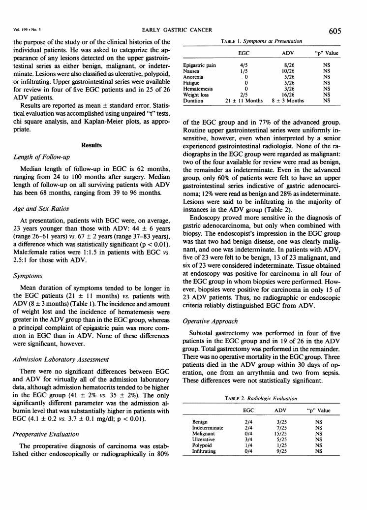
FIG. 1. Photomicrograph of earlygastric cancer confined to the mu-cosa (X250).
Tumor Location and Size
More than 70% of both EGC and ADV tumors wereantral in location. The EGC group had a predominanceof lesser curvature lesions, whereas ADV cancers wereevenly distributed between the lesser and greater curva-ture. EGC tumors tended to be smaller (1.7 ± 0.6 vs. 5.8± 0.7 cm in greatest diameter) although the differencewas not significant. Three patients among the ADV grouphad microscopic evidence of tumor at the margin of re-section; all developed recurrence by 4 months.
HistologyAll tumors were adenocarcinomas. An example ofEGC
is shown in Figure 1, demonstrating foci of adenocarci-noma in areas of atrophic gastritis. Three patients withEGC had tumor confined to the mucosa while, in two,tumor extended into the submucosa. In the ADV group,
TABLE 3. Correlation ofPositive Regional Lymph Nodeswith Depth of Gastric Wall Invasion
EGC ADV
Number %(+) Number %(+)Invasion of Nodes of Nodes
Mucosa 3 33%Submucosa 2 50%Muscularis propria 8 38%Serosa 6 83%Perigastric tissues 12 100%
tumor extension to the muscularis propria was noted ineight, to the serosa in six, and into the perigastric tissuein 12.
Lymph Node Metastases
Metastases to regional lymph nodes were found in twoof five patients with EGC and in 20 of 26 ADV, a dif-ference which was not significant statistically. Positivenodes were found at all levels oftumor invasion, althoughthe percentage of patients harboring lymph node metas-tases increased with increasing depth of invasion (Ta-ble 3).
Adjuvant Therapy
Thirty-eight per cent of the ADV group received post-operative chemotherapy and 15% received radiation ther-apy. In general, these treatments were begun after tumorrecurrence and did not appear to improve survival. Onepatient in the EGC group received a partial course ofchemotherapy; none received radiation therapy.
Recurrence
Twenty of 26 ADV patients (73%) developed eitherlocal or systemic recurrence of disease at a mean intervalof 17 ± 6 months after surgery. In contrast, only one
patient in the EGC group developed recurrence
(p < 0.01). This proved to be a suture line recurrence,found 2 years following the initial operation. The patient
606 Ann. Surg. * May 1984
EARLY GASTRIC CANCER
was successfully treated by subsequent near-total gas-
trectomy and is alive without detectable disease 3 years
following this procedure. In contrast, no patient who de-veloped recurrence in ADV is alive (p < 0.01 vs. EGC),mean survival in this group being 5 ± 9 months followingdetection of recurrent tumor.
Survival
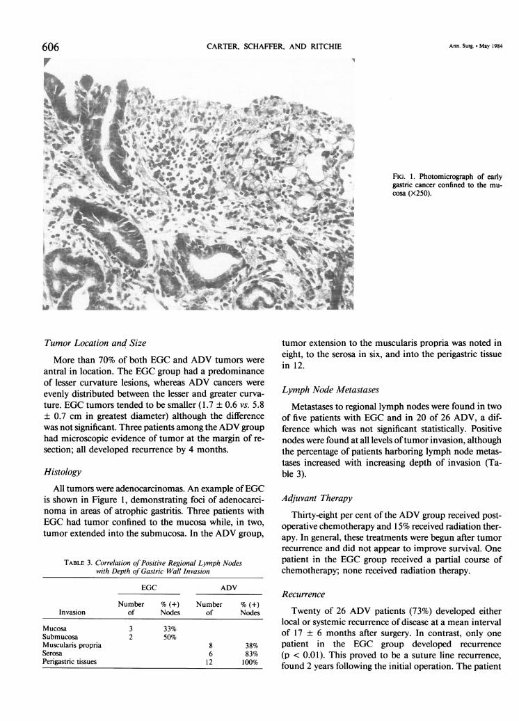
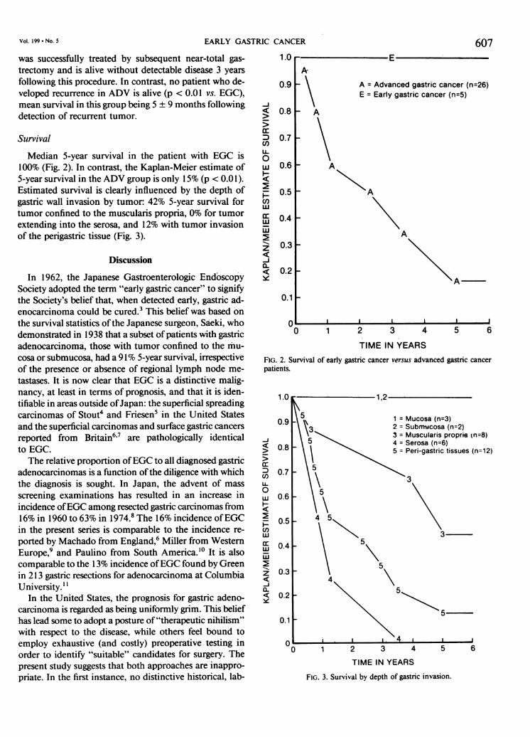
Median 5-year survival in the patient with EGC is100% (Fig. 2). In contrast, the Kaplan-Meier estimate of5-year survival in the ADV group is only 15% (p < 0.01).Estimated survival is clearly influenced by the depth ofgastric wall invasion by tumor: 42% 5-year survival fortumor confined to the muscularis propria, 0% for tumorextending into the serosa, and 12% with tumor invasionof the perigastric tissue (Fig. 3).
Discussion
In 1962, the Japanese Gastroenterologic EndoscopySociety adopted the term "early gastric cancer" to signifythe Society's belief that, when detected early, gastric ad-enocarcinoma could be cured.3 This belief was based on
the survival statistics ofthe Japanese surgeon, Saeki, whodemonstrated in 1938 that a subset of patients with gastricadenocarcinoma, those with tumor confined to the mu-
cosa or submucosa, had a 91% 5-year survival, irrespectiveof the presence or absence of regional lymph node me-
tastases. It is now clear that EGC is a distinctive malig-nancy, at least in terms of prognosis, and that it is iden-tifiable in areas outside ofJapan: the superficial spreadingcarcinomas of Stout4 and Friesen5 in the United Statesand the superficial carcinomas and surface gastric cancersreported from Britain',' are pathologically identicalto EGC.The relative proportion ofEGC to all diagnosed gastric
adenocarcinomas is a function ofthe diligence with whichthe diagnosis is sought. In Japan, the advent of mass
screening examinations has resulted in an increase inincidence ofEGC among resected gastric carcinomas from16% in 1960 to 63% in 1974.8 The 16% incidence ofEGCin the present series is comparable to the incidence re-
ported by Machado from England,6 Miller from WesternEurope,9 and Paulino from South America.'0 It is alsocomparable to the 13% incidence ofEGC found by Greenin 213 gastric resections for adenocarcinoma at ColumbiaUniversity. "
In the United States, the prognosis for gastric adeno-carcinoma is regarded as being uniformly grim. This beliefhas lead some to adopt a posture of "therapeutic nihilism"with respect to the disease, while others feel bound toemploy exhaustive (and costly) preoperative testing inorder to identify "suitable" candidates for surgery. Thepresent study suggests that both approaches are inappro-priate. In the first instance, no distinctive historical, lab-
1.0
0.9 _
-J
-j
ccnC/)
LL0
w
-
U.
cc
z
6iQ-
A = Advanced gastric cancer (n=26)E = Early gastric cancer (n=5)
0.8 _
0.7
0.6 _
0.5 _
0.4 _
0.3 _A
A
0.2 _
0.1
I I 1- I II I
0 1 2
TIME
3 4
IN YEARS
5 6
FIG. 2. Survival of early gastric cancer versus advanced gastric cancer
patients.
1.0 1,2
09 1 Mucosa'(n=3)
\3 2= Submrcosa (n=2)
3 = Muscularis proprie (n=8)-j0.
5 4 =Serosa (n=6)
> 08 5 Penr-gastric tissues (n=12)cc 5C: 0.7c/) 3
0 5
Z 0.6
0.54
)
w3ax: 0.4w
0.3
<~~~~a.
< 0.2
TIME IN YEARS
FIG. 3. Survival by depth of gastric invasion.
Vol. 199 * No. 5 607

608 CARTER, SCHAFFER, AND RITCHIE Ann. Surg. May 1984
oratory, radiographic, or endoscopic features reliably dis-tinguished the patients with early gastric cancer from thosewith more advanced disease. At presentation, only twosignificant differences were identified between thesegroups: age and admission albumin levels. Neither is spe-cific. Furthermore, larger series report very similar agedistribution between EGC and ADV; Johansen'2 noteda mean of 66 years in a series of 70 patients with EGCwhile Miller,' in a review of658 patients with EGC, foundthe disease most common in the sixth and seventh decadesof life. In addition, symptoms of patients with EGC arevague, often consisting only of epigastric fullness or in-digestion. The present study is in agreement with otherreports that indicate that epigastric pain is, indeed, themost common presenting symptom in EGC, whereas sig-nificant weight loss, hematemesis, or melena suggest (butdo not prove) the presence of more advanced carci-noma. 11,13
The diagnosis ofgastric adenocarcinoma is most readilymade by endoscopy when seven to ten biopsies are rou-tinely obtained.'4 However, endoscopic biopsies cannotdistinguish early from more advanced gastric cancer.Routine upper gastrointestinal series, at least as performedin the United States, is also relatively insensitive, althoughthe air-contrast techniques used in the mass screeningexaminations in Japan are reportedly more accurate.'5In the present series, neither the location, the appear-ance, nor the size of the lesions reliably distinguish EGCfrom ADV.
Adequate surgical resection is the mainstay oftherapy,with subtotal gastrectomy the treatment of choice forEGC of the antrum and total gastrectomy reserved forlesions high in the fundus or for patients with recurrentdisease.'3 Adjuvant chemotherapy or radiation therapydo not appear to improve survival of patients with EGC,even in those with nodal metastases.'6 Therefore, neithermodality can be recommended. Recurrence rates of 3%have been noted by others in EGC with a mean disease-free interval of 3 years.'6"7 Higher local recurrence rates(9%) have been reported for patients in whom the di-agnosis of adenocarcinoma was not suspected before sur-gery.'8 As a rule, recurrence tends to be local and can betreated successfully by repeat resection.
That "therapeutic nihilism" is also inappropriate issuggested by the superb survival statistics noted in thepresent (admittedly small) series, as well as those reportedfrom Japan by Kishimoto (97% 5-year survival for in-tramucosal cancers, 96% for submucosal cancers).'9 InWestern European Countries and in the United States,survival rates in EGC are somewhat lower: 87% in Ger-many,20 71% in Britain,'8 and 68% in New York." Unlikeother solid tumors of the gastrointestinal tract, depth ofgastric wall invasion and not metastases to regional nodes
appears to be the primary determinate of prognosis inboth EGC and ADV.
In summary, early gastric cancer behaves similarly inthe United States and Japan; for example, prognosis isexcellent even in the presence of regional lymph nodemetastases. Inability to distinguish early from advancedgastnc cancer before surgery justifies an aggressive surgicalapproach to patients with resectable gastric neoplasms.
Acknowledgment
The authors gratefully acknowledge the technical assistance of Ms.Catherine Norman.
References
1. Qizilbash AH, Stevenson GW. Early gastric cancer. Pathol Annu1979; 1:317-351.
2. Johansen AA. Early gastric cancer. In Morson BC, ed. Pathologyof the Gastrointestinal Tract. Current Topics in Pathology. NewYork: Springer-Verlag, 1976; 1-47.
3. Murakami T. Pathomorphological diagnosis. Definition and grossclassification. In Murakami T, ed. Early Gastric Cancer. GannMonograph 11. Tokyo: University of Tokyo Press, 1971; 53-55.
4. Stout AP. Superficial spreading type of carcinoma of the stomach.Arch Surg 1942; 44:651-657.
5. Friesen G, Dogherty MB, Remine WH. Superficial carcinoma ofthe stomach. Surgery 1961; 51:300-312.
6. Machado G, Davis JD, Tudway A, et al. Superficial carcinoma ofthe stomach. Br Med J 1976; 2:77-79.
7. Mason M. Surface carcinoma of the stomach. Gut 1965; 6:185-193.
8. Yamagata S, Hisamichi S. Epidemiology of cancer of the stomach.World J Surg 1979; 3:663-669.
9. Miller G, Kaufman M. Das magenfruhkarzinon in Europa. DtschMed Wochenschr 1975; 100:1946-1949.
10. Paulino F, Roselli A. Early gastric cancer: report of twenty-fivecases. Surgery 1979; 85:172-176.
11. Green PH, O'Toole KM, Weinberg LM, Goldfarb JP. Early gastriccancer. Gastroenterology 1981; 81:247-256.
12. Johansen AA. Early gastric cancer: a contribution to the pathologyand to gastric cancer histogenesis. Copenhagen: Poul Petri 1981;123.
13. Murakami T. Early cancer of the stomach. World J Surg 1979;3:685-692.
14. Graham D, Schwartz J, Cain G, Gyorky F. Prospective evaluationof biopsy number in the diagnosis of esophageal and gastriccarcinoma. Gastroenterology 1982; 82:228-231.
15. Maruyama M. Diagnostic limits for early gastric cancer by radiog-raphy. In Murakami T, ed. Early Gastric Cancer. Gann Mono-graph 11. Tokyo: University of Tokyo Press, 1971.
16. Lwanga T, Furukawa H, Kosaki G. Relapse of early gastric cancerand its prevention. J Clin Surg (Rinsho Geka) 1979; 31:29.
17. Yamada E. Surgical results for early gastric carcinoma. Int Surg1975; 60:139-142.
18. Fielding J, Ellis D, Jones B. Natural history of "early" gastric cancer:results of a 10 year regional survey. Br Med J 1980; 281:965-967.
19. Kishimoto H, Fukii T, Adachi H, Koga N. Resection line and long-term results in early cancer of the stomach. J Clin Surg (RinshoGeka) 1976; 31:541-545.
20. Gentsch HH, Groitl H, Giedl J. Results of surgical treatment ofearly gastric cancer in 113 patients. World J Surg 1981; 5:103-107.