the endoscopic diagnosis of early gastric cancer
DESCRIPTION
Endoscopic diagnosis of early gastric cancerTRANSCRIPT
© 2012 Hellenic Society of Gastroenterology www.annalsgastro.gr
The endoscopic diagnosis of early gastric cancer
Kenshi YaoFukuoka University Chikushi Hospital, Japan
Annals of Gastroenterology (2012) 25, 1-12
Department of Endoscopy, Fukuoka University Chikushi Hospital, Chikushino-city, Japan
Conflict of Interest: None
Correspondence to: Kenshi Yao, MD, PhD, Clinical Professor and Head, Department of Endoscopy, Fukuoka University Chikushi Hospital, 1-1-1 Zokumyoin Chikushino-city Fukuoka 818-8502 Japan, Tel.: +81 92 921 1011, e-mail: [email protected]
Received: 2 May 2012; accepted 2 July 2012
INVITED REVIEW
Abstract The aim of this article is to demonstrate the basic principles for the endoscopic diagnosis of early gastric cancer. The diagnostic process can be divided into two steps, detection and char-acterization. Detection requires good endoscopic technique, and thorough knowledge. With regard to technique, we should administer the optimum preparation to patients, including an antiperistaltic agent. Furthermore, in order to map the entire stomach we need to follow a standardized protocol, and we propose a systematic screening protocol for the stomach. With regard to knowledge, we should be able to identify high-risk background mucosa, and we should be aware of the indicators of a suspicious lesion. Chromoendoscopy and magnifying endoscopy are promising image-enhanced endoscopic techniques for characterization. The proposed criteria for a cancerous lesion are as follows: conventional endoscopic findings of 1) a well-demarcated lesion and 2) irregularity in color/surface pattern; vessel plus surface classification using magnifying endoscopy with narrow-band imaging findings of 1) irregular microvascular pattern with a demarcation line or 2) irregular microsurface pattern with a demarcation line. Conventional endoscopy and subsequent image-enhanced endoscopy can both contribute to the detection of early gastric cancer.
Keywords early gastric cancer, endoscopy, image-enhanced endoscopy, magnifying endoscopy with narrow-band imaging, systematic screening of the stomach
Ann Gastroenterol 2012; 25 (4): 1-12
Introduction
Outside Japan, endoscopic diagnosis of early gastric cancer is indeed difficult because it often shows only subtle changes on endoscopic examination, and endoscopists have few opportunities to familiarize themselves with systematic techniques and current knowledge. The endoscopic diagnosis of early gastric cancer is divided into two steps, detection and characterization. In this article, I will explain first how to detect suspicious lesions, and second how to characterize detected lesions to make an accurate diagnosis.
Basic principles for detecting early gastric cancer using conventional endoscopy
In order to detect lesions suspicious for early gastric cancer, we should familiarize ourselves with the basic principles of 1) technique and 2) knowledge.
Technique
Ideal preparation
The right preparation for an endoscopic examination is mandatory for minimizing time and effort removing mucus and froth from the mucosal surface during the procedure. Thirty minutes before the procedure, patients are asked to drink a mixture of water with mucolytic and defoaming agents. The formula in Japan is 100 mL of water with 20000 U pronase (Kaken Pharmaceutical, Tokyo, Japan), 1 g of sodium bicarbonate, and 10 mL of dimethylpolysiloxane (20 mg/mL, Horii Pharmaceutical Ind., Osaka Japan). Pronase, however, is not available for use in all countries. An alternative mixture comprises 100 mL of water mixed with 2 mL of acetylcysteine (200 mg/mL Parvolex, Celltech, UK; or Mucomyst, Bristol-Myers Squibb, USA), and 0.5 mL (40 mg/mL) activated dimethicone (Infacol, Forest Laboratories, UK).

2 K. Yao
Annals of Gastroenterology 25
Avoiding blind spots: systematic screening protocol for the stomach (SSS)
During the endoscopy, in order to avoid blind spots we should employ a standardized procedure to map the entire stomach. A basic technique for avoiding blind spots involves the following procedures: 1) extending the gastric wall by air insufflation, 2) rinsing mucus and the froth from the gastric mucosa through irrigation with water and a defoaming agent, and 3) mapping the entire stomach.
Let us consider why such steps are needed. 1) If we do not extend the gastric wall, we may fail to detect a cancer on the greater curvature, even a rather advanced cancer as shown in Figure 1, 2) If we do not rinse out mucus and froth, we may
Use of an antiperistaltic agent
In the physiological state, the gastric wall is always moving due to peristalsis. In order to detect subtle mucosal changes, we need to carefully scan the entire mucosal surface. For this purpose, peristalsis makes it difficult for the endoscopist to obtain static observations. Accordingly, we usually administer an anticholinergic agent such as 10 to 20 mg of scopolamine butylbromide (Buscopan) intramuscularly or intravenously just before inserting the endoscope, even for upper gastrointestinal (GI) screening endoscopies. If there are contraindications to the use of anticholinergic agents (e.g. cardiovascular disease), 1 mg of glucagon can be administered to inhibit peristalsis.
Figure 1 Endoscopic findings of advanced gastric cancer on the greater curvature of the lower gastric body. (A) With insufflation of a small amount of air, the appearance is normal. (B) However, when we insufflate more air and extend the gastric wall together with the greater curvature with gastric foils, a distinct lesion, suggestive of advanced gastric cancer, becomes evident. In fact, the histopathological diagnosis of the biopsied specimen was invasive signet-ring cell carcinoma
A Β
Figure 2 Proposed systematic screening protocol for the stomach (SSS). The SSS should be initiated as soon as we insert the scope into the gastric antrum. With the antegrade view, we should take endoscopic photos of 4 quadrants of the gastric antrum, body and middle-upper body. Then, with the retroflex view, we take endoscopic photos of 4 quadrants of the gastric fundus and cardia, and 3 quadrants of the gastric middle-upper body and incisura. Overall, the SSS series comprises 22 endoscopic photos Q, quadrant; L, lesser curvature; A, anterior wall; G, greater curvature; P, posterior wall; SSS, systematic screening protocol for the somach
The endoscopic diagnosis of early gastric cancer 3
Annals of Gastroenterology 25
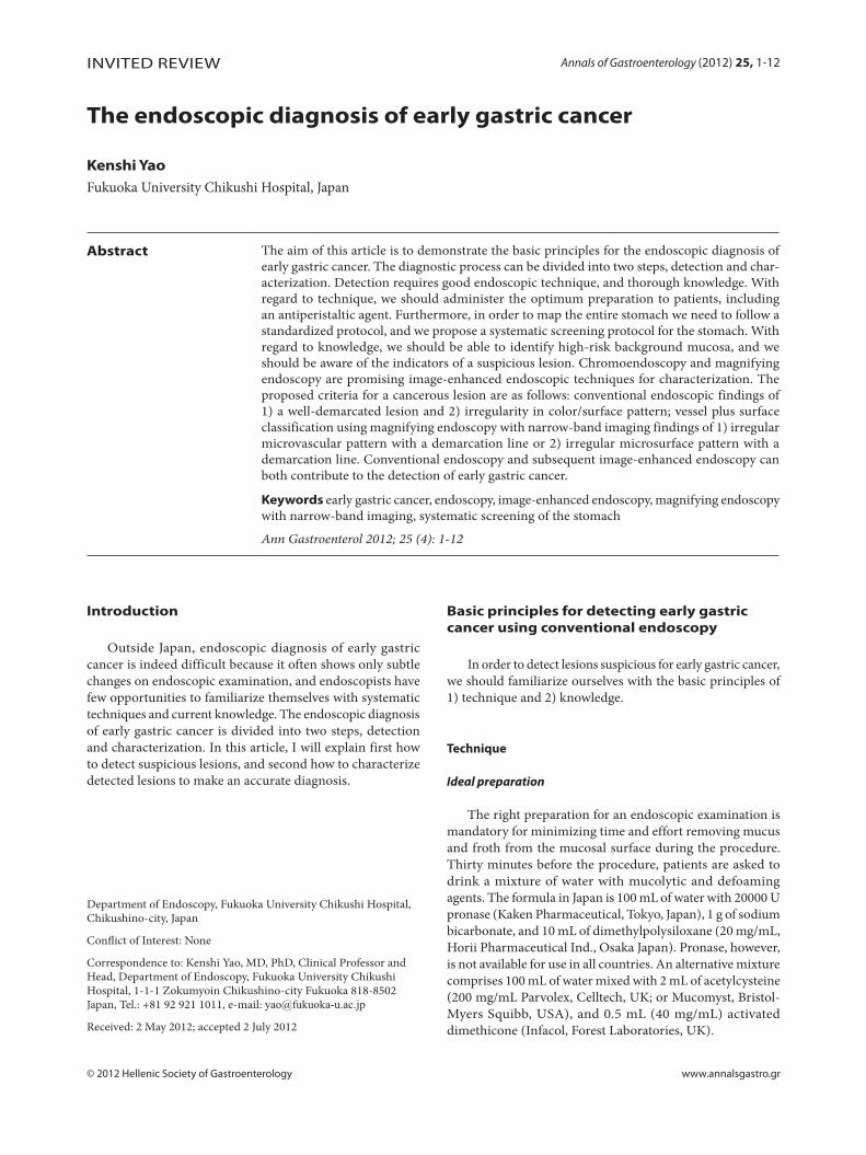
Figure 3 Magnified endoscopic findings of gastric fundic gland mucosa. Observation by magnified endoscopy focusing on gastric fundic gland mucosa (fundus and body) is indeed useful for estimating the risk of gastric cancer. (A, B) Normal gastric body mucosa. The microvascular pattern shows regular honeycomb-like subepithelial capillary network (SECN) pattern (brownish color) with regular arrangement of collect-ing venules (RAC) (cyan color). The microsurface pattern shows regular oval crypt-opening (CO) (brownish color) and oval marginal crypt epithelium (MCE) (whitish semitransparent part). (C) Helicobacter pylori-associated gastritis. When the gastric mucosa is accompanied with Helicobacter pylori-associated gastritis, both the microvascular and microsurface patterns show remarkable diffuse amendments compared with normal mucosa. Namely, the SECN pattern is dilated and the CV is not visualized in the inflamed mucosa, while MCE has a curved/oval shape which is different from normal morphology and CO is not visualized as a brownish pit. (D) Atrophic gastritis. When there is marked atrophy in the gastric body mucosa. The CV can be visualized again, however no honeycomb-like SECN pattern can be identified and neither MCE nor CO can be identified in remarkably atrophic mucosa. This type of appearance is also identified in autoimmune gastritis as well as common atrophic gastritis associated with Helicobacter pylori infectionSECN, subepithelial capillary network; RAC, regular arrangement of collecting venules; CD, crypt-opening; MCE, marginal crypt epithelium; CV, collecting volume
well miss the subtle findings that enable detection of early gastric cancers. 3) There is no standardized method of mapping the entire stomach that is accepted worldwide. In Japan, we developed a systematic screening protocol for the upper GI tract [1]. However, no well-designed studies have been completed. Other problems are that the number of pictures seems to be too big for overseas endoscopists to remember all the sites, and the protocol is too complicated to follow in clinical practice. Recently, the Japanese Society of Gastroenterological Cancer Screening (JSGCS) published a simplified protocol [2], nevertheless, it is still difficult to remember. On the other hand, the protocol proposed by the European Society of Gastrointestinal Endoscopy (ESGE) [3], includes only 4 pictures of the stomach. Endoscopists with longer procedure times, who take more than 4 pictures, detect more pathology than endoscopists with shorter procedure times and fewer pictures [4]. This suggests that longer examination times, and taking more pictures, may improve the rate of lesion detection [4]. From this point of view, we would like to propose a minimum required standard, a “systematic screening protocol for the stomach (SSS)”, as shown in Figure 2. In the SSS, pictures are arranged according to the order of the procedure, and we are able to take pictures of 4 or 3 quadrant views in either a clockwise or counter-clockwise manner. The SSS is a basic
concept showing the minimum required standard. If you find lesions, additional pictures can be taken. If your endoscopy system does not connect to an image-filing database system, the SSS should be used as check points.
Knowledge
Determining the risk for development of early gastric cancer from the endoscopic findings of the background gastric mu-cosa
As soon as we insert the scope into the stomach, we should determine using endoscopic inspection alone whether risk factors for gastric cancer are present in the background mucosa, such as Helicobacter pylori-associated gastritis, gastric atrophy or intestinal metaplasia [5-10]. If the appearance of the gastric mucosa is normal, with none of the abovementioned risk factors, lesions suspicious for gastric cancer are less likely. Magnified endoscopic observation, if available, is useful for determining whether the gastric mucosa is accompanied by such risk factors (Fig. 3, 4) [6, 7].
A
C
B
D
4 K. Yao
Annals of Gastroenterology 25
Basic principles for characterization of detected lesions
Characterization using conventional white light imaging (C-WLI) or chromoendoscopy (CE)
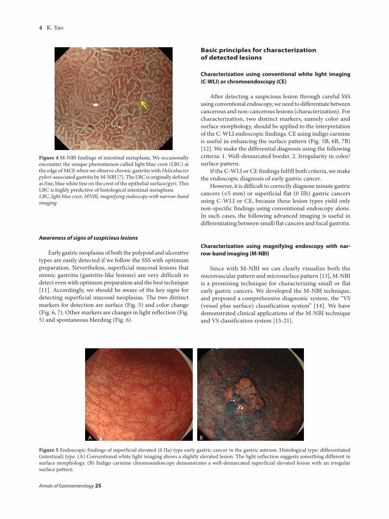
After detecting a suspicious lesion through careful SSS using conventional endoscopy, we need to differentiate between cancerous and non-cancerous lesions (characterization). For characterization, two distinct markers, namely color and surface morphology, should be applied to the interpretation of the C-WLI endoscopic findings. CE using indigo carmine is useful in enhancing the surface pattern (Fig. 5B, 6B, 7B) [12]. We make the differential diagnosis using the following criteria: 1. Well-demarcated border. 2. Irregularity in color/surface pattern.
If the C-WLI or CE findings fulfill both criteria, we make the endoscopic diagnosis of early gastric cancer.
However, it is difficult to correctly diagnose minute gastric cancers (≤5 mm) or superficial flat (0 IIb) gastric cancers using C-WLI or CE, because these lesion types yield only non-specific findings using conventional endoscopy alone. In such cases, the following advanced imaging is useful in differentiating between small/flat cancers and focal gastritis.
Characterization using magnifying endoscopy with nar-row-band imaging (M-NBI)
Since with M-NBI we can clearly visualize both the microvascular pattern and microsurface pattern [13], M-NBI is a promising technique for characterizing small or flat early gastric cancers. We developed the M-NBI technique, and proposed a comprehensive diagnostic system, the “VS (vessel plus surface) classification system” [14]. We have demonstrated clinical applications of the M-NBI technique and VS classification system [15-21].
Figure 5 Endoscopic findings of superficial elevated (0 IIa) type early gastric cancer in the gastric antrum. Histological type: differentiated (intestinal) type. (A) Conventional white light imaging shows a slightly elevated lesion. The light reflection suggests something different in surface morphology. (B) Indigo carmine chromoendoscopy demonstrates a well-demarcated superficial elevated lesion with an irregular surface pattern
A Β
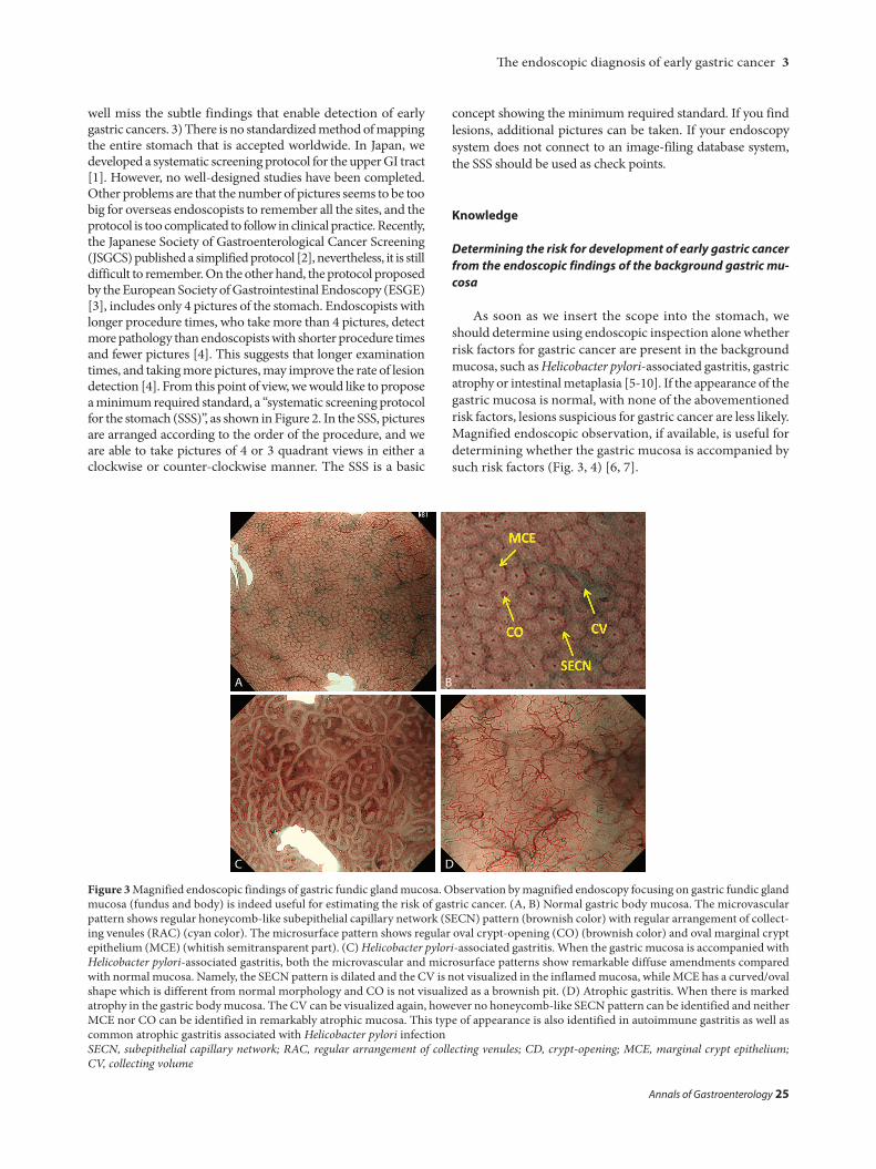
Figure 4 M-NBI findings of intestinal metaplasia. We occasionally encounter the unique phenomenon called light blue crest (LBC) at the edge of MCE when we observe chronic gastritis with Helicobacter pylori-associated gastritis by M-NBI [7]. The LBC is originally defined as fine, blue white line on the crest of the epithelial surface/gyri. This LBC is highly predictive of histological intestinal metaplasiaLBC, light blue crest; MNBI, magnifying endoscopy with narrow-band imaging
Awareness of signs of suspicious lesions
Early gastric neoplasias of both the polypoid and ulcerative types are easily detected if we follow the SSS with optimum preparation. Nevertheless, superficial mucosal lesions that mimic gastritis (gastritis-like lesions) are very difficult to detect even with optimum preparation and the best technique [11]. Accordingly, we should be aware of the key signs for detecting superficial mucosal neoplasias. The two distinct markers for detection are surface (Fig. 5) and color change (Fig. 6, 7). Other markers are changes in light reflection (Fig. 5) and spontaneous bleeding (Fig. 6).

The endoscopic diagnosis of early gastric cancer 5
Annals of Gastroenterology 25
Figure 7 Endoscopic findings of superficial depressed (0 IIc) type early gastric cancer in the gastric body. Histological type: undifferentiated (diffuse) type. (A) Conventional white light imaging demonstrates a pale depressed lesion. (B) Indigo carmine chromoendoscopy delineates a well-demarcated lesion with an irregular margin
Figure 8 A soft black hood is mounted on the tip of the magnifying endoscope
A
Figure 6 Endoscopic findings of superficial depressed (0 IIc) type early gastric cancer in the gastric cardia. Histological type: differentiated (intestinal) type. (A) Conventional white light imaging depicts a reddened depressed lesion with spontaneous bleeding. (B) Indigo carmine chromoendoscopy demonstrates a well-demarcated lesion with an irregular margin
A Β
Β
M-NBI technique using a soft black hood attachment
A soft black hood (MAJ-1988 for GIF-Q240Z, MAJ-1989 for GIF-H260Z and GIF-Q160Z; Olympus, Tokyo) is essential for optimal magnifying endoscopy. Prior to the examination, the hood is mounted on the tip of the endoscope to enable the endoscopist to consistently fix the mucosa at a distance
of approximately 2 mm, at which maximal magnification of the endoscopic image can be obtained (Fig. 8). Since the depth of the hood is very shallow, it does not disturb the visual field during non-magnifying observation. In addition, since the hood is soft, it does not injure the mucosa. In fact, when upper GI magnification endoscopy was performed in more than 600 screening endoscopy cases using this kind of soft black hood, there were no complications such as contact bleeding, nor any untoward incidents such as hood dislocation [16].
VS classification system for differential diagnosis between cancerous and noncancerous lesions
Microanatomies visualized in the stomach using M-NBI [22]
Since the gastric mucosa is composed of glandular epithelium, we should clarify the correlation between the microanatomy and actual images as visualized using M-NBI (Fig. 9). The basic microanatomical findings visualized using M-NBI are classified under the microvascular and microsurface pattern.
6 K. Yao
Annals of Gastroenterology 25
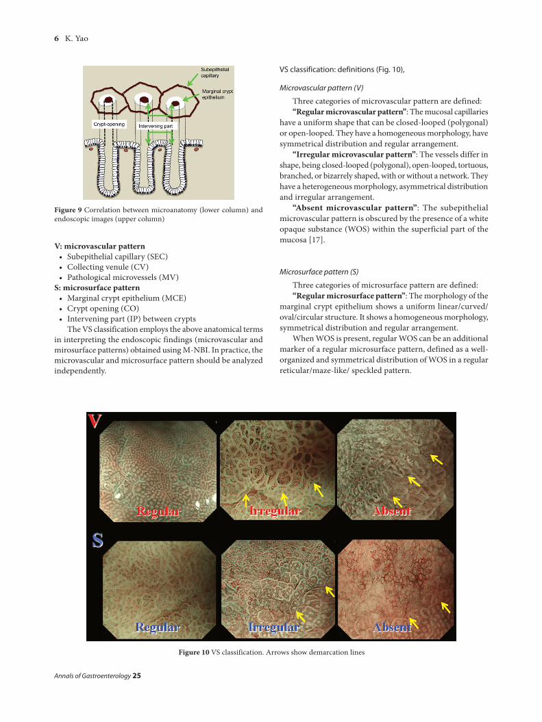
V: microvascular pattern• Subepithelial capillary (SEC)• Collecting venule (CV)• Pathological microvessels (MV)
S: microsurface pattern• Marginal crypt epithelium (MCE)• Crypt opening (CO)• Intervening part (IP) between crypts
The VS classification employs the above anatomical terms in interpreting the endoscopic findings (microvascular and mirosurface patterns) obtained using M-NBI. In practice, the microvascular and microsurface pattern should be analyzed independently.
Figure 9 Correlation between microanatomy (lower column) and endoscopic images (upper column)
VS classification: definitions (Fig. 10),
Microvascular pattern (V)
Three categories of microvascular pattern are defined:“Regular microvascular pattern”: The mucosal capillaries
have a uniform shape that can be closed-looped (polygonal) or open-looped. They have a homogeneous morphology, have symmetrical distribution and regular arrangement.
“Irregular microvascular pattern”: The vessels differ in shape, being closed-looped (polygonal), open-looped, tortuous, branched, or bizarrely shaped, with or without a network. They have a heterogeneous morphology, asymmetrical distribution and irregular arrangement.
“Absent microvascular pattern”: The subepithelial microvascular pattern is obscured by the presence of a white opaque substance (WOS) within the superficial part of the mucosa [17].
Microsurface pattern (S)
Three categories of microsurface pattern are defined:“Regular microsurface pattern”: The morphology of the
marginal crypt epithelium shows a uniform linear/curved/oval/circular structure. It shows a homogeneous morphology, symmetrical distribution and regular arrangement.
When WOS is present, regular WOS can be an additional marker of a regular microsurface pattern, defined as a well-organized and symmetrical distribution of WOS in a regular reticular/maze-like/ speckled pattern.
Figure 10 VS classification. Arrows show demarcation lines
The endoscopic diagnosis of early gastric cancer 7
Annals of Gastroenterology 25
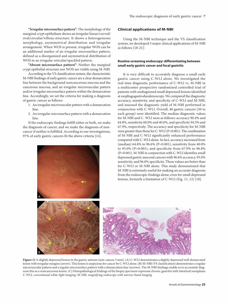
Figure 11 A slightly depressed lesion in the gastric antrum (non-cancer, 5 mm). (A) C-WLI demonstrates a slightly depressed well-demarcated lesion with irregular margins (arrow). This lesion is suspicious for cancer by C-WLI alone. (B) M-NBI (VS classification) demonstrates a regular microvascular pattern and a regular microsurface pattern with a demarcation line (arrows). The M-NBI findings enable us to accurately diag-nose this as a noncancerous lesion. (C) Histopathological findings of the biopsy specimen represent chronic gastritis with intestinal metaplasiaC-WLI, conventional white-light imaging; M-NBI, magnifying endoscopy with narrow-band imaging
“Irregular microsurface pattern”: The morphology of the marginal crypt epithelium shows an irregular linear/curved/ oval/circular/villous structure. It shows a heterogeneous morphology, asymmetrical distribution and irregular arrangement. When WOS is present, irregular WOS can be an additional marker of an irregular microsurface pattern, defined as a disorganized and asymmetrical distribution of WOS in an irregular reticular/speckled pattern.
“Absent microsurface pattern”: Neither the marginal crypt epithelial structure nor WOS are visible using M-NBI.
According to the VS classification system, the characteristic M-NBI findings of early gastric cancer are a clear demarcation line between the background noncancerous mucosa and the cancerous mucosa, and an irregular microvascular pattern and/or irregular microsurface pattern within the demarcation line. Accordingly, we set the criteria for making a diagnosis of gastric cancer as follows:
1. An irregular microvascular pattern with a demarcation line.
2. An irregular microsurface pattern with a demarcation line.
If the endoscopic findings fulfill either or both, we make the diagnosis of cancer, and we make the diagnosis of non-cancer if neither is fulfilled. According to our investigations, 97% of early gastric cancers fit the above criteria [14].
Clinical applications of M-NBI
Using the M-NBI technique and the VS classification system, we developed 3 major clinical applications of M-NBI as follows [18-21]:
Routine screening endoscopy: differentiating between small early gastric cancer and focal gastritis
It is very difficult to accurately diagnose a small early gastric cancer using C-WLI alone. We investigated the real-time diagnostic performance of C-WLI vs. M-NBI in a multicenter prospective randomized controlled trial of patients with undiagnosed small depressed lesions identified at esophagogastroduodenoscopy. We compared the diagnostic accuracy, sensitivity, and specificity of C-WLI and M-NBI, and assessed the diagnostic yield of M-NBI performed in conjunction with C-WLI. Overall, 40 gastric cancers (20 in each group) were identified. The median diagnostic values for M-NBI and C- WLI were as follows: accuracy 90.4% and 64.8%, sensitivity 60.0% and 40.0%, and specificity 94.3% and 67.9%, respectively. The accuracy and specificity for M-NBI were greater than those for C-WLI (P<0.001). The combination of M-NBI and C-WLI significantly enhanced performance compared with C-WLI alone. In fact, accuracy increased from (median) 64.8% to 96.6% (P=0.001), sensitivity from 40.0% to 95.0% (P=0.001), and specificity from 67.9% to 96.8% (P=0.001). M-NBI in conjunction with C-WLI identifies small depressed gastric mucosal cancers with 96.6% accuracy, 95.0% sensitivity, and 96.8% specificity. These values are better than for C-WLI or M-NBI alone. This study demonstrated that M-NBI is extremely useful for making an accurate diagnosis from the endoscopic findings alone, even for small depressed lesions, formerly a limitation of C-WLI (Fig. 11, 12) [18].
A
B C
8 K. Yao
Annals of Gastroenterology 25
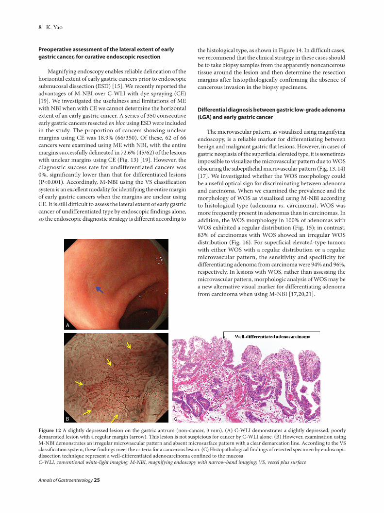
Figure 12 A slightly depressed lesion on the gastric antrum (non-cancer, 3 mm). (A) C-WLI demonstrates a slightly depressed, poorly demarcated lesion with a regular margin (arrow). This lesion is not suspicious for cancer by C-WLI alone. (B) However, examination using M-NBI demonstrates an irregular microvascular pattern and absent microsurface pattern with a clear demarcation line. According to the VS classification system, these findings meet the criteria for a cancerous lesion. (C) Histopathological findings of resected specimen by endoscopic dissection technique represent a well-differentiated adenocarcinoma confined to the mucosaC-WLI, conventional white-light imaging; M-NBI, magnifying endoscopy with narrow-band imaging; VS, vessel plus surface
A
B C
Preoperative assessment of the lateral extent of early gastric cancer, for curative endoscopic resection
Magnifying endoscopy enables reliable delineation of the horizontal extent of early gastric cancers prior to endoscopic submucosal dissection (ESD) [15]. We recently reported the advantages of M-NBI over C-WLI with dye spraying (CE) [19]. We investigated the usefulness and limitations of ME with NBI when with CE we cannot determine the horizontal extent of an early gastric cancer. A series of 350 consecutive early gastric cancers resected en bloc using ESD were included in the study. The proportion of cancers showing unclear margins using CE was 18.9% (66/350). Of these, 62 of 66 cancers were examined using ME with NBI, with the entire margins successfully delineated in 72.6% (45/62) of the lesions with unclear margins using CE (Fig. 13) [19]. However, the diagnostic success rate for undifferentiated cancers was 0%, significantly lower than that for differentiated lesions (P<0.001). Accordingly, M-NBI using the VS classification system is an excellent modality for identifying the entire margin of early gastric cancers when the margins are unclear using CE. It is still difficult to assess the lateral extent of early gastric cancer of undifferentiated type by endoscopic findings alone, so the endoscopic diagnostic strategy is different according to
the histological type, as shown in Figure 14. In difficult cases, we recommend that the clinical strategy in these cases should be to take biopsy samples from the apparently noncancerous tissue around the lesion and then determine the resection margins after histopthologically confirming the absence of cancerous invasion in the biopsy specimens.
Differential diagnosis between gastric low-grade adenoma (LGA) and early gastric cancer
The microvascular pattern, as visualized using magnifying endoscopy, is a reliable marker for differentiating between benign and malignant gastric flat lesions. However, in cases of gastric neoplasia of the superficial elevated type, it is sometimes impossible to visualize the microvascular pattern due to WOS obscuring the subepithelial microvascular pattern (Fig. 13, 14) [17]. We investigated whether the WOS morphology could be a useful optical sign for discriminating between adenoma and carcinoma. When we examined the prevalence and the morphology of WOS as visualized using M-NBI according to histological type (adenoma vs. carcinoma), WOS was more frequently present in adenomas than in carcinomas. In addition, the WOS morphology in 100% of adenomas with WOS exhibited a regular distribution (Fig. 15); in contrast, 83% of carcinomas with WOS showed an irregular WOS distribution (Fig. 16). For superficial elevated-type tumors with either WOS with a regular distribution or a regular microvascular pattern, the sensitivity and specificity for differentiating adenoma from carcinoma were 94% and 96%, respectively. In lesions with WOS, rather than assessing the microvascular pattern, morphologic analysis of WOS may be a new alternative visual marker for differentiating adenoma from carcinoma when using M-NBI [17,20,21].
The endoscopic diagnosis of early gastric cancer 9
Annals of Gastroenterology 25
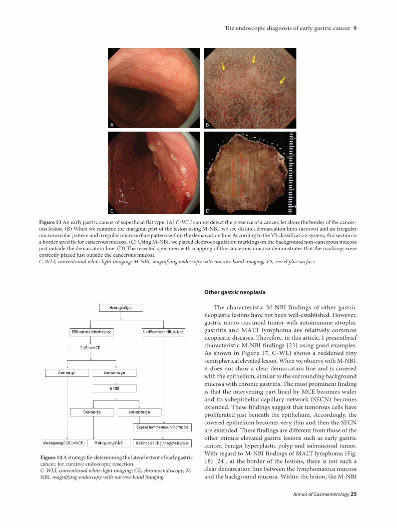
Figure 13 An early gastric cancer of superficial flat type. (A) C-WLI cannot detect the presence of a cancer, let alone the border of the cancer-ous lesion. (B) When we examine the marginal part of the lesion using M-NBI, we see distinct demarcation lines (arrows) and an irregular microvascular pattern and irregular microsurface pattern within the demarcation line. According to the VS classification system, this section is a border specific for cancerous mucosa. (C) Using M-NBI, we placed electrocoagulation markings on the background non-cancerous mucosa just outside the demarcation line. (D) The resected specimen with mapping of the cancerous mucosa demonstrates that the markings were correctly placed just outside the cancerous mucosaC-WLI, conventional white-light imaging; M-NBI, magnifying endoscopy with narrow-band imaging; VS, vessel plus surface
Figure 14 A strategy for determining the lateral extent of early gastric cancer, for curative endoscopic resectionC-WLI, conventional white-light imaging; CE, chromoendoscopy; M-NBI, magnifying endoscopy with narrow-band imaging
A Β
C D
Other gastric neoplasia
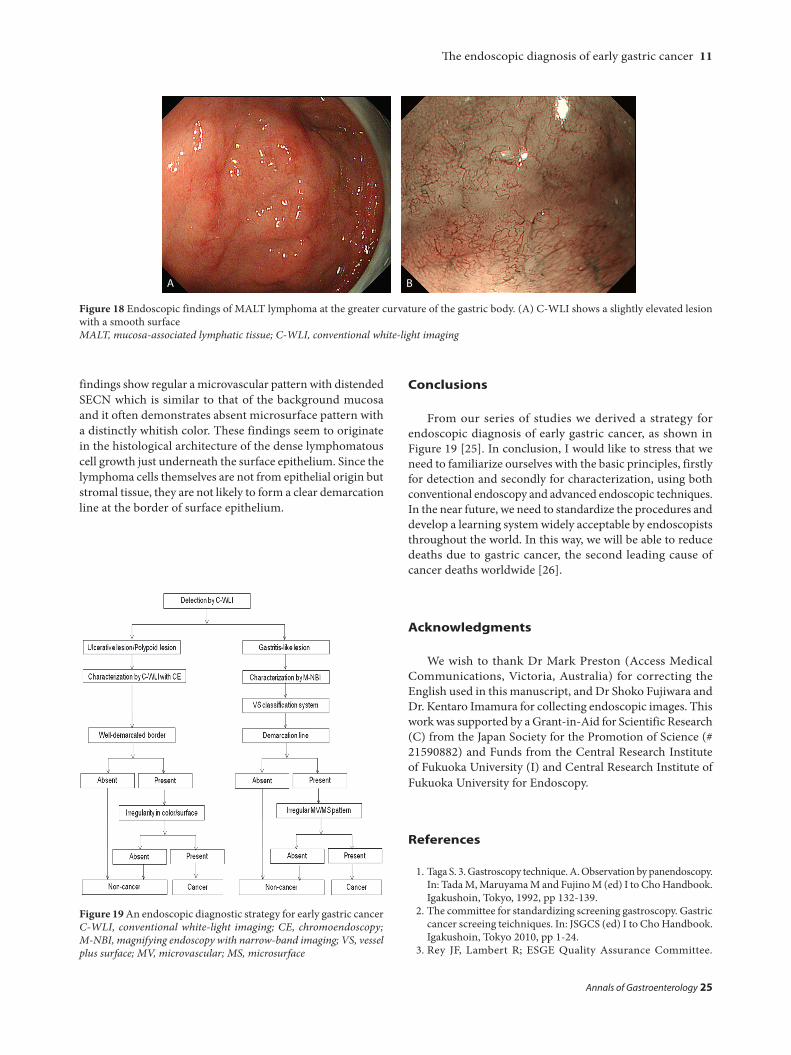
The characteristic M-NBI findings of other gastric neoplastic lesions have not been well-established. However, gastric micro-carcinoid tumor with autoimmune atrophic gastritis and MALT lymphoma are relatively common neoplastic diseases. Therefore, in this article, I presentbrief characteristic M-NBI findings [23] using good examples. As shown in Figure 17, C-WLI shows a reddened tiny semispherical elevated lesion. When we observe with M-NBI, it does not show a clear demarcation line and is covered with the epithelium, similar to the surrounding background mucosa with chronic gastritis. The most prominent finding is that the intervening part lined by MCE becomes wider and its subepithelial capillary network (SECN) becomes extended. These findings suggest that tumorous cells have proliferated just beneath the epithelium. Accordingly, the covered epithelium becomes very thin and then the SECN are extended. These findings are different from those of the other minute elevated gastric lesions such as early gastric cancer, benign hyperplastic polyp and submucosal tumor. With regard to M-NBI findings of MALT lymphoma (Fig. 18) [24], at the border of the lesions, there is not such a clear demarcation line between the lymphomatous mucosa and the background mucosa. Within the lesion, the M-NBI
10 K. Yao
Annals of Gastroenterology 25
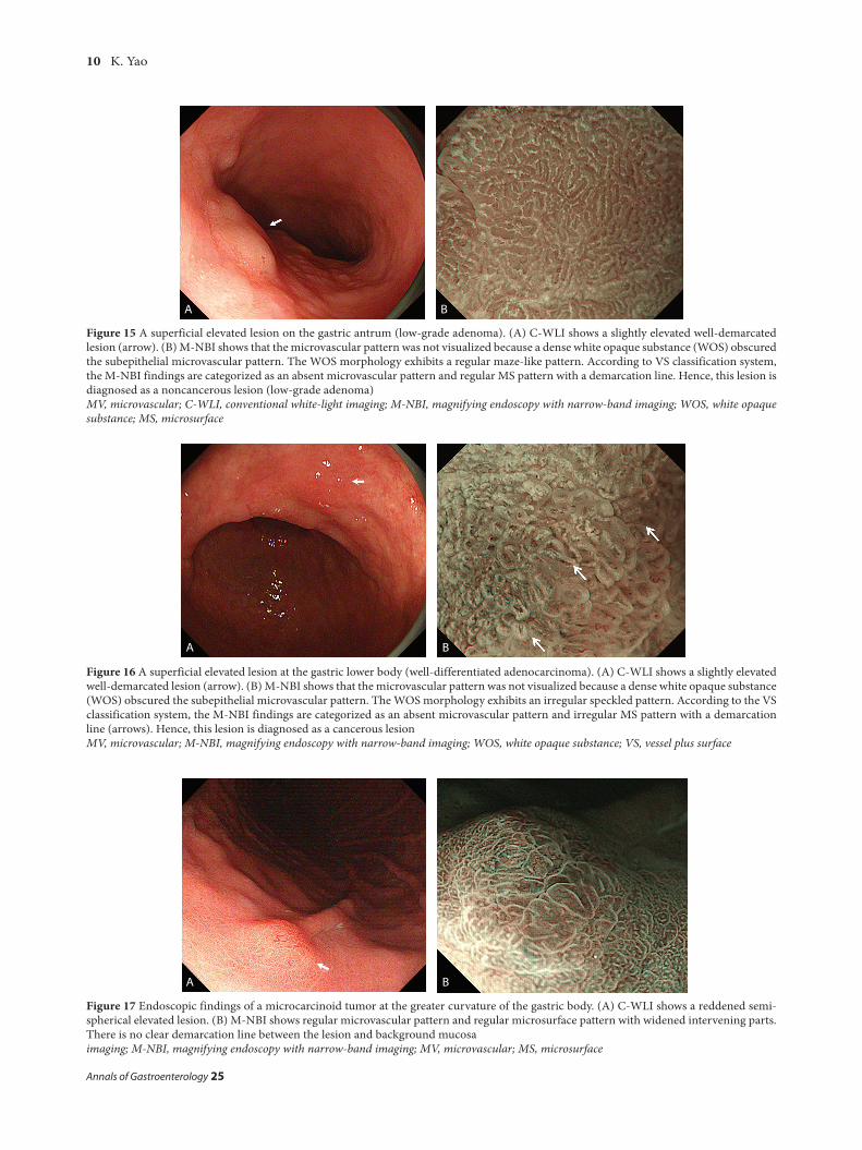
Figure 15 A superficial elevated lesion on the gastric antrum (low-grade adenoma). (A) C-WLI shows a slightly elevated well-demarcated lesion (arrow). (B) M-NBI shows that the microvascular pattern was not visualized because a dense white opaque substance (WOS) obscured the subepithelial microvascular pattern. The WOS morphology exhibits a regular maze-like pattern. According to VS classification system, the M-NBI findings are categorized as an absent microvascular pattern and regular MS pattern with a demarcation line. Hence, this lesion is diagnosed as a noncancerous lesion (low-grade adenoma)MV, microvascular; C-WLI, conventional white-light imaging; M-NBI, magnifying endoscopy with narrow-band imaging; WOS, white opaque substance; MS, microsurface
A Β
Figure 16 A superficial elevated lesion at the gastric lower body (well-differentiated adenocarcinoma). (A) C-WLI shows a slightly elevated well-demarcated lesion (arrow). (B) M-NBI shows that the microvascular pattern was not visualized because a dense white opaque substance (WOS) obscured the subepithelial microvascular pattern. The WOS morphology exhibits an irregular speckled pattern. According to the VS classification system, the M-NBI findings are categorized as an absent microvascular pattern and irregular MS pattern with a demarcation line (arrows). Hence, this lesion is diagnosed as a cancerous lesionMV, microvascular; M-NBI, magnifying endoscopy with narrow-band imaging; WOS, white opaque substance; VS, vessel plus surface
A Β
Figure 17 Endoscopic findings of a microcarcinoid tumor at the greater curvature of the gastric body. (A) C-WLI shows a reddened semi-spherical elevated lesion. (B) M-NBI shows regular microvascular pattern and regular microsurface pattern with widened intervening parts. There is no clear demarcation line between the lesion and background mucosaimaging; M-NBI, magnifying endoscopy with narrow-band imaging; MV, microvascular; MS, microsurface
A Β
The endoscopic diagnosis of early gastric cancer 11
Annals of Gastroenterology 25
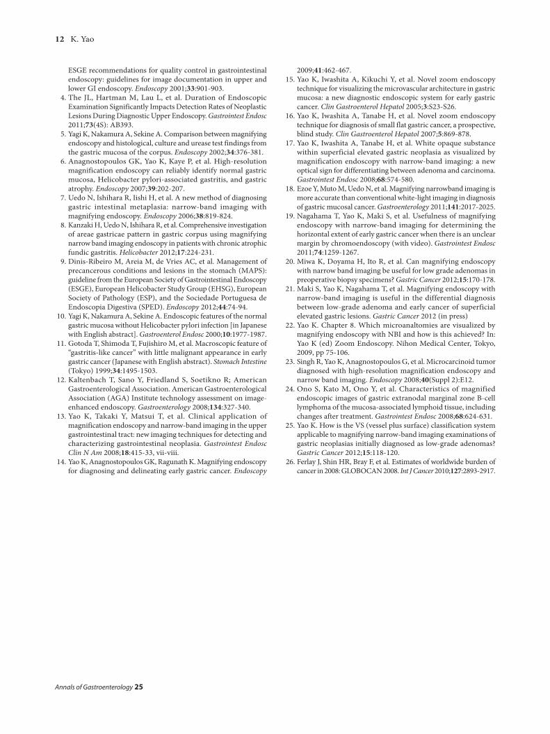
Figure 19 An endoscopic diagnostic strategy for early gastric cancerC-WLI, conventional white-light imaging; CE, chromoendoscopy; M-NBI, magnifying endoscopy with narrow-band imaging; VS, vessel plus surface; MV, microvascular; MS, microsurface
Figure 18 Endoscopic findings of MALT lymphoma at the greater curvature of the gastric body. (A) C-WLI shows a slightly elevated lesion with a smooth surfaceMALT, mucosa-associated lymphatic tissue; C-WLI, conventional white-light imaging
A Β
Conclusions
From our series of studies we derived a strategy for endoscopic diagnosis of early gastric cancer, as shown in Figure 19 [25]. In conclusion, I would like to stress that we need to familiarize ourselves with the basic principles, firstly for detection and secondly for characterization, using both conventional endoscopy and advanced endoscopic techniques. In the near future, we need to standardize the procedures and develop a learning system widely acceptable by endoscopists throughout the world. In this way, we will be able to reduce deaths due to gastric cancer, the second leading cause of cancer deaths worldwide [26].
Acknowledgments
We wish to thank Dr Mark Preston (Access Medical Communications, Victoria, Australia) for correcting the English used in this manuscript, and Dr Shoko Fujiwara and Dr. Kentaro Imamura for collecting endoscopic images. This work was supported by a Grant-in-Aid for Scientific Research (C) from the Japan Society for the Promotion of Science (# 21590882) and Funds from the Central Research Institute of Fukuoka University (I) and Central Research Institute of Fukuoka University for Endoscopy.
References
1. Taga S. 3. Gastroscopy technique. A. Observation by panendoscopy. In: Tada M, Maruyama M and Fujino M (ed) I to Cho Handbook. Igakushoin, Tokyo, 1992, pp 132-139.
2. The committee for standardizing screening gastroscopy. Gastric cancer screeing teichniques. In: JSGCS (ed) I to Cho Handbook. Igakushoin, Tokyo 2010, pp 1-24.
3. Rey JF, Lambert R; ESGE Quality Assurance Committee.
findings show regular a microvascular pattern with distended SECN which is similar to that of the background mucosa and it often demonstrates absent microsurface pattern with a distinctly whitish color. These findings seem to originate in the histological architecture of the dense lymphomatous cell growth just underneath the surface epithelium. Since the lymphoma cells themselves are not from epithelial origin but stromal tissue, they are not likely to form a clear demarcation line at the border of surface epithelium.
12 K. Yao
Annals of Gastroenterology 25
ESGE recommendations for quality control in gastrointestinal endoscopy: guidelines for image documentation in upper and lower GI endoscopy. Endoscopy 2001;33:901-903.
4. The JL, Hartman M, Lau L, et al. Duration of Endoscopic Examination Significantly Impacts Detection Rates of Neoplastic Lesions During Diagnostic Upper Endoscopy. Gastrointest Endosc 2011;73(4S): AB393.
5. Yagi K, Nakamura A, Sekine A. Comparison between magnifying endoscopy and histological, culture and urease test findings from the gastric mucosa of the corpus. Endoscopy 2002;34:376-381.
6. Anagnostopoulos GK, Yao K, Kaye P, et al. High-resolution magnification endoscopy can reliably identify normal gastric mucosa, Helicobacter pylori-associated gastritis, and gastric atrophy. Endoscopy 2007;39:202-207.
7. Uedo N, Ishihara R, Iishi H, et al. A new method of diagnosing gastric intestinal metaplasia: narrow-band imaging with magnifying endoscopy. Endoscopy 2006;38:819-824.
8. Kanzaki H, Uedo N, Ishihara R, et al. Comprehensive investigation of areae gastricae pattern in gastric corpus using magnifying narrow band imaging endoscopy in patients with chronic atrophic fundic gastritis. Helicobacter 2012;17:224-231.
9. Dinis-Ribeiro M, Areia M, de Vries AC, et al. Management of precancerous conditions and lesions in the stomach (MAPS): guideline from the European Society of Gastrointestinal Endoscopy (ESGE), European Helicobacter Study Group (EHSG), European Society of Pathology (ESP), and the Sociedade Portuguesa de Endoscopia Digestiva (SPED). Endoscopy 2012;44:74-94.
10. Yagi K, Nakamura A, Sekine A. Endoscopic features of the normal gastric mucosa without Helicobacter pylori infection [in Japanese with English abstract]. Gastroenterol Endosc 2000;10:1977-1987.
11. Gotoda T, Shimoda T, Fujishiro M, et al. Macroscopic feature of “gastritis-like cancer” with little malignant appearance in early gastric cancer (Japanese with English abstract). Stomach Intestine (Tokyo) 1999;34:1495-1503.
12. Kaltenbach T, Sano Y, Friedland S, Soetikno R; American Gastroenterological Association. American Gastroenterological Association (AGA) Institute technology assessment on image-enhanced endoscopy. Gastroenterology 2008;134:327-340.
13. Yao K, Takaki Y, Matsui T, et al. Clinical application of magnification endoscopy and narrow-band imaging in the upper gastrointestinal tract: new imaging techniques for detecting and characterizing gastrointestinal neoplasia. Gastrointest Endosc Clin N Am 2008;18:415-33, vii-viii.
14. Yao K, Anagnostopoulos GK, Ragunath K. Magnifying endoscopy for diagnosing and delineating early gastric cancer. Endoscopy
2009;41:462-467.15. Yao K, Iwashita A, Kikuchi Y, et al. Novel zoom endoscopy
technique for visualizing the microvascular architecture in gastric mucosa: a new diagnostic endoscopic system for early gastric cancer. Clin Gastroenterol Hepatol 2005;3:S23-S26.
16. Yao K, Iwashita A, Tanabe H, et al. Novel zoom endoscopy technique for diagnosis of small flat gastric cancer, a prospective, blind study. Clin Gastroenterol Hepatol 2007;5:869-878.
17. Yao K, Iwashita A, Tanabe H, et al. White opaque substance within superficial elevated gastric neoplasia as visualized by magnification endoscopy with narrow-band imaging: a new optical sign for differentiating between adenoma and carcinoma. Gastrointest Endosc 2008;68:574-580.
18. Ezoe Y, Muto M, Uedo N, et al. Magnifying narrowband imaging is more accurate than conventional white-light imaging in diagnosis of gastric mucosal cancer. Gastroenterology 2011;141:2017-2025.
19. Nagahama T, Yao K, Maki S, et al. Usefulness of magnifying endoscopy with narrow-band imaging for determining the horizontal extent of early gastric cancer when there is an unclear margin by chromoendoscopy (with video). Gastrointest Endosc 2011;74:1259-1267.
20. Miwa K, Doyama H, Ito R, et al. Can magnifying endoscopy with narrow band imaging be useful for low grade adenomas in preoperative biopsy specimens? Gastric Cancer 2012;15:170-178.
21. Maki S, Yao K, Nagahama T, et al. Magnifying endoscopy with narrow-band imaging is useful in the differential diagnosis between low-grade adenoma and early cancer of superficial elevated gastric lesions. Gastric Cancer 2012 (in press)
22. Yao K. Chapter 8. Which microanaltomies are visualized by magnifying endoscopy with NBI and how is this achieved? In: Yao K (ed) Zoom Endoscopy. Nihon Medical Center, Tokyo, 2009, pp 75-106.
23. Singh R, Yao K, Anagnostopoulos G, et al. Microcarcinoid tumor diagnosed with high-resolution magnification endoscopy and narrow band imaging. Endoscopy 2008;40(Suppl 2):E12.
24. Ono S, Kato M, Ono Y, et al. Characteristics of magnified endoscopic images of gastric extranodal marginal zone B-cell lymphoma of the mucosa-associated lymphoid tissue, including changes after treatment. Gastrointest Endosc 2008;68:624-631.
25. Yao K. How is the VS (vessel plus surface) classification system applicable to magnifying narrow-band imaging examinations of gastric neoplasias initially diagnosed as low-grade adenomas? Gastric Cancer 2012;15:118-120.
26. Ferlay J, Shin HR, Bray F, et al. Estimates of worldwide burden of cancer in 2008: GLOBOCAN 2008. Int J Cancer 2010;127:2893-2917.