molecular classification of gastric cancer: a new...
TRANSCRIPT
1
Molecular Classification of Gastric Cancer: A new paradigm
Authors: Manish A. Shah1, Raya Khanin2, Laura Tang3, Yelena Y. Janjigian1, David S.
Klimstra3, Hans Gerdes4, David P. Kelsen1
1Department of Medicine, Gastrointestinal Oncology Service, 2Bioinformatics, 3Department of
Pathology, 4Department of Medicine, Gastrointestinal Service
Supported by ASCO Career Development Award (Manish A. Shah, MD), NCI/CTEP (NCI# 5917), and NCI Contract for Early Drug Development (N01 CM 62206, David P. Kelsen, MD)
Corresponding Author:
Manish A.Shah, MD Memorial Sloan Kettering Cancer Center 1275 York Avenue Howard 910 New York NY 10065 Running Heading: Molecular Classification of Gastric Cancer Key Words: Gastric Cancer, cDNA Expression, classification, pathways Individual Author Awards: Manish A Shah: ASCO Career Development Award, FDA Orphan Disease Product Grant 1R01FD003755-01A1 David P Kelsen: NCI Contract N01 CM62206
Research. on November 19, 2018. © 2011 American Association for Cancerclincancerres.aacrjournals.org Downloaded from
Author manuscripts have been peer reviewed and accepted for publication but have not yet been edited. Author Manuscript Published OnlineFirst on March 23, 2011; DOI: 10.1158/1078-0432.CCR-10-2203
2
Translational Significance for the Future of Medicine
Considering gastric cancer as one disease has limited the rational development of new
prevention and therapeutic strategies. Herein, we examine cDNA expression data from gastric
adenocarcinoma samples from 36 individual primary tumors to examine a new classification of
gastric cancer. There is sufficient epidemiologic, histopathologic, anatomic and now molecular
evidence to categorize gastric cancer into three diseases – proximal non-diffuse, diffuse, and
distal non-diffuse gastric cancer. Differentially expressed genes distinguish each subtype of
gastric cancer from each other and from adjacent normal gastric mucosa. Our genomic
classifier tightly groups the defined gastric cancer subtypes into specific individual clusters with
high discrimination, suggesting that >85% of samples were classified correctly. In addition,
gene set analysis identifies several differentially regulated pathways between individual gastric
cancer subtypes. The ramifications of this classification are significant, including improving our
understanding of unique molecular drivers of each gastric cancer type, aiding in the
identification of novel biomarkers and targets of each disease, and ultimately helping to develop
new treatment paradigms for each gastric cancer type.
Research. on November 19, 2018. © 2011 American Association for Cancerclincancerres.aacrjournals.org Downloaded from
Author manuscripts have been peer reviewed and accepted for publication but have not yet been edited. Author Manuscript Published OnlineFirst on March 23, 2011; DOI: 10.1158/1078-0432.CCR-10-2203
3
Abstract
Purpose: Gastric cancer may be subdivided into three distinct subtypes –proximal, diffuse, and
distal gastric cancer– based on histopathologic and anatomic criteria. Each subtype is
associated with unique epidemiology. Our aim is to test the hypothesis that these distinct gastric
cancer subtypes may also be distinguished by gene expression analysis.
Experimental Design: Patients with localized gastric adenocarcinoma being screened for a
phase II preoperative clinical trial(NCI 5917) underwent endoscopic biopsy for fresh tumor
procurement. 4-6 targeted biopsies of the primary tumor were obtained. Macrodissection was
performed to ensure >80% carcinoma in the sample. HG-U133A GeneChip(Affymetrix) was
used for cDNA expression analysis, and all arrays were processed and analyzed using the
Bioconductor R-package.
Results: Between November 2003 and January 2006, 57 patients were screened to identify 36
patients with localized gastric cancer who had adequate RNA for expression analysis. Using
supervised analysis, we built a classifier to distinguish the three gastric cancer subtypes,
successfully classifying each into tightly grouped clusters. Leave-one-out cross validation error
was 0.14, suggesting that >85% of samples were classified correctly. Gene set analysis with
the False Discovery Rate set at 0.25 identified several pathways that were differentially
regulated when comparing each gastric cancer subtype to adjacent normal stomach.
Conclusions: Subtypes of gastric cancer that have epidemiologic and histologic distinction are
also distinguished by gene expression data. These preliminary data suggest a new classification
of gastric cancer with implications for improving our understanding of disease biology and
identification of unique molecular drivers for each gastric cancer subtype.
Research. on November 19, 2018. © 2011 American Association for Cancerclincancerres.aacrjournals.org Downloaded from
Author manuscripts have been peer reviewed and accepted for publication but have not yet been edited. Author Manuscript Published OnlineFirst on March 23, 2011; DOI: 10.1158/1078-0432.CCR-10-2203

4
Introduction
Gastric cancer is the second most common cause of cancer-related mortality worldwide
with 700,349 deaths annually, and is the third most common malignancy worldwide with
974,000 new cases in the year 2000(1). Gastric cancer has been considered a single
heterogenous disease with several epidemiologic and histopathologic characteristics; for the
purposes of medical management, gastric cancer is treated in a uniform fashion, without regard
to subtype. Pathologically, gastric adenocarcinoma may be distinguished according to the
Lauren’s classification as intestinal, diffuse, or mixed subtypes(2). Epidemiologically, intestinal
gastric cancer, particularly of the antrum, is strongly associated with chronic inflammation (i.e.
atrophic gastritis(3-4)) often as a consequence of chronic infection with H. pylori(5-6).
Conversely, inflammation is characteristically absent in the development of Lauren’s diffuse type
gastric cancer, particularly when as a result of a germline mutation in CDH1(7). Anatomically,
proximal gastric cancer may be classified as a third type of gastric cancer, as tumors of the
gastric cardia/gastroesophageal junction (GEJ), for which inflammation of a different type (i.e.
chronic gastric acid/bile reflux) may be the driving force for carcinogenesis(8-9). Proximal/GEJ
tumors are also usually not diffuse in histology, similar to distal non-diffuse gastric cancer.
As noted above, at present, the histopathologic, anatomic, and epidemiologic
distinctions that subdivide this disease are not taken into account in the clinical management of
the disease, for either initial potentially curative treatment or in palliation of advanced disease.
For patients with metastatic disease, the available cytotoxic agents are applied indiscriminately
to all disease subtypes, and with only modest success (reviewed(10)). In other epithelial
malignancies, such as breast(11-12) and lung adenocarcinoma(13), the identification of specific
molecular phenotypes have had profound implications for treatment strategies and continued
drug development(14-15). We hypothesize that gastric cancer represents at least three entirely
different malignancies arising in the same organ, each with different initiating pathologic
Research. on November 19, 2018. © 2011 American Association for Cancerclincancerres.aacrjournals.org Downloaded from
Author manuscripts have been peer reviewed and accepted for publication but have not yet been edited. Author Manuscript Published OnlineFirst on March 23, 2011; DOI: 10.1158/1078-0432.CCR-10-2203
5
processes, and each possibly having different tumor biology. If this is true, this disease
classification may lead to different treatment paradigms for individual gastric cancer subtypes.
Clinical indicators in support of this hypothesis include the suggestion that proximal
gastric tumors have a worse prognosis, stage for stage, when compared to distal tumors(16),
that Lauren’s diffuse gastric cancers appear to have a different pattern of spread and behaviour
than intestinal gastric adenocarcioma(17), and Her2 overexpression incidence is different
between intestinal and diffuse types of gastric cancer(18). We hypothesize that different tumors
arising from the stomach may be distinguished at the genomic level. The implications of this
new molecular classification would be significant as they would imply the presence of unique
molecular drivers and unique molecular pathways for each gastric cancer subtype that may be
exploited to identify prognostic and predictive biomarkers and to identify unique targets for
therapy. Herein, we present our preliminary evidence as a test set supporting a molecular
classification of gastric cancer into three individual diseases, supported by histopathologic and
epidemiologic characteristics.
Methods
Study Population
From May 2003 to January 2006, we screened patients with gastric or gastroesophageal
adenocarcinoma by endoscopic ultrasound, laparoscopy, CT scan and PET scan for enrolment
in an NCI-sponsored neoadjuvant clinical trial of irinotecan and cisplatin chemotherapy followed
by surgical resection(19). This protocol was reviewed and approved by the Institutional Review
Board of Memorial Sloan-Kettering Cancer Center and by the National Cancer Institute (NCI
#5917, NCT00062374). Written informed consent was obtained from each patient. All patients
without evidence of metastatic disease on CT scanning underwent pre-operative evaluation
including endoscopic evaluation with ultrasound during which an endoscopic biopsy was
performed for the procurement of fresh tumor tissue for RNA extraction and analysis. All
Research. on November 19, 2018. © 2011 American Association for Cancerclincancerres.aacrjournals.org Downloaded from
Author manuscripts have been peer reviewed and accepted for publication but have not yet been edited. Author Manuscript Published OnlineFirst on March 23, 2011; DOI: 10.1158/1078-0432.CCR-10-2203
6
endoscopic tumor biopsies were performed prior to initiation of any treatment for the
malignancy.
Endoscopy and Endoscopic Biopsy
All patients underwent standard video endoscopy using the Olympus gastroscope GIF-
160 (Olympus America, Melville, NY). Targeted biopsies of the gastric mass, ulcer edge or
thickened folds were obtained using the Bard Precisor EXL coated disposable biopsy forceps
(Bard International, Murray Hill, NJ). Four to 6 biopsies were performed for each patient, with
each biopsy usually measuring approximately 2-3 mm in diameter. Upon receiving the biopsy
tissue from the endoscopic biopsy forceps, a small sample was placed immediately into
buffered formalin (for histopathologic evaluation) or saline (for immediate freezing) while still in
the endoscopy suite. The specimens in saline were immediately transported to the Tumor
Procurement Lab where they were individually placed in OCT media and frozen at -80°C. The
time from obtaining the biopsy to OCT was less than 15 minutes. The formalin fixed samples
were submitted to the Pathology Department for routine processing.
Specimen Analysis for RNA Processing
OCT embedded biopsy samples were maintained below -20°C during processing.
Frozen section slides were made from OCT embedded biopsy samples and were then stained
with H&E and reviewed by reference pathologists (LT, DSK). The presence or absence of
invasive adenocarcinoma and the extent of malignant and non-malignant cell involvement of the
sample was recorded. We defined an adequate biopsy specimen as one having a proportion of
at least 80% carcinoma nuclei. Macrodissection was performed in a specimen dissection
chamber maintained at -20°C, using the marked H&E slide as a guide enabling us to remove
OCT and non-malignant tissue from the carcinoma. Following macrodissection, the remaining
biopsy sample that was then primarily gastric adenocarcinoma was sent in liquid nitrogen to the
Genome Core laboratory for RNA extraction and processing. Although samples varied in size
Research. on November 19, 2018. © 2011 American Association for Cancerclincancerres.aacrjournals.org Downloaded from
Author manuscripts have been peer reviewed and accepted for publication but have not yet been edited. Author Manuscript Published OnlineFirst on March 23, 2011; DOI: 10.1158/1078-0432.CCR-10-2203
7
(up to about 5 mm in diameter), most samples were approximately 2 mm in diameter. Samples
that were less than 1 mm were unable to be processed by macrodissection. These minute
samples were submitted for RNA extraction and processing only if they contained 100% cancer
on the H&E reference slides. In cases where individual patients had several biopsy samples
that were suitable for RNA processing, the samples were combined for RNA processing.
Throughout these tissue handling procedures, care was taken to use RNAase free gloves and
laboratory equipment to minimize contamination.
RNA isolation, probe preparation, and microarray hybridization
Total RNA was isolated from tumor specimens using RNeasy columns (Qiagen), and all
samples were treated on the column with RNase-free DNase. The quality of RNA was verified
before labeling by analyzing 20–50 nanograms (ng) of each sample using the RNA 6000
NanoAssay and a Bioanalyzer 2100 (Agilent). Samples with a 28S/18S ribosomal peak ratio of
1.8–2.0 were considered suitable for labeling. For samples meeting this standard, 1.2 - 2
micrograms (mcg) of total RNA (depending on availability) was used for cDNA synthesis using
an oligo-dT-T7 primer and the SuperScript Double-Stranded cDNA Synthesis Kit (Invitrogen).
Synthesis, linear amplification, and labeling of cRNA were accomplished by in vitro transcription
using the Message Amp aRNA Kit (Ambion) and biotinylated nucleotides (Enzo Diagnostics).
Ten mcg of labeled and fragmented cRNA were then hybridized to the Human Genome U133A
GeneChip (Affymetrix), which contained 22,215 oligonucleotide-based probe sets, at 45°C for
16 hours. Post hybridization staining, washing were processed according to manufacturer
(Affymetrix) guidelines. Finally, chips were scanned with a high-numerical Aperture and flying
objective (FOL) lens in the GS3000 scanner (Affymetrix). The image was quantified using MAS
5.1 (MicroArray Suite, Affymetrix) with the default parameters for the statistical algorithm and all
probe set scaling with a target intensity of 500.
Definition of Gastric Cancer Subtypes
Research. on November 19, 2018. © 2011 American Association for Cancerclincancerres.aacrjournals.org Downloaded from
Author manuscripts have been peer reviewed and accepted for publication but have not yet been edited. Author Manuscript Published OnlineFirst on March 23, 2011; DOI: 10.1158/1078-0432.CCR-10-2203
8
Three individual types of gastric cancer are strongly suggested by clinical and epidemiologic
data(20). Using these characteristics, we defined the subtypes of gastric cancer
histopathologically as follows:
(1) Proximal non-diffuse gastric cancer – The bulk of the tumor (>80%) is located in the
gastric cardia which may extend up to the gastroesophageal junction and small portion
of the distal esophagus. On histopathology, there is evidence of precursor glandular
dysplasia or in situ carcinoma in the setting of chronic inflammation usually without
atrophy. Tumor differentiation may range from well to poorly differentiated, but the
pattern of tumor infiltration should not be entirely diffuse.
(2) Diffuse Gastric Cancer – Tumor location may be anywhere in the stomach. On
histopathology, there is no apparent gastritis, neither severe chronic nor atrophic. The
pattern of infiltration is entirely diffuse without excessive extracellular mucin pools
(colloid carcinoma is not included). There should not be any component of gland-forming
intestinal-type carcinoma. The tumor is poorly differentiated signet ring cell-type either
with or without intracellular mucin.
(3) Distal non-diffuse Gastric Cancer – The bulk of the tumor is usually located in the distal
stomach and may extend up to the mid body of the stomach or down to the pylorus. On
histopathology, there is evidence of chronic gastritis with intestinal metaplasia and a
spectrum of glandular dysplasia and in situ carcinoma. The dominant pattern is a
moderately differentiated and intestinal type carcinoma without or with minor
components of poorly differentiated or de-differentiated carcinoma.
Patients were assigned to an individual subtype of gastric cancer based on the histopathologic
and anatomic definitions above and the expression arrays were analyzed.
Bioinformatics
The affymetrix (HG-133A/B) dataset that contains 38 gastric tumor samples and 31
normal samples taken from stomach tissues adjacent to cancerous tissues was downloaded
Research. on November 19, 2018. © 2011 American Association for Cancerclincancerres.aacrjournals.org Downloaded from
Author manuscripts have been peer reviewed and accepted for publication but have not yet been edited. Author Manuscript Published OnlineFirst on March 23, 2011; DOI: 10.1158/1078-0432.CCR-10-2203
9
from Gene Expression Omnibus (www.ncbi.nlm.nih.gov/geo/ GSE accession number
GSE13911) D’Errico et al(21). The GEO and MSKCC datasets contain 120 arrays that were
processed and analyzed using the Bioconductor suite of tools in R-statistical language
(www.bioconductor.org). All data were normalized using standard GCRMA functions with
default parameters. Normalized data was clustered using hierarchical clustering based on
Euclidian distance (hclust R-function) to verify that there is no batch effect between the two
data-sets (see supplemental figure 1). Indeed, normal and tumor samples were mostly grouped
together and not according to their batch. We next removed the tumor samples from the GEO
dataset as there is no information on their gastric cancer type, resulting in 73 samples for further
analysis (37 normal stomach and 36 gastric cancers). Probes that are present in at least half of
the normal samples or half of the tumor samples were retained for further analysis (23828
probes). For analysis at the gene level, multiple probes corresponding to one gene were
averaged, and probes without a gene symbol annotation were removed leaving 8740 unique
genes. Differential expression analysis was done using limma R-package. Probes/genes were
declared differentially expressed with fold-change cut-off greater than 2, and False Discovery
Rate (FDR)=0.01 (i.e. up to 1% of the significant differences are expected to be false positives).
For subtype classification only tumor samples from MSKCC were considered. To build the
features(=genes) that separate different subtypes of gastric cancer, the dataset was filtered and
genes with fold-changes greater than log2(1.5) in any pair-wise comparison (“proximal non-
diffuse” vs “diffuse”, “proximal non-diffuse” vs “distal non-diffuse, and “diffuse” vs “distal non-
diffuse”) were considered. An additional condition of (unadjusted) p-value<0.005 in any of 3
comparison was also used. The resulting dataset used for the learning classifier has data for
785 genes and 36 tumor samples. Results are similar if probes are selected based on other
criteria. To build the classifier, we opted for a supervised classification algorithm that
implements regularized regression with the optimal scoring algorithm(22-23). This algorithm
includes a procedure for finding gene signatures by ranking genes based on the fitted
Research. on November 19, 2018. © 2011 American Association for Cancerclincancerres.aacrjournals.org Downloaded from
Author manuscripts have been peer reviewed and accepted for publication but have not yet been edited. Author Manuscript Published OnlineFirst on March 23, 2011; DOI: 10.1158/1078-0432.CCR-10-2203
10
regression models. This methodology includes principal components, partial least squares, and
ridge regression models. It has been applied to several microarray studies in cancer(22) and it
is coded in R-package pdmclass. We used ridge regression methodology for the classification.
In addition, we used Gene Set Analysis (GSA)(24) for an exploratory analysis to determine if the
members of a given gene set were concordantly up or down regulated between gastric cancer
subtypes and normal stomach. GSA was run with default parameters and number of
permutations =500 to compute p-values, with FDR=0.25 as suggested in the GSEA manual
(http://www.broadinstitute.org/gsea/doc/GSEAUserGuideFrame.html). The gene set enrichment
analysis was carried out using the Molecular Signature Database (MSigDB) v2.5 released April
7, 2008. Functional analysis of differentially expressed genes was done using the DAVID tool
(http://david.abcc.ncifcrf.gov/home.jsp) using all human genes as a background set.(25)
Results
Patient and Tumor Characteristics
Between November 2003 and January 2006, 57 patients with localized gastric
adenocarcinoma based on CT scan imaging of the chest, abdomen, and pelvis underwent
endoscopic biopsy and tissue biopsy. Of these 57 patients, tumor biopsy samples from 41
patients (72%) were adequate for RNA processing and analysis. Subsequent staging
procedures (i.e. laparoscopy and FDG-PET scan) identified occult metastatic disease in 5
patients, leaving 36 patients with non-metastatic gastric cancer in the final study population (see
table 1). The majority of patients had locally advanced, poorly differentiated tumors, with
approximately 60% of the cases node positive on preoperative evaluation. Proximal non-diffuse
gastric cancers (n=12) were more commonly Lauren’s intestinal histology. Diffuse gastric
cancers (n=10) were more commonly anatomically located in the body or distal stomach, were
uniformly poorly differentiated and Lauren’s diffuse histology by definition. Distal non-diffuse
gastric cancers (n=14) were predominantly intestinal and mixed Lauren’s histology.
Research. on November 19, 2018. © 2011 American Association for Cancerclincancerres.aacrjournals.org Downloaded from
Author manuscripts have been peer reviewed and accepted for publication but have not yet been edited. Author Manuscript Published OnlineFirst on March 23, 2011; DOI: 10.1158/1078-0432.CCR-10-2203
11
Gastric Cancer Subtypes and Normal Stomach
Our aim was to examine whether genomic signatures would significantly differentiate
gastric cancer subtypes that were assigned solely based on anatomic and histopathologic
knowledge. We first compared gastric cancer versus normal stomach. We examined
expression data from two independent data sets (MSKCC data 36 tumor, 10 adjacent normal
stomach and D’Errico data(21) 38 tumor, 31 adjacent normal stomach). (21)Tumor samples
and normals from both datasets cluster primarily according to their malignancy status (normal,
tumor), and not according to the dataset (see supplemental Figure 1). Then, when evaluating
MSKCC gastric tumors (i.e. study population that was annotated according to gastric cancer
subtype) versus normal adjacent stomach, we identified a large number of genes that were
differentially expressed in each type of gastric cancer versus normal (limma analysis, FC cut-
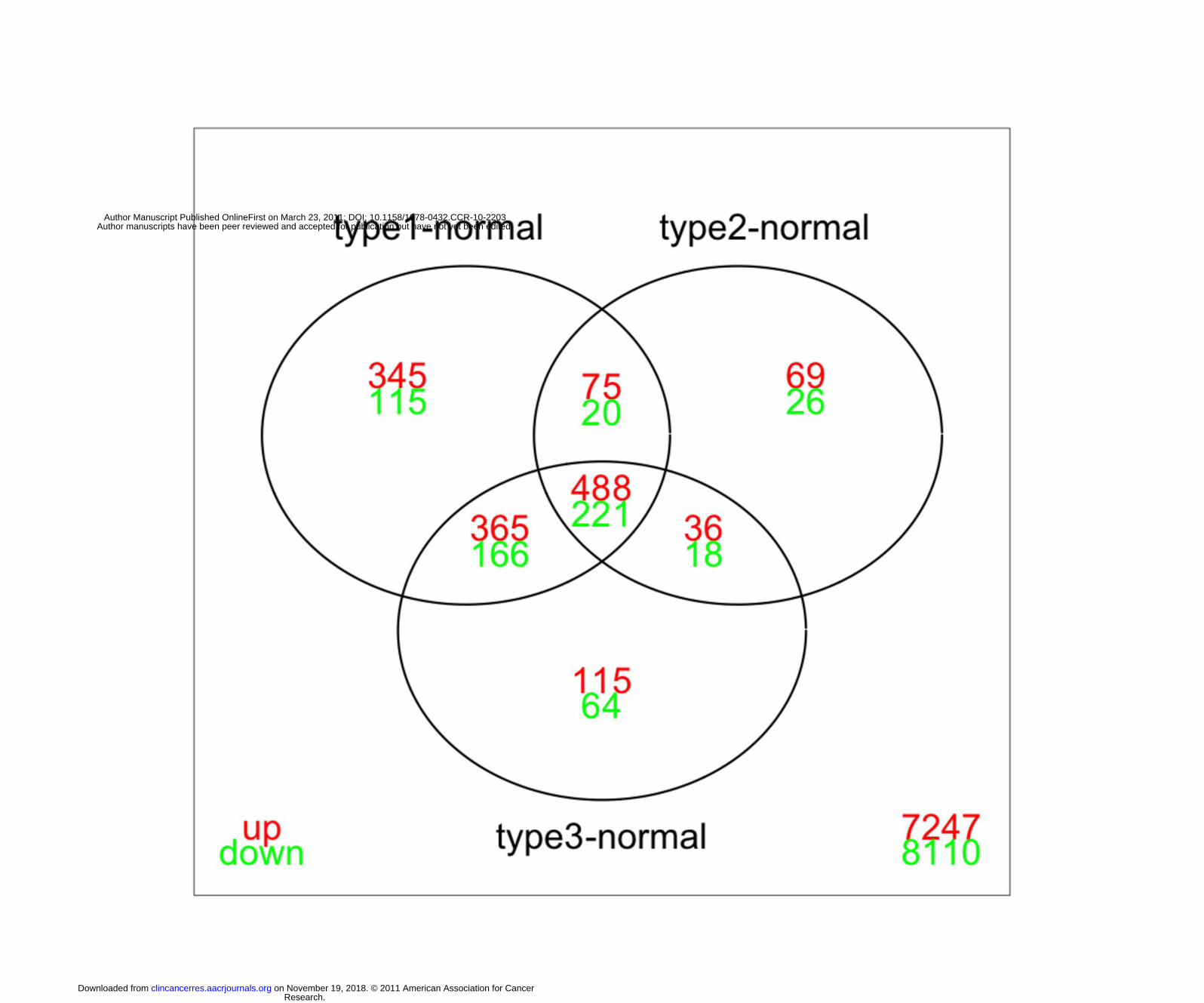
off=2, FDR=0.01; see Figure 2). We noted that while there is a large overlap in the genes
differentially expressed between gastric cancer types and normal, a significant number of genes
uniquely differentiate each subtype of gastric cancer from normal stomach. Direct comparison
of tumor subtypes (full dataset) yielded 3 genes (with FDR=0.05) that are differentially
expressed between proximal non-diffuse and diffuse gastric cancer subtypes, including PSCA
(prostate stem cell antigen) and PGA3 (pepsinogen A) which were both down-regulated >20
fold in proximal non-diffuse gastric cancer when compared with diffuse gastric cancer, and
TRIM32 which was upregulated (>2 fold) in proximal non-diffuse gastric cancer.
We then performed functional categories analysis on groups of differentially expressed
genes using the DAVID tool. Genes that were upregulated in all three gastric cancer types
versus normal are significantly enriched in many typical cancer-related categories including cell
cycle, cell proliferation, cell adhesion, platelet derived growth factor binding, and EGF-domain,
while genes that are downregulated in all gastric cancer types versus normal are enriched in
digestion, disease mutation, and lipid metabolism. Similarly, genes that are up regulated in
proximal and distal non-diffuse gastric cancer (but not diffuse gastric cancer) are enriched in
Research. on November 19, 2018. © 2011 American Association for Cancerclincancerres.aacrjournals.org Downloaded from
Author manuscripts have been peer reviewed and accepted for publication but have not yet been edited. Author Manuscript Published OnlineFirst on March 23, 2011; DOI: 10.1158/1078-0432.CCR-10-2203
12
numerous cell cycle and mitosis related categories, as well as p53 signaling pathways, while
genes downregulated in both non-diffuse gastric cancer subtypes are enriched in digestion,
drug metabolism, and response to various stimuli (nutrient levels, hormone stimulus, organic
substance).
Gene Expression Analysis and Development of Molecular Classification
We next focused on identifying gene signatures that can be used to classify gastric
cancer subtypes. We used ridge regression method on a smaller set of genes (785 genes; see
Methods). (22-23)
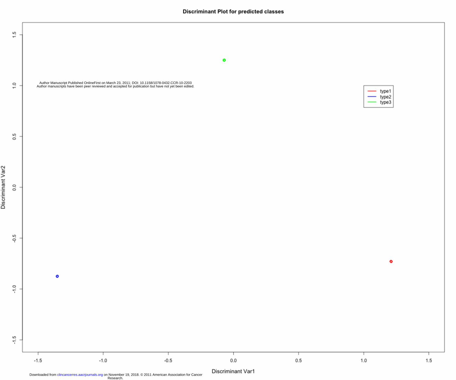
The classifier built on these genes separates the three subtypes quite well and the
samples are tightly grouped into three distinct groups (see Figure 2). The leave-one-out cross
validation error is 0.14 which implies that >85% samples are classified correctly. In our patient
population of 36 patients, 29 tumors were locally advanced (T3 or greater, or N+). When
limiting the analysis to these 29 cases, the leave-one-out cross validation error is 0.13, implying
that still > 85% of locally advanced samples are classified correctly. The top genes that
separate proximal non-diffuse and diffuse gastric cancer include PGA3, PSCA, XIST, SST,
ABCA8 (down regulated in proximal non-diffuse versus diffuse by over 2-fold), and PRF1,
CXCL9, CXCL10, IF144L, PLA2G2A (upregulated). Prostate stem cell antigen (PSCA), for
example, was over 20 fold diminished in proximal non-diffuse relative to diffuse gastric cancer
subtypes. The top genes that separate proximal non-diffuse from distal non-diffuse gastric
cancer include MSLN, IGJ, ENPP4, PLA2G2A (downregulated in proximal non-diffuse vs distal
non-diffuse) and PF4V1, HMBO1, CYP2J2, DSC3, and S100a12 (upregulated). Notably,
PLA2G2A is nearly 7-fold upregulated in proximal non-diffuse versus diffuse gastric cancer
(mean fold-change) and nearly 12 fold upregulated in distal non-diffuse versus diffuse gastric
cancer. The top genes that separate diffuse gastric cancer from distal non-diffuse gastric
cancer include ABCA8 (> 4 fold), HMBOX1, COCH, S100A12, CYP2J2 (up regulated in diffuse
Research. on November 19, 2018. © 2011 American Association for Cancerclincancerres.aacrjournals.org Downloaded from
Author manuscripts have been peer reviewed and accepted for publication but have not yet been edited. Author Manuscript Published OnlineFirst on March 23, 2011; DOI: 10.1158/1078-0432.CCR-10-2203
13
vs. distal non-diffuse), and IFI44L (4 fold), HOXA9, MSLN, and ENPP4 (down regulated in
diffuse vs. distal non-diffuse).
Gene Set Analysis
In addition, we applied the Gene Set Analysis tool to perform exploratory pathway
analysis by comparing each gastric cancer subtype to normal, as well as by performing direct
subtype comparisons. Pathway analysis may be indicative of underlying biological processes
Table 2 provides the list of pathways that were either upregulated or downregulated with
FDR=0.25. We observed that proximal and distal non-diffuse gastric cancers had a number of
upregulated pathways including glycolysis and gluconeogenesis when compared to normal
stomach, whereas no pathways were identified to be upregulated in diffuse gastric tumors.
Conversely, each gastric tumor subtype shows downregulation of the KEGG HSA05222 (small
lung cancer) pathway versus normal stomach. This pathway contains several tumor
suppressors, including p53, PTEN, RB and FHIT.
Discussion
Gastric cancer is a heterogeneous disease with differences in epidemiology and
histopathology that, when coupled with anatomic location, may be distinguished into at least 3
different cancers(20). We explored this hypothesis by examining the gene expression of
individual gastric cancer subtypes in a training set, performing a comparison with the expression
analysis of adjacent normal stomach as well as amongst individual gastric subtypes. We found
that individual diseases arising from the stomach defined a priori, namely “proximal non-diffuse”,
“diffuse”, and “distal non-diffuse” gastric cancer, have distinct gene expression. These findings
from our training set are both clinically consistent and have significance in the context of current
clinical management of gastric cancer. For example, two large global studies evaluating new
cytotoxic and biologic therapies have failed to demonstrate a survival benefit over the standard
Research. on November 19, 2018. © 2011 American Association for Cancerclincancerres.aacrjournals.org Downloaded from
Author manuscripts have been peer reviewed and accepted for publication but have not yet been edited. Author Manuscript Published OnlineFirst on March 23, 2011; DOI: 10.1158/1078-0432.CCR-10-2203
14
of care(26-27). The investigators suggested that this may be partially due to failure to
appreciate biological differences in gastric cancer and how these differences may affect
response to treatment.
When comparing gastric cancer subtypes to adjacent normal, we identified a significant
number of genes that uniquely distinguished individual gastric cancer subtypes from normal
stomach. These data provide support for the hypothesis that gastric cancer subtypes may be
distinguished molecularly.
Then, using supervised classification, we demonstrate a greater than 85% ability to
successfully distinguish gastric cancer subtypes by gene expression. This was the case when
examining all cases (both early and advanced) and also when limiting the cases to advanced
disease. One gene of interest that significantly distinguished proximal non-diffuse gastric
cancer from diffuse gastric cancer, prostate stem cell antigen (PSCA), has already been
implicated in gastric cancer. An intronic polymorphism (rs2294008) in PSCA, resulting in
reduced PSCA expression, is significantly associated with increased risk for diffuse gastric
cancer compared with an intestinal subtype (virtually all distal non-diffuse) in a Japanese
population(28-29). Expression of another gene, PLA2G2A, a secreted phospholipase, has
prognostic significance in gastric cancer. Specifically, tumors expressing high levels of
PLA2GA2 have improved survival compared with patients with low PLA2G2A expressing
tumors(30) Recently, it was demonstrated that PLA2G2A is a target of Wnt/β-catenin signalling
in gastric cancer cells, and is associated with negative regulation of genes associated with
invasion and metastasis(31). Using gene set analysis, a number of pathways were either up- or
downregulated when individual gastric cancer subtypes were compared to normal stomach. For
example, the glycoloysis pathway is upregulated in proximal and distal non-diffuse gastric
cancers relative to diffuse gastric cancer. Glycolysis is the process of converting glucose into
pyruvate and generating small amounts of ATP. It is a central pathway that produces important
precursor metabolites, and may explain the increased glucose metabolism of many cancers(i.e.
Research. on November 19, 2018. © 2011 American Association for Cancerclincancerres.aacrjournals.org Downloaded from
Author manuscripts have been peer reviewed and accepted for publication but have not yet been edited. Author Manuscript Published OnlineFirst on March 23, 2011; DOI: 10.1158/1078-0432.CCR-10-2203
15
Warburg effect(32)), and is commonly linked with FDG-avidity for PET scanning. Consistent
with this finding, diffuse gastric cancers (i.e. those that did not demonstrate upregulation of
glyocolysis on GSA) are commonly FDG non-Avid, unlike their non-diffuse counterparts.
Oncogenic pathways have been used previously to identify pathway signatures in
malignancy, and similar to this report, to identify pathway signatures in subtypes of malignancy
such as breast cancer(33-34). Ooi and colleagues examined a subset of oncogenic and tumor
suppressor pathways in gastric cancer, including the RAS pathway(35-36) which was identified
in our gene set analysis as downregulated in proximal non-diffuse gastric cancer when
compared with diffuse gastric cancer. These investigators suggested that combinations of
several pathways may provide greater predictive value for patient outcomes than individual
pathways themselves(35). Specifically, they noted three pathways were dysregulated in > 70%
of gastric cancers: proliferation/stem cell; NF-kB, and Wnt/Beta catenin. They validated the
pathways in gastric cancer derived cell lines(35), however the location of the primary tumor was
not included in their analysis – that is, whether or not the different pathways were associated
with proximal, diffuse, or distal gastric cancer subtypes. Similarly, in another gene expression
analysis, investigators identified three subgroups of cancer as “tumorigenic”, “reactive”, and
“gastric-like”(37). In their analysis, there was no association between intestinal and diffuse
Lauren classification, or between tumor sites. Notably, both studies suggest gastric cancer may
be subdivided genomically, with different prognoses, independent of stage(35, 37). However,
no study to date has incorporated epidemiological and histopathologic data together with
anatomic location, as we have done, to define subtypes of gastric cancer. Based on
epidemiology and pathology, we proposed a division of gastric cancer into three distinct types of
gastric cancer(20) . Chronic inflammation (e.g. from H. pylori infection) is required for the
development of distal gastric cancer, usually intestinal type(38), and a diet high in fruits or
vegetables is protective for this type of gastric cancer. Proximal gastric cancer is most
associated with obesity and gastroeophageal reflux disease(9), perhaps causing inflammation
Research. on November 19, 2018. © 2011 American Association for Cancerclincancerres.aacrjournals.org Downloaded from
Author manuscripts have been peer reviewed and accepted for publication but have not yet been edited. Author Manuscript Published OnlineFirst on March 23, 2011; DOI: 10.1158/1078-0432.CCR-10-2203
16
via different pathways than in distal non-diffuse gastric cancer, whereas diffuse gastric cancer
does not currently have established environmental or clinical risk factors(20). Our genomic
analysis data confirm a clear molecular distinction in these types of gastric cancer. The value
of this classification may be demonstrated even with currently defined biomarkers in gastric
cancer, namely Her-2-neu. This gene is amplified or overexpressed in approximately 25% of
gastric cancer cases, and overexpression convers sensitivity to Her-2 targeted therapy, and
importantly a significant survival advantage when patients with Her2 positive tumors are treated
with trastuzumab and chemotherapy(39). Her2 amplification or overexpression is not uniform
across gastric cancer subtypes, most prevalent in proximal or GEJ gastric cancer (~30% Her2
positivity rate) and least prevalent in diffuse type gastric cancer (~5% Her2 positivity rate).
Assessment of Her2 positivity rates, therefore, depend entirely on the constituent population
studied, and will be higher in areas were proximal gastric cancers prevail, and less frequent
where diffuse gastric cancers prevail.
These data are parallel to emerging analyses in other malignancies. For example,
recent approaches of molecular classification of breast cancer have identified three distinct
subclasses of breast cancer with both biologic and prognostic significance. These subclasses
are defined as estrogen receptor (ER) and/ or progesterone receptor (PR) positive tumors,
HER-2 amplified tumors, and ER/PR/HER2 (triple) negative tumors. The three breast cancer
subtypes have been reproducibly identified by gene expression profiling in multiple breast
cancer cohorts and exhibit consistent prognostic significance(11-12). Clinical implications of
this subclassification of breast cancer include the development of therapeutic strategies, such
as the use of PARP inhibition for triple negative disease(14, 40), as well as potentially significant
racial and ethnic ramifications, such as the identification of triple negative breast tumors as
more prevalent in premenopausal African American woman (39%)(41).
In summary, for the first time to our knowledge, we have demonstrated that
malignancies arising from the stomach that have epidemiologic and histologic distinction can
Research. on November 19, 2018. © 2011 American Association for Cancerclincancerres.aacrjournals.org Downloaded from
Author manuscripts have been peer reviewed and accepted for publication but have not yet been edited. Author Manuscript Published OnlineFirst on March 23, 2011; DOI: 10.1158/1078-0432.CCR-10-2203

17
also be distinguished by genomic/molecular analysis. These data have significant ramifications.
Our analysis suggests that (1) different types of cancers arise from the stomach, (2) there likely
exist unique molecular drivers that may be identified amongst specific genetic pathways that
distinguish each disease, and (3) the presence of different biomarkers and therapeutic targets
for each disease is also likely. We are performing a separate validation study to confirm the
classification error estimate of our classifier. However, these data provide corroborating
molecular evidence of a new classification for gastric cancer. Ultimately, such distinction will
allow us to begin to manage each of these diseases differently and uniquely. As we improve
our understanding of gastric cancer heterogeneity and its clinical consequences, our hope is to
improve patient outcomes with improved prevention, screening and treatment options, using
distinct biologic subtypes for improved application of targeted therapies.
Research. on November 19, 2018. © 2011 American Association for Cancerclincancerres.aacrjournals.org Downloaded from
Author manuscripts have been peer reviewed and accepted for publication but have not yet been edited. Author Manuscript Published OnlineFirst on March 23, 2011; DOI: 10.1158/1078-0432.CCR-10-2203
18
References:
1. Parkin DM, Bray FI, Ferlay J, Pisani P. Global cancer statistics: 2002. CA Cancer J Clin 2005; 55:74-108. 2. Lauren T. The two histologic main types of gastric carcinoma. Acta Pathol Microbiol Scand 1965; 64:34. 3. Yuo WC, Blot WJ, Li JY, et al. Precancerous gastric lesions in a population at high risk of stomach cancer. Cancer Res 1993; 53:1317-21. 4. Correa P, Haenszel W, Cuello C, et al. Gastric precancerous process in a high risk population: cross-sectional studies. Cancer Res 1990; 50:4731-6. 5. Peek RMJ, Blaser MJ. Helicobacter pylori and gastrointestinal tract adenocarcinomas. Nature Rev Cancer 2002; 2:28-37. 6. Uemura N, Okamoto S, Yamamoto S, et al. Helicobacter pylori infection and the development of gastric cancer. N Engl J Med 2001; 345:784-9. 7. Carneiro F, Huntsman DG, Smyrk TC, et al. Model of the early development of diffuse gastric cancer in E-cadherin mutation carriers and it's implications for patient screening. J Pathol 2004; 203:681-7. 8. Blot WJ, DeVesa SS, Kneller RW, Fraumeni JF. Rising incidence of adenocarcinoma of the esophagus and gastric cardia. Journal of the American Medical Association 1991:1287-9. 9. Crew KD, Neugut AI. Epidemiology of gastric cancer. World J Gastroenterol 2006; 12:354-62. 10. Power DG, Kelsen DP, Shah MA. Advanced gastric cancer - Slow but steady progress. Cancer Treat Rev 2010. 11. Perou CM, Sorlie T, Eisen MB, et al. Molecular portraits of human breast tumours. Nature 2000; 406. 12. Sorlie T, Perou CM, Tibshirani R, et al. Gene expression patterns of breast carcinomas distinguish tumor subclasses with clinical implications. Proc Natl Acad Sci U S A 2001; 98:10869-74. 13. Ding L, Getz G, Wheeler DA, et al. Somatic mutations affect key pathways in lung adenocarcinoma. Nature 2008; 455:1069-75. 14. O'Shaughnessy JO, Osborne C, Pippen J, Yoffe M. Efficacy of BSI-201, a poly (ADP-ribose) polymerase-1 (PARP1) inhibitor, in combination with gemcitabine/carboplatin (G/C) in patients with metastatic triple-negative breast cancer (TNBC): results of a randomized phase II trial. J Clin Oncol 2009; 27:abs 3. 15. Pao W, Miller V, Zakowski M, et al. EGF receptor gene mutations are common in lung cancers from "never smokers" and are associated with sensitivity of tumors to gefitinib and erlotinib. Proc Natl Acad Sci U S A 2004; 101:13306-11. 16. Sakaguchi T, Watanabe A, Sawada H, et al. Characteristics and clinical outcome of proximal-third gastric cancer. J Am Coll Surg 1998; 187:352-57. 17. Marrelli D, Roviello F, de Manzoni G, et al. Different patterns of recurrence in gastric cancer depending on Lauren's histologic type: longitudinal study. World J Surg 2002; 26:1160-5. 18. Hofman M, Stoss O, Shi D, Buttner R, van de Vijver M, al. e. Assessment of a Her2 scoring system for gastric cancer: results from a validation study. Histopathology 2008; 52:797-805. 19. Shah MA, Yeung H, Coit D, et al. A phase II study of preoperative chemotherapy with irinotecan(CPT) and cisplatin(CIS) for gastric cancer(NCI 5917): FDG-PET/CT predicts patient outcome. Amercan Society of Clinical Oncology, Annual Proceedings, Orlando, FL 2007:Anstract #4502.
Research. on November 19, 2018. © 2011 American Association for Cancerclincancerres.aacrjournals.org Downloaded from
Author manuscripts have been peer reviewed and accepted for publication but have not yet been edited. Author Manuscript Published OnlineFirst on March 23, 2011; DOI: 10.1158/1078-0432.CCR-10-2203
19
20. Shah MA, Kelsen DP. Gastric cancer: A primer on the epidemiology and biology of the disease and an overview of the medical management of advanced disease. J Natl Compr Canc Netw 2010; 8:437-47. 21. D'Errico M, de Rinaldis E, Blasi MF, et al. Genome-wide expression profile of sporadic gastric cancers with microsatellite instability. Eur J Cancer 2009; 45:461-69. 22. Ghosh D. Penalized discriminant methods for the classification of tumors from gene expression data. Biometrics 2003; 59. 23. Hastie T, Tibshirani R, Buja A. Flexible discriminant analysis by optimal scoring. J Am Statist Assoc 1994; 89:1255-70. 24. Efron B, Tibshirani R. On testing the significance of sets of genes. Ann Appl Stat 2007; 1:107-29. 25. Huang DW, Sherman BT, Lempicki RA. Systematic and integrative analysis of large gene lists using DAVID bioinformatics resources. Nature Protocols 2009; 4:44-57. 26. Ajani JA, Rodriguez W, Bodoky G, et al. Multicenter phase III comparison of cisplatin/S-1 with cisplatin/infusional fluorouracil in advanced gastric or gastroesophageal adenocarcinoma study: the FLAGS trial. J Clin Oncol 2010; 28:1547-53. 27. Kang YK, Ohtsu A, Van Cutsem E, et al. AVAGAST: A randomized, double-blind, placebo-controlled, phase III study of first-line capecitabine and cisplatin plus bevacizumab or placebo in patients with advanced gastric cancer (AGC). J Clin Oncol 2010; 28:absLBA4007. 28. Cancer SGoMGPf, Sakamoto H, Yoshimura K, Saeki N, Katai H, al. e. Genetic variation in PSCA is associated with susceptibility to diffuse-type gastric cancer. Nat Genet 2008; 40:730-40. 29. Saeki N, Gu J, Yoshida T, Wu X. Prostate stem cell antigen: A Jekyll and Hyde molecule? Clin Cancer Res 2010; 16:3533-8. 30. Leung SY, Chen XE, Chu KM, et al. Phospholipase A2 group IIA expression in gastric adenocarcinoma is associated with prolonged survival and less frequent metastases. Proc Natl Acad Sci U S A 2002; 99:16203-8. 31. Ganesan K, Ivanova T, Wu Y, et al. Inhibition of gastric cancer invasion and metastases by PLA2G2A, a novel beta-catenin/TCF target gene. Cancer Res 2008; 68:4277-86. 32. Vander Heiden MG, Cantley LC, Thompson CB. Understanding the Warburg effect: the metabolic requirements of cell proliferation. Science 2009; 324:1029-33. 33. Neve RM, Chin K, Fridlyand J, Yeh J, Baehner FL, al. e. A collection of breast cancer cell lines for the study of functionally distinct cancer subtypes. . Cancer Cell 2006; 10:515-27. 34. Becker M, Sommer A, Kratzschmar JR, Seidel H, Pohlenz HD, al. E. Distinct gene expression patterns in a tamoxifen-sensitive human mammary carcinoma xenograft and its tamoxifen-resistant subline MaCa 3366/TAM. Mol Cancer Ther 2005; 4:151-68. 35. Ooi CH, Ivanova T, Wu J, Lee MH, Tan IB, al. e. Oncogenic pathway combinations predict clinical prognosis in gastric cancer. PLos Genet 2009; 5:e1000676. 36. Hiyama T, Haruma K, Kitadai Y, Masuda H, Miyamoto M, al. e. K-ras mutation in helicobacter pylori-associated chronic gastritis in patietns with and without gastric cancer. . Int J Cancer 2002; 97:562-66. 37. Tay ST, Leong SH, Yu K, et al. A combined comparative genomic hybridization and expression microarray analysis of gastric cancer reveals novel molecular subtypes. Cancer Res 2003; 63:3309-16. 38. Correa P, Shiao YH. Phenotypic and genotypic events in gastric carcinogenesis. Cancer Res 1994; 54 (7 Suppl):1941s-43s. 39. Bang YJ, Van Cutsem E, Feyereislova A, et al. Trastuzumab in combination with chemotherapy versus chemotherapy alone for treatment of Her2-positive advanced gastric or gastro-oesophageal junction cancer (ToGA): a phase 3, open-label, randomised controlled trial. Lancet 2010; [epub ahead of print].
Research. on November 19, 2018. © 2011 American Association for Cancerclincancerres.aacrjournals.org Downloaded from
Author manuscripts have been peer reviewed and accepted for publication but have not yet been edited. Author Manuscript Published OnlineFirst on March 23, 2011; DOI: 10.1158/1078-0432.CCR-10-2203
20
40. Fong PC, Boss DS, Yap TA, al. e. Inhibition of poly(ADP-ribose) polymerase in tumors from BRCA mutation carriers. . N Engl J Med 2009; 361:123-34. 41. Carey LA, Perou CM, Livasy CA, et al. Race, breast cancer subtypes, and survival in the Carolina Breast Cancer Study. JAMA 2006; 295:2492-502.
Research. on November 19, 2018. © 2011 American Association for Cancerclincancerres.aacrjournals.org Downloaded from
Author manuscripts have been peer reviewed and accepted for publication but have not yet been edited. Author Manuscript Published OnlineFirst on March 23, 2011; DOI: 10.1158/1078-0432.CCR-10-2203
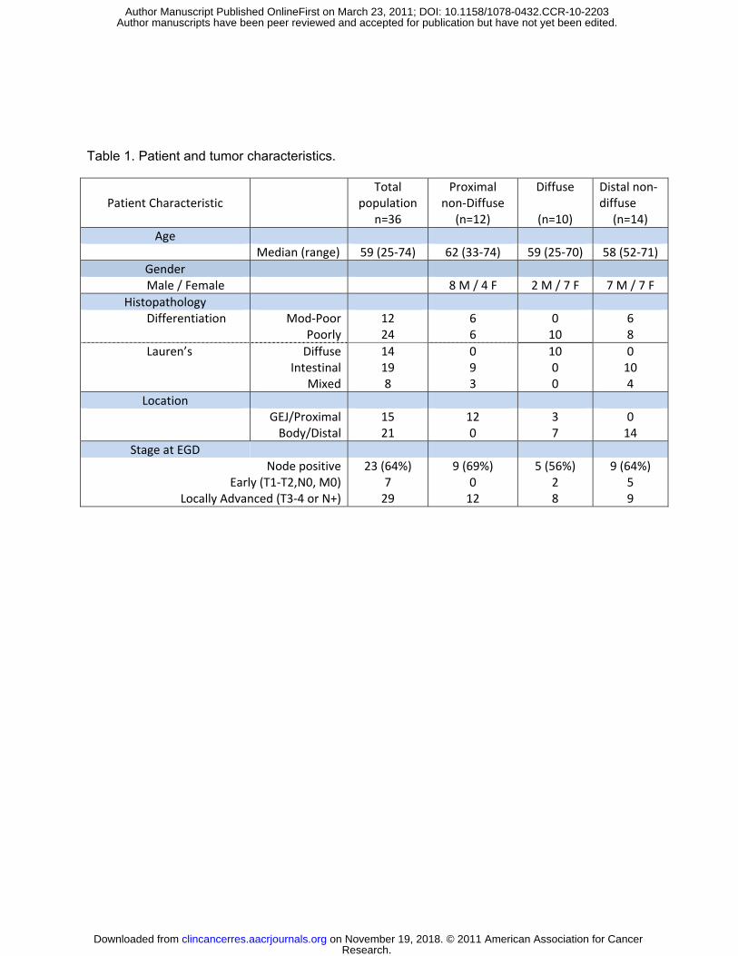
Table 1. Patient and tumor characteristics.
Patient Characteristic
Total population
n=36
Proximal non-Diffuse
(n=12)
Diffuse
(n=10)
Distal non-diffuse
(n=14) Age
Median (range) 59 (25-74) 62 (33-74) 59 (25-70) 58 (52-71) Gender Male / Female 8 M / 4 F 2 M / 7 F 7 M / 7 F
Histopathology Differentiation Mod-Poor
Poorly12 24
6 6
0 10
6 8
Lauren’s DiffuseIntestinal
Mixed
14 19 8
0 9 3
10 0 0
0 10 4
Location GEJ/Proximal
Body/Distal15 21
12 0
3 7
0 14
Stage at EGD Node positive
Early (T1-T2,N0, M0)Locally Advanced (T3-4 or N+)
23 (64%) 7
29
9 (69%) 0
12
5 (56%) 2 8
9 (64%) 5 9
Research. on November 19, 2018. © 2011 American Association for Cancerclincancerres.aacrjournals.org Downloaded from
Author manuscripts have been peer reviewed and accepted for publication but have not yet been edited. Author Manuscript Published OnlineFirst on March 23, 2011; DOI: 10.1158/1078-0432.CCR-10-2203

Figure 1. Venn Diagram demonstrating the distribution of probes that are significantly different between gastric cancer subtypes and normal stomach. Although there is a large overlap between differentially expressed genes in each subgroup comparison versus normal, a large number of genes uniquely differentiate each individual gastric cancer subtype and normal stomach. Analysis is done using limma (with fold-change cut-off 2, and FDR=0.01; values for multiple probe values for the same gene were averaged). Type 1 = proximal non diffuse, type 2 = diffuse, and type 3 = distal non-diffuse gastric cancer.
[insert figure 1 tiff]
Research. on November 19, 2018. © 2011 American Association for Cancerclincancerres.aacrjournals.org Downloaded from
Author manuscripts have been peer reviewed and accepted for publication but have not yet been edited. Author Manuscript Published OnlineFirst on March 23, 2011; DOI: 10.1158/1078-0432.CCR-10-2203

Figure 2. Discriminant Plot for Gene Signatures showing that the RNA expression signatures of samples from different gastric cancers are quite well separated and very tightly grouped into 3 types of gastric cancer (supplemental table 1): Proximal non-diffuse (type 1), Diffuse (type 2), and Distal non-diffuse (type 3). This sample classification was done using pdmclass R-function, using ridge regression. The leave-one-out cross validation error is 0.14, which implies that >85% samples are classified correctly. The output from pdmClass cross-validation confusion matrix is provided to the right of the discriminant plot.
[insert Fig 2 Tiff File]
True Object Type 1 Type 2 Type 3Type 1 10 0 0 Type 2 0 8 1 Type 3 2 2 13
Research. on November 19, 2018. © 2011 American Association for Cancerclincancerres.aacrjournals.org Downloaded from
Author manuscripts have been peer reviewed and accepted for publication but have not yet been edited. Author Manuscript Published OnlineFirst on March 23, 2011; DOI: 10.1158/1078-0432.CCR-10-2203
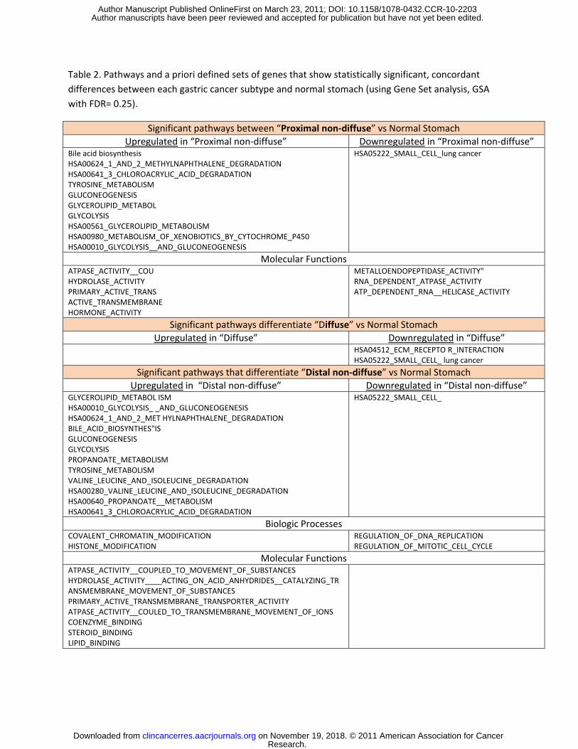
Table 2. Pathways and a priori defined sets of genes that show statistically significant, concordant differences between each gastric cancer subtype and normal stomach (using Gene Set analysis, GSA with FDR= 0.25).
Significant pathways between “Proximal non-diffuse” vs Normal Stomach Upregulated in “Proximal non-diffuse” Downregulated in “Proximal non-diffuse”
Bile acid biosynthesis HSA05222_SMALL_CELL_lung cancer HSA00624_1_AND_2_METHYLNAPHTHALENE_DEGRADATION HSA00641_3_CHLOROACRYLIC_ACID_DEGRADATION TYROSINE_METABOLISM GLUCONEOGENESIS GLYCEROLIPID_METABOL GLYCOLYSIS HSA00561_GLYCEROLIPID_METABOLISM HSA00980_METABOLISM_OF_XENOBIOTICS_BY_CYTOCHROME_P450 HSA00010_GLYCOLYSIS__AND_GLUCONEOGENESIS
Molecular Functions ATPASE_ACTIVITY__COU METALLOENDOPEPTIDASE_ACTIVITY" HYDROLASE_ACTIVITY RNA_DEPENDENT_ATPASE_ACTIVITY PRIMARY_ACTIVE_TRANS ATP_DEPENDENT_RNA__HELICASE_ACTIVITY ACTIVE_TRANSMEMBRANE HORMONE_ACTIVITY
Significant pathways differentiate “Diffuse” vs Normal Stomach Upregulated in “Diffuse” Downregulated in “Diffuse”
HSA04512_ECM_RECEPTO R_INTERACTION HSA05222_SMALL_CELL_ lung cancer
Significant pathways that differentiate “Distal non-diffuse” vs Normal Stomach Upregulated in “Distal non-diffuse” Downregulated in “Distal non-diffuse”
GLYCEROLIPID_METABOL ISM HSA00010_GLYCOLYSIS_ _AND_GLUCONEOGENESIS HSA00624_1_AND_2_MET HYLNAPHTHALENE_DEGRADATION BILE_ACID_BIOSYNTHES"IS GLUCONEOGENESIS GLYCOLYSIS PROPANOATE_METABOLISM TYROSINE_METABOLISM VALINE_LEUCINE_AND_ISOLEUCINE_DEGRADATION HSA00280_VALINE_LEUCINE_AND_ISOLEUCINE_DEGRADATION HSA00640_PROPANOATE__METABOLISM HSA00641_3_CHLOROACRYLIC_ACID_DEGRADATION
HSA05222_SMALL_CELL_
Biologic Processes COVALENT_CHROMATIN_MODIFICATION HISTONE_MODIFICATION
REGULATION_OF_DNA_REPLICATION REGULATION_OF_MITOTIC_CELL_CYCLE
Molecular Functions ATPASE_ACTIVITY__COUPLED_TO_MOVEMENT_OF_SUBSTANCES HYDROLASE_ACTIVITY____ACTING_ON_ACID_ANHYDRIDES__CATALYZING_TRANSMEMBRANE_MOVEMENT_OF_SUBSTANCES PRIMARY_ACTIVE_TRANSMEMBRANE_TRANSPORTER_ACTIVITY ATPASE_ACTIVITY__COULED_TO_TRANSMEMBRANE_MOVEMENT_OF_IONS COENZYME_BINDING STEROID_BINDING LIPID_BINDING
Research. on November 19, 2018. © 2011 American Association for Cancerclincancerres.aacrjournals.org Downloaded from
Author manuscripts have been peer reviewed and accepted for publication but have not yet been edited. Author Manuscript Published OnlineFirst on March 23, 2011; DOI: 10.1158/1078-0432.CCR-10-2203
Published OnlineFirst March 23, 2011.Clin Cancer Res Manish A Shah, Raya Khanin, Laura H Tang, et al. Molecular Classification of Gastric Cancer: A new paradigm
Updated version
10.1158/1078-0432.CCR-10-2203doi:
Access the most recent version of this article at:
Material
Supplementary
http://clincancerres.aacrjournals.org/content/suppl/2011/05/05/1078-0432.CCR-10-2203.DC1
Access the most recent supplemental material at:
Manuscript
Authoredited. Author manuscripts have been peer reviewed and accepted for publication but have not yet been
E-mail alerts related to this article or journal.Sign up to receive free email-alerts
Subscriptions
Reprints and
To order reprints of this article or to subscribe to the journal, contact the AACR Publications
Permissions
Rightslink site. Click on "Request Permissions" which will take you to the Copyright Clearance Center's (CCC)
.http://clincancerres.aacrjournals.org/content/early/2011/03/23/1078-0432.CCR-10-2203To request permission to re-use all or part of this article, use this link
Research. on November 19, 2018. © 2011 American Association for Cancerclincancerres.aacrjournals.org Downloaded from
Author manuscripts have been peer reviewed and accepted for publication but have not yet been edited. Author Manuscript Published OnlineFirst on March 23, 2011; DOI: 10.1158/1078-0432.CCR-10-2203