
CLINICAL REPORTADULT BRAIN
Dural Arteriovenous Fistulas: A Characteristic Pattern of Edemaand Enhancement of the Medulla on MRI
X A.Z. Copelan, X A. Krishnan, X H. Marin, and X R. Silbergleit
ABSTRACTSUMMARY: Medullary edema with enhancement is rarely reported at initial MR imaging in intracranial dural arteriovenous fistulas. Wereport a series of 5 patients with dural arteriovenous fistulas, all of whom demonstrated a characteristic pattern of central medullaryedema and medullary enhancement at initial MR imaging. Cognard type V dural arteriovenous fistula, defined by drainage into theperimedullary veins and the veins surrounding the brain stem, is a rare yet well-described pathologic entity. Even more rarely reported,however, is its clinical presentation with predominantly bulbar symptoms and MR imaging findings of central medullary edema withenhancement. This constellation of findings frequently leads to a convoluted clinical picture, prompting work-up for alternative diseaseprocesses and delaying diagnosis. Because an expedited diagnosis is critical in preventing poor outcomes, it is paramount to make thereferring physician and neuroradiologist more cognizant of this rare-yet-characteristic imaging manifestation of dural arteriovenousfistula.
ABBREVIATION: DAVF � dural arteriovenous fistula
Intracranial dural arteriovenous fistulas (DAVFs) result from a
meshwork of anomalous communications between dural arter-
ies and dural venous sinuses or cortical veins, without an inter-
vening capillary network or nidus, and account for approximately
10%–15% of intracranial vascular malformations.1 The rare Cog-
nard type V DAVF is defined by its drainage into veins around the
brain stem and further caudally into the perimedullary veins.2
Consequently, this subtype often presents with symptoms related
to swelling of the cervical cord, including a slowly progressive
myelopathy initially involving the upper limbs. The imaging find-
ings include an enlarged cervical cord and engorged perimedul-
lary veins.
What is less commonly understood and described here is the
clinical presentation with bulbar symptoms related to brain stem
involvement.3 The imaging findings within the brain stem are
even more rarely reported, though intuitively, a brain MR may be
the initial imaging test ordered by clinicians in these patients. MR
imaging may reveal central medullary edema with occasional in-
tense medullary enhancement.4 Engorged perimedullary veins
may not be evident. These findings may lead the clinician further
astray and prompt a work-up for a neoplasm and infectious or
inflammatory processes. Ultimately, improper management and,
in some reported instances, biopsy for a suspected neoplasm may
occur.5
We report a series of 5 patients who presented at 2 nearby
academic institutions from 2012 to 2016 with DAVFs demon-
strating medullary edema and enhancement at initial imaging.
Most interesting, the unique pattern of edema was nearly identical
in all 5 cases and, retrospectively, has been anecdotally mentioned
previously.5-7 All 5 patients underwent CT and MR imaging ex-
aminations, and 2 patients underwent MR spectroscopy/perfu-
sion because there was concern for a neoplasm. All patients even-
tually underwent conventional angiography.
This multicenter retrospective study was approved by the in-
stitutional review board of each institution with data compiled
into a single Health Insurance Portability and Accountability Act–
compliant data base.
Cases
Case 1. A 59-year-old man presented with new-onset dizziness
and severe nausea and vomiting. He reported positional vertigo
that had commenced 5 weeks before presentation. MR imaging
Received July 30, 2017; accepted after revision September 16.
From the Department of Diagnostic Radiology and Medical Imaging (A.Z.C., A.K.,R.S.), Beaumont Health - Royal Oak, Oakland University William Beaumont Schoolof Medicine, Royal Oak, Michigan; and Department of Radiology (H.M.), Henry FordHospital, Detroit, Michigan.
Several of these cases were previously presented at: Annual Meeting of the Ameri-can Society of Neuroradiology and the Foundation of the ASNR Symposium, May21-26, 2016; Washington, DC.
Please address correspondence to Alexander Z. Copelan, MD, Hospital of the Uni-versity of Pennsylvania, Radiology Department, Neuroradiology Division, 3400Spruce St, Philadelphia, PA 19104; e-mail: [email protected]
http://dx.doi.org/10.3174/ajnr.A5460
238 Copelan Feb 2018 www.ajnr.org

revealed a relatively diffuse T2 hyperintense signal within the me-
dulla, with peripheral and internal linear areas of sparing, as well
as associated mild medullary enhancement (Fig 1A–F). CTA was
also performed, and no vascular malformation was identified. MR
imaging with spectroscopy/perfusion was performed several days
later to exclude neoplasms (Fig 1G–J). This repeat MR imaging
FIG 1. A 59-year-old man who presented with new-onset dizziness and severe nausea and vomiting. Initial MR imaging demonstrates mild hyperintensesignal on axial DWI (b � 1000) (white arrows, A) without definite restricted diffusion on the corresponding ADC map (B). There is relatively diffusehyperintense signal abnormality within the medulla on axial T2WI with fat suppression (C and D) with sparing of the periphery and several internal linearregions (black arrows). This pattern of sparing was not initially recognized. E, Axial FLAIR imaging demonstrates relatively diffuse hyperintense signalabnormality within the medulla (short white arrow). Axial postcontrast T1WI (F) demonstrates mild patchy enhancement (long white arrow). G, Coronalpostcontrast T1WI reveals a dilated perimedullary vein (white arrow) extending inferiorly from the patchy enhancement within the medulla (blackarrow) and coursing caudally along the upper cervical cord. MR spectroscopy with TEs of 135 (H) and 35 (I) ms demonstrates no significant elevation incholine with decreased NAA. MR perfusion imaging (J) reveals slightly decreased relative CBV within the medulla. Suspicion was raised for an underlyingDAVF. Left external carotid injection on digital subtraction angiography arterial phase lateral (K) and delayed venous phase anteroposterior (L) viewsdemonstrates a DAVF in the wall of the left superior petrosal sinus (long white arrow) fed by meningeal branches of the occipital (short white arrow),ascending pharyngeal (long black arrow), and middle meningeal (short black arrows) arteries, with retrograde venous drainage via the petrosal vein tothe pial perimedullary veins running to the cervical cord as the anterior and posterior spinal veins (black dashed arrows).
AJNR Am J Neuroradiol 39:238 – 44 Feb 2018 www.ajnr.org 239
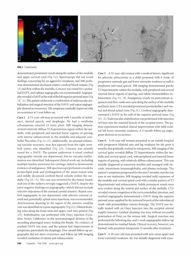
demonstrated prominent vessels along the surface of the medulla
and upper cervical cord (Fig 1G). Spectroscopy did not reveal
findings concerning for an aggressive neoplasm, and MR perfu-
sion demonstrated decreased relative cerebral blood volume (Fig
1J) and flow within the medulla. Concern was raised for a poten-
tial DAVF, and catheter angiography was recommended. Angiogra-
phy revealed a DAVF in the wall of the left superior petrosal sinus (Fig
1K, -L). The patient underwent a combination of endovascular em-
bolization and surgical resection of the DAVF, and repeat angiogra-
phy showed no recurrence. His symptoms markedly improved with
no recurrence at 3-year follow-up.
Case 2. A 72-year-old man presented with 3 months of imbal-ance, slurred speech, and dysphagia. He had a vestibularschwannoma resected 15 years prior. MR imaging demon-strated relatively diffuse T2 hyperintense signal within the me-dulla, with peripheral and internal linear regions of sparing,with intense enhancement in the medulla and adjacent cere-bellar flocculus (Fig 2A–D). Additionally, an unusual enhanc-ing vascular structure, near but separate from the right verte-bral artery, was identified (Fig 2D). Concern was initiallyraised for a DAVF. The patient underwent 4-vessel cerebralangiography outside our department, but no vascular malfor-mation was identified. Subsequent clinical work-up, includingmultiple lumbar punctures for cytology, failed to demonstrateevidence of malignancy. MR spectroscopy/perfusion revealed alactate/lipid peak and prolongation of the mean transit timeand mildly decreased cerebral blood volume within the me-dulla (Fig 2E, -F). The case was reviewed by the tumor board,and one of the authors strongly suggested a DAVF, despite theprior negative findings on angiography (which did not includeselective injections of the external carotid arteries). Repeat cere-bral angiography in our department with selective external ca-rotid and, potentially, spinal artery injections, was recommended.Arteriovenous shunting in the region of the anterior condylarvein was identified on repeat angiography (Fig 2G). Dilated drain-ing veins along the brain stem and upper cord were present (Fig2H). Embolization was performed with Onyx injection (Covi-dien, Irvine, California) in the neuromeningeal division of theascending pharyngeal artery feeding the fistula. No evidence ofresidual DAVF was seen, and the patient had improvement insymptoms, particularly his dysphagia. Five-month follow-up an-giography did not show recurrence, and follow-up MR imagingrevealed resolution of edema and enhancement.
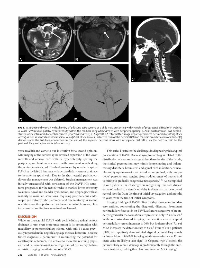
Case 3. A 35-year-old woman with a medical history significant
for pilocytic astrocytoma as a child presented with 4 weeks of
progressive unsteady gait and lower extremity weakness as well as
dysphonia and nasal speech. MR imaging demonstrated patchy
T2 hyperintensity within the medulla, with peripheral and several
internal linear regions of sparing, and subtle intramedullary en-
hancement (Fig 3A, -B). Serpiginous vessels on postcontrast se-
quences and flow voids were seen along the surface of the medulla
and brain stem. CTA revealed prominent perimedullary and ven-
tral and dorsal spinal veins (Fig 3C). Cerebral angiography dem-
onstrated a DAVF in the wall of the superior petrosal sinus (Fig
3D, -E). Endovascular embolization was performed with injection
of Onyx into the mastoid branch of the occipital artery. The pa-
tient experienced marked clinical improvement with mild resid-
ual left lower extremity weakness. A 3-month follow-up angio-
gram showed no recurrence.
Case 4. A 64-year-old woman presented to an outside hospital
with progressive bilateral arm and leg weakness for the prior 6
months that gradually evolved to tetraparesis. MR imaging of the
cervical spine revealed diffuse T2 hyperintensity within the me-
dulla and cervical spinal cord, with peripheral and internal linear
regions of sparing, with relatively diffuse enhancement. This was
initially diagnosed as transverse myelitis and managed with ste-
roids, intravenous immunoglobulin, and plasma exchange. The
patient’s symptoms progressed for the next 5 months, and she was
seen at our institution. MR imaging revealed mild expansion of
the medulla and cervical spinal cord with a similar pattern of T2
hyperintensity and enhancement. Subtle prominent vessels were
now evident along the ventral pial surface of the medulla. CTA
revealed venous congestion at the cervicomedullary junction, and
cerebral angiography demonstrated a DAVF along the superior
petrosal sinus supplied by the tentorial branch of the inferolateral
trunk with perimedullary venous drainage. The DAVF was ini-
tially treated with an Onyx injection into the occipital arterial
supply; however, residual shunting was seen without successful
penetration of Onyx on the venous side. Surgical resection was
performed the following day, and 1-week angiographic follow-up
demonstrated no residual fistula. Clinical recovery, however, was
limited, with persistent tetraparesis 12 months after treatment.
Case 5. A 50-year-old man presented with new-onset upper and
lower extremity weakness. He was initially diagnosed with trans-
FIG 1. Continued.
240 Copelan Feb 2018 www.ajnr.org

FIG 2. A 72-year-old man with 2- to 3-month history of imbalance and dysphagia. A and B, Axial T2WI with fat suppression from initial MR imagingdemonstrates diffuse hyperintense signal in the medulla with segments of linear sparing (short white arrows), creating a geographic pattern, as well asan unusual vascular structure (long white arrow). C and D, Axial postcontrast T1-weighted fat-suppression imaging demonstrates intense enhancementwithin the medulla (short white arrow) and again depicts the unusual vascular structure (long white arrow), which appears separate from the vertebralarteries. Dynamic susceptibility contrast MR perfusion imaging reveals prolongation in mean transit time (short white arrow, E), and MR spectroscopywith a TE of 35 ms (F) reveals a lactate/lipid peak (long white arrow) without significant elevation in choline. G, Lateral view from a selective rightascending pharyngeal injection on DSA demonstrates arteriovenous shunting with early opacification of the anterior condylar vein (short white arrow)supplied by feeders from the neuromeningeal division of the ascending pharyngeal artery (long white arrow). H, More delayed lateral image demon-strates dilated veins along the surface of the brain stem and upper cord (short white arrow), draining both superiorly along the anterolateral surface ofthe pons toward the petrosal vein (long white arrow) and inferiorly toward the anterior spinal vein (long black arrow). The tortuous vein (short blackarrow) likely corresponds to the anomalous venous structure seen on the original MR imaging.
AJNR Am J Neuroradiol 39:238 – 44 Feb 2018 www.ajnr.org 241
verse myelitis and came to our institution for a second opinion.
MR imaging of the cervical spine revealed expansion of the lower
medulla and cervical cord with T2 hyperintensity, sparing the
periphery, and faint enhancement with prominent vessels along
the ventral cervical cord. Cerebral angiography revealed a spinal
DAVF in the left C1 foramen with perimedullary venous drainage
to the anterior spinal vein. Due to the short arterial pedicle, en-
dovascular management was deferred. Surgical management was
initially unsuccessful with persistence of the DAVF. His symp-
toms progressed for the next 6 weeks to marked lower extremity
weakness, bowel and bladder dysfunction, and dysphagia, with an
inability to maintain secretions, requiring percutaneous endo-
scopic gastrostomy tube placement and tracheostomy. A second
operation was then performed and was successful; however, clin-
ical examination findings remained unchanged.
DISCUSSIONWhile an intracranial DAVF with perimedullary spinal venous
drainage is rare, even more uncommon is its presentation with
medullary or pontomedullary edema, with only 31 cases previ-
ously reported in the English language medical literature. Because
timely diagnosis is paramount in minimizing the potential for
catastrophic outcomes, it is critical to make the referring physi-
cian and neuroradiologist more cognizant of this rare-yet-char-
acteristic imaging manifestation of a DAVF.
This series illustrates the challenges in diagnosing this atypical
presentation of DAVF. Because symptomatology is related to the
distribution of venous drainage rather than the site of the fistula,
the clinical presentation may mimic demyelinating and inflam-
matory disorders, brain stem and spinal cord infarction, or neo-
plasms. Symptom onset may be sudden or gradual, with our pa-
tients’ presentations ranging from sudden onset of nausea and
vomiting to gradually progressive tetraparesis.8-12 As exemplified
in our patients, the challenges in recognizing this rare disease
entity often lead to a significant delay in diagnosis, on the order of
several months from the time of initial investigation and months
to years from the time of initial symptoms.
Imaging findings of DAVF often overlap more common dis-
ease entities, convoluting the diagnostic dilemma. Prominent
perimedullary flow voids on T2WI, a feature suggestive of an un-
derlying vascular malformation, are present in only 37% of cases.4
With contrast-enhanced imaging, the detection rate of atypical
perimedullary vessels increases to 76% but is often subtle.5 Use of
MRA increases the detection rate to 85%.5 Four of our 5 patients
(80%) retrospectively demonstrated atypical perimedullary vessels
or flow voids on initial MR imaging, subtle in some instances. Prom-
inent veins are likely a later sign.5 In Cognard type V lesions, the
perimedullary venous drainage is predominantly through the ante-
rior spinal veins, making them less prominent on MR imaging.4
FIG 3. A 35-year-old woman with a history of pilocytic astrocytoma as a child now presenting with 4 weeks of progressive difficulty in walking.A, Axial T2WI reveals patchy hyperintensity within the medulla (long white arrow) with peripheral sparing. B, Axial postcontrast T1WI demon-strates subtle intramedullary enhancement (short white arrow). C, Sagittal CTA reformatted image depicts prominent perimedullary (long blackarrow) as well as ventral and dorsal spinal veins (short black arrows). Selective DSA of the occipital (D) and mastoid branch via microcatheter (E)demonstrates the fistulous connection in the wall of the superior petrosal sinus with retrograde pial reflux via the petrosal vein to theperimedullary and spinal veins (black arrows).
242 Copelan Feb 2018 www.ajnr.org

Central medullary or pontomedullary edema has been re-
ported in 73% of patients with Cognard type V DAVFs.5 All our
patients demonstrated a nearly identical pattern of geographic
central medullary edema with sparing of the periphery as well as
internal linear segments in a tigroid pattern. In this series, not all
cases of Cognard type V DAVFs were reviewed; rather, only those
cases demonstrating central medullary edema were included, so
the proportion of total cases of type V DAVFs from our institu-
tions with the aforementioned specific imaging features is un-
known. This peripheral sparing appearance has been mentioned
in case reports.13 This pattern contrasts with the infiltrative pro-
cess seen in brain stem tumors. Maintenance of the normal signal
of the peripheral subpial tissue may be secondary to direct drain-
age of the extracellular vasogenic edema into the CSF through the
permeable pia mater.14,15 Perivascular spaces may also play a role
in draining the peripheral tissue.16
Of the 20 cases in the literature reporting on the presence of
medullary contrast enhancement for Cognard type V DAVFs, 11
cases (55%) demonstrated enhancement. Medullary enhance-
ment was demonstrated in all our cases, 4 of which were initially
scanned on a 1.5T scanner and 1 on a 3T scanner, with 3 different
vendors used. Medullary enhancement was demonstrated in all
our cases. The combination of medullary edema and enhance-
ment may lead to the consideration of neoplastic or inflammatory
etiologies, as was the case in 3 of our 5 cases, and has reportedly led
to inappropriate brain stem biopsies.5 Two of our patients under-
went MR perfusion/spectroscopy because a neoplasm was consid-
ered. MR perfusion and spectroscopy in DAVF are also uncom-
monly reported. A prolonged mean transit time with decreased
cerebral blood volume may be present secondary to venous con-
gestion with ischemia, seen in both of our patients.17,18 Addition-
ally, spectroscopy may reveal increased lactate, which was dem-
onstrated in 1 of our patients.
Four of our 5 patients had true intracranial DAVFs. Case 5
revealed the site of the fistula at the craniocervical junction along
the C1 foramen. In 3 of our 4 cases of true intracranial DAVFs,
conventional angiography revealed the site of the fistula to be
along the superior petrosal sinus, and in 1 case, the anterior con-
dylar vein. The goal of treatment is closure of the draining vein
proximally as it exits the fistula. Successful endovascular emboli-
zation with Onyx was performed in 2 of our patients; combined
embolization and an operation, in 2 patients; and 2 operations, in
1 patient. As in case 2, the medullary edema might resolve follow-
ing treatment.5
Bulbar symptoms were present in 3 of our 5 (60%) cases: case
2 (slurred speech and dysphagia), case 3 (dysphonia and nasal
speech), and case 5 (inability to maintain secretions). A systematic
review of the literature on Cognard type V DAVFs identified bul-
bar symptoms in 31% of patients and found no significant differ-
ence in prognosis between those with versus those without bulbar
symptoms.19 In our series, 2 of 3 patients with clinical improve-
ment and 1 of 2 patients without clinical improvement had bulbar
symptoms, further exemplifying a lack of relationship between
the presence of bulbar symptoms and prognosis.
Three of our 5 patients (60%) had near-complete resolution of
symptoms and angiographically complete occlusion of the DAVF.
Unfortunately, the 2 patients with the most significant delay in
diagnosis and treatment did not show significant clinical im-
provement following treatment, further exemplifying the impor-
tance of early diagnosis and management.
CONCLUSIONSThis relatively unusual-but-characteristic pattern of medullary
edema with areas of sparing and patchy enhancement should
prompt scrutiny for atypical perimedullary vessels. If no such ves-
sels are identified, MRA/CTA or conventional angiography
should be recommended. Of utmost importance, selective injec-
tion of the external carotid arteries is mandated at the time of
conventional angiography to avoid false-negative results as was
the case in 1 of our patients.
Disclosures: Richard Silbergleit—UNRELATED: Consultancy: Relievant Medsystems.
REFERENCES1. Kwon BJ, Han MH, Kang HS, et al. MR imaging findings of intra-
cranial dural arteriovenous fistulas: relations with venous drainagepatterns. AJNR Am J Neuroradiol 2005;26:2500 – 07 Medline
2. Cognard C, Gobin YP, Pierot L, et al. Cerebral dural arteriovenousfistulas: clinical and angiographic correlation with a revised classi-fication of venous drainage. Radiology 1995;194:671– 80 CrossRefMedline
3. van Rooij WJ, Sluzewski M, Beute GN. Intracranial dural fistulaswith exclusive perimedullary drainage: the need for complete cere-bral angiography for diagnosis and treatment planning. AJNR Am JNeuroradiol 2007;28:348 –51 Medline
4. Haryu S, Endo T, Sato K, et al. Cognard type V intracranial duralarteriovenous shunt: case reports and literature review with specialconsideration of the pattern of spinal venous drainage. Neurosur-gery 2014;74:E135– 42; discussion E142 CrossRef Medline
5. Roelz R, Van Velthoven V, Reinacher P, et al. Unilateral contrast-enhancing pontomedullary lesion due to an intracranial dural ar-teriovenous fistula with perimedullary spinal venous drainage: theexception that proves the rule. J Neurosurg 2015;123:1534 –39CrossRef Medline
6. Ricolfi F, Manelfe C, Meder JF, et al. Intracranial dural arterio-venous fistulae with perimedullary venous drainage: anatomical,clinical and therapeutic considerations. Neuroradiology 1999;41:803–12 CrossRef Medline
7. Brunereau L, Gobin YP, Meder JF, et al. Intracranial dural arterio-venous fistulas with spinal venous drainage: relation between clin-ical presentation and angiographic findings. AJNR Am J Neurora-diol 1996;17:1549 –54 Medline
8. Gaensler EH, Jackson DE Jr, Halbach VV. Arteriovenous fistulas ofthe cervicomedullary junction as a cause of myelopathy: radio-graphic findings in two cases. AJNR Am J Neuroradiol 1990;11:518 –21 Medline
9. Hahnel S, Jansen O, Geletneky K. MR appearance of an intracranialdural arteriovenous fistula leading to cervical myelopathy. Neurol-ogy 1998;51:1131–35 CrossRef Medline
10. Li J, Ezura M, Takahashi A, et al. Intracranial dural arteriovenousfistula with venous reflux to the brainstem and spinal cord mimick-ing brainstem infarction: case report. Neurol Med Chir (Tokyo)2004;44:24 –28 CrossRef Medline
11. Cahan LD, Higashida RT, Halbach VV, et al. Variants of radiculo-meningeal vascular malformations of the spine. J Neurosurg 1987;66:333–37 CrossRef Medline
12. Hassler W, Thron A. Flow velocity and pressure measurements inspinal dural arteriovenous fistulas. Neurosurg Rev 1994;17:29 –36CrossRef Medline
13. Hurst RW, Grossman RI. Peripheral spinal cord hypointensity on T2-weighted MR images: a reliable imaging sign of venous hypertensivemyelopathy. AJNR Am J Neuroradiol 2000;21:781–86 Medline
AJNR Am J Neuroradiol 39:238 – 44 Feb 2018 www.ajnr.org 243

14. Nicholas DS, Weller RO. The fine anatomy of the human spinalmeninges: a light and scanning electron microscopy study. J Neuro-surg 1988;69:276 – 82 CrossRef Medline
15. Hutchings M, Weller RO. Anatomical relationships of the pia materto cerebral blood vessels in man. J Neurosurg 1986;65:316 –25CrossRef Medline
16. Fleury J, Gherardi R, Poirier J. Pathology of perivascular spaces inthe central nervous system [in French]. Ann Pathol 1984;4:249 –57Medline
17. Doege CA, Tavakolian R, Kerskens CM, et al. Perfusion and diffu-sion magnetic resonance imaging in human cerebral venousthrombosis. J Neurol 2001;248:564 –71 CrossRef Medline
18. Kim DJ, Krings T. Whole-brain perfusion CT patterns of brain ar-teriovenous malformations: a pilot study in 18 patients. AJNR Am JNeuroradiol 2011;32:2061– 66 CrossRef Medline
19. El Asri AC, El Mostarchid B, Akhaddar A, et al. Factors influencing theprognosis in intracranial dural arteriovenous fistulas with perime-dullary drainage. World Neurosurg 2013;79:182–91 CrossRef Medline
244 Copelan Feb 2018 www.ajnr.org


