diversity of lactic acid bacteria … · bacteria bacteriophages from dairy environmement ... phage...
TRANSCRIPT

UNIVERSITÀ DEGLI STUDI DI NAPOLI
FEDERICO II
DIVERSITY OF LACTIC ACID
BACTERIA BACTERIOPHAGES
FROM DAIRY
ENVIRONMEMENT
Paola Zinno

UNIVERSITÀ DEGLI STUDI DI NAPOLI FEDERICO II
Tesi di Dottorato di Ricerca in
Scienze e Tecnologie delle Produzioni Agro-Alimentari
XXI ciclo
DIVERSITY OF LACTIC ACID BACTERIA BACTERIOPHAGES
FROM DAIRY ENVIRONMENT
Il Tutor: Il Dottorando:
Prof. Gianluigi Mauriello Dott.ssa Paola Zinno
Coordinatore
Prof. Giancarlo Barbieri

I
CONTENTS
CHAPTER 1
Introduction
General introduction 1
Baccteriophages 2
The Streptococcus thermophilus bacteriophages 5
Bacteriophage /host interaction 6
Genome organization 11
Transcription 14
Integration/excision 15
Replication 17
DNA packaging 18
Head proteins 21
Tail proteins 22
Lysis cassette 22
Bacteriophage defense strategies 23
Improving sanitation and manufacturing processes 24
Phage inhibitory media 24
Strain- rotation 24
Molecular strategies 26
Outlook of thesis 27
CHAPTER 2
Investigation on the presence of LAB bacteriophages in Natural Whey Cultures used
for traditional water-buffalo Mozzarella Cheese manufacture
INTRODUCTION 28
EXPERIMENTAL PROCEDURES 29
NWC samples 29
Enumeration, bacterial strain isolation and culture conditions 29
Analysis of the NWC samples for the presence of phages 30
Detection of Streptococcus thermophilus phages by PCR 30
RESULTS 31
Enumeration of lactic acid bacteria 31

II
Analysis for the presence of bacteriophages 33
Detection of Streptococcus thermophilus phages by PCR 33
DISCUSSION 33
CHAPTER 3
Characterization of Streptococcus thermophilus Lytic Bacteriophages from Mozzarella
Cheese Plants
INTRODUCTION 36
EXPERIMENTAL PROCEDURES 37
Bacterial strains, bacteriophages and culture conditions 37
Phage multiplication 38
Host range 39
Phage DNA isolation 39
Restriction analysis 40
Determination of DNA packaging mechanism 40
Amplification and sequencing of the antireceptor variable region 41
RESULTS 42
Host range 42
Restriction analysis 44
Determination of DNA packaging mechanism 44
Bacteriophage clustering based on VR2 sequences 45
DISCUSSION 47
CHAPTER 4
Evidence of a temperate bacteriophage from lysogenic Streptococcus macedonicus AI4
INTRODUCTION 50
EXPERIMENTAL PROCEDURES 52
Induction of bacteriophages 52
Screening for indicator strains 52
Determination of DNA packaging mechanism 53
Dot blot hybridization 54
RESULTS 54
Evidence of temperate bacteriophages and screening of indicator strains 54
Determination of DNA packaging 56

III
Dot blot hybridization 56
DISCUSSION 57
CHAPTER 5
Genomic sequence of S. macedonicus temperate phage PZ1ϕ by genome walking
method - work in progress
INTRODUCTION 59
EXPERIMENTAL PROCEDURES 60
Construction of Genome WalkerTM Libraries 60
PCR amplification 61
RESULTS and DISCUSSIONS 62
REFERENCES 64

Chapter 1
INTRODUCTION

1
General introduction
Lactic Acid Bacteria (LAB) have always played a very important role in diary industry for
their fermentative capacities. LAB comprise a wide range of genera and include lactococci,
lactic streptococci and lactobacilli. When these microorganisms are present in the food
products, they also have a function as biopreservatives and probiotic characteristic
(Salminen et al., 1996).
Beside Lactococcus lactis, Streptococcus thermophilus is the major starter bacterium in
cheese industry (Brussow, 2001). It widely occurs in commercial starter cultures as well as
in natural milk or whey cultures traditionally used in the manufacture of several protected
designation of origin (PDO) and artisanal cheeses (Coppola et al., 1988). They are used
during the manufacture of Italian-style cheese varieties such as Asiago, Mozzarella,
Parmigiano and ripened cheeses including Limburger, Port du Salut and Trappist (Olson
1969). S. thermophilus is also used with Lactococcus lactis for the prduction of Cheddar
cheese. This microorganism is perhaps best known for its use in combination with
Lactobacillus delbrueckii spp. bulgaricus, during the manufacture of yogurt. It is well
known these two microorganisms share a synergistic relationship when mixed, growing
faster and producing more lactic acid. The application of lactic acid bacteria, specially S.
thermophilus, in large-scale diary fermentation was an incentive to the unravelling of
molecular mechanism and genetics underlying some of its significant industrial traits. This
has resulted in the improvement of existing strains and the development of novel strains by
genetic modification (McKay and Baldwin, 1990).
The recognition that the bacteriophages could be a main risk to starter culture performances
led to the research aimed to the development of strategies to effectively protect starter
strains.

2
In recent years significant progress has been made in the elucidation of genetic organization
of several S. thermophilus phage genomes. The aim of this overview is to describe the
current state-of-art about S. thermophilus bacteriophage research and exploitation of this
knowledge in both fundamental and applied research.
Bacteriophages
Virus that infect bacteria, bacteriophages (from Greek ϕαγειν= to eat, bacteriophage =
bacteria eater) are obligate intracellular molecular parasites belonging to Myoviridea,
Podoviridea or Siphoviridea. On the basis of specific features of their heads and tails the
bacteriophages can be differentiated in three basic morphotypes (Ackerman et al., 1984,
Figure 1). Phage of morphotype A is characterized by a contractile tail allowing active
injection of their genome into the host cells. Morphotypes B and C have respectively, a long
and small non-contractile tail.
The latter two groups are further subdivided on the basis of the size and shape of their
capsids as either small-isometric, prolated-headed or elongated bacteriophages. Although
Figure 1: Basic morphotypes of compositive viruses belonging to Myoviridea (A), Siphoviridea (B) and Podoviridea (C).

3
phages can possess specific appendages such as a collar, a baseplate,
whiskers, spikes and/or fibers, taxonomically not significant structures
(Figure 2).
The genome of tailed phages constitutes either a single- or double–
stranded DNA or single stranded RNA molecule, encapsulated in a head
(capsid) composed of proteins. Furthermore, the bacteriophages can be distinguished on the
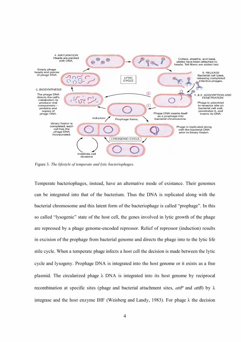
basis of their specific life cycle after infection (Figure 3). Lytic (or virulent) bacteriophages
have only one strategy for the replication. At the beginning of infection a phage adsorbs to
its host and injects its DNA in linear form from the phage capsid to the inside of a cell.
During the latent period of a lytic cycle, phage DNA is replicated and the phage genes are
transcribed in strict order: proteins of tailed phages are assembled through separate
pathways into heads, tails and fibers (Ackermann and Dubow, 1987). Phage genome
replication often leads to accumulation of head-to-tail arrays of phage genomes. This so
called concatemeric DNA is then cut by headful cutting during the packaging process into
DNA-free proheads. All cuts occur either precisely at cos-sites, or the first cut occurs at the
packaging sequence pac and then the cutting process continues by imprecise headful
measuring leading to terminal redundancy (Black 1989.) Phage heads and tails are connected
together to finish phage maturation and phages are released due to cell bursts or lysis as a
result of cell wall degradation by a phage lysozyme (Ackermann and DuBow, 1987). A
virulent phage can only initiate a lytic cycle.
Figure 2
4
Temperate bacteriophages, instead, have an alternative mode of existance. Their genomes
can be integrated into that of the bacterium. Thus the DNA is replicated along with the
bacterial chromosome and this latent form of the bacteriophage is called “prophage”. In this
so called “lysogenic” state of the host cell, the genes involved in lytic growth of the phage
are repressed by a phage genome-encoded repressor. Relief of repressor (induction) results
in excision of the prophage from bacterial genome and directs the phage into to the lytic life
stile cycle. When a temperate phage infects a host cell the decision is made between the lytic
cycle and lysogeny. Prophage DNA is integrated into the host genome or it exists as a free
plasmid. The circularized phage λ DNA is integrated into its host genome by reciprocal
recombination at specific sites (phage and bacterial attachment sites, attP and attB) by λ
integrase and the host enzyme IHF (Weisberg and Landy, 1983). For phage λ the decision
Figure 3. The lifestyle of temperate and lytic bacteriophages.

5
between the lytic cycle and the lysogenic state depends on relative levels of two antagonistic
phage regulatory proteins, Cro and cI. After spontaneous induction or induction by DNA
damaging agents like mitomycin C, prophages sometimes escape the control of the cI
repressor, excise and initiate the lytic cycle (Ackermann and DuBow, 1987). Furthermore,
there is the chronic bacteriophage life ciycle. A chronically infecting phage can release
progeny phages into extracellular environment without killing its host (Maniloff et al.,
1981). In this case the phage and the host coexist and bacteriophages are shed over the
infection.
By the genetic organization of the conserved head cluster, it has recently been proposed a
new LAB phage classification (Brussow and Desiere, 2001, Proux et al., 2002). According to
this new proposal the sequenced LAB phages could be divided into five genera: c2-, sk1-,
Sfi11-, Sfi21- and r1t-like phages (Desiere et al., 2002, Proux et al., 2002).
The Streptococcus thermophilus bacteriophages
S. thermophilus phages have the same basic morphology belonging to the B1 Syphoviridae
group of Caudovirales order (Brussow et al., 1994) with few exceptions appear to be very
similar with isometric heads (generally 42 to 63 nm in diameter), tails 200-300 nm long, no
obvious collars and only small base plates, often with a central fibre. Variants with larger
heads, shorter tails and exceptionally long tails (polytails) (Reinbold et al., 1982) are also
known to occur. S. thermophilus phages have a linear double-stranded DNA genome
ranging in size from 30 to 45 kb (Brussow et al., 1998). DNA-DNA hybridization studies
showed homology between all S. thermophilus phages, including virulent and temperate
phages.

6
Brussow and Bruttin (1995) proposed classification of S. thermophilus phages into four lytic
groups based on host range and type-specific antisera. An alternative approach for
classifying S. thermophilus phages is originated from the analysis of phage genomes and it
is based on their evolutionary descendent. Mercenier (1990) proposed that all S.
thermophilus phages are derived from a common ancestor. Corroborating this idea is the
cloning DNA fragment from ΦS1, which hybridized to all S. thermophilus phage isolates.
The diversity in phage genomes observed by different researchers may result from multiple
rearrangements occurring within the phage population. Brussow et al. (1994b) proposed that
the evolution of S. thermophilus phages takes place by means of module exchange between
phage genomes, a hypothesis that was proposed previously for other bacteriophages. Le
Marrec et al. (1997) proposed a further classification of S. thermophilus phages into two
groups based on the number of major structural protein (MSP) and the mode of DNA
packaging. The first group comprises phages with two MPS and cohesive genome
extremities (cos-type). The second cluster includes phages with three MPS and a DNA
packaging scheme that proceeds via a headful mechanism (pac-type).
Bacteriophage /host interaction
The obligatory use by the LAB bacteriophages of their hosts is reflected by the numerous
phage/host interactions (Garvey et al. 1994). These interactions are often subject to change
due to development of new defence mechanisms by host cells. The natural defence
mechanisms in LAB against phage infection are often plasmid-coded and can be classified
into four main categories:
• adsorption interference;
• injection blocking;

7
• restriction/ modification (R/M) systems;
• abortive-infection (Abi).
The adsorption interference is the mechanism prevailing in spontaneous phage resistant
mutants of thermophilic LAB, whereas R/M systems are widely represented within
Lactococcus genus (Moineau S., 1999). It was proven that the modification and restriction
enzymes both recognize the same target, a specific nucleotide sequence. The modification
enzyme is the DNA methyltransferase that methylates specific bases within the target
sequence. In the absence of the specific methylation in the target sequence, DNA becomes
sensitive to the restriction endonuclease. When the DNA lacking the appropriate
modification imprint, it enters a restriction proficient cell. Therefore, the DNA is recognized
as foreign and degraded by the endonuclease. Restriction/modification systems are classified
into three types on the basis of their composition and cofactor requirements, the nature of
their target sequence, and the position of the site of DNA cleavage with respect to the target
sequence (Murray, 2000). In the first R/M system, designated type I, the enzymes are
hetero-oligomeric. They require ATP hydrolysis for the restriction and cut at sites remote
from the recognition sequences. In the R/M system type II, the endonucleases and the
methyltransferases are separate enzymes and they cut the DNA within the recognition
sequence. The type III is characterized by hetero-oligomeric endonucleases, which require
ATP for restriction and they cut the DNA close to recognition sequence.

8
By adsorption interference, most phage resistant strains achieve their resistance through a
loss or a modification of phage receptors due to mutation in corresponding genes (Riipinen
et al., 2007).
Furthermore, Duplessis and Moineau (2001) studied the phage genetic determinant
(antireceptor) involved in the recognition of S. thermophilus hosts. Most of the information
on the antireceptor and phage-host receptor interaction comes from studies carried out on
coliphages, such as T-even (Myoviridae family) and lambdoid (Siphoviridae) phages. For
T4 phage, the major host range determinant is the gene product of orf37, which encodes the
large subunit of distal tail fibre. Specific interaction between the tip of these long fibres and
receptors on the surface of Escherichia coli host bacterium result in rapid and efficient
phage adsorption (Goldberg et al., 1994). The C terminal extremity of gp37 can recognize a
variety of bacterial receptor molecules, such as OmpC, OmpF, and E. coli B
lipopolysaccharide (Tétart et al., 1998). In the phage λ, the gene product J constitutes the
Figure 4. Restriction/Modification system (modified from Murray 2000)

9
receptor-binding protein that allows phage adsorption to the surface of E. coli K-12 by
interaction with the outer membrane protein LamB. The J protein is the target of λ-
neutralizing antibodies as well as a structural component of the tail fibre. Analysing lambda
mutants it was shown that host range mutations occurred in the last distal (5-10 %) portion
of gene J (gpJ) (Werts et al., 1994). Wang et al. (2000) demonstrated only 249 amino acids
at carboxy-terminal part of the antireceptor protein is involved in host specificity.
Duplessis and Moineau (2001) characterized the gene of S. thermophilus phage DT1 that
encodes the structural protein involved in host recognition. Bio-informatic analysis by
Tremblay and Moineau (1999) suggested the gene product of orf18 as the antireceptor: it has
the same position within its genome as the anti-receptor of phage lambda gene J, the pI and
molecular weight of ORF18 are similar to the those gpJ of λ and when compared with
homologues, orf18 has an organization that resembles some T-even antireceptor genes with
conserved and non-conserved regions. Furthermore, they sequenced the orf18 of S.
thermophilus DT1 and of other six lytic, cos-type phages. By amino acid alignment, they
could understand that these open reading frames (ORFs) were divided into three domains:
the first domain of the seven ORF18s corresponded to the amino-terminal portion of the
protein up to the collagen-like repeats. This portion of 491 amino acids resulted highly
conserved among the seven phages, with 83-100% amino acid identity and the first domain
of these phages was identical also at the nucleotide level.
The second domain could be present or absent depending on the phage and has a length of
approximately 400 amino acids. This domain resulted flanked by two motifs called
collagen-like repeats. This second domain was called VR1 containing. The third domain,
which corresponds to the carboxy-terminal part of the protein, starts after fourth collagen-
like repetition and has a length of approximately 400 amino acids. It also showed an internal

10
variable region VR2 of about 145 amino acids and it resulted the most divergent region
within the deduced ORF18 (Figure 5). The overall comparisons of the seven orf18s revealed
that the variations could be attributed to point short mutations, short deletions or insertions.
The comparisons of host range of the seven phages with their corresponding ORF18s
revealed that the ORF18 of phage DT1 has a genetic organization similar to that of S.
thermophilus phage MD4 and both has very distinct host ranges. To provide biological
evidence that orf18 is involved in host recognition, Duplessis and Moineau (2001) used the
distinctive features of these two phages for the construction of chimeric phage in which the
orf18 of phage DT1 was swapped for the orf18 of phage MD4. By this construction, DT1
acquired the host range of MD4. Prior to this, a spontaneous deletion mutants of phage
Sfi21, called D3, was isolated by routine serial propagation. In this case, the second domain
between the collagen-like repeats III and I was deleted in the mutant, indicating these
recombination hotspots contribute to the allelic diversity within the population (Bruttin and
Brussow, 1996). The nucleotide sequences encoding the collagen-like reapeats suggested to
be hotspots for recombination-mediates gene shuffling (Desière et al., 1998). These motifs
consist of repeated amino acids triplets where glycine is the first residue in each triplet
(Beck and Brodsky, 1998).
Comparisons of other VR2 regions characterized in the same study also revealed that
additional phage factors are involved in the host specificity of other S. thermophilus phages.
Although VR2 is clearly responsible for the specificity of phages MD4 and DT1 to their S.
thermophilus hosts, this finding cannot be generalized to other S. thermophilus. The VR1
region may also interact with the VR2 region to confer host specificity. However, the phage
receptors in S. thermophilus are currently unknown. A first effort consists in submitting a
strain to mutagenesis with the thermolabile insertional vector pG+host9:ISS1 (Lucchini et

11
al., 2000). Vector insertion into four different sites led to a phage resistant phenotype, but all
mutated strains adsorbed the phages, suggesting the elements involved in phage adsorbtion
remain unidentified. Unfortunately, no biological evidence has allowed the identification of
the phage antireceptor. It is also important noting that the collagen-like repeats are found in
sugar-binding proteins, such as maltose-binding protein. Furthermore, it is interesting to
speculate that ORF18 could bind to a carbohydrate component of the S. thermophilus cell
wall.
Genome organization
Similar to most other bacteriophages, the DNA content of streptococci bacteriophages, is
rather small. Genomes analyzed are double–stranded DNA linear molecule ranging in size
from 30 to 45 kb (Brussow et al., 1998). The DNA sequence data are rapidly accumulating
4for S. thermophilus phages . However, the genomes sequenced so far were from isolated in
Figure 5: Alignment and comparison of the ORF18 of seven S. thermophilus phages. VR1 and VR2 indicate variable region 1 and 2. Homologous sequences (>80) are indicated by similar colours. The collagen- like repeats are represented in red as motifs I, II, III, and IV (Duplessis and Moineau, 2001).

12
two countries (France and Germany) and in the same ecological niche (yogurt) (Tremblay
and Moineau ,1999). As stated by Brussow et al. (1998), a greater coverage of phages from
other areas is needed because evolution and population studies could be influenced by a
strong geographical and ecological bias.
The first complete genome sequenced for S. thermophilus phage, was that of temperate
phage O1205. It belonged to the 3MSP/pac-group and was isolated from the lysogenic strain
CNRZ1205 used in the yogurt production in France. Phage O1205 has a genome size of
43075 bp organized in 57 open reading frames (ORFs) (Stanely et al. 1997). Later, the
structural genes and the lysis module of another phage of 3MSP/pac group have also been
determined in Sfi11 by Lucchini et al. (1998). The lytic phage Sfi11, also from France,
differed from the phage O1205 at nucleotidic level by about 10% and the majority of the
changes were point mutations and only one gene differed substantially in the two phages.
Nucleic acid sequences are also available for two other French yogurt isolates but of
2MSP/cos group, the temperate phage Sfi21 and the lytic phage Sfi19 (Brussow et al.,
1994b; Bruttin and Brussow, 1996; Bruttin et al., 1997; Desière et al., 1997, 1998). The
sequences of two phages differed only for 10% and these differences were due to punctual
mutations as well as short deletion and insertions (Desière et al., 1998).
Tremblay and Moineau (1999) reported the first complete genomic characterization of a S.
thermophilus lytic phage, DT1, isolated from a North American mozzarella cheese plant.
The phage DT1 is recognized member of 2MSP/cos group and has a linear DNA of 34820
bp with 46 ORFs of more 40 codons. They underlighted that all these ORFs, with exclusion
of ORF8, ORF27 and ORF35, were preceded by potential Shine-Delgarno (SD) sequences
complementary to the 3’ end of the 16S rRNA of S. thermophilus. These SD sequences were
at an appropriate distance from one of the common initiation codons (AUG, UUG, GUG).

13
The 46 ORFs were compared with database and significant homologies were detected only
with the phages of Gram-positive bacteria, including Lactobacillus casei (Garcia et al.,
1997), Lactobacillus delbrueckii (Mikkonen and Alatossava, 1994), Lactobacillus
plantarum (Kodaira et al.,1997) Lactococcus lactis (Arendt et al., 1994) Leuconostoc oenos
(Sutherland et al., 1994), Staphilococcus aureus, Streptococcus thermophilus (Brussow et
al., 1994b; Desière et al., 1997;1998; Neve et al., 1998; Stanley et al., 1997). Based on
amino acid homology, putative functions were assigned to five ORFs and they included the
terminase (ORF4), holin (ORF24), lysine (ORF25), helicase (ORF33) and primase (ORF36)
(Tremblay and Moineau, 1999). It was also compared the genome of phage DT1 with partial
sequence available for two phages of 2MSP/cos group, showing a great homology over 17-
kb region for the lytic phage Sfi19 and a 29-kb region of temperate phage Sfi21. The
percentage of nucleotide identity for 15 putative genes of phage Sfi19 ranged from 67 to
93%. For phage Sfi21 the percentage extended over 66 to 91 in 21 shared genes. All three
phages showed a similar genomic organization and detailed investigation between two lytic
phages Sfi19 and DT1 showed that most discrepancies were due to point mutation and they
were not located at specific base position. Small insertion and deletions also occurred within
some predicted genes such as orf15 (minor tail protein) and orf18 (host specificity protein).
Instead, the most conserved regions of phage DT1 when compared to the putative genes of
Sfi19, resulted to be the orf13 (major tail protein) and orf21 (unknown function).
The lambdoid phages genome was divided into 11 major segments of functional genes
occurring in the same chromosomal order (Casjens et al., 1992), instead the genome of S.
thermophilus phage could be separated into 4 large fragments (Tremblay and Moineau,
1999). The first region (heterologous) consists of DNA packaging machinery and major
structural proteins. The second segment (homologous) consists of late morphogenesis genes

14
and lysis cassette. The third division (heterologous) is made of the lysogeny module and the
fourth (homologous) corresponds to replication genes. Also for S. thermophilus phages the
gene involved in similar functions seems to be clustered. Although the organization of many
bacteriophages genomes is similar, the lytic phages have differently oriented clusters of
genes required for lytic growth, whereas the temperate phages appear to have organized
their genetic determinants in one large cluster.
Transcription
Transcription of lactic acid bacteria bacteriophages DNA follows a model where early,
middle and late genes are sequentially transcribed as cluster.
Three temporal classes of messenger RNA transcription in lactococcal temperate phage
TP901-1 were identified. Short leftward (repressor to integrase genes) and longer rightward
early transcripts (DNA replication genes) were initiated in the genetic switch region. Short
middle transcript of unknown attribution overlapped the end of the early region. Related to
this region was initiated a long late transcript which covered two thirds of the genome that
encodes structural and lysis genes (Madsen and Hammer, 1998). Detailed transcription maps
were also developed for virulent Lactococcus lactis phage sk1, where three classes of
transcript were differentiated based on their time of appearance (Chandry et al., 1994). Early
transcripts covered the entire leftward oriented 10 kb of the sk1 genome. Middle transcripts
were transcribed from a 2 kb genome region near the right cos-site. Instead, late transcripts
were derived from the 16-kb genome region of rightward oriented gene; they started next the
left of cos-site and terminated in the same genome region as the oppositely oriented early
transcripts. Early transcription was carried out by the host RNA polymerase whereas middle
and late transcription depended on a phage-encoded protein. The late promotor lacked a 35

15
consensus sequence and the late transcripts were processed by RNase E (Brussow, 2001).
Generally, the lysogeny replacement and bacteriophage DNA replication modules are
expressed as early genes (Duplessis et al. 2005). During the infection of S. thermophilus
phage Sfi21 and Sfi19 the genes from the DNA replication module and gene encoding a
Cro-like repressor are transcribed as middle genes (Ventura and Brussow, 2004). The
middle region of Lactobacillus gasseri phage adh covers the genes from the ori to the end of
putative terminase gene (Altermann and Henrich, 2003). The middle gene cluster of S.
thermophilus phage 2972 and DT1 covers all the genes from the packaging genes to the end
of the head morphogenesis module. The corresponding genes in the Sfi21 and Sfi19 genome
are clustered as late genes (Ventura and Brussow, 2004; Duplessis et al., 2005)
Integration/excision
In temperate diary phages the gene functions necessary for the establishment and
maintenance of the lysogeny are organized into a compact lysogeny module (Lucchini et al.
1999). The integration pathway of lactoccoccal phage followed the Campbell et al. (1983)
model (Figure 6) of site-specific recombination for phage λ between two specific attachment
sites (attB, attP). In the phage λ, induction of the prophage results in recombination between
the two junction sites attL and attR created by the integration process and leads to the
excision of the prophage DNA. Usually, the LAB phages use the tyrosine integrases for
prophage integration (Brussow, 2001). In S. thermophilus phages Sfi21 no excisionase gene
was identified upstream of the int gene. The upstream orf203 encoded a superinfection
immunity function that protected the lysogen against infection by many virulent S.
thermophilus phages (Bruttin et al., 1997). Prophage integration was widely studied in many
Lactobacillus, Lactocococcus and S. thermophilus phages (Sfi21, O1205). The attP site is

16
found downstream of the int gene with exclusion of Sfi 21, which showed attP overlaps the
3’-end of the int gene.
In the Lactobacillus phage mv4 the 17 bp common core sequence overlaps the 3’ end of
tRNASer. A nonreplicative vector based on attP and int from mv4 could integrate into a wide
range of Gram positive bacterial hosts. The consequential isolation and sequencing of the
plasmid integration site demonstrated integration into protein-coding genes and intergenic
DNA in these heterologous hosts. Sequencing of the integration sites showed a flexibility of
Figure 6: Integration and excision mechanisms of DNA l phage in E. coli.
17
integrase, in agreement with results obtained for other dairy bacteriophages. The phage
Sfi21 integrase, in fact, mediated phage genome deletions and deletions in plasmids into
which it was cloned (Brussow, 2001).
Replication
The knowledge of phage DNA replication and gene expression is still limited for S.
thermophilus phages. Many studies about the replication deal to control phage infections.
For instance, phage resistance mechanism has been engineered with phage genetic elements,
such as the origin of replication (ori) and antisense RNA genes (Coffey and Ross, 2002). A
DNA replication module containing genes encoding putative single stranded DNA binding
proteins, a topoisomerase I, a methilase and a replisome organizer protein was identified in
Lactococcus phage Tuc2009. It was also identified the origin of the replication as both
region capable of supporting plasmids replication and region that interfere with phage
growth by binding replisome organizers. The replisome organizer from Tuc2009 contained
160 bp repeat conferring phage growth inibition. McGrath et al. (1999), by mutation
analysis, demonstrated the importance of the repeats for the resistance phenotype.
An alternative putative DNA replication module was identified in S. thermophilus phages.
Three predicted proteins (Desière et al., 1997) showed nucleoside triphosphate binding
motifs. One of them showed in also a DEAH box motif. A tree analysis classified it as a
distant member of the helicase superfamily (Brussow 2001).
Phage oris are mainly characterized by noncoding region containing several inverted and
direct repeats (McGrath et al., 1999). The gene organization in the vicinity of ori is
relatively conserved among S. thermophilus phages. This region is essentially composed of
genes expressed early after the start of infection, such as those encoding the helicase and

18
primase. The ori of phage DT1 is located between orf36 and orf37 (Tremblay and Moineau,
1999). A non-coding region containing direct and indirect repeats, typical of phage oris, was
found between these two open reading frames (Lamothe et al., 2005). Cloning of different
restriction fragments containing this putative ori into ori-probe vector was undertaken in
E.coli and S. thermophilus in order to find a DNA fragment from DT1 genome capable of
sustaining plasmid replication. Despite several efforts Lamothe et al. (2005) could not
obtain a stable plasmid containing DNA from phage DT1 that could replicate autonomously
in S. thermophilus. They explained it by the presence of early genes in the vicinity of the ori.
DNA packaging
One of the most interesting issues in the field of phage morphogenesis is the mechanism by
which the DNA is packaged into a preformed protein shell (prohead or procapsid). The
genome packaging can be accomplished by unit-length or headful mechanism (Figure 6).
The first one uses the cos site on the phage genome as a stop and start signal during each
round of DNA packaging. This sequence, together with adjacent regions, is recognized by
the terminase that cleaves off unit length chromosomes from the concatameric DNA
molecule (Figure 7a). The resulting protruding self-complementary single-stranded termini
(called cohesive ends) allow circularization and ligation of phage DNA (Figure 7b) and the
genome is encapsitated in the procapsid (Figure 7c and Figure 7d).After its injection in the
cells, the circular DNA molecule becomes the substrate for replication or integration. This
mechanism has been well characterized in the phage λ, T3 and T7 (Oliveira et al., 2005).

19
In the headful mechanism (Figure 8), the packaging starts with the recognition of a specific
sequence, called pac, leading to initial endonucleolytic cut (pac cleavege). When a threshold
amount of the genome has been packaged, the terminase introduces a sequence-indipendent
cut (headful cleavage). The termination cut separates the first headful from the concatamer,
which serves now as a substrate to fill a second pro-head. Subsequent encapsidation begins
at the end created by the previous event. This mechanism leads to the generation of
terminally redundant and partially circularly permutated DNA molecules. The DNA
packaging machinery thus uses two substrates for packaging: a pac sequence in the first
packaging cycle, and a DNA end generated by headful cleavage in the following
encapsidation cycles. The control to the specificity by the heedful cleavage apparatus is
Left cohesive end Right cohesive end
cos site
a)
b)
c) d)
Figure 7: Unit-lenght mechanism.

20
essential to ensure the end of the reaction that can lead to more than 12 sequential packaging
cycles along a single substrate concatemer (Oliveira et al., 2005).
Usually, S. thermophilus pahges are divided in cos- or pac- type and the mechanism was
determined by restriction analysis. Le Marrec et al. (1997) analyzed 30 S. thermophilus
phages by restriction analysis with enzymes EcoRV, HindIII and PvuII. By the use of
various restriction enzymes it was found that all phage genomes contain sub-molar
fragments and comparing heated and non-heated digests of linear phage DNA, it was
possible to distinguish the phages within two groups. In fact, when the digests were heated
for 10 minutes at 80°C the loss of one band and the appearance of two smaller bands in the
restriction profile indicated that these genomes were packaged by cos-mechanism. On the
contrary, when the treatments by heating did not give two smaller bands the genome was
packaged by pac-mechanism. Upon examination, it was also found that the cos-containing
and pac-containing phages possessed two and three major structural proteins, respectively.
Further results suggested the presence of specific conserved DNA regions for each group,
pac pac pac pac
pac
pac
pac
Sequences-indipendent cut
Subsequent encaencapsidation
Start of DNA packaging
Figure 8: Headful mechanism

21
which could conceivably encompass the genes specifying structural proteins and the
packaging machinery. Tremblay and Moineau (1999) suggested that the availability of two
packaging systems and two sets of structural proteins in S. thermophilus phages could
represent an adaptive response to a particular dairy environment or to a specific host. The
latter is of interest because lactic acid bacteria are known to possess an arsenal of anti-phage
systems (Dinsmore and Klaenhammer, 1995). Because some of these phage proteins share
homology to other lactic acid bacteriophages, horizontal transfer could be a possible
mechanism for creating this diversity (Tremblay and Moineau, 1999).
Head proteins
The cos-site temperate S. thermophilus phage Sfi21, the lactococcal phage BK5-T and the
Lb. gasseri phage adh have a shared gene organization for the head module indicated by the
presence of the small and large terminase portal protein protease-major head protein (mhp)
(Desière et al., 2000). It was reported that in adh (Altermann and Henrich, 2003) and Sfi21
phage (Bruttin at al., 1997), the N-terminal sequence of the mature mhp in the phage
particle started at aa position 104 and 105 respectively of the predicted protein. The mph
from lactococcal phage c2 was identified by a combination N-terminal sequencing and
immunogold electron microscopy and the comparison with the gene sequence suggested that
the mhp was cleaved at aa position 205 just downstream of a protein domain with a strong
coiled-coil prediction. The mhp showed to be covalently cross-linked into trimers and
hexamers (Lubbers et al., 1995). The mhp was also identified for pac- S. thermophilus
phage O1205 where the N-terminus of the mature protein corresponded to the predicted
gene sequence.

22
Tail proteins
The genome organization of the putative head-to tail joining and tail genes is remarkably
well conserved in temperate cos-site and pac-site dairy phages (Brussow, 2001). The gene
coding the major tail protein has been identified in many dairy phages by N-terminal
sequencing combined with electron microscopy. In many dairy phages the tail
morphogenesis module encoded an unusually long multi domain protein which covers up to
13% of the total coding capacity of the phages. Its topological position corresponded to that
of the λ gene H encoding the tape measure protein. The construction of an in-frame deletion
or duplication of 2% in this gene from Lactococcus lactis phage TP901-1 shortened or
lengthened the phage tail by approximately 30%, respectively (Pedersen et al., 2000).
The comparison of closely related S. thermophilus phages that differ in host range linked a
single gene at the topological position of the λ J gene with the host range phenotype both in
cos- and pac-site phages. Lucchini et al. (1999) by multiple alignments divided four
streptococcal proteins identified in C-terminal within two highly conserved domains
separated by a variable domain. The first highly conserved domain showed conspicuous
collagen-like repeats (Desière et al., 1998). Similarly the receptor-recognizing protein in E.
coli T4 phage also showed a hypervariable region separated by conserved domains
containing oligoglycine streches (Tètart et al., 1998).
Lysis cassette
Two different classes of lytic enzymes were found in dairy bacteriophages. The
Lactobacillus phages mv1, LL-H and phi-g1e and Lactococcus phages LC-3 and Tuc2009
possessed muraminidase, whereas S. thermophilus phage and Lactococcus phages BK5-T

23
and US3 contained amidases (Brussow, 2001). Both the classes of lytic enzymes have a
two-domain structure: the N-terminal half containing the enzymatic activity and the C-
terminal half that constitutes the substrate binding domain. Sheenan et al. (1996)
demonstrated that the rearrangement of the domains by genetic engineering allowed the
creation of chimeric lysins with new properties. In the S. thermophilus phages the lysine is
actually preceded by two holins, a type I and a type II holin, defined by three and two
transmembrane domains, respectively (Brussow, 2001). Sheenan et al. (1999) showed that
the expression of the type I holin resulted in a modest decrease in cell viability, whereas
expression of type II holin killed the cells. Furthermore, expression of the holin from phage
phi-g1e yelded empty ghost cells with intact cell walls (Oki et al. 1997). In the S.
thermophilus phage DT1 the holin (ORF24) and lysin (ORF25) of phage DT1 were closely
related to lysis cassettes of other Gram-positive phages. The lysine of DT1 was considerably
shorter (70 to 80 amino acids) than other analogous lysins (Tremblay and Moineau, 1999).
The N- terminal DT1 phage lysine was more conserved than the C-terminal part. Loessner et
al. (1995) reported that in the Siphoviridae family, the catalytic activity of the phage lysine
is located at N-terminal end, whereas the target recognition is the C-terminal domain.
Although the lysin genes of other S. thermophilus phages were preceded by two holin genes,
only one gene with holin characteristics was found in phage DT1.
Bacteriophage defense strategies
To minimize the impact of phage infection, the dairy industry designed several
countermeasures. In this section will be discussed briefly phage defence strategies, as
specific actions that, when properly directed, reduce the number or type of phages in diary
environment.

24
Improving sanitation and manufacturing processes
The use of higher quality milk substrate and the pasteurization and sanitation regimes are
critical to control phage contamination within dairy facilities. However, most phages are not
completely inactivated by standard pasteurization treatments and when survive they could
infect starter cultures leading to the failure of fermentation (Binetti and Reinheimer, 2000).
To inactivate the phages during the routine sanitation sodium hypochlorite (100 ppm) and
peracetic acid (0.15%) are resulted very effective. Other biocides, as 75-100% ethanol and
isopropanol, exhibit suboptimal biocidal activity and are generally used only in laboratory
settings (Binetti and Reinheimer, 2000).
To minimize the risk of phage attack, special care should be taken in the starter room. When
the bulk starter is contaminated the cultures can be lysed. Other methods, such as the use of
closed cheese vats and concentrated direct vat inoculation, eliminating the need for bulk
starter systems, have also used to reduce the impacts of phage contamination. Finally, the
problems associated with scale up propagations can be largely limited using frozen
concentrates, which eliminate the need for intermediate transfers (Klaenhammer, 1984).
Phage inhibitory media
The use of phage inihibitory media has been widely adopted. These media are supplemented
with phosphates and citrates in order to chelate divalent cations, particularly calcium. When
the calcium is not available, proliferation of most bacteriophages is inhibited.
Strain- rotation
The repeated use of the same defined starter culture under non-aseptic processing conditions
amplifies the phage proliferation and their infection in dairy environment. Starter cultures

25
can be defined or undefined strain composition and can be used with or without culture or
strain rotation (Cogan et al., 1991). The starter cultures usually contain few well-
characterized phage-unrelated strains and posses defined fermentation performance.
In defined systems, rotation is a process whereby sensitive strains are replaced with one or
more non-lysogenic and phage-unrelated strains with similar fermentative properties.
In recent years the use of multiple-strain starters has provided an alternative to traditional
rotation programs. Multiple-strain starters are composed of three to six selected strains of
lactic streptococci that are used continuously in the plant as part of the phage-monitoring
program. The success of the multiple-strain starter is dependent upon selection of phage-
unrelated strains that resist attack by phages present in plants targeted for use of culture.
An alternative method to traditional strain rotation is a plasmid intracellular rotation
developed in lactococci, but also used in S. thermophilus strains (Durmaz and Klaenhamer,
1995, O’ Sullivan et al., 1998). By this process a number of phage-resistent derivatives of a
single strain are built by introducing a variety of phage defence plasmids of different
natures and specificity (e.g abortive infection and restriction and modification systems) and
then they are commonly rotated. Since the most widely accepted approach for the
introduction of heterologous DNA is the use of conjugation (Klaenhammer and Fitzgerald,
1994), when used properly, these rotation strategies could significantly extend the longevity
of strains in the diary environment. Although the strain rotation constitutes a main tool to
defence from phage infection it was documented that the concurrent use of large number of
phage unrelated strains at one time would increase the size of the available gene pool and
might stimulate the emergence of new virulent phages by mutation or recombination (Hull,
1985).

26
Molecular strategies
The specificity of phage adsorption to cell surface receptors has been well studied in E. coli
and other Gram-negative bacteria. In Gram positive bacteria phage adsorption almost always
involves the cell surface carbohydrates and specific studies have been carried out for phages
of Lactococcus species and recently for S. thermophilus. Quiberoni et al. (2000) have
characterized the S. thermophilus phage receptor by purifying cell walls from two S.
thermophilus strains, YSD10 and BJ15, by treatments with sodium dodecyl sulphate and
proteinase K. These treatments did not reduce the adsorption of phages CYM and 0BJ to the
cell walls of YSD10 and BJ15, respectively. However, phage binding was reduced when the
cell envelopes were treated with mutanolysin or 5% trichloroacetic acid, suggesting that the
phage receptor component is part of the peptidoglycan or another polymer closely linked to
it. In further experiments the authors tested the ability of several saccharides to inactivate
both phages. These assays indicated that the phage CYM was adsorbed to a component
involving glucosammine and rhamnose, while glucosamine and ribose interfered with
adsorption of phage 0BJ. In L. lactis, the adsorption of c2-type phages involves the phage
tail protein that adsorbs to a carbohydrate component (rhamnose) of the cell wall and then
the phage particle becomes irreversibly anchored to a membrane associated infection protein
(Pip) (Geller et al.,1993; Monteville et al., 1994).
The generation of bacteriophage insensitive mutants (BIM) by spontaneous mutation or
chemical mutagenesis is widely studied (Coffey and Ross, 2002). The random introduction
of pecific mutation could confer partial or total resistance to phages. Although is easy to
isolate BIMs, sometimes they show a variety of negative qualities and consequently they are
excluded during product manufacture. Other problems commonly associated with the use of
BIMs are frequent reversion to the phage sensitive phenotype and insensitive to closely
27
related phages. Furthermore, it is often difficult to localize the genes that have been mutated,
since they may be located anywhere in the bacterial genome. To improve the use of BIMs,
Lucchini et al. (2000) describes the use of pG+host9Iss1 based insertional mutagenesis to
identify genes involved in bacteriophage sensitivity. By this mutagenesis the genes
interrupted by the integrated plasmid are cloned and the vector sequences can be removed
from the chromosome by recombination while leaving a single integrated copy of ISS1 in
the chromosome. Using this method four encoded loci involved in bacteriophage sensitivity
were identified (Lucchini et al., 2000). The orf394 that encoded a putative transmembrane
protein and the gene product gp394 were identified. When mutated the gp394 conferred
complete resistance to all S. thermophilus phages tested. Other authors (Garbutt et al., 1997)
suggested gp394 as analogous to the lactococcal Pip, which is essential for infection of L.
lactis c2-like bacteriophages.
Outlook of thesis
Bacteriophage infection is still a main cause of fermentation failures and consequently of
economic losses for diary industry, in which lactic acid bacteria are widely used.
This study focuses on research of lytic and temperate phages from mozzarella cheese whey
and their characterization in order to provide efficient tools for development and selection of
phage resistant starters and to offer knowledge to apply a well organized antiphages
strategy. Chapter 2 is focused on investigation LAB bacteriophages from Natural Whey
Cultures used for mozzarella cheese manufacture. In Chapter 3, S. thermophilus
bacteriophages are characterized in terms of phenotypic and genotypic features. Finally, the
chapters 4 and 5 describe the evidence of a S. macedonicus temperate phage and a progress
on its DNA sequencing by genome walking.

Chapter 2
Investigation on the presence of LAB bacteriophages in
Natural Whey Cultures used for traditional water-buffalo
Mozzarella Cheese manufacture

28
SUMMARY
Twenty-two natural whey cultures (NWCs) used for water-buffalo Mozzarella cheese manufactures were analysed for the presence of bacteriophages. One-hundred and two thermophilic and mesophilic LAB isolates from the same NWC sample were tested as indicators towards filtered NWC. No bacteriophage was found by this procedure in the samples tested.
INTRODUCTION
Most of the bacteriophage-related problems have occurred with massive industrial use of
defined strain starter cultures. Consequently, the dairy industry has developed starter cultures
systems to minimize the impact of phage infection (Brussow et al., 1994; Klaenhammer and
Fitzgerald, 1994; Ravin et al., 2002).
Undefined cultures, both mesophilic and thermophilic, which contain unknown numbers of
strains are usually less affected by the presence of virulent phages (Filosofo et al., 1995;
Brussow, 2001). Natural (artisanal) whey cultures (NWCs), commonly used to produce
water-buffalo mozzarella cheese in the South of Italy, are a typical example of undefined
cultures. NWCs are commonly prepared daily in the cheese plants under non-aseptic
conditions, without any protection against phages (Zago et al., 2006).
Water buffalo mozzarella cheese is a typical “pasta filata cheese” from Southern Italy having
high moisture (55 to 62 %) and high fat in DM ( >45%) and characterized by soft body and
juicy appearance and by a pleasant, fresh, sour, and slightly nutty flavour (Mauriello et al.,
2003) and the natural whey cultures, from the manufacture of the previous day, is used as
starter. For undefined starter cultures only minor effects on the ability to produce lactic acid
are normally observed and the coexistence of phage and sensitive strains without evidence of
acidifying activity failures has been shown in natural whey cultures for Grana Padano and
Provolone cheese (Zago et al., 2005).

29
In this study, twenty-two NWCs were collected from water-buffalo mozzarella cheese plants
of Caserta and Salerno provinces and investigated for the presence of phages active against
mesophilic and thermophilic LAB strains.
EXPERIMENTAL PROCEDURES
NWC samples
NWC samples were collected from twenty-two different local water-buffalo mozzarella
cheese plants in provinces of Caserta and Salerno. The samples collected did not show
evidence of acidifying activity failures. Samples were cooled in ice immediately after
collection for transportation and then kept at -18 °C until being used.
Enumeration, bacterial strains isolation and culture conditions
Standard enumeration methods were used to determine the LAB populations in the samples:
serial decimal dilutions were made and microbial populations were target in duplicate as
follows: M17 agar (Oxoid) incubated in aerobic condition at 30 °C for 48 h; M17 agar
incubated in anaerobic condition at 43 °C for 24 h; De Man, Rogosa, Sharpe (MRS) agar
(Oxoid) incubated in aerobic condition at 30 °C for 48 h, MRS agar incubated in anaerobic
condition at 43 °C for 24h. Colonies of LAB strains were randomly isolated from countable
M17 and MRS plates, purified, tested by KOH 3% and H2O2 3% and stored at –40 °C in
M17 and MRS broth with 15% glycerol.

30
Analysis of the NWC samples for the presence of phages
The whey samples were analyzed to verify the presence of phages: the samples were
centrifuged (10 min at 8000 g) and filtered by 0.45 µm pore size filter (Minisart®-plus,
Sartorius AG, Goettingen, Germany). The filtrates were used to investigate by means the spot
test and by turbidity test. Briefly, the log phase of pure cultures (isolated from the same
samples) were mixed with M17 or MRS soft agar (0.5 % w/v) and plated as a thin top layer
on M17-Ca/Mg or MRS-Ca/Mg (10 mM CaCl2, 10 mM MgCl2) agar (1% w/v) plates.
Aliquots of 10 µl of filtrates suspected containing phages were spotted on the plates. After
incubation at the optimal conditions of growth for bacterial strains tested, the presence or
absence of lysis zones was recorded. The turbidity test was carried out inoculating M17-
Ca/Mg or MRS-Ca/Mg broth with each strain at 1% and adding 1 ml of filtered NWC
followed by incubation at optimal condition of growth for bacterial strains assayed. In order
to enhance phages amplification, this procedure was cyclically repeated three times. The tubes
were examined visually at regular time intervals.
Detection of Streptococcus thermophilus phages by PCR
To detect S. thermophilus phages directly in whey samples, it was carried out a simple
protocol developed by Binetti et al. (2005), amplifying the variable region VR2 of
antireceptor gene of tail morphogenesis module (orf18). The PCR reactions were performed
in a total volume of 50 µL containing 125 µM deoxynucleoside triphosphate, 5 µM
concentrations of the 2 primers, HOST1 and HOST5 (Table 1) 2.5 U of Taq DNA
polymerase, Taq buffer (20 mM Tris-HCl pH 8.4, 1.5 mM magnesium chloride, 50 mM
potassium chloride), and 1 µL of each whey sample. Whey sample inoculated with S.
thermophilus phage DT1 at 105 PFU/ml was used as positive control. The PCR products were

31
separated on a 1% agarose gel in TAE buffer (40 mM Tris-acetate, 1 mM EDTA), stained
with ethidium bromide, and visualized under UV light.
Primer Sequence, 5’- 3’ Reference
HOST1 GAATGATACTGCTGGCAGTATTTCGGTTGG Binetti et al., 2005
HOST5 CAGTCATGTAGCTATCGATGAAATTCCAACG Binetti et al.,2005
RESULTS
Enumeration of lactic acid bacteria
The results obtained by the enumeration of lactic acid bacteria in NWC samples analyzed are
shown in table 2. The LAB load was quite heterogeneous: the mesophilic bacteria on M17
agar ranged from 1.1 104 (sample AB) to 1.0 108 (sample BM), while the mesophilic load on
MRS ranged from 1.5 104(sample AA) to 1.8 107 (samples BI and BL). The thermophilic
bacteria on M17 agar ranged from 1.5 10 5 (sample AM) and 2.2 107 (sample BF) while the
thermophilic strains grown on MRS agar ranged from 1.2 106 (sample AB) to 3.0 107 (sample
BL).
Table 1: Primers used for detection of S. thermophilus phages.

32
M17 CFU/ml
MRS CFU/ ml NWC Sample
30 °C 43° C 30 °C 43° C AA 4.0 105 2.1 106 1.5 104 1.7 107 AB 1.1 104 1.5 106 1.1 105 1.2 106 AC 2.3 105 1.3 106 1.5 106 2.3 107 AD 1.5 106 1.2 107 1.3 105 1.3 107 AE 2.0 106 1. 3 106 1.3 106 1.4 106 AF 2.3 105 2.1 107 2.6 104 2.2 106 AG 1.6 106 1.5 107 1.1 105 1.0 107 AH 2.0 106 2.3 106 2.5 106 2.1 107 AI 1.0 106 1.8 106 2.8 104 2.1 106 AL 1.6 104 1.5 107 3.2 10 5 1.1 107 AM 1.5 105 1.5 105 1.2 105 1.5 107 BA 3.0 106 3.1 106 1.0 106 2.7 107 BB 2.1 105 1.8 105 2.1 107 2.0 107 BC 4.3 106 1.3 105 1.3 107 3.3 106 BD 1. 0 106 1.0 107 1.1 106 2.8 107 BE 1.0 105 1.0 106 1.0 107 1.3 107 BF 4.3 107 2.2 107 4.6 106 1.2 107 BG 1.1 105 1.8 107 2.1 106 1.4 107 BH 1.6 106 1.3 107 1.5 107 1.7 107 BI 2.0 105 1.0 105 1.8 108 1.3 106 BL 1.0 107 5.5 105 1.8 10 8 3.0 107 BM 1. 0 108 1.2 106 1.8 107 5.5 106
Table 2: Mesophilic and thermophilic lactic acid bacteria loads in NWC samples.

33
Analysis for the presence of bacteriophages
For the detection of phages an aliquot of the natural whey starter, deprived of the bacterial
cells by filtration, was used to infect 102 mesophilic and thermophilic lactic acid bacteria
previously isolated from the same starters. The filtrates were used to investigate by means
both spot and turbidity test. By these experiments it was not possible to make in evidence the
presence of bacteriophages.
Detection of Streptococcus thermophilus phages by PCR
The method described by Binetti et al. (2005) was used as tool for phage detection in this
study. Since by this specific PCR amplification, the detection limit for phage-contaminated
milk is 105 PFU/ml, a whey sample inoculated with the same concentration of S.
thermophilus phage DT1 was used as positive control. The PCR did not show the presence of
an amplification product for each NWC sample assayed.
DISCUSSION
Natural whey cultures are widely used in Italy for many typical cheese manufactures and in
particular for pasta filata cheeses production in South of Italy. Although they are prepared
daily in cheese plants under non-aseptic conditions they show low susceptibility to the phage
attack. It is probably due to their complex bacterial composition in which susceptible strains
are rapidly substituted by resistant population. In these natural starters the lytic phages often
coexist with bacterial strains (Daly and Fitzgerald, 1989) and both undergo to a constant
selective pressure.

34
When the acidification is delayed or arrested during dairy fermentation due to a suspected
phage infection, the first common methods to assay the whey sample for the presence of
virulent phages consists to apply standard microbiological methods (Svensson and
Christiansson, 1991).
In previous studies (Filosofo et al., 1995; Zago et al., 2005) it is reported that NWC samples,
which did not show acidification failure, contained bacteriophages that was possible source
of infection for the following manufactures. The isolation and the characterization of these
bacteriophages could represent an efficient knowledge to implement strategies to prevent the
loss of product.
Zago et al. (2005) investigated twenty-eight samples of natural whey starters used for the
production of various Italian long-ripened cheeses to evaluate the presence of phage active
against thermophilic lactobacilli. They reported that the phages of Lactobacillus helveticus
and L. delbrueckii subsp. lactis were found in 16 out of 28 samples of natural whey starters
which did not show delay or failure in the acidifying activity. Out of the strains tested in the
same study the 15% of L. helveticus and early the 30% of L. delbrueckii subsp. lactis lysed
when natural whey starter filtrates were added. Furthermore, only 15 of the 54 L. helveticus
phages and 19 of the 27 L. delbrueckii subsp. lactis phages were able to lyse host strains by
testing on agar medium and to form lysis plaques.
In this study twenty-two whey samples, where there was not evidence of acidification failure,
collected in different cheese plants, are analyzed for the presence of bacteriophages by
standard microbiological approach. These methods did not result capable to detect the phages
and an alternative strategy was implemented.
Zago et al. (2006) optimised a PCR-based system amplifying an internal fragment of the
major tail protein (MTP) gene (g17) for a specific detection of Lb. delbrueckii subsp.lactic
phages from undefined starter cultures of Italian hard cheeses. Prior of this, Brussow (1994)

35
described a PCR protocol to detect S. thermophilus phages in whey samples. Finally,
Duplessis and Moineau (2001) characterized the antireceptor gene (orf18) of the tail
morphognesis module and designated the variable region VR2 as responsible for host
specificity. This region was found in all bacteriophages and it is flanked by highly conserved
region. Based on these data, Binetti et al. (2005) developed a PCR methods that allows S.
thermophilus phages to be detected, providing a sensitive system useful to the dairy industry.
Since S. thermophilus is the most technologically important lactic acid bacteria in mozzarella
cheese manufacture, to detect the S. thermophilus phages directly in whey samples is
considered interestingly.
In this study the PCR protocol suggested by Binetti et al. (2005) was applied by using the
natural whey starter as template to detect the S. thermophilus phages. The absence of
amplification fragments suggested that the limit of detection of this protocol is not suitable to
these samples.
Our results confirmed that the richness in the microbial composition of these natural whey
starter cultures, characterized by the presence of an unknown number of LAB strains
represents the first natural barrier against fatal phage infection. The quick replacement of
phage sensitive strains by phage resistant mutants and the contribution of a microflora with
different phage resistance patterns, probably help to renovate the starter activity.

Chapter 3
Characterization of Streptococcus thermophilus Lytic
Bacteriophages from Mozzarella Cheese Plants

36
SUMMARY
In this study 26 Streptococcus thermophilus bacteriophages isolated from mozzarella cheese plants were characterized in terms of their host range, DNA resctriction profile, DNA packaging mechanisms and the antireceptor variable region VR2. The DNA restriction analysis was carried out by using EcoRV, PstI and HindIII. The bacteriophages were classified into two main groups of S. thermophilus phages (cos- and pac-type) using a multiplex PCR method based on the amplification of conserved regions in the genes coding for the major structural protein. All the phages belong to the cos-type group, whereas only one of them gave a PCR fragment distinctive of pac-type group. Furthermore, the amplification of the variable region of the antireceptor gene VR2 allowed to classify the phages and verify the correlation between typing profile and host range.
INTRODUCTION
Phage attack is a main cause of fermentation failure during the manufacture of mozzarella
cheese. Dairy fermentations are vulnerable to phage infection for several reasons: i)
contaminating phages are dispersed in fluid milk; ii) repeated use of defined culture under
non-aseptic processing conditions provides a constant host for phage proliferation
(Klaenhammer and Fitzgerald, 1994; Neve et al., 1995); and iii) lysogenic bacteria may also
be a phage source. The dairy industry has implemented many methods to reduce the
consequences of phage infection such as ordinary disinfection of equipment, direct vat
inoculation, propagation of starter cultures in phage inhibitory media, strain rotations and
application of phage-resistant multiple strain starters (Everson, 1991). Streptococcus
thermophilus strains are predominant in starter cultures used in the mozzarella cheese
production and it is well known that they are often susceptible to phage attack resulting in
slow lactic acid fermentation and loss of product quality. Since, beside Lactococcus lactis,
Streptococcus thermophilus is considered the most technologically important lactic acid
bacteria by dairy industry, the characterization of its lytic phages is an important tool for the
selection of efficient starter cultures. Removing the sensitive strains and replacing them with

37
strains resistant to the phage infection is an efficient approach for correct management of a
defined culture rotation system.
Streptococcus thermophilus phages belong to B1 Bradley’s group, having a hexagonal capsid
and a long noncontractile tail. They are also divided into two groups (cos and pac-types)
based on the number of major structural proteins and the encapsidation mechanism of double-
stranded DNA (Le Marrec et al., 1997). It is well known that each bacteriophage can infect
different bacterial strains and the definition of the host range is an important feature of
bacteriophages to assess. The phage-hosts interactions were widely studied, and Duplessis
and Moineau (2001) identified the phage genetic determinant (antireceptor) likely to be
involved in the recognition of Streptococcus thermophilus hosts. They, in fact, characterized
the antireceptor gene (orf18) of the tail morphogenesis module, finding the variable region
VR2 responsible for the host specificity. The VR2 sequence was also used to classify
Streptococcus thermophilus phages and verify the correlation between typing profile and host
range (Binetti et al., 2005).
EXPERIMENTAL PROCEDURES
Bacterial strains, bacteriophages and culture conditions
Bacterial strains and bacteriophages used in this study are reported in Table 1 and were from
Chr. Hansen culture collection. Phages were isolated from an abnormal mozzarella cheese
manufactures. All Streptococcus thermophilus strains out of AT1 were isolated from milk
samples, instead Streptococcus macedonicus and Streptococcus thermophilus AT1 were
isolated from natural whey cultures used for traditional mozzarella cheese manufacture.
Bacterial strains were identified by sequence analysis of the 16S rRNA gene.
38
Host strains were conserved as frozen at -80 °C in M17 (Oxoid) with 2% lactose,
supplemented with 15% glycerol, and routinely cultured overnight in M17 broth at 42 °C.
Phage stocks were prepared by addition of phages to an actively growing M17-Ca broth (M17
supplemented with10 mM CaCl2) culture of the appropriate host. Host cultures were incubate
at 42°C until lysis was complete. Unlysed cells were removed by centrifugation at 8000 g 14
for 10 min and the surnatants were filtered by 0.45 µm pore size filters (Minisart® plus
Sartorius AG, Goettingen, Germany). These phage preparations were then stored at -80°C
with 15% glycerol. Phage enumeration (pfu/mL) was performed by the double-layer plaque
titration method (Svensson and Christiansson, 1991), using M17-Ca agar and incubated at
42°C.
Phage multiplication
The multiplication of each bacteriophage was carried out according to the method follow
described.
Overnight S. thermophilus host bacteria culture was inoculated (1%) into 10 mL of M17-Ca
broth, infected with its virulent phage suspension at a multiplicity of infection from 0.1 to 1
and incubated at 42°C until complete lysis occurred. Then, the incubation was prolonged and
1 mL of bacterial culture was added at 1 h intervals for 4-5 times, in order to obtain a further
amplification of the phage population.
Host range
The sensitivity of Streptococcus thermophilus and Streptococcus macedonicus strains to 26
phages was determined. Briefly, 0,1 mL of the log phase of each cultures was mixed with
M17 soft agar (0.5 % wt/vol) and plated as a thin top layer on M17-Ca/Mg (10 mM aCl2, 10

39
mM MgCl2 ) agar (1% wt/vol) plates. Aliquots of 10 µL of lysates were spotted on the plates.
After incubation at the conditions of growth, the presence or absence of lysis zones was
recorded.
Phage DNA isolation
The DNA isolation was carried out by centrifugation of 1 mL of fresh phage lysate at 10000 g
for 10 min. The RNase/DNase (1 mg mL-1) was added and the preparation was incubated at
37 °C for 30 min. The supernatant was transferred to another tube and 100 µL of an SDS
mixture (0.5 M Tris-HCl, 0.25 M EDTA, 2.5% SDS) were added. The solution was mixed for
few seconds and then was incubated at 65°C for 30 min. Then, 125 µL of 8 M potassium
acetate were added, and the preparation was mixed and placed on ice for 30 min. After
centrifugation at 16100 g for 30 minutes each sample was extracted twice with phenol-
chloroform (1:1). The DNA was precipitated with an equal volume of isopropanol and each
pellet was resuspended in 20 µL of water. Phage DNA was quantified by electrophoresis on
agarose (0.7 wt/vol) gel. The visualization by ethidium bromide staining was performed
according to the standard protocols (Sambrook et al.1989).
Restriction analysis
Purified phage DNA was digested by using three endonucleases (HindIII, PstI, EcoRV)
(FastDigest™ Fermentas) used according to the manufacture’s instructions. Restricted phages
DNA were electrophoresed in a 0.8 agarose gel in TAE buffer (40 mM Tris-acetate, 1 mM
EDTA), stained with ethidium bromide and visualized under UV illumination.

40
Determination of DNA packaging mechanism
To rapidly classify Streptococcus thermophilus phages within one of 2 groups (cos– and pac-
type) Quiberoni et al. (2006) have developed a multiplex PCR by using two pairs of primers
(one per phage group Table 2) designed from the conserved regions of the gene coding for the
major capsid protein in the phages for which the complete genome is available. The PCR
reactions were performed in a total volume of 50 µL containing 125 µM deoxynucleoside
triphosphate, 5 µM concentrations of the 4 primers, 2.5 U of Taq DNA polymerase, Taq
buffer (20 mM Tris-HCl pH 8.4, 1.5 mM magnesium chloride, 50 mM potassium chloride),
and 1 µL of the phage lysate. Phages DT1 (Tremblay and Moineau, 1999) was used as cos-
type positive control. Phage DT1 was propagated on Streptococcus thermophilus SMQ-301.
A negative control (without the template) was included for all PCR assays to eliminate the
possibility of contamination.
The conditions of PCR amplifications were set as follows: 5 min at 94°C, followed by 35
cycles (45 s at 94°C, 45 s at 53°C, 1 min at 73°C), and a final step of 5 min at 73°C. The PCR
products were separated on a 2% agarose gel in TAE buffer (40 mM Tris-acetate, 1 mM
EDTA), stained with ethidium bromide, and visualized under UV light. The fragments of 170
bp and 427 bp were distinctive of the cos-type and pac -type group, respectively.
Amplification and sequencing of the antireceptor variable region
The amplification of the variable region (VR2) involved in host recognition was performed as
suggested by Binetti et al. (2005). The PCR reactions were performed in a total volume of
100 µL containing 125 µM deoxynucleoside triphosphate, 5 µM concentrations of the 2
primers, HOST1 and HOST5 (Table 2), 2.5 U of Taq DNA polymerase, Taq buffer (20 mM
Tris pH 8.4, 1.5 mM magnesium chloride, 50 mM potassium chloride), and 2 µL of the phage
41
lysate. The PCR products were separated on a 1% agarose gel in TAE buffer (40 mM Tris-
acetate, 1 mM EDTA), stained with ethidium bromide, and visualized under UV light.
The PCR products, whose sizes were between 700 and 800 bp, were purified by using
QIAquick PCR purification kit (QIagen, Milan, Italy) and sequencing was performed by
Deoxy terminator cycle sequencing kit (Perkin-Elmer Applied Biosystems). Research for
DNA similarity was performed with the National Centre of Biotechnology Information
GenBank (Altschul et al. 1997). Phylogenetic analysis was performed using MEGA version
4.0 (Tamura et al., 2007) after multiple alignment by the ClustalW 1.8 programme
(Thompson et al., 1994 ). Distance matrix and neighbour-joining methods (Saitou and Nei,
1987) were applied for a dendrogram construction.
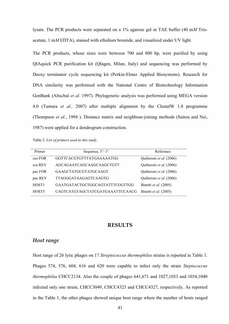
Table 2. List of primers used in this study.
Primer Sequence, 5’- 3’ Reference
cos FOR GGTTCACGTGTTTATGAAAAATGG Quiberoni et al. (2006)
cos REV AGCAGAATCAGCAAGCAAGCTGTT Quiberoni et al. (2006)
pac FOR GAAGCTATGCGTATGCAAGT Quiberoni et al. (2006)
pac REV TTAGGGATAAGAGTCAAGTG Quiberoni et al. (2006)
HOST1 GAATGATACTGCTGGCAGTATTTCGGTTGG Binetti et al. (2005)
HOST5 CAGTCATGTAGCTATCGATGAAATTCCAACG Binetti et al. (2005)
RESULTS
Host range
Host range of 26 lytic phages on 17 Streptococcus thermophilus strains is reported in Table 1.
Phages 574, 576, 604, 616 and 620 were capable to infect only the strain Steptococcus
thermophilus CHCC2134. Also the couple of phages 641,671 and 1027,1033 and 1034,1040
infected only one strain, CHCC3049, CHCC4323 and CHCC4327, respectively. As reported
in the Table 1, the other phages showed unique host range where the number of hosts ranged

42
from just 1 to 4. Out of the 11 above reported, 6 other bacteriophages showed to be virulent
against only 1 strain, 3 against 2 strains, 4 against 3 strains and 2 against 4 strains. On the
other hand, all Streptococcus thermophilus strains, with exclusion of the strain Streptococcus
thermophilus AT1, were infected from at least one bacteriophage. No phage was able to infect
the three Streptococcus macedonicus strains (data not shown). Streptococcus thermophilus
CHCC2134 showed to be very sensitive to phage infection, resulting host for seven different
bacteriophages. Instead, the strains Streptococcus thermophilus CHCC4131, CHCC4325,
CHCC3048 and CHCC6592, resulted susceptible to only the phages 671, 1028, 1032 and
1042, respectively. All the other strains resulted sensitive to 2, 3 or 4 bacteriophages.

Table 1. Host range of 26 bacteriophages against 17 Streptococcus thermophilus strains.
Bacteriophages
Strains 574 575 576 577 591 596 603 604 605 607 611 616 620 641 642 654 671 1027 1028 1032 1033 1034 1036 1040 1041 1042
CHCC3063 - - - - - - - - - - + - - - + + - - - - - - + - - -
CHCC2070 - + - - - + - - - - - - - - - - - - - - - - - - + -
CHCC2130 - - - - + - - - - + - - - - - - - - - - - - - - - -
CHCC2133 - - - - + - + - + - - - - - - - - - - - - - - - - -
CHCC2134 + - + - - + - + + - - + + - - - - - - - - - - - - -
CHCC2389 - - - + - - - - - - - - - - - - - - + + - - - - - -
CHCC3048 - - - - - - - - - - - - - - - - - - - + - - - - - -
CHCC3049 - - - - - - - - - - - - - + + - + - - + - - - - - -
CHCC3050 - - - - - - - - - - - - - - + - - - - - - - + - - -
CHCC4131 - - - - - - - - - - - - - - - - - - - - - - - - - -
CHCC4323 - - - - - - - - - - - - - - - - - + + + + - - - - -
CHCC4325 - - - - - - - - - - - - - - - - - - + - - - - - - -
CHCC4327 - - - - - - - - - - - - - - - - - - - - - + - + + -
CHCC4460 - - - - - - - - - - + - - - + - - - - - - - + - - -
CHCC4895 - - - - - + - - - - - - - - - - - - - - - - - - + -
CHCC6592 - - - - - - - - - - - - - - - - - - - - - - - - - +
St At1 - - - - - - - - - - - - - - - - - - - - - - - - - -

44
Restriction analysis
The DNA restriction analysis showed only HindIII and EcoRV as capable to give a pattern
with 8-12 bands, suitable for the differentiation of the 26 bacteriophages, while the enzyme
PstI gave patterns with less than 3 bands (Figure 1). Most phages had a unique profile, instead
the phages 604, 620 and 654 showed a similar pattern with 6 or 8 bands when their DNA
were digested by HindIII and EcoRV, respectively. However, while the phages 604 and 620
had the same host range (they infected only Streptococcus thermophilus CHCC2134), the
phage 654 revealed a different host range.
Determination of DNA packaging mechanism
All the bacteriophages, with exclusion of 1042 and 575, showed an amplification fragment of
170 bp, indicating they belonged to the cos-type group. On the other hand only the
bacteriophage 1042 yelded a PCR fragment of 427 bp, distinctive of the pac-type phage
group. Surprisingly, the phage 575 able to infect only the strain Streptococcus thermophilus
CHCC2070 (Table 1), showed both 170 bp and 427 bp amplification fragments (Figure 2).
1 2 3 4 5 6 M
Figure 1. Representative restriction profiles of the Streptococcus thermophilus bacteriophages 574 and 575 digested by Hind III (lanes 1 and 2), PstI (lanes 3 and 4) and EcoRV (lanes 5 and 6). Lane M, I Kb plus.

45
Bacteriophage clustering based on VR2 sequences
The antireceptor variable region VR2 was amplified for all bacteriophages assayed with
exclusion of the phages 577, 607 642 and 1028. The PCR analysis showed an amplification
fragment of 700-800 bp (Figure 3).
The cluster analysis of VR2 sequences from both bacteriophages used in this study and some
Streptococcus thermophilus phages available in GenBank database gave a dendrogram where
the bacteriophages of this study were grouped in 6 clusters at 90% similarity level (Figure 4).
The cluster A grouped 7 bacteriophages at 100% similarity, the cluster B grouped 7
bacteriophages at about 92% similarity, the clusters C, D and F at 100% similarity grouped 2,
3 and 2 phages, respectively. Finally, only the phage 1041 belonged to the cluster E.
1 M 2 3
Figure 2. Multiplex PCR for the detection of the cos- and pac-type groups of Streptococcus thermophilus phages. Lane 1, representative cos-type phage 574; Lane 2, bacteriophage 575 showing both amplification products; Lane 3, pac-type phage 1042; lane M, 1 kb ladder.
Figure 3 Amplification of the antireceptor VR2 variable region of the bacteriophages in this study .Lanes from 1 to 9: phages 574, 575, 591, 596, 603,604,616,671, 1036, M 1kb plus ladder
1 2 3 4 5 6 7 8 9 M

46
Similarity (%)
A
B
C
D
E
F
Figure 4. Dendrogram (neighbor-joining method) showing the % of similarity of 27 Streptococcus thermophilus bacteriophages on the basis of antireceptor VR2 sequences

47
DISCUSSION
The increasing demand for dairy products in recent years has required increases in both
production capacity and process efficiency. In this optic the use of starter culture properly
meets these dairy industry needs. However, the repeated use of defined starter cultures under
non-aseptic processing conditions amplifies the phage proliferation and their infection ability
in dairy environments. The characterization of Streptococcus thermophilus phages is an
important tool for the selection of phage-resistant dairy starter cultures. In this study 26
phages isolated from mozzarella cheese whey samples were analyzed on the basis of their
host range, DNA packaging mechanism and DNA restriction profile. Furthermore, the phages
were clustered considering the antireceptor variable region VR2 sequences and a correlation
with host range was ascertained.
The phage population assayed showed 19 different host ranges suggesting a good variability
in terms of infection capability. Interestingly, AT1 was the sole strain isolated from natural
whey culture used for the production of traditional mozzarella cheese (data not shown). This
environment is characterized by a very high microbial diversity, which probably represents a
natural barrier against phage infection (Carminati et al., 1997). This strain could be taken in
account for the assessment of other industrial performances such as acidifying and proteolytic
activity. On the contrary, the strain CHCC2134, as well as other multi-sensible strains, should
be eliminated as a hypothetic starter culture because of the high risk of phage infection. Our
results on host range were generally in agreement with Binetti et al. (2005) who described the
host ranges of 15 bacteriophages against 14 Streptococcus thermophilus strains, all from dairy
environment. They reported the phages had different host ranges, and the number of hosts
ranged from just one to four strains. By contrast, only 16 Streptococcus thermophilus strains
out of 36 could be found as sensitive to a number of bacteriophages ranging from 1 to 4, and

48
as many as 9 strains were sensitive to more of 11 among 27 bacteriophages tested (Le Marrec
et al. 1997).
The high variability within the phage population was confirmed by restriction profile analysis.
In fact, most phages showed a unique pattern, as obtained by using HindIII and EcoRV
enzymes. On the contrary, as reported in results, restriction profiles obtained by using PstI
were not suitable for the differentiation of the 26 bacteriophages. Only the phages 604, 620
and 654 showed similar patterns when their DNA was digested by HindIII ( 6 bands) and
EcoRV (8 bands) (data not shown). The similarity among these phages was confirmed by the
results of VR2 sequence analysis, where all the three phages belonged to the cluster A (Figure
3). However, while the phages 604 and 620 showed also the same host range being able to
infect the host Streptococcus thermophilus CHCC2134 (Table 1), the phage 654 infected a
different host, i.e. Streptococcus thermophilus CHCC3063 strain. On the other hand, the
phages 574 and 576, belonging to the cluster A obtained by VR2 sequence analysis, showed
same host range (infected Streptococcus thermophilus CHCC2134) but different restriction
profiles.
The phylogenetic tree showed 6 clusters at 90% similarity level. The bacteriophages that
showed a good correlation between host range and VR2 sequence were 604, 574, 576 and
620, which belonged to the cluster A, 1027 and 1033, which belonged to the cluster B and
1034 and 1040 which belonged to the cluster F. The phages 641 and 671 had the same host
range but they belonged to two very distant clusters. In agreement with Binetti et al. (2005)
these results might indicate further phage factors are involved in host specificity.
Furthermore, the multiplex PCR allowed distinguishing the cos-type and the pac-type phages.
In this study only the bacteriophage 1042 and the temperate phage from Streptococcus
thermophilus CHCC2070 gave an amplification fragment distinctive of pac-type group. All
the other bacteriophages belong to the cos-type. As reported by Quiberoni et al. (2006),

49
discriminating the pac-type and cos-type phages could be interesting to remove Streptococcus
thermophilus strains sensitive to both phages group. In this study the unique pac-type phage
1042 infected only Streptococcus thermophilus strain CHCC6592, whereas all the other
strains were sensitive to cos-type phages, confirming previous study demonstrating that rarely
Streptococcus thermophilus strains were infected by members of both phage groups
(Lévesque et al., 2005) Surprisingly, the bacteriophage 575 that infected only Streptococcus
thermophilus strain CHCC2070 was shown to have both PCR products for cos-type and pac-
type group. Inducing the Streptococcus thermophilus CHCC2070 by Mitomycin C the strain
resulted lysogenic and the PCR performed with its lysate yielded an amplification fragment
distinctive of the pac-type group. Definitively, the results suggested that the bacteriophage
575 lysate contained both its DNA (providing the cos-type fragment) and CHCC2070
prophage (providing the pac-type fragment).
In conclusion, the high diversity among bacteriophages highlights the serious risk of phage
infection during fermented food processing. On the other hand, we registered the presence of
a high percentage of low sensitive Streptococcus thermophilus strains. Testing a hypothetic
starter culture against a phage population, characterized by a well determined high variability,
is an efficient tool to exclude bacterial strains bringing on possible fermentation failure.

Chapter 4
Evidence of a temperate bacteriophage from lysogenic
Streptococcus macedonicus AI4
50
SUMMARY
The bacterial strains used in this study were investigated for the presence of temperate phages by induction with mitomycin C. Only the Streptococcus thermophilus CHCC2070 and Streptococcus macedonicus AI4 showed to be lysogenic and the temperate phage PZ1ϕ from Streptococcus macedonicus AI4 was choosen for detailed investigation. PZ1ϕ seems to belong to Bradley’s phage group B1, having a hexagonal head and a long, non-contractile tail.
INTRODUCTION
Streptococcus macedonicus, named cause of its place, Macedonia (Greece) where it was first
found, was isolated during a survey of the LAB microbiota of naturally fermented Greek
Kasseri cheese (Tsakalidou et al., 1998). S. macedonicus seems to be associated with
traditional dairy manufacture produced with raw milk without the use of selected starter
cultures (Mora et al., 2003). Whereas the first strains of S. macedonicus have been isolated
from Greek Kasseri cheese, it’s reported that the other strains have been isolated from very
different cheese: French cheese (Callon et al. 2004) and Italian cheese designated as PDO or
traditional cheese, as Asiago, Montasio, Ragusano and Mozzarella cheese (Lombardi et al.
2004; Pacini et al., 2006).
The species S. macedonicus belongs to the thermophilic homofermentative LAB strains. It is
nutritionally fastidious (multiple amino acid auxotrophies, restricted proteolityc system, etc.),
requiring the addition of appropriate nitrogen sources. Strains of S. macedonicus are
moderately acidifying and proteolytic probably due to weak (phospho)-β-galactosidase
activity and extracellular proteolytic activity, respectively (Georgalaki et al. 2000). This does
not allow S. macedonicus to completely acidify milk as the sole starter strain. However,
strains of S. macedonicus do precipitate in fat hydrolysis, citrate consumption and casein
hydrolysis in milk, underlining their usefulness as adjunct or co-culture (Georgalaki et al.,
2000). Furthermore, it was found that some S. macedonicus strains displayed antimicrobial

51
activity towards Clostridium tyrobutyricum, but no activity was detected against Listeria
innocua. These antagonistic activities of S. macedonicus strains point towards their use to
combat spoilage and late loss in hard and semi-hard cheeses. The strain S. macedonicus
ACA-DC 198, for example, produces a food-grade lantibiotic, named macedonicin.
Lantibiotics are a class of LAB bacteriocins that are post-traslationally modified, including
the formation of didhydro amino acids and thioether amino acids; bacteriocins are defined as
ribosomally synthesized antibacterial peptides that kill closely related species (De Vuyst and
Vandamme, 1994). S. macedonicus is a food-grade streptococcus, isolated from cheese, wich
does non exhibit potential pathogenicity traits. The exact role of S. macedonicus in cheese
making has still to be elucidated. However, its natural occurrence in different European
cheeses, probably originating from raw milk used, its potential contribution to cheese
ripening, in particular with respect to milk fat hydrolysis and peptidolytic activity, and its
potential role to combat or late loss during ripening strengthen its importance as non-starter
LAB and adjunct culture in cheese manufacture (De Vuyst and Tsakalidou, 2008) has been
recently investigated. To know its sensibility to bacteriophages could be interesting to
evaluate a criterion to select efficient strains.
EXPERIMENTAL PROCEDURES
Induction of bacteriophages
Induction of temperate phages was performed on all bacterial strains used in this study, by
addition of Mitomycin C at final concentration of 0.2 µg/mL when the cultures had an
OD600nm= 0.2. The presence of temperate phages was indicated by decrease of culture
turbidity up to total clarification of the broth monitored by spectrophotometer measures at 600

52
nm. The lysate obtained was centrifuged at 8000 g for 10 min to remove bacterial cell debris,
the supernatant was filtered through 0.45 µm membrane and stored at 4°C.
Screening for indicator strains
The possible sensitivity of 60 S. macedonicus strains isolated from samples collected during
Provolone del Monaco making process (Aponte et al., 2008), was determined in two ways: i)
a spot test on agar medium involved depositing drops of lysate of temperate phages or of
appropriate dilutions on M17-Ca++ agar, with previously poured in a double layer, the second
soft layer having been inoculated with the strain tested. ii) M17-Ca++ broth was inoculated
with each strain at 1%, 0.5 ml of PZ1ϕ lysate was added followed by incubation at 37°C. The
tubes were examined visually at regular intervals. Control and lysate containing cultures was
transferred at 2% to fresh M17-Ca++ medium and a second or third transfer was performed.
Determination of DNA packaging mechanism
To rapidly classify S. macedonicus temperate phages within one of 2 groups (cos– and pac-
type) it was used the Quiberoni et al (2006) multiplex PCR protocol by using two pairs of
primers (one per phage group Table 1) designed from the conserved regions of the gene
coding for the major capsid protein in the phages for which the complete genome is available.
The PCR reactions were performed in a total volume of 50 µL containing 125 µM
deoxynucleoside triphosphate, 5 µM concentrations of the 4 primers, 2.5 U of Taq DNA
polymerase, Taq buffer (20 mM Tris-HCl pH 8.4, 1.5 mM magnesium chloride, 50 mM
potassium chloride). The templates were constituted by i) 1 µL of the temperate phage lysate
ii)10 ng of bacterial total DNAof S. macedonicus AI4 iii) 10 ng of bacterial total DNA of S.

53
gallolyticus subsp macedonicus 18488T. Phage DT1 (Tremblay and Moineau, 1999) was used
as cos-type positive control. Phage DT1 was propagated on S. thermophilus SMQ-301.
Negative control (without the template) was included for all PCR assays to eliminate the
possibility of contamination.
The conditions of PCR amplifications were set as follows: 5 min at 94°C, followed by 35
cycles (45 s at 94°C, 45 s at 53°C, 1 min at 73°C), and a final step of 5 min at 73°C. The PCR
products were separated on a 2% agarose gel in TAE buffer (40 mM Tris-acetate, 1 mM
EDTA), stained with ethidium bromide, and visualized under UV light. The fragments of 170
bp and 427 bp were distinctive of the cos-type and pac -type group, respectively.

54
Primer Sequence, 5’- 3’ Reference
cos FOR GGTTCACGTGTTTATGAAAAATGG Quiberoni et al. (2006)
cos REV AGCAGAATCAGCAAGCAAGCTGTT Quiberoni et al. (2006)
pac FOR GAAGCTATGCGTATGCAAGT Quiberoni et al. (2006)
pac REV TTAGGGATAAGAGTCAAGTG Quiberoni et al. (2006)
Dot blot hybridization
Hybridazation assays and labelling of DNA to be used as probe were performed by using the
enhanced chemiluminescence (ECL) system (Amersham) according to the supplier’s
recommendations. S. macedonicus AI4 total DNA and the DNA from PZ1ϕ was spotted on
nylon membrane (Hybond-N+ Amersham) and probed with DT1 DNA S. thermophilus
phage.
RESULTS
Evidence of temperate bacteriophages and screening of indicator strains
Among the bacterial strains tested for the presence of temperate phages, Streptococcus
thermophilus CHCC2070 and Streptococcus macedonicus AI4 exhibited cell lysis (Figure 1)
suggesting temperate bacteriophages from these microorganisms were induced. All the S.
thermophilus and S. macedonicus strains were tested as possible host for these temperate
phages, but no bacterial strain showed to be host for the phages.
The temperate phage from S. macedonicus named PZ1ϕ was observed by transmission
electron microscopy (Figure 2). PZ1ϕ seems to belong to Bradley´s phage group B1, having a
Table 1: List of primers used for multiplex PCR

55
hexagonal head of about 64 nm in diameter and a non-contractile tail of about 187 nm in
length.
S. macedonicus AI4
0,000
0,500
1,000
1,500
2,000
2,500
0,0 2,0 4,0 6,0 8,0
time (h)
Ab
s 600 n
m
Control
AI4 mc
S. thermophilus 2070
0,000
0,500
1,000
1,500
2,000
2,500
0,0 2,0 4,0 6,0 8,0
time (h)
Ab
s 600 n
m
Control
CHCC7020 mc
Determination of DNA packaging
The multiplex PCR performed on the lysate of the PZ1ϕ gave an amplification product of
170 bps, indicating the temperate phage belong to the cos-type group. The PCR performed
Figure 1 Effect of mitomycin C (mc) on S. macedonicus AI4(■ ) and S. thermophilus CHCC2070 (■). Control without mc (♦ )
Figure 2 Temperate bacteriophage PZ1ϕ from Streptococcus macedonicus AI4.

56
on the prophage of S. macedonicus AI4 yielded a product of the same size. No amplification
product was found for S. gallolyticus subsp. macedonicus 18488T (Figure 3).
Dot blot hybridization
The dot blot hybridization analysis confirmed the similarity with S. thermophilus phages
(Figure 4). However when the amount of DNA of S. macedonicus AI4 was diluted the signal
was weak.
Figure 3. Multiplex PCR for determination of DNA packaging. M 1kb ladder; 1 S. macedonicus AI4; 2 S. gallolyticus subsp. macedonicus 18488.T
M 1 2 3
Figure 4: Dot blot signal of S. macedonicus AI4 total DNA hybridizated with S. thermophilus DT1 probe.

57
DISCUSSIONS
Lactic Acid Bacteria constitute the majority in volume and value of the commercial starter
with the largest amount being applied in dairy industry (Hansen, 2002). Among the
streptococci sensu lato, S. thermophilus strains are widely used in dairy manufactures and in
the last decade several dairy S. thermophilus-like microogranisms have been identified, due
to their unusual carbohydratate fermentation pattern and their different genotype as compared
with those of S. thermophilus strains sensu stricto (Moschetti et al., 1998). S. macedonicus
(Tsakalidou et al., 1998) and S. waius ( Flint et al., 1999) have been described in detail,
which has allowed some clarification of the taxonomically undefined group of S.
thermophilus-like microrganisms. Whether these strains are discovered in natural whey
starters could influence the stability of fermented dairy products other aspects need to be
studied further.
Since the lysogenic starter strains are considered a major source of bacteriophages in cheese
plants, the induction of all bacterial strains used in this study was carried out to evidence the
presence of temperate phages. Only S. thermophilus CHCC2070 and S. macedonicus AI4
showed to be lysogenic. The importance of the role of S. macedonicus strains as adjunct
culture (De Vuyst and Tsakalidou, 2008) in cheese manufacture has addressed the attention to
its temperate phage. By electron transmission microscopy, S. macedonicus phage PZ1ϕ
appears to belong to group B according to Bradley’s classification having a hexagonal head
and a long non-contractile tail. However, while it seems the lysogenic starter strains are main
source of phages in dairy environments (Zhang et al, 2006), the difficulty in finding indicator
strains is contradictory to this notion. Sixty S. macedonicus strains isolated from different
sources were tested as indicator strains, but no bacterial cell was found to be a host. The
multiplex PCR indicated that the temperate phage belong to the cos-type group and the
similarity with the S. thermophilus phage was confirmed by dot blot hybridization.

58
At the best of our knowledge, this is the first evidence for a phage within the S. macedonicus
species.

Chapter 5
Genomic sequence of S. macedonicus temperate phage
PZ1ϕ by genome walking method - work in progress.

59
SUMMARY
The difficulties in finding indicator strains for S. macedonicus temperate phage PZ1ϕ and the impossibility to amplify the phage and to extract an adequate amount of its DNA, led to an alternative method to sequence unknown DNA regions. The PCR-based genome walking method was used starting from a known DNA cos fragment of PZ1ϕ . By taking multiple steps using new primers based on the sequence obtained in previous steps, it is possible to reveal unknown DNA sequences. The research need to be continued because of too small fragments obtained at this time.
INTRODUCTION
The genome walking method is the systematic identification of unknown regions flanking a
known DNA sequence. Isolation of such unknown flanking regions can be obtained by
screening a genomic library with a known DNA as probe. However, this is time consuming
for preparation of libraries and their screening to obtain the desired DNA fragment (Rishi et
al., 2004). By using the polymerase chain reaction (PCR) is possible to isolate unknown
flanking regions of known DNA sequence, efficiently and fast. Different PCR methods, such
as inverse PCR (Ocham et al., 1988), and adaptor-specific PCR (Siebert et al., 1995) have
been used to isolate the unknown flanking regions. In both cases the genomic DNA is
digested by restriction enzymes and ligated to self or to adaptor. The ligated product is then
used as template to amplify flanking regions using PCR. The GenomeWalker TM Universal
kit (Coletech, Palo Alto, CA, USA) is used by starting the known cos- fragment of
PZ1ϕ. The first step is to construct pools of uncloned, adaptor-ligated genomic DNA
fragments. The genome walker takes just two days after library preparation and consists of
two PCR amplifications per library (Figure 1). The primary PCR uses the outer adaptor
primer (AP1) provided in the kit and outer genes-specific primer (in this case that used for
amplification of cos- fragment). The primary PCR mixture is then diluted and used as

60
template for a secondary, nested PCR by using a nested specific primer. This produces
generally a single, major PCR product from at least one library.
The genome walking method enables to create uncloned libraries for walking by PCR in any
genomic DNA. In less than a week the method provides access to the genomic DNA
sequences adjacent to a known DNA sequence in any species. The method allows to walk
simply by taking multiple steps using new primers based on the sequence obtained in
previous steps. However, the genome walking has some limitation in amplifying (either
smaller fragment amplification or no amplification) the desired size of flanking DNA, when
the restriction site is close to the upstream location of the gene specific primer in the genome
( Rishi et. al., 2004). To overcome this problem it could be possible to use different
restriction enzymes, so that there is a grater chance to obtain templates wherein the location
of the restriction site is distant from the gene-specific primer.
In this study the genome walking method is applied to determine the genome of PZ1ϕ by
using the total DNA from S. macedonicus AI4 starting from the cos fragment.
EXPERIMENTAL PROCEDURES
Construction of Genome WalkerTM Libraries
For each library construction the DNA of S. macedonicus AI4 strain was digested by
restriction enzymes DraI, EcoRV, PvuII, separately, provided from kit, used according to
the manufacture’s instructions. Restricted phages DNA were electrophoresed in a 0.8%
agarose gel in TAE buffer (40 mM Tris-acetate, 1 mM EDTA), stained with ethidium
bromide and visualized under UV illumination. The digested DNA was ethanol precipitated
after phenol:chloroform (1:1) extraction. The DNA pellet was in water and 0.5 µg of

61
completely digested DNA was used to ligate with the “Genome Walker Adaptor” according
to manufacturer’s protocol.
PCR amplification
The PCR reactions were carried out in a total volume of 50 µl containing the following to a
final concentration: 125 µM deoxynucleoside triphosphate, 5 µM concentrations of the 4
primers, 2.5 U of Taq DNA polymerase, Taq buffer (20 mM Tris-HCl pH 8.4, 1.5 mM
magnesium chloride, 50 mM potassium chloride), and 1 µl of DNA coming from each library
and using the primers as described in table 2. The list of primers used for these amplifications
are listed in table 3, where the AP1for was provided by Genome WalkerTM . The PCR
conditions were 94°C, 25 s; 67 °C 3 min for 7 cycles; 94 °C 25 s, 67 °C 3 min for 32 cycles
amd an additional one cile at 67°C for 10 minuts. The PCR products were purified by using
QIAquick PCR purification kit (QIagen, Milan, Italy) and sequencing was performed by
Deoxy terminator cycle sequencing kit (Perkin-Elmer Applied Biosystems).
DNA library Sample No. 1 PCR primers 2 PCR primers 1 DraI 1 COSrev & AP1 WALK1rev & AP1 2 EcoRV 2 COSrev &AP1 WALK1rev &AP1 3 PvuII 3 COSrev &AP1 COSrev &AP1
Primer Sequence, 5’- 3’ Reference
COS rev AGCAGAATCAGCAAGCAAGCTGTT Quiberoni et al. (2006)
AP1 for GTAATACGACTCACTATAGGGC Genome WalkerTM
WALK1 rev GACCTTTCTTGTCATGTGTCTTCG This study
Table 1 Schematic plan used for first and second step during genome walking.
Table 2: List of primers used in this study.

62
RESULTS and DISCUSSION
To identify the genomic DNA sequences adjacent to cos segment of S. macedonicus
prophage AI4 a genome walking method was applied. By using the Genome WalkerTM it
was possible to identify the upstream region that result in amplification of fragments of less
than 500 bp in two libraries created by using DraI and EcoRV, but no amplification
fragments was observed in the PvuII library (Figure 1). Since the first PCR produced a
single major product from at least one library, the following nested PCR was not performed.
The amplifications of the small fragments could be due to the presence of a restriction site in
the genome close to the designed primer and the negative result in the PvuII library may be
due to an absence of a restriction site close the gene-specific primer. Since the location of
the restriction site relative to the gene-specific primer in the genome is not known, the use of
different enzymes-digested libraries could result in amplifications of fragments of different
sizes and relatively larger fragments in some libraries. The first step yielded a PCR product
of 500 bp from DraI library and the cos fragment was contained in this longer new fragment.
The second reverse primer WALK1 designed basing on the sequence obtained in previous
step was used to walk on the genome and gave a PCR product of about 200 bp. However,
the too small fragments obtained do not allow to obtain rapidly the complete sequence,
considering that a phage genome ranges in size from 30 to 45 kb.

63
cos 2
cos 2
cos 1
cos 1
1 2 3 M
Figure 1. The GenomeWalkerTM method, the samples loaded are listed in the table 1 and correspond to the results obtained from the first PCR. M: 1kb plus ladder.

64
REFERENCES
Ackermann, H-W., Cantor, E.D., Jarvis, A.W., Lembke, J., and Mayo, J. (1984). New species
definitions in phages of gram-positive cocci. Intervirology 22: 181.
Ackermann and DuBow M.S. (1987). Viruses of Prokaryotes 1 & 2. CRC Press, Boca Raton. Altermann E. and Henrich B. (2003). Transcript map of the temperate Lactobacillus gasseri
bacteriophage adh. Microbiology 149: 2987-2999.
Altermann E, Kein J. R. and Henrich B. (1999) Primary structure and features of the genome
of theLactobacillus gasseri temperate bacteriophage adh. Gene 236: 333-346.
Altschul, S., T. Madden, A. Schäffer, J. Zhang, Z. Zhang, W. Miller, and D. Lipman. (1997).
Gapped BLAST and PSI-BLAST: a new generation of protein database search
programs. Nucleic Acids Res. 25:3389-3402.
Aponte M., Fusco V., Andolfi R. and Coppola S. (2008). Lactic acid bacteria occurring
during manufacture and ripening of Provolone del Monaco cheese: Detection by
different analytical approaches. Int. Dairy J.18 : 403-413.
Arendt E. K., Daly C., Fitzegerald, G.F.,and de Guchte, M. (1994). Molecular
characterization of lactococcal bacteriophage Tuc2009 and identification and analysis of
genes encoding lysin, a putative holin, and two structural proteins. App. Environ.
Microbiol. 60:1875-1883.
Beck, K., and B. Brodsky. (1998). Supercoiled protein motifs: the collagen triple-helix and
the α-helical coiled coil. J. Struct. Biol. 122:17-29.
Binetti A. and Reinheimer (2000). Thermal and chemical inactivation of indigenous
Streptococcus thermophilus bacteriophages isolated from Argentinean dairy plants. J.
Food Prot. 63:509-515.

65
Binetti, A. G., B. Del Rio, M. C. Martin, and M. A. Alvarez (2005). Detection and
characterization of Streptococcus thermophilus bacteriophages by use of the antireceptor
gene sequence. Appl. Environ. Microbiol. 71:6096–6103.
Black LW (1989) DNA packaging in dsDNA bacteriophages. Annu Rev Microbiol 43: 267-292.
Brussow H. (2001). Phages of dairy bacteria. Annals Reviews of Microbiology 55:283-303.
Brussow H. and Bruttin A. (1995). Characterization of a temperate Streptococcus
thermophilus bacteriophage and its genetic relationship with lytic phages. Virology
212:632-640.
Brussow H., Bruttin A., Desiére F., Lucchini S. and Foley S. (1998). Molecular ecology
and evolution of Streptococcus thermohilus bacteriophages .Virus Genes 16:95-109.
Brüssow H. and Desiere F. (2001). Comparative genomics and the evolution of
Siphoviridae: insights from dairy phages. Mol Microbiol 39: 213-222.
Brussow H., Frémot M., Bruttin A., Siidoti J, Constable A, et al. (1994a) Detection and
classification of Streptococcus thermophilus bacteriophages isolated from industrial
milk fermentation. App. Environ. Microbiol. 60:4537-4543.
Brussow H, Probst A., Frémot M. and J Sidoti (1994b). Distinct Streptococcus
thermophilus bacteriophages share an extremely conserved DNA fragment. Virology,
200: 854-857.
Bruttin, A., Desière, F., Lucchini, S., Foley, S.,and Brussow, H. (1997). Characterization
of the lysogeny DNA module from the temperate Streptococcus thermophilus
bateriophage Sfi21. Virology 233:136-148.
Bruttin A., Foley S., Brussow H. (1997). The site-specific integration system of the
temperate Streptococcus thermophilus bacteriophage Sfi21. Virology 237:148-158.

66
Bruttin A. and Brussow, H. (1996). Site-specific spontaneous deletions in three genome
regions of a temperate Streptococcus thermophilus phage. Virology 219: 96-104.
Callon, C., Millet, L., and Montel, M. C. (2004). Diversity of lactic acid bacteria isolated
from AOC Salers cheese. Journal of Dairy Research 71: 231–244.
Campbell, A., and Botstein, D. (1983) Evolution of the lambdoid phages. In: Lambda II
(R.W. Hendrix, J.W. Roberts, F.W. Stahl, and R.A. Weisberg, 365-380, Cold Spring
Harbor Laboratory Press, Cold Spring Harbor, NY.
Carminati D., L. Mazzuccotelli, G. Giraffa and Neviani E. (1997). Incidence of inducible
bacteriopahge in Lactobacillus helveticus strain isolated from natural whey starter
cultures. Journal of Dairy Science 80: 1505-1511.
Casjens, S., Hatful, G., and Hendrix, R. (1992). Evolution of dsDNA tailed-bacteriophage
genome. Semin. Virology 3:383-397.
Chandry P. S. Davidson B.E. Hiller A.J. (1994). Temporal transcription map of
Lactococcus lactis bacteriophage sk1. Microbiol. 140 :2251-2261.
Coffey A., and Ross R. P. (2002). Bacteriophage-resistance systems in dairy industry starter
strains: molecular analysis to application. Antoine van Leeuwenhoeck 82: 303-321.
Cogan, T. M., N. Peitersen, and R. L. Sellars. (1991). Starter systems. Bulletin of the
International Dairy Federation, no. 263/1991. Practical phage control. International
Dairy Federation. Brussels. 16-23
Coppola S., E. Parente, S. Dumontet and A. La Peccerella (1988). The microflora of
natural whey cultures utilized as starter in the manufacture of Mozzarella cheese
from water-buffalo milk. Le Lait, 68:295-310.
Daly, C and Fitzgerald G.,F. (1982) Microbiology. D.Shelessinger, ed. 213-216 .
Desière F., Lucchini, S., and Brussow, H.(1998). Evolution of Streptococcus thermophilus
bacteriophage genome by modular exchanges followed by point mutations and small
deletions and insertions. Virology 241: 345-356.

67
Desière F., Lucchini, S., Bruttin, A., Zwahlen, M. C., and Brussow H. (1997). A highly
conserved DNA replication module from Streptococcus thermophilus phages is
similar in sequence and topology to a module from Lactococcus lactis phage.
Virology 234:372-382.
Desiere F., Lucchini S., Canchaya C., Ventura M. and Brussow H (2002) Comparative
genomics ofphages and prophages in lactic acid bacteria. Antonie van Leeuwenhoek
82: 73-91.
Desiere F., McShan W.H., van Sinderen D., Ferretti J.J. and Brussow H. (2001)
Comparative genomics reveals close genetic relationships between phages from dairy
bacteria and pathogenic streptococci: evolutionary implications for prophage-host
interactions. Virology 288: 325-341.
Desière F., Pridmore R. D., Brussow H., (2000). Comparative genomics of the late gene
cluster from Lctobacillus phages. Virology 275:294-305.
De Vuyst L. and Vandamme E. J.,( 1994). Bacteriocins of lactic bacteria: microbiology,
genetics and application London UK: Blackie Academic and Professional.
Dinsmore P. K. and Klaenhammer T. R., (1995) Molecular characterization of a genomic
region in Lactococcus bacteriophage that is involved in its sensitivity to the phage
defense mechanism AbiA. J. Bacteriol. 179:2949-2957.
Diaz-Ruiz, G., Guyot, J. P., Ruiz-Teran, F., Morlon-Guyot, J., and Wacher, C. (2003).
Microbial and physiological characterization of weakly amylolytic but fast-growing
lactic acid bacteria: A functional role in supporting microbial diversity in pozol, a
Mexican fermented maize beverage App Environ. Microbiol. 69:4367–4374.

68
Dupont K, Janzen T, Vogensen F.K., Josephsen J. and Stuer-Lauridsen B. (2004)
Identification of Lactococcus lactis genes required for bacteriophage adsorption.
Appl. Environ. Microbiol. 70: 5825-5832.
Durmaz E. and Klaenhammer T. R (1995). A starter culture rotation strategy incorporating
paired restriction/modification and abortive infection bacteriophagerefenses in a single
Lactococcus lactis strain. Appl. Environ. Microbiol. 61:1266-1273.
Everson T.C. (1991). Control of phage in the dairy plant. Practical phage control FIL-IDF
Bullettin 263:16-23.
Filosofo T., Lombardi A., Cislaghi S., Ceccarelli P., Pirovano F., Minissi S., et al. (1995).
Phage attacks cause defects in whey cultures. Il latte 20: 1120-1123.
Flint, S. H., Ward, L. J. H. and Brooks, J. D. (1999). Streptococcus waius sp. nov., a
thermophilic streptococcus from a biofilm. Int. J. Systematic Bacteriology 49: 759–767.
Garbutt, K. C., Kraus J. and Geller B. L. (1997). Bacteriophage resistance in Lactococcus
lactis engineered by replacement of a gene for a bacteriophage receptor. J. Dairy Sci.
80:1512-1519.
Geller, B. L., Ivey R. G., Trempy J. E., and Hettinger-Smith B. (1993). Cloning of a
chromosomal gene required for phage infection in Lactococcus lactis subsp. lactis C2.
J.Bacteriol. 175:5510 5519.
Georgalaki M. D., Sarantinopoulos P., Ferriera E S, De Vuyst L., Kalantzopoulos G., and
Tsakalidou E. (2000) Biochemical properties of Streptococcus macedonicus strains
isolated from Greek Kasseri cheese. Journal of Applied Microbiology. 88:817-825.
Goldberg, E., Grinius, L. and Letellier, L. (1994) Recognition attachment and injection. In
Molecular Biology of Bacteriophages T4. Karam, J.D.(ed) Washington, DC:
American Society for Microbiology 347-356.

69
Hansen, E. B. (2002). Commercial bacterial starter cultures for fermented foods of the
future. Int. J. Food Microbiology, 78: 119–131.
Hull, R. R. (1985). Recent developments in the genetics of lactic acid bacteria. CSIRO
Food Res. Q. 45:40-46.
Klaenhammer, T.R. (1984) Interactions of bacteriophages with lactic streptococci. In:
Advances in Applied Microbiology 30: 1-29.
Kodaira, K.I., Oki, M., Kakikawa, M., Watanabe, N., Hirakawa, M., Yamada, K., and
Taketo, A. (1997).Genome structure of the Lactobacillus temperate phage gle: the
whole genome sequence and promoter/repressor system. Gene 187:45-43.
Lamothe G., Lèvesque C., Bissonnette F., Cochu A., Vadeboncoeur C. et al (2004).
Characterization of the cro-oriRegion of the Streptococcus thermophilus virulent
bacteriophage DT1. App.Environ. Microbiol. 71:1237-1246.
Le Marrec C., D. van Sinderen, L. Walsh, E. Stanley, E. Vlegels, S. Moineau, Heinze P,
Fitzgerald G. and Fayard B. (1997). Streptococcus thermophilus bacteriophages can
be divided into two distinct groups based on mode of packaging and structural
protein composition. Appl. Environ. Microbiol. 63:3246–3253.
Loessner M.W., Waterfield N.R., Beresford T.P. J., Le Page R.W.F., and JarvisA.W.
(1995). Heterogeneous endolysins in Listeria monocytogenes bacteriophages: a new
class of enzymes and evidence for conserved holin genes within the siphoviridale
lysis cassette. Mol.Microbiol. 16: 1231-1241.
Lombardi, A., Gatti, M., Rizzotti, L., Torriani, S., Andrighetto, C., and Giraffa, G. (2004).
Characterization of Streptococcus macedonicus strains isolated from artisanal Italian
raw milk cheeses. Int. Dairy J., 14, 967–976.
Lucchini S., Desière F., Brussow H (1999) similarly organized lysogeny modules in
temperate Siphoviridae from low GC content Gram-positive bacteria. Virology
263:427-435.

70
Lucchini S., Sidoti, J. and Brussow H. (2000). Broad-range bacteriophage resistance in
Streptococcus thermophilus by insertional mutagenesis. Virology 275:267-277.
Madsen P.L. and Hammer K. (1998). Temporal transcription of the lactococcal temperate
phages TP901-1 and DNA sequence of the early promoter region. Microbiol.
144:2203-2215.
Maniloff, J., Cadden S. P. and Putzrath R. M.(1981). Maturation of an enveloped budding
phage: mycoplasmavirus L2. Prog. Clin. Biol. Res. 64:503-513.
Mauriello G., Moio L., Genovese A and Ercolini D. (2003) Relationships between
flavouring capabilities, bacterial composition and geographical origin of natural
whey cultures used for traditional water buffalo mozzarella cheese manufacture. J.
Dairy Sci. 86: 486-497.
Moschetti, G., Blaiotta, G., Aponte, I., Catzeddu, P., Villani, F., and Coppola, S. (1998).
Random amplified polymorphic DNA and amplified ribosomal DNA spacer
polymorphism—powerful methods to differentiate Streptococcus thermophilus
strains. J. App. Microbiol 85: 25–36.
McGrath S., Seegers J.F., Fitzgerald G.and van Sinderen D. (1999). Molecular
characterization of a phage-encoded resistance system in Lactococcus lactis. Appl.
Environ. Microbiol. 65: 1891-1899.
McKay, L.L., and Baldwin, K.A. (1990) Applications for biotechnology: present and
future improvements in lactic acid bacteria. FEMS Microbiol Rev 87: 3-14.
Mercenier A. (1990). Molecular genetics of Streptococcus thermophilus. FEMS Microbiol.
Rev., 87:61-78.

71
Mikkonen M. and Alatossava T. (1994). Characterization of the genome region encoding
structural proteins of Lactobacillus delbrueckii subsp. lactis phage LL-H. Gene 151:
53-59
Moineau S. Application of phage resistance in lactic acid bacteria. In: Konings, W. N.,
Kuipers, O.P., Huis Veld, J.H.J. (1999) . (Eds) Proceedings if the Sixstg Symposium
on Lactic Acid Bacteria: Genetics, Metabolism and Applications. Kluwer Academic
Publishers, Veldhoven, The Netherlands, 377-382.
Mora D., M.G. Fortina, C. Parini, G. Ricci, M. Gatti, G. Giraffa, Manachini P.L. (2002).
Genetic diversity and technological properties of Streptococcus thermophilus strains
isolated from dairy products. J. Appl. Microbiol. 93278-287
Murray N.,(2000) Type I restriction systems: sophisticated molecular machines.
Microbiology and Molecular Biology Review 64 (2): 412-434.
Neve, H., Zenz K.I., Desière, F., Koch A., Heller, K.J., and Brussow, H. (1998).
Comparison of the lysogeny modules from the temperate Streptococcus thermophilus
bacteriophages TP-J34 and Sfi21: Implications for the modular theory of phage
evolution. Virology 241: 61-72.
Ocham H., Gerber A. S. and Hart D.L. (1988). Genetic application of an inverse
polymerase chain reaction. Genetics 120: 621-623.
Oliveira L., Alonso J.C., Tavares P., (2005). A defined in vitro system for DNA packaging by
the bacteriophage SPP1:insights into the heedful packaging mechanism. J. Mol. Biol.
353:529-539.
Olson, N. F. (1969). Ripened semisoft cheeses. Pfizer Cheese Monographs, vol. 4. Pfizer,
Inc., New York, NY.
O’Sullivan, D., Coffey A., Fitzgerald G. F., Hill C. and Ross R. P. (1998). Design of a phage-
insensitive lactococcal dairy starter via sequential transfer of naturally occurring
conjugative plasmids. Appl. Environ. Microbiol. 64:4618-4622.

72
Pacini, F., Cariolato, D., Andrighetto, C., and Lombardi, A. (2006). Occurrence of
Streptococcus macedonicus in Italian cheeses. FEMS Microbiology Letters, 261, 69–73.
Pedersen M., OstergaardS., BrescianiJ., Vongensen F.K. (2000). Mutational analysis of
two structural genes of the temperate lactococcal bacteriophage TP901-1involved in
tail length deermination and baseplate assembly. Virology 276:315-328.
Proux C, van Sinderen D, Suarez J, Garcia P, Ladero V, Fitzgerald GF, Desiere F and
Brüssow H. (2002) The dilemma of phage taxonomy illustrated by comparative
genomics of Sfi21-like Siphoviridae in lactic acid bacteria. J Bacteriol 184: 6026-
6036.
Quiberoni A., D. Tremblay, Ackermann W., Moineau S. and Reinheimer J. A. (2006).
Diversity of Streptococcus thermophilus Phages in a Large-Production Cheese Factory
in Argentina J. Dairy Sci. 89:3791–3799.
Ravin V., Räisänen L. and Alatossava T. (2002). A conserved C-terminal region in Gp71 of
the small isometric-head phage LL-H and ORF474 of the prolate-head phage JCL1032
is implicated in specificity of adsorption to its host, Lactobacillus delbrueckii. J
Bacteriol 184: 2455-2459.
Reinbold G.W., Reddy M.S. and Hammond E.G. (1982) Journal food protection 45, 119.
Riipinen, K., Räisänen, L., Alatossava, T., (2007). Integration of the Group C phage
JCL1032 of Lactobacillus delbruecckii subp. lactis and complex phage resistance of
the host. Journal Applied Microbiology 103: 2465-2475.
Rishi A.S. , Nelson N. D. and Goyal A., (2004). Genome walking of large fragments: an
improved method. J.Biotec, 111: 9-15
Saitou N. and Nei M.(1987). The neighbour-joining method: new method for
reconstructing phylogenetic trees. Molecular Biology and Evolution 12:823-833.

73
Salminen, S., Isolauri, E., and Salminen, E. (1996) Clinical uses of probiotics for
stabilizing the gut mucosal barrier: successful strains and future challenges. Antonie
van Leeuwenhoek 70: 347-358.
Sambrook J., E.F Fritsch and T. Maniatas. (1989). Molecular cloning: A laboratory manual.
Cold Spring Harbor Laboratory.
Sanger F., S. Nicklen and A. Coulson. (1977). DNA sequencing with chain-terminating
inhibitors. Proc. Natl Acad Sci USA 74:5463-5467.
Sheenan M.M.,Garcia JL., Lopez R., Garcia P. (1996). Analysis of the catalatyc domain of the
lysine of lactococcal bacteriophage Tuc2009 by chimeric gene assembly. FEMS
Micribiol. Lett. 140:23-28
Sheenan M.M., Stanley E., Fitzgerald G.F., van Sinderen D.(1999). Identification and
characterization of a lysis module present in a large proportion of bacteriophages
infecting Streptococcus thermophilus. App.Environ. Microbiol. 65:569-577.
Siebert P.D., Chenhick A., Kellogg D.E. Lukyanov K.A. , Lukyanov S.A., (1995). An
improved method for walking uncloned DNA.Nucl.Acids Res. 23:1087-1088.
Stanley E., Fitzgerald G.F., Le Marrec C., Fayard B., and van Sindersen D. (1997).
Sequence analysis and characterization of O1205, a temperate bacteriophage
infecting Streptococcus thermophilus CNRZ 1205. Microbiology 143:3417-3429.
Stadhouders,J.and Leenders G.J.M. (1984). Spontaneously developed mixed-strain cheese
starters. They behavior towards phages and their use in Dutch cheese industry.
Netherlands milk and dairy journal 38:157-181.
Sutherland, M., van Vuuren H. J., and Howe M. M. (1994). Cloning, sequence and in vitro
transcription/translation analysis of a 3.2-kb EcoRI-HindIII fragment of Leuconostoc
oenos bacteriophage L10. Gene 148 125-129.
Svensson, U. and A. Christiansson. (1991). Methods for phage monitoring, Bulletin of the
International Dairy Federation 263: 29-39.

74
Tamura, K., J. Dudley, M. Nei, and S. Kumar. (2007). MEGA4: Molecular Evolutionary
Genetics Analysis (MEGA) software version 4.0. Mol. Biol. Evol. 24:1596-1599
Tétart, F., Desplats ,C., and Krish H.M. (1998) Genome plasticity in the distal tail fibre
locus of T-even bacteriophage: recombination between conserve motifs swaps
adhesion specificity Journal Molecular Biology 282:543-556.
Thompson, J. D., D. G. Higgins, and T. J. Gibson. 1994. Clustal W: improving the sensitivity
of progressive multiple sequence alignment through sequence weighting, position-
specific gap penalties and weight matrix choice. Nucleic Acids Res. 22:4673-4680.
Tremblay, D. M., and S. Moineau (1999). Complete genomic sequence of the lytic
bacteriophage DT1 of Streptococcus thermophilus.Virology 255:63–76.
Tsakalidou, E., Zoidou, E., Pot B., Wssill L., Ludwig W., Devriese L.A., et al. (1998).
Identification of streptococci from Greek Kasseri cheese and description of
Streptococcus macedonicus sp.nov. International Journal of Systematic Bacteriology,
48: 519-527.
Ventura M. and Brüssow H. (2004) Temporal transcription map of the virulent Streptococcus
thermophilus bacteriophage Sfi19. Appl Environ Microbiol 70: 5041-5046.
Wang, J., Hofnung, M., and Charbit A (2000). The C-terminal portion of the tail fiber
protein of bacteriophages lambda is responsible for binding to lamB, its receptor at
the surface of Escherichia coli K-12. Journal bacteriology 182: 508-512.
Weigel C. and Seitz H. (2006). Bacteriophage replication modules. FEMS Microbiol. Rev 322-381.
Weisberg R.A. and Landy A. (1983). Site-specific recombination in phage lambda. In:
Hendrix RW, Roberts JW, Stahl FW & Weisberg RA (eds) Lambda II, Cold Spring
Harbor Laboratory, NewYork, 211-250.

75
Werts, C., Michel, V., Hofnung, M., and Charbit, A. (1994). Adsorption of bacteriophage
lambda on the lamB protein of Escherichia coli K-12: point mutation in gene J of
lambda responsible for extended host range. Journal bacteriology 176:941-947.
Zago M., Comaschi L., Neviani E and Carminati D. (2005). Investigation on the presence
of bacteriophage in natural whey starter used for the production of Italian long-
ripened cheeses. Milchwissenchaft 60:171-174.
Zago M., De Lorentis A., Carminati D., Comaschi L and Giraffa G. (2006) Detection and
identification of Lactobacillus delbrueckii subp. lactis bacteriophages by PCR. J.
Dairy Research. 73: 1-8.
Zhang X., J. Kong and Y. Qu. (2006). Isolation and characterization of a Lactobacillum
fermentum temperate bacteriophage from Chinese yogurt. Journal of Applied
Microbiology 101:857-863.