bacteriophages self assembly and applications. bacteriophages: definition & history...
Post on 20-Dec-2015
237 views
TRANSCRIPT

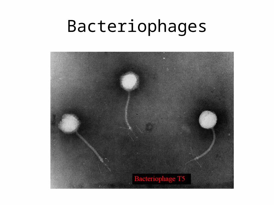
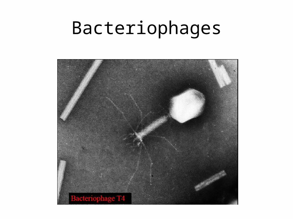
Bacteriophages
Self assembly and Applications
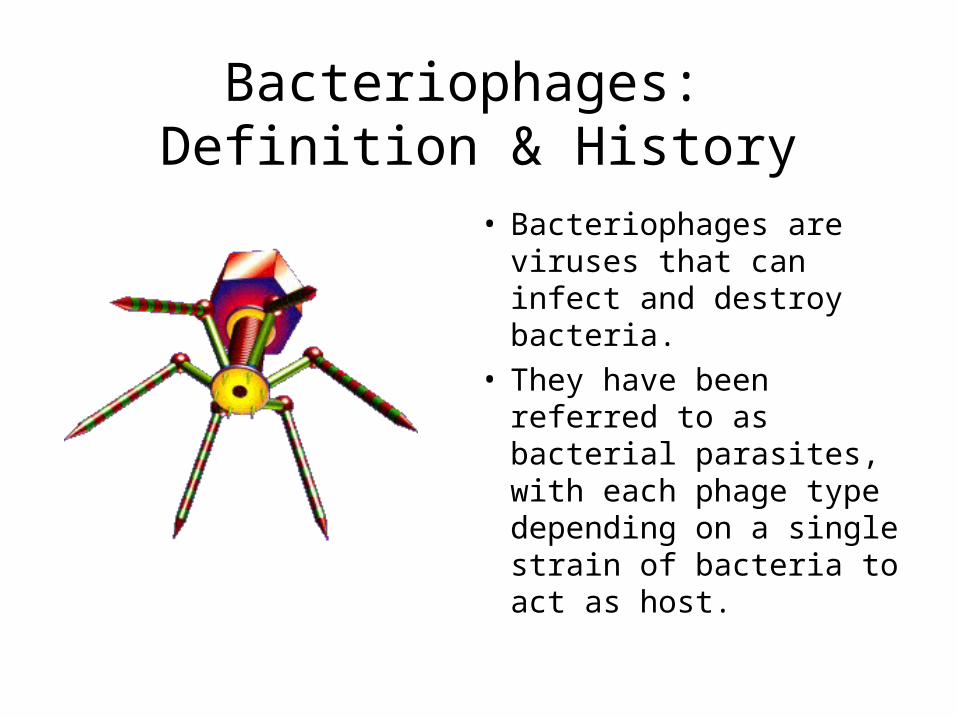
Bacteriophages: Definition & History
• Bacteriophages are viruses that can infect and destroy bacteria.
• They have been referred to as bacterial parasites, with each phage type depending on a single strain of bacteria to act as host.
Bacteriophages: Classification
• Based on two major criteria:
phage morphology (electron microscopy)
nucleic acid properties
Bacteriophages: Classification
• At present, over 5000 bacteriophages have been studied by electron microscopy and can be divided into 13 virus families.
5
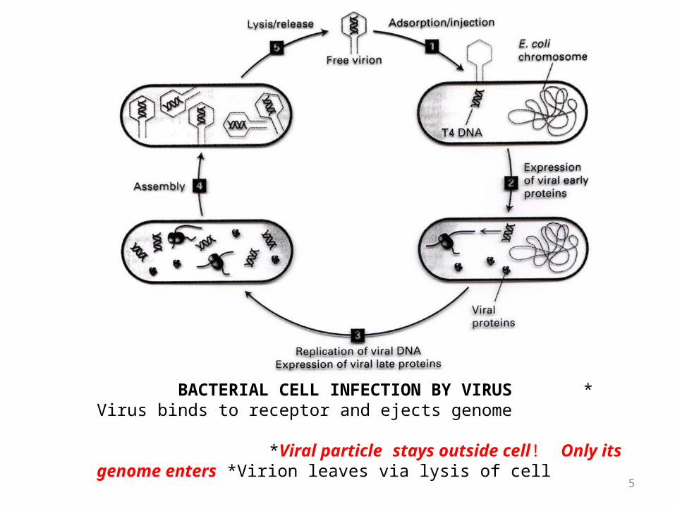
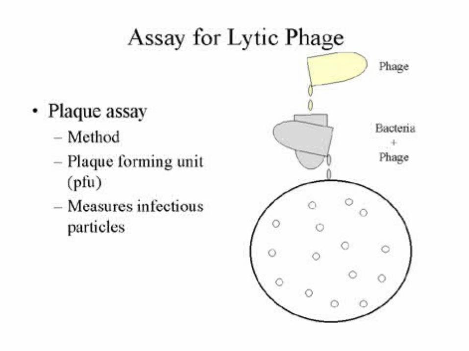
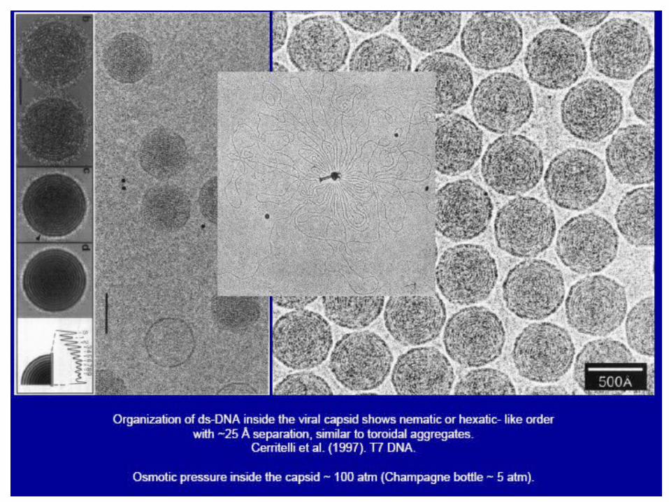
BACTERIAL CELL INFECTION BY VIRUS * Virus binds to receptor and ejects genome *Viral particle stays outside cell! Only its genome enters *Virion leaves via lysis of cell
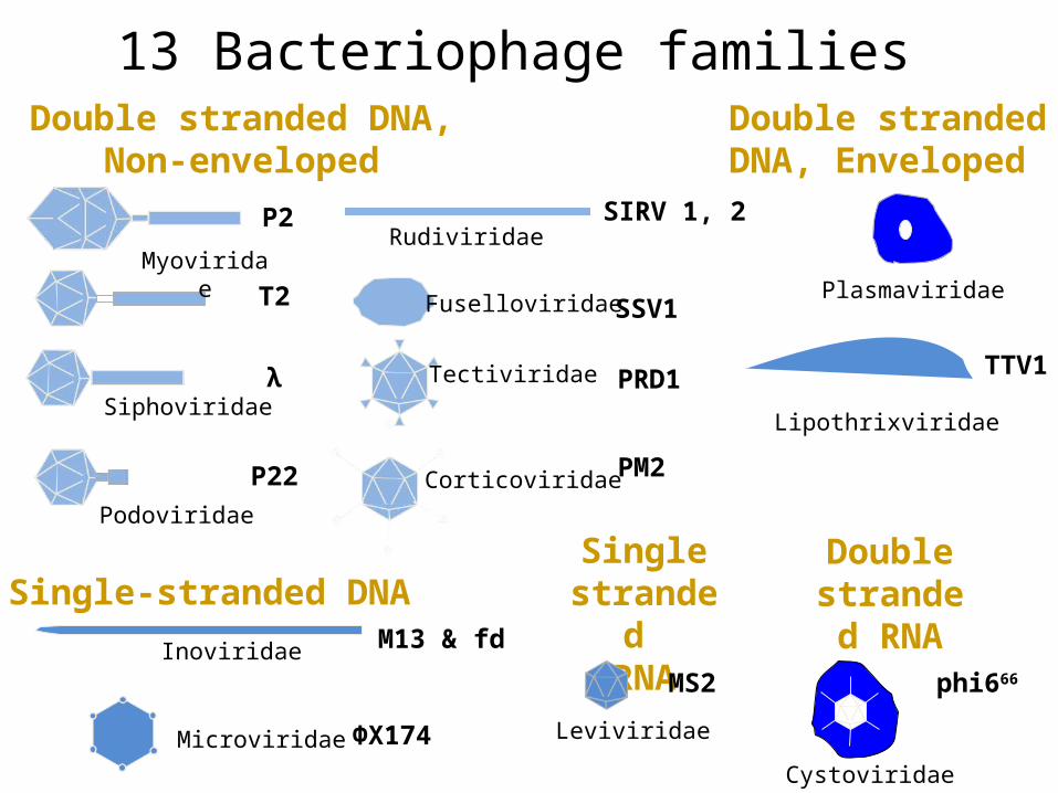
Double stranded DNA, Enveloped
Double stranded DNA,Non-enveloped
Myoviridae
Siphoviridae
Podoviridae
P2
T2
λ
P22
Tectiviridae PRD1
Corticoviridae PM2
Single-stranded DNA
Inoviridae M13 & fd
Microviridae ΦX174 Leviviridae
Single strande
d RNAMS2
Lipothrixviridae
TTV1
Fuselloviridae SSV1Plasmaviridae
Double stranded RNA
phi666
Cystoviridae
RudiviridaeSIRV 1, 2
13 Bacteriophage families
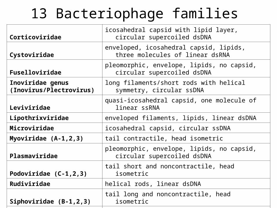
13 Bacteriophage familiesCorticoviridae
icosahedral capsid with lipid layer, circular supercoiled dsDNA
Cystoviridaeenveloped, icosahedral capsid, lipids, three molecules of
linear dsRNA
Fuselloviridaepleomorphic, envelope, lipids, no capsid, circular
supercoiled dsDNA
Inoviridae genus(Inovirus/Plectrovirus)
long filaments/short rods with helical symmetry, circular ssDNA
Leviviridae quasi-icosahedral capsid, one molecule of linear ssRNA
Lipothrixviridae enveloped filaments, lipids, linear dsDNA
Microviridae icosahedral capsid, circular ssDNA
Myoviridae (A-1,2,3) tail contractile, head isometric
Plasmaviridaepleomorphic, envelope, lipids, no capsid, circular
supercoiled dsDNA
Podoviridae (C-1,2,3) tail short and noncontractile, head isometric
Rudiviridae helical rods, linear dsDNA
Siphoviridae (B-1,2,3) tail long and noncontractile, head isometric
Tectiviridaeicosahedral capsid with, linear dsDNA, "tail" produced for
DNA injection
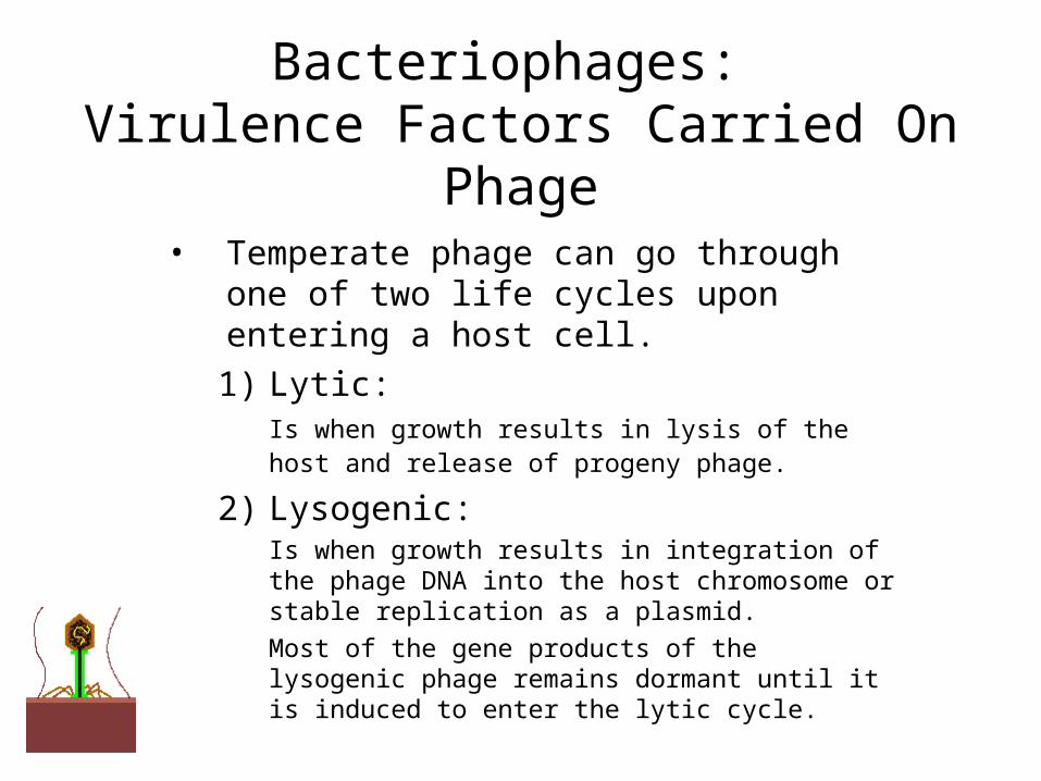
Bacteriophages: Virulence Factors Carried On Phage
• Temperate phage can go through one of two life cycles upon entering a host cell.
1) Lytic:Is when growth results in lysis of the host and release of progeny phage.
2) Lysogenic:Is when growth results in integration of the phage DNA into the host chromosome or stable replication as a plasmid. Most of the gene products of the lysogenic phage remains dormant until it is induced to enter the lytic cycle.
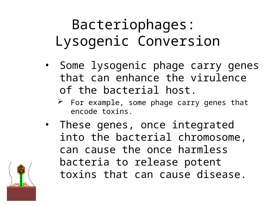
Bacteriophages: Lysogenic Conversion
• Some lysogenic phage carry genes that can enhance the virulence of the bacterial host. For example, some phage carry genes that encode toxins.
• These genes, once integrated into the bacterial chromosome, can cause the once harmless bacteria to release potent toxins that can cause disease.
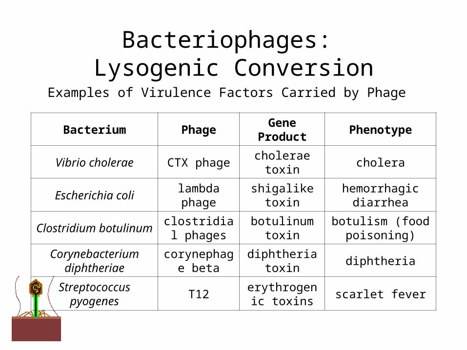
Bacteriophages: Lysogenic Conversion
Examples of Virulence Factors Carried by Phage
Bacterium PhageGene
ProductPhenotype
Vibrio cholerae CTX phage cholerae toxin cholera
Escherichia colilambda phage
shigalike toxinhemorrhagic
diarrhea
Clostridium botulinumclostridial phages
botulinum toxin
botulism (food poisoning)
Corynebacterium diphtheriae
corynephage beta
diphtheria toxin
diphtheria
Streptococcus pyogenes
T12erythrogenic
toxinsscarlet fever
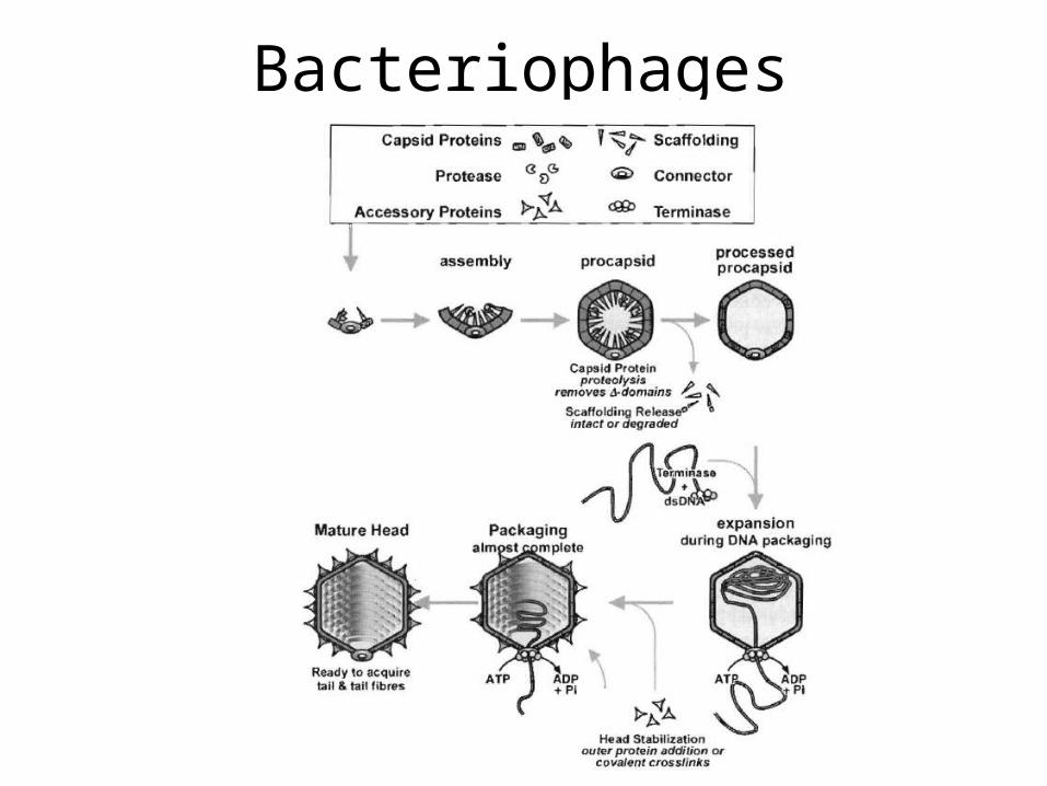
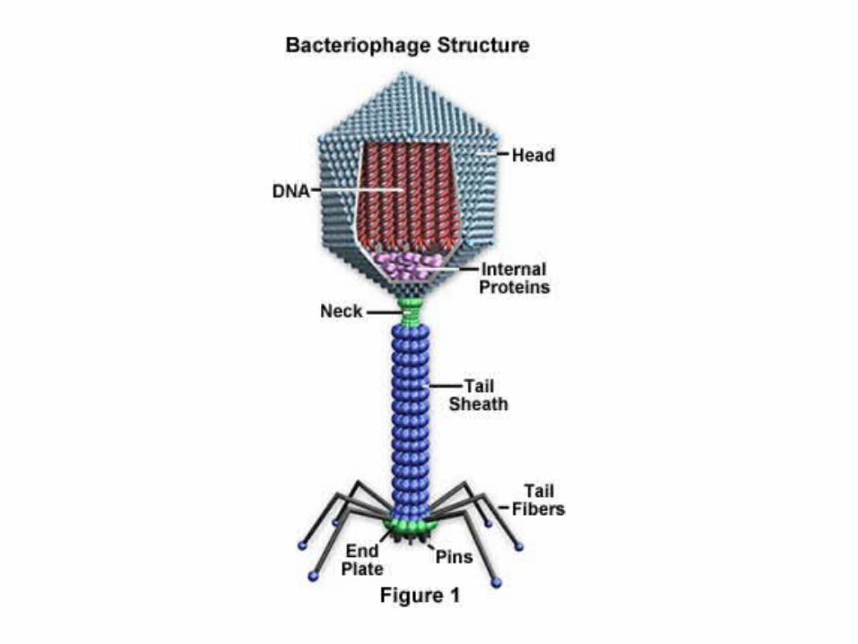
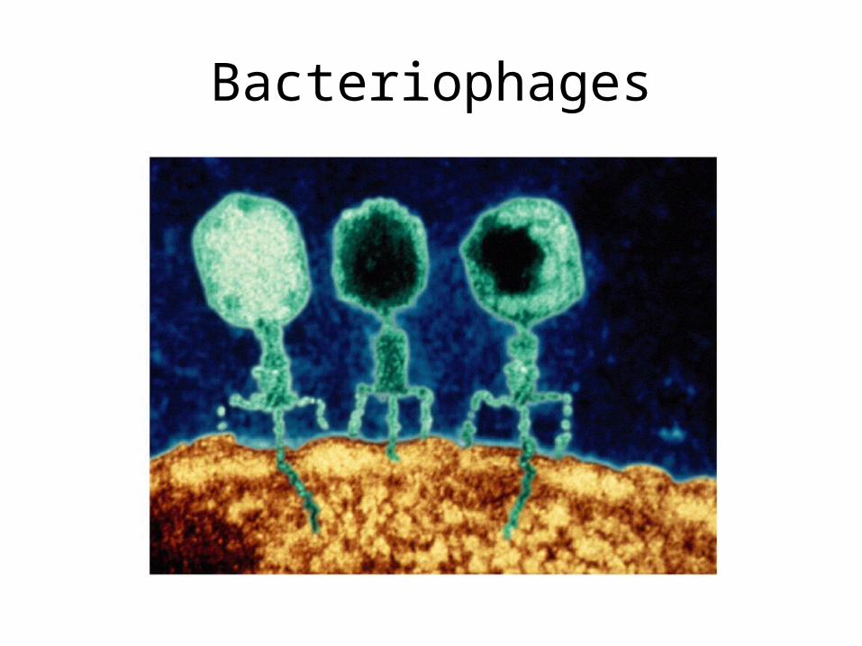
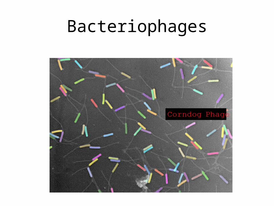
Bacteriophages
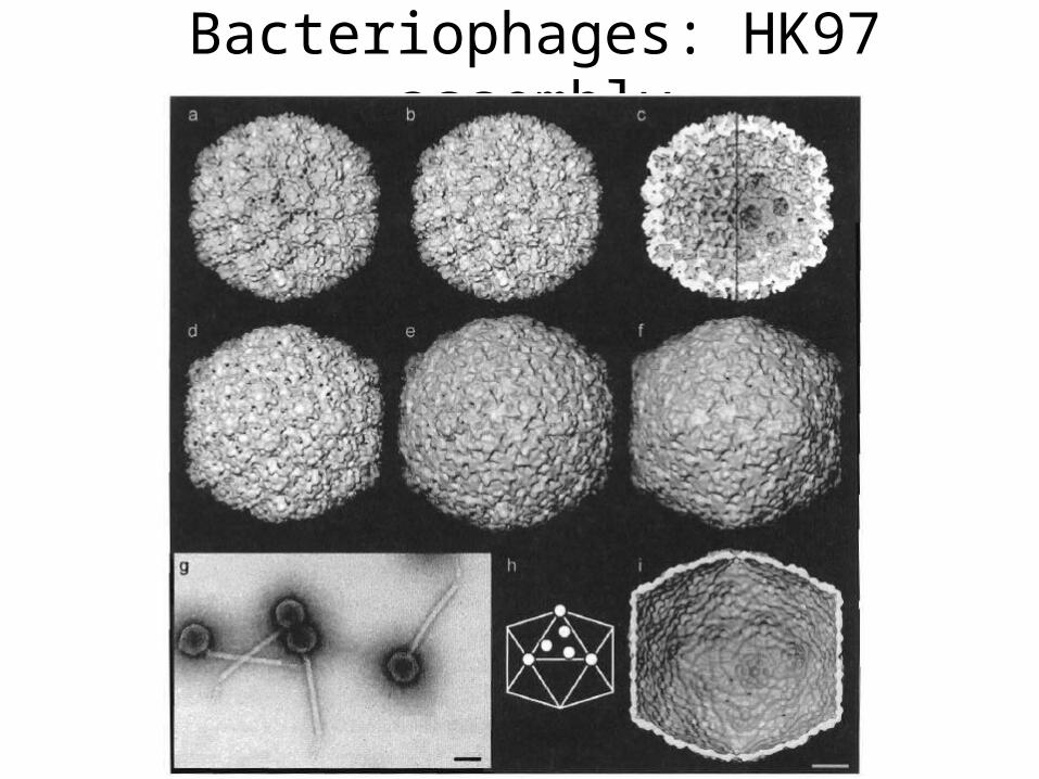
Bacteriophages: HK97 assembly
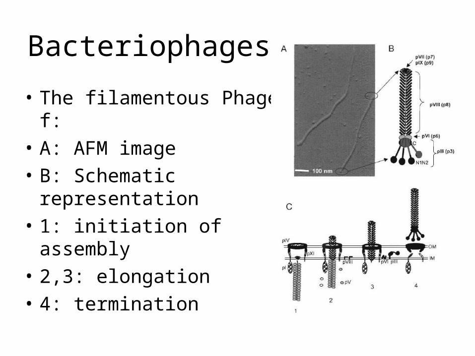
Bacteriophages
• The filamentous Phage f:• A: AFM image• B: Schematic representation• 1: initiation of assembly• 2,3: elongation• 4: termination
Bacteriophages
Bacteriophages
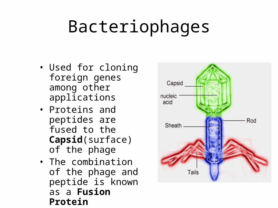
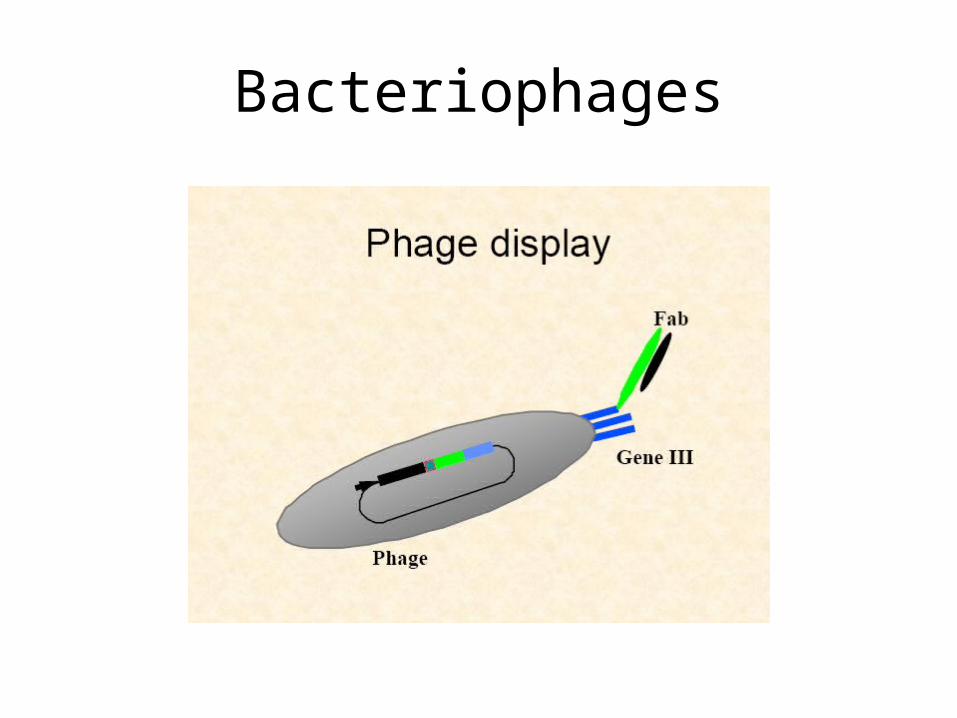
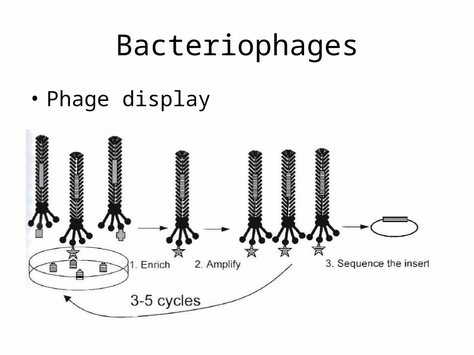
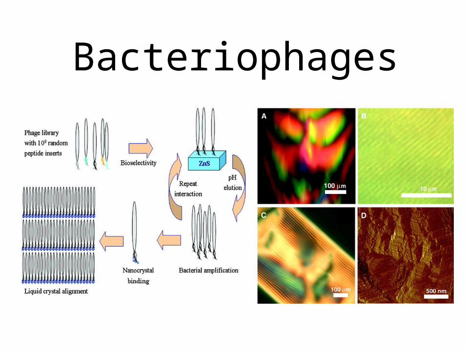
• Used for cloning foreign genes among other applications
• Proteins and peptides are fused to the Capsid(surface) of the phage
• The combination of the phage and peptide is known as a Fusion Protein
Bacteriophages
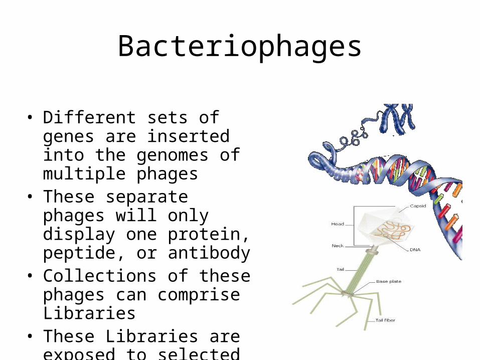
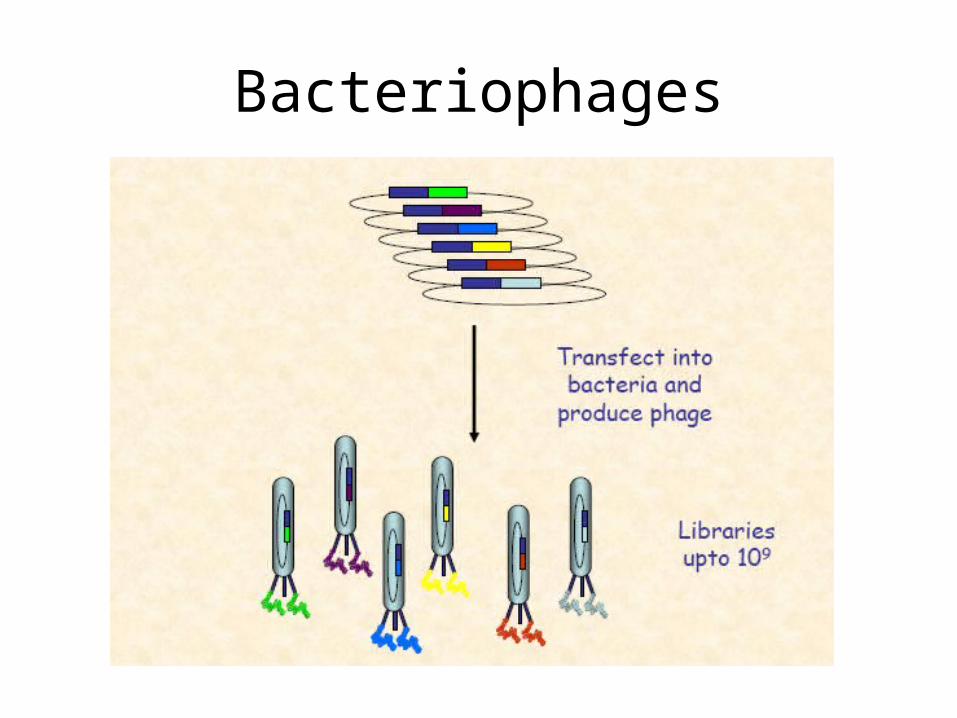
• Different sets of genes are inserted into the genomes of multiple phages
• These separate phages will only display one protein, peptide, or antibody
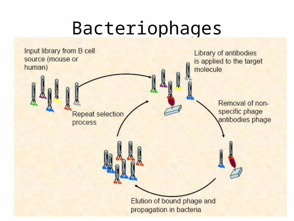
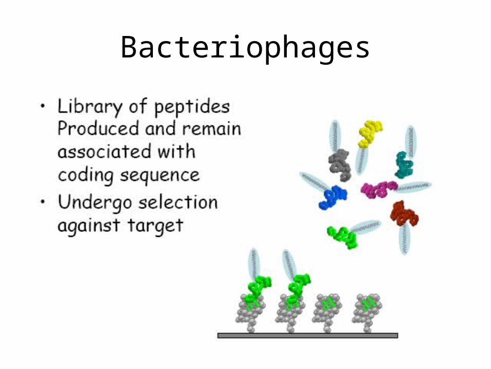
• Collections of these phages can comprise Libraries
• These Libraries are exposed to selected targets and only some phages will interact with targets
Bacteriophages
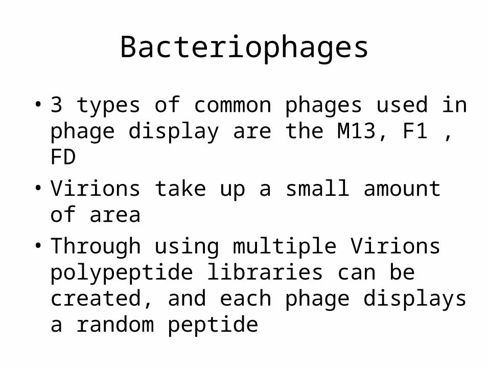
• 3 types of common phages used in phage display are the M13, F1 , FD
• Virions take up a small amount of area• Through using multiple Virions polypeptide
libraries can be created, and each phage displays a random peptide
Bacteriophages
Bacteriophages
Bacteriophages
Bacteriophages
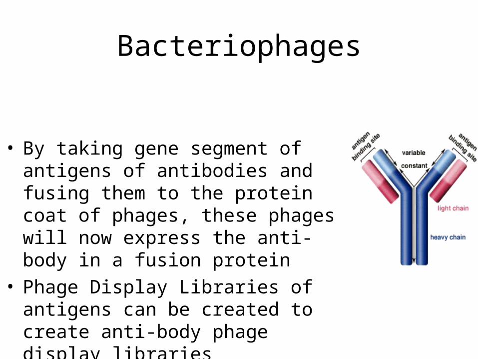
• By taking gene segment of antigens of antibodies and fusing them to the protein coat of phages, these phages will now express the anti-body in a fusion protein
• Phage Display Libraries of antigens can be created to create anti-body phage display libraries
Bacteriophages
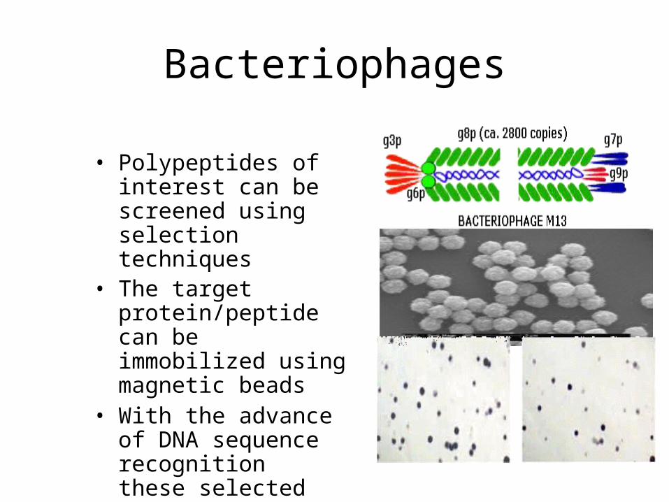
• Polypeptides of interest can be screened using selection techniques
• The target protein/peptide can be immobilized using magnetic beads
• With the advance of DNA sequence recognition these selected sequences can be identified easily
Bacteriophages
Bacteriophages
• Phage display
Bacteriophages
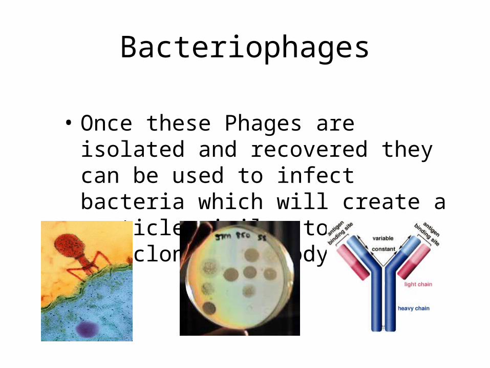
• Once these Phages are isolated and recovered they can be used to infect bacteria which will create a particle similar to a monoclonal antibody
Bacteriophages
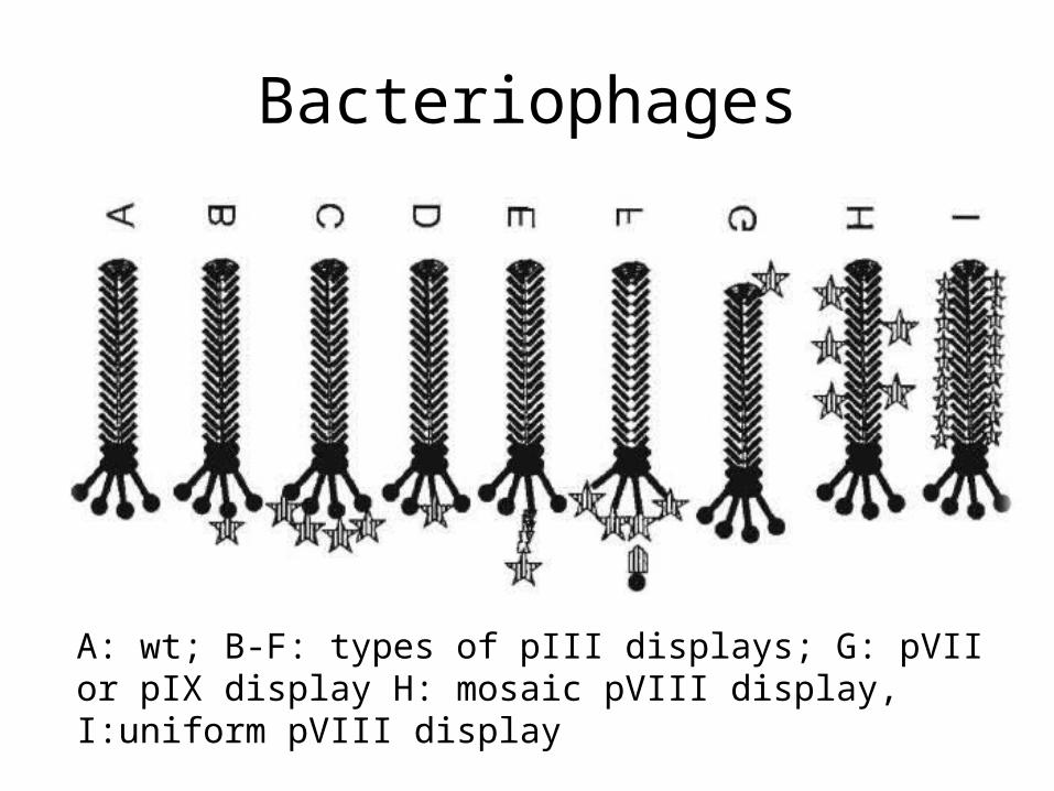
A: wt; B-F: types of pIII displays; G: pVII or pIX display H: mosaic pVIII display, I:uniform pVIII display
Bacteriophages
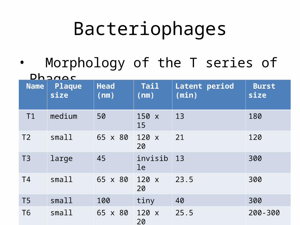
• Morphology of the T series of Phages
Name Plaque size Head (nm) Tail (nm) Latent period (min) Burst size
T1 medium 50 150 x 15 13 180
T2 small 65 x 80 120 x 20 21 120
T3 large 45 invisible 13 300
T4 small 65 x 80 120 x 20 23.5 300
T5 small 100 tiny 40 300
T6 small 65 x 80 120 x 20 25.5 200-300
T7 large 45 invisible 13 300
Bacteriophages
Bacteriophages
Bacteriophages
Bacteriophages
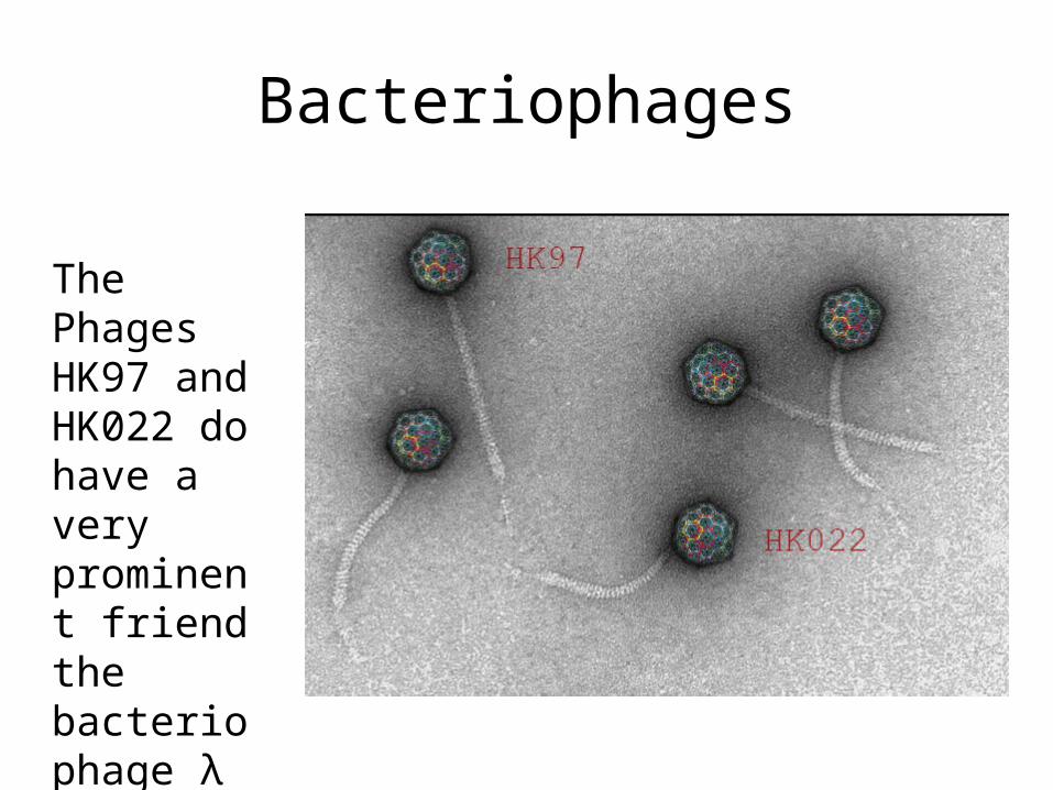
The Phages HK97 and HK022 do have a very prominent friend the bacteriophage λ
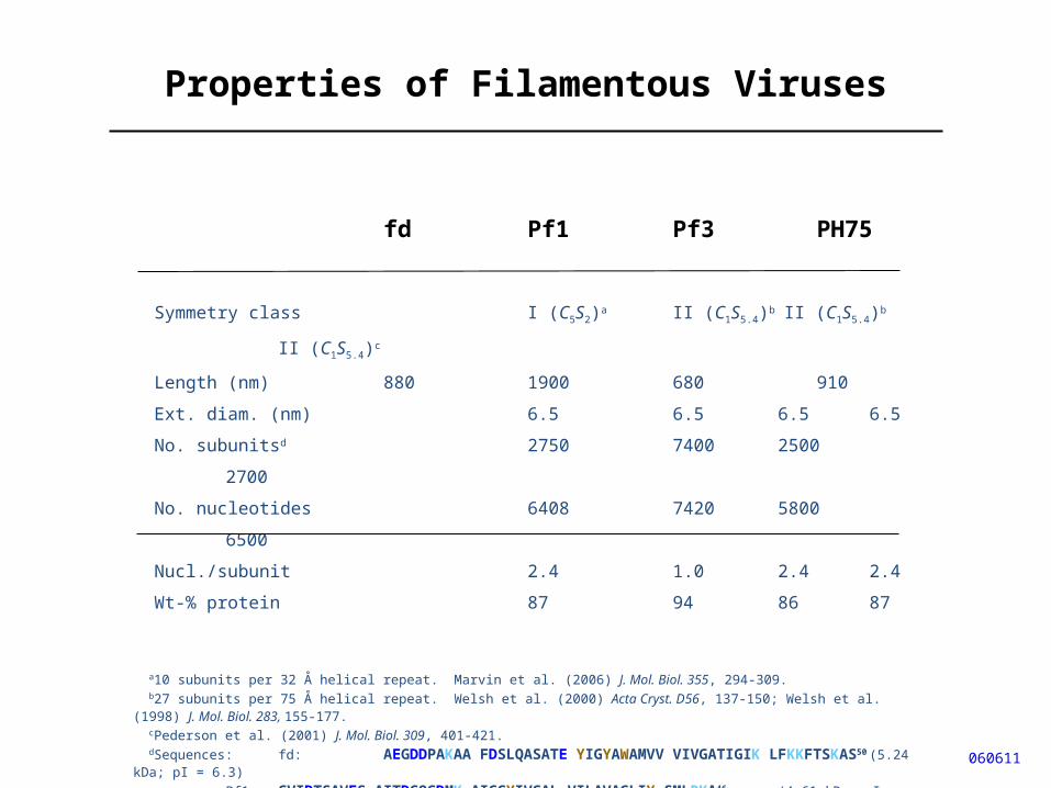
Properties of Filamentous Viruses
fd Pf1 Pf3 PH75
Symmetry class I (C5S2)a II (C1S5.4)b II (C1S5.4)b II (C1S5.4)c
Length (nm) 880 1900 680 910
Ext. diam. (nm) 6.5 6.5 6.5 6.5
No. subunitsd 2750 7400 2500 2700
No. nucleotides 6408 7420 5800 6500
Nucl./subunit 2.4 1.0 2.4 2.4
Wt-% protein 87 94 86 87
a10 subunits per 32 Å helical repeat. Marvin et al. (2006) J. Mol. Biol. 355, 294-309. b27 subunits per 75 Å helical repeat. Welsh et al. (2000) Acta Cryst. D56, 137-150; Welsh et al. (1998) J. Mol. Biol. 283, 155-177. cPederson et al. (2001) J. Mol. Biol. 309, 401-421. dSequences: fd: AEGDDPAKAA FDSLQASATE YIGYAWAMVV VIVGATIGIK LFKKFTSKAS50 (5.24 kDa; pI = 6.3)
Pf1: GVIDTSAVES AITDGQGDMK AIGGYIVGAL VILAVAGLIY SMLRKA46 (4.61 kDa; pI = 4.7)Pf3: MQSVITDVTG QLTAVQADIT TIGGAIIVLA AVVLGIRWIK AQFF44 (4.63 kDa; pI = 5.7)PH75: MDFNPSEVAS QVTNYIQAIA AAGVGVLALA IGLSAAWKYA KRFLKG46 (4.81 kDa; pI = 9.4)
060611
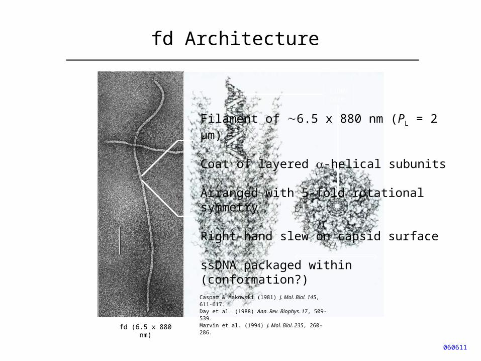
fd Architecture
060611
fd (6.5 x 880 nm)
1/100th virion length
ssDNAcore
65 Å
Caspar & Makowski (1981) J. Mol. Biol. 145, 611-617.Day et al. (1988) Ann. Rev. Biophys. 17, 509-539.Marvin et al. (1994) J. Mol. Biol. 235, 260-286.
Filament of 6.5 x 880 nm (PL = 2 μm)
Coat of layered -helical subunits
Arranged with 5-fold rotational symmetry
Right-hand slew on capsid surface
ssDNA packaged within (conformation?)
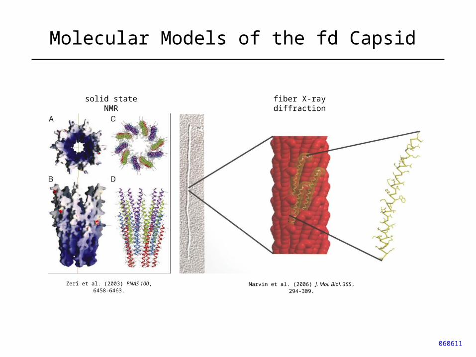
Molecular Models of the fd Capsid
060611
Zeri et al. (2003) PNAS 100, 6458-6463.
solid state NMR
Marvin et al. (2006) J. Mol. Biol. 355, 294-309.
fiber X-ray diffraction
Bacteriophages