differentiation of human, animal and synthetic hair by atr
TRANSCRIPT

University at Albany, State University of New York University at Albany, State University of New York
Scholars Archive Scholars Archive
Chemistry Honors College
5-2015
Differentiation of Human, Animal and Synthetic Hair by ATR FTIR Differentiation of Human, Animal and Synthetic Hair by ATR FTIR
Spectroscopy Spectroscopy
Jeremy Manheim University at Albany, State University of New York
Follow this and additional works at: https://scholarsarchive.library.albany.edu/honorscollege_chem
Part of the Chemistry Commons
Recommended Citation Recommended Citation Manheim, Jeremy, "Differentiation of Human, Animal and Synthetic Hair by ATR FTIR Spectroscopy" (2015). Chemistry. 5. https://scholarsarchive.library.albany.edu/honorscollege_chem/5
This Honors Thesis is brought to you for free and open access by the Honors College at Scholars Archive. It has been accepted for inclusion in Chemistry by an authorized administrator of Scholars Archive. For more information, please contact [email protected].

1
Differentiation of human, animal and synthetic hair by ATR FTIR Spectroscopy
An honors thesis presented to the
Department of Chemistry,
University at Albany, State University Of New York
in partial fulfillment of the requirements
for graduation with Honors in Chemistry
and
graduation from the Honors College.
Jeremy Manheim
Research Mentor: Kyle Doty, B.S. and Greg McLaughlin, Ph.D.
Research Advisor: Igor K. Lednev, Ph.D.
April, 2015

2
Abstract
Hair fibers are ubiquitous to every environment and are the most commonly found form
of trace evidence at crime scenes. The primary difficulty forensic examiners face after retrieving
a hair sample is determining who it came from. Currently, the methodology of microscopic
examination of potential hair evidence is absent of statistical probability and is inherently
subjective. Another method, involving DNA analysis, takes months to conduct and the majority
of times is unsuccessful due to its degradation and absence from the hair. Here, Attenuated Total
Reflectance (ATR) Fourier Transform Infrared (FTIR) Spectroscopy coupled with advanced
statistics was used to identify a hair sample within a specific confidence solely from its spectrum.
Ten spectra were collected for each of ten human, cat, and dog donors and a single
synthetic fiber for 310 total spectra. A spectrum is collected by simply placing a single strand or
patch of hair, without preparation, directly across the crystal (500μm) of the instrument. Two
Partial Least Squares-Discriminant Analysis (PLS-DA) models were constructed: one to
differentiate natural hair fibers from synthetic fibers and the second discriminating human hair
from dog and cat hair. Both internal models were successful in separating the desired class from
another; synthetic hair was completely separated from actual hair in the binary approach and all
human samples were predicted as human in the species specific model.
The species specific training model was tested by loading spectra from ten external
donors (three human, two cat and five dog) and examined the model’s ability to correctly assign
these spectra. The external validation confirmed our model’s ability to correctly classify a
sample as human as well as properly predict spectra that are not human. It also showed that a
breed of dog not accounted for in the training data set was entirely misclassified as cat, but more
importantly led to the possibility that different breeds of dog can be separated based on their hair
spectra. This preliminary investigation sheds light on the next step of the discrimination process
to identify the gender and race of a human hair, as well as the identification of different hair
dyes. Overall, the method is able to quantitatively identify a sample of hair as human with a high
degree of confidence and is of ample importance to the field of forensic science. The method
can be conducted without the need of a specialist, is non-destructive, is extremely quick and
requires no sample preparation.

3
Acknowledgements
I would like to extend a special thank you to Professor Igor Lednev for all of his support
and guidance throughout my research experience. His dedication to my development as a
researcher and success at the undergraduate level has allowed me to continue realizing my full
potential, and has inspired me to pursue my Ph.D. I would also like to thank my mentors Kyle
Doty and Greg McLaughlin for their patience and assistance in teaching me everything I needed
to know to carry out my research thesis. I would especially like to thank Dr. Jeffrey Haugaard for
helping me reach this milestone in my life. Last but not least, my mom and dad, for supporting
all of my career and academic decisions, and for helping me stay focused on my dreams.
4
Table of Contents
Abstract……………………………………………………………………………………………2
Acknowledgements………………………………………………………………………………..3
Introduction………………………………………………………………………………………..5
Materials and Method……………………………………………………………………………..9
Results……………………………………………………………………………………………11
Discussion………………………………………………………………………………………..21
References………………………………………………………………………………………..24

5
1. Introduction
Hair fibers are ubiquitous to every environment and are a common form of trace evidence
found at crime scenes. The primary difficulty forensic examiners face after retrieving a hair
sample is determining its origin; if it came from a human or an animal and, if human, what is the
race, gender and type of body hair (e.g. head, pubic, underarm, etc.). Light microscopy is the
most commonly employed method for the investigation of hairs in forensic laboratories[1].
Transmitted light and polarized light microscopes are traditionally used to analyze and identify
the morphology of a natural fiber[2]. A comparison microscope is used when comparing
unknown natural hairs, or fibers, recovered from a crime scene to those of a known origin.[3]
Hair classification is dependent on the expertise of the forensic examiner, the quality of the hair
sample and the instrumentation used[1]. DNA analysis is another common method employed for
the identification of an unknown hair sample. DNA testing is an extensive and costly procedure
that requires sophisticated techniques, time and resources[4]. Since hair is so abundant, crime
scene investigators collect many unknown fibers for analysis that could have come from a
human, an animal or even a wig. The ability to quickly identify a hair fiber as human, animal or
synthetic, with statistical support, would be of tremendous assistance to forensic investigations.
Based upon the probability theory, evidence including fingerprints, body fluids, and hair
are considered as circumstantial[5]. Fingerprints and body fluids have established probability
standards recognized by the criminal justice system that account for points of comparison
between known and unknown samples of evidence[5]. The issue preventing the same type of
standards for hair analysis is that the method is unable to directly associate the number of
different properties between two hairs and the probability that the samples did or did not come
from the same individual[6]. Additionally, two examiners who analyze the same hairs may
describe the hairs in slightly different ways, placing varying emphasis on certain characteristics,

6
and often use different descriptive words in their findings[7]. Furthermore, hair comparisons may
contain prejudice or bias, on the forensic expert’s part, due to interactions with criminal justice
personnel[5]. In particular, police and attorneys may have preconceived beliefs on a suspect’s
guilt, and if these attitudes are expressed to the examiner, it can greatly affect their conclusions
when analyzing hair evidence.
Hair is important to the investigation process because it may contain DNA and, in some
cases, it is the only evidence available linking a criminal to the crime scene. In the 2009 report,
“Strengthening Forensic Science in the United States: A Path Forward,” it was concluded that
there are no accepted statistics about the frequency with which certain hair characteristics are
distributed within a population and that hair comparisons for individualization have no scientific
support without nuclear DNA[8]. In early 2013, the F.B.I. began a review of over 2000
convictions based on hair evidence[9]. Of the first 310 cases, DNA analysis revealed that 72 of
the convictions were grounded on faulty hair evidence[9]. One case involved a man named
Claude Jones who was executed in 2000 after being convicted of killing the owner of a bar. His
conviction stemmed from the belief that a hair recovered from the crime scene was his. As part
of the F.B.I.’s review, DNA from the hair proved to not have come from Claude Jones[10].
Although this was only one case, there are many more examples where innocent people were
wrongly convicted based on improper conclusions drawn by examiners, which reinforces the
need for new methods to accurately analyze hair evidence.
Despite its increasing popularity, the process of extracting DNA from a hair fiber is an
extensive procedure that does not always generate usable results[11-14]. The majority of the
genetic material in hair is located in its root which is generally absent from the hair shaft (i.e. the
portion of hair that grows out of the skin)[4]. However, collected hairs absent of the root or
follicle material may undergo exhaustive and laborious mitochondrial DNA analysis, even

7
though success is not guaranteed[4]. DNA analysis is extremely costly and time consuming, not
to mention that most laboratories are currently backlogged. A method for determining the
identity of an unknown fiber quickly, with a high degree of certainty, and eliminating examiner
bias would be extremely useful and cost-effective for the field of forensic science.
ATR FTIR spectroscopy is a technique rising in popularity for analytical and biological
purposes. It has been employed for the analysis of biomedical samples[15], paint[16, 17],
fingerprints[18] and ink[19]. The attributes of ATR FTIR spectroscopy are very attractive for
forensics because of its rapid and non-destructive nature, its ease-of-use and minimal to no
sample preparation. An infrared spectrum displays the vibrational characteristics of a sample
based on the different absorption frequencies of the individual functional groups[20]. The ATR
attachment allows for analysis of solid samples, often with no sample preparation[21]. The
advantage of combining ATR FTIR spectroscopy with chemometrics is its ability to enhance the
selectivity of the instrument and create classification models[16, 22, 23].
Two published studies demonstrate the use of FTIR and chemometrics to differentiate the
spectra from different types of hair. Espinoza et al. applied infrared spectroscopy and advanced
statistics to the forensic identification of elephant and giraffe hair[24]. They visually observed a
difference in the elephant and giraffe hair spectra at a very prominent peak (1032 cm-1), which is
due to surface cystine oxides and the presence of cysteic acid. Through the discriminant analysis
of their spectral data they demonstrated a performance index of 91.8%, which specifies how well
their algorithm can differentiate between elephant and giraffe hair. Another group combined
FTIR microscopy and chemometrics to differentiate Asian hair samples and black Caucasian
hairs[25]. Using Principle Component Analysis (PCA), they were able to separate the three
female Asian hair samples from the three female Caucasian hair samples demonstrating their
ability to discriminate between hair from two different races.

8
Our lab has used Raman spectroscopy, in conjunction with advanced statistics, for
differentiation purposes when spectra are visually similar. Some of these studies include body
fluid identification[26], distinguishing between species’ blood[27], species’ bones[28], and
mixtures of semen and blood[29]. However, Raman spectroscopy is not an advantageous method
to use for hair analysis due to the significant fluorescence interference, as shown in the
literature[30, 31]. For this reason our approach was to use ATR FTIR to analyze hair samples.
Similar work has been done as part of two theses projects, “Vibrational spectroscopy of keratin
fibres: A forensic approach” by Helen Panayiotou[32] and “A forensic investigation of single
human hair fibres using FTIR-ATR spectroscopy and chemometrics” by Paul Barton[33], at
Queensland University of Technology in Australia. Our study is an expansion upon their work,
primarily Panayiotou’s 2004 thesis, in a few different ways. First, they treat their hair samples by
flattening with a roller[32] prior to analysis whereas we have analyzed all hairs without any
sample preparation. Second, our data analysis was performed using a different statistical
algorithm better suited for class separation, PLS-DA, and we used ATR FTIR spectroscopy for
data collection, rather than Panayiotou’s approach of using FTIR micro-spectroscopy in the
transflection mode. With ATR FTIR, there is no need for sample preparation and allows for the
potential opportunity of on-field analysis due to the availability of portable instruments[34].
Finally, our sample size for species differentiation is over fourteen times larger, focusing on
humans, dogs and cats.
Our analysis for the present study is bimodal where the first model discriminates natural
hair from synthetic and the second discriminates human hair from other common natural hair
sources (i.e. dog and cat hairs). Hair samples were collected from a synthetic wig and a diverse
population of humans, dogs, and cats. The spectra were differentiated using Partial Least
Squares-Discriminant Analysis (PLS-DA) classification models which were built from a training

9
dataset of human, dog, and cat spectra. An external validation step was also carried out to test the
model’s ability to accurately predict a sample to its actual class.
2. Methods and materials
2.1 ATR FTIR spectrometer and hair samples
A PerkinElmer Spectrum 100 FTIR spectrometer with an attenuated total reflectance
(ATR) attachment was used for data collection for all experiments. Spectra were collected over a
range of 650-4000 cm-1 with 10 scans per sample. For each donor, ten averaged spectra were
collected. The chemical composition of hair, primarily its proteins, is subject to change after
being exposed to various chemical reactions such as bleaching, waving, straightening and
extensive sunlight exposure[30, 35-37]. Of the many variables that can influence the chemical
make-up of hair only chemically treated (i.e. dye, bleaching, etc.) hairs were excluded from this
study. A single hair was placed over the diamond/ZnSe crystal of the instrument in order to
obtain a spectrum with optimal signal. For animal donors consisting of only fur hairs, multiple
hairs were required because they are fine and shorter compared to that of an animal’s guard
(outer) hair[38]. For each donor, ten spectra were acquired at various points along several hair
fibers, and each spectrum was treated as its own sample. In the case where multiple fur hairs
were placed over the crystal, spectra were obtained over different patches of the fur hair.
Spectra from ten different human, dog and cat hair samples were collected as well as
from one polyester synthetic hair fiber. The race, gender, and age of the human donors, as well
as the breed of dog and cat, were taken into consideration for sample collection. These individual
characteristics can be seen in Table 1.

10
Table 1: The background information of the thirty human, dog and cat donors used in the
training data set for all PLS-DA models.
Donor # Human (age) Dog Cat
1 Asian female (18) Barbet Maine Coon
2 Caucasian female (20) Maltese Ragdoll
3 Caucasian male (20) A Cocker Spaniel Domestic Short Hair (Grey A)
4 Caucasian male (20) B Dachmund Mini Domestic Short Hair (Black A)
5 Caucasian female (40) Pug Domestic Short Hair (black-and-white A)
6 Hispanic female (20) Golden Retriever Domestic Short Hair (White)
7 Hispanic male (20) Unknown Dog Domestic Short Hair (Brown)
8 African American female (21) Yorkshire Terrier Domestic Short Hair (Black B)
9 Egyptian male (20) Briard Domestic Short Hair (Grey B)
10 Ecuadorian male (20) Beagle Domestic Short Hair (black-and-white B)
2.2 Data preparation and statistical treatment
All data preparation and statistical models were performed with the PLS Toolbox 7.0.3
(Eigenvector Research, Inc.) operating in MATLAB version R2010b. The model for
differentiating natural hair from synthetic hair was built using the full spectrum collected (650-
4000 cm-1). All 310 spectra were imported into a dataset; the dataset was preprocessed using
transmittance log, second-order derivative, normalization by total area and finally mean
centering. The model created for discriminating human hair from animal hair (species specific)
was built using spectra truncated to the data range of 650-1827 cm-1. The 300 total spectra
(excluding the ten synthetic fiber spectra) were imported into a data matrix and preprocessed the
same way as the binary model. All models were cross-validated using the venetian blinds
method.

11
2.3 External validation
The training model was tested by loading external donors (three human, two cat and five dog)
into the model to test its ability to correctly predict the identity (class) of an untrained sample.
All external samples were preprocessed in the same manner as the training data but not included
as part of the training dataset used to build the models.
3. Results
The main objectives of this study were to discriminate natural hair from a synthetic fiber
and differentiate human hair from animal hair using chemometric modeling of ATR FTIR
spectroscopic data. Preliminary experimentation determined the model selection and data
processing steps. PLS-DA models were chosen to build simple classification models using the
infrared spectra of a synthetic fiber and human, dog, and cat hair. The number of latent variables
for each model was selected by choosing a local minimum of total data variance captured using a
scree plot (not shown). The PLS-DA models were constructed in two fashions, first by
classifying each spectrum as either natural or synthetic and secondly, focusing on the individual
species, to determine if a more specific assignment could be made. The second model was used
to make class predictions of 10 external natural hair donors that were not accounted for in the
training dataset.

12
3.1: Natural hair v. synthetic hair (binary)
The prominent features of an infrared spectrum of natural hair correspond to specific
vibrational modes of the amino acids and lipids present[39]. The averaged raw spectra for
human, dog, cat and synthetic hair, as shown in Figure 1, reveal visual differences between
natural hair and synthetic hair. These differences include the absence of the Amide A peak at
3300 cm-1 and the more intense CH3/CH2 (alkane stretching) peak at 2950 cm-1 in the averaged
synthetic hair fiber spectrum. Additionally, various spectral inconsistences exist between the
two hair types in the fingerprint region (650-1827cm-1) including peaks at ~1400 and ~1450 cm-1
for synthetic hair and peaks at ~1520 and ~1620 cm-1 only present in natural hair spectra. These
peaks most likely correspond to C=N and C=O respectively[32]. Due to these spectral
differences, the polyester synthetic hair spectrum can be visually differentiated from a spectrum
of natural hair quite easily.
Figure 1: The raw mean spectra of human, cat, dog, and synthetic hair samples.
100015002000250030003500400070
75
80
85
90
95
100
Wavenumber (cm-1)
Rela
tive
In
ten
sity (
a.u
.)
human
cat
dog
synthetic

13
For statistical analysis, all 310 spectra from the 31 donors were used to build a Partial
Least Squares-Discriminant Analysis (PLS-DA) model using four latent variables. Before the
model was built, all spectra were preprocessed as described in Section 2.2. Initially, the human,
cat, and dog samples were grouped together as one class (natural hair) and compared to synthetic
hair in a binary approach. Under cross-validation (CV) all of the synthetic hair samples were
correctly classified as seen in Figure 2. A cross validation model works by treating all of the
trained spectra as unknowns, and tries to properly predict them. The results of perfect separation
between the synthetic hair and natural hair were not surprising since the averaged natural hair
spectra looked visually different from the averaged synthetic hair spectrum. These results
demonstrate that our model can efficiently discriminate samples of natural hair from synthetic
hair with 100% accuracy.
Figure 2: Cross-validated synthetic hair class predictions for all 310 spectra analyzed in the
binary model (natural v. synthetic). All spectra above the red threshold line are predicted to the
synthetic class and all below are predicted as not synthetic.
50 100 150 200 250 300
Spectrum Number
CV
Pred
icte
d C
lass
Syn
thet
ic
Human
Cat
Dog
Synthetic

14
3.2: Human, cat, and dog hair (species specific)
The 300 spectra of the human, cat, and dog hairs were truncated to 650-1827 cm-1. An
artifact around 2350 cm-1, consistent with atmospheric CO2[40], is not a vibrational mode of hair
and to ensure that the air did not influence our results we only analyzed the specified region.
Although not shown here, the full hair spectrum was also analyzed and the results were very
similar, informing us that the air artifact would not significantly alter our results. From visual
inspection, all natural hair spectra shown below in Figure 3 appear to be identical in terms of the
number of spectral features and their location. For this reason we utilized classification statistical
analysis in an attempt to extract any differences which could not be visualized.
Figure 3: The truncated, raw mean spectra of human, cat, and dog hair samples.
A second PLS-DA model was built to analyze the training dataset classified by their
species of origin: human, cat or dog. The spectra were preprocessed in the same way as in the
binary approach and ten latent variables were selected to build the model. According to the strict
8001000120014001600180070
75
80
85
90
95
100
Wavenumber (cm-1)
Rela
tive inte
nsity
(a.u
.)
Human
Cat
Dog

15
class predictions, which assigns a sample to its nearest class and samples with a large uncertainty
are unassigned, all of the human and cat spectra in the training dataset were correctly assigned to
their proper class. Only one dog (Cocker Spaniel) spectrum was predicted incorrectly, as
unassigned. Using this approach both the human and cat classes showed 100% correct
classification while the dog class showed a marginally lower rate at 99%. Although these strict
predictions are informative, cross-validated analysis provides more reliable classification results.
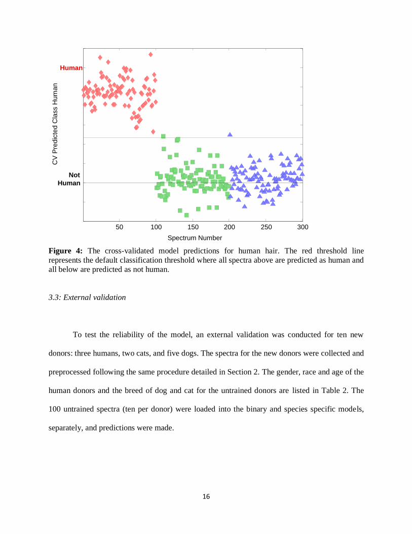
Figure 4 shows the cross-validation prediction plot which illustrates the probability that a
given spectrum will be classified as human. All of the human hair spectra lie above the
classification threshold (red dotted) line, signifying a 100% correct class prediction rate.
However, one cat (Ragdoll) and one dog (Barbet) spectrum are above the threshold line and are
therefore false positive predictions. This means that 90% of the spectra from the Ragdoll and
Barbet donors were correctly classified as opposed to 100% classification rate for all other
donors’ spectra. The single misclassified spectra could be due to any sudden instrument
movements or background contaminants since nine out of ten spectra along the same hair fiber
were properly predicted. Overall this represents a correct classification rate of 99% for both the
cat and dog classes as not human under cross-validation. These results conclude that our model
has no false negative assignments and is capable of predicting a sample of human hair as human
with 100% accuracy.
16
Figure 4: The cross-validated model predictions for human hair. The red threshold line
represents the default classification threshold where all spectra above are predicted as human and
all below are predicted as not human.
3.3: External validation
To test the reliability of the model, an external validation was conducted for ten new
donors: three humans, two cats, and five dogs. The spectra for the new donors were collected and
preprocessed following the same procedure detailed in Section 2. The gender, race and age of the
human donors and the breed of dog and cat for the untrained donors are listed in Table 2. The
100 untrained spectra (ten per donor) were loaded into the binary and species specific models,
separately, and predictions were made.
50 100 150 200 250 300
Not
Human
Human
CV
Pre
dic
ted C
lass H
um
an
Spectrum Number

17
Table 2: The new donors collected for the external validation
Examining the prediction plot for the species specific model demonstrates how well the
model correctly predicted for the human class. Figure 5 shows complete separation between the
classes and all 130 human spectra (100 in the training set and 30 external) lie above the threshold
(red dotted) line, with one external sample lying close to it. In addition, all of the cat and dog
spectra (training and external) are well below the threshold line signifying zero false positive
predictions for the human class. Therefore, the accuracy of the model for predicting a hair
sample as human or nonhuman under strict class prediction conditions was 100%.
External Donor (Class) Description
Human Hispanic Female, age 24
Human Caucasian Female, age 22
Human Caucasian Male, age 30
Cat Calico
Cat Domestic Short Hair (Grey C)
Dog Maltese B
Dog Maltese C
Dog Maltese D
Dog Golden Retriever B
Dog Pomeranian

18
Figure 5: Human class predictions for all 400 samples analyzed, including untrained donors, in
the species specific PLS-DA model. Red line represents the default classification threshold
where all spectra above are predicted to the human class and all spectra below are predicted as
not human.
For the species specific model, under strict class predictions (Figure 6), twenty-nine of
the thirty external human spectra were correctly predicted as human; the other spectrum was
unassigned. All twenty external cat hair samples were correctly classified as cat. In addition, all
of the external Golden Retriever (dog) donors were classified correctly as dog. However, of the
three external Maltese donors (B, C, and D), three samples from donor C were misclassified (two
of which were unassigned and one predicted as cat) and eight samples from donor D were
misclassified (four were unassigned and four were misclassified as cat). Lastly, all ten spectra
from the internal Pomeranian donor were misclassified as cat.
50 100 150 200 250 300 350 400-0.4
Not
Human
Human
Spectrum Number
Cla
ss P
redic
tio
n H
um
an
HumanCatDogExternalCatExternalDogExternalHuman

19
Figure 6: Strict class predictions for the external validation samples loaded into the species
specific PLS-DA model. Deviations from each class’ horizontal line represent a
misclassification.
In order to understand why some dog spectra were misclassified as cat, differences in hair
between individual dogs of various breeds was investigated. The results for this test revealed that
all of the individual dogs (ten spectra from each dog) of ten different breeds, with the exception
of three spectra, were correctly assigned to their correct class (Figure 7). All of the breeds of dog
appear to be differentiating from one another, which still does not explain why the Pomeranian
breed in particular was the only breed being misclassified within our model. However, since only
one donor from each breed of dog was analyzed, the differences observed could be an individual
difference or breed differentiation. More dog donors from each breed would need to be collected
in order to make any definite conclusions from these results.
Unassigned
Human
50 100 150 200 250 300 350 400
0
0.5
1
1.5
2
2.5
3
Spectrum Number
Cla
ss P
redic
tion (
Str
ict)
Cat
DogHumanCatDogExternalCatExternalDogExternalHuman

20
Figure 7: Strict class predictions for the individual dog donors. Deviations from each class’
horizontal line represent a misclassification.
To further investigate the Pomeranian dog breed misclassification, we also created a
second binary PLS-DA to differentiate dog hairs from cat hairs and included the Pomeranian dog
donor in the dog class. An influence plot was analyzed, which groups spectra similar to each
other within a 95% confidence interval and all spectra plotted outside the interval are considered
extremely different. Analysis of the model revealed all ten Pomeranian spectra had higher
Hotelling T2 values, and were grouped together, separate from all other hair spectra. Hotelling T2
values are directly related to the amount of variation in each sample. So, higher Hotelling T2
values suggest that those spectra are somehow inherently different than the other spectra, yet
similar within themselves because of their close grouping. Although the Pomeranian dog was
shown to be different from the other dog and cat spectra, it still does not explain its
misclassification as cat, but rather illuminates the unique characteristics in the chemical spectra
for the Pomeranian dog donor.

21
Figure 8: The influence plot for the dog v. cat binary model. All spectra to the left of the vertical
blue line are within a 95% confidence interval.
4. Discussion
The differentiation of human hair from cat and dog hair is difficult to observe from their
raw spectra, unlike for synthetic hair from natural hair. The species specific PLS-DA model was
able to successfully make this differentiation based on the shape of each component latent
variable (Figure 9). These variables represent the prominent discriminating factors between
spectra and are denoted by different peaks in the fingerprint region. This observation implies that
there is more than one characteristic peak which differentiates the individual classes of hair from
one another; latent variables one, two and three as shown in Figure 9 depict where these
characteristic peaks are. The dominant features are in the regions 1739-1742 cm-1 (C=O stretch),
1467-1477 cm-1 (CH2 bend) and 1230 cm-1(amide III). One possible explanation for the model’s

22
discriminatory power is based on the different combinations of amino acids that form keratin, the
structural protein from which hair is constructed. Hair is chemically composed of 65-95%
proteins and the content of the different proteins vary among different donors[41]. The 2004
study conducted by Panayiotou at Queensland University of Technology determined differences
between the relative intensity areas of various peaks for seven different samples: human, cat,
dog, horse, cow, feather, and wool[32]. As it pertains to our project, that study found that human
and dog hairs have lower Amide I (α-helix) content than cats and that humans have lower Amide
II (α-helix) content than cats and dogs. Based off this research and the complex nature of keratin,
it can help explain the subtle differences identified by the species specific model. Here, we show
that spectra collected from multiple points along a donor’s single hair fiber can still be predicted
as its correct species (class).
The most important results are that a sample of human hair can be quickly and non-
destructively analyzed, and subsequently identified with a high degree of confidence. Our rapid
analysis and superb probability prediction results have been accomplished without human bias,
and could potentially be of great use for the forensic investigative process. The non-destructive
nature of using ATR FTIR spectroscopy makes this method ideal for the forensic identification
of an unknown hair sample. The developed methodology has the potential to differentiate
gender, race, other animal species, and even hair dyes. Furthermore, the presence of portable
FTIR instrumentation supports the idea that on-field analysis of a hair fiber is feasible.
Conclusions
The combination of ATR FTIR spectroscopy and chemometrics was demonstrated to be a
powerful tool toward the differentiation of hair samples from three species. Two PLS-DA
models were constructed: one focusing on the differentiation of natural hair fibers from synthetic

23
fibers and the second discriminating human hair from animal hair. Both models were successful
in separating the desired class from another; synthetic hair was completely separated from
natural hair in the binary approach and all human samples were predicted as human in the
species specific model. The external validation step confirmed our model’s ability to correctly
predict a sample as human with zero false positives. A larger sample size for the dog class would
help account for the misclassified Pomeranian donor, but this is beyond the scope of this project.
Of the many variables that can alter one’s hair chemistry, only chemically treated hairs
(i.e. dye, bleaching, etc.) were excluded. All other potential external interferences (e.g. sun
damage, type of shampoo, physical treatment, etc.) were not taken into account for this study and
did not preclude a high differentiation efficiency of the method. Overall, this demonstrates the
significance of the model’s unique ability to quantitatively identify a sample of hair as human
with a high degree of confidence. But, most importantly, the method can be conducted without
the need of a trained expert, is non-destructive, requires no sample preparation, with rapid
identification, making it of ample importance to the field of forensic science.

24
References
[1] D.W. Deedrick, Part 1: Hair Evidence, in Hairs, Fibers, Crime, and Evidence, Forensic Sci.
Comm. 2 (2000).
[2] SWGMAT, Forensic Human Hair Examination Guidelines, Forensic Sci. Comm. 7 (2005).
[3] S.R. Tridico, M.M. Houck, K.P. Kirkbride, M.E. Smith, B.C. Yates, Morphological
identification of animal hairs: Myths and misconceptions, possibilities and pitfalls, Forensic Sci.
Int. 238 (2014) 101-107.
[4] C. Hughes, Challenges in DNA Testing and Forensic Analysis of Hair Samples, Forensic
Mag. (2013).
[5] L.S. Miller, Procedural bias in forensic science examinations of human hair, Law and Human
Behavior. 11 (1987) 157-163.
[6] J. Taupin, Forensic hair morphology comparison–a dying art or junk science? Sci. & Justice
44 (2004) 95-100.
[7] C.T. Oien, Forensic Hair Comparison: Background Information for Interpretation. Forensic
Sci. Comm. 11 (2009).
[8] H. Edwards, C. Gotsonis, Strengthening forensic science in the United States: a path forward,
Washington, DC, National Academy Press (2009).
[9] M. Doyle, FBI announces review of 2,000 cases featuring hair samples, McClatchy DC
(2013).
[10] D. Mann, DNA Tests Undermine Evidence in Texas Execution, Observer (2010).
[11] M.C. Grieve, G. Wiggins, Fibers under fire: suggestions for improving their use to provide
forensic evidence, J. Forensic Sci. 46 (2001) 835-843.
[12] K. Takayanagi, H. Asamura, K. Tsukada, M. Ota, S. Saito, H. Fukushima, Investigation of
DNA extraction from hair shafts. International Congress Series, Elsevier. 1239 (2003) 759-764.
[13] K.A. Roberts, C. Calloway, Mitochondrial DNA amplification success rate as a function of
hair morphology, J. Forensic Sci. 52 (2007) 40-47.
[14] C.F. Bengtsson, M. E. Olsen, L.Ø. Brandt, M.F. Bertelsen, E. Willerslev, D.J. Tobin, A.S.
Wilson, M.T. Gilbert, DNA from keratinous tissue. Part I: Hair and nail, Annals of Anatomy-
Anatomischer Anzeiger. 194 (2012) 17-25.
[15] S. Kazarian, K. Chan, Applications of ATR-FTIR spectroscopic imaging to biomedical
samples, Biochim. et Biophys. Acta (BBA)-Biomembranes. 1758 (2006) 858-867.

25
[16] C. Muehlethaler, G. Massonnet, P. Esseiva, The application of chemometrics on Infrared
and Raman spectra as a tool for the forensic analysis of paints, Forensic Sci. Int. 209 (2011) 173-
182.
[17] M. Szafarska, M. Woźniakiewicz, M. Pilch, J. Zięba-Palus, P. Kościelniak, Computer
analysis of ATR-FTIR spectra of paint samples for forensic purposes, J. Mol. Struc. 924 (2009)
504-513.
[18] N.J. Crane, E.G. Bartick, R.S. Perlman, S. Huffman, Infrared spectroscopic imaging for
noninvasive detection of latent fingerprints, J. Forensic Sci. 52 (2007) 48-53.
[19] W. Dirwono, J.S. Park, M.R. Agustin-Camacho, J. Kim, H.M. Park, Y. Lee, K.B. Lee,
Application of micro-attenuated total reflectance FTIR spectroscopy in the forensic study of
questioned documents involving red seal inks, Forensic Sci. Int. 199 (2010) 6-8.
[20] J. McMurry, Organic Chemistry, Thomson Brooks/Cole (2004) 407.
[21] D.A. Skoog, F.J. Holler, S.R. Crouch, Principles of Instrumental Analysis, Thomson
Brooks/Cole (2007) 471-472.
[22] N.A. Sinkov, P.M. Sandercock, J.J. Harynuk, Chemometric classification of casework arson
samples based on gasoline content. Forensic Sci. Int. 235 (2014) 24-31.
[23] PerkinElmer, ATR FT-IR Imaging of Human Hair Cross-Section, PerkinElmer, Inc. (2006).
[24] E.O. Espinoza, B.W. Baker, T.D. Moores, D. Voin, Forensic identification of elephant and
giraffe hair artifacts using HATR FTIR spectroscopy and discriminant analysis. Endang. Species
Res. 9 (2008) 239-246.
[25] L. Rintoul, H. Panayiotou, S. Kokot, G. George, G. Cash, R. Frost, T. Bui, P. Fredericks,
Fourier transform infrared spectrometry: a versatile technique for real world samples, Analyst.
123 (1998) 571-577.
[26] K. Virkler, I.K. Lednev, Analysis of body fluids for forensic purposes: from laboratory
testing to non-destructive rapid confirmatory identification at a crime scene, Forensic Sci. Int.
188 (2009) 1-17.
[27] G. McLaughlin, K.C. Doty, I.K. Lednev, Discrimination of human and animal blood traces
via Raman spectroscopy, Forensic Sci. Int. 238 (2014) 91-95.
[28] G. McLaughlin, I.K. Lednev, Spectroscopic discrimination of bone samples from various
species, Amer. J. Anal. Chem. 3 (2012) 161-167.
[29] V. Sikirzhytski, A. Sikirzhytskaya, I.K. Lednev, Advanced statistical analysis of Raman
spectroscopic data for the identification of body fluid traces: Semen and blood mixtures,
Forensic Sci. Int. 222 (2012) 259-265.

26
[30] A. Kuzuhara, Analysis of structural change in keratin fibers resulting from chemical
treatments using Raman spectroscopy, Biopolymers. 77 (2005) 335-344.
[31] P. Carpenter, F. Bell, Experiments in Raman spectroscopy of hair: exciting light and
molecular orientation, J. Cosmet. Sci. 60 (2009) 199-204.
[32] H. Panayiotou, Vibrational spectroscopy of keratin fibres: A forensic approach, Queensland
University of Technology (2004).
[33] P.M. Barton, A forensic investigation of single human hair fibres using FTIR-ATR
spectroscopy and chemometrics, Queensland University of Technology (2011).
[34] A.J. Rein, J. Seelenbinder, Handheld and Portable FTIR Spectrometers for the Analysis of
Materials: Taking the Lab to the Sample, American Laboratory (2013).
[35] A. Kuzuhara, Analysis of internal structure changes in black human hair keratin fibers
resulting from bleaching treatments using Raman spectroscopy, J. Mol. Struc. 1047 (2013) 186-
193.
[36] N. Nishikawa, Y. Tanizawa, S. Tanaka, Y. Horiguchi, T. Asakura, Structural change of
keratin protein in human hair by permanent waving treatment, Polymer. 39 (1998) 3835-3840.
[37] C.M. Pande, FT-Raman spectroscopy: applications in hair research, J. Soc. Cosmet. Chem.
45 (1994) 257-268.
[38] D.W. Deedrick, S.L. Koch, Microscopy of Hair Part 1: A Practical Guide and Manual for
Human Hairs, Forensic Sci. Comm. 6 (2004).
[39] K.S. Kim, H.K. Park, Analysis of aging effects on chemical property of human hair by
Fourier transform infrared spectroscopy, Skin Research and Technology 19 (2013) e325-e331.
[40] J.W. Robinson, E.M.S. Frame, G.M. Frame II, Undergraduate instrumental analysis, CRC
Press (2004).
[41] C.R. Robbins, Chemical Composition of Different Hair Types, in Chemical and Physical
Behavior of Human Hair, Springer (2012) 105-176.