diabetic myonecrosis in a cystic fibrosis...
TRANSCRIPT
Diabetic myonecrosis in a cystic fibrosis patient
Benjamin T. Kopp, MD1*, Stephen Kirkby MD1, Don Hayes, Jr., MD1, Kevin M. Flanigan,
MD2
1Section of Pediatric Pulmonology, Nationwide Children’s Hospital, Columbus, OH.
Department of Pediatrics, The Ohio State University, Columbus, OH
2Division of Neurology, Nationwide Children's Hospital, Columbus, OH
This work was presented in a poster format at the American Thoracic Society
Conference in San Francisco, California on May 21, 2012 by Dr. Kopp.
The authors have no financial disclosures or grants for this manuscript
*Address correspondence to: Benjamin Kopp, Nationwide Children's Hospital, Section
of Pulmonary Medicine, Columbus, OH 43205; tel. 614-722-4766; fax 614-722-4755;
e-mail: [email protected]
Running Title: Diabetic myonecrosis in CF
RESPIRATORY CARE Paper in Press. Published on January 29, 2013 as DOI: 10.4187/respcare.02299
Epub ahead of print papers have been peer-reviewed and accepted for publication but are posted before being copy edited and proofread, and as a result, may differ substantially when published in final version in the online and print editions of RESPIRATORY CARE.
Copyright (C) 2013 Daedalus Enterprises
Abstract:
Cystic fibrosis-related diabetes is an increasingly common co-morbidity in cystic fibrosis
(CF) patients with scarce data on end-stage complications in the CF population. We
report the case of a 32 year old with poorly controlled diabetes presenting with sub-
acute leg pain and focal quadriceps tenderness. The patient was found to have diabetic
myonecrosis through careful neuromuscular testing and imaging after an extensive
workup. This first reported case of diabetic myonecrosis in CF highlights the need for
pulmonary physicians to recognize this diabetic complication in CF patients, which is
associated with a poor long term prognosis and existing microvascular complications.
Keywords: Cystic fibrosis, diabetes mellitus, leg pain, microvascular,
myonecrosis, non-compliance
RESPIRATORY CARE Paper in Press. Published on January 29, 2013 as DOI: 10.4187/respcare.02299
Epub ahead of print papers have been peer-reviewed and accepted for publication but are posted before being copy edited and proofread, and as a result, may differ substantially when published in final version in the online and print editions of RESPIRATORY CARE.
Copyright (C) 2013 Daedalus Enterprises
Introduction:
Pharmacologic, nutritional, and screening advancements in cystic fibrosis (CF) have led
to dramatic improvements in life expectancy. However, due to advancing patient age,
the incidence of co-morbidities is increasing. In particular, the most common co-
morbidity, CF-related diabetes mellitus (CFRD), is present in about 20% of adolescents
and nearly 50% of adults with CF 1 . Concerns over plateauing patient life expectancy
rates have led to interest in the impact of CFRD on CF morbidity and mortality. We
present a case with a rare, but important complication of poorly controlled diabetes in a
CF patient that is potentially under-recognized by respiratory clinicians.
Case Report:
A 32-year-old female with CF with a baseline FEV1 of 37% predicted, poorly controlled
CFRD with a hemoglobin A1C of 11.1, chronic renal insufficiency with resolved
microalbuminuria, and osteoporosis presented with a one month history of fatigue,
weight loss, dyspnea, and right thigh pain. The patient had baseline concerns of
medication compliance as noted by her extremely elevated hemoglobin A1C. At
presentation, she noted onset of the pain over one week, and originally attributed it to
muscle strain. Over the ensuing month the pain became severe, and she noted
increasingly exquisite tenderness to palpation of her thigh. She denied any weakness
or functional loss, and she was able to continue working.
She was admitted to the hospital for evaluation. She was afebrile and her examination
showed no skin or nail bed abnormalities. Her cranial nerve examination was normal,
RESPIRATORY CARE Paper in Press. Published on January 29, 2013 as DOI: 10.4187/respcare.02299
Epub ahead of print papers have been peer-reviewed and accepted for publication but are posted before being copy edited and proofread, and as a result, may differ substantially when published in final version in the online and print editions of RESPIRATORY CARE.
Copyright (C) 2013 Daedalus Enterprises
without cranial neuropathies. She was thinly-muscled throughout. She had marked
tenderness to palpation of the right anterior and superior thigh muscles, with a thigh
circumference that was 5 mm larger on the right. Isolated muscle testing showed 5/5
strength in all groups except at shoulder abduction (4+ bilaterally), hip flexion (4+ on the
right, and 5 on the left), knee extension (4+ on the right, 5 on the left), and great toes
abduction (4/5 bilaterally). Pinprick sensation was reduced in a stocking fashion,
normalizing at the mid-calf, and was notably normal across the thighs bilaterally.
Vibration was diminished in a stocking-glove fashion and ankle jerks were absent
bilaterally.
Plain films of the right leg revealed no acute fractures. A bone scan revealed no occult
infection or injury, but increased radiotracer uptake at the patella. An initial knee MRI
demonstrated a small right knee joint effusion, but an MRI of the thighs demonstrated
abnormal enhancing T2 signal within the anterior thigh musculature (Figures 1,2). At
the time of admission, her serum CK was normal at 69 u/L (nl=37-289 u/l), rheumatoid
factor was <7 IU/ml, lactate dehydrogenase 538 U/L and white blood cell count
10.7K/cu mm. Her erythrocyte sedimentation rate was elevated at 53 mm/h.
Given her clinical course including a history of uncontrolled diabetes with microvascular
complications, the characteristic myonecrosis as demonstrated by examination and MRI
imaging, and the absence of laboratory evidence for another infectious or rheumatologic
cause, a diagnosis of diabetic myonecrosis was made. Along with treatment with
intravenous antibiotics and chest physiotherapy for a pulmonary exacerbation, she was
RESPIRATORY CARE Paper in Press. Published on January 29, 2013 as DOI: 10.4187/respcare.02299
Epub ahead of print papers have been peer-reviewed and accepted for publication but are posted before being copy edited and proofread, and as a result, may differ substantially when published in final version in the online and print editions of RESPIRATORY CARE.
Copyright (C) 2013 Daedalus Enterprises
placed on bed rest with temporary avoidance of physical activity. She required
narcotics for pain control (oxycodone BID), and was encouraged to maintain strict
glycemic control. Her acute symptoms resolved over approximately 2 weeks, but she
has had chronic leg pain requiring long term pain clinic follow-up. Her distal lower
extremity sensory abnormalities persist due to diabetic neuropathy and are treated with
gabapentin and she has no known ophthalmologic abnormalities. Additionally, her
glycemic control has only modestly improved to a hemoglobin A1C of 10.1 despite
physician interventions.
Discussion:
First reported in 1965 2, diabetic myonecrosis has since been reported in approximately
100 cases worldwide, but to our knowledge this is the first case associated with CFRD.
A rare complication of uncontrolled diabetes, myonecrosis is essential to recognize as
an ominous sign of worsening diabetes because many patients die within 5 years of
onset due to existing or worsening microvascular complications 3. Diabetic myonecrosis
is caused by muscle infarction leading to acute muscle pain in the absence of trauma,
usually located in the quadriceps. Pain is typically unilateral, but is bilateral in up to 8%
of cases 4. The pathogenesis is still unknown with some groups advocating diabetic
microangiopathy, while others speculate an association with hypercoagulable factors 5.
Serum markers, including creatine kinase, are normal in approximately 50% of cases 4.
Radiologic imaging characteristic of the diagnosis include MRI imaging with increased
T2-weighted signal in the affected muscle secondary to edema and to inflammatory
changes from infarction 6. Bedside ultrasound can be a useful imaging technique to rule
RESPIRATORY CARE Paper in Press. Published on January 29, 2013 as DOI: 10.4187/respcare.02299
Epub ahead of print papers have been peer-reviewed and accepted for publication but are posted before being copy edited and proofread, and as a result, may differ substantially when published in final version in the online and print editions of RESPIRATORY CARE.
Copyright (C) 2013 Daedalus Enterprises
out other causes of leg pain and detail muscle architecture 7. Needle electromyography
(EMG) is often done clinically if the diagnosis is uncertain, but results can be variable
and non-specific, limiting the diagnostic use in this setting unless the diagnosis is in
doubt. Muscle biopsy may help to exclude other syndromes such as focal nodular
myositis, which may present with similar radiographic and clinical features8, but in the
appropriate clinical setting it is not necessary for diagnosis and is not routinely
recommended due to associated complications such as delayed wound healing 9.
Other diagnostic considerations to exclude in making the diagnosis include infections
such as pyomyositis and necrotizing fasciitis, tumor, thrombosis, dermatomyositis, and
diabetic lumbosacral radiculoplexus neuropathy, as several of these can be acutely life-
threatening and could require a tissue biopsy.
Treatment of diabetic myonecrosis is aimed at improving glycemic levels along with
control of generally exquisite pain through non-steroidal anti-inflammatories and opioid
analgesics. Strict bed rest helps improve the self-limiting nature of diabetic
myonecrosis, and physical therapy has been reported to prolong symptoms in about
14% of cases. Recurrences occur in nearly half of the cases, with a new muscle
affected in 82% of these recurrences 9. The use of anti-coagulants has been suggested,
but not proven to be of benefit 10. Although short term prognosis for patients affected
with diabetic myonecrosis is good, 5-year survival rates are poor due to existing
microvascular complications of uncontrolled diabetes 3. Additionally, females with
CFRD have decreased overall survival rates compared to males even in the absence of
diabetic myonecrosis, putting our particular subject at even greater risk11.
RESPIRATORY CARE Paper in Press. Published on January 29, 2013 as DOI: 10.4187/respcare.02299
Epub ahead of print papers have been peer-reviewed and accepted for publication but are posted before being copy edited and proofread, and as a result, may differ substantially when published in final version in the online and print editions of RESPIRATORY CARE.
Copyright (C) 2013 Daedalus Enterprises
Macrovascular complications of CFRD are currently rare in CF, presumably due to low
cholesterol levels from cystic fibrosis transmembrane conductance regulator ion
channel defects or fat malabsorption. However, microvascular defects are common in
patients with diabetic symptoms greater than 10 years in duration 12, and upwards of
27% of CF patients on insulin have been reported to have other microvascular
complications such as diabetic retinopathy 13. It is therefore essential that respiratory
caregivers recognize the clinical syndrome of diabetic myonecrosis, and understand
that it may signal impending microvascular catastrophe and be alert for this diagnosis in
patients with existing microvascular complications. Screening for CFRD is
recommended to start at age 10 with an annual oral glucose tolerance test to help
identify patients early and prevent development of microvascular complications.14
Quarterly hemoglobin A1C measurements with a goal less than 7% are recommend to
prevent microvascular complications in CF14. Annual neurologic, ophthalmologic, and
proteinuria screenings are also recommended starting 5 years after the diagnosis of
CFRD. Although our patient had a history of resolved microalbuminuria, there is a high
incidence (21%) in patients with CFRD compared to type 1 diabetics, which may reflect
other disease and medication factors in CF15.
In summary, diabetic myonecrosis is a rare, but ominous complication of uncontrolled
diabetes that respiratory physicians need to be aware of in patients with poorly
controlled CFRD who present with leg pain.
RESPIRATORY CARE Paper in Press. Published on January 29, 2013 as DOI: 10.4187/respcare.02299
Epub ahead of print papers have been peer-reviewed and accepted for publication but are posted before being copy edited and proofread, and as a result, may differ substantially when published in final version in the online and print editions of RESPIRATORY CARE.
Copyright (C) 2013 Daedalus Enterprises
Figure Legends:
Figure 1: T2-weighted axial magnetic resonance imaging of the anterior thigh showing
diffusely abnormal signal intensity within the thigh musculature. The white arrow points
to highest signal within the vastus lateralis. Diffuse sub-cutaneous tissue edema is also
noted.
Figure 2: Coronal magnetic resonance imaging of the thigh shows diffuse abnormal
signal intensity, greatest in the right vastus lateralis as shown by white arrow.
RESPIRATORY CARE Paper in Press. Published on January 29, 2013 as DOI: 10.4187/respcare.02299
Epub ahead of print papers have been peer-reviewed and accepted for publication but are posted before being copy edited and proofread, and as a result, may differ substantially when published in final version in the online and print editions of RESPIRATORY CARE.
Copyright (C) 2013 Daedalus Enterprises
Acknowledgements: Thanks to Drs. Long, O’Donovan, and Adler for radiologic input.
Abbreviations:
CF Cystic Fibrosis
CFRD Cystic fibrosis-related diabetes mellitus
EMG electromyography
RESPIRATORY CARE Paper in Press. Published on January 29, 2013 as DOI: 10.4187/respcare.02299
Epub ahead of print papers have been peer-reviewed and accepted for publication but are posted before being copy edited and proofread, and as a result, may differ substantially when published in final version in the online and print editions of RESPIRATORY CARE.
Copyright (C) 2013 Daedalus Enterprises

References:
1. Moran A, Dunitz J, Nathan B, Saeed A, Holme B, Thomas W. Cystic fibrosis-related
diabetes: current trends in prevalence, incidence, and mortality. Diabetes Care
2009;32(9):1626-1631.
2. Angervall L SB. Tumoriform focal muscular degeneration in two diabetic patients.
Diabetologia 1965;1:39-42.
3. Rocca PV, Alloway JA, Nashel DJ. Diabetic muscular infarction. Semin Arthritis Rheum
1993;22(4):280-287.
4. Trujillo-Santos AJ. Diabetic muscle infarction: an underdiagnosed complication of long-
standing diabetes. Diabetes Care 2003;26(1):211-215.
5. Palmer GW, Greco TP. Diabetic thigh muscle infarction in association with
antiphospholipid antibodies. Semin Arthritis Rheum 2001;30(4):272-280.
6. Jelinek JS, Murphey MD, Aboulafia AJ, Dussault RG, Kaplan PA, Snearly WN. Muscle
infarction in patients with diabetes mellitus: MR imaging findings. Radiology
1999;211(1):241-247.
7. Nagdev A, Murphy M, Sisson C. Bedside ultrasound for the detection of diabetic
myonecrosis. Am J Emerg Med 2008;26(8):969 e963-964.
8. Jelinek J, Kransdorf MJ. MR imaging of soft-tissue masses. Mass-like lesions that
simulate neoplasms. Magn Reson Imaging Clin N Am 1995;3(4):727-741.
9. Chester CS, Banker BQ. Focal infarction of muscle in diabetics. Diabetes Care
1986;9(6):623-630.
RESPIRATORY CARE Paper in Press. Published on January 29, 2013 as DOI: 10.4187/respcare.02299
Epub ahead of print papers have been peer-reviewed and accepted for publication but are posted before being copy edited and proofread, and as a result, may differ substantially when published in final version in the online and print editions of RESPIRATORY CARE.
Copyright (C) 2013 Daedalus Enterprises
10. Bjornskov EK, Carry MR, Katz FH, Lefkowitz J, Ringel SP. Diabetic muscle infarction:
a new perspective on pathogenesis and management. Neuromuscul Disord 1995;5(1):39-
45.
11. Milla CE, Billings J, Moran A. Diabetes is associated with dramatically decreased
survival in female but not male subjects with cystic fibrosis. Diabetes Care
2005;28(9):2141-2144.
12. Schwarzenberg SJ, Thomas W, Olsen TW, Grover T, Walk D, Milla C, et al.
Microvascular complications in cystic fibrosis-related diabetes. Diabetes Care
2007;30(5):1056-1061.
13. Andersen HU, Lanng S, Pressler T, Laugesen CS, Mathiesen ER. Cystic fibrosis-related
diabetes: the presence of microvascular diabetes complications. Diabetes Care
2006;29(12):2660-2663.
14. Moran A, Brunzell C, Cohen RC, Katz M, Marshall BC, Onady G, et al. Clinical care
guidelines for cystic fibrosis-related diabetes: a position statement of the American
Diabetes Association and a clinical practice guideline of the Cystic Fibrosis Foundation,
endorsed by the Pediatric Endocrine Society. Diabetes Care 2010;33(12):2697-2708.
15. van den Berg JM, Morton AM, Kok SW, Pijl H, Conway SP, Heijerman HG.
Microvascular complications in patients with cystic fibrosis-related diabetes (CFRD). J
Cyst Fibros 2008;7(6):515-519.
RESPIRATORY CARE Paper in Press. Published on January 29, 2013 as DOI: 10.4187/respcare.02299
Epub ahead of print papers have been peer-reviewed and accepted for publication but are posted before being copy edited and proofread, and as a result, may differ substantially when published in final version in the online and print editions of RESPIRATORY CARE.
Copyright (C) 2013 Daedalus Enterprises
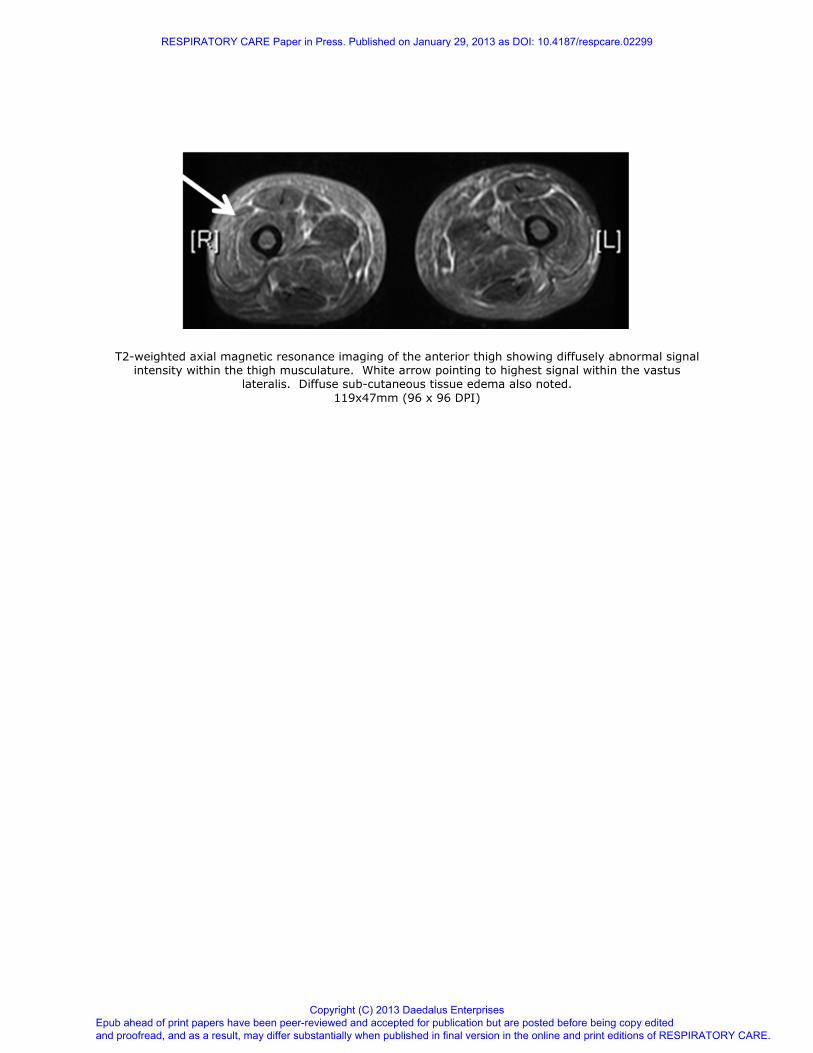
T2-weighted axial magnetic resonance imaging of the anterior thigh showing diffusely abnormal signal intensity within the thigh musculature. White arrow pointing to highest signal within the vastus
lateralis. Diffuse sub-cutaneous tissue edema also noted.
119x47mm (96 x 96 DPI)
RESPIRATORY CARE Paper in Press. Published on January 29, 2013 as DOI: 10.4187/respcare.02299
Epub ahead of print papers have been peer-reviewed and accepted for publication but are posted before being copy edited and proofread, and as a result, may differ substantially when published in final version in the online and print editions of RESPIRATORY CARE.
Copyright (C) 2013 Daedalus Enterprises
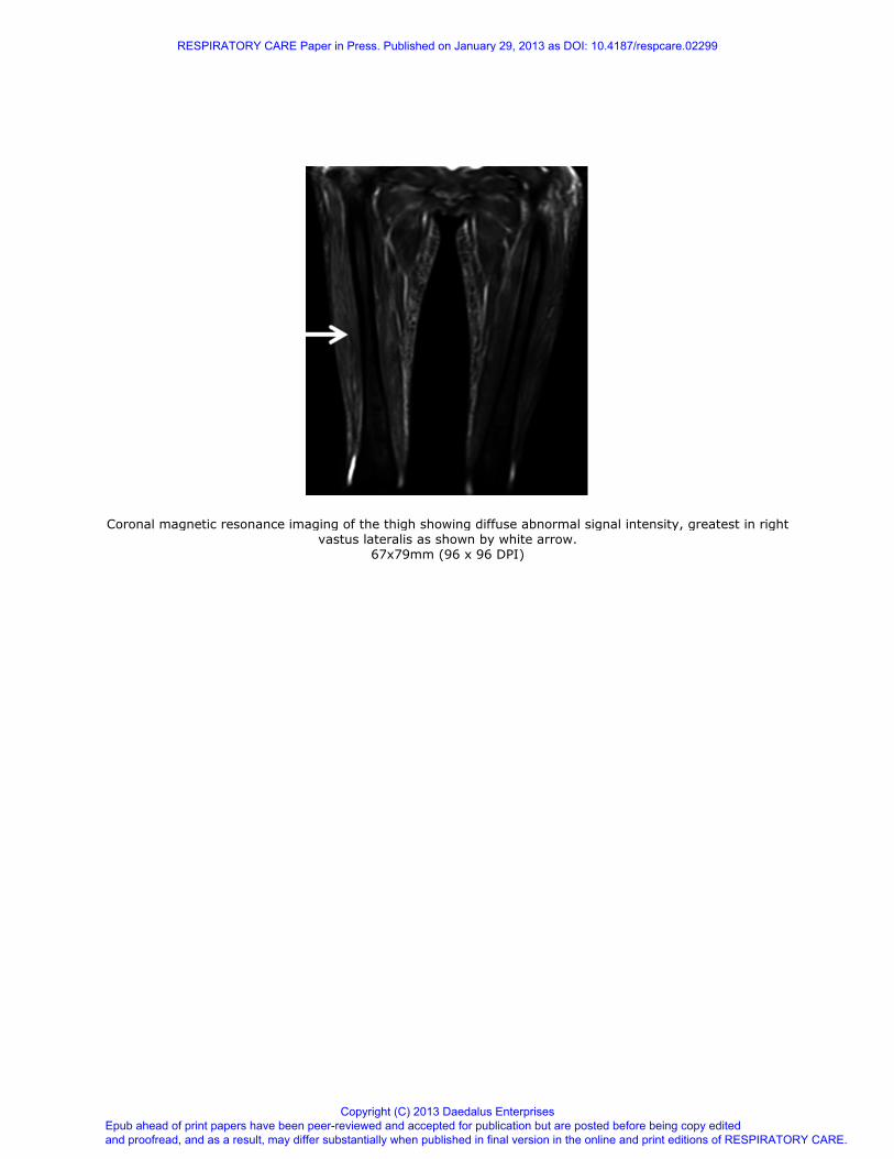
Coronal magnetic resonance imaging of the thigh showing diffuse abnormal signal intensity, greatest in right vastus lateralis as shown by white arrow.
67x79mm (96 x 96 DPI)
RESPIRATORY CARE Paper in Press. Published on January 29, 2013 as DOI: 10.4187/respcare.02299
Epub ahead of print papers have been peer-reviewed and accepted for publication but are posted before being copy edited and proofread, and as a result, may differ substantially when published in final version in the online and print editions of RESPIRATORY CARE.
Copyright (C) 2013 Daedalus Enterprises