comparison of commercial and non-commercial endotracheal...
TRANSCRIPT
Comparison of Commercial and Non-Commercial Endotracheal Tube
Securing Devices
Daniel F. Fisher, MS, RRT1, Christopher T. Chenelle, BS
2, Andrew Marchese, MS
3,
Joseph Kratohvil, LPN, RRT4, Robert M. Kacmarek, PhD, RRT, FAARC
5
1Assistant Director, Respiratory Care Services, Massachusetts General Hospital, Boston,
MA, USA. 2 Research Assistant, Department of Respiratory Care Services, Massachusetts General
Hospital, Boston, MA, USA. 3 Research Assistant, Department of Respiratory Care Services, Massachusetts General
Hospital, Boston, MA, USA and Computer Science and Artificial Intelligence
Laboratory, Massachusetts Institute of Technology, Cambridge, MA, USA. 4 Clinical Support Coordinator, Respiratory Care Services, Massachusetts General
Hospital, Boston, MA, USA. 5 Professor of Anesthesia, Department of Anesthesiology, Critical Care and Pain
Medicine, and Director of Respiratory Care Services, Massachusetts General Hospital
and Harvard Medical School, Boston, MA, USA.
This work is attributed to the Department of Respiratory Care, Massachusetts General
Hospital, Boston, MA, USA
All of the authors listed contributed equally in the preparation of this manuscript
This work has been presented as posters in the AARC 58th International Respiratory
Convention & Exhibition, Nov 10th – 13
th, New Orleans, Louisiana, USA
Corresponding author
Daniel F. Fisher, MS, RRT [email protected]
Massachusetts General Hospital Fax: (617) 724-4495
55 Fruit Street, Blake 652 Tel: (617) 724-1797
Boston, MA 02114
Abstract word count: 286
Body of manuscript count: 4534
This project was funded by a gift from Hollister, Libertyville, IL, USA.
Robert M. Kacmarek has received research grants from Covidien, Hamilton, General
Electric, Newport, and Dräger, honorarium for lecturing from Covidien and Maquet and
is a consultant for Newport.
Abbreviated Title: Comparison of Endotracheal Tube Holders
RESPIRATORY CARE Paper in Press. Published on December 24, 2013 as DOI: 10.4187/respcare.02951
Epub ahead of print papers have been peer-reviewed and accepted for publication but are posted before being copy edited and proofread, and as a result, may differ substantially when published in final version in the online and print editions of RESPIRATORY CARE.
Copyright (C) 2013 Daedalus Enterprises

Comparison of Commercial and Non-Commercial Endotracheal
Tube Securing Devices
Daniel F. Fisher, MS, RRT;
Christopher T. Chenelle, BS;
Andrew Marchese, MS;
Joseph Kratohvil, LPN, RRT;
Robert M. Kacmarek, PhD, RRT, FAARC
ABSTRACT
INTRODUCTION: Tracheal intubation is used to establish a secure airway in patients
who require mechanical ventilation. Unexpected extubation can have serious
complications including airway trauma and death. Various methods and devices have
been developed to maintain endotracheal tube (ETT) security. Associated complications
include pressure ulcers due to decreased tissue perfusion. Device consideration includes
ease of use, rapid application, and low exerted pressure around the airway.
METHODS: Sixteen ETT holders were evaluated under a series of simulated clinical
conditions. ETT security was tested by measuring distance displaced after a tug. Nine
out of the 16 devices could be evaluated for speed of moving the ETT to the opposite side
of the mouth. Sensors located on a mannequin measured applied forces when the head
was rotated vertically or horizontally. Data were analyzed using multivariate ANOVA
with p < 0.05 as significant.
RESULTS: Median displacement of the ETT by the Tug test was 0 cm, IQR 0.0 - 0.10
cm, p < 0.0001. The mean time to move the ETT from one side of the mouth to the other
ranged from 1.25+ 0.2 s to 34.4 + 3.4 s, p < 0.0001.
RESPIRATORY CARE Paper in Press. Published on December 24, 2013 as DOI: 10.4187/respcare.02951
Epub ahead of print papers have been peer-reviewed and accepted for publication but are posted before being copy edited and proofread, and as a result, may differ substantially when published in final version in the online and print editions of RESPIRATORY CARE.
Copyright (C) 2013 Daedalus Enterprises

Forces applied to the face with a vertical head lift ranged < 0.2 N to a maximum
of 3.52 N, p < 0.0001. Forces applied to the face with a horizontal rotation ranged < 0.2
N to 3.52 N, p < 0.0001. Commercial devices produced greater force than non-
commercial.
CONCLUSION: Non-commercial airway holders exert less force onto the patient’s face
than commercial devices. Airway stability is affected by the type of securing
device.Many of the commercial holders allow for a rapid, but secure movement of the
artificial airway from one side of the mouth to the other.
Abstract word count: 286
Manucript word count: 4534
RESPIRATORY CARE Paper in Press. Published on December 24, 2013 as DOI: 10.4187/respcare.02951
Epub ahead of print papers have been peer-reviewed and accepted for publication but are posted before being copy edited and proofread, and as a result, may differ substantially when published in final version in the online and print editions of RESPIRATORY CARE.
Copyright (C) 2013 Daedalus Enterprises

INTRODUCTION:
The purpose of an artificial airway is to relieve upper airway obstruction,
facilitate suctioning, allow effective ventilation and prevent aspiration. Unintended
removal or dislodgement of the endotracheal tube (ETT) can have harmful effects
ranging from localized trauma, and aspiration of oral/gastric secretions, to death as a
result of a compromised airway1 Alternatively, extended pressure from securing the ETT
on the surrounding tissue can lead to pressure sores and mucosal damage. This is a direct
result of the securing device causing pressure points decreasing local tissue perfusion2.
In addition, ETT can be inadvertently advanced to the carina or into a mainstem
bronchus.
The Joint Commission has made pressure ulcer prevention a national patient
safety goal (http://www.jointcommission.org/, Accessed May 2, 2012). Pressure ulcers
can range from discomfort to disfiguring sores. Current American Heart Association
(AHA) guidelines suggest that ETT should be secured with “tape or a commercial
device.”3 There are a myriad of devices that are designed to secure the ETT. All of these
devices and techniques have similar goals: to keep the artificial airway secure, and to
keep the patient safe by maintaining an intact airway and minimizing the chance of an
unplanned extubation.
When considering the type of device to secure the airway: ease of use, efficiency
in keeping the airway secure, and the ability to reposition the ETT to prevent pressure
ulcer formation should be considered. Methods of securing an ETT vary from straps of
tape or cotton string, to mechanical devices with integrated securing and movement
mechanisms. The purpose of this study was to evaluate a wide array of commercially
RESPIRATORY CARE Paper in Press. Published on December 24, 2013 as DOI: 10.4187/respcare.02951
Epub ahead of print papers have been peer-reviewed and accepted for publication but are posted before being copy edited and proofread, and as a result, may differ substantially when published in final version in the online and print editions of RESPIRATORY CARE.
Copyright (C) 2013 Daedalus Enterprises

available devices and traditional ETT securing techniques under simulated clinical
conditions. The conditions tested were designed to evaluate the ability of the holder to
keep the ETT in place, the rapidity of relocating the ETT from one side of the mouth to
the other, and finally how much force is transmitted to surrounding areas covered by the
device as the head is moved.
METHODS
Sixteen unique ETT holding devices or securing methods were subjected to four
separate tests to evaluate performance in simulated daily activities of the mechanically
ventilated acutely ill patient. The tests performed were, (1) static tug test, subjecting the
ETT secured by one of the holders to a momentary force pulling on the ETT in an
attempt to remove the ETT from the airway; (2) ETT movement, determination of the
time required to move an ETT from one side of the mouth to the opposite side of the
mouth; (3) vertical movement, determination of the pressure applied by the device to the
face and neck during simulated raising and lowering of the head 30 degrees; and (4)
horizontal movement, determination of the pressure applied by the device to the face and
neck during simulated turning of the head from side to side in a 70 degree arc. All
commercial devices were supplied by the manufacturer. (Table 1). Some of the holders
evaluated incorporated a band of material that wrapped around the back of the neck for
stability. For those holders, the tension along the band was set to allow two fingers to
pass snugly between the band and the surface of the mannequin head parallel to the head.
RESPIRATORY CARE Paper in Press. Published on December 24, 2013 as DOI: 10.4187/respcare.02951
Epub ahead of print papers have been peer-reviewed and accepted for publication but are posted before being copy edited and proofread, and as a result, may differ substantially when published in final version in the online and print editions of RESPIRATORY CARE.
Copyright (C) 2013 Daedalus Enterprises

Airway Model
An anatomically correct adult intubation model (Airway Larry #25000033
Laerdal, Wappingers Falls, NY) was used to simulate the head and upper airway. An 8.0
mm internal diameter (ID) ETT (Covidien Mallinckrodt Hi-LO) was inserted into the
trachea and secured by each of the 16 devices/methods evaluated. The cuff was inflated
to 25 cm H2O establishing a minimal leak.4 Cuff pressure was checked and re-
established immediately before each experimental trial.
Static Tug Test:
The intubation mannequin was orally intubated at the beginning of each series of
evaluations with every device. Prior to each intubation, the upper airway was lubricated
with silicone spray (Laerdal Airway Lubricant, Cat No. 25-20-90, Laerdal Wappingers
Falls, NY) to simulate lubrication of the airway with secretions and allow for a greater
freedom of movement than was available with dry plastic-on-plastic.
The ETTs were secured with each of the 16 devices/methods. After securing the
ETT, the cuff was slowly inflated to a pressure of 25 cm H2O monitored by a pressure
manometer with an incorporated luer-lok connection (Instrumentation Industries BE 148-
7, Bethel Park, PA). Using modified test apparatus as previously described,5, 6
the ETT
was connected to a pre-stressed 80-lb test nylon line (Mason Tackle, Otisville, MI). The
opposite end of the line was threaded through a pulley and attached to a 578 g weight.
The resulting angle formed between the pulley and the intubation head was 30 degrees
equal to the head of bed elevation angle suggested in the ventilator associated pneumonia
RESPIRATORY CARE Paper in Press. Published on December 24, 2013 as DOI: 10.4187/respcare.02951
Epub ahead of print papers have been peer-reviewed and accepted for publication but are posted before being copy edited and proofread, and as a result, may differ substantially when published in final version in the online and print editions of RESPIRATORY CARE.
Copyright (C) 2013 Daedalus Enterprises
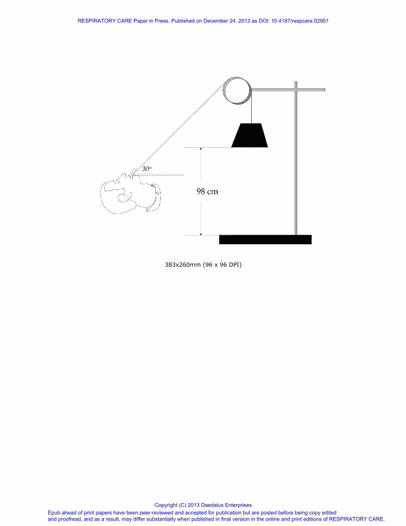
(VAP) bundles7 (Figure 1). A reference mark was made on the side of the ETT denoting
tooth position at the beginning of the test.
During a pilot test for this study, the weight was applied without a tug and tube
movement recorded. No discernable movement occurred. To prevent bias toward
adhesives, the face of the mannequin was covered with a new sheet of plastic film
(Blenderm, 3M) at the start of testing on each new device. This was to prevent any
potential interaction between the adhesive and residual cleaner, as well as providing a
better surface for adherence than the outer covering of the intubation head.
The weight was then dropped from a height of 98 cm and allowed to free fall
before stopping abruptly at 16 cm above the tabletop producing a jolt of approximately
5.7 Newtons on the ETT in the direction away from the patient as an attempt to extubate
the model. The distance the ETT moved was then recorded. This process was repeated
5 drops per run, and 5 runs per series, using 4 devices per evaluation resulting in a
possible 100 drops for each holder evaluated. Following the fifth drop and measurement,
the cuff was deflated, the ETT repositioned, and the securing method refastened. At the
end of the fifth run in each series, the securing device was removed; the ETT was placed
back into the starting position, secured with a new device from the same manufacture,
and the complete series of runs repeated. A new, previously unused device was used
during each series for each securing technique.
If during testing, the ETT became dislodged, or moved > 5 cm it was considered
an extubation and testing was stopped for that cycle (This distance is considerably
greater than the 20 mm suggested by others.6). The ETT was then repositioned and
refastened with a new device/method and a complete series of drops repeated with a new
RESPIRATORY CARE Paper in Press. Published on December 24, 2013 as DOI: 10.4187/respcare.02951
Epub ahead of print papers have been peer-reviewed and accepted for publication but are posted before being copy edited and proofread, and as a result, may differ substantially when published in final version in the online and print editions of RESPIRATORY CARE.
Copyright (C) 2013 Daedalus Enterprises

device/method. If the ETT was dislodged completely again, the series with the greatest
number of measurements would be used for analysis. When the distance moved was less
than 5 cm, the distance moved was recorded, the initial tube position re-established and
testing continued.
Side to Side ETT Movement:
For this portion of the evaluation, six of these devices were excluded due to a
common design feature which prevented lateral tube movement; an integrated bite block.
Another device was excluded because it was an optional head strap that would have no
impact on lateral tube position. The specific devices excluded from this evaluation were:
AMBU (Velcro), AMBU (silicone strap), Thomas tube holder (Laerdal), Precision
Medical, Portex Quickstrap, Teleflex, and Marpac 320 with headstrap. The nine
remaining devices/methods were evaluated for speed in moving the ETT from one corner
of the mouth to other. For the commercial devices, the manufacturer’s instructions were
followed. With the non commercial methods the knots evaluated were tied as described
by a web-based knot reference (http://www.animatedknots.com/, Accessed May 2, 2012).
Prior to testing the investigator practiced each knot until it could be rapidly tied without
assistance.
At the start of the testing, the ETT was secured adjacent to one corner of the
mannequin’s mouth. The investigator moving the airway would signal to the timing
investigator when to start the stopwatch. When the airway was repositioned, the
repositioning investigator would signal when to cease timing. This action was repeated a
RESPIRATORY CARE Paper in Press. Published on December 24, 2013 as DOI: 10.4187/respcare.02951
Epub ahead of print papers have been peer-reviewed and accepted for publication but are posted before being copy edited and proofread, and as a result, may differ substantially when published in final version in the online and print editions of RESPIRATORY CARE.
Copyright (C) 2013 Daedalus Enterprises
total of 10 times for each method. To limit variation in technique the same person (DF)
performed all repositioning and the same person kept time (JK) with all devices and
techniques.
If there was a problem during the movement, or either investigator felt that
conditions were not optimal, or the results were considered to be outside of 1 standard
deviation when compared with the other trials of the same device/method, the trial was
discarded and repeated using the same device/method.
Rotating Head Study:
The intubation head was removed from the stock torso and placed on a computer-
controlled platform that has independent motion on 2 axes (Figure 2). Digital encoders
were attached to each drive motor to provide spatial reference for the axis of rotation.
Control of each motor was governed via a graphical interface (Labview, National
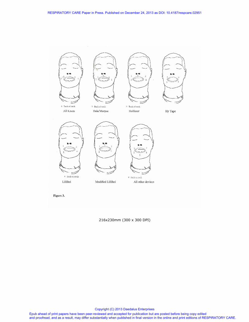
Instruments). Five force sensing resistors (FSR; Interlink Electronics, Camarillo, CA)
were placed around the mouth and nose (Figure 3). An additional sensor was placed on
the back of the neck. Sensor data from the FSRs were recorded as a voltage every 100
milliseconds using an analog to digital converter and output as a text file. During
analysis, the voltage was converted to Newtons (N) of force based upon the separate
calibration curves for each sensor which were developed before any measurements.
The upper airway was lubricated with silicone spray and the head was orally
intubated in the same fashion as previously described. The ETT was secured using a new
device/method for each trial. The secured ETT was then connected to a ventilator circuit
(Hudson RCI model #780-32) supported by a cross arm.
RESPIRATORY CARE Paper in Press. Published on December 24, 2013 as DOI: 10.4187/respcare.02951
Epub ahead of print papers have been peer-reviewed and accepted for publication but are posted before being copy edited and proofread, and as a result, may differ substantially when published in final version in the online and print editions of RESPIRATORY CARE.
Copyright (C) 2013 Daedalus Enterprises
The vertical elevation test was carried out by having the head lift from 3 to 30
degrees simulating the head being raised from a flat to a semi-recumbent position at a
rate of 12 rotations every minute. For the horizontal rotational testing the head moved in
a 70 degree arc at a rate of 5 rotations every minute. Run time for each axis was 10
minutes in order to allow for stabilization of the system. Due to the design features of
each ETT fixation device/method, the FSRs were moved to be under potential high
pressure points as determined by the investigators during pilot studies. Specific sensor
locations for each device are in the supplementary material. (Supplementary Figure 1)
The system was started with the head in the “neutral” position, 0 degrees
elevation and 0 degrees rotation from midline. During this startup, the system would
recalibrate position. Testing occurred only with one axis movement at any time.
Data was recorded at a rate of 100 ms, which included all sensor voltages, angular
position, and time. This recording was saved into a text file for later analysis.
Data Analysis:
Data was checked for normality using the Shapiro-Wilk test. All normally
distributed data were expressed as mean plus or minus standard deviation.
Nonparametric data was analyzed using multivariate ANOVA with p < 0.05. Post-hoc
analysis was performed using the Tukey HSD test. All data analysis was performed
using R statistical software, (version 3.0.1, R Project for Statistical Computing).
RESPIRATORY CARE Paper in Press. Published on December 24, 2013 as DOI: 10.4187/respcare.02951
Epub ahead of print papers have been peer-reviewed and accepted for publication but are posted before being copy edited and proofread, and as a result, may differ substantially when published in final version in the online and print editions of RESPIRATORY CARE.
Copyright (C) 2013 Daedalus Enterprises
Static Tug Test
Distance moved was collected for each of the 100 trials per device/technique.
Tube displacement > 5 cm was considered extubation; the remaining successful drops for
the holder were evaluated.
Side to Side ETT Movement
The time needed to move the ETT was compared using a one-way ANOVA with
Tukey HSD between devices and within each device.
Rotating Head Study
The system was allowed to stabilize one minute. The next 5 complete cycles were
collected and the voltage readings of the FSRs were converted into Newtons based from
calibration curves specific for each of the six sensors. When any device or technique did
not have a portion go around the neck of the intubation head, or touch a portion of the
face such as the chin, those sensors were not included in the analysis.
With the vertical axis movement, data was collected at 3, 15, and 30 degree
positions. The horizontal axis movement data was collected at -35, 0, and 35 degree
positions. Five data points at each of the three angles were selected for each device.
Since five of the same type of ETT holders were used during each evaluation a total of 25
data points for each angle for each device were analyzed. If 5 data points at a specific
angle were not available, the closest data point from the preceding or following angle
were recorded to bring the total to 5. We used repeated measures multivariate ANOVA
to compare the effects of device, angle of rotation/elevation, type of device; commercial
RESPIRATORY CARE Paper in Press. Published on December 24, 2013 as DOI: 10.4187/respcare.02951
Epub ahead of print papers have been peer-reviewed and accepted for publication but are posted before being copy edited and proofread, and as a result, may differ substantially when published in final version in the online and print editions of RESPIRATORY CARE.
Copyright (C) 2013 Daedalus Enterprises

or non-commercial, and the trial run between devices as well as comparing each device to
itself. For all evaluation a p < 0.05 was considered significant.
Results:
Static Tug Test
There were 1600 potential observations. During testing 17 complete
dislodgements were noted (2 with the Portex Quickstrap, 15 with Precision Medical #PM
1110). To compensate for the stretching of the new material, the first series of five drops
for each holder was removed (Run Series 1). Two of the 17 extubations occurred in this
series, both for the PM 1110 device and were omitted from analysis. This resulted in a
total of 1265/1280 observations. The median displacement for this group was 0.00 cm,
IQR 0.0 -0.10 cm. The holders with the least movement (median, IQR) were Anchor
Fast (0.0 cm; 0.0 – 0.00 cm), Hy Tape (0.0 cm, 0.0 – 0.00 cm), and the modified Lillihei
method (0.0, 0.0 – 0.00 cm), and the holder with the greatest displacement was the
Precision Medical 0.2 cm, IQR 0 - 1.1 cm; p < 0.0001, (Table 2). The displacement
distance (median, IQR) for both commercial and non-commercial devices were the same
(0 cm, 0.0 – 0.10 cm), but all of the extubations that occurred during testing happened
with the commercial devices and none occurred with the non-commercial devices, p =
0.0009. Table 2.
Tube Repositioning Study
An average of 13.8 + 12.3 seconds was required to move the ETT for the 9
devices/methods evaluated. The shortest period of time was documented for the Marpac
RESPIRATORY CARE Paper in Press. Published on December 24, 2013 as DOI: 10.4187/respcare.02951
Epub ahead of print papers have been peer-reviewed and accepted for publication but are posted before being copy edited and proofread, and as a result, may differ substantially when published in final version in the online and print editions of RESPIRATORY CARE.
Copyright (C) 2013 Daedalus Enterprises

320 (0.82 + 0.14 s), and Hollister Anchor Fast (1.25 + 0.20 s). The difference between
these two devices was not significant, p = 0.9999. The two slowest methods for
movement were Hy Tape (32.0 + 4 s), and the modified Lillihei method (34.4 + 3.44 s),
again, the differences between these two devices was not significant, p = 0.51 (Table 3).
Commercially available devices took less time to move the ETT when compared with
non-commercial techniques, (5.58 + 6.57 s vs. 17.91 + 12.48 s respectively) p < 0.0001.
Rotational Head Studies
There were 1200 observations for sensors 1 through 4. Both sensors 5 and 6 had
1050 observations each. Sensor 5 was the chin placement, and sensor 6 was the back of
the neck. This is because three holders had designs which did not cover those sensor
areas on the mannequin (Hy Tape – no sensor 5 or 6, Lillihei method – no sensor 5,
Modified Lillihei method – no sensor 5 or 6), see Figure 3.
Vertical Head Lift
Forces measured varied among each sensor for all of the ETT holders, p < 0.0001.
The greatest force recorded at any time was 3.52 N (AMBU # 320264040) at sensor 1.
The least force recorded was < 0.2 N; this measured force was beyond the resolution for
the sensors so a more precise measurement is unavailable. Grouping the forces into the
three angles (3, 15, and 30 degrees), force readings at individual sensors varied among
device, 1 through 4 p < 0.0001, sensor 6, p = 0.002 and sensor 5, p = 0.07. Summary
force readings mean + SD at each sensor for each device are shown in Supplementary
RESPIRATORY CARE Paper in Press. Published on December 24, 2013 as DOI: 10.4187/respcare.02951
Epub ahead of print papers have been peer-reviewed and accepted for publication but are posted before being copy edited and proofread, and as a result, may differ substantially when published in final version in the online and print editions of RESPIRATORY CARE.
Copyright (C) 2013 Daedalus Enterprises

Tables 1a-c. Commercial devices exerted more force than non-commercial methods; all
sensors p < 0.0001 see Figure 4.
When the individual devices were analyzed, the AMBU holder # 320264040, the
Marpac 320 with headgear, Cow Hitch, Lillihei method and Teleflex #1065 did not show
any difference between forces measured among the sensors.
Horizontal Head Turning
Force readings were different for each sensor with every device, p < 0.0001. The
greatest force recorded at any time was 3.40 N (AMBU # 320264040) at sensor 1. The
least force recorded was < 0.2 N; this measured force was beyond the resolution for the
sensors so a more precise measurement is unavailable. Grouping the data into the 3
angles (-35, 0, and 35 degrees), the forces measured for all devices showed wide
variation, p < 0.0001 with the exception of the Lillihei method p = 0.06826. Summary
force readings, mean + SD are listed in Supplementary Tables 2a-c.
Commercially available devices exerted more pressure at all sensors than non-
commercial devices with the exception of sensor 4. It registered higher forces with non-
commercial device than commercial devices, Figure 5.
Discussion:
This is the most comprehensive study to date, examining a large variety of ETT
securing devices under four different simulated clinical conditions. No single device
design performed well in all of the evaluations.
RESPIRATORY CARE Paper in Press. Published on December 24, 2013 as DOI: 10.4187/respcare.02951
Epub ahead of print papers have been peer-reviewed and accepted for publication but are posted before being copy edited and proofread, and as a result, may differ substantially when published in final version in the online and print editions of RESPIRATORY CARE.
Copyright (C) 2013 Daedalus Enterprises

The force exerted on the patient’s face by many of the commercial securing
devices may result in discomfort and the formation of pressure ulcers. Non-commercial
techniques use materials (tape, string) that are more form-fitting to the patient’s face and
therefore do not have the same pressure point issues as seen with the commercial devices.
Barnason 8 looked prospectively, at the impact of two securing techniques on
patient comfort and skin integrity. The techniques used to secure the airway were the
Lillihei method and cotton twill with a Cow Hitch knot. Pressure on the patient’s face
was based more on descriptive findings rather than quantifiable measurements. Their
conclusion was both techniques were equally effective in preventing oral mucosal
breakdown which is consistent with our findings.
Resistance to movement after the ETT has been subjected to an unplanned
tugging motion is crucial to the function of any ETT holder. In general the non-
commercial devices, because of variation in gripping ability did not perform as well as
the commercial devices.
Two separate studies 5, 9
used a similar technique as we did for creating dynamic
torque on the ETT by dropping a weight via a pulley. Murdoch and Hollgate’s design
was for the force generated to be perpendicular to the intubation mannequin. They
considered a movement of the ETT > 20 mm a major displacement. The two techniques
evaluated were the Laerdal Thomas Tube Holder and twill tape tied with a reef (square)
knot. During testing, they noted that in 61% of the trials the tape allowed ETT movement
of > 20 mm whereas none of the trials with the Laerdal device met failure criteria. An
explanation for this discrepancy may be that the reef knot was a poor choice and that a
better holding knot would have been the Rolling Hitch.
RESPIRATORY CARE Paper in Press. Published on December 24, 2013 as DOI: 10.4187/respcare.02951
Epub ahead of print papers have been peer-reviewed and accepted for publication but are posted before being copy edited and proofread, and as a result, may differ substantially when published in final version in the online and print editions of RESPIRATORY CARE.
Copyright (C) 2013 Daedalus Enterprises

Lovett used a PVC pipe as an intubation model and then saturated the holders
once applied with saline to simulate oral secretions5. They measured the actual force
generated when dropping 2.5, 5, and 10 lb weights and measuring the distance the ETT
moved after 6 and 15 drops. This method provided the average distance moved after a
sequence of drops rather than the effects of each drop. Although there was a difference
in models, PVC pipe vs. intubation mannequin the performance was similar to what we
experienced for the three holders that were common for both studies (Lillihei, Precision
Medical, and Thomas Tube Holder)
The overall poorest performer during the static tug test, Precision Medical,
utilized a thin securing strap which stretched significantly. It is the elasticity of the strap
that allowed for a large displacement of the ETT. This finding is similar to that by
Carlson.9 In fact, the strap was so compliant; it was difficult to secure the airway with
only a “two-finger tight” assessment, two fingers snugly fitting between the face of the
mannequin and the securing strap. This issue was evident to some degree with other
devices employing a similar design where there was too much material requiring multiple
layers of wrapping, or in extreme conditions, tightening beyond the recommended two-
finger test. Devices in this class were the AMBU ETT holder with blue silicone strap,
Smiths-Portex Quickstrap, and Dale Stabilok.
Several manufacturers secured the ETT, by compressing the ETT between a
clamp or screw. These mechanisms had an influence on the cross-sectional shape of the
airway. This distortion may have an influence on airway resistance, but it was not within
the scope of this investigation to measure airway resistance.
RESPIRATORY CARE Paper in Press. Published on December 24, 2013 as DOI: 10.4187/respcare.02951
Epub ahead of print papers have been peer-reviewed and accepted for publication but are posted before being copy edited and proofread, and as a result, may differ substantially when published in final version in the online and print editions of RESPIRATORY CARE.
Copyright (C) 2013 Daedalus Enterprises

Several commercial devices are designed to allow for quick tube position
relocation, and in fact outperformed non-commercial devices. A common design feature
for the commercially-prepared holders was a track where the ETT could be guided from
one point to another. The non-commercial holders all required disassembly and
reassembly of the securing technique; two techniques (modified Lillihei method, and Hy
Tape) required a completely new holder to be fashioned after moving the airway. This
resulted in the airway being unsecured and susceptible to displacement, either by the
practitioner, or by the patient.
Causes of unplanned extubation
There are two classifications of unplanned extubation (UEX): patient initiated,
and practitioner initiated. Both categories of UEX place the patient at some risk
depending upon their clinical status. It has been one of the primary goals of ETT fixation
to secure the ETT in a manner where the airway is unlikely to become dislodged, yet
flexible enough not to cause damage to surrounding tissue. In one multicenter study
examining UEX, the lack of a “strong” fixation device was found to be one of the risk
factors identified.10
UEX can lead to co-morbidity including increased ventilator days, and increased
mortality 10, 11
. UEX has been associated with an increased likelihood of transfer to
chronic care facilities.11 Among the factors affecting transfer are skin integrity,
complexity of the ETT securing device, oral care, speed of application of the device to
the ETT, and patient comfort. No one device can address every factor, nor is it possible
to test for every contingency.
RESPIRATORY CARE Paper in Press. Published on December 24, 2013 as DOI: 10.4187/respcare.02951
Epub ahead of print papers have been peer-reviewed and accepted for publication but are posted before being copy edited and proofread, and as a result, may differ substantially when published in final version in the online and print editions of RESPIRATORY CARE.
Copyright (C) 2013 Daedalus Enterprises
Ability to relocate the ETT Position
Devices with an integrated bite block prevent the ETT from being repositioned
which may lead to pressure ulcers in the mouth and surrounding tissue. Kuhn reported on
the development of a necrotic region on the tongue of a patient after only eight hours of
intubation.12 The bite block incorporated with some devices can also interfere with
providing adequate oral hygiene by preventing access to the oral cavity. There is at least
one report of an added bite block interfering with cuff function.13
Limitations:
There are several limitations to this study. First, this was a bench study and the
surface of the mannequin may have behaved differently with the various adhesives used
by some of the manufacturers than natural skin. This property was standardized with the
application of the Blenderm product whenever the adhesive properties were being
stressed during the static tug. Nonetheless, the skin of the intubation head does not have
the same tensile qualities of real skin and thus may have impacted the pressure readings
measured when securing the ETT.
Secondly, all readings were done at ambient temperature (~ 68o F) without oral
secretions saturating the securing devices/methods. Previous studies examined the
securing properties of various devices both in simulation and in-vitro, identified oral
secretions as factors affecting function.5, 8, 9, 14
These studies were either of small sample
size, or anecdotal discussions of various ETT holders. In the study by Carlson9,
extubation force was determined using fresh, < 24 hour old refrigerated cadavers and
RESPIRATORY CARE Paper in Press. Published on December 24, 2013 as DOI: 10.4187/respcare.02951
Epub ahead of print papers have been peer-reviewed and accepted for publication but are posted before being copy edited and proofread, and as a result, may differ substantially when published in final version in the online and print editions of RESPIRATORY CARE.
Copyright (C) 2013 Daedalus Enterprises

actual force was measured using a strain gauge. Studies by both Arrott and Barnason
were either descriptive or observational in nature.8, 15
In clinical practice, there is no
opportunity to pre-stress the neck strap as occurred during the static tug test, so actual
ETT displacement distances may be greater than reported here.
Thirdly, during the tube movement, the goal was to move the airway as quickly as
possible. It is likely that under clinical circumstances, concern for the safety of the
patient would have a slowing effect on tube movement. As a result, timing was most
likely shorter than during actual clinical conditions.
Fourthly, the movement of the ETT further into the airway, i.e. a mainstem
bronchus, was not evaluated in this study. This is an important issue and should be
considered for future studies.
Finally, this study was not designed to measure the convenience factor in
application of the devices, which can have an impact on choice of device/method.
Conclusion:
Conclusions for this study are as follows: 1) Non-commercial airway
holders exert less force onto the patient’s face than commercial devices; 2) Airway
stability is affected by the type of securing device selected; and 3) many of the
commercial securing devices allow for a rapid, but secure movement of the artificial
airway from one side of the mouth to the other. However, at this time there is no ideal
device or method for securing ETT.
RESPIRATORY CARE Paper in Press. Published on December 24, 2013 as DOI: 10.4187/respcare.02951
Epub ahead of print papers have been peer-reviewed and accepted for publication but are posted before being copy edited and proofread, and as a result, may differ substantially when published in final version in the online and print editions of RESPIRATORY CARE.
Copyright (C) 2013 Daedalus Enterprises

References
1. da Silva PSL, Fonseca MCM. Unplanned Endotracheal Extubations in the
Intensive Care Unit. Anesth Analg 2012;114(5):1003-1014.
2. Zaratkiewicz S, Teegardin C, Whitney JD. Retrospective review of the reduction
of oral pressure ulcers in mechanically ventilated patients: a change in practice.
Crit Care Nurse 2012;35(3):247-254.
3. American Heart Association . Part 7.1: Adjuncts for airway control and
ventilation. Circulation 2005;112(24_suppl):IV-51 - IV -57.
4. Pitts R, Fisher D, Sulemanji D, Kratohvil J, Jiang Y, Kacmarek R. Variables
affecting leakage past endotracheal tube cuffs: A bench study. Intens Care Med
2010;36:2066-2073.
5. Lovett PB, Flaxman A, Stürmann KM, Bijur P. The insecure airway: A
comparison of knots and commercial devices for securing endotracheal tubes.
BMC Emerg Med 2006;6(1):7.
6. Murdoch E, Holdgate A. A comparison of tape-tying versus a tube holding device
for securing endtotracheal tubes in adults. Anaesth Intens Care 2007;35:730-735.
7. Dodek P, Keenan S, Cook D, Heyland D, Jacka M, Hand L, et al. Evidence-based
clinical practice guideline for the prevention of ventilator-associated pneumonia.
Ann Intern Med 2004;141:305-313.
8. Barnason S, Graham J, Wild MC, Jensen LB, Rasmussen D, Schulz P, et al.
Comparison of two endotracheal tube securement techniques on unplanned
extubation, oral mucosa, and facial skin integrity. Heart Lung 1998;27(6):409-
417.
RESPIRATORY CARE Paper in Press. Published on December 24, 2013 as DOI: 10.4187/respcare.02951
Epub ahead of print papers have been peer-reviewed and accepted for publication but are posted before being copy edited and proofread, and as a result, may differ substantially when published in final version in the online and print editions of RESPIRATORY CARE.
Copyright (C) 2013 Daedalus Enterprises
9. Carlson J, Mayrose J, Krause R, Jehle D. Extubation Force: Tape Versus
Endotracheal Tube Holders. Ann Emerg Med 2007;50(6):686-691.
10. Boulain T. Unplanned extubations in the adult intensive care unit: a prospective
multicenter study. Association des Reanimateurs du Centre-Ouest. Am J Resp
Crit Care 1998;157(4 Pt 1):1131-1137.
11. Epstein SK, Nevins ML, Chung J. Effect of unplanned extubation on outcome of
mechanical ventilation. Am J Resp Crit Care 2000;161(6):1912-1916.
12. Kuhn MA, Zeitler DM, Myssiorek DJ. Tongue necrosis: A rare complication of
oral intubation. Laryngoscope 2010;120(S4):S125-S249.
13. Adams JR, Hoffman J, Lavelle J, Mireles-Cabodevila E. Pilot Balloon
Malfunction Caused by Endotracheal Tube Bite Blockers. [epub ahead of print]
Respir Care 2013. doi: 10.4187/respcare.02474.
14. Levy H, Griego L. A comparative study of oral endotracheal tube securing
methods. Chest 1993;104(5):1537-1540.
15. Arrott JJ, Talley AW. Endotracheal tube holder. Anesth Analg 1974;53(1):70-71.
RESPIRATORY CARE Paper in Press. Published on December 24, 2013 as DOI: 10.4187/respcare.02951
Epub ahead of print papers have been peer-reviewed and accepted for publication but are posted before being copy edited and proofread, and as a result, may differ substantially when published in final version in the online and print editions of RESPIRATORY CARE.
Copyright (C) 2013 Daedalus Enterprises

Legends to Figures
Figure 1. Static Tug setup. The angle between the intubation head and the pulley was
30 degrees simulating a patient in the semi-fowler’s position. The weight was dropped
and allowed to fall freely until stopping abruptly causing a jolt.
Figure 2. Illustration of the mannequin setup for the vertical and horizontal movement
evaluation. The platform uses gear motors and encoders to pivot the mannequin head in
two dimensions. Control for the motors is via a LabView interface.
Figure 3. Sensor Placement for Rotating Head Study Numbers indicate sensor
placement. Sensor 6 was on the back of neck for those holders that had a strap.
Figure 4. Graph of pooled forces measured during the vertical head lift at each sensor
between commercial and non-commercial ETT holders.
Figure 5. Graph of pooled forces measured during the horizontal head rotation at each
sensor between commercial and non-commercial ETT holders.
RESPIRATORY CARE Paper in Press. Published on December 24, 2013 as DOI: 10.4187/respcare.02951
Epub ahead of print papers have been peer-reviewed and accepted for publication but are posted before being copy edited and proofread, and as a result, may differ substantially when published in final version in the online and print editions of RESPIRATORY CARE.
Copyright (C) 2013 Daedalus Enterprises

Table 1: Devices/Techniques used
Commercial Methods
Device Manufacturer
AMBU ET Tube Holder (blue strap) # 320264040 AMBU
AMBU ET Tube Holder (white strap) # 320264041 AMBU
Stabilock ETT Holder Dale
Anchor Fast Hollister
Thomas Endotracheal Tube Holder Laerdal
Marpac 320 Marpac
Marpac 320 with optional headgear Marpac
Quickstrap Endotracheal Tube Holder Portex
Endotracheal Tube Holder # PM1110 Precision Medical
Cushioned Endotracheal Tube Holder #1065 Teleflex
Non-Commercial Methods
Device Material Used
Clove Hitch Cotton Twill
Cow Hitch Cotton Twill
Rolling (Magnus) Hitch Cotton Twill
Hy Tape Hy Tape ¼ inch
Lillihei method using cloth tape Covidien Cloth Tape
Modified Lillihei Method Covidien Cloth Tape
RESPIRATORY CARE Paper in Press. Published on December 24, 2013 as DOI: 10.4187/respcare.02951
Epub ahead of print papers have been peer-reviewed and accepted for publication but are posted before being copy edited and proofread, and as a result, may differ substantially when published in final version in the online and print editions of RESPIRATORY CARE.
Copyright (C) 2013 Daedalus Enterprises
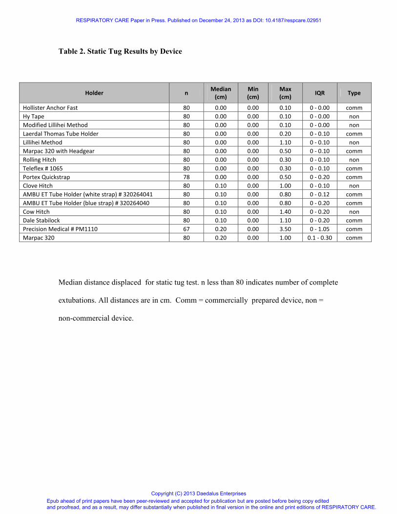
Table 2. Static Tug Results by Device
Holder n Median
(cm)
Min
(cm)
Max
(cm) IQR Type
Hollister Anchor Fast 80 0.00 0.00 0.10 0 - 0.00 comm
Hy Tape 80 0.00 0.00 0.10 0 - 0.00 non
Modified Lillihei Method 80 0.00 0.00 0.10 0 - 0.00 non
Laerdal Thomas Tube Holder 80 0.00 0.00 0.20 0 - 0.10 comm
Lillihei Method 80 0.00 0.00 1.10 0 - 0.10 non
Marpac 320 with Headgear 80 0.00 0.00 0.50 0 - 0.10 comm
Rolling Hitch 80 0.00 0.00 0.30 0 - 0.10 non
Teleflex # 1065 80 0.00 0.00 0.30 0 - 0.10 comm
Portex Quickstrap 78 0.00 0.00 0.50 0 - 0.20 comm
Clove Hitch 80 0.10 0.00 1.00 0 - 0.10 non
AMBU ET Tube Holder (white strap) # 320264041 80 0.10 0.00 0.80 0 - 0.12 comm
AMBU ET Tube Holder (blue strap) # 320264040 80 0.10 0.00 0.80 0 - 0.20 comm
Cow Hitch 80 0.10 0.00 1.40 0 - 0.20 non
Dale Stabilock 80 0.10 0.00 1.10 0 - 0.20 comm
Precision Medical # PM1110 67 0.20 0.00 3.50 0 - 1.05 comm
Marpac 320 80 0.20 0.00 1.00 0.1 - 0.30 comm
Median distance displaced for static tug test. n less than 80 indicates number of complete
extubations. All distances are in cm. Comm = commercially prepared device, non =
non-commercial device.
RESPIRATORY CARE Paper in Press. Published on December 24, 2013 as DOI: 10.4187/respcare.02951
Epub ahead of print papers have been peer-reviewed and accepted for publication but are posted before being copy edited and proofread, and as a result, may differ substantially when published in final version in the online and print editions of RESPIRATORY CARE.
Copyright (C) 2013 Daedalus Enterprises

Table 3. Time needed to move the ETT by Device
Holder Mean (sec) SD (sec)
Marpac 320 0.82 0.14
Hollister Anchor Fast 1.25 0.20
Cow Hitch 2.79 0.34
Clove Hitch 4.21 0.54
Rolling Hitch 7.48 1.16
Bow * 8.48 0.83
Dale Stabilock 14.67 1.10
Lillihei Method 20.84 5.51
Hy Tape 32.02 4.00
Modified Lillihei Method 34.42 3.44
Mean + SD time is seconds to move the ETT from one side of the mouth to the other.
* See text for description of knots.
RESPIRATORY CARE Paper in Press. Published on December 24, 2013 as DOI: 10.4187/respcare.02951
Epub ahead of print papers have been peer-reviewed and accepted for publication but are posted before being copy edited and proofread, and as a result, may differ substantially when published in final version in the online and print editions of RESPIRATORY CARE.
Copyright (C) 2013 Daedalus Enterprises
383x260mm (96 x 96 DPI)
RESPIRATORY CARE Paper in Press. Published on December 24, 2013 as DOI: 10.4187/respcare.02951
Epub ahead of print papers have been peer-reviewed and accepted for publication but are posted before being copy edited and proofread, and as a result, may differ substantially when published in final version in the online and print editions of RESPIRATORY CARE.
Copyright (C) 2013 Daedalus Enterprises

302x148mm (96 x 96 DPI)
RESPIRATORY CARE Paper in Press. Published on December 24, 2013 as DOI: 10.4187/respcare.02951
Epub ahead of print papers have been peer-reviewed and accepted for publication but are posted before being copy edited and proofread, and as a result, may differ substantially when published in final version in the online and print editions of RESPIRATORY CARE.
Copyright (C) 2013 Daedalus Enterprises
For Peer Review
216x230mm (300 x 300 DPI)
RESPIRATORY CARE Paper in Press. Published on December 24, 2013 as DOI: 10.4187/respcare.02951
Epub ahead of print papers have been peer-reviewed and accepted for publication but are posted before being copy edited and proofread, and as a result, may differ substantially when published in final version in the online and print editions of RESPIRATORY CARE.
Copyright (C) 2013 Daedalus Enterprises

179x141mm (96 x 96 DPI)
RESPIRATORY CARE Paper in Press. Published on December 24, 2013 as DOI: 10.4187/respcare.02951
Epub ahead of print papers have been peer-reviewed and accepted for publication but are posted before being copy edited and proofread, and as a result, may differ substantially when published in final version in the online and print editions of RESPIRATORY CARE.
Copyright (C) 2013 Daedalus Enterprises