pilot balloon malfunction caused by endotracheal tube bite...
TRANSCRIPT

Pilot Balloon Malfunction Caused by Endotracheal Tube Bite Blockers
Jacob R. Adams, DO; Justin Hoffman, RRT; Joe Lavelle, RRT; Eduardo Mireles-
Cabodevila, MD.
Affiliation of authors: Respiratory Institute, Division of Pulmonary, Allergy and
Critical Care Medicine, Cleveland Clinic, Cleveland, OH
Address Correspondence to:
Eduardo Mireles-Cabodevila, MD
Cleveland Clinic Main Campus
Mail Code G62
9500 Euclid Avenue
Cleveland, OH 44195
The authors report no conflict of interest in the content of this case report.
Key Words:
Airway extubation, Intratracheal intubation, Laryngeal edema
RESPIRATORY CARE Paper in Press. Published on June 12, 2013 as DOI: 10.4187/respcare.02474
Epub ahead of print papers have been peer-reviewed and accepted for publication but are posted before being copy edited and proofread, and as a result, may differ substantially when published in final version in the online and print editions of RESPIRATORY CARE.
Copyright (C) 2013 Daedalus Enterprises
Introduction
A patient’s bite is a frequent cause of endotracheal tube (ETT) obstruction. ETT
obstructions affect the volume and pressure delivered by the ventilator. Bite-related
ETT occlusions are a frequent cause of ventilator alarm activation, and are known to
cause respiratory failure, negative pressure pulmonary edema or ETT tube/pilot
tube damage. Clinicians have used objects (syringes, tongue depressors, gauze) or
devices intended for other goals (i.e. oropharyngeal airways or intermolar devices)
to prevent the patient from biting the ETT. However, these devices are often
makeshift, not designed for prolonged use and come with numerous side effects
such as, accidental dislodgement, ulcers, aspiration, ischemia and/or injury to the
temporomandibular joint. Thus, several commercial bite blocks, which encase and
protect the endotracheal tube, are now commonly used. These devices are made of a
relatively hard plastic that keeps its form in spite of the body temperature or patient
bites. They have a low profile and may double as tube holders. These devices come
in single sizes for adults or children. They are easy to use and thus are becoming
ubiquitous. The placement of these devices is mainly intuitive, and although
instructions and policies may exist, its placement and troubleshooting comes from
experience.
Case Summary
A 78 year-old, 152cm tall woman was transferred to our intensive care unit for
evaluation of acute respiratory failure. At the outside hospital she was treated for
community acquired pneumonia. While admitted, she was noted to have jaw
stiffness with limited opening leading to an inability to eat or drink. She developed
worsening respiratory failure and hypoxemia and was electively intubated. The
intubation was described as very difficult due to limited mouth opening, even with
the use of neuromuscular blockers. Intubation was achieved with a 7.0 mm, cuffed
ETT (MallinckrodtTM, Mansfield, MA), fixed at 21 cm at the teeth. Her past medical
history was significant for hypertension, hyperlipidemia and osteoporosis.
Her physical exam was relevant for the presence of limited mouth opening, less than
2 cm. The neck mobility was limited in all directions. She was awake and
interactive. Her lung exam demonstrated scant bilateral basilar crackles. Her heart
rate was regular and rhythmic, and there was trace lower extremity edema. The
endotracheal tube was easily compressed by the teeth, thus a B&B Universal Bite
Block® (B&B Medical Technologies Inc., Orangevale, CA) was placed by the
respiratory therapist and secured with an Anchor Fast Oral Endotracheal Tube
Fastener (Hollister Inc., Libertyville, IL).
Ear, Nose and Throat and dentistry consultation concluded that the limited mouth
opening was due to severe temporomandibular joint disease. The patient was
RESPIRATORY CARE Paper in Press. Published on June 12, 2013 as DOI: 10.4187/respcare.02474
Epub ahead of print papers have been peer-reviewed and accepted for publication but are posted before being copy edited and proofread, and as a result, may differ substantially when published in final version in the online and print editions of RESPIRATORY CARE.
Copyright (C) 2013 Daedalus Enterprises
unable to open her mouth more than 2 finger widths, voluntarily or under paralysis.
After four days of therapy, her respiratory status improved. She passed a
spontaneous breathing trial, was awake and interactive. Part of our extubation
checklist includes the presence of a cuff leak (1,2). She did not have any leak by
ventilator volume or auscultation. Due to the absence of a cuff leak and the concern
for a difficult re-intubation, the extubation was aborted. Endoscopic exam of the
larynx was inconclusive due to partial visualization of the laryngeal structures. The
patient was given intravenous steroids (Dexamethasone 8mg IV every 8hrs) for
presumed laryngeal edema.
The next day a cuff leak test was again performed. No leak was elicited. As part of
routine protocol, cuff pressures were measured all averaging 20 cm H2O. The team
was discussing the possibility of a tracheostomy given her difficult airway. The
respiratory therapist, recalling similar events, decided to check the pilot balloon
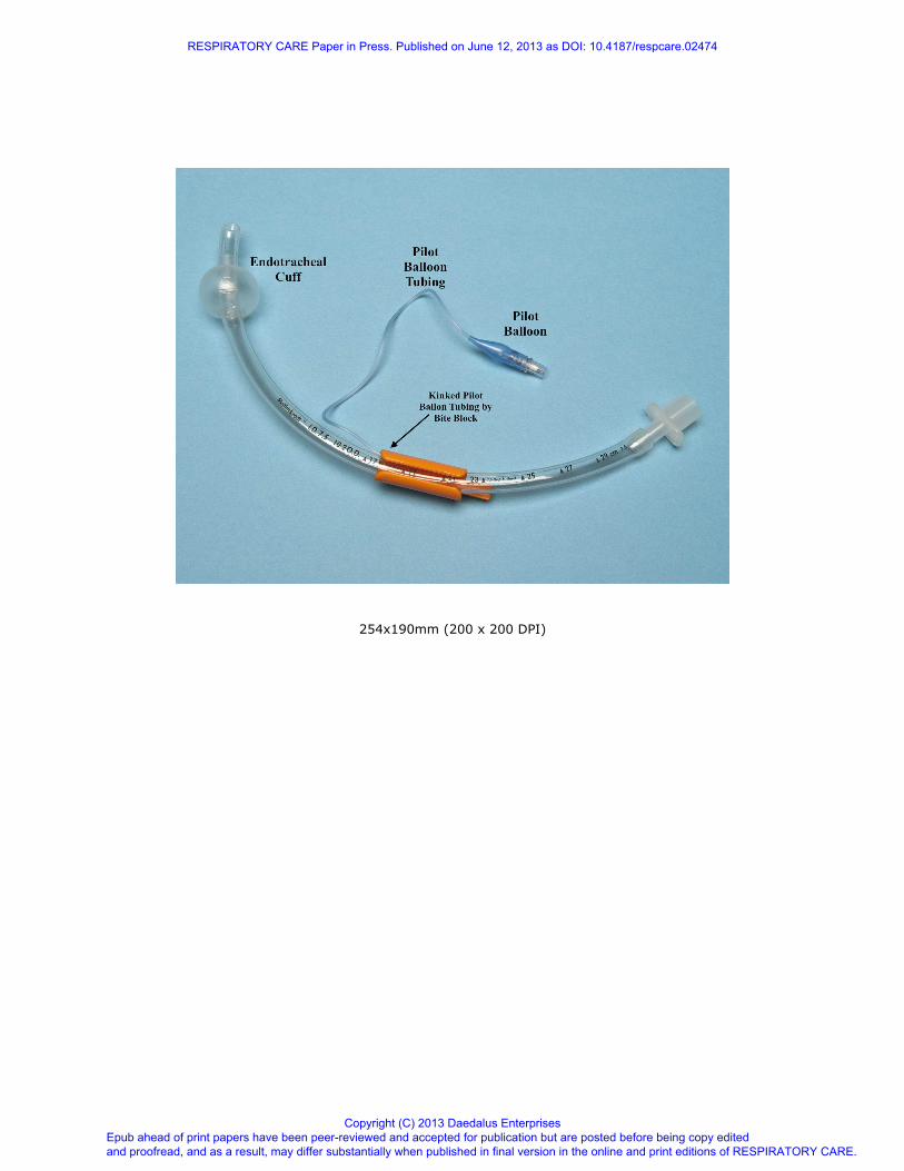
line. The bite block had inadvertently kinked the pilot balloon tubing at the junction
of the ETT (Figure 1); as a result we saw a completely deflated pilot balloon while
the ETT cuff remained inflated (Figure 2). After removal of the bite block, a cuff leak
became evident. The patient was extubated over a tube exchanger. She continued to
recover and was eventually discharged to the floor without further problems.
Discussion
Our case report highlights the need for specific steps to care for patients that have a
bite block in place. We demonstrated that the bite block placement or its migration
could kink the pilot balloon tubing, which if not identified, can result in unnecessary
interventions.
A kinked pilot tube may cause two scenarios:
1) Persistent under inflation of the cuff. The pilot balloon is fully inflated, the
pressure gauge would be normal, but the ETT cuff is deflated or
underinflated. This may lead to persistent air leak, inability to ventilate
and aspiration of subglotic secretions. (3,4)
2) Persistent inflation or over inflation of the cuff. The pilot balloon is deflated
or is fully inflated, the pressure gauge would be normal, but the ETT cuff
is inflated or overinflated. The result may be an absent cuff leak test,
inaccurate cuff pressures or difficulty removing the ETT (5,6).
Although we have experienced both scenarios, there is a paucity of reports referring
to this problem. A systematic review of the literature revealed both situations
scarcely being reported (Table 1). The stand-alone bite blocks that encase the ETT
were the most common device associated with pilot balloon occlusion. (3,4,5). Each
of the reported episodes had clinical consequences, which is likely why they reached
publication. The reports by Alkire (3) and Brock-Utne (4) describe large air-leaks,
desaturations and difficulty ventilating the patients. In both cases, the pilot balloon
was completely inflated while the ETT’s cuff was deflated. In a case similar to ours,
RESPIRATORY CARE Paper in Press. Published on June 12, 2013 as DOI: 10.4187/respcare.02474
Epub ahead of print papers have been peer-reviewed and accepted for publication but are posted before being copy edited and proofread, and as a result, may differ substantially when published in final version in the online and print editions of RESPIRATORY CARE.
Copyright (C) 2013 Daedalus Enterprises
Gleich (5) et al and Singh et al (6) report unintended kinking of the pilot tubing
leading to persistent inflation of the ETT cuff, which resulted in difficulty removing
the ETT. Our extubation protocol includes a cuff leak test, where an absence of a
leak triggers an alert to the physician to decide whether the tube is removed. Our
patient had a difficult airway, thus the decision to provide steroids for 24 hrs (7).
Bite blocks can be grouped into those that serve as individual bite blocks encasing
the ETT, and those that are part of an ETT holder. All of these bite blocks come in a
variety of sizes ranging from pediatric to adult. The cases reported in the literature
all happened with individual bite blocks. Those integrated with the ETT holder may
have the advantage that they do not encase the ETT (or pilot balloon) and the bite
blocker is not as long as the individual units. Further, as they are attached to the face
of the patient, migration is less likely.
When we consider the usual depth of insertion of an ETT, the bite block length may
go over the pilot balloon take off. The device we used was the Adult size Universal
Bite Block (B&B Medical Technologies Inc.) which measures 4.4cm in length. The
pilot balloon take-off occurs at different lengths of the ETT according to its size. In
Table 2 we demonstrate patients that may be at risk according to the combinations
of pilot balloon take-off, ETT length placements at the lip and an individual bite
blocker (Universal Bite Block (adult)™). As it can be seen, there is more risk in
patients with short ETT insertion, distance to the lips and larger ETT.
Each bite block comes with specific instructions on its placement. In the particular
of this case, the bite block may be positioned to avert the pilot balloon kinking,
however, this does not allow the respiratory therapist to visualize the length of the
tube at the patient’s lip. Turning the bite block, to visualize the numbering, will kink
the pilot balloon when advancing the bite block (Figure 1). Indeed, the B&B’s
policies and procedures for insertion (8) indicate that the pilot balloon should be
placed between the bite block and the ETT. This may prevent the kinking at the
pilot balloon take off. The insert reads: “Should cuff filling problems occur, gently
pull the pilot balloon line taut to remove any kinks.” However, this would not solve
the problem when the kinking is at the pilot line takeoff.
Whenever a pilot balloon is inflated, a clue to obstruction of the tubing is the
amount of air needed to fill the ETT cuff. When small volumes are used, or pressure
rapidly rises in the pressure manometer while inflating, suspicion should rise for an
obstruction of the pilot balloon line. Whenever the balloon is deflated, a clue to an
obstruction is low volumes being removed with the syringe.
Teaching Points
• Endotracheal tube bite blocks may cause kinking of pilot balloon tubing.
• Every blocker has its particulars, but patients at risk may be those with
larger tubes and short ETT insertion distance to the lips.
RESPIRATORY CARE Paper in Press. Published on June 12, 2013 as DOI: 10.4187/respcare.02474
Epub ahead of print papers have been peer-reviewed and accepted for publication but are posted before being copy edited and proofread, and as a result, may differ substantially when published in final version in the online and print editions of RESPIRATORY CARE.
Copyright (C) 2013 Daedalus Enterprises

• Obstruction of the pilot balloon tubing may cause persistent inflation or
under inflation of the cuff.
• The clues that signal kinking of the pilot balloon tubing are small amounts of
air (1-2 mL) required to deflate or inflate the cuff.
• Routine assessment of the intubated patient must include evaluation of the
pilot balloon and tubing.
• Finally, we suggest that the ETTs should be free of any additional device
when performing an extubation.
RESPIRATORY CARE Paper in Press. Published on June 12, 2013 as DOI: 10.4187/respcare.02474
Epub ahead of print papers have been peer-reviewed and accepted for publication but are posted before being copy edited and proofread, and as a result, may differ substantially when published in final version in the online and print editions of RESPIRATORY CARE.
Copyright (C) 2013 Daedalus Enterprises
References
1. Miller RL, Cole RP. Association between reduced cuff leak volume and
postextubation stridor. Chest 1996;110(4):1035-1040.
2. Ochoa ME, Marin Mdel C, Frutos-Vivar F, Gordo F, Latour-Perez J, Calvo E, Esteban
A Cuff-leak test for the diagnosis of upper airway obstruction in adults: A systematic
review and meta-analysis. Intensive Care Med 2009;35(7):1171-1179.
3. Alkire MT. Ventilatory compromise secondary to occlusion of an endotracheal
tube's balloon air channel by a malpositioned bite block. Anesthesiology
1998;88(5):1419.
4. Brock-Utne AJ. The universal bite-block: A word of caution. Anesth Analg
2006;103(2):495-496.
5. Gleich SJ, Mauermann WJ, Torres NE. An unusual cause of a difficult extubation.
Respir Care 2008;53(3):376.
6. Singh M, Rautela RS, Kumar S. Laryngeal tube pilot balloon kinking in the
presence of a bite block. Can J Anaesth 2009;56(11):880-881.
7. McCaffrey J, Farrell C, Whiting P, Dan A, Bagshaw SM, Delaney AP. Corticosteroids
to prevent extubation failure: A systematic review and meta-analysis. Intensive Care
Med 2009;35(6):977-986.
8. http://bandb-medical.com/wp-content/uploads/2012/10/11160P1.pdf
Figure 1:
Figure 1 depicts the location at which the bite block typically kinks the pilot balloon
tubing.
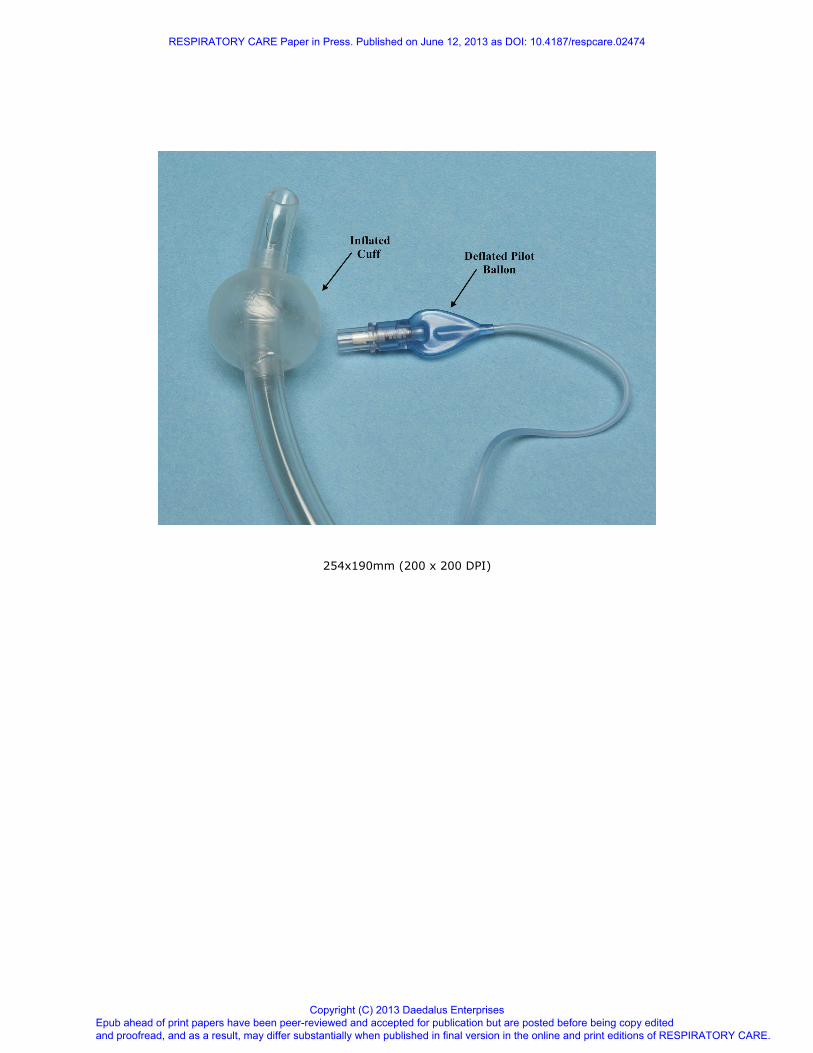
Figure 2
Figure 2 demonstrates that when the pilot balloon tubing is kinked, as in Figure 1,
then deflation of the pilot balloon will result in a completely inflated cuff.
RESPIRATORY CARE Paper in Press. Published on June 12, 2013 as DOI: 10.4187/respcare.02474
Epub ahead of print papers have been peer-reviewed and accepted for publication but are posted before being copy edited and proofread, and as a result, may differ substantially when published in final version in the online and print editions of RESPIRATORY CARE.
Copyright (C) 2013 Daedalus Enterprises
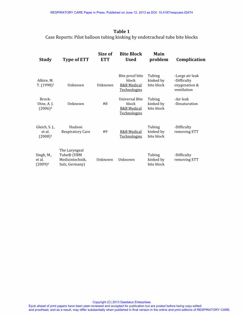
Table 1
Case Reports: Pilot balloon tubing kinking by endotracheal tube bite blocks
Study
Type of ETT
Size of
ETT
Bite Block
Used
Main
problem
Complication
Alkire, M.
T. (1998)3
Unknown
Unknown
Bite proof bite
block
B&B Medical
Technologies
Tubing
kinked by
bite block
-Large air leak
-Difficulty
oxygenation &
ventilation
Brock-
Utne, A. J.
(2006)4
Unknown
#8
Universal Bite
block
B&B Medical
Technologies
Tubing
kinked by
bite block
-Air leak
-Desaturation
Gleich, S. J.,
et al.
(2008)5
Hudson
Respiratory Care
#9
B&B Medical
Technologies
Tubing
kinked by
bite block
-Difficulty
removing ETT
Singh, M.,
et al.
(2009)6
The Laryngeal
Tube® (VBM
Medizintechnik,
Sulz, Germany)
Unknown
Unknown
Tubing
kinked by
bite block
-Difficulty
removing ETT
RESPIRATORY CARE Paper in Press. Published on June 12, 2013 as DOI: 10.4187/respcare.02474
Epub ahead of print papers have been peer-reviewed and accepted for publication but are posted before being copy edited and proofread, and as a result, may differ substantially when published in final version in the online and print editions of RESPIRATORY CARE.
Copyright (C) 2013 Daedalus Enterprises
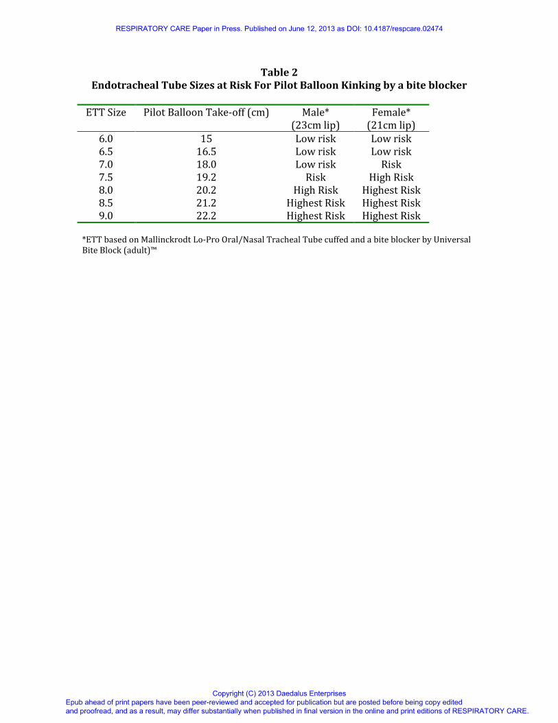
Table 2
Endotracheal Tube Sizes at Risk For Pilot Balloon Kinking by a bite blocker
ETT Size Pilot Balloon Take-off (cm) Male*
(23cm lip)
Female*
(21cm lip)
6.0 15 Low risk Low risk
6.5 16.5 Low risk Low risk
7.0 18.0 Low risk Risk
7.5 19.2 Risk High Risk
8.0 20.2 High Risk Highest Risk
8.5 21.2 Highest Risk Highest Risk
9.0 22.2 Highest Risk Highest Risk
*ETT based on Mallinckrodt Lo-Pro Oral/Nasal Tracheal Tube cuffed and a bite blocker by Universal
Bite Block (adult)™
RESPIRATORY CARE Paper in Press. Published on June 12, 2013 as DOI: 10.4187/respcare.02474
Epub ahead of print papers have been peer-reviewed and accepted for publication but are posted before being copy edited and proofread, and as a result, may differ substantially when published in final version in the online and print editions of RESPIRATORY CARE.
Copyright (C) 2013 Daedalus Enterprises
254x190mm (200 x 200 DPI)
RESPIRATORY CARE Paper in Press. Published on June 12, 2013 as DOI: 10.4187/respcare.02474
Epub ahead of print papers have been peer-reviewed and accepted for publication but are posted before being copy edited and proofread, and as a result, may differ substantially when published in final version in the online and print editions of RESPIRATORY CARE.
Copyright (C) 2013 Daedalus Enterprises