developmental cognitive neuroscience2015)dcn.pdf · 18 e.h. telzer et al. / developmental cognitive...
TRANSCRIPT

Developmental Cognitive Neuroscience 14 (2015) 16–22
Contents lists available at ScienceDirect
Developmental Cognitive Neuroscience
jo ur nal ho me pag e: ht tp : / /www.e lsev ier .com/ locate /dcn
Sleep variability in adolescence is associated with alteredbrain development
Eva H. Telzera,b,∗, Diane Goldenbergc, Andrew J. Fulignic,d, Matthew D. Liebermanc,d,Adriana Gálvanc,e
a Department of Psychology, University of Illinois, Urbana-Champaign, United Statesb Beckman Institute for Science and Technology, University of Illinois, Urbana-Champaign, United Statesc Department of Psychology, University of California, Los Angeles, United Statesd Department of Psychiatry and Biobehavioral Sciences, University of California, Los Angeles, United Statese Brain Research Institute, University of California, Los Angeles, United States
a r t i c l e i n f o
Article history:Received 15 March 2015Received in revised form 16 May 2015Accepted 20 May 2015Available online 28 May 2015
Keywords:SleepAdolescenceBrain developmentDTI
a b s t r a c t
Despite the known importance of sleep for brain development, and the sharp increase in poor sleep duringadolescence, we know relatively little about how sleep impacts the developing brain. We present the firstlongitudinal study to examine how sleep during adolescence is associated with white matter integrity. Wefind that greater variability in sleep duration one year prior to a DTI scan is associated with lower whitematter integrity above and beyond the effects of sleep duration, and variability in bedtime, whereas sleepvariability a few months prior to the scan is not associated with white matter integrity. Thus, variabilityin sleep duration during adolescence may have long-term impairments on the developing brain. Whitematter integrity should be increasing during adolescence, and so sleep variability is directly at odds withnormative developmental trends.
© 2015 The Authors. Published by Elsevier Ltd. This is an open access article under the CC BY-NC-NDlicense (http://creativecommons.org/licenses/by-nc-nd/4.0/).
Inadequate sleep is endemic in adolescence (Colrain and Baker,2011). In addition to receiving less than optimal average sleep pernight, variability in sleep duration and sleep–wake rhythms peakduring adolescence (Dahl and Lewin, 2002; Thorleifsdottir et al.,2002). Children and adolescents often need more sleep than adults,which is attributed to the putative function sleep serves in brainmaturation (Iglowstein et al., 2003; Huber and Born, 2014). Indeed,researchers have proposed that the primary purpose of sleep is thepromotion of brain development (Roffwarg et al., 1996; Jan et al.,2010; Dahl and Lewin, 2002). Unfortunately, adolescents may notreceive the quality environmental context needed for restorativesleep and optimal neuronal development (Dahl and Lewin, 2002).
Experimental research utilizing animal models and humanadults has shown that impaired sleep, including sleep depriva-tion and variability in sleep–wake cycles, alters synaptic plasticityresulting in transcriptional alterations in protein synthesis, reducedcerebral metabolism, gray matter loss in cortical and subcor-tical structures, as well as neurogenesis as a result of increasedcirculating levels of the adrenal stress hormone, corticosterone
∗ Corresponding author at: Department of Psychology, University of Illinois, 603East Daniel Street, Champaign, IL 61820, United States. Tel.: +1 217 300 0383.
E-mail address: [email protected] (E.H. Telzer).
(Mirescu et al., 2006; Kopp et al., 2006; Halbower et al., 2006).Moreover, there is compelling evidence from high density EEGstudies in youth measuring electrical activity of the brain showingthat poor sleep can influence neural function, relating to impair-ments in memory formation and learning, executive functions, andemotional well-being (see Jan et al., 2010). Because sleep, and REMsleep in particular, facilitates memory consolidation, learning, andemotional development (Sejnowski and Destexhe, 2000; Tononiand Cirelli, 2006; Pugin et al., 2014; Huber and Born, 2014; Jan et al.,2010), poor sleep in adolescence may be associated with a host ofnegative outcomes via alterations in brain development and func-tion (Dahl and Lewin, 2002). Yet, despite the important role of sleepin promoting brain development, we know little about how varia-tions in sleep during adolescence are associated with alterations inthe developing brain.
The adolescent years represent a key developmental periodwhen the brain undergoes significant remodeling, which is accom-panied by changes in the neurophysiological features of sleep,memory systems, socioemotional processing, and emotion regula-tion (Nelson et al., 2005; Spear, 2000). Myelination of frontocorticaland frontostriatal white matter tracts continues through ado-lescence and into adulthood (Asato et al., 2010; Giorgio et al.,2008, 2010). Cellular maturation of white matter facilitates fasterand more efficient neural transmission and cognitive processing
http://dx.doi.org/10.1016/j.dcn.2015.05.0071878-9293/© 2015 The Authors. Published by Elsevier Ltd. This is an open access article under the CC BY-NC-ND license (http://creativecommons.org/licenses/by-nc-nd/4.0/).
E.H. Telzer et al. / Developmental Cognitive Neuroscience 14 (2015) 16–22 17
(Giedd, 2008). Thus, relative delayed maturation and subtlealterations in the microstructure of white matter fibers duringdevelopment can affect neurocognition and behavior, includingbehavioral problems (Li et al., 2005), substance use (Achesonet al., 2014; Bava et al., 2013), and poor cognitive performance(Chaddock-Heyman et al., 2013; Nagy et al., 2004). During periodsof brain development, synaptic activity exhibits high levels of plas-ticity and connectivity and is therefore particularly influenced byenvironmental inputs (Jan et al., 2010). Moreover, stressors can sig-nificantly modify brain development (Kaufman et al., 2000). Sleepmay therefore have an especially large impact on the develop-ing adolescent brain. While sleep problems may not induce neuralchanges in white matter that are immediately evident, chronic orfrequent sleep problems may have accumulating effects, impairingneuronal integrity, decreasing neurogenesis, and altering structuralplasticity over time.
Adolescents demonstrate considerable variability in their sleeppatterns, with intra-individual differences in sleep duration oftenexceeding between-person differences. Such variability in sleep isdetrimental for health outcomes, perhaps more so than is chroni-cally low sleep duration (Acebo and Carskadon, 2002). For example,adolescents with high day-to-day variability of sleep durationassessed by daily sleep logs over two weeks report greater depres-sion, anxiety, fatigue, and lower subjective well-being controllingfor average sleep duration (Fuligni and Hardway, 2006; Lemolaet al., 2013). Daily diary measurements offer an ideal method forassessing sleep. Single-time surveys do not provide estimates ofthe daily variability in adolescents’ sleep that go beyond differ-ences between weekdays and weekends (Fuligni and Hardway,2006). Moreover, daily reporting minimizes the amount of errorthat occurs in retrospective reporting of events and allows for adirect estimation of the daily variability in adolescents’ sleep time.
In the current study, adolescents completed daily diary check-lists two times, one year apart. In order to examine how sleep wasassociated with the developing brain, adolescents underwent a DTIscan a few months following the second wave of daily diaries. DTIoffers the ability to indirectly study the microstructural compo-nents of white matter. Fractional anisotropy (FA), a measure of fiberbundle organization and microstructural integrity of white mat-ter (WM), is a widely used general index of axonal integrity (Boraet al., 2012). By measuring sleep at two time points, once a fewmonths prior to the DTI scan and once 1.5 years prior to the scan,we were able to test whether sleep problems have more imme-diate or long-term effects on brain development. Sleep problemsmay take time to impact the development of the brain, as alter-ations in myelination may not occur immediately. Indeed, earlylife stressors are associated with later reductions in FA (Paul et al.,2008; Teicher et al., 2010). Moreover, animal models have shownthat stressors can differentially impact short-term and long-termneural processing. For instance, in rats, caffeine treatment is asso-ciated with short-term decreases in slow-wave activity (SWA), anelectrophysiological signature of slow, synchronized, oscillatoryneocortical activity that occurs during deep sleep, but the longer-term effects are reversed (i.e., increases in SWA) and persist overtime (Olini et al., 2013). Thus, we expected to find more long-termassociations between poor sleep and white matter integrity.
1. Methods
1.1. Participants
At the first and second waves of data collection, 48 adolescents(27 female) completed daily diary checklists (wave 1: Mage = 14.8years; wave 2: Mage = 15.9 years). A few months after comple-ting the second wave of daily diaries, adolescents completed aDTI scan (Mage = 16.3 years). All participants were right-handed,
free of metal, reported no current medication except birth control,and did not report being diagnosed with any mood or sleep disor-der. Parents provided written consent and adolescent participantscompleted written assent in accordance with UCLA’s InstitutionalReview Board.
1.2. Procedures
At waves 1 and 2, interviewers visited the home of participants.Adolescents were provided with 14 days of diary checklists to com-plete every night before going to bed for two subsequent weeks. Thethree-page diary checklists took approximately 5–10 min to com-plete each night. Participants were instructed to fold and seal eachcompleted diary checklist and to stamp the seal with an electronictime stamper each night. The time stamper imprinted the currentdate and time and was programmed with a security code such thatadolescents could not alter the correct time and date. Participantswere told that if inspection of the data indicated that they hadcompleted the checklists correctly and on time, each family wouldalso receive two movie passes. At the end of the two-week period,interviewers returned to the home to collect the diary checklists.Adolescents received $30 for participating. The time-stamper mon-itoring and incentives resulted in a high rate of compliance, with96% of the potential diaries being completed. On average, adoles-cents completed 13.7 diaries at wave 1 and 13.0 diaries at wave2. One adolescent provided fewer than 3 diaries at wave 2 andso is excluded from analyses examining wave 2 associations. Atboth waves, the diaries were collected during the fall semester ofschool across two weeks when school was in session. Waves 1 and2 occurred during the same time of year, as close to 1 year apart aspossible, in order to control for time of year effects. Within 1–3months following the second wave of diaries, adolescents com-pleted a DTI scan.
1.2.1. Daily sleepEach day, adolescents reported the time they went to bed the
prior night (“what time did you go to bed last night?”) and thetime they woke up that morning (“what time did you get up frombed this morning?”). They then indicated the total time they slept(“how much time did you sleep last night?”). From these questions,we created four variables of interest, sleep duration, sleep durationvariability, weekend–weekday sleep duration, and bedtime variabil-ity. Using the daily item on total sleep time, sleep duration wascalculated by taking the average minutes of sleep attained eachnight across the 14 days and sleep duration variability was cal-culated by taking the standard deviation in minutes of sleep timeattained each night across the 14 days. Weekend–weekday sleepduration was calculated by taking the difference in total sleep timebetween weekend and weekdays. Positive scores indicate greatersleep time on weekends whereas negative scores indicate greatersleep time on weekdays. Finally, bedtime variability was calculatedby the standard deviation in bedtimes across the 14 days. Scoreswere calculated at wave 1 and wave 2.
1.3. DTI image acquisition and processing
Imaging data were collected using a 3 Tesla Siemens Tri-oMRI scanner. Diffusion weighted images were acquired usingan echo-planar imaging (EPI) sequence (64 directions, TR/TE =7200/93 ms, 50 slices; slice thickness = 2 mm, FOV = 190 mm, voxelsize = 2 mm × 2 mm × 2 mm). This sequence also provides a T2weighted volume (B0).
DTI processing and voxelwise statistical analysis were per-formed with FSL v4.1.6 (www.fmrib.ox.ac.uk/fsl). Datasets werecorrected for head motion, eddy current distortion, and signal lossusing the FMRIB Diffusion Toolbox. Four participants displayed

18 E.H. Telzer et al. / Developmental Cognitive Neuroscience 14 (2015) 16–22
Table 1Correlations between sleep variables at wave 1 and wave 2.
Variable 1. 2. 3. 4. 5. 6. 7. 8. 9. 10.
1. Sleep duration W1 12. Sleep variability W1 −.06 13. Weekend–weekday W1 .20 .43*** 14. Bedtime W1 −.26 .00 −.20 15. Bedtime variability W1 −.15 .10 −.24 −.23 16. Sleep duration W2 .37** −.39** −.07 −.16 −.40** 17. Sleep variability W2 .12 .18 .21 .14 .02 −.12 18. Weekend–weekday W2 .16 .08 .41** −.21 −.03 −.20 .29* 19. Bedtime W2 −.17 .06 −.19 .63*** .12 −.27 .17 −.28 110. Bedtime variability W2 .01 .14 −.05 −.12 −.04 −.05 .41*** −.12 .06 1
* p < .05.** p < .01.
*** p < .005.
>2 mm of translational motion. Excluding these individuals did notalter results, and therefore they were retained in analyses. Degreeof diffusion was assessed with fractional anisotropy (FA).
Voxelwise statistical analysis of the FA data was carried outusing TBSS [Tract-Based Spatial Statistics (Smith et al., 2006)],within the FSL toolbox (Smith et al., 2004). First, FA images werecreated by fitting a tensor model to the raw diffusion data usingFDT, and then brain-extracted using BET (Smith, 2002). All subjects’FA data were then aligned into a common space using the nonlin-ear registration tool FNIRT (Andersson et al., 2007a,b), which usesa b-spline representation of the registration warp field (Rueckertet al., 1999). Next, the mean FA image was created and thinnedto create a mean FA skeleton, which represents the centers ofall tracts common to the group. Each subject’s aligned FA datawere then projected onto this skeleton and the resulting datawere used in voxelwise cross-subject analyses. To minimize partial-volume effects and areas of high inter-subject variability, valueswere thresholded at FA > 0.2. Data from each point on the skeletonformed the basis of voxelwise statistical comparisons.
DTI-based voxelwise statistics were carried out using FSLRandomize with 500 permutations and a standard GLM design.Randomize (FMRIB’s Software Library, www.fmrib.ox.ac.uk/fsl/)uses a permutation based statistical inference that does not rely ona Gaussian distribution (Nichols and Holmes, 2002). A single-groupaverage with covariate design was applied to assess voxelwisedifferences among individuals based on behavioral variables ofinterest (e.g., sleep variability), which were demeaned and individ-ually entered as regressors in the GLM model. A statistical thresholdof p < .05, corrected for multiple comparisons with familywise errorcorrection (FWE) and Threshold-Free Cluster Enhancement (TFCE),was used for analyses. TFCE helps identify cluster-like structuresin images without definition of an initial cluster-forming thresh-old or carrying out a large amount of data smoothing (Smith andNichols, 2009). Identification of the significant white-matter tractsrevealed by TBSS was based on the Johns Hopkins University (JHU)– ICBM-DTI-81 white-matter labels atlas and the white-mattertractography atlas (Wakana et al., 2004; Hua et al., 2008). For visu-alization purposes for the figures, the skeletonized results werethickened using the tbss fill command. The images are displayedin radiological convention. When sex was included as a covariate,all the results below remain the same.
2. Results
2.1. Descriptives
At wave 1, adolescents went to bed at 11:00pm on average(range 8:20 pm to 1:45 am; bedtime variability = 1.44 h). Ado-lescents attained an average of 499 min (8.3 h) of sleep pernight, ranging from 274 to 647 min. Adolescents’ sleep duration
Table 2Regions of FA that correlated negatively with sleep duration variability at wave 1,controlling for sleep duration at wave 1.
Region x y z Voxels
L Posterior limb internal capsule −27 −26 17 3869a
L Retrolenticular part of theinternal capsule
−30 −37 17 a
L Anterior thalamatic radiation −25 −52 26 a
L Superior coronoa radiata −27 −7 21 a
L Posterior corona radiata −27 −24 19 a
L Cingulate gyrus −16 −55 31 a
L Splenium of the corpus callosum −16 −44 10 a
R Posterior limb internal capsule 25 −15 10 1476b
R Retrolenticular part of theinternal capsule
32 −35 15 b
R Superior coronoa radiata 26 −19 28 b
R Posterior corona radiata 27 −40 24 b
R Splenium of the corpus callosum 14 −41 10 b
R Posterior thalamatic radiation 34 −59 16 295L Superior longitudinal fasciculus −54 −12 23 198L Superior longitudinal fasciculus −26 −21 44 157R Corticospinal tract 19 −31 52 84R Superior longitudinal fasciculus 32 −42 29 44R Anterior limb of the internal
capsule12 −2 13 24
Note: x, y, and z refer to MNI coordinates; Voxels refers to the number of voxels ineach significant cluster; L and R refer to left and right hemispheres. Regions thatshare the same superscript are part of the same cluster. All regions are significantat p < .05, corrected.
variability (i.e., one’s own standard deviation in sleep time across14 days) was 93 min on average, ranging from 36 to 152 min.Weekend–weekday sleep duration was 60 min (ranging from −133to +198), indicating that adolescents attained 1 h more sleep onaverage on weekends than weekdays.
At wave 2, adolescents went to bed at 11:10 pm on aver-age (range 8:50 pm to 2:10 am; bedtime variability = 1.33 h).Adolescents attained an average of 493 min (8.2 h) of sleepper night, ranging from 397 to 596 min. Adolescents’ sleepduration variability at wave 2 was 95 min on average, ran-ging from 21 to 199 min. Weekend–weekday sleep durationwas 48 min on average (ranging from −135 to +228).
Within samples t-tests revealed no significant differences inany of the sleep variables between wave 1 and wave 2. In addi-tion, we examined gender differences in sleep patterns. Malesand females only differed in average sleep time at wave 2, suchthat females (M = 480 min, SD = 49) attained less sleep than males(M = 510 min, SD = 39) on average, t(46) = 2.3, p < .05. Next, we ranbivariate correlations between the sleep estimates at waves 1 and 2.As shown in Table 1, greater sleep duration at wave 1 was associatedwith greater sleep duration at wave 2. Greater variability in sleeptime at wave 1 was associated with greater weekend–weekday

E.H. Telzer et al. / Developmental Cognitive Neuroscience 14 (2015) 16–22 19
sleep duration at wave 1 and less total sleep time at wave 2.Weekend–weekday sleep durations at waves 1 and 2 were cor-related. Bedtime at wave 1 and wave 2 were highly correlated.Bedtime variability at wave 1 was associated with less sleep dura-tion at wave 2. Bedtime variability at wave 2 was highly correlatedwith sleep duration variability at wave 2.
2.2. Association between sleep and FA
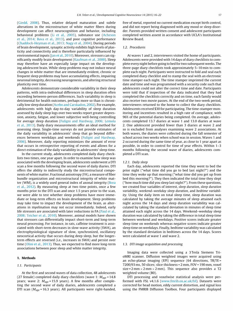
White matter microstructure was assessed using fractionalanisotropy (FA), a measure of the directional coherence of braintissue that provides an estimate of white matter integrity. Our firstanalysis tested how sleep duration variability and sleep durationat wave 1 correlated with FA. We simultaneously entered sleepduration variability and sleep duration as regressors to test howeach was associated with white matter microstructure above andbeyond the effect of the other. As shown in Table 2 and Fig. 1, greatervariability in sleep duration across two weeks was associated withless FA in several white matter tracts. These clusters were located inassociation tracts (e.g., superior longitudinal fasciculus), projectiontracts (e.g., anterior thalamatic radiata, anterior corona radiata, cor-ticospinal tract, internal capsule), and the interhemispheric tract
(corpus callosum). For descriptive purposes, we plotted several ofthese associations (Fig. 2).
Next, to examine whether the associations with sleep durationvariability and FA were accounted for by weekend–weekday differ-ences in sleep time, we entered weekend–weekday sleep durationas a regressor into the same model with sleep duration variabilityand sleep duration. Sleep duration variability continued to predictlower FA and weekend–weekday sleep duration was not asso-ciated with FA. Lastly, we tested whether the associations withsleep duration variability and FA were accounted for by variabil-ity in bedtime. We simultaneously entered bedtime variability,sleep duration variability, and sleep duration as regressors. Bed-time variability was not associated with FA, whereas sleep durationvariability continued to predict lower FA.
Finally, we examined how sleep at wave 2 was associated withFA. We simultaneously entered sleep duration variability and sleepduration as regressors to test how each was associated with whitematter microstructure above and beyond the effect of the other.Sleep duration variability and sleep duration at wave 2 were notassociated with FA. In addition, we examined weekend–weekdaysleep duration and bedtime variability in parallel models to thoserun at wave 1. None of these sleep indexes were associated withFA.
Fig. 1. Regions of FA that correlated negatively with sleep variability at wave 1. Significant results are displayed on the study-specific mean FA map and the study-specificmean FA skeleton. Note: Right = left.
Fig. 2. Significant correlations between FA and sleep variability at wave 1. Scatterplots showing a visual depiction of the relation between sleep variability at wave 1 and FA inthe (a) splenium of the corpus callosum, (b) posterior limb of the internal capsule, (c) superior longitudinal fasciculus, and (d) corticospinal tract. **p < .005. Note: Right = left.

20 E.H. Telzer et al. / Developmental Cognitive Neuroscience 14 (2015) 16–22
3. Discussion
Adolescence is a developmental period marked by increases inpoor sleep coupled with significant reorganization of the brain, andextensive emotional, behavioral, and cognitive changes. The linksbetween poor sleep and behavioral and psychological outcomes(e.g., academic difficulties, poor impulse control, problems withemotion regulation, negative affect) have been well established(Fuligni and Hardway, 2006; Anderson et al., 2008; O’Brien andMindell, 2005; McKnight-Eily et al., 2011). Yet, despite the knownimportance of sleep for brain development (Roffwarg et al., 1996;Jan et al., 2010; Dahl and Lewin, 2002), we still know relativelylittle about how insufficient sleep during adolescence may alterthe developing brain. This is the first study to our knowledge toexamine how sleep during adolescence is longitudinally associatedwith white matter integrity. We find that greater variability in sleepduration 1.5 years prior to the DTI scan is associated with lower FAabove and beyond the effects of sleep duration and variability inbedtime, whereas sleep variability a few months prior to the scanis not associated with FA. These effects suggest that variability insleep duration, regardless of the duration in the total amount ofsleep or large fluctuations in the time teenagers are going to bed,may have long-term effects on the developing brain.
The amount of daily variability in adolescents’ sleep time wasstriking, averaging over 1.5 h. Sleep variability measured 1.5 yearsprior to the scan was associated with significantly reduced FA infrontocortical and frontostriatal tracts, association tracts, projec-tion tracts, and the interhemispheric tract. FA in these tracts shouldbe increasing during adolescence (Asato et al., 2010; Giorgio et al.,2008, 2010). Thus, sleep variability is directly at odds with nor-mative developmental trends. Notably, sleep variability predictedreduced FA above and beyond average sleep time, suggesting thatvariability in sleep duration may be more important for whitematter development than does receiving chronically low levels ofaverage sleep time. This finding is consistent with prior researchshowing that variability in sleep predicts adolescents’ psychologi-cal functioning more so than does average sleep time (Fuligni andHardway, 2006; Lemola et al., 2013).
Our findings suggest that high levels of sleep variability mayimpair the development of white matter development. Develop-mental increases in myelination during adolescence facilitate fasterand more efficient neural transmission and cognitive processingwhich, in turn, is related to better behavioral performance andlearning (e.g., Jacobus et al., 2013). Here we find potentially long-term consequences of poor sleep, relating to lower FA in theparietocortical, frontocortical and frontostriatal tracts. Specifically,we found that greater sleep variability was associated with reducedFA in projection and association tracts including the internal cap-sule (IC), anterior thalamatic radiation (ATR), posterior thalamaticradiation (PTR), cingulum, superior corona radiata, posterior coronaradiata, and superior longitudinal fasciculus (SLF). The IC is tra-versed by axonal fibers that connect the thalamus to the prefrontalcortex and forms part of a circuit linking the frontal lobe and basalganglia. The ATR connects the dorsomedial and anterior thalamicnuclei with the prefrontal cortex (Schmahmann and Pandya, 2006).The cingulum connects the anterior cingulate cortex to other frontalsites as well as to the amygdala, nucleus accumbens, and thala-mus (Goldman-Rakic et al., 1984). Deficits in the cingulate and ATRtracts result in the disintegration of the fronto-striato-thalamiccircuitry and the disruption of functional connectivity betweenregions linked by these tracts. The PTR is a projection fiber fromthe posterior part of the thalamus to the occipital cortex, whichincludes axons connecting the cerebral cortex, basal ganglia, andthalamus. The PTR is part of the cortico-thalamo-cortical path-way, which is considered to play a key role in central informationprocessing and overall cognitive control (Sherman and Guillery,
2002; Chaddock-Heyman et al., 2013). The posterior and superiorcorona radiata contain reciprocal prefrontal–striatal connectionsand projection fibers. These tracts are central to detection of salientrewarding cues, positive affect, and higher order processing nec-essary to effectively monitor conflict and resist impulsive actions(Jacobus et al., 2013). Finally, the superior longitudinal fasciculus isa long association fiber connecting the prefrontal to parietal cortex.The SLF plays an important role in higher-level cognitive processes,including attention and executive functions. Together, these tractscarry important connections between the prefrontal cortex andsubcortical regions, and are therefore essential for cognitive andexecutive functioning and emotion regulation (Asato et al., 2010).Importantly, these tracts are still undergoing significant develop-ment during adolescence (Asato et al., 2010; Giorgio et al., 2008,2010). Thus, variability in sleep duration may impair cognitive andsocioemotional well-being by altering the development of whitematter integrity in these tracts. However, we did not measure cog-nitive or emotional outcomes or the consolidation of memories andlearning. Thus, future research should use longitudinal techniquesto examine functional outcomes at both neural and behavioral lev-els in order to effectively examine how sleep and alterations inwhite matter are associated with developmental aspects of affect,cognition, and behavior.
Adolescents in this study averaged over 8 h of sleep per night,which is similar to what has been obtained in other studies withthis age group (Wolfson and Carskadon, 1998; Fuligni and Hardway,2006) and is close to the number of hours recommended for ado-lescents (Wolfson and Carskadon, 1998). Yet, some participantsattained as few as 4.5 h of sleep on average. Interestingly, averagesleep duration was not associated with white matter integrity. Inaddition, the effects of sleep variability on brain development werenot due to large differences in sleep duration on weekends versusweekdays or to variability in bedtime. This suggests that day-to-day variability in the duration of sleep within the week may beparticularly detrimental for youth.
3.1. Limitations
Our study is correlational and does not provide evidence aboutthe direction or causality of the effects. Thus, it is possible that alter-ations in white matter integrity result in greater variability in sleep.However, because variability in sleep measured a few months priorto the scan was not associated with FA, whereas variability in sleepmeasured 1.5 years prior to the scan was associated with lower FA,our findings suggest that sleep problems may not induce neuralchanges in white matter that are immediately evident. Rather, sleepproblems may have accumulating effects that impair neural devel-opment over time. Therefore, it may take time for sleep patterns tohave an effect on structural changes in the brain. We have previ-ously found that poor quality sleep has immediate consequencesfor functional brain activation in adolescence, such that insufficientsleep is related to lower activation in the PFC and heightened acti-vation in the striatum, which, in turn, are associated with greaterrisk taking behavior (Telzer et al., 2013). Here we show that theconsequences of poor sleep for structural brain development mayhave more accumulating and long-term effects. It is also possi-ble that adolescents’ brains were more malleable and sensitive toenvironmental stressors at wave 1, and so poor sleep may havehad immediate changes on neural circuitry at age 14.8 years thatpersisted, neural changes we did not find at wave 2 when the ado-lescents were older and potentially less sensitive to environmentalstressors.
We utilized adolescents’ self-reported sleep. Although dailydiaries generally avoid retrospective errors in recall and providea means for estimating daily variability in adolescents’ sleep thatcannot be captured with retrospective reports, daily self-reports

E.H. Telzer et al. / Developmental Cognitive Neuroscience 14 (2015) 16–22 21
are still subjective measures that can be prone to reporting biases.Thus, using a device such as an actigraph to monitor participants’motor movements throughout the night could provide more objec-tive measures of sleep as well as a wider variety of sleep measures.For instance, actigraphy provides measures of sleep quality, includ-ing sleep efficiency, sleep latency onset, and wake after sleep onset,measures which could provide a deeper understanding of the roleof sleep on brain development. Thus, our study is limited in thatwe only assessed the time adolescents went to bed and the timespent sleeping across study days. While this allowed us to cap-ture several important indexes of sleep (i.e., sleep duration, sleepvariability, weekend–weekday sleep, and variability in bedtime),we were not able to assess other measures of the quality of adoles-cents’ sleep. Future studies should continue to carefully unpack therole of multiple sleep indexes on adolescents’ brain developmentand behavioral functioning in order to fully understand when andwhy chronically low sleep duration versus high variability in sleepduration impact youths’ development.
Finally, DTI is an effective measure for assessing white mat-ter integrity. Fractional anisotropy (FA), the most widely usedparameter of DTI (Assaf and Pasternak, 2008), is sensitive to avariety of parameters including fiber density, axonal diameter,and regional myelination (Beaulieu, 2002). However, changes inFA values during normal development largely reflect changes inaxonal myelination and can therefore be used as an indirect mea-surement of myelin level (Mädler et al., 2008). Nonetheless, theseother indexes included in FA also contribute to observed anisotropy(Assaf and Pasternak, 2008). Thus, future research should takeadvantage of methodological advances in estimating white mat-ter integrity to fully understand the specific mechanisms that arealtered as a function of poor sleep. In addition, future researchshould unpack how sleep may contribute to alterations in otherstructural brain measures, including gray matter development andMR elastography (Mariappan et al., 2010).
In conclusion, we utilized longitudinal reports of daily sleepduring adolescence and find that variability in adolescents’ sleeppatterns can have implications for their brain development.Attaining low-variability sleep therefore is essential not only forshort-term functioning and well-being, but also for longer-termneurobiological development. Parents, practitioners, and poli-cymakers should pay close attention to the daily changes inadolescents’ sleep patterns and identify ways to decrease variableamounts of sleep time, which can potentially have lasting implica-tions for the developing teenage brain.
Conflict of interest
We have no biomedical, financial, or potential conflicts of inter-est.
Authors’ note
Support for this study was provided by the NICHD(R01HD057164-S, Fuligni), NSF (NSF 1023293, Telzer), and aUniversity of California Institute for Mexico and the United StatesDissertation Research Grant (Telzer).
References
Acebo, C., Carskadon, M.A., 2002. Influence of irregular sleep patterns on wakingbehavior. In: Carskadon, M.A. (Ed.), Adolescent Sleep Patterns: Biological, Social,and Psychological Influences. Cambridge University Press, Cambridge, UK, pp.220–235.
Acheson, A., Wijtenburg, S.A., Rowland, L.M., Winkler, A.M., Gaston, F., Mathias, C.W.,Fox, P.T., Lovallo, W.R., Wright, S.N., Hong, L.E., Dougherty, D.M., Kochunov, P.,2014. Assessment of whole brain white matter integrity in youths and youngadults with a family history of substance-use disorders. Hum. Brain Mapp. 35(11), 5401–5413.
Anderson, B., Storfer-Isser, A., Taylor, H.G., Rosen, C.L., Redline, S., 2008. Associa-tions of executive function with sleepiness and sleep duration in adolescents.Pediatrics 123, e701–e707.
Andersson, J.L.R., Jenkinson, M., Smith, S., from www.fmrib.ox.ac.uk/analysis/techrep 2007a. Non-linear optimisation FMRIB technical report TR07JA1.
Andersson, J.L.R., Jenkinson, M., Smith, S., from www.fmrib.ox.ac.uk/analysis/techrep 2007b. Non-linear registration, aka Spatial normalisation FMRIB tech-nical report TR07JA2.
Asato, M.R., Terwilliger, R., Woo, J., Luna, B., 2010. White matter development inadolescence: a DTI study. Cereb. Cortex 20 (9), 2122–2131.
Assaf, Y., Pasternak, O., 2008. Diffusion tensor imaging (DTI)-based whitematter mapping in brain research: a review. J. Mol. Neurosci. 34 (1),51–61.
Bava, S., Jacobus, J., Thayer, R.E., Tapert, S.F., 2013. Longitudinal changes in whitematter integrity among adolescent substance users. Alcohol. Clin. Exp. Res. 37,E181–E189.
Beaulieu, C., 2002. The basis of anisotropic water diffusion in the nervous system–atechnical review. NMR Biomed. 15 (7/8), 435–455.
Bora, E., Yücel, M., Fornito, A., Pantelis, C., Harrison, B.J., Cocchi, L., Lubman, D.I.,2012. White matter microstructure in opiate addiction. Addiction Biology 17(1), 141–148.
Chaddock-Heyman, L., Erickson, K.I., Voss, M.W., Powers, J.P., Knecht, A.M., Pontifex,M.B., Drollette, E.S., Moore, R.D., Raine, L.B., Scudder, M.R., Hillman, C.H., Kramer,A.F., 2013. White matter microstructure is associated with cognitive control inchildren. Biol. Psychol. 94 (1), 109–115.
Colrain, I.M., Baker, F.C., 2011. Changes in sleep as a function of adolescent develop-ment. Neuropsychol. Rev. 21 (1), 5–21.
Dahl, R.E., Lewin, D.S., 2002. Pathways to adolescent health sleep regulation andbehavior. J. Adolesc. Health 31 (6), 175–184.
Fuligni, A.J., Hardway, C., 2006. Daily variation in adolescents’ sleep, activities, andpsychological well-being. J. Res. Adolesc. 16 (3), 353–378.
Giedd, J.N., 2008. The teen brain: insights from neuroimaging. J. Adolesc. Health 42(4), 335–343.
Giorgio, A., Watkins, K.E., Douaud, G., James, A.C., James, S., De Stefano, N., Matthews,P.M., Smith, S.M., Johansen-Berg, H., 2008. Changes in white matter microstruc-ture during adolescence. Neuroimage 39 (1), 52–61.
Giorgio, A., Watkins, K.E., Chadwick, M., James, S., Winmill, L., Douaud, G., De Stefano,N., Matthews, P.M., Smith, S.M., Johansen-Berg, H., James, A.C., 2010. Longitudi-nal changes in grey and white matter during adolescence. Neuroimage 49 (1),94–103.
Goldman-Rakic, P.S., Selemon, L.D., Schwartz, M.L., 1984. Dual pathways connect-ing the dorsolateral prefrontal cortex with the hippocampal formation andparahippocampal cortex in the rhesus monkey. Neuroscience 12 (3), 719–743.
Halbower, A.C., Degaonkar, M., Barker, P.B., Earley, C.J., Marcus, C.L., Smith, P.L.,Prahme, M.C., Mahone, E.M., 2006. Childhood obstructive sleep apnea associateswith neuropsychological deficits and neuronal brain injury. PLoS Med. 3 (8),e301.
Hua, K., Zhang, J., Wakana, S., Jiang, H., Li, X., Reich, D.S., Mori, S., 2008. Tractprobability maps in stereotaxic spaces: analyses of white matter anatomy andtract-specific quantification. Neuroimage 39 (1), 336–347.
Huber, R., Born, J., 2014. Sleep, synaptic connectivity, and hippocampal memoryduring early development. Trends Cogn. Sci. 18 (3), 141–152.
Iglowstein, I., Jenni, O.G., Molinari, L., Largo, R.H., 2003. Sleep duration from infancyto adolescence: reference values and generational trends. Pediatrics 111 (2),302–307.
Jacobus, J., Squeglia, L.M., Infante, M.A., Bava, S., Tapert, S.F., 2013. White matterintegrity pre- and post marijuana and alcohol initiation in adolescence. BrainSci. 3 (1), 396–414.
Jan, J.E., Reiter, R.J., Bax, M.C., Ribary, U., Freeman, R.D., Wasdell, M.B., 2010. Long-term sleep disturbances in children: a cause of neuronal loss. Eur. J. Paediatr.Neurol. 14 (5), 380–390.
Kaufman, J., Plotsky, P.M., Nemeroff, C.B., Charney, D.S., 2000. Effects of early adverseexperiences on brain structure and function: clinical implications. Biol. Psychi-atry 48 (8), 778–790.
Kopp, C., Longordo, F., Nicholson, J.R., Lüthi, A., 2006. Insufficient sleep reversiblyalters bidirectional synaptic plasticity and NMDA receptor function. J. Neurosci.26 (48), 12456–12465.
Lemola, S., Ledermann, T., Friedman, E.M., 2013. Variability of sleep duration isrelated to subjective sleep quality and subjective well-being: an actigraphystudy. PLOS ONE 8 (8), e71292.
Li, T.Q., Mathews, V.P., Wang, Y., Dunn, D., Kronenberger, W., 2005. Adolescentswith disruptive behavior disorder investigated using an optimized MR diffusiontensor imaging protocol. Ann. N. Y. Acad. Sci. 1064 (1), 184–192.
Mädler, B., Drabycz, S.A., Kolind, S.H., Whittall, K.P., MacKay, A.L., 2008. Is diffusionanisotropy an accurate monitor of myelination? Correlation of multicomponentT2 relaxation and diffusion tensor anisotropy in human brain. Magn. Reson.Imaging 26 (7), 874–888.
Mariappan, Y.K., Glaser, K.J., Ehman, R.L., 2010. Magnetic resonance elastography: areview. Clin. Anat. 23 (5), 497–511.
McKnight-Eily, L.R., Eaton, D.K., Lowry, R., Croft, J.B., Presley-Cantrell, L., Perry, G.S.,2011. Relationships between hours of sleep and health-risk behaviors in USadolescent students. Prev. Med. 53 (4), 271–273.
Mirescu, C., Peters, J.D., Noiman, L., Gould, E., 2006. Sleep deprivation inhibits adultneurogenesis in the hippocampus by elevating glucocorticoids. Proc. Natl. Acad.Sci. U. S. A. 103, 19170–19175.

22 E.H. Telzer et al. / Developmental Cognitive Neuroscience 14 (2015) 16–22
Nagy, Z., Westerberg, H., Klingberg, T., 2004. Maturation of white matter is associatedwith the development of cognitive functions during childhood. J. Cogn. Neurosci.16 (7), 1227–1233.
Nelson, E.E., Leibenluft, E., McClure, E., Pine, D.S., 2005. The social re-orientationof adolescence: a neuroscience perspective on the process and its relation topsychopathology. Psychol. Med. 35 (2), 163–174.
Nichols, T.E., Holmes, A.P., 2002. Nonparametric permutation tests for functionalneuroimaging: a primer with examples. Hum Brain Mapp 15, 1–25.
O’Brien, E.M., Mindell, J.A., 2005. Sleep and risk-taking behavior in adolescents.Behav. Sleep Med. 3 (3), 113–133.
Olini, N., Kurth, S., Huber, R., 2013. The effects of caffeine on sleep and maturationalmarkers in the rat. PLOS ONE 8 (9), e72539.
Paul, R., Henry, L., Grieve, S.M., Guilmette, T.J., Niaura, R., Bryant, R., Bruce, S.,Williams, L.M., Richard, C.C., Cohen, R.A., Gordon, E., 2008. The relationshipbetween early life stress and microstructural integrity of the corpus callosum ina non-clinical population. Neuropsychiatr. Dis. Treat. 4 (1), 193–201.
Pugin, F., Metz, A.J., Wolf, M., Achermann, P., Jenni, O.G., Huber, R., 2014. Localincrease of sleep slow wave activity after three weeks of working memorytraining in children and adolescents. Sleep 38 (4), 607–614.
Roffwarg, H.P., Muzio, J.N., Dement, W.C., 1996. Ontogenetic development of thehuman sleep-dream cycle. Science 152, 604–619.
Rueckert, D., Sonoda, L.I., Hayes, C., Hill, D.L.G., Leach, M.O., Hawkes, D.J., 1999.Non-rigid registration using free-form deformations: Application to breast MRimages. IEEE Transactions on Medical Imaging 18 (8), 712–721.
Schmahmann, J.D., Pandya, D.N., 2006. Fiber Pathways of the Brain. Oxford UniversityPress, New York.
Sejnowski, T.J., Destexhe, A., 2000. Why do we sleep? Brain Res. 886 (1), 208–223.Sherman, S.M., Guillery, R.W., 2002. The role of the thalamus in the flow of infor-
mation to the cortex. Philos. Trans. R. Soc. Lond. Ser. B: Biol. Sci. 357 (1428),1695–1708.
Smith, S.M., Jenkinson, M., Johansen-Berg, H., Rueckert, D., Nichols, T.E., Mackay, C.E.,Behrens, T.E., 2006. Tract-based spatial statistics: voxelwise analysis of multi-subject diffusion data. Neuroimage 31 (4), 1487–1505.
Smith, S.M., Jenkinson, M., Woolrich, M.W., Beckmann, C.F., Behrens, T.E., Johansen-Berg, H., Matthews, P.M., 2004. Advances in functional and structural MR imageanalysis and implementation as FSL. Neuroimage 23, S208–S219.
Smith, S.M., 2002. Fast robust automated brain extraction. Human Brain Mapping17 (3), 143–155.
Smith, S.M., Nichols, T.E., 2009. Threshold-free cluster enhancement: addressingproblems of smoothing, threshold dependence and localisation in cluster infer-ence. Neuroimage 44 (1), 83–98.
Spear, L.P., 2000. The adolescent brain and age-related behavioral manifestations.Neurosci. Biobehav. Rev. 24 (4), 417–463.
Telzer, E.H., Fuligni, A.J., Lieberman, M.D., Gálvan, A., 2013. The effects of poor qual-ity sleep on brain function during risk taking in adolescence. NeuroImage 71,275–283.
Teicher, M.H., Samson, J.A., Sheu, Y.S., Polcari, A., McGreenery, C.E., 2010. Hurtfulwords: association of exposure to peer verbal abuse with elevated psychiatricsymptom scores and corpus callosum abnormalities. Am. J. Psychiatry 167 (12),1464–1471.
Thorleifsdottir, B., Bjornsson, J.K., Benediktsdottir, B., Gislason, T., Kristbjarnarson, H.,2002. Sleep and sleep habits from childhood to young adulthood over a 10-yearperiod. J. Psychosom. Res. 5, 529–537.
Tononi, G., Cirelli, C., 2006. Sleep function and synaptic homeostasis. Sleep Med. Rev.10 (1), 49–62.
Wakana, S., Jiang, H., Nagae-Poetscher, L.M., Van Zijl, P.C., Mori, S., 2004. Fibertract–based atlas of human white matter anatomy. Radiology 230 (1), 77–87.
Wolfson, A.R., Carskadon, M.A., 1998. Sleep schedules and daytime functioning inadolescents. Child Dev. 69, 875–887.