development of a real-time microchip pcr system for
TRANSCRIPT

Development of a Real-Time Microchip PCR System forPortable Plant Disease DiagnosisChiwan Koo1., Martha Malapi-Wight2., Hyun Soo Kim3, Osman S. Cifci3, Vanessa L. Vaughn-Diaz2,
Bo Ma3, Sungman Kim3, Haron Abdel-Raziq3, Kevin Ong2, Young-Ki Jo2, Dennis C. Gross2,
Won-Bo Shim2*, Arum Han1,3*
1 Department of Biomedical Engineering, Texas A&M University, College Station, Texas, United States of America, 2 Department of Plant Pathology and Microbiology,
Texas A&M University, College Station, Texas, United States of America, 3 Department of Electrical and Computer Engineering, Texas A&M University, College Station,
Texas, United States of America
Abstract
Rapid and accurate detection of plant pathogens in the field is crucial to prevent the proliferation of infected crops.Polymerase chain reaction (PCR) process is the most reliable and accepted method for plant pathogen diagnosis, howevercurrent conventional PCR machines are not portable and require additional post-processing steps to detect the amplifiedDNA (amplicon) of pathogens. Real-time PCR can directly quantify the amplicon during the DNA amplification without theneed for post processing, thus more suitable for field operations, however still takes time and require large instruments thatare costly and not portable. Microchip PCR systems have emerged in the past decade to miniaturize conventional PCRsystems and to reduce operation time and cost. Real-time microchip PCR systems have also emerged, but unfortunately allreported portable real-time microchip PCR systems require various auxiliary instruments. Here we present a stand-alonereal-time microchip PCR system composed of a PCR reaction chamber microchip with integrated thin-film heater, a compactfluorescence detector to detect amplified DNA, a microcontroller to control the entire thermocycling operation with dataacquisition capability, and a battery. The entire system is 2561668 cm3 in size and 843 g in weight. The disposablemicrochip requires only 8-ml sample volume and a single PCR run consumes 110 mAh of power. A DNA extraction protocol,notably without the use of liquid nitrogen, chemicals, and other large lab equipment, was developed for field operations.The developed real-time microchip PCR system and the DNA extraction protocol were used to successfully detect sixdifferent fungal and bacterial plant pathogens with 100% success rate to a detection limit of 5 ng/8 ml sample.
Citation: Koo C, Malapi-Wight M, Kim HS, Cifci OS, Vaughn-Diaz VL, et al. (2013) Development of a Real-Time Microchip PCR System for Portable Plant DiseaseDiagnosis. PLoS ONE 8(12): e82704. doi:10.1371/journal.pone.0082704
Editor: William R. Abrams, New York University, United States of America
Received June 7, 2013; Accepted October 26, 2013; Published December 12, 2013
Copyright: � 2013 Koo et al. This is an open-access article distributed under the terms of the Creative Commons Attribution License, which permits unrestricteduse, distribution, and reproduction in any medium, provided the original author and source are credited.
Funding: This work was supported by the United States Department of Agriculture (USDA) grant #2009-55605-05005. The funder had no role in study design,data collection and analysis, decision to publish, or preparation of the manuscript.
Competing Interests: Arum Han is an editor for PLOS ONE. This does not alter the authors’ adherence to all the PLOS ONE policies on sharing data andmaterials.
* E-mail: [email protected] (AH); [email protected] (WBS)
. These authors contributed equally to this work.
Introduction
Crop production worldwide is highly vulnerable to emerging
and variable pathogens that can cause devastating economic losses
and threaten global food security [1]. The introduction of exotic
plant pathogenic microbes has continually challenged US agricul-
ture, e.g., recent threats to citrus production in Florida, soybean
production in the Midwest, and native stands of oaks and forests in
California [2]. The ornamental industry in Texas is vulnerable to
importation of quarantined pathogens as a result of the large-scale
movement of soil and plant material from out-of-state as well as
offshore producers [3]. Federal and state regulators recently
responded to the introduction of Phytophthora ramorum and Ralstonia
solanacearum race 3 biovar 2 (R3B2) on ornamentals to quarantine
and eradicate infected plants. To meet these new and growing
challenges caused by plant pathogenic microbes, there is a critical
need for the development of innovative technologies for rapid,
accurate, and cost-effective detection and identification of such
etiologic agents in field settings.
A variety of commercial kits and methods are currently
available for microbial pathogen detection and diagnosis such as
ELISA kit (Invitrogen, Grand Island, NY, USA) and ViaLightTM
kit (Lonza, Basel, Switzerland). The majority of these test kits on
the market is antigen/antibody-based immunoassays and lack
high-throughput capability or high sensitivity. Nucleic acid-based
pathogen detection technologies can overcome these limitations
with the use of DNA amplification for species-specific and
pathovar-specific detection of microbes [4]. These DNA-based
detection systems include polymerase chain reaction (PCR),
strand-displacement amplification (SDA), nucleic acid sequence-
based amplification (NASBA), rolling-circle amplification (RCA)
and the Q-beta replicase reaction [5]. Notably, PCR has been
widely accepted and used for pathogen detection and diagnostics
due to its ease of use, accuracy and availability. However,
conventional bench-top PCR systems are not suitable for portable
operations not only due to their size and cost, but also due to the
requirement of gel electrophoresis in a laboratory setting to
quantify and detect the amplified DNA (amplicon). A more
PLOS ONE | www.plosone.org 1 December 2013 | Volume 8 | Issue 12 | e82704

advanced form of PCR, i.e., real-time PCR, has emerged as the
diagnostic tool of choice in recent years to rapidly identify and
quantify critical pathogenic agents [6–9]. While conventional PCR
and real-time PCR systems are both widely accepted and popular
in plant disease diagnostics, due to their need for sample
preparation, power supply, and logistics, they are not considered
for on-field operation.
Recent advances in microfluidics and micro/nano fabrication
technologies led to the development of various miniaturized PCR
systems suitable for biological applications [5,10–12]. These
systems utilized unique characteristics of microtechnologies, such
as small sample/reagent volume, large surface to volume ratio,
and compact size for fast and accurate analysis of biological
samples at low cost. These microchip PCR systems typically have
very small reaction chambers that contain micro- to nano-liter
scale reagents, significantly reducing the time for each thermal
cycling step. However, these systems do not employ real-time
detection, and thus require additional methods or devices such as
gel electrophoresis for the quantification of amplicons. In recent
years, several real-time microchip PCR systems integrated with
optical detection systems have been developed with promising
results, but mostly for clinical diagnosis applications [13–22],
Julich et al. has reported the one real-time microchip PCR system
for plant pathogen detection [23], but the system still requires
various auxiliary instruments such as a laptop, fluidic pump,
electrical readout and temperature regulation system which are
larger than the laptop size.
Another roadblock in field deployable real-time PCR systems is
the lack of suitable high quality DNA extraction protocols that can
allow the users to circumvent the requirement for laboratory
instruments. Typically, diseased plant samples are brought into a
diagnostic lab where liquid nitrogen is used to grind and extract
DNA from the samples [24–26]. One of the key needs for a truly
portable plant pathogen diagnosis microchip PCR system is to
develop a method that would allow users to prepare PCR-grade
DNA samples without the standard in-lab practices that requires
many different chemicals and liquid nitrogen.
Together, these limitations prevent microchip PCR systems
from becoming a practical portable system of choice that can be
deployed for on-field operation in plant pathogen diagnosis. Here,
we present a portable real-time microchip PCR system to meet the
future demand for on-field plant pathogen detection. The
uniqueness of our system is the development of a highly compact
fluorescence detector and a battery-powered microcontroller unit
for PCR, rendering our instrument compact and portable. The
entire system is 2561668 cm3 in size and weighs under 850 g,
substantially smaller compared to other real-time microchip PCR
systems that typically require a computer and several auxiliary
instruments. In addition, a DNA sample preparation method was
developed to allow users to rapidly and conveniently prepare DNA
Figure 1. Illustration of the portable real-time microchip PCR system. (A) An overview of the real-time microchip PCR system, including aPCR microchip, a fluorescence detector housing coupled to an LED and a PMT, a cooling fan to accelerate cooling, and a microcontroller unit (MCU)for thermocycle control and fluorescence signal acquisition. (B) The PCR microchip is composed of a heater layer and a reaction chamber layerbonded together. The widest line of the heater is located at the center of the chamber, and the widths gets bigger by 1.25 times from the outermosttrace to the center trace, thus are 1280, 1600, 2000, and 2500 mm. The gap between the spiral heater lines is 630 mm. The diameter and depth of thecircular reaction chamber is 7.8 mm and 80 mm, respectively. White dotted line in the image shows the location of the reaction chamber and theinlet/outlet. (C) Schematic of the compact fluorescence detector housing and optical components inside the housing.doi:10.1371/journal.pone.0082704.g001
Portable Real-Time PCR for Pathogens Diagnosis
PLOS ONE | www.plosone.org 2 December 2013 | Volume 8 | Issue 12 | e82704

samples for real-time PCR in the field. The system was successfully
demonstrated by detecting six different plant pathogens with
100% efficiency with a detection limit of 5 ng/8 ml sample. This
real-time microchip PCR system and field deployable DNA
extraction protocol will provide momentous inroads to the
development of a truly portable, rapid, and practical microchip
PCR system for on-field plant pathogen detection and diagnostics.
Materials and Methods
Real-time microchip PCR System Design & FabricationThe real-time microchip PCR system is composed of three
functional units: a PCR microchip for amplifying PCR samples, an
optical setup for detecting amplified DNA in real time, and a
control/data acquisition part for monitoring and controlling the
entire real-time PCR process (Figure 1A).
PCR microchip. The PCR microchip was made up of two
25.4625.460.5 mm3 glass slides, where the bottom slide has a
stationary reaction chamber holding the PCR sample and the top
slide has a thin film heater to precisely and uniformly control the
reaction chamber temperature (Figure 1B). The circular reaction
chamber in the bottom glass slide (diameter: 7.8 mm, depth:
80 mm) holds 8-ml of PCR sample to be amplified. Two different
size holes were made as the inlet and outlet (2 mm and 1 mm in
diameter, respectively) of the reaction chamber, in order to load
samples into the reaction chamber through the 2 mm hole and to
extract amplified samples from the reaction chamber through the
1 mm hole for off-chip validation. When loading samples through
a 2 mm hole, samples can easily fill the entire reaction chamber
due to capillary force. However when injecting samples through a
1 mm hole that tightly fits to a pipette tip (1–200 ml, VWR, Sugar
Land, TX, USA), samples tend to flow only along one side of the
reaction chamber from time to time and did not fill the entire
reaction chamber so that an air bubble is formed in the chamber.
This air bubble expands during thermocycling, making it difficult
to properly measure the fluorescence of liquid sample. When
extracting samples, the 1–200 ml pipette tip was tightly plugged
into the 1 mm hole and vacuum was applied to completely extract
the sample in the reaction chamber. By forming a tight seal
between the 1 mm hole and the pipette tip, amplified samples
were efficiently extracted.
To obtain a uniform temperature distribution over the entire
reaction chamber during thermocycling, a square spiral shaped
thin-film electrode (heater) was designed, similar to the one
reported by Kim et al. [27]. The width of the outermost trace was
1280 mm, and the widths of the traces were widened by a ratio of
1.25 in succeeding traces towards the middle of the chamber (trace
width from outside to middle: 1280, 1600, 2000 and 2500 mm).
The gap between neighboring traces in the square spiral heater
design was 630 mm. Gold (Au) was selected as the thin-film heater
material, with chromium (Cr) being used as the adhesion layer
between gold and the glass slide. The temperature profile of the
PCR microchip when heated by the on-chip thin-film heater was
simulated by a commercial finite element method (FEM) software
(COMSOL MultiphysicsH, COMSOL Inc., Los Angeles, CA,
USA). Time-series thermal simulations were conducted while
applying various voltages between the two ends of the heater traces
to optimize the heater design to achieve uniform temperature.
The microchip was fabricated using standard microfabrication
technologies. Briefly, for the heater fabrication, Cr and Au layers
(25 and 200 nm thick) were deposited on a clean borofloat glass
(50.8650.860.5mm3) using an e-beam evaporator. Photoresist
(MicropositTM S1818, Rohm-Haas-Shipley Company, Inc., Marl-
borough, MA, USA) was spin-coated on it, and the square spiral
heater was patterned on the photoresist layer using photolithog-
raphy. Selective metal etch using Cr and Au etchants was followed
by photoresist removal. For the PCR chamber fabrication, the
circular chamber design was patterned on a Cr/Au deposited glass
slide using the same procedure as described above, using
photolithography and metal etching. To build an 80 mm deep
chamber in the glass slide, the glass slide selectively covered with
the Cr/Au layer (etch mask) was etched using hydrofluoric acid
(49% HF, J.T. Baker, USA). Prior to this step, an adhesive tape
(MicroAmpH Optical Adhesive Film, Applied Biosystems, Beverly,
MA, USA) was attached to the back of the glass slide, allowing
only the front side of the glass slide to be etched. The remaining
metal etch mask layer was then removed by Cr and Au etchants
and rinsed with deionized (DI) water. Since each glass slide
(50.8650.8mm2) contains four identical PCR chips
(25.4625.4mm2), a laser micromachining equipment (PLS
6.120D, Universal Laser Systems, Inc., Scottsdale, AZ, USA)
was used to cut the single glass slides into four pieces. Before
assembling the heater and the reaction chamber glass slides, inlet
and outlet holes were drilled on the reaction chamber glass slide
using a platinum coated drill bit (UKAM Industrial Superhard
Tools, Valencia, CA, USA). To prevent possible cracking of the
glass slides during drilling, a supporting glass slide was attached to
the reaction chamber glass slide using wax (CrystalBondTM, SPI,
West Chester, PA, USA). After drilling the holes, the supporting
glass slide was removed and the remaining wax on the reaction
chamber glass slide was cleaned with acetone. Bonding between
the heater glass slide and the PCR chamber glass slide was
conducted using a UV curable adhesive (LoctiteH 3492, Henkel,
Rocky Hill, CT, USA). The final PCR microchip was then
sterilized using an autoclave before use.
Compact fluorescence detector. The optical setup used in
the real-time microchip PCR system is based on the detection of
fluorescence dyes that intercalate with DNA, where fluorescent
intensity is proportional to the amount of amplified DNA in the
reaction chamber of the PCR microchip. The dye is excited with
light emitted from an LED, and the emission light passing through
series of filters and lenses to eliminate excitation light is detected
through a photomultiplier tube (PMT) (Figure 1C). SYBR Green
dye (QIAGEN, Valencia, CA, USA) was used as a fluorescence
dye, which absorbs blue light (497 nm) and emits green light
(520 nm). As a light source to excite the SYBR Green, a blue LED
(470 nm, NSPB310B, Nichia, Tokushima, Japan) was used. The
excitation filter (ET470/40x, Chroma Technologies, Brattleboro,
VT, USA) was placed after the blue LED to further narrow down
the spectrum, and a dichroic mirror (495DCLP, Chroma
Technologies) was used to reflect the excitation light vertically to
the PCR microchip placed on top of the optical housing. An
aspheric lens (352330-A, Thorlabs, Inc., Newton, NJ, USA) was
placed between the dichroic mirror and the PCR microchip to
focus the excitation light onto the reaction chamber. The emission
filter (ET535/50 m, Chroma Technologies) was placed before the
PMT to transmit only the emitted light from the PCR sample. As a
fluorescence detector, a compact PMT (H10721, 56262 cm3,
Hamamatsu, Hamamatsu City, Japan) was utilized because of its
superior sensitivity compared to photodiodes even though the size
and cost is higher. Since the amplicon detection is based on
fluorescent intensity, drift in baseline readout of the PMT could
result in inaccurate measurement of amplicon fluorescent inten-
sity. Therefore, the PMT was warmed up for 1 hr by applying a
working voltage of 5 V before any real-time PCR runs so that the
degree of this drift becomes negligible.
The housing for enclosing all of the above optical components
was fabricated using a 3D material printer (ULTRAH, Envisiontec
Portable Real-Time PCR for Pathogens Diagnosis
PLOS ONE | www.plosone.org 3 December 2013 | Volume 8 | Issue 12 | e82704

Inc., Dearborn, MI, USA). The overall size of the optical detector
housing was 48668677 mm3. To characterize the performance of
the fabricated compact fluorescence detector, eight different
concentration of fluorescein isothiocyanate (FITC) dye from
0 nM (DI water) to 2000 nM were loaded into the reaction
chambers of PCR microchips placed on top of the compact
fluorescence detector housing and the fluorescent intensity was
measured.
Microcontroller for PCR thermocycle control and dataacquisition. In order to create a smaller and truly portable real-
time PCR system, a compact battery-operated microcontroller
unit (MCU) board was developed to control the thermocycling of
the microchip as well as data acquisition and data display. The
developed board is composed of an MCU and a custom-built
printed circuit board (PCB) for the MCU. An 8-bit CMOS flash
model microcontroller (PIC16F877, Microchip Technology Inc.,
Austin, TX, USA) was selected as the MCU and was programmed
using the CCS-C language on the MPLAB IDE software suite
(Microchip Technology Inc.) to regulate all operations of the
system (thermocycling control, LED excitation light control, data
acquisition from the PMT, and data display). Figure S1 shows the
PCB schematic.
For thermocycling control, a proportional-integral-derivative
(PID) control scheme was used, which measures the difference
between current and target temperatures, and then change the
current temperature to minimize this difference. To read the
temperature of the reaction chamber, a fine-tip thermocouple (K-
type, OMEGA, Stamford, CT, USA) was attached on the glass
slide of the microchip (1 mm apart from the reaction chamber)
using thermal grease (Thermalcote, Aavid Thermalloy, Concord,
NH, USA) to form a tight thermal contact. Temperature reading
from the thermocouple was converted and amplified to 5 mV/uCby an analog to digital (A/D) converter (AD8495, Analog Devices,
Norwood, MA, USA), and this value was transmitted to an analog
port of the MCU. This temperature information was then used to
process the PID control for adjusting the pulse-width modulation
(PWM) duty cycle of the current flow. By controlling a PSMNR5-
40PS transistor (NXP semiconductor, Eindhoven, Netherlands),
which was used as a switch, the MCU could control the current
flow from a voltage source (15 V) to the heater. The PWM control
signal from the MCU was sent to the gate of the transistor to
switch the current flow. For faster cooling of the PCR microchip
during thermocycling to minimize the total run time, a cooling fan
(GB1206PHV1-AY, Digi-Key, Thief River Falls, MN, USA) was
attached on top of the fluorescence detector housing as illustrated
in Figure 1A and connected to the voltage source (15 V) and
another PSMNR5-40PS transistor to let the MCU control turn on
and off the cooling fan.
For fluorescent intensity measurement, the blue LED was
turned on for 1 sec and then off by the MCU controlled 3.2 V
voltage source during each thermocycle to minimize photobleach-
ing. The emission light from the LED-excited amplicon was read
by the PMT, PMT output current converted to a voltage value by
an op-amp (OPA350, Texas Instruments, Dallas, TX, USA), and
then acquired through the analog input port of the MCU.
For data display, a 128664 pixel graphic LCD display screen
(NHD-12864WG-BTGH-T#N, Newhaven Display, Elgin, IL,
USA) controlled by the MCU was used. This display shows all
necessary PCR process parameters such as the current tempera-
ture of the microchip, the current step and cycle number, the
current PMT voltage reading of the present cycle’s fluorescence
intensity, the increased amount of the current PMT voltage
compared to the initial PMT voltage and a graph of the PMT
voltage vs. cycle number in real time.
The overall real-time microchip PCR system requires 5 V for
the microcontroller, A/D converter, PMT, op-amp, and LCD
display, and 15 V for the heater and cooling fan. For the LED and
the gain-control of the PMT, 3.2 V and 0.6 V are used,
respectively. To make the system portable, a 2200 mAH 15 V Li-
ion battery (Tenergy, Fremond, CA, USA) in conjunction with a 5
V voltage regulator (LM7805, Fairchild Semiconductor, San Jose,
CA, USA) provides all voltages and power to the system. A voltage
divider using two resistors was used to provide 3.2 V and 0.6 V
from the 5V regulator. All circuit components are placed on a
PCB designed using an electronic design automation (EDA)
software tool (open source program, www.kicad-pcb.org) and
fabricated by a PCB manufacturer (Advanced Circuits, Aurora,
CO, USA). In parallel, during the initial device characterization
process, a laptop-based version using NI modules (NI 9481 and NI
9219, National Instruments, Austin, TX, USA) was also built. An
NI relay module (NI 9481) was used to control (on/off) the heater
of the PCR microchip, the cooling fan, and the LED light. An NI
data acquisition module (NI 9219) was used to monitor the
microchip temperature and the PMT output voltage.
Plant Pathogens and Sample PreparationPlant pathogen strains. As a proof-of-concept, we selected
Fusarium species and Pseudomonas species as fungal and bacterial
pathogens, respectively, for this study (Table 1). Fungal strains
stored in 30% glycerol at –80uC were activated on V8 juice agar
(200 ml of V8 juice, 3 g of CaCO3, and 20 g of agar per liter) at
25uC for 7 days. Bacteria strains stored in 20% glycerol at –80uCwere cultured on King’s B (KB) agar medium at 26uC for 2 days
prior to experiments. Fungal strains were inoculated in 100 ml of
yeast extract-peptone-dextrose (YEPD) broth with constant
shaking (100 rpm) at 25uC under 14 h light/10 h dark photocycle.
Fungal mycelia (50 mg) was harvested and used to extract DNA
samples for PCR. For bacterial DNA samples, bacterial colonies
were inoculated into 5 ml of nutrient broth-yeast extract (NBY)
broth with shaking (180 rpm) at 26uC to an OD600 of 0.6 (,56108
CFU/ml). Approximately 50 mg of bacterial cell mass was used
for DNA extraction. In addition to Pseudomonas species, bacterial
pathogen Burkholderia glumae was grown in liquid TNB (5 g trypton,
2.5 g yeast extract, 1 g dextrose, 8.5 g NaCl, and 4 g KNO3 per
liter) culture until the bacterial cells reached late log phase (18 h,
OD600 , 1.2).
DNA extraction. Fungal and bacterial genomic DNA was
isolated using the Plant/Fungi DNA Isolation Kit (Norgen Biotek
Corp., Thorold, ON, Canada) following the manufacturer’s
protocol with modifications to meet our needs for field application.
A typical plant/microbe DNA extraction protocol calls for the use
of liquid nitrogen and fine grinding of tissue materials. Since such
steps are not suitable for field operation, our aim was to implement
a modification that can provide a field-compatible but highly
efficient DNA extraction method. Fusarium species and Pseudomonas
syringae pv. syringae (Pss) B728a cells were macerated without liquid
nitrogen in 500 ml of lysis buffer (1:10 ratio, w/v), and treated with
100 (l of lysis additive buffer. The solution was vortexed and
incubated at 65uC for 15 min, mixed with 100 (l of binding
solution and incubated on ice for 10 min. Cell debris was
precipitated by centrifugation for 10 min at 14,000 rpm, and the
supernatant was mixed with an equal volume of 70% ethanol. The
DNA-containing supernatant was added onto a column for
binding, subsequently centrifuged, and washed twice with 500 ml
of washing buffer. DNA was eluted by adding 50 ml of elution
buffer, centrifuged for 2 min at 2,000 rpm, and for 1 min at 14,000
rpm. For maximum elution, another 50 ml of elution buffer was
added to the column, and centrifuged for 2 min at 14,000 rpm.
Portable Real-Time PCR for Pathogens Diagnosis
PLOS ONE | www.plosone.org 4 December 2013 | Volume 8 | Issue 12 | e82704
Other conventional extraction methods such as standard genomic
DNA extraction method using liquid nitrogen and phenol/
chloroform [28] and DNeasyH Plant Mini Kit (Qiagen, Valencia,
CA, USA) were used to compare the DNA yield. B. glumae DNA
extraction was performed in our laboratory following the
established CTAB protocol [29,30]. DNA quality and yield were
determined on agarose gels and with a spectrophotometer.
PCR Analysis MethodPrimer design and conventional Quantitative Real
Time (qPCR) analysis. Fusarium oxysporum f. sp lycopersici (Fol)
isolates were subject to PCR analysis with primers designed to
target a conserved locus SIX1 (Secreted In Xylem 1) [31]. All
PCR primers used in this study are listed in Table 2. Here, we
modified the primer set published by Van Der Does et al. [32] in
order to generate a smaller amplicon for detection in our
microchip system. Primers P12-F2B [32] and FOL-SIX1-R1 were
used to amplify a 260-bp fragment from the SIX1 coding region.
For F. verticillioides (Fv), b-tubulin gene (TUB2) was targeted for
detection using b-tub-rt1 and b-tub-rt2 primer set [33]. Primer
specificity and sensitivity were tested in a real-time PCR system
(Cepheid Smart Cycler system, Sunnyvale, CA, USA). PCR was
carried out in a 20 ml volume, containing 10 ml of 26QuantiTect
SYBRH Green PCR Master Mix (Qiagen), 20 pM of each primer
and genomic DNA. Thermal cycling conditions consisted of 15
min at 95uC (hot-start step), followed by 35 cycles of 15 sec at
94uC (denaturation step), 30 sec at 60uC (annealing step) and 30
sec at 72uC (extension step).
Pss B728a, P. syringae pv. tabaci (P.s. tabaci) ATCC 11528 and P.
syringae pv. tomato (Pst) DC3000 were subjected to PCR with
primers designed to target the conserved genes syrB2 (syringomy-
cin biosynthesis), cmaA (coronatine biosynthesis) and tblA (tabotoxin
biosynthesis), respectively [34,35]. cmaA, syrB2, and tblA amplicons
were 177-, 160-, and 200-bp in size, respectively [35]. PCR was
carried out in a 20-ml reaction mixture containing 10 ml of the
SYBRH Green ERTM SuperMix Universal (Invitrogen, Grand
Island, NY, USA), 8.16 ml nuclease-free water, 0.4 ml of each
primer (200 nM), and 0.04 ml of ROX reference dye and genomic
DNA. Thermal cycling conditions consisted of 20 sec at 95uC,
followed by 40 cycles of 3 sec at 95uC, 30 sec at 60uC, and 30 sec
at 72uC.
Real-time PCR of B. glumae was carried out with primers BF and
BR [29] to amplify the rhs family gene (YD repeat protein) that
produces a 138-bp amplicon. SYBRH Green PCR Master Mix
(10 ml), 4 pmol of each primer, and 1 ml of extracted genomic
DNA were combined for a total reaction mix of 20 ml. Real-time
PCR was carried out with the following conditions: 20 sec at 95uCfollowed by 40 cycle of 95uC for 3 sec and 58uC for 30 sec, and a
dissociation cycle of 95uC for 15 sec, 60uC for 1 min and 95uC for
15 sec.
Real-time microchip PCR. All samples for the PCR
mixture (SYBRH green, primers, and DNA sample) were stored
at –20uC and thawed on ice before use. A 20-ml PCR sample was
prepared by mixing 13.5 ml of a QuantiTect SYBRH Green PCR
mix, 1 ml of forward and reverse primers, 1 ml of 10%
polyvinylpyrrolidone (PVP), 1 ml of 1 mg/ml bovine serum
albumin (BSA) and 2.5 ml of DNA sample. Sample (8 ml in
volume) was loaded into the reaction chamber of the PCR
microchip using a pipette and the microchip was placed on top of
the fluorescence detector housing (Figure 2A). Both the inlet and
outlet of the PCR microchip were sealed with green septa rubbers
(Thermogreen LB-2, Sigma-Aldrich, St. Louis, MO, USA) to
prevent evaporation during PCR. A top cover with a cooling fan
was then placed on top of the microchip (Figure 2A) and tightened
by screws in order to tightly seal the microchip (Figure 2B). Thus,
the entire microchip is enclosed inside a plastic casing, providing a
dark environment to minimize background optical noise during
fluorescence detection.
Thermal cycling condition was 15 min at 95uC (hot-start step),
followed by 35 cycles of 50 sec at 94uC (denaturation step), 60 sec
at 60uC (annealing step) and 60 sec at 72uC (extension step). The
emission signal from the PCR microchip was monitored in real
time through the LCD display on the PCB. To verify the PCR
results, amplified samples were extracted from the microchip and
were subjected to electrophoresis in a 1.2% agarose gel.
Results and Discussion
Simulation & Fabrication ResultsThermocycle characterization of the PCR microchip. The
temperature distribution over the entire PCR heater, obtained
from COMSOL MultiphysicsH simulation, showed very good
uniformity (Figure 3A). Detailed temperature profile inside the
chamber area was further analyzed (Figure 3B), which shows
uniform temperature within 1uC variation, demonstrating the
capability of the heater. When a voltage of 15 V was applied, the
PCR sample inside the microchip reached 95uC from room
temperature within 11 sec. The time needed for the annealing
temperature (60uC) to reach the extension temperature (72uC) was
3 sec, and time needed from the extension temperature to the
denaturation temperature (94uC) was 4 sec. The cooling time from
the extension temperature to the annealing temperature was 60
sec without a cooling fan and 14 sec when using a cooling fan.
Since the cooling-time difference was significant (46 sec) and
corresponds to 27 min difference in the overall assay time over 35
thermocycles, we decided to use a cooling fan in our real-time
microchip PCR system. The total PCR time for 35 cycles (a single
cycle: 50 sec at denaturation, 60 sec at annealing, and 60 sec at
extension), including a 15 min initial hot-start step at 95uC, was 2
hr. 78% (99 min) of the total PCR time was taken for steps in 35
cycles, 12% (15 min) for the initial hot-start step, and 10% (12 min)
for heating and cooling between each step. The total PCR time
can be reduced by shortening the heating and cooling time
between each step by using a smaller reaction chamber, however
the reduction in time would only be 6 min if the heating and
cooling time is reduced by 50%.
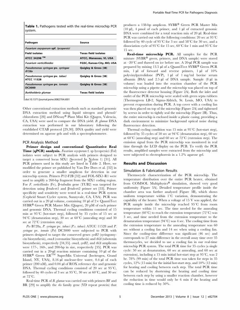
Table 1. Pathogens tested with the real-time microchip PCRsystem.
Pathogen Source
Fusarium oxysporum spf. lycopersici
Field isolates Texas field isolates
ATCC 34298 TM ATCC, Manassas, VI, USA
Fusarium verticillioides FGSC, Kansas City, MO, USA
Pseudomonas syringae pv. syringaeB728a
Quigley & Gross [38]
Pseudomonas syringae pv. tabaciATCC 11528
Quigley & Gross [38]
Pseudomonas syringae pv. tomatoDC3000
Quigley & Gross [38]
Burkholderia glumae Texas field isolate
doi:10.1371/journal.pone.0082704.t001
Portable Real-Time PCR for Pathogens Diagnosis
PLOS ONE | www.plosone.org 5 December 2013 | Volume 8 | Issue 12 | e82704

Compact fluorescence detector. When using nine different
concentrations of FITC dye from 0 nM to 2000 nM, the PMT
reading shows linear output (R2 = 0.9991) from 600 mV to
925 mV (Figure S2). Thus, the compact fluorescence detector
shows sufficient sensitivity for low limit of detection as well as
sufficient linearity for quantification.
Overall system assembly. Figure 4A shows the real-time
microchip PCR system with a compact fluorescence detector, fully
controlled by an MCU module and powered by a battery. The
complete system fits in a 2561668 cm3 box and weighs less than
843 g. Critical processing information and results, including
fluorescent readout from the PMT, current thermocycle number,
current thermocycle step, and temperature of the reaction
chamber were displayed on the LCD display (Figure 4B). The
MCU module consumed about 110 mAh for one PCR run that
includes one hot-start cycle and 35 PCR thermocycles, meaning
that more than 20 PCR runs can be conducted from a single
battery charge. The compact system size and efficient energy
consumption of our system makes this system more suitable for on-
field plant pathogen diagnostics than other portable real-time PCR
systems that still require additional instrumentations such as a
laptop or additional control modules [14–15].
DNA Preparation and PCR PerformanceDevelopment of a DNA extraction method feasible for
field application. We modified a variety of publicly and
commercially available DNA extraction methodologies and kits to
be usable in the absence of liquid nitrogen. Our test results show
that a commercial Plant/Fungi DNA Isolation Kit when used with
our unique modifications explained in the materials and methods
(section 2.2) provided high quality and sufficient yield of genomic
DNA from fungi and bacteria. With these modifications, we were
able to improve the DNA yield close to the levels with either liquid
nitrogen or chemical usage, obtaining 13 mg and 65 mg of DNA
per 50 mg of fungal and bacterial biomass, respectively (Table 3).
While the quantity of microbial genomic DNA extracted by the
improved method is not as effective as conventional in-lab
protocols, the yield obtained was sufficient for PCR based
diagnostics.
Primer specificity and sensitivity. Fol is a soil-borne
fungus that causes tomato wilt worldwide. Three physiological
Table 2. List of primers used in this study.
Primer Sequence (59R 39) Target Amplicon (bp) Reference
P12-F2B TATCCCTCCGGATTTTGAGC SIX1 260 Van Der Does et al. [32]
FOL-SIX1-R1 TCGTTTCCAGGAAAGCTGC SIX1 This work
b-tub-rt1 CAGCGTTCCTGAGTTGACCCAACAG TUB2 200 Choi et al. [33]
b-tub-rt2 CTGGACGTTGCGCATCTGATCCTCG TUB2 Choi et al. [33]
BF CCGCGCTGTTCATGAGGGATAA RHS 138 Kim et al. [29]
BR CGGGCGGAACGACGGTAAGT RHS Kim et al. [29]
cmaAF ACCGTGATGTTTACCTCTGGCA cmaA 177 Winsor et al.[35]
cmaAR GCAGCGGTACCCAAACTTCAAA cmaA Winsor et al.[35]
syrB2F TCCTTATCGATCTGCAACTGGCGA syrB2 160 Winsor et al.[35]
syrB2R AATGGTTGCCTGCAGTTCATTCCC syrB2 Winsor et al.[35]
tblAF ACCCAGACTTAGAGCTCAAGCCAA tblA 200 Winsor et al.[35]
tblAR TTCGATCTTGAAGCCAGCCTCAGT tblA Winsor et al.[35]
doi:10.1371/journal.pone.0082704.t002
Figure 2. Photographs of the compact fluorescence detector housing assembly for real-time detection of amplified DNA samples.(A) A PCR chip with a thermocouple placed on top of the optical detector housing (bottom part of the image) and a cover integrated with a coolingfan and having septa rubbers to seal the inlet and outlet of the PCR chip (top part of the image). This cover also completely encloses the PCR chip toprevent ambient light from affecting the reading of the PMT. (B) The fully assembled housing that encloses the PCR microchip. The cooling fan can beseen on top of the housing, as well as screws that provide the tight seal.doi:10.1371/journal.pone.0082704.g002
Portable Real-Time PCR for Pathogens Diagnosis
PLOS ONE | www.plosone.org 6 December 2013 | Volume 8 | Issue 12 | e82704

races of Fol have been reported worldwide, where race 1 is most
common in countries with warm climates. However, molecular
diagnosis of these races is challenging since only the genome
sequence of Fol race 2 is publicly available (http://www.
broadinstitute.org/). Fol SIX1 gene encodes for a virulence factor
towards tomato plants, which is secreted in the xylem of the host
during colonization [32]. Significantly, Six1 has been shown to be
a key protein required for full virulence of the pathogen [36] and is
a suitable target for diagnostics. Figure 5 shows that PCR analyses
targeting a small fragment of SIX1, using primer sets P12-F2B and
FOL-SIX1-R1 resulted in a clear and specific amplicon for Fol
strains. In addition, no false-positive amplicons were observed
when tested with other Fusarium species (Figure 5), as has been
previously reported by Lievens et al. [31]. Using the DNA
extraction method described earlier and primer sets P12-F2B and
FOL-SIX1-R1, we determined that the limit of detection is 10 pg
of gDNA/ real-time PCR.
There are over 50 pathovars of P. syringae that vary in host range
and symptomology [34,37]. For accurate diagnosis of disease
caused by P. syringae, primers that can detect these pathovars with a
high rate of specificity are critical. The gene syrB2, selected for the
detection of Pss B728a, is known to be conserved among most P.
syringae strains that produce syringomycin, syringotoxin, or
syringostatin [34]. Table S1 shows the specificity of syrB2 primers
for the detection of Pss strains utilizing PCR and real-time PCR.
The syrB2 primers were also screened against P. syringae strains that
were not producers of syringomycin, syringotoxin or syringosta-
tion. These strains were used as negative controls that produced no
amplicons on an agarose gel (data not shown).
These results demonstrate that the primers designed for Fol and
Pss are suitable for a reliable and sensitive identification method.
Furthermore, due to the small size of amplicons used for
conventional real-time PCR analysis (section 3.1 in the materials
and methods), overall PCR running time can be decreased,
resulting in faster detection of the pathogens.
Performance of the portable real-time microchip PCRsystem. To demonstrate the performance of our developed real-
time microchip PCR system, we investigated the success rate and
the detection limit of the system with Fol and Fv genomic DNA
samples.
When loading sample, bubble formation occasionally occurred,
but at a very low rate of about one per 20–30 loadings due to
capillary force as well as the hydrophilicity of the chamber. When
bubble formation was recognized, the PCR mixture was pulled
and pushed from the reaction chamber using the same pipette with
Figure 3. Simulated temperature profile of the microchip. (A) COMSOL MultiphysicsH simulation showing uniform temperature distribution inthe PCR chamber region when heated to 94uC (White dotted line shows the position of the reaction chamber). (B) Temperature profile across A-A’shows the uniformity of the temperature in the chamber region of the PCR microchip within 1uC variation when the PCR microchip is heated to 94uC.doi:10.1371/journal.pone.0082704.g003
Figure 4. Photograph of the portable real-time microchip PCR system. (A) The portable real-time microchip PCR system controlled by anMCU and powered by a battery. The entire size is 1662869 cm3, and the total weight is 843 g. (B) The LCD of the MCU board displays severalinformation about the real time PCR such as the number of cycle, current PCR step, current temperature, fluorescence intensity, the increasingamount of the present cycle’s fluorescence intensity compared to the first cycle’s fluorescence intensity, and the graph showing the trace of thefluorescence intensity of each cycle.doi:10.1371/journal.pone.0082704.g004
Portable Real-Time PCR for Pathogens Diagnosis
PLOS ONE | www.plosone.org 7 December 2013 | Volume 8 | Issue 12 | e82704
its tip placed in the inlet hole. In general, 2–4 repeated push/
pulling motion using the pipette successfully removed the bubble.
Firstly, a negative control sample (no DNA) was amplified
through 35 thermocycles, and the PMT showed voltage increase of
0.0260.02 V between the 1st thermocycle and 35th thermocycle.
The experiment was repeated five times. The voltage sum of the
average and the standard deviation (0.04 V) from this negative
control was considered as a threshold line to determine the
presence of amplified DNA for all future tests. Subsequently, ten
PCR tests with FOL genomic DNA (300 ng/sample) were
conducted. Increases in PMT output voltage (0.1360.2 V) of
more than the threshold voltage were obtained from all ten tests,
indicating that the portable real-time microchip PCR system
successfully amplified and detected the target DNA consistently.
The Fv genomic DNA (300 ng/sample) was tested seven times
with 100% success rate, showing average increase in PMT output
of 0.3260.13 V. The Pss B728a DNA (75 ng/sample) was tested
ten times and also showed 100% success rate through the PMT
output increase (0.160.08 V). After these test results, we
continued with P.s. tabaci ATCC 11528 (115 ng/sample), Pst
DC3000 (166 ng/sample), and B. glumae (300 ng/sample and
150 ng/sample), each with four repetitions. All tests showed 100%
success rate (Table 4).
To determine the detection limit of our system, seven different
concentrations of Fv DNA – 300, 180, 100, 50, 12, 5, and 1 ng/
sample – were tested with four repetitions. The PMT voltage
increase versus the number of thermocycles was plotted (Figure 6a)
and cycle of quantification (Cq) values were extracted from the
graph. The Cq value was defined as the thermocycle number
when the PMT output increased over the previously determined
threshold line of 0.04 V. PMT outputs from the concentrations of
300, 180, 100, 50, 12, and 5 ng/sample all became larger than the
threshold, while the PMT output from the 1 ng/sample was not
over the threshold. Thus, the detection limit of our developed real-
time microchip PCR system was determined to be 5 ng/sample.
We expect that this detection limit can be further improved in the
future by using a higher power excitation light source, more
efficient filter sets (transmission efficiency larger than the currently
used 80%), and a more sensitive PMT with lower background
noise (luminous sensitivity larger than the currently used 350 mA/
lm), all currently commercially available.
Table 3. Comparison of fungal and bacterial genomic DNA extracted by a variety of methods with or without the use of liquidnitrogen, phenol and chloroform.
Organism a Kit Liquid nitrogen Phenol/ Chloroform Modifications b Yield (mg) c
Fol Standard method Yes Yes None 33±2.03
Standard method No Yes Yes 8.5±0.8
Norgen No No None 6.7 ±0.8
Norgen modification (ourmethod)
No No Yes 12.6±2.02
Pss Standard method No Yes None 52.4±1.18
DNeasy No No None 2.2±0.6
Norgen modification (ourmethod)
No No Yes 64.76±7.0
aFusarium oxysporum f. sp. lycopersici (FOL) and Pseudomona syringae pv. syringae (Pss) genomic DNA was isolated from 50 mg of wet fungal and bacterial biomass,respectively.bModifications as described in the materials and methods (section 2.2).cValues are the means of three biological replicates 6 SE.doi:10.1371/journal.pone.0082704.t003
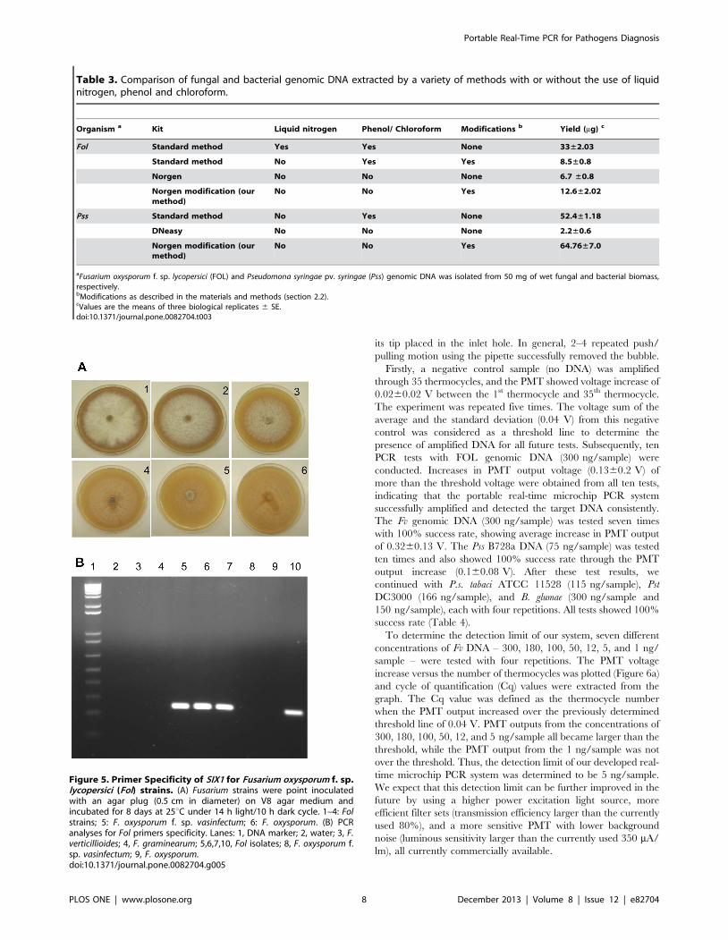
Figure 5. Primer Specificity of SIX1 for Fusarium oxysporum f. sp.lycopersici (Fol) strains. (A) Fusarium strains were point inoculatedwith an agar plug (0.5 cm in diameter) on V8 agar medium andincubated for 8 days at 25uC under 14 h light/10 h dark cycle. 1–4: Folstrains; 5: F. oxysporum f. sp. vasinfectum; 6: F. oxysporum. (B) PCRanalyses for Fol primers specificity. Lanes: 1, DNA marker; 2, water; 3, F.verticillioides; 4, F. graminearum; 5,6,7,10, Fol isolates; 8, F. oxysporum f.sp. vasinfectum; 9, F. oxysporum.doi:10.1371/journal.pone.0082704.g005
Portable Real-Time PCR for Pathogens Diagnosis
PLOS ONE | www.plosone.org 8 December 2013 | Volume 8 | Issue 12 | e82704

The Cq values of Fv DNA 300, 180, 100, 50, 12, and 5 ng/
sample were 18, 19, 20, 22, 24, and 29, respectively, which were
correlated to the amount of DNA (Figure 6b). As the amount of
DNA in the sample solution decreased, corresponding Cq values
increased, which indicate that more thermocycles are required for
the target DNA to be sufficiently amplified to have fluorescent
signals higher than those from the negative control (Table 4).
Throughout our experiments, resulting amplicons from the real-
time PCR microchips were extracted and subjected to conven-
tional gel electrophoresis. All successful real-time microchip PCR
runs as determined by PMT output voltage increase also showed
strong gel bands, validating our real-time microchip PCT result.
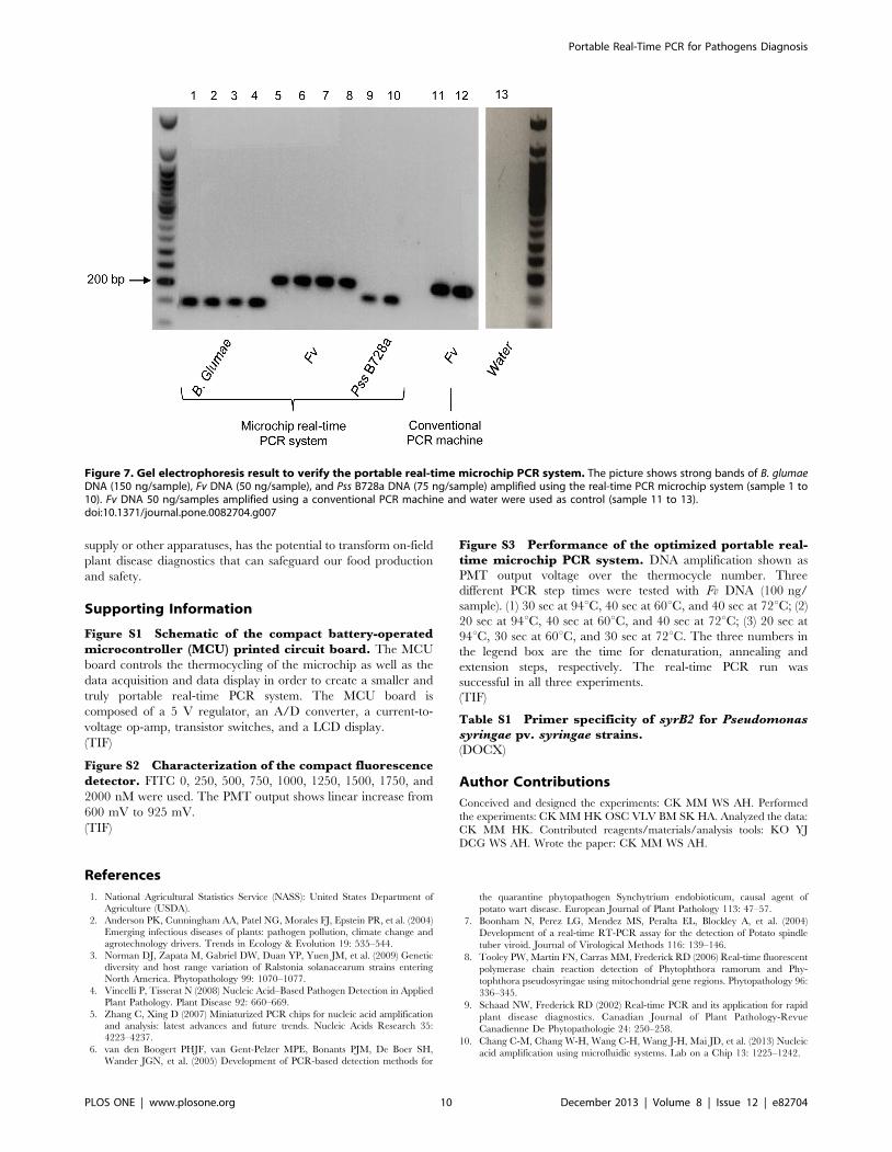
Figure 7 shows examples of such confirmation results, including
real-time microchip PCR results from Fv DNA (50 ng/sample), Pss
B728a DNA (75 ng/sample), and B. glumae DNA (150 ng/sample).
This confirmation also allows us to reduce the PCR reaction
chamber volume of the microchip in the future, since the current
chamber volume (8 ml) was simply determined based on the
needed sample amount for off-chip gel electrophoresis conforma-
tion.
Since we have initially used a much longer step time (50 sec at
95uC, 60 sec at 60uC, 60 sec at 72uC) to amplify our particular
plant pathogen samples compared to those of other microchip
PCR systems (typically using 15 sec at 95uC, 30 sec at 60uC, 30 sec
at 72uC), we attempted to further optimize and reduce this PCR
step time to reduce the overall assay time. Successful amplification
of Fv DNA was obtained by using as short as 20 sec at 94uC, 30 sec
at 60uC, and 30 sec at 72uC (Figure S3). This resulted in the
reduction in the total operation time to 1.2 hours, including the
initial hot-start step (15 min), a 0.8 hour reduction, thus making it
much more suitable for field applications.
Conclusion
We developed a portable and standalone real-time microchip
PCR system equipped with a microcontroller for controlling the
entire PCR operation and with a compact fluorescence detector
for acquiring fluorescence emission intensity for on-field plant
disease diagnostics. This battery-powered system was fully
operational with no need for an external computer or auxiliary
power source. With these specifications, we were able to develop a
real-time microchip PCR system that is 2561668 cm3 in size and
843 g in weight, substantially increasing the prospect of a fully
functional yet truly portable real-time microchip PCR system. The
MCU runs 35 thermocycles and detects amplicons in real time
with low power consumption (110 mAh), enabling more than 22
runs with a single battery charge. We also developed efficient
DNA extraction protocols for field operations, notably without the
use of liquid nitrogen and other large lab equipment. Multiple
fungal and bacterial genomic DNA samples were tested with our
portable real-time microchip PCR system, and successful DNA
amplification resulted in increased fluorescent signal through the
PMT. We were extremely encouraged by the consistency of
success in DNA amplification and detection (100% success rate
and validation through gel electrophoresis). The detection limit of
our system was 5 ng/sample DNA, with room for further
improvement. Significantly, our portable lab-on-a-chip real-time
PCR system, without the need for external computer, power
Table 4. Analysis of pathogen DNAs using the real-timemicrochip PCR system.
Sample Cq Success rate
Fol 300 ng/sample 18 100% (n = 10)
Pss B728a 75 ng/sample 23 100% (n = 10)
P.s. tabaci ATCC11528 115 ng/sample 23 100% (n = 4)
P.s.t. DC3000 166 ng/sample 17 100% (n = 4)
B. glumae 300 ng/sample 18 100% (n = 4)
B. glumae 150 ng/sample 21 100% (n = 4)
Negative - - (n = 5)
doi:10.1371/journal.pone.0082704.t004
Figure 6. Performance of the portable real-time microchip PCR system using different concentrations of Fv DNA. (A) Amplication plotof PMT voltage output over the thermocycle number. Using 300, 180, 100, 50, 12, and 5 ng/sample, the Cq was 18, 19, 20, 22, 24, and 29, respectively(enlarged graph shows the Cq). The error bar of the negative control (0 ng) is the standard deviation. (B) Correlation graph of the DNA amount andthe Cq (R2.0.947).doi:10.1371/journal.pone.0082704.g006
Portable Real-Time PCR for Pathogens Diagnosis
PLOS ONE | www.plosone.org 9 December 2013 | Volume 8 | Issue 12 | e82704
supply or other apparatuses, has the potential to transform on-field
plant disease diagnostics that can safeguard our food production
and safety.
Supporting Information
Figure S1 Schematic of the compact battery-operatedmicrocontroller (MCU) printed circuit board. The MCU
board controls the thermocycling of the microchip as well as the
data acquisition and data display in order to create a smaller and
truly portable real-time PCR system. The MCU board is
composed of a 5 V regulator, an A/D converter, a current-to-
voltage op-amp, transistor switches, and a LCD display.
(TIF)
Figure S2 Characterization of the compact fluorescencedetector. FITC 0, 250, 500, 750, 1000, 1250, 1500, 1750, and
2000 nM were used. The PMT output shows linear increase from
600 mV to 925 mV.
(TIF)
Figure S3 Performance of the optimized portable real-time microchip PCR system. DNA amplification shown as
PMT output voltage over the thermocycle number. Three
different PCR step times were tested with Fv DNA (100 ng/
sample). (1) 30 sec at 94uC, 40 sec at 60uC, and 40 sec at 72uC; (2)
20 sec at 94uC, 40 sec at 60uC, and 40 sec at 72uC; (3) 20 sec at
94uC, 30 sec at 60uC, and 30 sec at 72uC. The three numbers in
the legend box are the time for denaturation, annealing and
extension steps, respectively. The real-time PCR run was
successful in all three experiments.
(TIF)
Table S1 Primer specificity of syrB2 for Pseudomonassyringae pv. syringae strains.(DOCX)
Author Contributions
Conceived and designed the experiments: CK MM WS AH. Performed
the experiments: CK MM HK OSC VLV BM SK HA. Analyzed the data:
CK MM HK. Contributed reagents/materials/analysis tools: KO YJ
DCG WS AH. Wrote the paper: CK MM WS AH.
References
1. National Agricultural Statistics Service (NASS): United States Department ofAgriculture (USDA).
2. Anderson PK, Cunningham AA, Patel NG, Morales FJ, Epstein PR, et al. (2004)
Emerging infectious diseases of plants: pathogen pollution, climate change and
agrotechnology drivers. Trends in Ecology & Evolution 19: 535–544.
3. Norman DJ, Zapata M, Gabriel DW, Duan YP, Yuen JM, et al. (2009) Geneticdiversity and host range variation of Ralstonia solanacearum strains entering
North America. Phytopathology 99: 1070–1077.
4. Vincelli P, Tisserat N (2008) Nucleic Acid–Based Pathogen Detection in Applied
Plant Pathology. Plant Disease 92: 660–669.
5. Zhang C, Xing D (2007) Miniaturized PCR chips for nucleic acid amplificationand analysis: latest advances and future trends. Nucleic Acids Research 35:
4223–4237.
6. van den Boogert PHJF, van Gent-Pelzer MPE, Bonants PJM, De Boer SH,
Wander JGN, et al. (2005) Development of PCR-based detection methods for
the quarantine phytopathogen Synchytrium endobioticum, causal agent of
potato wart disease. European Journal of Plant Pathology 113: 47–57.
7. Boonham N, Perez LG, Mendez MS, Peralta EL, Blockley A, et al. (2004)
Development of a real-time RT-PCR assay for the detection of Potato spindle
tuber viroid. Journal of Virological Methods 116: 139–146.
8. Tooley PW, Martin FN, Carras MM, Frederick RD (2006) Real-time fluorescent
polymerase chain reaction detection of Phytophthora ramorum and Phy-
tophthora pseudosyringae using mitochondrial gene regions. Phytopathology 96:
336–345.
9. Schaad NW, Frederick RD (2002) Real-time PCR and its application for rapid
plant disease diagnostics. Canadian Journal of Plant Pathology-Revue
Canadienne De Phytopathologie 24: 250–258.
10. Chang C-M, Chang W-H, Wang C-H, Wang J-H, Mai JD, et al. (2013) Nucleic
acid amplification using microfluidic systems. Lab on a Chip 13: 1225–1242.
Figure 7. Gel electrophoresis result to verify the portable real-time microchip PCR system. The picture shows strong bands of B. glumaeDNA (150 ng/sample), Fv DNA (50 ng/sample), and Pss B728a DNA (75 ng/sample) amplified using the real-time PCR microchip system (sample 1 to10). Fv DNA 50 ng/samples amplified using a conventional PCR machine and water were used as control (sample 11 to 13).doi:10.1371/journal.pone.0082704.g007
Portable Real-Time PCR for Pathogens Diagnosis
PLOS ONE | www.plosone.org 10 December 2013 | Volume 8 | Issue 12 | e82704

11. Park S, Zhang Y, Lin S, Wang T-H, Yang S (2011) Advances in microfluidic
PCR for point-of-care infectious disease diagnostics. Biotechnology Advances 29:830–839.
12. Zhang Y, Ozdemir P (2009) Microfluidic DNA amplification—A review.
Analytica Chimica Acta 638: 115–125.13. Cady NC, Stelick S, Kunnavakkam MV, Yuxin L, Batt CA (2004) A microchip-
based DNA purification and real-time PCR biosensor for bacterial detection. pp.1191-1194 vol.1193.
14. Focke M, Stumpf F, Faltin B, Reith P, Bamarni D, et al. (2010) Microstructuring
of polymer films for sensitive genotyping by real-time PCR on a centrifugalmicrofluidic platform. Lab on a Chip 10: 2519–2526.
15. Xiang Q, Xu B, Li D (2007) Miniature real time PCR on chip with multi-channel fiber optical fluorescence detection module. Biomedical Microdevices 9:
443–449.16. Lund-Olesen T, Dufva M, Dahl J, Collas P, Hansen M (2008) Sensitive on-chip
quantitative real-time PCR performed on an adaptable and robust platform.
Biomedical Microdevices 10: 769–776.17. Lee JG, Cheong KH, Huh N, Kim S, Choi JW, et al. (2006) Microchip-based
one step DNA extraction and real-time PCR in one chamber for rapid pathogenidentification. Lab on a Chip 6: 886–895.
18. Shi X, Lin L-I, Chen S-y, Chao S-h, Zhang W, et al. (2011) Real-time PCR of
single bacterial cells on an array of adhering droplets. Lab on a Chip 11: 2276–2281.
19. Verdoy D, Barrenetxea Z, Berganzo J, Agirregabiria M, Ruano-Lopez JM, et al.(2012) A novel Real Time micro PCR based Point-of-Care device for Salmonella
detection in human clinical samples. Biosensors and Bioelectronics 32: 259–265.20. Chen D, Mauk M, Qiu X, Liu C, Kim J, et al. (2010) An integrated, self-
contained microfluidic cassette for isolation, amplification, and detection of
nucleic acids. Biomed Microdevices 12: 705–719.21. Qiu X, Chen D, Liu C, Mauk MG, Kientz T, et al. (2011) A portable, integrated
analyzer for microfluidic - based molecular analysis. Biomed Microdevices 13:809–817.
22. Liu C, Mauk MG, Hart R, Bonizzoni M, Yan G, et al. (2012) A low-cost
microfluidic chip for rapid genotyping of malaria-transmitting mosquitoes. PLoSOne 7: e42222.
23. Julich S, Riedel M, Kielpinski M, Urban M, Kretschmer R, et al. (2011)Development of a lab-on-a-chip device for diagnosis of plant pathogens.
Biosensors & Bioelectronics 26: 4070–4075.24. Moller EM, Bahnweg G, Sandermann H, Geiger HH (1992) A simple and
efficient protocol for isolation of high molecular weight DNA from filamentous
fungi, fruit bodies, and infected plant tissues. Nuclear Acids Research 20: 6115–6116.
25. Woloshuk CP, Yousibova GL, Rollins JA, Bhatnagar D, Payne GA (1995)Molecular characterization of the afl-1 locus in Aspergillus flavus. Applied and
Environmental Microbiology 61: 3019–3023.
26. Xu JR, Hamer JE (1996) MAP kinase and cAMP signaling regulate infection
structure formation and pathogenic growth in the rice blast fungus Magnaporthe
grisea. Genes & Development 10: 2696–2706.
27. Kim J, Gale BK. Geometric Optimization of a Thin Film ITO Heater to
Generate a Uniform Temperature Distribution; 2006; Tokyo, Japan. pp. 293–
295.
28. Wilson K (2001) Preparation of Genomic DNA from Bacteria. Current
Protocols in Molecular Biology: John Wiley & Sons, Inc.
29. Kim BK, Cho MK, Kim MO, Choi HJ, Kang MJ, et al. (2012) Rapid and
Specific Detection of Burkholderia glumae in Rice Seed by Real-Time Bio-PCR
Using Species-Specific Primers Based on an rhs Family Gene. Plant Disease 96:
577–580.
30. Sayler RJ, Cartwright RD, Yang Y (2006) Genetic Characterization and Real-
Time PCR Detection of Burkholderia glumae, a Newly Emerging Bacterial
Pathogen of Rice in the United States. Plant disease 90: 603–610.
31. Lievens B, Houterman PM, Rep M (2009) Effector gene screening allows
unambiguous identification of Fusarium oxysporum f. sp. lycopersici races and
discrimination from other formae speciales. FEMS Microbiology Letters 300:
201–215.
32. van der Does HC, Lievens B, Claes L, Houterman PM, Cornelissen BJC, et al.
(2008) The presence of a virulence locus discriminates Fusarium oxysporum
isolates causing tomato wilt from other isolates. Environmental Microbiology 10:
1475–1485.
33. Choi Y-E, Shim W-B (2008) Identification of Genes Associated with Fumonisin
Biosynthesis in Fusarium verticillioides via Proteomics and Quantitative Real-
Time PCR. J Microbiol Biotechnol 18: 648–657.
34. Bender CL, Alarcon-Chaidez F, Gross DC (1999) Pseudomonas syringae
Phytotoxins: Mode of Action, Regulation, and Biosynthesis by Peptide and
Polyketide Synthetases. Microbiol Mol Biol 63: 266–292.
35. Winsor GL, Lam DKW, Fleming L, Lo R, Whiteside MD, et al. (2011)
Pseudomonas Genome Database: improved comparative analysis and popula-
tion genomics capability for Pseudomonas genomes. Nucleic Acids Res 39:
D596–600.
36. Rep M, Meijer M, Houterman PM, van der Does HC, Cornelissen BJC (2005)
Fusarium oxysporum Evades I-3-Mediated Resistance Without Altering the
Matching Avirulence Gene. Molecular Plant-Microbe Interactions 18: 15–23.
37. Feil H, Feil WS, Chain P, Larimer F, DiBartolo G, et al. (2005) Comparison of
the complete genome sequences of Pseudomonas syringae pv. syringae B728a
and pv. tomato DC3000. Proceedings of the National Academy of Sciences of
the United States of America 102: 11064–11069.
38. Quigley NB, Gross DC (1994) Syringomycin Production Among Strains of
Pseudomonas syringae pv. syringae: Conservation of the syrB and syrD Genes
and Activation of Phytotoxin Production by Plant Signal Molecules. Molecular
Plant-Microbe Interactions 7: 78–90.
Portable Real-Time PCR for Pathogens Diagnosis
PLOS ONE | www.plosone.org 11 December 2013 | Volume 8 | Issue 12 | e82704