development of a quantitative method for …
TRANSCRIPT
Bull. Acad. Vét. France — 2017 - Tome 170 - N°2 http://www.academie-veterinaire-defrance.org
COMMUNICATION
DEVELOPMENT OF A QUANTITATIVE METHOD FOR ULTRASONOGRAPHIC
ASSESSMENT OF RECURRENT LARYNGEAL NEUROPATHY IN HORSES
MISE AU POINT D’UNE MÉTHODE QUANTITATIVE D’ÉVALUATION
ÉCHOGRAPHIQUE DE L’HÉMIPLÉGIE LARYNGÉE CHEZ LE CHEVAL
Par Lucile VAVASSEUR(1), Antoine LECHARTIER(2), Céline ROBERT(3) & Céline MESPOULHES(3) (Communication présentée le 08 Juin 2017
Manuscrit accepté le 02 Mai 2017)
Plus accessible sur le terrain, moins invasive et mieux corrélée à l’endoscopie à l’effort que l’endosco-pie au repos, il ne manque à l’exploration échographique du larynx par voie externe que des critères échographiques de suspicion objectifs pour en faire un outil clé du dépistage de l’hémiplégie laryngée chez le cheval. L’objectif de cette étude est de définir un paramètre échographique combinant une bonne concordance inter-échographistes et une bonne aptitude à discriminer les chevaux sains des chevaux présentant un défaut d’abduction du cartilage aryténoïde gauche à l’effort. La moyenne, sur trois images acquises en vue transversale, du ratio entre l’échogénicité moyenne d’un disque de 1130 pixels placé dans le muscle crico-aryténoïdien latéral et celle d’un disque de 1130 pixels placé dans le muscle vocal est le paramètre garantissant la meilleure concordance inter-échographistes. Ce paramètre présente par ailleurs une très bonne corrélation avec l’examen endoscopique à l’effort, considéré comme le Gold Standard.
Mots-clés : hémiplégie laryngée, neuropathie, larynx, cornage, échographie musculaire, pathologie, appareil respiratoire, équidé, cheval.
Résumé
(1) Docteur Vétérinaire, 61 rue Aristide Briand 61200 ARGENTAN, Email : [email protected]
(2) Clinique Vétérinaire Équine de Méheudin, 61150 Ecouché Email : [email protected]
(3) École Nationale Vétérinaire d’Alfort Email : [email protected] Email : [email protected]
More affordable and less invasive than resting endoscopy, ultrasonography of the equine larynx also provides a better correlation with dynamic endoscopy. With appropriate objective ultrasonographic suspicion criteria, its use for early screening would then allow subjecting only suspected horses to a dynamic endoscopic exam for confirmation. In this study, we tried to determine a quantitative ultra-sonographic parameter that would both take concordant values between two different operators and reliably differentiate healthy horses from horses showing a decreased abduction of the left arytenoid cartilage at dynamic endoscopic evaluation. Calculated from three images in transverse section, the parameter that ensures the best inter-operator concordance is the mean ratio between mean echogenicity of a 1130 pixels disc located in the cricoarytenoideus lateralis muscle and mean echogenicity of a 1130 pixels disc located in the vocalis muscle. This parameter also provides a very good correlation with dynamic endoscopic exam, which is the gold standard.
Key words: recurrent laryngeal neuropathy, larynx, roaring noise, muscular ultrasonography, pathology, respiratory tract, equine, horse.
AbstrAct
DOI : 10.4267/2042/62280
118

Bull. Acad. Vét. France — 2017 - Tome 170 - N°2 http://www.academie-veterinaire-defrance.org
COMMUNICATION
INTRODUCTION
Recurrent laryngeal neuropathy (RLN) is a frequent endosco-pically diagnosed cause of poor performance in sport and race horses (Martin et al. 2000; Ducharme & Cheetham, 2014). Resting endoscopic exam allows the assessment of the degree of laryngeal hemiplegia using a seven grades scale (ranging from grade 1 to 4) as defined by the Havemeyer consensus conference (Dixon et al. 2004). As part of a routine screening protocol as at public sales, the clinical relevance of the resting endoscopic laryngeal grading system has been questioned (Garrett et al. 2010). In practice, this exam is often complemented by a dyna-mic endoscopic exam, which is considered as the gold standard for the assessment of RLN (Ducharme & Cheetham, 2014). Besides, RLN induces a denervation atrophy of all intrinsic laryngeal muscles, excepted for the cricothyroideus muscle that is innervated by the ramus externus of the laryngeus cranialis nerve (Duncan et al. 1974). Beyond these muscles, the cricoarytenoideus lateralis muscle is the first one to undergo neurogenic atrophy and is histologically the most severely affected in RLN (Cahill & Goulden, 1986; Duncan et al. 1974). Recent studies highlighted that abnormal arytenoid movement during dynamic endoscopic exam was statistically associated with relative hyperechogenicity of the cricoarytenoideus lateralis muscle (Garrett et al. 2010; Chalmers et al. 2012). Subjective hyperechogenicity of the left cricoarytenoideus lateralis muscle had a sensibility of 94.59% and a specificity of 94.54% for the detection of RLN (Chalmers et al. 2012). These findings are consistent with reports in human medicine, in which quantitative measures of muscle echogenicity have been proposed as methods for enhancing detection of neu-romuscular pathologies.
The only study about quantitative assessment of the left cricoary-tenoideus lateralis muscle echogenicity identified a significantly different echogenicity between left and right cricoarytenoideus lateralis muscles in Havemeyer grade B and C horses. Yet, no significant difference was observed in left cricoarytenoideus late-ralis raw echogenicity between the grade A and grade B horses (Chalmers et al. 2012).
As transcutaneous ultrasonography is a less invasive and a more affordable technique than endoscopy, its use to detect horses likely to present a laryngeal asymmetry during exercise seems relevant. However, the use of a quantitative parameter to assess cricoarytenoideus lateralis echogenicity is essential for an objective evaluation.
The aim of this study was to propose and validate a new method for ultrasonographic assessment of RLN based on cricoarytenoi-deus lateralis muscle echogenicity quantification
MATERIALS AND METHODS
Patient Population
Four groups of five horses with no history of laryngeal or pharyngeal surgery were included in the study. Five healthy horses that had shown a fully normal abduction of the ary-tenoid cartilages during the endoscopic exam performed at rest composed the Havemeyer grade 1 group. The three other groups included horses referred to the Alfort Veterinary School or to the Clinic of Grosbois by practitioners for left-sided signs of RLN at rest. The Havemeyer grade IV group was composed of five horses showing a complete immobility of the arytenoid cartilage and vocal folds at rest. Horses showing a Havemeyer grade III at rest (full abduc-tion not obtained or maintained) were divided in groups two and three. Group two included horses with a grade A at exercise and group three included horses with grades B and C at exercise (a decreased abduction of the arytenoid cartilage). Following Barakzai & Dixon observations (2011), a Havemeyer grade I at rest was considered as a Havemeyer grade A at exercise and Havemeyer grade IV at rest was considered as a Havemeyer grade C at exercise.
Ultrasonography
Acquisition
Ultrasonographic examination of the laryngeal region was performed using the lateral window (Chalmers et al. 2006). The horses were examined in stocks and sedated with detomidine at the dose of 0.01 mg per kg intra-venously. The head was placed on a 1.20 meter high device in order to main-tain extension of the neck (figure 1). The laryngeal region was systematically clipped, soaked with seventy percent isopropyl alcohol and ultrasonographic gel was applied on each side before the exam.
Figure 1 : Preparation of the horse and position of the probe.
DOI : 10.4267/2042/62280
119

Bull. Acad. Vét. France — 2017 - Tome 170 - N°2 http://www.academie-veterinaire-defrance.org
COMMUNICATION
Two authors previously trained to laryngeal ultrasonography (AL and LV) performed independently on each horse the acquisition of:
- three longitudinal plane ultrasonographic images of the right lateral acoustic window;
- three transversal plane ultrasonographic images of the right lateral acoustic window;
- three longitudinal plane ultrasonographic images of the left lateral acoustic window;
- three transversal plane ultrasonographic images of the left lateral acoustic window.
The ultrasonographic exams were performed with a portable SONOSITE M-TURBO® (FUJIFILM Sonosite, Paris, FRANCE) machine connected to a 7.5-13 MHz linear transducer 2.5 cm broad. The depth was set at 31 mm. The musculoskeletal exam type in 2D imaging mode and the “general” setting mode, which provides an optimal balance between resolution and penetra-tion, were chosen. The overall gain was standardized and kept constant for all the exams.
Data analysis
Each exam (twelve images) was exported in Jpeg format to a USB storage device to compose forty files (twenty horses x two operators) that were randomized and blinded by one of the two operators (LV). Each file was, at first, named after the name of the horse (Name) followed by the operator’s ini-tials (LV or AL). Then, a number between one and forty was
randomly associated to each file and the correspondence between the names of the exams and their numbers were consigned in a table for result interpretation. The image processing was performed by one single operator (LV) with a dedicated greyscale quantification program (EchoQuant, BPLC, Maisons-Alfort, France) developed on Matlab 7.5.0 (MathWorks, Natick, Massachusetts).
In order to allow the most precise and accurate description of the cricoarytenoideus lateralis muscle, the Regions Of Interest (ROI) were set outside the artefact areas, especially outside the shadow cones caused by a possible thyroid carti-lage mineralization. Their dimension was set to a 1130 pixels disc which is the maximum size allowing to define three distinct areas without artefact in this muscle in our study. Similar ROI were set in the vocalis muscle in order to esta-blish a ratio of echogenicity between the cricoarytenoideus lateralis muscle and a reference structure present in the same acoustic window.
Two protocols of image processing were used. The first one, which was designed to enhance precision, consisted of the largest sample of pixels allowed, that is three distinct 1130 pixel discs in the cricoarytenoideus lateralis muscle and two distinct 1130 pixel discs in the vocalis muscle. The second one, which was designed to enhance accuracy, consisted of a smaller but best quality sample: only one 1130 pixel disc excluding artefact, set in the most representative zone of the
cricoarytenoideus lateralis muscle, and one similar excluding artefact ROI set in the vocalis muscle and vertically aligned to the cricoarytenoideus lateralis ROI were considered (figure 2).
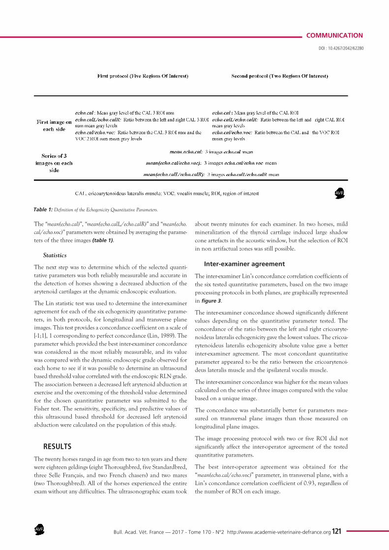
The 256 grey levels repartition matrix was used to determine the mean gray level of each ROI and to obtain three quantitative parameters: (1) the “echo.cal” parameter was defined, on both sides, as the mean gray level of the cri-coarytenoideus lateralis (based on three ROI in the first protocol and one ROI in the second protocol) ; (2) the “echo.calL/echo.calR” parameter was calculated as the ratio between the left and right cricoarytenoideus lateralis ROI mean gray levels in both protocols ; (3) the “echo.cal/echo.voc” parameter was calcu-lated, on both sides, as the ratio between the cricoarytenoideus lateralis ROI and the ipsilateral vocalis ROI mean gray levels in both protocols. Those three parameters were defined on the first of the three images taken on each side in each plane (either longitudinal or transverse).
Figure 2 : Regions of interest selection on the longitudinal (a) and transverse (d) plane ultrasound images of the cricoarytenoideus lateralis muscle and vocalis muscle, according to the first (b, e) and the second (c, f) protocols.TC, thyroid cartilage; CC, cricoid cartilage; AC, arytenoid cartilage; CAL, cricoarytenoideus lateralis muscle; VOC, vocalis muscle.
DOI : 10.4267/2042/62280
120
Bull. Acad. Vét. France — 2017 - Tome 170 - N°2 http://www.academie-veterinaire-defrance.org
COMMUNICATION
The “mean(echo.cal)”, “mean(echo.calL/echo.calR)” and “mean(echo.cal/echo.voc)” parameters were obtained by averaging the parame-ters of the three images (table 1).
Statistics
The next step was to determine which of the selected quanti-tative parameters was both reliably measurable and accurate in the detection of horses showing a decreased abduction of the arytenoid cartilages at the dynamic endoscopic evaluation.
The Lin statistic test was used to determine the inter-examiner agreement for each of the six echogenicity quantitative parame-ters, in both protocols, for longitudinal and transverse plane images. This test provides a concordance coefficient on a scale of [-1;1], 1 corresponding to perfect concordance (Lin, 1989). The parameter which provided the best inter-examiner concordance was considered as the most reliably measurable, and its value was compared with the dynamic endoscopic grade observed for each horse to see if it was possible to determine an ultrasound based threshold value correlated with the endoscopic RLN grade. The association between a decreased left arytenoid abduction at exercise and the overcoming of the threshold value determined for the chosen quantitative parameter was submitted to the Fisher test. The sensitivity, specificity, and predictive values of this ultrasound based threshold for decreased left arytenoid abduction were calculated on the population of this study.
RESULTS
The twenty horses ranged in age from two to ten years and there were eighteen geldings (eight Thoroughbred, five Standardbred, three Selle Français, and two French chasers) and two mares (two Thoroughbred). All of the horses experienced the entire exam without any difficulties. The ultrasonographic exam took
about twenty minutes for each examiner. In two horses, mild mineralization of the thyroid cartilage induced large shadow cone artefacts in the acoustic window, but the selection of ROI in non artifactual zones was still possible.
Inter-examiner agreement
The inter-examiner Lin’s concordance correlation coefficients of the six tested quantitative parameters, based on the two image processing protocols in both planes, are graphically represented in figure 3.
The inter-examiner concordance showed significantly different values depending on the quantitative parameter tested. The concordance of the ratio between the left and right cricoaryte-noideus lateralis echogenicity gave the lowest values. The cricoa-rytenoideus lateralis echogenicity absolute value gave a better inter-examiner agreement. The most concordant quantitative parameter appeared to be the ratio between the cricoarytenoi-deus lateralis muscle and the ipsilateral vocalis muscle.
The inter-examiner concordance was higher for the mean values calculated on the series of three images compared with the value based on a unique image.
The concordance was substantially better for parameters mea-sured on transversal plane images than those measured on longitudinal plane images.
The image processing protocol with two or five ROI did not significantly affect the inter-operator agreement of the tested quantitative parameters.
The best inter-operator agreement was obtained for the “mean(echo.cal/echo.voc)” parameter, in transversal plane, with a Lin’s concordance correlation coefficient of 0.93, regardless of the number of ROI on each image.
Table 1: Definition of the Echogenicity Quantitative Parameters.
DOI : 10.4267/2042/62280
121

Bull. Acad. Vét. France — 2017 - Tome 170 - N°2 http://www.academie-veterinaire-defrance.org
COMMUNICATION
Correlation with the dynamic endoscopy results
The average of the three transversal plane images ratios between the mean gray levels of one 1130 pixel disc located in the left cricoarytenoideus lateralis muscle and one 1130 pixel disc located in the left vocalis muscle obtained by each examiner were com-pared with the resting or dynamic endoscopic grade for each horse (figure 4). For readability, this quantitative parameter has been abbreviated to “CAL/VOC ratio”.
On this figure, the CAL/VOC ratios calculated for each of the two operators appear on the x and y-axis respectively. The data
points clustering around the y=x coordinates axis illustrates the high inter operator correlation for this parameter (Lin concor-dance correlation coefficient: 0.93). In this study, all the horses showing a decreased left arytenoid abduction at exercise (grades B or C) have a CAL/VOC ratio greater than 0.6, or even greater than 0,8 whereas all the horses showing normal left arytenoid abduction at exercise (grade A) have a CAL/VOC ratio below 0.8 or even below 0,6. The threshold value of 0.6 allows the discrimination of horses likely to show decreased left arytenoid abduction at exercise with a sensibility of 1, a specificity of 0.90, a predictive positive value of 0.91 and a negative predictive value of
1. The statistic association between a decreased left arytenoid abduction at exercise and a CAL/VOC ratio greater than 0.6 is significant with a p-value of 0.000119 according to the Fisher test.
DISCUSSIONIn this study on twenty horses, the ratio between the mean gray level of one 1130 pixel disc located in the left cricoarytenoideus lateralis muscle and the mean gray level of one 1130 pixel disc located in the left vocalis muscle on three images provided both a very good inter-examiner concordance (Lin concor-dance correlation coefficient: 0.93) and an excellent correlation with dynamic endoscopy for RLN dia-gnosis. The association between a value greater than 0.6 for this quantitative parameter and decreased left arytenoid abduction at exercise was statistically
Figure 3: Inter-examiner Lin’s concor-dance correlation coefficients of the tested quantitative parameters and their confidence interval, calculated on twenty horses, in both longitudinal (L) and transversal (T) planes, taking two and five regions of interest (ROI) in account. The “echo.cal” parameter was defined, on both sides, as the mean gray level of the cricoarytenoideus lateralis; the “echo.calL/echo.calR” parameter was calcu-lated as the ratio between the left and right cricoarytenoideus lateralis ROI mean gray levels; the “echo.cal/echo.voc” parameter was calculated, on both sides, as the ratio between the cricoarytenoideus lateralis ROI and the ipsilateral vocalis ROI mean gray levels. The “mean(echo.cal)”, “mean(echo.calL/echo.calR)” and “mean(echo.cal/echo.voc)” parameters were obtained by averaging the parameters of the three images.
Figure 4: Mean ratio between the cricoarytenoideus lateralis and the ipsilateral vocalis muscles’ echogenicity calculated on three images acquired by each one of the two operators (LV and AL) in the left lateral acoustic window for the four groups of horses.
DOI : 10.4267/2042/62280
122

Bull. Acad. Vét. France — 2017 - Tome 170 - N°2 http://www.academie-veterinaire-defrance.org
COMMUNICATION
significant according to the Fisher test (p-value<0.001). This study thus allows proposing a very reliable ultrasonographic quantitative parameter to predict RLN associated with exercise intolerance in horses.
The relevance of quantitative assessment of the cricoarytenoideus lateralis echogenicity for RLN diagnosis has previously been described (Chalmers et al. 2012). A recent report evaluated the relationship between ultrasonographic appearance of the cri-coarytenoideus lateralis muscle and its histological composition after right recurrent laryngeal transection. Increased cricoa-rytenoideus lateralis muscle echogenicity was associated with histological markers of muscle atrophy including reduced mean fiber diameter, increased collagen content, increased fiber den-sity, and as a later stage change, increased fat content (Maurtis et al. 2003; Chalmers-Chaudhry, 2014). In naturally occurring RLN, progressive alterations in fiber diameter and infiltration with fibrous connective tissue and fat increase the number of reflections within the muscle and therefore the mean gray value of the muscle in the ultrasound image (Cahill & Goulden, 1986; Pillen, 2010; Chalmers et al. 2012). Quantitative gray-scale ana-lysis has been shown to improve the reliability and sensitivity of muscle ultrasound for the detection of neuromuscular disorders in children compared to visual evaluation alone (Pillen, 2006).
To our knowledge, this is the first study that tests inter-examiner agreement of quantitative parameters for cricoarytenoideus lateralis muscle hyperechogenicity assessment. One major limi-tation of echogenicity quantification indeed, is the repeatability of this measurement. Even in a standardized protocol, some remaining intrinsic and extrinsic variation factors make the raw echogenicity quantitative measurement hardly repeatable. The thickness and the ultrasonic permeability of the overlying tissues impact the cricoarytenoideus lateralis muscle raw echogenicity. Moreover, the image global echogenicity varies with the skin properties and preparation, but also with the scanning plane which cannot be fully standardized. In order to provide a real added-value compared to subjective assessment, quantitative muscle ultrasound using gray-scale analysis requires a standardi-zation of the scanning protocol. In this study, extrinsic factors were controlled by standardizing the acquisition protocol, but the biggest innovation is the use of an echogenicity ratio between the cricoarytenoideus lateralis and a reference structure part of the same acoustic window. Echogenicity ratios have been established previously between the left and right cricoarytenoideus lateralis muscles or between the cricoarytenoideus lateralis muscle and the ipsilateral thyrohyoideus muscle imaged in the midventral window (Chalmers et al. 2006; Chalmers et al. 2012). In both cases, the cricoarytenoideus lateralis muscle and the reference structure were part of different acoustic windows and were shown on separate images. In the present study, the use of a ratio between two adjacent structures both visible in the caudolateral window, reduced the bias due to the image global echogenicity variability depending on skin property and preparation or plane imaging. The choice of the vocalis muscle appears relevant based on its adjacent location to the cricoarytenoideus lateralis muscle.
Located between the same cartilaginous structures, immediately deeper to the cricoarytenoideus lateralis muscle, the vocalis muscle undergoes the same artifactual echogenicity variations, like shadow cones due to thyroid mineralizations. The echoge-nicity ratio between the cricoarytenoideus lateralis muscle and the ipsilateral vocalis muscle can thus be expected to provide a less biased evaluation of the cricoarytenoideus lateralis muscle structure than its absolute echogenicity.
Conversely, the echogenicity ratio between left and right cricoa-rytenoideus lateralis muscles (“echo.calL/echo.calR”), which is cal-culated between two structures from different acoustic windows, has a poorer inter-examiner agreement than the cricoarytenoi-deus lateralis muscle’s raw echogenicity (“echo.cal”). In this case, there is no reason why the ratio should lower the disagreement between the two operators because there is no effect on the bias due to the image global echogenicity variability. Instead of a compensative effect, this combination of two disagreements might have a cumulative effect.
As the only laryngeal intrinsic muscle innerved by the laryngeus cranialis nerve, the cricothyroideus muscle initially seemed the perfect reference structure. Unfortunately, only a small part of this muscle could be visualized in this study, and thereafter, the vocalis muscle was preferred to the cricothyroideus muscle. Because it is also innerved by the laryngeus recurrens nerve, the vocalis muscle structure should be altered in case of RLN. In this study, B and C Havermeyer grade horses did not have more hype-rechoic left vocalis muscle compared to the right vocalis muscle than A grade horses. The vocalis muscle echogenicity therefore seems less altered than the cricoarytenoideus lateralis echogeni-city in the context of RLN. This observation is consistent with earlier studies that suggest that the cricoarytenoideus lateralis muscle is the first muscle to undergo neurogenic atrophy in RLN and the most severely affected histologically (Cahill & Goulden, 1986; Duncan et al. 1974).
The vocalis muscle location, deeper to the cricoarytenoideus late-ralis muscle, artifactually reduces its echogenicity with increasing cricoarytenoideus lateralis echogenicity, as a result of the shadow cone effect. The lower degree of alteration of the vocalis muscle compared to the cricoarytenoideus lateralis muscle on the one hand, and the artifactual setoff of its eventual RLN associated hyperechogenicity by the shadow cone effect on the other hand, make the vocalis muscle a perfectly acceptable reference structure for cricoarytenoideus lateralis echogenicity quantification.
The use of standardized sized regions of interest in each muscle was preferred to the calculation of the mean grey level on the wholes cricoarytenoideus lateralis and vocalis muscles for three main reasons. Using fixed sized samples of pixels ensures that all the means are calculated on the same number of elements, and therefore ensures that ratios are calculated between sets of the same dimension. The constraint to vertically align the two identically shaped regions of interest on each image strongly res-tricted the choice given to the operator while positioning them. Otherwise, the operator would have had to follow the contour
DOI : 10.4267/2042/62280
123

Bull. Acad. Vét. France — 2017 - Tome 170 - N°2 http://www.academie-veterinaire-defrance.org
COMMUNICATION
of both muscles. But considering a muscle’s contour while calcu-lating its mean echogenicity is an issue, as the ultrasonographic image results of the ultrasounds reflection on interfaces between two elements of different acoustic impedance. Therefore, the contour is hyperechoic compared to the center of the muscle (Pillen, 2010). And this phenomenon is even stronger at the vocalis muscle insertion on the arytenoid cartilage.
The small patient population is certainly a major limitation of this pilot study. The recruitment of horses was limited to the caseload of the two clinics were the study was conducted, and no additional case was obtained from field practitioner’s that were not informed on this survey. No randomization of age, and breed could be applied to this small sample of twenty horses. This is the main reason why those promising results require further studies on a larger population to validate the reliability of the quantitative parameter described for RLN screening in horses.
Quantitative assessment of the cricoarytenoideus lateralis echoge-nicity also has limitations. The simple qualitative or semi-quan-titative assessment of the cricoarytenoideus lateralis muscle architecture previously described (Chalmers et al. 2012) also provides very relevant information as RLN also induces an alte-ration of the muscular echotexture pattern. As the perimysium surrounding muscle fascicles is highly reflective, muscle archi-tecture such as the disruption of the fascicle structure can easily be visualized (Pillen, 2010). More widely, the lateral acoustic window allows the subjective assessment of the thyroid, cricoid and arytenoid cartilages and the evaluation of other laryngeal affections like arytenoid chondritis and laryngeal dysplasia (Chalmers et al. 2006). A significant proportion of horses affec-ted with laryngeal dysplasia show an incomplete abduction of one arytenoid cartilage (Ducharme & Cheetham, 2014). The qualitative ultrasonographic assessment of the larynx is therefore complementary to the quantitative assessment proposed here and strongly recommended for horses with suspected clinical upper airway disease (Chalmers-Chaudhry, 2014).
Currently, the image processing with the definition of ROI and calculation of gray levels for standardization of the measurements limits the availability of quantitative echogenicity assessment to research. Recent assumption that image standardization may be less necessary than previously thought supports the development of quantitative assessment of the cricoarytenoideus lateralis
echogenicity for field practice (Chalmers-Chaudhry, 2014). In a study about alterations in laryngeal ultrasonographic findings over time in an equine neurectomy model, fixed machine settings resulted in compromised image quality on the cricoarytenoideus lateralis muscle because of the operator disability to adjust the equipment to optimize the image (Chalmers-Chaudhry, 2014). Conversely, when the operators had the freedom to change machine settings during real time imaging, it still resulted in a quantifiable difference in greyscale values between groups (Chalmers-Chaudhry, 2014).
One remaining difficulty of quantitative muscle echogenicity ana-lysis is transposition of normal values obtained with one ultra-sound device to measurements obtained with another machine. Because each ultrasound device has its own characteristics that are incorporated in the machine and cannot be adjusted, no user-adjustable system settings can guarantee the same range of muscle echo intensities on two different machines. Some authors assumed that normal values established with one device can be reliably used on another after establishing and implementing this correction factor (Pillen, 2010).
Finally, this study confirms previously published data about the relevance of quantitative assessment of cricoarytenoideus lateralis muscle echogenicity for RLN diagnosis. The ratio between the cricoarytenoideus lateralis muscle and the ipsilateral vocalis muscle especially seems to be a very promising parameter, although the required data processing is an obstacle to its use in field practice. If future studies confirm these data at a larger scale, this method could be a useful additional diagnostic tool for RLN. Laryngeal ultrasonography will never replace the dynamic endoscopic exam, which is the gold standard for RLN diagnosis. However, quantitative evaluation of the cricoaryte-noideus lateralis muscle echogenicity could be performed by first intention, especially for screening purpose like pre-sales evaluations. Dynamic endoscopic exam would be performed on second intention in horses with abnormal or sub-normal ultra-sonographic values or with clinical sign of exercise intolerance. Moreover, recent data support the ability of ultrasonography to detect subclinical affection, affirming that muscle atrophy is progressive, and that underlying histological evidence of muscle atrophy precedes the clinical presence of abductor dysfunction that characterizes the disease (Chalmers-Chaudhry, 2014).
ACKNOWLEDGMENTSThe authors are very thankful to Dr Fabrice ROSSIGNOL for his support during data acquisition and article review,
to Philippe POURCELOT for allowing the use of the ECHOQUANT program in this study and to Loïc DESQUILBET for his precious advice on statistical analysis.
DOI : 10.4267/2042/62280
124

Bull. Acad. Vét. France — 2017 - Tome 170 - N°2 http://www.academie-veterinaire-defrance.org
COMMUNICATION
BIBLIOGRAPHIE• Barakzai SZ & Dixon PM. Correlation of resting
and exercising endoscopic findings for horses with dynamic laryngeal collapse and palatal dys-function. Equine Vet J. 2011;43:18-23.
• Cahill JI & Goulden BE. Equine laryngeal hemiplegia. Part IV. Muscle pathology. N Z Vet J. 1986; 34:186-90.
• Chalmers HJ, Cheetham J, Yeager AE, Ducharme NG. Ultrasonography of the equine larynx. Vet Radiol Ultrasound 2006; 47:476-81.
• Chalmers HJ, Yeager AE, Cheetham J, Ducharme NG. Diagnostic sensitivity of subjec-tive and quantitative laryngeal ultrasonography for recurrent laryngeal neuropathy in horses. Vet Radiol Ultrasound 2012; 53:660-6.
• Chalmers-Chaudhry HJ. The Use of Ultrasonography for Assessment of the Equine Intrinsic Laryngeal Muscles [PhD Thesis]. University of Guelph, Ontario, Canada, March 2014.132p.
• Dixon P, Robinson E, Wade JF, editors.
Workshop Summary. Proceedings of a
workshop on Equine Recurrent Laryngeal Neuropathy. 2003 Sep 7-10; Stratford-upon-Avon, UK. Newmarket: R & W Publications; 2004; 93-7.
• Ducharme NG & Cheetham J. Abnormalities
of the upper airway. In: Hinchcliff KW, Kaneps AJ, Geor RJ, editors. Equine sports medicine
and surgery. Basic and clinical sciences of the equine athlete. Second edition. Philadelphia: Saunders; 2014. p. 549-86.
• Duncan ID, Griffiths IR, McQueen A, Baker GO. The pathology of equine laryngeal hemiplegia. Acta Neuropathol. 1974; 27:337-48.
• Garrett KS, Pierce SW, Embertson RM, Stromberg AJ. Endoscopic evaluation of ary-tenoid function and epiglottic structure in Thoroughbred yearlings and association with racing performance at two to four years of age: 2,954 cases (1998–2001). J Am Vet Med Assoc. 2010; 236:669-73.
• Garrett KS, Woodie JB, Embertson RM. Association of treadmill upper airway endosco-
pic evaluation with results of ultrasonography and resting upper airway endoscopic evalua-tion. Equine Vet J. 2010; 43:365-71.
• Lin L.I-K. A concordance correlation coeffi-cient to evaluate reproducibility. Biometrics. 1989;45:255-68.
• Martin BB, Reef VB, Parente EJ, Sage AD. Causes of poor performance of horses during training, racing or showing: 348 cases (1992-1996). J Am Vet Med Assoc. 2000; 216:554-8.
• Maurtis NM, Bollen AE, Windhausen A, DeJager AEJ, VanDerHoeven JH. Muscle ultra-sound analysis: normal values and differentia-tion between myopathies and neuropathies. Ultrasound Med Biol. 2003; 29:215-25.
• Pillen S. Skeletal muscle ultrasound. Eur J Transl Myol. 2010; 1:145-55.
• Pillen S, Keimpema M, Nievelstein RAJ, Verrips A, Kruijsbergen-Raijmann W, Zwarts MJ. Skeletal muscle ultrasonography: visual versus quantitative evaluation. Ultrasound Med Biol. 2006; 32:1315-21.
DOI : 10.4267/2042/62280
125