developing a quantitative method for determining
TRANSCRIPT
RESEARCH POSTER PRESENTATION DESIGN © 2011
www.PosterPresentations.com
Introduction. Aging of post-mitotic cells is associated with the
accumulation of lipofuscin, which can lead to deleterious changes in
the body and increased susceptibility to certain diseases. This study
focused on measuring lipofuscin in the aging retina and determining
whether the absence of immunoproteasome affected this age-
related process. We hypothesize that lipofuscin increased with
aging and the absence of immunoproteasomes.
Objective I. Develop a method to quantify lipofuscin content in
retinal pigment epithelial (RPE) cells.
Objective II. Compare the accumulation of lipofuscin in mice of
different ages and in wild type (WT) and immunoproteasome (L7M1)
knockout (KO) mice.
Materials and Methods. RPE cells from mice of different ages,
including wild type and L7M1 KO strains, were used. The cells were
processed for lipid extracts containing lipofuscin. The lipid extracts
were then used to measure lipofuscin content using fluorescence
spectroscopy.
Results. The optimal method for measuring lipofuscin was
developed, including homogenization with PBS buffer, and extraction
under dark condition. Fluorescence intensity increases with age in
KO mice, but decreases with age in WT mice. The intensity was also
observed to be higher in KO compared with WT. Intensity-average-
emission-maximum (IAEM) values were found to vary within different
age groups.
Conclusion. Our method of quantifying lipofuscin could detect
differences in content between retinas from mice of different ages
and between strains. The higher content of lipofuscin in KO mice
supports our hypothesis. Varied IAEM suggests different fluorescent
species developed with aging.
ABSTRACT
Quantitative Method Development Materials. Mouse RPE cells with a C57BL/6 genetic background.
Tissue/Cell Homogenization. RPE cells from two pairs of eyes
were homogenized using glass homogenizer with teflon pestle with
PBS.
Lipid Extraction. The homogenates were mixed with chloroform
and methanol at 1:2 ration and chloroform and PBS at 1:1 ratio.
The solution was centrifuged with 3727 m/s2 G force for 15
minutes. The organic phase (lipids) was isolated and dried with
argon (Bligh and Dyer, 1959). The dried extract was dissolved in
methanol. The procedure was performed in dark condition.
Fluorescence Spectroscopy. The dissolved extract was scanned
inside a fluorometer cell using FluoroMax-2 and excited at
wavelengths of 290 and 350 nm to generate emission spectra with
1 s/1 nm integration time.
Lipofuscin Content Comparison Materials. RPE cells from mice of different ages (2-3 months, 8-9
months, and 12-14.5 months) and strains (WT and L7M1 KO).
Testing RPE Cells with Optimized Conditions of Fluorescence
Measurement. RPE cells were homogenized and extracted with
the optimized procedure described above. Lipofuscin in the
extracts were quantitated using fluorescence spectroscopy.
Data & Statistical Analysis. The total intensity of the emission
spectra and the Intensity-average-emission-maximum (IAEM) for
each sample were calculated. One-way ANOVA (Tukey post-hoc
test) and t-test were used to test for significance differences
between mice of different ages and strains, respectively.
MATERIALS AND METHODS
Quantitative Method Development
- Homogenization using PBS gives higher yield than using
Tris/Urea/DNAse.
- Extraction procedure was performed in dark condition to reduce
light exposure, preventing unwanted reactions.
- Fluorescence spectroscopy was more sensitive for measuring
lipofuscin content in RPE compared to spotting TLC plates with lipid
extracts and performing densitometry.
Lipofuscin Content Comparison
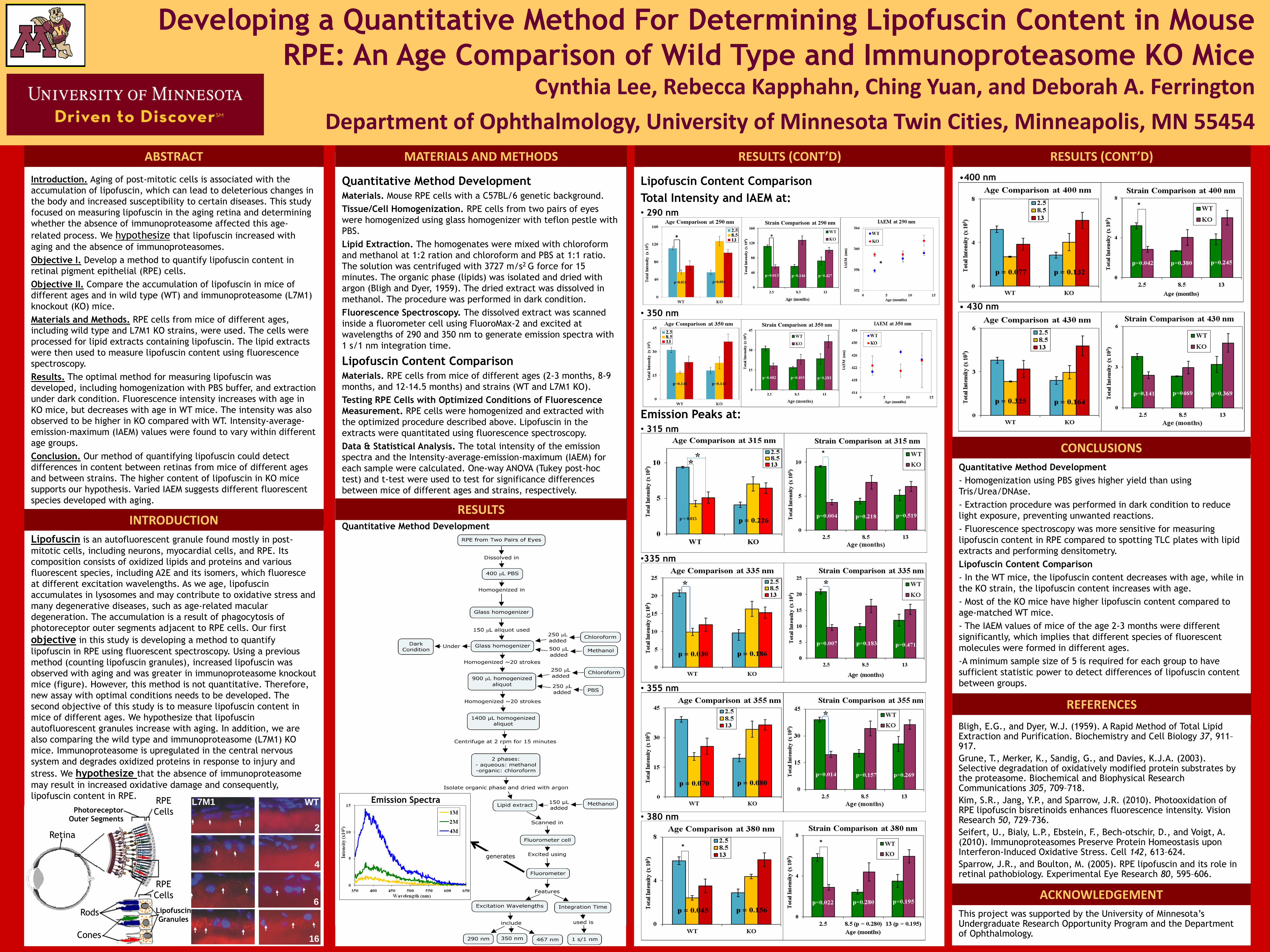
- In the WT mice, the lipofuscin content decreases with age, while in
the KO strain, the lipofuscin content increases with age.
- Most of the KO mice have higher lipofuscin content compared to
age-matched WT mice.
- The IAEM values of mice of the age 2-3 months were different
significantly, which implies that different species of fluorescent
molecules were formed in different ages.
-A minimum sample size of 5 is required for each group to have
sufficient statistic power to detect differences of lipofuscin content
between groups.
Bligh, E.G., and Dyer, W.J. (1959). A Rapid Method of Total Lipid Extraction and Purification. Biochemistry and Cell Biology 37, 911–917.
Grune, T., Merker, K., Sandig, G., and Davies, K.J.A. (2003). Selective degradation of oxidatively modified protein substrates by the proteasome. Biochemical and Biophysical Research Communications 305, 709–718.
Kim, S.R., Jang, Y.P., and Sparrow, J.R. (2010). Photooxidation of RPE lipofuscin bisretinoids enhances fluorescence intensity. Vision Research 50, 729–736.
Seifert, U., Bialy, L.P., Ebstein, F., Bech-otschir, D., and Voigt, A. (2010). Immunoproteasomes Preserve Protein Homeostasis upon Interferon-Induced Oxidative Stress. Cell 142, 613–624.
Sparrow, J.R., and Boulton, M. (2005). RPE lipofuscin and its role in retinal pathobiology. Experimental Eye Research 80, 595–606.
Lipofuscin is an autofluorescent granule found mostly in post-
mitotic cells, including neurons, myocardial cells, and RPE. Its
composition consists of oxidized lipids and proteins and various
fluorescent species, including A2E and its isomers, which fluoresce
at different excitation wavelengths. As we age, lipofuscin
accumulates in lysosomes and may contribute to oxidative stress and
many degenerative diseases, such as age-related macular
degeneration. The accumulation is a result of phagocytosis of
photoreceptor outer segments adjacent to RPE cells. Our first
objective in this study is developing a method to quantify
lipofuscin in RPE using fluorescent spectroscopy. Using a previous
method (counting lipofuscin granules), increased lipofuscin was
observed with aging and was greater in immunoproteasome knockout
mice (figure). However, this method is not quantitative. Therefore,
new assay with optimal conditions needs to be developed. The
second objective of this study is to measure lipofuscin content in
mice of different ages. We hypothesize that lipofuscin
autofluorescent granules increase with aging. In addition, we are
also comparing the wild type and immunoproteasome (L7M1) KO
mice. Immunoproteasome is upregulated in the central nervous
system and degrades oxidized proteins in response to injury and
stress. We hypothesize that the absence of immunoproteasome
may result in increased oxidative damage and consequently,
lipofuscin content in RPE.
Lipofuscin Content Comparison
Total Intensity and IAEM at: • 290 nm
• 350 nm
Emission Peaks at: • 315 nm
•335 nm
• 355 nm
• 380 nm
Cynthia Lee, Rebecca Kapphahn, Ching Yuan, and Deborah A. Ferrington1
Department of Ophthalmology, University of Minnesota Twin Cities, Minneapolis, MN 554541
Quantitative Method Development
L7M1 WT
2mo
3.5mo
6mo
16mo
L7M1 WT
2mo
3.5mo
6mo
16mo
2
WT L7M1
4
6
16
This project was supported by the University of Minnesota’s Undergraduate Research Opportunity Program and the Department of Ophthalmology.
RESULTS (CONT’D)
•400 nm
• 430 nm
INTRODUCTION
Developing a Quantitative Method For Determining Lipofuscin Content in Mouse1
RPE: An Age Comparison of Wild Type and Immunoproteasome KO Mice1
p=0.051 p=0.091
*
*
* *
p = 0.015
*
ACKNOWLEDGEMENT
*
RESULTS (CONT’D)
RESULTS
Retina
RPE
Cells
Rods
Cones
RPE
Cells
Photoreceptor
Outer Segments
Lipofuscin
Granules
CONCLUSIONS
REFERENCES
Emission Spectra
generates