method for detailed and quantitative glycoproteomic
TRANSCRIPT
Subscriber access provided by - Access paid by the | UC Davis Libraries
Journal of Proteome Research is published by the American Chemical Society. 1155Sixteenth Street N.W., Washington, DC 20036Published by American Chemical Society. Copyright © American Chemical Society.However, no copyright claim is made to original U.S. Government works, or worksproduced by employees of any Commonwealth realm Crown government in the courseof their duties.
Article
Glyco-analytical multispecific proteolysis (Glyco-AMP): A simplemethod for detailed and quantitative glycoproteomic characterization
Serenus Hua, Chloe Y Hu, Bum Jin Kim, Sarah M. Totten, Myung Jin Oh, NayoungYun, Charles Chuks Nwosu, Jong Shin Yoo, Carlito B. Lebrilla, and Hyun Joo An
J. Proteome Res., Just Accepted Manuscript • DOI: 10.1021/pr400442y • Publication Date (Web): 09 Sep 2013
Downloaded from http://pubs.acs.org on September 13, 2013
Just Accepted
“Just Accepted” manuscripts have been peer-reviewed and accepted for publication. They are postedonline prior to technical editing, formatting for publication and author proofing. The American ChemicalSociety provides “Just Accepted” as a free service to the research community to expedite thedissemination of scientific material as soon as possible after acceptance. “Just Accepted” manuscriptsappear in full in PDF format accompanied by an HTML abstract. “Just Accepted” manuscripts have beenfully peer reviewed, but should not be considered the official version of record. They are accessible to allreaders and citable by the Digital Object Identifier (DOI®). “Just Accepted” is an optional service offeredto authors. Therefore, the “Just Accepted” Web site may not include all articles that will be publishedin the journal. After a manuscript is technically edited and formatted, it will be removed from the “JustAccepted” Web site and published as an ASAP article. Note that technical editing may introduce minorchanges to the manuscript text and/or graphics which could affect content, and all legal disclaimersand ethical guidelines that apply to the journal pertain. ACS cannot be held responsible for errorsor consequences arising from the use of information contained in these “Just Accepted” manuscripts.
Glyco-analytical multispecific proteolysis (Glyco-AMP): A
simple method for detailed and quantitative glycoproteomic
characterization
Serenus Hua,1,2 Chloe Y. Hu,3 Bum Jin Kim,1 Sarah M. Totten,3 Myung Jin Oh,1
Nayoung Yun,1 Charles C. Nwosu,3 Jong Shin Yoo,1,4 Carlito B. Lebrilla,3*† and
Hyun Joo An1,2*†
1. Graduate School of Analytical Science and Technology, Chungnam National University,
Daejeon, Korea
2. Cancer Research Institute, Chungnam National University, Daejeon, Korea
3. Department of Chemistry, University of California, Davis, CA
4. Division of Mass Spectrometry Research, Korea Basic Science Institute, Ochang, Korea
* To whom correspondence should be addressed:
Hyun Joo An, email: [email protected], Tel: 82 42-821-8547, Fax: 82 42-821-8551
Carlito B. Lebrilla, email: [email protected], Tel: 1 530-752-0504, Fax: 1 530-752-8995
† Hyun Joo An and Carlito B. Lebrilla contributed equally to this paper as co-corresponding
authors.
Page 1 of 41
ACS Paragon Plus Environment
Journal of Proteome Research
123456789101112131415161718192021222324252627282930313233343536373839404142434445464748495051525354555657585960
KEYWORDS: multispecific proteases, non-specific proteases, site-specific glycosylation,
glycoproteomics, biopharmaceutical glycoproteins, glycan isomers
ABBREVIATIONS: Glyco-AMP, glyco-analytical multispecific proteolysis; Hex, hexose;
HexNAc, N-acetylhexosamine; Man, mannose; GlcNAc, N-acetylglucosamine; Fuc, fucose; G1F,
Hex4HexNAc4Fuc; NeuAc, N-acetylneuraminic acid; OAc, O-acetylation
Page 2 of 41
ACS Paragon Plus Environment
Journal of Proteome Research
123456789101112131415161718192021222324252627282930313233343536373839404142434445464748495051525354555657585960
ABSTRACT
Despite recent advances, site-specific profiling of protein glycosylation remains a significant
analytical challenge for conventional proteomic methodology. To alleviate the issue, we propose
glyco-analytical multispecific proteolysis (Glyco-AMP) as a strategy for glycoproteomic
characterization. Glyco-AMP consists of rapid, in-solution digestion of an analyte glycoprotein
(or glycoprotein mixture) by a multispecific protease (or protease cocktail). Resulting
glycopeptides are chromatographically separated by isomer-specific porous graphitized carbon
nano-LC, quantified by high-resolution MS, and structurally elucidated by MS/MS. To
demonstrate the consistency and customizability of Glyco-AMP methodology, the glyco-
analytical performances of multispecific proteases subtilisin, pronase, and proteinase K were
characterized in terms of quantitative accuracy, sensitivity, and digestion kinetics. Glyco-AMP
was shown be effective on glycoprotein mixtures as well as glycoproteins with multiple
glycosylation sites, providing detailed, quantitative, site- and structure-specific information about
protein glycosylation.
Page 3 of 41
ACS Paragon Plus Environment
Journal of Proteome Research
123456789101112131415161718192021222324252627282930313233343536373839404142434445464748495051525354555657585960
INTRODUCTION
Proteins are commonly decorated with long carbohydrate chains, known as glycans, during
normal biosynthesis. Over 60% of the known proteome is believed to be glycosylated.1 As the
importance of glycosylation towards protein structure and function becomes ever more apparent,
glycomic methods for profiling the detached glycan component of a glycoprotein have
undergone extensive innovation and development.2-4 However, glycoproteomic methods that
characterize both glycosylation site and glycan structure are still in their infancy. Until now, the
most commonly-used methods have been adapted from traditional proteomics5-7 and often do not
sufficiently address the complexities of characterizing a post-translational modification as
intricate as glycosylation.
Whereas peptides can be identified and characterized simply by their linear sequence,
glycans are branched, with different linkage possibilities between each monosaccharide residue.
When peptide and glycan moieties both exist on a single molecule, as with glycopeptides,
peptide sequence isomers and glycosylation site isomers further increase the already significant
structural complexity.8 The abundance of isomeric glycopeptides encountered during
glycoproteomic analysis is problematic for mass spectrometry, which cannot distinguish between
isomers without the assistance of additional analytical technologies. Consequently,
glycoproteomic methods incorporating isomer-specific chromatographic separation and/or
tandem MS are crucial for effective glycoprotein characterization.9
When analyzing peptides with post-translational modifications such as glycosylation, peptide
length must be considered. Large glycopeptides are not only harder to detect by mass
spectrometry (due to decreased ionization efficiency as well as instrumental mass limitations) but
Page 4 of 41
ACS Paragon Plus Environment
Journal of Proteome Research
123456789101112131415161718192021222324252627282930313233343536373839404142434445464748495051525354555657585960
can also incorporate multiple glycosylation sites, severely complicating or obfuscating site-
specific analysis.10-12 Proteases with high substrate specificity, such as trypsin, are especially
prone to creating these large glycopeptides, since their specificity severely limits potential
cleavage sites.13 This is exacerbated by the well-known phenomenon of glycoprotein trypsin
resistance, wherein large glycosyl modifications sterically hinder protease access to a
glycoprotein cleavage site, resulting in a missed cleavage.14, 15
While the amino acid sequences of some glycoproteins may be fortuitously rich in tryptic
cleavage sites, many are not so easily dissected.16, 17 The glycoproteomics community is
therefore in great need of alternative proteases and proteolytic digestion strategies that can be
broadly applied to enhance and/or replace conventional trypsin-based methodology. One such
strategy is multispecific (or non-specific) proteolysis, which utilizes proteases or protease
cocktails with multiple substrate specificities to hydrolyze peptide bonds at a number of different
sites on a glycoprotein. The resulting digest contains glycopeptides of a roughly consistent size,
irrespective of the glycoprotein's amino acid sequence or steric hindrance by the glycan moiety.18,
19
Early forays into glycoprotein characterization via multispecific proteolysis involved MS-
only analysis of single glycoprotein digests.20 Sensitivity and specificity were relatively low, due
to interference from ion-suppressing unglycosylated peptides. Later iterations of the strategy
used various chemical methods to immobilize the multispecific proteases on solid supports,
removing a significant amount of the peptide interference originating from protease autolysis.21,
22 However, the most dramatic improvements in glycoproteomics have come just recently, with
the incorporation of nano-LC separation into established mass spectrometric methods.8, 9, 23 Use
of nano-LC/MS not only boosts sensitivity by vastly reducing interference from ion-suppressing
Page 5 of 41
ACS Paragon Plus Environment
Journal of Proteome Research
123456789101112131415161718192021222324252627282930313233343536373839404142434445464748495051525354555657585960
peptides, but also enables differentiation of isobaric or even isomeric molecules. Additionally,
condensation of the in-spectrum dynamic range enables increased use of MS/MS to supplement
accurate mass MS, adding an extra layer of certainty to glycopeptide identifications.
In this study, isomer-sensitive porous graphitized carbon nano-LC/MS and nano-LC/MS/MS
are used to interrogate glycopeptide mixtures digested in solution by various multispecific
proteases. Because the glycoprotein digestions are carried out in solution, rather than by bead-
immobilized proteases, sample preparation is vastly simplified while experimental variation is
minimized. Additionally, the in-solution protease digestions inherently proceed faster than bead-
immobilized protease digestions due to more favorable kinetics.24, 25 Though in-solution protease
digests do contain higher levels of signal-suppressing peptides than immobilized protease digests,
nano-LC successfully separates the peptide contaminants from the glycopeptide analytes— in
essence, solving the problem with analytical technology rather than chemical derivatization.
Whereas previous studies have concentrated on the analytical performance of individual
multispecific proteases, commonly pronase, we demonstrate the general applicability of glyco-
analytical multispecific proteolysis (Glyco-AMP) by performing detailed and quantitative
characterization of site-specific protein glycosylation using a variety of multispecific proteases.
The accuracy and sensitivity of the nano-LC/MS profiling methods are evaluated. Basic
digestion kinetics and activity of multispecific proteases subtilisin, pronase, and proteinase K are
tracked and compared. Site-specific glycoform characterization is demonstrated to be effective
on multiply glycosylated glycoproteins as well as glycoprotein mixtures. Isomer separation by
porous graphitized carbon nano-LC, combined with MS/MS structural elucidation, reveals fine
details about the structure and originating site of the glycopeptide.
Page 6 of 41
ACS Paragon Plus Environment
Journal of Proteome Research
123456789101112131415161718192021222324252627282930313233343536373839404142434445464748495051525354555657585960
METHODS
Materials and reagents
All multispecific proteases (porcine elastase, papain, porcine pepsin, pronase, proteinase K,
subtilisin from Bacillus licheniformis, and thermolysin) were obtained from Sigma-Aldrich (St
Louis, MO), as were glycoproteins bovine ribonuclease B and human plasma vitronectin.
Prostate-specific antigen was obtained from Lee BioSolutions (St Louis, MO). Infliximab was
obtained from Janssen Biotech (Horsham, PA). Darbepoetin alfa was obtained from Amgen
(Thousand Oaks, CA). Graphitized carbon cartridges were obtained from Grace Davison
(Deerfield, IL). Solvents were of LC-MS grade. All other materials and reagents were of
analytical grade or higher.
Digestion of glycoproteins by multispecific proteolysis
In general, 50 µg of glycoprotein and 50 µg of protease were dissolved in 100 µL of 100 mM
phosphate buffer (pH 7.5) and incubated at 37 ºC for 20 minutes. Exceptions occurred during: a)
initial protease screening, for which the optimal digestion buffer and temperature for each
protease was specified by the manufacturer's product notes; b) kinetics studies, for which
multiple digestion timepoints were taken; and c) sensitivity studies, for which the amount of
glycoprotein was varied.
Glycopeptide enrichment with graphitized carbon SPE
Digested glycopeptides were enriched by graphitized carbon solid-phase extraction according to
previously optimized procedures.26 Briefly, an automated liquid handler (Gilson, Middleton, WI)
Page 7 of 41
ACS Paragon Plus Environment
Journal of Proteome Research
123456789101112131415161718192021222324252627282930313233343536373839404142434445464748495051525354555657585960
conditioned graphitized carbon cartridges with water; loaded aqueous glycopeptide solutions;
washed with water; then eluted glycopeptides with 40% acetonitrile and 0.05% trifluoroacetic
acid (v/v) in water. Samples were dried in vacuo.
Chromatographic separation and MS analysis
Samples were reconstituted in water and analyzed using an HPLC-Chip Quadrupole Time-of-
Flight (Chip/Q-TOF) MS system (Agilent Technologies, Santa Clara, CA) comprising an
autosampler (maintained at 6 oC), capillary pump, nano pump, HPLC-Chip/MS interface, and a
6520 Q-TOF MS detector. The chip used consisted of a 9 × 0.075 mm i.d. enrichment column
and a 43 × 0.075 mm i.d. analytical column, both packed with 5 μm porous graphitized carbon as
the stationary phase, with an integrated nano-ESI spray tip. Chromatographic separation was
performed according to previously optimized procedures 2. Briefly, following sample loading, a
rapid glycopeptide elution gradient was delivered at 0.4 μL min-1 using solutions of (A) 3.0%
acetonitrile and 0.1% formic acid (v/v) in water, and (B) 90.0% acetonitrile and 0.5% formic
acid (v/v) in water, at the following proportions and time points: 5% to 32.8% B, 0 min to 13.3
min; and 32.8% to 35.9% B, 13.3 min to 16.5 min. The columns were then flushed with 100% B,
and the analytical column was re-equilibrated with 5% B while the enrichment column was re-
equilibrated with 0% B. The drying gas temperature was set at 325 °C with a flow rate of 4 L
min-1 (2 L of filtered nitrogen gas and 2 L of filtered dry compressed air).
MS spectra were acquired in positive ionization mode over a mass range of m/z 500-2000
with an acquisition time of 1.5 seconds per spectrum. MS/MS spectra were acquired in positive
ionization mode over a mass range of m/z 100-3000 with an acquisition time of 1.5 seconds per
spectrum. Following an MS scan, precursor compounds were automatically selected for MS/MS
Page 8 of 41
ACS Paragon Plus Environment
Journal of Proteome Research
123456789101112131415161718192021222324252627282930313233343536373839404142434445464748495051525354555657585960
analysis by the acquisition software based on ion abundance and charge state (z = 2, 3, or 4) and
isolated in the quadrupole with a mass bandpass FWHM (full width at half maximum) of 1.3 m/z.
In general, collision energies for CID fragmentation were calculated for each precursor
compound based on the following formula:
collision
/3.6 V 4.8 V
100 Da
m zV
Here, Vcollision is the potential difference across the collision cell. The slope and offset values of
the energy-m/z ramp could be changed as needed to produce more or less fragmentation.8
LC/MS data processing and glycopeptide identification
After data acquisition, raw LC/MS data was processed using the Molecular Feature Extractor
algorithm included in the MassHunter Qualitative Analysis software (version B.04.00 SP2,
Agilent Technologies). MS peaks were filtered with a signal-to-noise ratio of 5.0 and parsed into
individual ion species. Using expected isotopic distribution, charge state information, and
retention time, all ion species associated with a single compound (e.g. the doubly protonated ion,
the triply protonated ion, and all associated isotopologues) were summed together, and the
neutral mass of the compound was calculated. Using this information, a list of all compound
peaks in the sample was generated, with abundances represented by chromatographic peak areas.
Glycopeptides were identified using an in-house software tool based on the GlycoX
algorithm.27 Taking into account the mass of a potential glycopeptide, the amino acid
sequence(s) of the protein or proteins it could be derived from, the type of glycosylation (N or O),
and a given mass tolerance (here, 5 ppm), the algorithm generated a list of potential amino acid
and glycan compositions associated with a given compound. Results were further filtered based
on known glycosylation patterns and/or other biological rules.28
Page 9 of 41
ACS Paragon Plus Environment
Journal of Proteome Research
123456789101112131415161718192021222324252627282930313233343536373839404142434445464748495051525354555657585960
RESULTS AND DISCUSSION
Screening for multispecific proteases with high glycopeptide-generating activity
A number of common, commercially-available proteases were initially screened for their ability
to digest glycoproteins into (detectable) glycopeptides. Elastase, papain, pepsin, pronase,
proteinase K, subtilisin, and thermolysin were each evaluated by combining 50 µg of protease
with 50 µg of well-characterized glycoprotein ribonuclease B (at a total digestion volume of 100
µL). Digestion buffers and conditions were chosen based on the manufacturers' suggested
protocols29-35— for example, the thermolysin digestion took place at 75 ºC in 50 mM Tris and
0.5 mM calcium chloride, while the pepsin digestion took place at 37 ºC in 10 mM hydrochloric
acid. Digestions were carried out for 20 minutes each, after which the digests were cleaned by
graphitized carbon solid-phase extraction and analyzed by nano-LC/MS/MS.
The glycopeptide content of each digest was evaluated by screening MS/MS spectra for
glycopeptide-associated fragment ions with m/z 163.06 (Hex+H)+, m/z 204.09 (HexNAc+H)+,
and m/z 366.14 (HexNAc1Hex1+H)+. Spectra with significant levels (over 50% base peak
abundance) of these fragment ions were assumed to be of glycopeptides. The number and ion
intensity of glycopeptide spectra were taken as indicative of the number of glycopeptides.
Based on these preliminary evaluations, the subtilisin, pronase, and proteinase K digests were
found to contain the highest abundances of glycopeptides. Subtilisin and proteinase K are serine
proteases (of bacterial and fungal origin, respectively) that are evolutionarily unrelated yet show
a high degree of structural homology, particularly at the catalytic triad.36 Pronase is an enzyme
Page 10 of 41
ACS Paragon Plus Environment
Journal of Proteome Research
123456789101112131415161718192021222324252627282930313233343536373839404142434445464748495051525354555657585960
cocktail of bacterial origin containing mostly serine proteases but also some additional endo- and
exoproteases. None of these proteases are themselves glycosylated. All are stable over a wide pH
and temperature range, with optimal activity in slightly alkaline conditions at around 40 ºC.
Sensitive and quantitative glycoform profiling through multispecific proteolysis
To evaluate the quantitative accuracy of Glyco-AMP, subtilisin, pronase, and proteinase K were
each applied to analyze bovine ribonuclease B (RNase B). RNase B is a well-characterized
glycoprotein with a known glycosylation profile, thus providing a convenient means of
validation. The relative abundances of each RNase B glycoform were compared after parallel 20
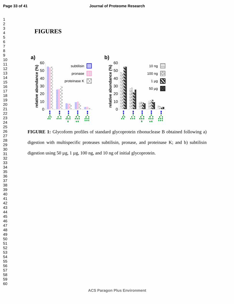
minute digestions by subtilisin, pronase, and proteinase K. Overall profiles in Figure 1a are
obtained by summing up the abundances of observed glycopeptides associated with each
glycoform. To minimize differences in glycopeptide ionization efficiencies, the peptide moieties
used for quantitation are kept constant across glycoforms.8 High mannose type glycan
Man5GlcNAc2 (55%) is the most abundant glycan attached to RNase B, followed by
Man6GlcNAc2 (27%), Man8GlcNAc2 (9%), Man7GlcNAc2 (7%), and Man9GlcNAc2 (3%); these
results correlate highly with previous studies quantifying the glycoforms of RNase B.37-39 All
protease digests result in similar profiles, with the relative abundances of individual glycoforms
varying by less than 2% between proteases. For subtilisin, major peptide moieties (exceeding
10% abundance) consist of NLTK (66%) and NLT (13%), while the remaining 21% consists of
minor peptide moieties SRNL, KSRNL, NL, and SRNLT (in decreasing order of abundance).
For pronase, major peptide moieties consist of SRN (52%) and RN (21%), while the remaining
27% consists of minor peptide moieties NLT, NLTK, NLTKDR, N, and NL. For proteinase K,
Page 11 of 41
ACS Paragon Plus Environment
Journal of Proteome Research
123456789101112131415161718192021222324252627282930313233343536373839404142434445464748495051525354555657585960
major peptide moieties consist of NLTK (40%), SRNLT (27%), NLT (14%), and SRNL (13%),
while the remaining 6% consists of minor peptide moieties SRNLTKD and NL. Here, as earlier
with infliximab, the subtilisin appears to exhibit the highest cleavage specificity (resulting in a
simpler peptide mixture), while the pronase and proteinase K both exhibit much lower cleavage
specificity (resulting in a more complex peptide mixture).
The sensitivity of Glyco-AMP was also tested by digesting different amounts of RNase B
with subtilisin. The relative abundances of each RNase B glycoform were compared after
parallel 20 minute digestions from initial glycoprotein amounts of 10 ng, 100 ng, 1 µg, and 50 µg.
All initial glycoprotein amounts result in similar profiles, summarized in Figure 1b, with the
relative abundances of individual glycoforms varying by less than 5% between digests. As in
Figure 1a, these results correlate highly with previous studies quantifying the glycoforms of
RNase B.37-39
Since RNase B has a molecular weight of approximately 15 kDa, 10 ng of RNase B
corresponds to 670 femtomoles. Additionally, not all of the sample was injected into the LC/MS
system— because the autosampler needle could not physically reach the bottom of the sample
vial, only 8 µL (out of 25 µL) of glycopeptide solution were drawn from the vial for analysis.
Despite all of this, RNase B glycoform Man9GlcNAc2 was still detected at 5% relative
abundance, corresponding to the detection (from within a mixture) of just 10 femtomoles of the
glycoform.
Characterization of subtilisin, pronase, and proteinase K enzyme activity
Page 12 of 41
ACS Paragon Plus Environment
Journal of Proteome Research
123456789101112131415161718192021222324252627282930313233343536373839404142434445464748495051525354555657585960
To better understand the glyco-analytical performance of subtilisin, pronase, and proteinase K,
in-depth kinetic studies were performed using infliximab (Remicade®) as a test substrate.
Infliximab is a chimeric monoclonal antibody (mAb) made up of a mouse-derived antigen-
binding (Fab) region and a human IgG1-derived constant (Fc) region. Glycosylation is present
exclusively on the IgG1-derived constant region, and closely imitates (but exhibits some
significant differences from) the glycosylation pattern of IgG. Multispecific proteolysis of
infliximab is kinetically similar to multispecific proteolysis of pure IgG1 (without interference
from IgG isotypes 2-4) or, more relevantly, other mAbs with an IgG1-derived constant region.
Thus, the conclusions derived from these experiments may be applied directly to future
glycoproteomic analyses of IgG1-derived mAbs such as adalimumab (Humira®), bevacizumab
(Avastin®), or rituximab (Rituxan®), all of which possess perfectly conserved IgG1-like amino
acid sequences around the N-glycosylation site. These experiments also provide a rough
indication of how each protease will perform when applied to glycoproteomic analyses of more
complex glycoproteins with multiple glycosylation sites.
To evaluate and characterize each protease, the abundances of the generated glycopeptides
were tracked across six time points: 20 min, 60 min, 2 h, 4 h, 8 h, and 18 h. These six time points
were tested in each of the three selected proteases: pronase, proteinase K, and subtilisin. All 18
digests were conducted, processed, and analyzed in parallel. Glycopeptides were identified by
nano-LC/MS/MS and quantified by nano-LC/MS.
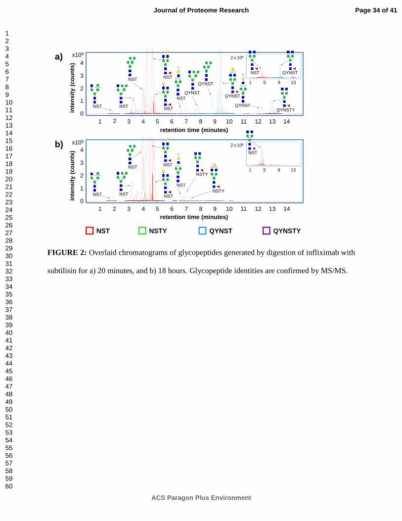
Figure 2 shows the overlaid chromatograms of glycopeptides separated by nano-LC (and
identified by accurate mass MS and MS/MS). Figure 2a shows the glycopeptides generated by
20 minutes of digestion by subtilisin, whereas Figure 2b shows the glycopeptides generated by
18 hours of digestion by subtilisin. These results demonstrate one of the unique advantages of
Page 13 of 41
ACS Paragon Plus Environment
Journal of Proteome Research
123456789101112131415161718192021222324252627282930313233343536373839404142434445464748495051525354555657585960

multispecific proteolysis; that is, the ability to modulate digestion time (as well as enzyme
concentration) in order to customize the size or length of the peptide moiety on each
glycopeptide.21, 22 The 20 minute digestion (Figure 2a) results predominantly in longer
glycopeptides with peptide moiety sequences of QYNST (the major product) and QYNSTY (a
minor product), identifying the originating protein as IgG1-like rather than IgG2-like. Shorter
glycopeptides with peptide moiety sequences of NST and NSTY are also present, supporting this
identification. In contrast, the 18 hour digestion (Figure 2b) results almost entirely in short
glycopeptides possessing the NST peptide moiety, with an extremely minor contribution from
glycopeptides with a peptide moiety sequence of NSTY.
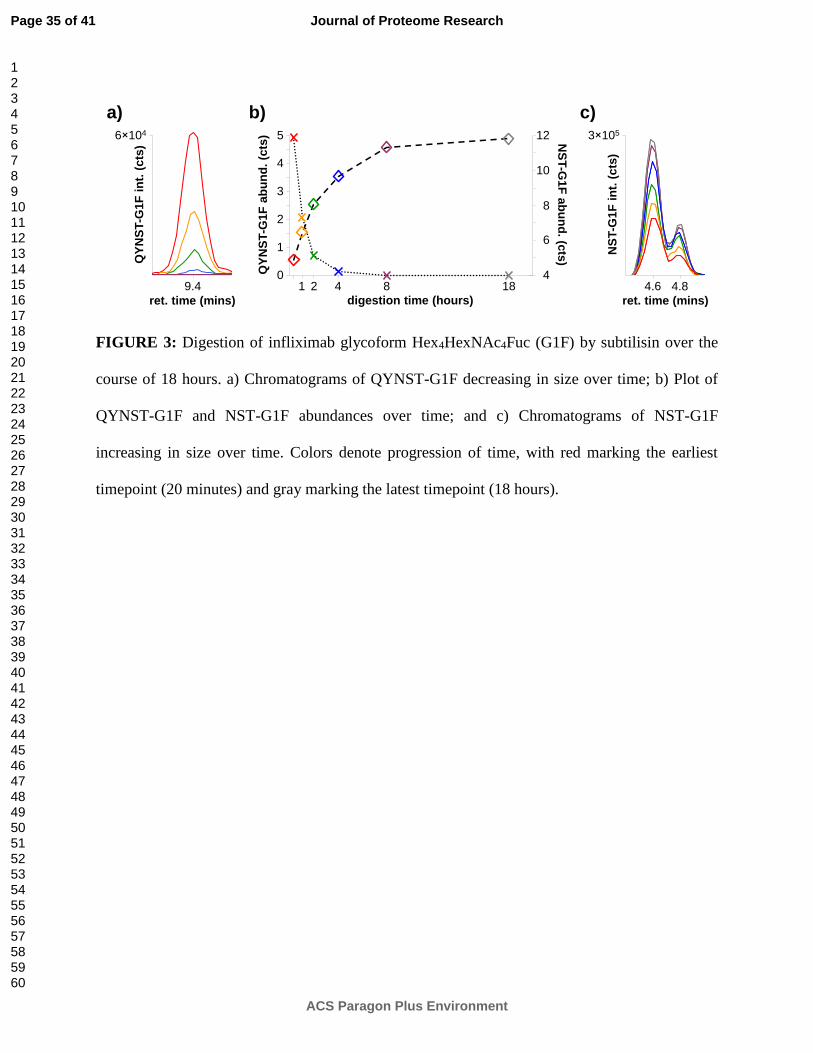
Figure 3 provides a preliminary glimpse of the kinetics of a subtilisin digestion, showing the
abundance of glycopeptide QYNST-Hex4HexNAc4Fuc (i.e. QYNST-G1F) decreasing over time
while the abundance of glycopeptide NST-Hex4HexNAc4Fuc (i.e. NST-G1F) increases over time.
Figure 3b plots the absolute abundances of these two glycopeptides at six different points during
the 18-hour digestion, while Figures 3a and 3c show their chromatograms at these points.
The chromatograms of glycopeptide NST-G1F (Figure 3c) highlight the ability of porous
graphitized carbon to separate glycopeptide isomers (provided that they are sufficiently small).
Here, the only difference between the two glycopeptide isomers is their G1F glycan moiety.
Previous glycan structural studies using the same isomer-sensitive porous graphitized carbon
stationary phase indicate that on the earlier-eluting G1F isomer, the antennal galactose is
attached to the alpha-1,6-linked branch of the glycan, whereas on the later-eluting G1F isomer,
the antennal galactose is attached to the alpha-1,3-linked branch.40 Based on these results, the
predominant G1F isomer present on infliximab is the one in which the antennal galactose is
attached to the alpha-1,6-linked branch.
Page 14 of 41
ACS Paragon Plus Environment
Journal of Proteome Research
123456789101112131415161718192021222324252627282930313233343536373839404142434445464748495051525354555657585960
In contrast to glycopeptide NST-G1F, glycopeptide QYNST-G1F (Figure 3a) appears to
have too long of a peptide moiety for effective isomer separation by porous graphitized carbon
nano-LC. However, as mentioned earlier, the presence of a longer peptide moiety (such as
QYNST) is still desirable for certain applications because it increases the specificity of the
glycosylation site assignment— in this case, by differentiating between IgG isotypes. Detection
of glycopeptides with QYNST peptide moieties differentiates glycoforms of infliximab (which is
an IgG1 homolog) from glycoforms of IgG2 homologs such as panitumumab (Vectibix®) or
denosumab (Prolia®/Xgeva®). In instances of multiply-glycosylated proteins or protein
mixtures where multiple sites of glycosylation exist, precise control of peptide moiety length
enables researchers to select digestion times that would create glycopeptide markers with high
specificity for a particular site of glycosylation. These markers could then be used to quantify
selected glycoforms of a specific glycoprotein, with implications for biosimilar batch analysis or
biomarker research.
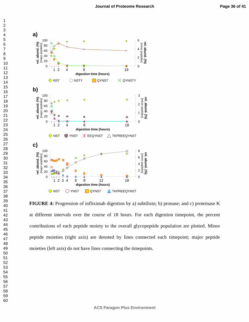
Figure 4 tracks the progression over time of infliximab digestion by subtilisin (Figure 4a),
pronase (Figure 4b), and proteinase K (Figure 4c). For each digestion timepoint, the percent
contributions of each peptide moiety to the overall glycopeptide population are plotted. These
charts may be used to estimate the rate of glycopeptide production at a given time point and can
help optimize a digestion for generation of a target glycopeptide.
In subtilisin (Figure 4a), as discussed earlier, the major peptide moieties are initially
QYNST and NST, with minor contributions from QYNSTY and NSTY. As the digestion
progresses, abundances of QYNST and QYNSTY drop drastically, eventually becoming
undetectable, while abundances of NST rise correspondingly. However, the contribution from
NSTY is more interesting. For the first two hours of the digestion, abundances of NSTY rise,
Page 15 of 41
ACS Paragon Plus Environment
Journal of Proteome Research
123456789101112131415161718192021222324252627282930313233343536373839404142434445464748495051525354555657585960
very nearly imitating the reaction plot of NST (proportionately, at much lower levels). However,
after the first two hours, abundances of NSTY drop, more closely imitating the reaction plots of
QYNST and QYNSTY. Presumably, the initial rise is fed largely by digestion of QYNSTY
peptide moieties (or even larger precursors) to form NSTY. Simultaneously, NSTY peptide
moieties are being digested to form NST. Approximately two hours later, the rate of NSTY
formation slows (due to lack of substrate) and is overtaken by the rate of NSTY digestion,
resulting in the observed decreases in abundance of NSTY peptide moieties. By the 18 hour
digestion timepoint, 96% of all infliximab glycopeptides have NST peptide moieties, while the
remaining 4% have NSTY peptide moieties. During the observed course of the reaction, total
detected glycopeptide abundances (in ion counts) increase only slightly, by just 4%, indicating
that, after the 20 minute timepoint, subtilisin is mainly acting on already-digested short
glycopeptides, and very little undigested infliximab remains.
In pronase (Figure 4b), the major peptide moieties are initially NST and YNST, with minor
contributions from TKPREEQYNST and EEQYNST. As the digestion progresses, abundances
of TKPREEQYNST and EEQYNST drop drastically; abundances of YNST decrease at a slower
rate; and abundances of NST increase correspondingly. The slower rate of decrease for YNST
peptide moieties may be due in part to digestion of TKPREEQYNST and EEQYNST to form
new YNST; however, abundances of YNST appears to decrease quite slowly even after
abundances of TKPREEQYNST and EEQYNST peptide moieties have dropped below
detectable levels. A more likely explanation is that, due to enzyme autolysis, the pronase is
losing activity as the digestion progresses. This would explain the lack of significant decreases in
YNST abundances (as well as the lack of corresponding increases in NST abundances) after the
four hour digestion timepoint. Even after 18 hours of digestion by pronase, only 84% of all
Page 16 of 41
ACS Paragon Plus Environment
Journal of Proteome Research
123456789101112131415161718192021222324252627282930313233343536373839404142434445464748495051525354555657585960

infliximab glycopeptides have NST peptide moieties, while the remaining 16% have YNST
peptide moieties. During the observed course of the reaction, total detected glycopeptide
abundances (in ion counts) increase only slightly, by just 8%, indicating that, after the 20 minute
timepoint, pronase is mainly acting on already-digested short glycopeptides, and very little
undigested infliximab remains.
In proteinase K (Figure 4c), the major peptide moieties are initially QYNST,
TKPREEQYNST, and NST, with a minor contribution from YNST. As the digestion progresses,
abundances of QYNST and TKPREEQYNST generally decrease, while abundances of NST and
YNST generally increase. Due to the curious behavior of the digestion during the initial two
hours, additional timepoints have been added to the analysis. In all, timepoints are shown for 20
min, 40 min, 60 min, 90 min, 2 h, 3 h, 4 h, 6 h, 8 h, 12 h, and 18 h of digestion.
In contrast to the subtilisin and pronase digestions, total detected glycopeptide abundances
(in ion counts) increase dramatically, by 150%, during the initial two hours of the proteinase K
digestion, indicating that proteinase K is still digesting either whole infliximab or large pieces
thereof up until this timepoint. After two hours, there are no further increases in total detected
glycopeptide abundance, indicating that from this timepoint on, proteinase K is mainly acting on
already-digested short glycopeptides, and that very little undigested infliximab remains.
The initial rise in abundance of QYNST and TKPREEQYNST peptide moieties is most
likely related to the continuing digestion of whole infliximab during this period. After two hours,
all of the whole infliximab has been digested into (glyco)peptides of various sizes, and
abundances of QYNST and TKPREEQYNST begin to drop. Correspondingly, the rate of
generation of NST and YNST peptide moieties increases. After 18 hours of digestion by
proteinase K, the rate of change has slowed, but not stopped. The dominant peptide moiety at
Page 17 of 41
ACS Paragon Plus Environment
Journal of Proteome Research
123456789101112131415161718192021222324252627282930313233343536373839404142434445464748495051525354555657585960
this time is NST, comprising 89% of all infliximab glycopeptides; however, detectable
abundances of YNST (8%) and QYNST (3%) peptide moieties are still present.
When selecting the appropriate multispecific protease for an application, sensitivity may also
be a consideration. Total detected glycopeptide abundances (in ion counts) were used to evaluate
the yield, and thus sensitivity, afforded by each protease digest. For subtilisin, the total detected
glycopeptide abundance was 4.2 × 10⁷ counts at 20 minutes, increasing to 4.4 × 10⁷ counts by
the end of the digestion. For pronase, the total detected glycopeptide abundance was 7.7 × 10⁷
counts at 20 minutes, increasing to 8.4 × 10⁷ counts by the end of the digestion. For proteinase K,
the total detected glycopeptide abundance was 1.0 × 10⁷ counts at 20 minutes, increasing rapidly
to 2.5 × 10⁷ counts after two hours and remaining steady until the end of the digestion.
When determining the optimal digestion conditions for a particular application, multiple
factors come into play. Specificity of the peptide moiety for a single site of glycosylation;
digestion yield (i.e. sensitivity); and digestion purity (i.e. the degree of signal splitting) should all
be considered. For example, at the 18 hour digestion timepoints in Figure 4, all three proteases
offer their own unique advantages. Proteinase K (Figure 4c) offers the highest degree of
glycosite specificity, since the digest contains glycopeptides with three overlapping peptide
moieties, two of which cross-confirm that the originating IgG-like protein is homologous to the
IgG1 isotype, rather than the IgG2 isotype. Pronase (Figure 4b) offers the highest glycopeptide
yield, increasing the sensitivity of the analysis and decreasing the amount of analyte glycoprotein
necessary. Subtilisin (Figure 4a) offers the highest glycopeptide purity, with almost all of the
glycopeptides condensed to a single peptide moiety, geometrically reducing the complexity of
the digest. Depending on the specific application, researchers have an unlimited menu of options
with which to customize their Glyco-AMP digestion conditions for optimal results.
Page 18 of 41
ACS Paragon Plus Environment
Journal of Proteome Research
123456789101112131415161718192021222324252627282930313233343536373839404142434445464748495051525354555657585960
Site-specific profiling with protein mixtures and multiple glycosylation sites
The main purpose of using multispecific proteolysis to digest glycoproteins is to obtain site-
specific information about their glycosylation, particularly when there is more than one
glycosylation site involved. This can occur when more than one glycoprotein is present in a
mixture (e.g. as a result of co-precipitation or co-elution); or, when a glycoprotein has multiple
glycosylation sites. In-solution multispecific proteolysis is demonstrated to be effective in both
of these scenarios.
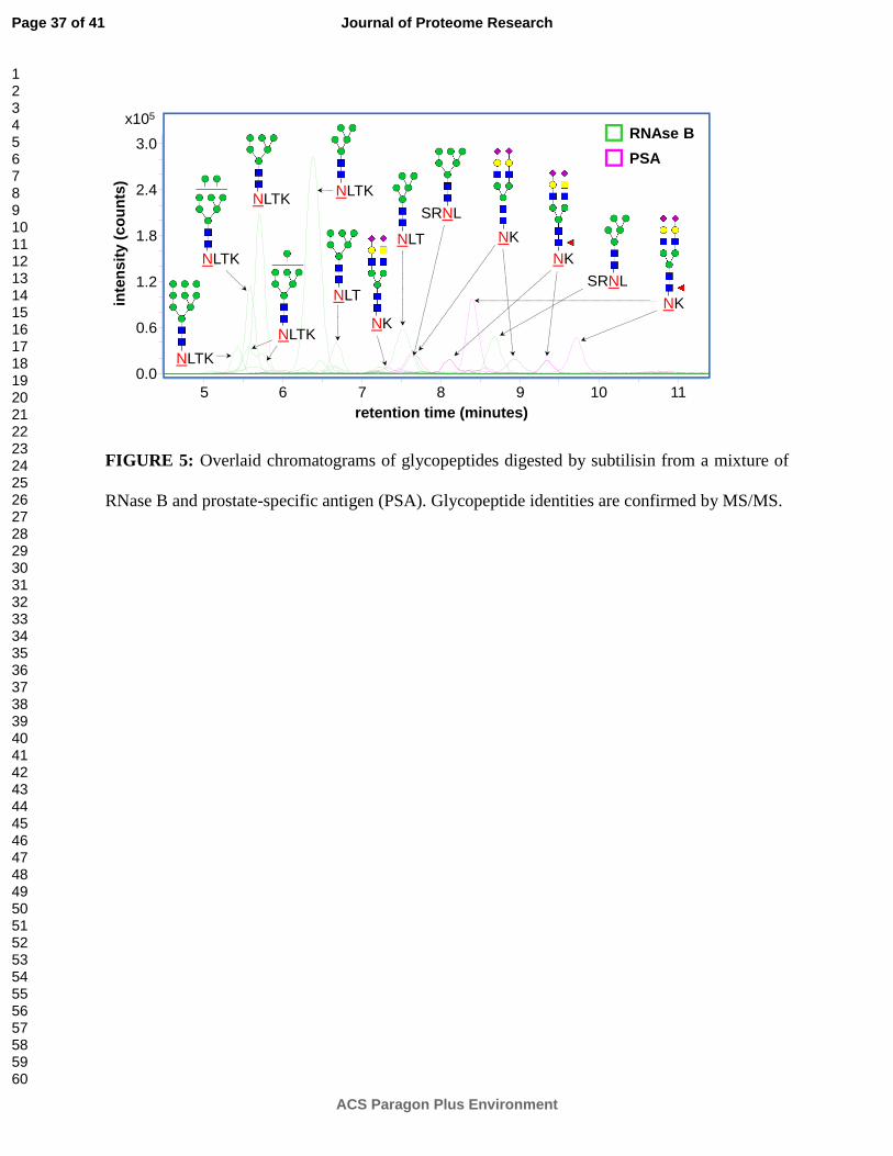
Figure 5 shows the chromatograms of glycopeptides digested by subtilisin from a mixture of
RNase B and prostate-specific antigen (PSA). PSA is a commonly-used clinical biomarker for
prostate cancer;41, 42 however, in recent years, its specificity has been called into question.43, 44
Closer examination of its glycosylation may provide a means of increasing the specificity of PSA
as a biomarker. Unfortunately, since PSA is present at relatively low levels in serum, isolation by
either immunoprecipitation or chromatography may well result in contamination by co-
precipitating or co-eluting proteins, necessitating site-specific analysis to confirm the origin of
glycosylation. Here, site-specific peptide moieties unambiguously identify each glycopeptide as
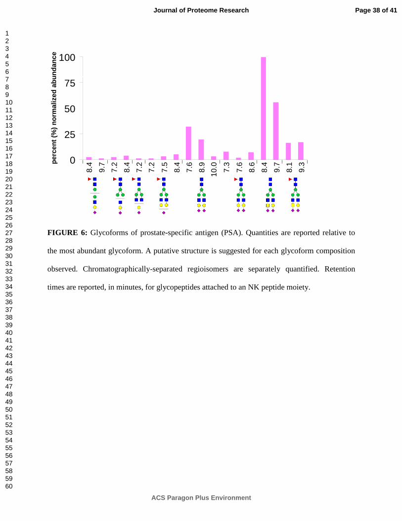
originating from either RNase B or PSA. PSA glycoforms detected during this analysis (Figure
6) support and expand upon previous studies of PSA glycosylation.45, 46 For example, multiple
regioisomeric glycoforms of PSA are baseline-resolved by porous graphitized carbon nano-LC,
providing structural specificity to the analysis. These regioisomers most likely arise from
differential linkages of the terminal sialic acids (which can be either alpha-2,3- or alpha-2,6-
Page 19 of 41
ACS Paragon Plus Environment
Journal of Proteome Research
123456789101112131415161718192021222324252627282930313233343536373839404142434445464748495051525354555657585960
linked to the preceding galactoses) and may be confirmed in future experiments by linkage-
specific glycosidase digestion.40
Pronase digestion of vitronectin, a glycoprotein with three sites of glycosylation, reveals the
detailed glycosylation profile shown in Figure 7. Recent research has indicated that vitronectin,
an abundant serum glycoprotein, may be involved in the early stages of cancer.47, 48 However,
few studies have examined vitronectin glycosylation,49 and none have been able to isolate
glycoforms at specific sites of glycosylation until now. Part of the difficulty may have to do with
the proteomics community's traditional reliance on trypsin— three of vitronectin's glycosylation
sites are located in the near vicinity of tryptic cleavage sites, with two in a P1' position,
immediately adjacent to a tryptic cleavage site, and another one located in a P2' position. Steric
hindrance of trypsin at these sites would cause missed cleavages, leading to excessively large
glycopeptides that would be difficult to detect or characterize.
In Figure 7, multispecific proteolysis is employed to sidestep the issue by creating
glycopeptides that are small enough for easy detection as well as isomeric separation by porous
graphitized carbon nano-LC, yet still retain their specificity for a single site of glycosylation. For
each site of glycosylation, glycan occupation was confirmed by at least two overlapping peptide
moieties. For 86Asn, glycopeptides were detected with peptide moieties NN (91%), NNAT (6%),
and NAT (3%). For 169Asn, glycopeptides were detected with peptide moieties NGS (89%) and
KNGS (3%). For 242Asn, glycopeptides were detected with peptide moieties NISDG (79%) and
NIS (21%). Multiple regioisomeric glycan moieties, arising from differences in either
monosaccharide linkage or position, were chromatographically separated and differentiated;
however, de novo identification was not possible based on CID MS/MS data alone. As this is the
first site-specific analysis of vitronectin glycosylation with any kind of isomer separation, further
Page 20 of 41
ACS Paragon Plus Environment
Journal of Proteome Research
123456789101112131415161718192021222324252627282930313233343536373839404142434445464748495051525354555657585960
experimentation with linkage-specific glycosidases (or other structural elucidation methods) will
be necessary in order to determine the exact structural characteristics of the isomeric glycoforms.
Isomer-specific LC/MS/MS characterization of site-specific glycoforms
One of the major advantages of multispecific proteolysis is its ability to generate small
glycopeptides that can be isomerically separated by nano-LC and then structurally characterized
by MS/MS. Figure 8 further explores this concept by displaying isomer-specific MS/MS spectra
of two chromatographically separated O-glycopeptide isomers originating from a pronase digest
of darbepoetin alfa (aka novel erythropoiesis stimulating protein, or NESP). Darbepoetin alfa is a
commonly-used biotherapeutic analogue of erythropoietin, the glycoprotein that stimulates red
blood cell production.
The glycopeptide isomers in Figure 8 are each comprised of a tri-O-acetylated
Hex1HexNAc1NeuAc2 O-glycan moiety attached to peptide moiety SPPDAASAAP. Extensive
CID fragmentation of both the glycan and peptide moieties not only confirms the composition of
the glycopeptides, but also reveals stark differences in the MS/MS fingerprint of each isomer.
For example, the fragments at m/z 204.09 (HexNAc+H)+, m/z 274.09 (NeuAc-H2O+H)+, m/z
292.10 (NeuAc+H)+, m/z 366.14 (Hex1HexNAc1+H)+, m/z 657.23 (Hex1HexNAc1NeuAc1+H)+,
and m/z 1461.62 (SPPDAASAAP+HexNAc1NeuAc1+2OAc+H)+) are present in high abundance
in MS/MS of the isomer eluting at 14.5 minutes (Figure 8a), yet conspicuously absent in
MS/MS of the isomer eluting at 15.6 minutes (Figure 8b).
Closer examination suggests that discrepancies between the isomers in Figures 8a and 8b
stem from differing degrees of O-acetylation on their sialic acids. In Figure 8a, the fragments at
Page 21 of 41
ACS Paragon Plus Environment
Journal of Proteome Research
123456789101112131415161718192021222324252627282930313233343536373839404142434445464748495051525354555657585960

m/z 1461.62 (SPPDAASAAP+HexNAc1NeuAc1+2OAc+H)+ and m/z 1346.54
(SPPDAASAA+HexNAc1NeuAc1+2OAc+H)+ suggest the HexNAc-attached NeuAc as the site
of two out of three total O-acetyl modifications on this isomer. These fragments are completely
absent from Figure 8b; however, Figure 8b does show a high abundance of the fragment at m/z
538.18 (Hex1NeuAc1+2OAc+H)+, which conversely suggests the Hex-attached NeuAc as the site
of two out of three total O-acetyl modifications on this isomer. Based on these data, we propose
that the isomer shown in Figure 8a has one O-acetyl modification on the Hex-attached NeuAc
and two O-acetyl modifications on the HexNAc-attached NeuAc, while the isomer shown in
Figure 8b has two O-acetyl modifications on the Hex-attached NeuAc and one O-acetyl
modification on the HexNAc-attached NeuAc.
Previous studies of darbepoetin alfa glycosylation have found a maximum of two O-acetyl
modifications per sialic acid for both N- and O-glycosylation,50, 51 in concurrence with the
proposed structures for the two isomers shown here. Of course, additional structural differences
are also possible between these two isomers— for instance, either of the two sialic acids on each
glycopeptide might be connected to the rest of the molecule by a different linkage.
Complementary structural elucidation methods such as exoglycosidase digestion and/or non-
ergodic MS/MS may provide further information.52, 53
O-acetylation increases the half-life and potency of recombinant erythropoietins by
interfering with in vivo enzymatic desialylation of the glycans present on the drug.54 Additionally,
O-acetylated sialic acids act as binding substrates for several viruses, including common cold
viruses such as influenza C as well as certain coronaviruses.55, 56 Thus, the level of fine structural
detail provided by nano-LC/MS/MS of multispecific protease digests could be of great import to
both producers and regulators of biopharmaceutical drugs and vaccines.
Page 22 of 41
ACS Paragon Plus Environment
Journal of Proteome Research
123456789101112131415161718192021222324252627282930313233343536373839404142434445464748495051525354555657585960
CONCLUSION
Though subtilisin, pronase, and proteinase K have previously been described as "non-specific"
proteases, detailed and extensive characterization reveals that these proteases neither lack
specificity, nor digest proteins at random. Instead, they may best be described as "multispecific"
proteases, possessing multiple substrate specificities, such that they hydrolyze peptide bonds at a
large, but finite number of sites on a (glyco)protein. As demonstrated, the interplay of these
different substrate specificities can be analytically exploited to optimize glycoprotein digestion
and glycopeptide generation, providing a valuable new class of tools for the burgeoning field of
glycoproteomics.
Analogously to the thousands of restriction enzymes available to genomicists, a large
protease toolbox provides greater flexibility and experimental customizability to the field of
proteomics, and particularly to the characterization of post-translational modifications such as
glycosylation. Glyco-AMP packages this flexibility into a rapid, easy-to-use methodology that
can be broadly applied to a variety of complex glycoproteins as well as glycoprotein mixtures.
Using the simple, in-solution digestion protocol, researchers avoid laborious protease
immobilization reactions while increasing quantitative reproducibility. In addition, researchers
can control peptide moiety length by adjusting the multispecific digestion kinetics, allowing
them to choose the degrees of glycosylation site specificity and glycan structure specificity
necessary for their experiment.
Multispecific proteases, as well as the glyco-AMP strategy in general, find maximum utility
in detailed, structure-specific studies of the glycoproteome. In contrast to trypsin, which is most
Page 23 of 41
ACS Paragon Plus Environment
Journal of Proteome Research
123456789101112131415161718192021222324252627282930313233343536373839404142434445464748495051525354555657585960
effective at sketching an outline of whole glycoproteomes, glyco-AMP and related strategies are
crucial for filling in the fine details about specific targeted glycoproteins or mixtures. As a result,
glyco-AMP is an ideal method for detailed characterization of potential glycoprotein biomarkers
as well as glycosylated biopharmaceutical products, providing unparalleled site- and structure-
specific information.
ACKNOWLEDGMENTS
We are grateful for the support provided by the 2012 University-Institute Cooperation Program
via the National Research Foundation of Korea as well as the Converging Research Center
Program (2012K001505 for H. J. An) via the Ministry of Education, Science and Technology.
Page 24 of 41
ACS Paragon Plus Environment
Journal of Proteome Research
123456789101112131415161718192021222324252627282930313233343536373839404142434445464748495051525354555657585960
REFERENCES
1. Apweiler, R.; Hermjakob, H.; Sharon, N., On the frequency of protein glycosylation, as
deduced from analysis of the SWISS-PROT database. Biochimica et Biophysica Acta (BBA) -
General Subjects 1999, 1473, (1), 4-8.
2. Hua, S.; Williams, C. C.; Dimapasoc, L. M.; Ro, G. S.; Ozcan, S.; Miyamoto, S.; Lebrilla, C.
B.; An, H. J.; Leiserowitz, G. S., Isomer-Specific Chromatographic Profiling Yields Highly
Sensitive And Specific Potential N-Glycan Biomarkers For Epithelial Ovarian Cancer. J
Chromatogr A 2013, (0).
3. Hua, S.; An, H. J.; Ozcan, S.; Ro, G. S.; Soares, S.; DeVere-White, R.; Lebrilla, C. B.,
Comprehensive native glycan profiling with isomer separation and quantitation for the
discovery of cancer biomarkers. Analyst 2011, 136, (18), 3663-3671.
4. Hua, S.; Lebrilla, C.; An, H. J., Application of nano-LC-based glycomics towards biomarker
discovery. Bioanalysis 2011, 3, (22), 2573-2585.
5. Ahn, Y.; Shin, P.; Ji, E.; Kim, H.; Yoo, J., A lectin-coupled, multiple reaction monitoring
based quantitative analysis of human plasma glycoproteins by mass spectrometry. Analytical
and Bioanalytical Chemistry 2012, 402, (6), 2101-2112.
6. Miyoshi, E.; Nakano, M., Fucosylated haptoglobin is a novel marker for pancreatic cancer:
Detailed analyses of oligosaccharide structures. PROTEOMICS 2008, 8, (16), 3257-3262.
7. Kurogochi, M.; Amano, M.; Fumoto, M.; Takimoto, A.; Kondo, H.; Nishimura, S.-I.,
Reverse Glycoblotting Allows Rapid-Enrichment Glycoproteomics of Biopharmaceuticals
and Disease-Related Biomarkers. Angewandte Chemie International Edition 2007, 46, (46),
8808-8813.
Page 25 of 41
ACS Paragon Plus Environment
Journal of Proteome Research
123456789101112131415161718192021222324252627282930313233343536373839404142434445464748495051525354555657585960
8. Hua, S.; Nwosu, C.; Strum, J.; Seipert, R.; An, H.; Zivkovic, A.; German, J. B.; Lebrilla, C.,
Site-specific protein glycosylation analysis with glycan isomer differentiation. Anal Bioanal
Chem 2012, 403, (5), 1291-1302.
9. Hua, S.; An, H. J., Glycoscience aids in biomarker discovery. BMB Rep 2012, 45, (6), 323-30.
10. Dallas, D. C.; Martin, W. F.; Hua, S.; German, J. B., Automated glycopeptide analysis—
review of current state and future directions. Briefings in Bioinformatics 2012.
11. Kim, J. Y.; Kim, S.-K.; Kang, D.; Moon, M. H., Dual Lectin-Based Size Sorting Strategy to
Enrich Targeted N-Glycopeptides by Asymmetrical Flow Field-Flow Fractionation: Profiling
Lung Cancer Biomarkers. Analytical Chemistry 2012, 84, (12), 5343-5350.
12. Schlosser, A.; Vanselow, J. T.; Kramer, A., Mapping of Phosphorylation Sites by a Multi-
Protease Approach with Specific Phosphopeptide Enrichment and NanoLC−MS/MS
Analysis. Analytical Chemistry 2005, 77, (16), 5243-5250.
13. Pompach, P.; Chandler, K. B.; Lan, R.; Edwards, N.; Goldman, R., Semi-Automated
Identification of N-Glycopeptides by Hydrophilic Interaction Chromatography, nano-
Reverse-Phase LC–MS/MS, and Glycan Database Search. Journal of Proteome Research
2012, 11, (3), 1728-1740.
14. Wu, Z. L.; Ethen, C.; Hickey, G. E.; Jiang, W., Active 1918 pandemic flu viral
neuraminidase has distinct N-glycan profile and is resistant to trypsin digestion. Biochemical
and Biophysical Research Communications 2009, 379, (3), 749-753.
15. Fujihara, J.; Yasuda, T.; Kunito, T.; Fujii, Y.; Takatsuka, H.; Moritani, T.; Takeshita, H.,
Two N-Linked Glycosylation Sites (Asn18 and Asn106) Are Both Required for Full
Enzymatic Activity, Thermal Stability, and Resistance to Proteolysis in Mammalian
Page 26 of 41
ACS Paragon Plus Environment
Journal of Proteome Research
123456789101112131415161718192021222324252627282930313233343536373839404142434445464748495051525354555657585960
Deoxyribonuclease I. Bioscience, Biotechnology, and Biochemistry 2008, 72, (12), 3197-
3205.
16. Giménez, E.; Ramos-Hernan, R.; Benavente, F.; Barbosa, J.; Sanz-Nebot, V., Analysis of
recombinant human erythropoietin glycopeptides by capillary electrophoresis electrospray–
time of flight-mass spectrometry. Analytica Chimica Acta 2012, 709, (0), 81-90.
17. Wang, D.; Hincapie, M.; Rejtar, T.; Karger, B. L., Ultrasensitive Characterization of Site-
Specific Glycosylation of Affinity-Purified Haptoglobin from Lung Cancer Patient Plasma
Using 10 μm i.d. Porous Layer Open Tubular Liquid Chromatography−Linear Ion Trap
Collision-Induced Dissociation/Electron Transfer Dissociation Mass Spectrometry.
Analytical Chemistry 2011, 83, (6), 2029-2037.
18. Swaney, D. L.; Wenger, C. D.; Coon, J. J., Value of Using Multiple Proteases for Large-
Scale Mass Spectrometry-Based Proteomics. Journal of Proteome Research 2010, 9, (3),
1323-1329.
19. Seipert, R. R.; Dodds, E. D.; Lebrilla, C. B., Exploiting Differential Dissociation Chemistries
of O-Linked Glycopeptide Ions for the Localization of Mucin-Type Protein Glycosylation.
Journal of Proteome Research 2008, 8, (2), 493-501.
20. An, H. J.; Peavy, T. R.; Hedrick, J. L.; Lebrilla, C. B., Determination of N-Glycosylation
Sites and Site Heterogeneity in Glycoproteins. Analytical Chemistry 2003, 75, (20), 5628-
5637.
21. Clowers, B. H.; Dodds, E. D.; Seipert, R. R.; Lebrilla, C. B., Site Determination of Protein
Glycosylation Based on Digestion with Immobilized Nonspecific Proteases and Fourier
Transform Ion Cyclotron Resonance Mass Spectrometry. Journal of Proteome Research
2007, 6, (10), 4032-4040.
Page 27 of 41
ACS Paragon Plus Environment
Journal of Proteome Research
123456789101112131415161718192021222324252627282930313233343536373839404142434445464748495051525354555657585960
22. Dodds, E. D.; Seipert, R. R.; Clowers, B. H.; German, J. B.; Lebrilla, C. B., Analytical
Performance of Immobilized Pronase for Glycopeptide Footprinting and Implications for
Surpassing Reductionist Glycoproteomics. Journal of Proteome Research 2008, 8, (2), 502-
512.
23. Froehlich, J. W.; Barboza, M.; Chu, C.; Lerno, L. A.; Clowers, B. H.; Zivkovic, A. M.;
German, J. B.; Lebrilla, C. B., Nano-LC–MS/MS of Glycopeptides Produced by Nonspecific
Proteolysis Enables Rapid and Extensive Site-Specific Glycosylation Determination.
Analytical Chemistry 2011, 83, (14), 5541-5547.
24. He, P.; Greenway, G.; Haswell, S., Development of enzyme immobilized monolith micro-
reactors integrated with microfluidic electrochemical cell for the evaluation of enzyme
kinetics. Microfluid Nanofluid 2010, 8, (5), 565-573.
25. Calleri, E.; Temporini, C.; Gasparrini, F.; Simone, P.; Villani, C.; Ciogli, A.; Massolini, G.,
Immobilized trypsin on epoxy organic monoliths with modulated hydrophilicity: Novel
bioreactors useful for protein analysis by liquid chromatography coupled to tandem mass
spectrometry. Journal of Chromatography A 2011, 1218, (49), 8937-8945.
26. Kronewitter, S. R.; de Leoz, M. L. A.; Peacock, K. S.; McBride, K. R.; An, H. J.; Miyamoto,
S.; Leiserowitz, G. S.; Lebrilla, C. B., Human Serum Processing and Analysis Methods for
Rapid and Reproducible N-Glycan Mass Profiling. Journal of Proteome Research 2010, 9,
(10), 4952-4959.
27. An, H. J.; Tillinghast, J. S.; Woodruff, D. L.; Rocke, D. M.; Lebrilla, C. B., A New
Computer Program (GlycoX) To Determine Simultaneously the Glycosylation Sites and
Oligosaccharide Heterogeneity of Glycoproteins. Journal of Proteome Research 2006, 5,
(10), 2800-2808.
Page 28 of 41
ACS Paragon Plus Environment
Journal of Proteome Research
123456789101112131415161718192021222324252627282930313233343536373839404142434445464748495051525354555657585960
28. Kronewitter, S. R.; An, H. J.; de Leoz, M. L.; Lebrilla, C. B.; Miyamoto, S.; Leiserowitz, G.
S., The development of retrosynthetic glycan libraries to profile and classify the human
serum N-linked glycome. PROTEOMICS 2009, 9, (11), 2986-2994.
29. Bieth, J.; Spiess, B.; Wermuth, C. G., The synthesis and analytical use of a highly sensitive
and convenient substrate of elastase. Biochemical Medicine 1974, 11, (4), 350-357.
30. Glazer, A. N.; Smith, E. L., 14 Papain and Other Plant Sulfhydryl Proteolytic Enzymes. In
The Enzymes, Paul, D. B., Ed. Academic Press: 1971; Vol. Volume 3, pp 501-546.
31. Ryle, A., Pepsins, gastricsins and their zymogens. Methods of enzymatic analysis 1984, 228-
233.
32. Juhasz, P.; Martin, S. A., The utility of nonspecific proteases in the characterization of
glycoproteins by high-resolution time-of-flight mass spectrometry. International Journal of
Mass Spectrometry and Ion Processes 1997, 169–170, (0), 217-230.
33. Sweeney, P. J.; Walker, J. M., Proteinase K (EC 3.4. 21.14). Enzymes of Molecular Biology
1993, 16, 305-317.
34. Guntelberg, A.; Ottesen, M., Purification of the proteolytic enzyme from Bacillus subtilis.
Comptes rendus des travaux du Laboratoire Carlsberg. Série chimique 1954, 29, (3-4), 36.
35. Hanzawa, S.; Kidokoro, S.-I.; Flickinger, M. C., Thermolysin. In Encyclopedia of Industrial
Biotechnology, John Wiley & Sons, Inc.: 2009.
36. Fischer, D.; Wolfson, H.; Lin, S. L.; Nussinov, R., Three-dimensional, sequence order-
independent structural comparison of a serine protease against the crystallographic database
reveals active site similarities: Potential implications to evolution and to protein folding.
Protein Science 1994, 3, (5), 769-778.
Page 29 of 41
ACS Paragon Plus Environment
Journal of Proteome Research
123456789101112131415161718192021222324252627282930313233343536373839404142434445464748495051525354555657585960
37. Thobhani, S.; Yuen, C.-T.; Bailey, M. J. A.; Jones, C., Identification and quantification of N-
linked oligosaccharides released from glycoproteins: An inter-laboratory study. Glycobiology
2009, 19, (3), 201-211.
38. Thaysen-Andersen, M.; Mysling, S.; Højrup, P., Site-Specific Glycoprofiling of N-Linked
Glycopeptides Using MALDI-TOF MS: Strong Correlation between Signal Strength and
Glycoform Quantities. Analytical Chemistry 2009, 81, (10), 3933-3943.
39. Fu, D.; Chen, L.; O'Neill, R. A., A detailed structural characterization of ribonuclease B
oligosaccharides by 1H NMR spectroscopy and mass spectrometry. Carbohydrate Research
1994, 261, (2), 173-186.
40. Aldredge, D.; An, H. J.; Tang, N.; Waddell, K.; Lebrilla, C. B., Annotation of a Serum N-
Glycan Library for Rapid Identification of Structures. Journal of Proteome Research 2012,
11, (3), 1958-1968.
41. Catalona, W. J.; Richie, J. P.; Ahmann, F. R.; Hudson, M. A.; Scardino, P. T.; Flanigan, R.
C.; deKernion, J. B.; Ratliff, T. L.; Kavoussi, L. R.; Dalkin, B. L., Comparison of digital
rectal examination and serum prostate specific antigen in the early detection of prostate
cancer: results of a multicenter clinical trial of 6,630 men. The Journal of Urology 1994, 151,
(5), 1283-1290.
42. Partin, A. W.; Catalona, W. J.; Southwick, P. C.; Subong, E. N. P.; Gasior, G. H.; Chan, D.
W., Analysis of percent free prostate-specific antigen (PSA) for prostate cancer detection:
Influence of total psa, prostate volume, and age. Urology 1996, 48, (6, Supplement 1), 55-61.
43. Schröder, F. H.; Hugosson, J.; Roobol, M. J.; Tammela, T. L. J.; Ciatto, S.; Nelen, V.;
Kwiatkowski, M.; Lujan, M.; Lilja, H.; Zappa, M.; Denis, L. J.; Recker, F.; Berenguer, A.;
Määttänen, L.; Bangma, C. H.; Aus, G.; Villers, A.; Rebillard, X.; van der Kwast, T.;
Page 30 of 41
ACS Paragon Plus Environment
Journal of Proteome Research
123456789101112131415161718192021222324252627282930313233343536373839404142434445464748495051525354555657585960
Blijenberg, B. G.; Moss, S. M.; de Koning, H. J.; Auvinen, A., Screening and Prostate-
Cancer Mortality in a Randomized European Study. New England Journal of Medicine 2009,
360, (13), 1320-1328.
44. Heidenreich, A.; Bellmunt, J.; Bolla, M.; Joniau, S.; Mason, M.; Matveev, V.; Mottet, N.;
Schmid, H.-P.; van der Kwast, T.; Wiegel, T.; Zattoni, F., EAU Guidelines on Prostate
Cancer. Part 1: Screening, Diagnosis, and Treatment of Clinically Localised Disease.
European Urology 2011, 59, (1), 61-71.
45. Peracaula, R.; Tabarés, G.; Royle, L.; Harvey, D. J.; Dwek, R. A.; Rudd, P. M.; de Llorens,
R., Altered glycosylation pattern allows the distinction between prostate-specific antigen
(PSA) from normal and tumor origins. Glycobiology 2003, 13, (6), 457-470.
46. Tabarés, G.; Radcliffe, C. M.; Barrabés, S.; Ramírez, M.; Aleixandre, R. N.; Hoesel, W.;
Dwek, R. A.; Rudd, P. M.; Peracaula, R.; de Llorens, R., Different glycan structures in
prostate-specific antigen from prostate cancer sera in relation to seminal plasma PSA.
Glycobiology 2006, 16, (2), 132-145.
47. Kenny, H. A.; Kaur, S.; Coussens, L. M.; Lengyel, E., The initial steps of ovarian cancer cell
metastasis are mediated by MMP-2 cleavage of vitronectin and fibronectin. The Journal of
Clinical Investigation 2008, 118, (4), 1367-1379.
48. Hurt, E. M.; Chan, K.; Duhagon Serrat, M. A.; Thomas, S. B.; Veenstra, T. D.; Farrar, W. L.,
Identification of Vitronectin as an Extrinsic Inducer of Cancer Stem Cell Differentiation and
Tumor Formation. STEM CELLS 2010, 28, (3), 390-398.
49. Ogawa, H.; Yoneda, A.; Seno, N.; Hayashi, M.; Ishizuka, I.; Hase, S.; Matsumoto, I.,
Structures of the N-Linked Oligosaccharides on Human Plasma Vitronectin. European
Journal of Biochemistry 1995, 230, (3), 994-1000.
Page 31 of 41
ACS Paragon Plus Environment
Journal of Proteome Research
123456789101112131415161718192021222324252627282930313233343536373839404142434445464748495051525354555657585960
50. Stübiger, G.; Marchetti, M.; Nagano, M.; Grimm, R.; Gmeiner, G.; Reichel, C.; Allmaier, G.,
Characterization of N- and O-glycopeptides of recombinant human erythropoietins as
potential biomarkers for doping analysis by means of microscale sample purification
combined with MALDI-TOF and quadrupole IT/RTOF mass spectrometry. Journal of
Separation Science 2005, 28, (14), 1764-1778.
51. Oh, M. J.; Hua, S.; Kim, B. J.; Jeong, H. N.; Jeong, S. H.; Grimm, R.; Yoo, J. S.; An, H. J.,
Analytical platform for glycomic characterization of recombinant erythropoietin
biotherapeutics and biosimilars by MS. Bioanalysis 2013, 5, (5), 545-559.
52. Du, Y.; Wang, F.; May, K.; Xu, W.; Liu, H., LC–MS analysis of glycopeptides of
recombinant monoclonal antibodies by a rapid digestion procedure. Journal of
Chromatography B 2012, 907, (0), 87-93.
53. Dodds, E. D., Gas-phase dissociation of glycosylated peptide ions. Mass Spectrometry
Reviews 2012, 31, (6), 666-682.
54. Llop, E.; Gutiérrez-Gallego, R.; Segura, J.; Mallorquí, J.; Pascual, J. A., Structural analysis
of the glycosylation of gene-activated erythropoietin (epoetin delta, Dynepo). Analytical
Biochemistry 2008, 383, (2), 243-254.
55. Rogers, G. N.; Herrler, G.; Paulson, J. C.; Klenk, H. D., Influenza C virus uses 9-O-acetyl-N-
acetylneuraminic acid as a high affinity receptor determinant for attachment to cells. Journal
of Biological Chemistry 1986, 261, (13), 5947-5951.
56. Schultze, B.; Herrler, G., Bovine coronavirus uses N-acetyl-9-O-acetylneuraminic acid as a
receptor determinant to initiate the infection of cultured cells. Journal of General Virology
1992, 73, (4), 901-906.
Page 32 of 41
ACS Paragon Plus Environment
Journal of Proteome Research
123456789101112131415161718192021222324252627282930313233343536373839404142434445464748495051525354555657585960
FIGURES
0
10
20
30
40
50
60
rela
tive a
bu
nd
an
ce (
%)
pronase
proteinase K
subtilisin
a)
0
10
20
30
40
50
60
rela
tive a
bu
nd
an
ce (
%)
100 ng
1 µg
10 ng
50 µg
b)
FIGURE 1: Glycoform profiles of standard glycoprotein ribonuclease B obtained following a)
digestion with multispecific proteases subtilisin, pronase, and proteinase K; and b) subtilisin
digestion using 50 µg, 1 µg, 100 ng, and 10 ng of initial glycoprotein.
Page 33 of 41
ACS Paragon Plus Environment
Journal of Proteome Research
123456789101112131415161718192021222324252627282930313233343536373839404142434445464748495051525354555657585960
x105
0
1
2
3
4
retention time (minutes)
1 2 3 4 5 6 7 8 9 10 11 12 13 14
2 x 10⁶
1 5 9 13
inte
nsit
y (
co
un
ts)
x105
0
1
2
3
4
1 2 3 4 5 6 7 8 9 10 11 12 13 14
retention time (minutes)
1 5 9 13
2 x 10⁶
inte
nsit
y (
co
un
ts)
a)
b)
QYNST
QYNSTY
QYNST
NST
NST
QYNST
NST
NST
NST
NST
QYNST
QYNSTNST
NST
NST
NST
NSTY
NSTY
NST NSTY QYNST QYNSTY
NST
NST
NST
NST
FIGURE 2: Overlaid chromatograms of glycopeptides generated by digestion of infliximab with
subtilisin for a) 20 minutes, and b) 18 hours. Glycopeptide identities are confirmed by MS/MS.
Page 34 of 41
ACS Paragon Plus Environment
Journal of Proteome Research
123456789101112131415161718192021222324252627282930313233343536373839404142434445464748495051525354555657585960
0
1
2
3
4
5
2 8 184
6
8
10
12
1 4
QY
NS
T-G
1F
ab
un
d.
(cts
) NS
T-G
1F
ab
un
d. (c
ts)
6 104
9.4
QY
NS
T-G
1F
in
t. (
cts
)3 105
4.6
NS
T-G
1F
in
t. (
cts
)
4.8digestion time (hours)ret. time (mins) ret. time (mins)
a) b) c)
FIGURE 3: Digestion of infliximab glycoform Hex4HexNAc4Fuc (G1F) by subtilisin over the
course of 18 hours. a) Chromatograms of QYNST-G1F decreasing in size over time; b) Plot of
QYNST-G1F and NST-G1F abundances over time; and c) Chromatograms of NST-G1F
increasing in size over time. Colors denote progression of time, with red marking the earliest
timepoint (20 minutes) and gray marking the latest timepoint (18 hours).
Page 35 of 41
ACS Paragon Plus Environment
Journal of Proteome Research
123456789101112131415161718192021222324252627282930313233343536373839404142434445464748495051525354555657585960
0
20
40
60
80
100
0
2
4
6
2 8 181 4
rel.
ab
un
d. (%
)[m
ajo
r p
ep
tid
es]
rel. a
bu
nd
. (%)
[min
or p
ep
tides]
digestion time (hours)
NST NSTY QYNST QYNSTY
a)
0
1
2
3
0
20
40
60
80
100
2 8 181 4
rel.
ab
un
d. (%
)[m
ajo
r p
ep
tid
es]
rel. a
bu
nd
. (%)
[min
or p
ep
tides]
digestion time (hours)
NST YNST EEQYNST TKPREEQYNST
b)
0
2
4
6
8
0
20
40
60
80
100
rel.
ab
un
d. (%
)[m
ajo
r p
ep
tid
es]
2 8 181 43 6 12
rel. a
bu
nd
. (%)
[min
or p
ep
tides]
digestion time (hours)
NST QYNST TKPREEQYNSTYNST
c)
FIGURE 4: Progression of infliximab digestion by a) subtilisin; b) pronase; and c) proteinase K
at different intervals over the course of 18 hours. For each digestion timepoint, the percent
contributions of each peptide moiety to the overall glycopeptide population are plotted. Minor
peptide moieties (right axis) are denoted by lines connected each timepoint; major peptide
moieties (left axis) do not have lines connecting the timepoints.
Page 36 of 41
ACS Paragon Plus Environment
Journal of Proteome Research
123456789101112131415161718192021222324252627282930313233343536373839404142434445464748495051525354555657585960
0.0
0.6
1.2
1.8
2.4
3.0
5 6 7 8 9 10 11
NLTK
NLTKNLTK
NLT
SRNL
SRNL
NLT
NLTK
NK
NK
RNAse B
PSA
NK
NKNLTK
inte
nsit
y (
co
un
ts)
retention time (minutes)
x105
FIGURE 5: Overlaid chromatograms of glycopeptides digested by subtilisin from a mixture of
RNase B and prostate-specific antigen (PSA). Glycopeptide identities are confirmed by MS/MS.
Page 37 of 41
ACS Paragon Plus Environment
Journal of Proteome Research
123456789101112131415161718192021222324252627282930313233343536373839404142434445464748495051525354555657585960
0
25
50
75
100p
erc
en
t (%
) n
orm
alized
ab
un
dan
ce
8.4
9.7
7.2
8.4
7.2
7.2
7.5
8.4
7.6
8.9
10
.0
7.3
7.6
8.6
8.4
9.7
8.1
9.3
FIGURE 6: Glycoforms of prostate-specific antigen (PSA). Quantities are reported relative to
the most abundant glycoform. A putative structure is suggested for each glycoform composition
observed. Chromatographically-separated regioisomers are separately quantified. Retention
times are reported, in minutes, for glycopeptides attached to an NK peptide moiety.
Page 38 of 41
ACS Paragon Plus Environment
Journal of Proteome Research
123456789101112131415161718192021222324252627282930313233343536373839404142434445464748495051525354555657585960
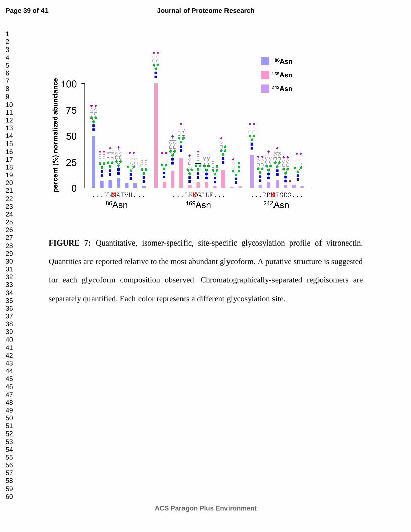
FIGURE 7: Quantitative, isomer-specific, site-specific glycosylation profile of vitronectin.
Quantities are reported relative to the most abundant glycoform. A putative structure is suggested
for each glycoform composition observed. Chromatographically-separated regioisomers are
separately quantified. Each color represents a different glycosylation site.
Page 39 of 41
ACS Paragon Plus Environment
Journal of Proteome Research
123456789101112131415161718192021222324252627282930313233343536373839404142434445464748495051525354555657585960
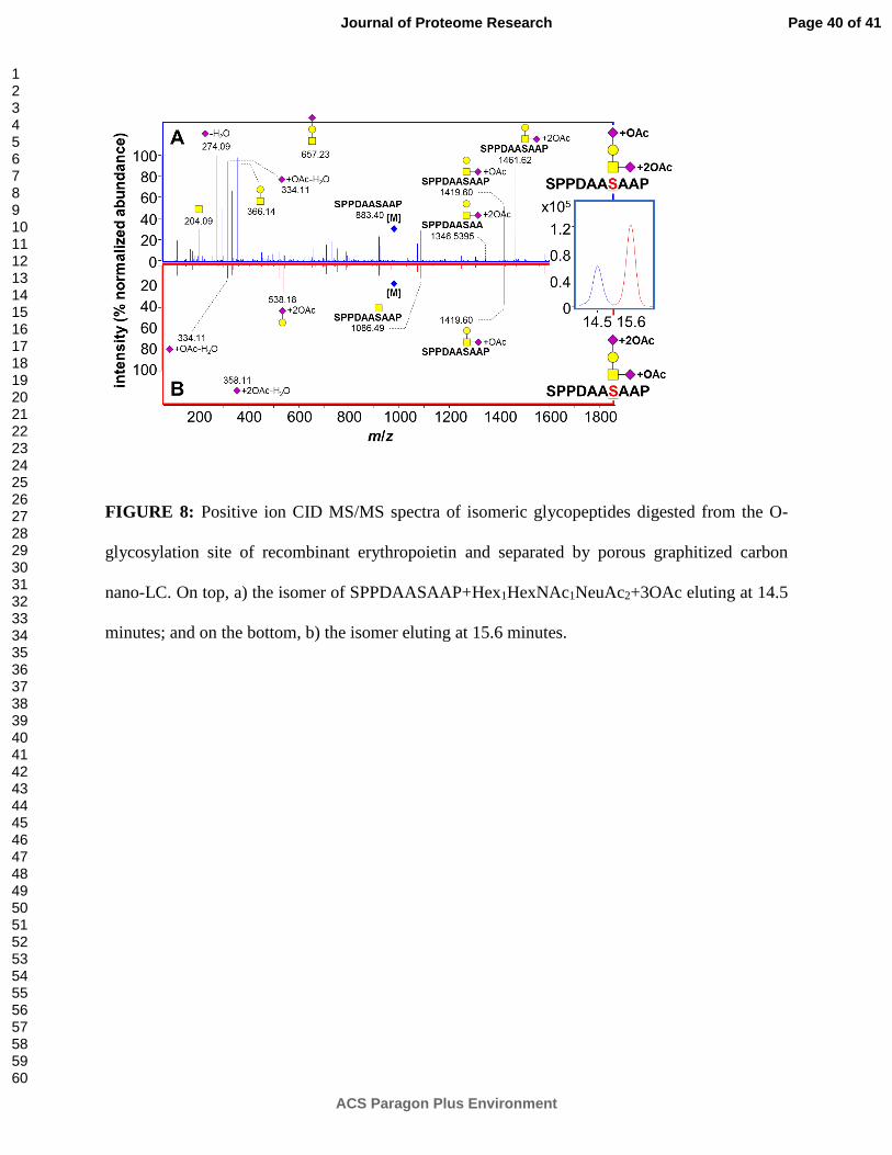
FIGURE 8: Positive ion CID MS/MS spectra of isomeric glycopeptides digested from the O-
glycosylation site of recombinant erythropoietin and separated by porous graphitized carbon
nano-LC. On top, a) the isomer of SPPDAASAAP+Hex1HexNAc1NeuAc2+3OAc eluting at 14.5
minutes; and on the bottom, b) the isomer eluting at 15.6 minutes.
Page 40 of 41
ACS Paragon Plus Environment
Journal of Proteome Research
123456789101112131415161718192021222324252627282930313233343536373839404142434445464748495051525354555657585960
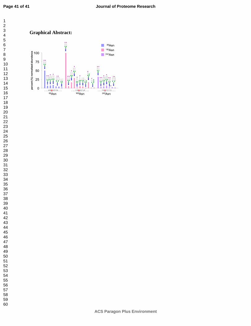
Graphical Abstract:
Page 41 of 41
ACS Paragon Plus Environment
Journal of Proteome Research
123456789101112131415161718192021222324252627282930313233343536373839404142434445464748495051525354555657585960