dairy chemistry and biochemistry
DESCRIPTION
dairy, milk, cheese, butter,TRANSCRIPT
Contents
Preface General references on dairy chemistry
1 Production and utilization of milk 1.1 Introduction 1.2 Composition and variability of milk 1.3 Classification of mammals 1.4 Structure and development of mammary tissue 1.5 Ultrastructure of the secretory cell 1.6 Techniques used to study milk synthesis
1.6.1 Arteriovenous concentration differences 1.6.2 Isotope studies 1.6.3 Perfusion of isolated gland 1.6.4 Tissue slices 1.6.5 Cell homogenates 1.6.6 Tissue culture
Production and utilization of milk 1.7 Biosynthesis of milk constituents 1.8 1.9 Trade in milk products References Suggested reading
2 Lactose 2.1 2.2
2.3 2.4
2.5 2.6
Introduction Chemical and physical properties of lactose 2.2.1 Structure of lactose 2.2.2 Biosynthesis of lactose 2.2.3 Lactose equilibrium in solution 2.2.4 Significance of mutarotation 2.2.5 Solubility of lactose 2.2.6 Crystallization of lactose 2.2.7 Problems related to lactose crystallization Production of lactose Derivatives of lactose 2.4.1 Enzymatic modification of lactose 2.4.2 Chemical modifications 2.4.3 Fermentation products Lactose and the Maillard reaction Nutritional aspects of lactose 2.6.1 Lactose intolerance 2.6.2 Galactosaemia
xiii xv
1 1 1 3 4 7 8 8 9 9
10 10 10 11 11 18 20 20
21 21 23 23 23 25 27 27 28 31 39 42 42 43 50 54 56 58 61
vi CONTENTS
3
2.7 Determination of lactose concentration 2.7.1 Polarimetry 2.7.2 Oxidation and reduction titration 2.7.3 Colorimetric methods 2.7.4 Chromatographic methods 2.7.5 Enzymatic methods
References Suggested reading
Milk lipids 3.1 Introduction 3.1 3.3 3.4 3.5 3.6 3.7 3.8
3.9
3.10
3.1 1
3.12 3.13 3.14 3.15
3.16
Factors that affect the fat content of bovine milk Classes of lipids in milk Fatty acid profile of milk lipids Synthesis of fatty acids in milk fat Structure of milk lipids Milk fat as an emulsion Milk fat globule membrane 3.8.1 Isolation of the fat globule membrane 3.8.2 Gross chemical compositlion of FGM 3.8.3 The protein fraction 3.8.4 The lipid fraction 3.8.5 Other membrane components 3.8.6 Membrane structure 3.8.7 Secretion of milk lipid globules Stability of the milk fat emulsion 3.9.1 Emulsion stability in general 3.9.2 The creaming process in milk Influence of processing operations on the fat globule membrane 3.10.1 Milk supply: hydrolytic rancidity 3.10.2 Mechanical separation of milk 3.10.3 Homogenization 3.10.4 Heating Physical defects in milk and cream 3.11.1 Free fat Churning Freezing Dehydration Lipid oxidation 3.15.1 3.15.2 Antioxidants in milk 3.15.3 Spontaneous oxidation 3.15.4 Other factors that affect lipid oxidation in milk and
dairy products 3.15.5 Measurement of lipid oxidation Rheology of milk fat 3.16.1 3.16.2 Process parameters
Pro-oxidants in milk and milk products
Fatty acid profile and distribution
References Suggested reading Appendices
62 62 63 64 65 65 65 66
67 67 68 71 75 81 87 90 92 93 94 94 95 97 97
100 1 04 104 106
108 108 111 113 116 117 118 118 126 126 127 130 132 133
133 134 134 134 137 140 141 141
CONTENTS vii
4 Milk proteins 4.1 4.2
4.3
4.4
4.5
4.6
4.7
4.8
4.9 4.10 4.11 4.12 4.13
Introduction Heterogeneity of milk proteins 4.2.1 Other protein fractions Preparation of casein and whey proteins 4.3.1 Acid (isoelectric) precipitation 4.3.2 Centrifugation 4.3.3 Centrifugation of calcium-supplemented milk 4.3.4 Salting-out methods 4.3.5 Ultrafiltration 4.3.6 4.3.7 Precipitation with ethanol 4.3.8 Cryoprecipitation 4.3.9 Rennet coagulation 4.3.10 Other methods for the preparation of whey proteins Heterogeneity and fractionation of casein 4.4.1 4.4.2 Microheterogeneity of the caseins 4.4.3 Nomenclature of the caseins Some important properties of the caseins 4.5.1 Chemical composition 4.5.2 Secondary and tertiary structures 4.5.3 Molecular size 4.5.4 Hydrophobicity 4.5.5 Influence of Ca2+ on caseins 4.5.6 Action of rennets on casein 4.5.7 Casein association 4.5.8 Casein micelle structure Whey proteins 4.6.1 Preparation 4.6.2 Heterogentity of whey proteins P-Lactoglobulin 4.7.1 Occurrence and microheterogeneity 4.7.2 Amino acid composition 4.7.3 Primary structure 4.7.4 Secondary structure 4.7.5 Tertiary structure 4.7.6 Quaternary structure 4.7.7 Physiological function 4.7.8 Denaturation a-Lactal bumin 4.8.1 Amino acid composition 4.8.2 Genetic variants 4.8.3 Primary structure 4.8.4 Secondary and tertiary structure 4.8.5 Quaternary structure 4.8.6 Other species 4.8.7 Biological function 4.8.8 Blood serum albumin Immunoglobulins (Ig) Minor milk proteins Non-protein nitrogen Comparison of human and bovine milks
Gel filtration (gel permeation chromatography)
Resolution of caseins by electrophoresis
Metal binding and heat stability
146 146 149 150 152 152 153 153 153 153 154 154 154 154 155 155 159 160 162 163 163 175 178 178 179 179 180 180 186 186 186 187 188 188 189 189 189 190 191 192 192 192 192 192 193 193 193 194 194 195 195 199 199 200
... V l l l CONTENTS
4.14 Synthesis and secretion of milk proteins 4.14.1 Sources of amino acids 4.14.2 4.14.3 Synthesis of milk proteins 4.14.4 4.14.5 4.14.6 Secretion of milk-specific proteins 4.14.7 Secretion of immunoglobulins
4.15.1 Industrial production of caseins 4.15.2 Novel methods for casein production 4.15.3 Fractionation of casein 4.1 5.4 Functional (physicochemical) properties of caseins 4.15.5 Applications of caseins 4.15.6 Whey proteins 4.15.7 Casein-whey protein co-precipitates Biologically active proteins and peptides in milk 4.16.1 Lactoperoxidase 4.16.2 Lactotransferrin 4.16.3 Immunoglobulins 4.16.4 Vitamin-binding proteins 4.16.5 Growth factors 4.16.6 Bifidus factors 4.16.7 Milk protein hydrolysates
Amino acid transport into the mammary cell
Modifications of the polypeptide chain Structure and expression of milk protein genes
4.15 Functional milk proteins
4.16
References Suggested reading Appendices
5 Salts of milk 5.1 5.2 5.3 5.4 5.5
5.6 5.7
5.8
Introduction Method of analysis Composition of milk salts Secretion of milk salts Factors influencing variation in salt composition 5.5.1 Breed of cow 5.5.2 Stage of lactation 5.5.3 Infection of the udder 5.5.4 Feed Interrelations of milk salt constituents Partition of milk salts between colloidal and soluble phases 5.7.1 5.7.2 Soluble salts 5.7.3 5.7.4 Colloidal milk salts Changes in milk salts equilibria induced by various treatments 5.8.1 Addition of acid or alkali 5.8.2 Addition of various salts 5.8.3 Effect of changes in temperature 5.8.4 Changes in pH induced by temperature 5.8.5 Etfect of dilution and concentration 5.8.6 Etfect of freezing
Methods used to separate the colloidal and soluble phases
Measurement of calcium and magnesium ions
20 1 20 1 203 203 205 206 207 209 210 21 1 215 216 218 219 219 227 228 229 229 230 230 23 1 23 1 232 234 236 237
239 239 239 240 242 243 24 3 244 247 247 247 249 249 250 254 256 260 260 26 1 26 1 262 262 263 263 264
References Suggested reading
CONTENTS ix
6 Vitamins in milk and dairy products 6.1 Introduction 6.2 Fat-soluble vitamins
6.2.1 Retinol (vitamin A) 6.2.2 Calciferols (vitamin D) 6.2.3 Tocopherols and related compounds (vitamin E) 6.2.4 Phylloquinone and related compounds (vitamin K)
6.3.1 Thiamin (vitamin B,) 6.3.2 Riboflavin (vitamin B2) 6.3.3 Niacin 6.3.4 Biotin 6.3.5 Panthothenic acid 6.3.6 6.3.7 Folate 6.3.8
6.3 B-group vitamins
Pyridoxine and related compounds (vitamin B6)
Cobalamin and its derivatives (vitamin B12) 6.4 Ascorbic acid (vitamin C) References Suggested reading Appendices
7 Water in milk and dairy products 7.1 Introduction 7.2 General properties of water 7.3 Water activity 7.4 Water sorption 7.5 Glass transition and the role of water in plasticization 7.6 Non-equilibrium ice formation 7.7 Role of water in stickiness and caking of powders and
crystallization of lactose 7.8 Water and the stability of dairy products References Suggested reading
8 Enzymology of milk and milk products 8.1 Introduction 8.2 Indigenous enzymes of bovine milk
8.2.1 Introduction 8.2.2 Proteinases (EC 3 . 4 ~ ) 8.2.3 8.2.4 Phosphatases 8.2.5 Lysozyme (EC 3.2.1.17) 8.2.6 N-Acetyl-P-D-glucosaminidase (EC 3.2.1.30) 8.2.7 8.2.8 Xanthine oxidase (EC 1.2.3.2) 8.2.9 Sulphydryl oxidase (EC 1.8.3.-) 8.2.10 Superoxide dismutase (EC 1.15.1.1) 8.2.11 Catalase (EC 1.11.1.6) 8.2.12 Lactoperoxidase (EC 1.1 1.1.7) 8.2.13 Other enzymes
Lipases and esterases (EC 3.1.1.-)
y-Glutamyl transpeptidase (transferase) (EC 2.3.2.2)
265 265 266 266 269 272 274 275 275 277 279 28 1 281 282 285 287 289 291 29 1 29 1
294 294 294 301 305 311 312
313 313 316 316
317 317 317 317 318 322 324 327 328 328 328 330 330 331 331 333
X CONTENTS
8.3 Exogenous enzymes in dairy technology 8.3.1 Introduction 8.3.2 Proteinases 8.3.3 P-Galactosidase 8.3.4 Lipases 8.3.5 Lysozyme 8.3.6 Catalase 8.3.7 Glucose oxidase 8.3.8 Superoxide dismutase 8.3.9 Exogeneous enzymes in food analysis
References Suggested reading
9 Heat-induced changes in milk 9.1 9.2
9.3
9.4 9.5 9.6
9.7
9.8
9.9 9.10
Introduction Lipids 9.2.1 Physiochemical changes 9.2.2 Chemical changes Lactose 9.3.1 Formation of lactulose 9.3.2 Formation of acids 9.3.3 Maillard browning Milk salts Vitamins Proteins 9.6.1 Enzymes 9.6.2 Denaturation of other biologically active proteins 9.6.3 Denaturation of whey proteins 9.6.4 Effect of heat on caseins Heat stability of milk 9.7.1 Effect of processing operations on heat stability Effect of heat treatment on rennet coagulation of milk and related properties Age gelation of sterilized milk Heat-induced changes in flavour of milk
References Suggested reading
10 Chemistr and biochemistry of cheese and fermente B milks 10.1 Introduction 10.2 Rennet-coagulated cheeses
10.2.1 Preparation and treatment of cheesemilk 10.2.2 Conversion of milk to cheese curd 10.2.3 Acidification 10.2.4 Moulding and shaping 10.2.5 Salting 10.2.6 Manufacturing protocols for some cheese varieties 10.2.7 Cheese ripening 10.2.8 Cheese flavour 10.2.9 Accelerated ripening of cheese
10.3 Acid-coagulated cheeses 10.4 Processed cheese products
10.4.1 Processing protocol
333 333 336 338 338 339 339 340 341 342 345 346
347 341 349 349 351 352 354 354 356 360 360 360 360 363 363 368 369 372
373 374 376 371 378
379 379 380 380 382 394 391 398 402 403 416 418 419 421 424
CONTENTS xi
10.5 Cheese analogues 10.6 Cultured milks References Suggested reading Appendices
11 Physical properties of milk 11.1 Ionic strength 11.2 Density 11.3 Redox properties of milk 11.4 Colligative properties of milk 11.5 Interfacial tension 11.6 Acid-base equilibria 11.7 Rheological properties
11.7.1 Newtonian behaviour 11.7.2 Non-Newtonian behaviour 11.7.3 Rheology of milk gels 11.7.4 Rheological properties of milk fat
11.8 Electrical conductivity 11.9 Thermal properties of milk 11.10 11.1 1 References Suggested reading
Interaction of light with milk and dairy products Colour of milk and milk products
421 428 432 433 434
437 438 438 439 443 447 449 453 453 454 455 456 456 457 458 459 460 46 1
463
Dairy Chemistry and Biochemistry
P.F. FOX and P.L.H. McSWEENEY Department of Food Chemistry
University College Cork, Ireland
BLACKIE ACADEMIC & PROFESSIONAL An Imprint of Chapman 8 Hall
London 1 Weinheim . New York * Tokyo Melbourne . Madras

Published by Blackie Academic & Professional, an imprint of Thomson Science, 2-6 Boundary Row, London SE1 SHN, UK
Thomson Science, 2-6 Boundary Row, London SE18HN, UK Thomson Science, 115 Fifth Avenue, New York NY 10003, USA
Thomson Science, Suite 750, 400 Market Street, Philadelphia, PA 19106, USA Thomson Science, Pappelallee 3, 69469 Weinheim, Germany
First edition 1998 0 1998 Thomson Science
Thomson Science is a division of International Thomson Publishing I@P* Typeset in 10/12pt Times by Doyle Graphics, Tullamore, Ireland Printed in Great Britain by St Edmundsbury Press Ltd, Bury St Edmunds, Suffolk
ISBN 0 412 72000 0 All rights reserved. No part of this publication may be reproduced, stored in a retrieval system or transmitted in any form or by any means, electronic, mechanical, photocopying, recording or otherwise, without the prior written permission of the publishers. Applications for permission should be addressed to the rights manager at the London address of the publisher.
The publisher makes no representation, express or implied, with regard to the accuracy of the information contained in this book and cannot accept any legal responsibility or liability for any errors or omissions that may be made. A catalogue record for this book is available from the British Library
Library of Congress Catalog Card Number: 97-77281
@ Printed on acid-free text paper, manufactured in accordance with ANSI/NISO 239.48-1992 (Permanence of Paper).
Preface
Milk has been the subject of scientific study for about 150years and, consequently, is probably the best characterized, in chemical terms, of our major foods. It is probably also the most complicated and serves as the raw material for a very large and diverse family of food products. Dairy science has existed as a university discipline for more than 100 years; it is the oldest sector of food science (and technology), with the exception of brewery science. Since dairy chemistry is a major facet of dairy science, it might be expected to have been the subject of numerous books. This is, in fact, not so. During the past 40years, as far as we are aware, only six books or series on dairy chemistry have been published in English, i.e. Principles of Dairy Chemistry (Jenness and Paton, 1959), Dairy Chemistry and Physics (Walstra and Jenness, 1984), Fundamentals of Dairy Chemistry (Webb and Johnson, 1964; Webb, Johnson and Alford, 1974; Wong et al., 19SS), Developments in Dairy Chemistry (Fox, four volumes, 1982, 1983, 1985, 1989), Advanced Dairy Chemistry (Fox, three volumes, 1992, 1995, 1997) and Handbook of Milk Composition (Jensen, 1995). Of these, Principles of Dairy Chemistry and Dairy Chemistry and Physics were written essentially for senior undergrad- uate students. The other four books/series were focused principally on lecturers, researchers, senior postgraduate students and senior production management. Thus, at present there is a lack of books written at senior undergraduate/junior postgraduate level specializing in dairy chemistry/ science. This book is intended to fill that gap and should be as useful to graduates working in the dairy industry as it is to those still studying.
The book assumes a knowledge of chemistry and biochemistry but not of dairy chemistry. As the title suggests, the book has a stronger biochemical orientation than either Principles of Dairy Chemistry or Dairy Chemistry and Physics. In addition to a fairly in-depth treatment of the chemistry of the principal constituents of milk, i.e. water, lactose, lipids, proteins (including enzymes), salts and vitamins, various more applied aspects are also covered, e.g. heat-induced changes, cheese, protein-rich products and the applications of enzymes in dairy technology. The principal physical properties are also described.
To facilitate the reader, the structure of various molecules mentioned frequently in the text are given in appendices but we emphasize that a good general knowledge of chemistry and biochemistry is assumed. The chemical composition of the principal dairy products is also included.

xiv PREFACE
The book does not cover the technology of the various dairy products, although brief manufacturing protocols for some products are included to facilitate discussion; however, a number of textbooks on various aspects of dairy technology are referenced. Neither are the chemical analyses, microbi- ology and nutritional aspects of dairy products covered, except in a very incidental manner. The effects of dairy husbandry on the composition and properties of milk are discussed briefly, as is the biosynthesis of milk constituents; in both cases, some major textbooks are referenced.
We hope that the book will answer some of your questions on the chemistry and biochemistry of milk and milk products and encourage you to undertake more extensive study of these topics.
The highly skilled and enthusiastic assistance of Ms Anne Cahalane and Ms Brid Considine in the preparation of the manuscript and of Professor D.M. Mulvihill and Dr N. O’Brien for critically and constructively review- ing the manuscript are gratefully acknowledged and very much appreciated.
P.F. Fox P.L.H. McSweeney

General references on dairy chemistry
Alais, C . (1974) Science du Lait. Principes des Techniques Laitieres, 3rd edn, SEP Editions, Paris.
Fox, P.F. (ed.) (1982-1989) Developments in Dairy Chemistry, Volumes 1, 2, 3 and 4, Elsevier Applied Science Publishers, London.
Fox, P.F. (ed.) (1992-1997) Advanced Dairy Chemistry, Volumes 1, 2 and 3, Elsevier Applied Science Publishers and Chapman & Hall, London.
Jenness, R. and Patton, S . (1959) Principles of Dairy Chemistry, John Wiley & Sons, New York.
Jensen, R.G. (ed.) (1995) Handbook of Milk Composition, Academic Press, San Diego.
Walstra, P. and Jenness, R. (1984) Dairy Chemistry and Physics, John Wiley & Sons, New York.
Webb, B.H. and Johnson, A.H. (eds) (1964) Fundamentals of Dairy Chemis- try, AVI, Westport, CT, USA.
Webb, B.H., Johnson, A.H. and Alford, J.A. (eds) (1974) Fundamentals of Dairy Chemistry, 2nd edn, AVI, Westport, CT, USA.
Wong, N.P., Jenness, R., Keeney, M. and Marth, E.H. (eds) (1988) Funda- mentals of Dairy Chemistry, 3rd edn, Van Norstrand Reinhold, New York.

Dairy Chemistry and Biochemistry
P.F. FOX and P.L.H. McSWEENEY Department of Food Chemistry
University College Cork, Ireland
BLACKIE ACADEMIC & PROFESSIONAL An Imprint of Chapman 8 Hall
London 1 Weinheim . New York * Tokyo Melbourne . Madras
Published by Blackie Academic & Professional, an imprint of Thomson Science, 2-6 Boundary Row, London SE1 SHN, UK
Thomson Science, 2-6 Boundary Row, London SE18HN, UK Thomson Science, 115 Fifth Avenue, New York NY 10003, USA
Thomson Science, Suite 750, 400 Market Street, Philadelphia, PA 19106, USA Thomson Science, Pappelallee 3, 69469 Weinheim, Germany
First edition 1998 0 1998 Thomson Science
Thomson Science is a division of International Thomson Publishing I@P* Typeset in 10/12pt Times by Doyle Graphics, Tullamore, Ireland Printed in Great Britain by St Edmundsbury Press Ltd, Bury St Edmunds, Suffolk
ISBN 0 412 72000 0 All rights reserved. No part of this publication may be reproduced, stored in a retrieval system or transmitted in any form or by any means, electronic, mechanical, photocopying, recording or otherwise, without the prior written permission of the publishers. Applications for permission should be addressed to the rights manager at the London address of the publisher.
The publisher makes no representation, express or implied, with regard to the accuracy of the information contained in this book and cannot accept any legal responsibility or liability for any errors or omissions that may be made. A catalogue record for this book is available from the British Library
Library of Congress Catalog Card Number: 97-77281
@ Printed on acid-free text paper, manufactured in accordance with ANSI/NISO 239.48-1992 (Permanence of Paper).

Preface
Milk has been the subject of scientific study for about 150years and, consequently, is probably the best characterized, in chemical terms, of our major foods. It is probably also the most complicated and serves as the raw material for a very large and diverse family of food products. Dairy science has existed as a university discipline for more than 100 years; it is the oldest sector of food science (and technology), with the exception of brewery science. Since dairy chemistry is a major facet of dairy science, it might be expected to have been the subject of numerous books. This is, in fact, not so. During the past 40years, as far as we are aware, only six books or series on dairy chemistry have been published in English, i.e. Principles of Dairy Chemistry (Jenness and Paton, 1959), Dairy Chemistry and Physics (Walstra and Jenness, 1984), Fundamentals of Dairy Chemistry (Webb and Johnson, 1964; Webb, Johnson and Alford, 1974; Wong et al., 19SS), Developments in Dairy Chemistry (Fox, four volumes, 1982, 1983, 1985, 1989), Advanced Dairy Chemistry (Fox, three volumes, 1992, 1995, 1997) and Handbook of Milk Composition (Jensen, 1995). Of these, Principles of Dairy Chemistry and Dairy Chemistry and Physics were written essentially for senior undergrad- uate students. The other four books/series were focused principally on lecturers, researchers, senior postgraduate students and senior production management. Thus, at present there is a lack of books written at senior undergraduate/junior postgraduate level specializing in dairy chemistry/ science. This book is intended to fill that gap and should be as useful to graduates working in the dairy industry as it is to those still studying.
The book assumes a knowledge of chemistry and biochemistry but not of dairy chemistry. As the title suggests, the book has a stronger biochemical orientation than either Principles of Dairy Chemistry or Dairy Chemistry and Physics. In addition to a fairly in-depth treatment of the chemistry of the principal constituents of milk, i.e. water, lactose, lipids, proteins (including enzymes), salts and vitamins, various more applied aspects are also covered, e.g. heat-induced changes, cheese, protein-rich products and the applications of enzymes in dairy technology. The principal physical properties are also described.
To facilitate the reader, the structure of various molecules mentioned frequently in the text are given in appendices but we emphasize that a good general knowledge of chemistry and biochemistry is assumed. The chemical composition of the principal dairy products is also included.
xiv PREFACE
The book does not cover the technology of the various dairy products, although brief manufacturing protocols for some products are included to facilitate discussion; however, a number of textbooks on various aspects of dairy technology are referenced. Neither are the chemical analyses, microbi- ology and nutritional aspects of dairy products covered, except in a very incidental manner. The effects of dairy husbandry on the composition and properties of milk are discussed briefly, as is the biosynthesis of milk constituents; in both cases, some major textbooks are referenced.
We hope that the book will answer some of your questions on the chemistry and biochemistry of milk and milk products and encourage you to undertake more extensive study of these topics.
The highly skilled and enthusiastic assistance of Ms Anne Cahalane and Ms Brid Considine in the preparation of the manuscript and of Professor D.M. Mulvihill and Dr N. O’Brien for critically and constructively review- ing the manuscript are gratefully acknowledged and very much appreciated.
P.F. Fox P.L.H. McSweeney
General references on dairy chemistry
Alais, C . (1974) Science du Lait. Principes des Techniques Laitieres, 3rd edn, SEP Editions, Paris.
Fox, P.F. (ed.) (1982-1989) Developments in Dairy Chemistry, Volumes 1, 2, 3 and 4, Elsevier Applied Science Publishers, London.
Fox, P.F. (ed.) (1992-1997) Advanced Dairy Chemistry, Volumes 1, 2 and 3, Elsevier Applied Science Publishers and Chapman & Hall, London.
Jenness, R. and Patton, S . (1959) Principles of Dairy Chemistry, John Wiley & Sons, New York.
Jensen, R.G. (ed.) (1995) Handbook of Milk Composition, Academic Press, San Diego.
Walstra, P. and Jenness, R. (1984) Dairy Chemistry and Physics, John Wiley & Sons, New York.
Webb, B.H. and Johnson, A.H. (eds) (1964) Fundamentals of Dairy Chemis- try, AVI, Westport, CT, USA.
Webb, B.H., Johnson, A.H. and Alford, J.A. (eds) (1974) Fundamentals of Dairy Chemistry, 2nd edn, AVI, Westport, CT, USA.
Wong, N.P., Jenness, R., Keeney, M. and Marth, E.H. (eds) (1988) Funda- mentals of Dairy Chemistry, 3rd edn, Van Norstrand Reinhold, New York.
1 Production and utilization of milk
1.1 Introduction
Milk is a fluid secreted by the female of all mamalian species, of which there are more than 4000, for the primary function of meeting the complete nutritional requirements of the neonate of the species. In addition, milk serves several physiological functions for the neonate. Most of the non- nutritional functions of milk are served by proteins and peptides which include immunoglobulins, enzymes and enzyme inhibitors, binding or car- rier proteins, growth factors and antibacterial agents. Because the nutri- tional and physiological requirements of each species are more or less unique, the composition of milk shows very marked inter-species differences. Of the more than 4000 species of mammal, the milks of only about 180 have been analysed and, of these, the data for only about 50 species are considered to be reliable (sufficient number of samples, representative sampling, adequate coverage of the lactation period). Not surprisingly, the milks of the principal dairying species, i.e. cow, goat, sheep and buffalo, and the human are among those that are well characterized. The gross compo- sition of milks from selected species is summarized in Table 1.1; very extensive data on the composition of bovine and human milk are contained in Jensen (1995).
1.2 Composition and variability of milk
In addition to the principal constituents listed in Table 1.1, milk contains several hundred minor constituents, many of which, e.g. vitamins, metal ions and flavour compounds, have a major impact on the nutritional, technologi- cal and sensoric properties of milk and dairy products. Many of these effects will be discussed in subsequent chapters.
Milk is a very variable biological fluid. In addition to interspecies differences (Table 1.1), the milk of any particular species varies with the individuality of the animal, the breed (in the case of commercial dairying species), health (mastitis and other diseases), nutritional status, stage of lactation, age, interval between milkings, etc. In a bulked factory milk supply, variability due to many of these factors is evened out, but some variability will persist and will be quite large in situations where milk
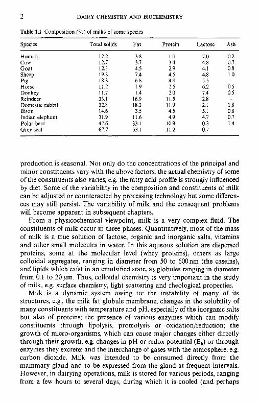
2 DAIRY CHEMISTRY AND BIOCHEMISTRY
Table 1.1 Composition (%) of milks of some species
Species Total solids Fat Protein Lactose Ash
Human 12.2 3.8 1 .o 7.0 0.2 c o w 12.7 3.7 3.4 4.8 0.7 Goat 12.3 4.5 2.9 4.1 0.8 Sheep 19.3 1.4 4.5 4.8 1.0 Pig 18.8 6.8 4.8 5.5 -
Horse 11.2 1.9 2.5 6.2 0.5 Donkey 11.7 1.4 2.0 7.4 0.5 Reindeer 33.1 16.9 11.5 2.8 - Domestic rabbit 32.8 18.3 11.9 2.1 1.8 Bison 14.6 3.5 4.5 5.1 0.8 Indian elephant 31.9 11.6 4.9 4.1 0.7 Polar bear 47.6 33.1 10.9 0.3 1.4 Grey seal 67.7 53.1 11.2 0.7 -
production is seasonal. Not only do the concentrations of the principal and minor constituents vary with the above factors, the actual chemistry of some of the constituents also varies, e.g. the fatty acid profile is strongly influenced by diet. Some of the variability in the composition and constituents of milk can be adjusted or counteracted by processing technology but some differen- ces may still persist. The variability of milk and the consequent problems will become apparent in subsequent chapters.
From a physicochemical viewpoint, milk is a very complex fluid. The constituents of milk occur in three phases. Quantitatively, most of the mass of milk is a true solution of lactose, organic and inorganic salts, vitamins and other small molecules in water. In this aqueous solution are dispersed proteins, some at the molecular level (whey proteins), others as large colloidal aggregates, ranging in diameter from 50 to 600nm (the caseins), and lipids which exist in an emulsified state, as globules ranging in diameter from 0.1 to 20 pm. Thus, colloidal chemistry is very important in the study of milk, e.g. surface chemistry, light scattering and rheological properties.
Milk is a dynamic system owing to: the instability of many of its structures, e.g., the milk fat globule membrane; changes in the solubility of many constituents with temperature and pH, especially of the inorganic salts but also of proteins; the presence of various enzymes which can modify constituents through lipolysis, proteolysis or oxidation/reduction; the growth of micro-organisms, which can cause major changes either directly through their growth, e.g. changes in pH or redox potential (EJ or through enzymes they excrete; and the interchange of gases with the atmosphere, e.g. carbon dioxide. Milk was intended to be consumed directly from the mammary gland and to be expressed from the gland at frequent intervals. However, in dairying operations, milk is stored for various periods, ranging from a few hours to several days, during which it is cooled (and perhaps
PRODUCTION AND UTILIZATION OF MILK 3
heated) and agitated to various degrees. These treatments will cause at least some physical changes and permit some enzymatic and microbiological changes which may alter the processing properties of milk. Again, it may be possible to counteract some of these changes.
1.3 Classification of mammals
The essential characteristic distinguishing mammals from other animal species is the ability of the female of the species to produce milk in specialized organs (mammary glands) for the nutrition of its newborn.
1. Prototheria. This subclass contains only one order, Monotremes, the species of which are egg-laying mammals, e.g. duck-billed platypus and echidna, and are indigenous only to Australasia. They possess many (perhaps 200) mammary glands grouped in two areas of the abdomen; the glands do not terminate in a teat and the secretion (milk) is licked by the young from the surface of the gland.
2. Marsupials. The young of marsupials are born live (viviparous) after a short gestation and are ‘premature’ at birth to a greater or lesser degree, depending on the species. After birth, the young are transferred to a pouch where they reach maturity, e.g. kangaroo and wallaby. In marsu- pials, the mammary glands, which vary in number, are located within the pouch and terminate in a teat. The mother may nurse two offspring, differing widely in age, simultaneously from different mammary glands that secrete milk of very different composition, designed to meet the different specific requirements of each offspring.
3. Eutherians. About 95% of all mammals belong to this subclass. The developing embryo in utero receives nourishment via the placental blood supply (they are referred to as placental mammals) and is born at a high, but variable, species-related state of maturity. All eutherians secrete milk, which, depending on the species, is more or less essential for the development of the young; the young of some species are born sufficiently mature to survive and develop without milk.
The number and location of mammary glands varies with species from two, e.g. human, goat and sheep, to 14-16 for the pig. Each gland is anatomically and physiologically separate and is emptied via a teat.
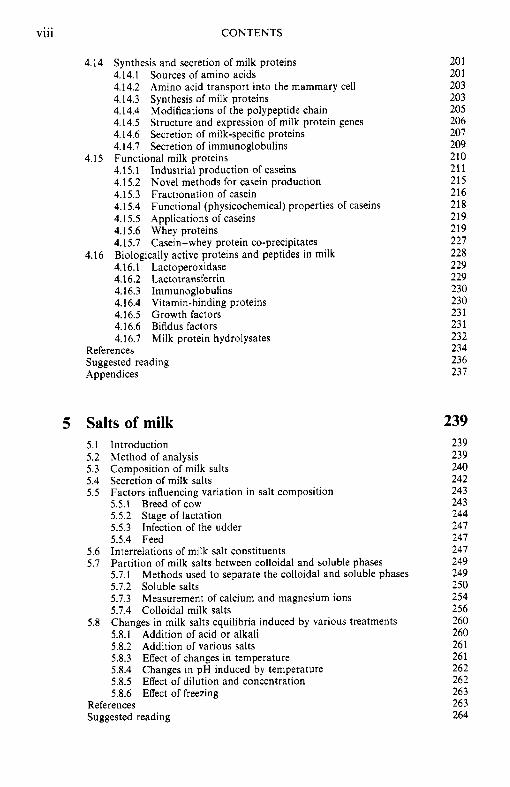
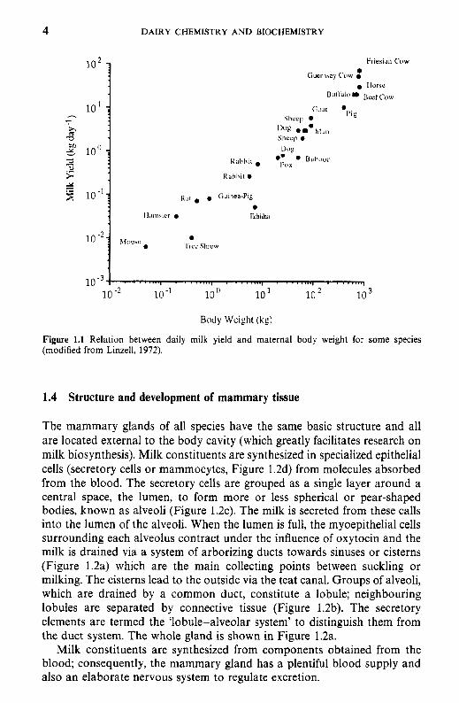
The wide interspecies variation in the composition (Table 1.1) and the chemistry of the constituents of milk, as discussed elsewhere, renders milk species-specific, i.e., designed to meet the requirements of the young of that species. There is also a surprisingly good relationship between milk yield and maternal body weight (Figure 1.1); species bred for commercial milk production, e.g. dairy cow and goat, fall above the line.
The class Mammalia is divided into three subclasses:
4 DAIRY CHEMISTRY AND BIOCHEMISTRY
R , ~ , . . Oumea-Pig
Il.,lll*lcr . 1khidii:i
3 3 10.'
Body Wcight (kg)
Figure 1.1 Relation between daily milk yield and maternal body weight for some species (modified from Linzell, 1972).
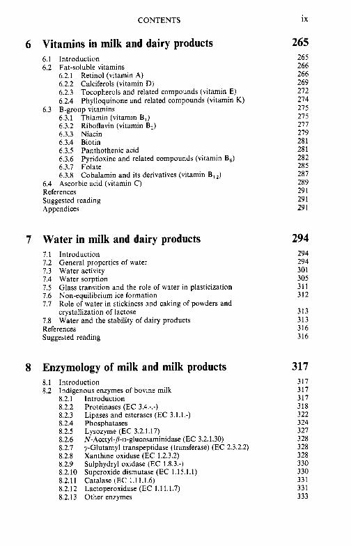
1.4 Structure and development of mammary tissue
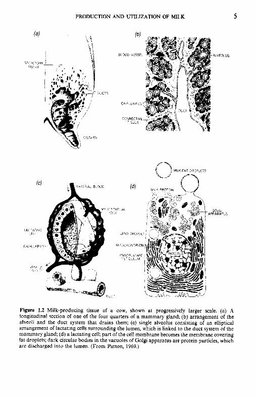
The mammary glands of all species have the same basic structure and all are located external to the body cavity (which greatly facilitates research on milk biosynthesis). Milk constituents are synthesized in specialized epithelial cells (secretory cells or mammocytes, Figure 1.2d) from molecules absorbed from the blood. The secretory cells are grouped as a single layer around a central space, the lumen, to form more or less spherical or pear-shaped bodies, known as alveoli (Figure 1.2~) . The milk is secreted from these calls into the lumen of the alveoli. When the lumen is full, the rnyoepithelial cells surrounding each alveolus contract under the influence of oxytocin and the milk is drained via a system of arborizing ducts towards sinuses or cisterns (Figure 1.2a) which are the main collecting points between suckling or milking. The cisterns lead to the outside via the teat canal. Groups of alveoli, which are drained by a common duct, constitute a lobule; neighbouring lobules are separated by connective tissue (Figure 1.2b). The secretory elements are termed the 'lobule-alveolar system' to distinguish them from the duct system. The whole gland is shown in Figure 1.2a.
Milk constituents are synthesized from components obtained from the blood; consequently, the mammary gland has a plentiful blood supply and also an elaborate nervous system to regulate excretion.
PRODUCTION AND UTILIZATION OF MILK 5
WPI.LAR1ES
C0:NECTIbE ISSUE
N LK PRVEIN
GOLGi \ PPAHATUS
Figure 1.2 Milk-producing tissue of a cow, shown at progressively larger scale. (a) A longitudinal section of one of the four quarters of a mammary gland; (b) arrangement of the alveoli and the duct system that drains them; (c) single alveolus consisting of an elliptical arrangement of lactating cells surrounding the lumen, which is linked to the duct system of the mammary gland; (d) a lactating cell; part of the cell membrane becomes the membrane covering fat droplets; dark circular bodies in the vacuoles of Golgi apparatus are protein particles, which are discharged into the lumen. (From Patton, 1969.)
6 DAIRY CHEMISTRY A N D BIOCHEMISTRY
3 t-”
10
0 0 100 200
Days
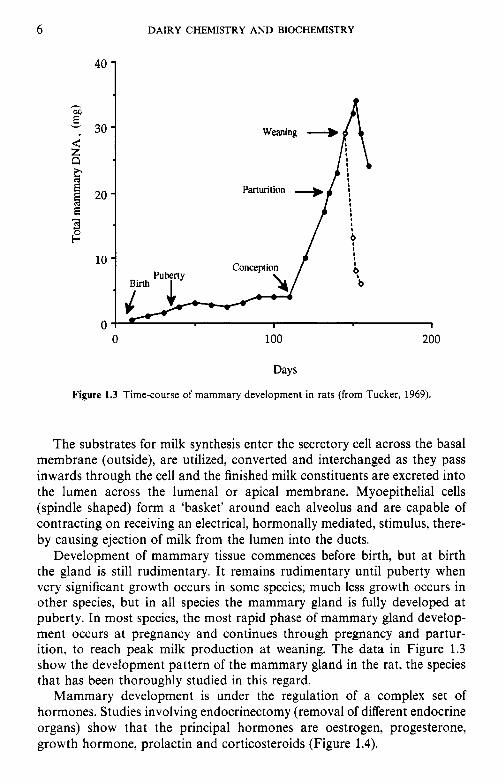
Figure 1.3 Time-course of mammary development in rats (from Tucker, 1969).
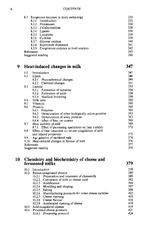
The substrates for milk synthesis enter the secretory cell across the basal membrane (outside), are utilized, converted and interchanged as they pass inwards through the cell and the finished milk constituents are excreted into the lumen across the lumenal or apical membrane. Myoepithelial cells (spindle shaped) form a ‘basket’ around each alveolus and are capable of contracting on receiving an electrical, hormonally mediated, stimulus, there- by causing ejection of milk from the lumen into the ducts.
Development of mammary tissue commences before birth, but at birth the gland is still rudimentary. It remains rudimentary until puberty when very significant growth occurs in some species; much less growth occurs in other species, but in all species the mammary gland is fully developed at puberty. In most species, the most rapid phase of mammary gland develop- ment occurs at pregnancy and continues through pregnancy and partur- ition, to reach peak milk production at weaning. The data in Figure 1.3 show the development pattern of the mammary gland in the rat, the species that has been thoroughly studied in this regard.
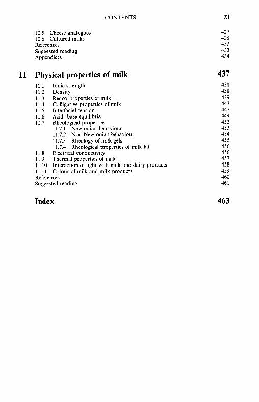
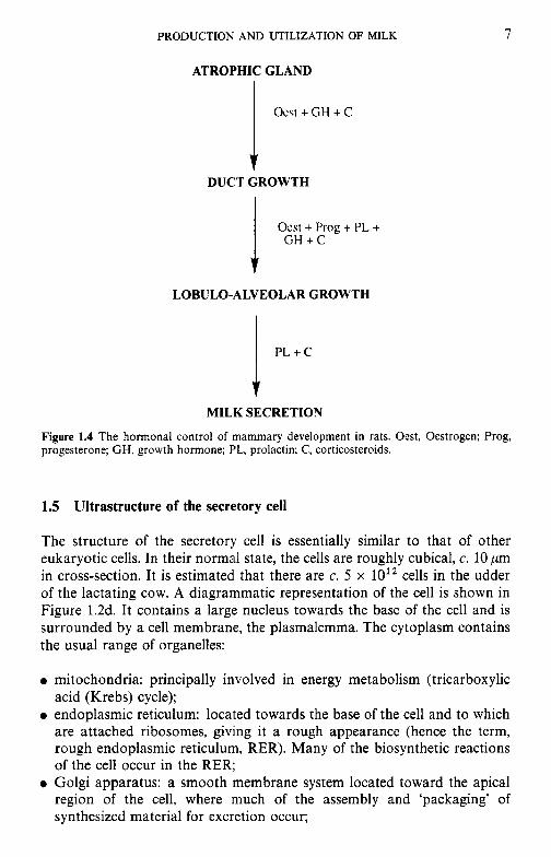
Mammary development is under the regulation of a complex set of hormones. Studies involving endocrinectomy (removal of different endocrine organs) show that the principal hormones are oestrogen, progesterone, growth hormone, prolactin and corticosteroids (Figure 1.4).
PRODUCTION AND UTILIZATION OF MILK 7
ATROPHIC GLAND
Ocst + GH + C
DUCT GROWTH
LOBULO-ALVEOLAR GROWTH
MILK SECRETION
Figure 1.4 The hormonal control of mammary development in rats. Oest, Oestrogen; Prog, progesterone; GH, growth hormone; PL, prolactin; C, corticosteroids.
1.5 Ultrastructure of the secretory cell
The structure of the secretory cell is essentially similar to that of other eukaryotic cells. In their normal state, the cells are roughly cubical, c. 10 pm in cross-section. It is estimated that there are c. 5 x 10’’ cells in the udder of the lactating cow. A diagrammatic representation of the cell is shown in Figure 1.2d. It contains a large nucleus towards the base of the cell and is surrounded by a cell membrane, the plasmalemma. The cytoplasm contains the usual range of organelles:
0 mitochondria: principally involved in energy metabolism (tricarboxylic acid (Krebs) cycle);
0 endoplasmic reticulum: located towards the base of the cell and to which are attached ribosomes, giving it a rough appearance (hence the term, rough endoplasmic reticulum, RER). Many of the biosynthetic reactions of the cell occur in the RER;
0 Golgi apparatus: a smooth membrane system located toward the apical region of the cell, where much of the assembly and ‘packaging’ of synthesized material for excretion occur;
a DAIRY CHEMISTRY AND BIOCHEMISTRY
0 lysosomes: capsules of enzymes (usually hydrolytic) distributed fairly uniformly throughout the cytoplasm.
Fat droplets and vesicles of material for excretion are usually apparent toward the apical region of the cell. The apical membrane possesses microvilli which serve to greatly increase its surface area.
1.6 Techniques used to study milk synthesis
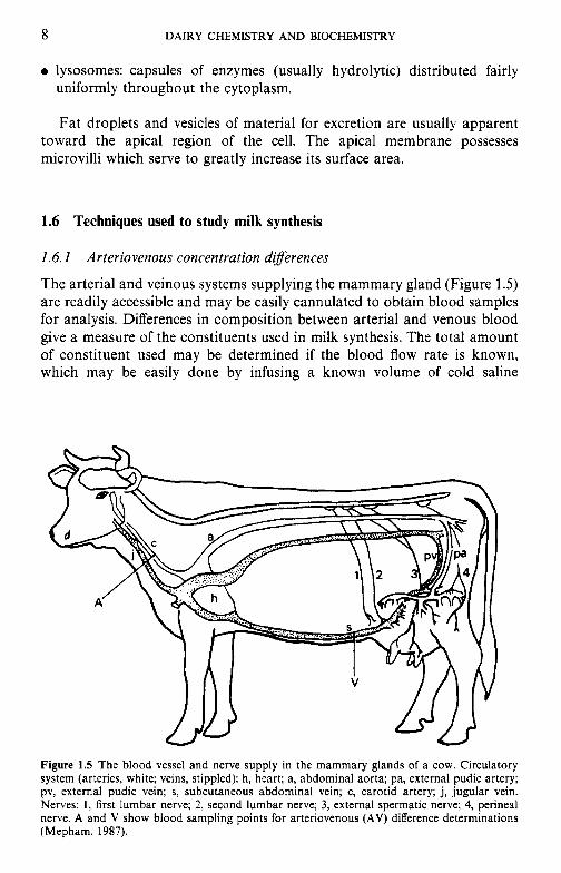
1.6.1 Arteriovenous concentration diferences
The arterial and veinous systems supplying the mammary gland (Figure 1.5) are readily accessible and may be easily cannulated to obtain blood samples for analysis. Differences in composition between arterial and venous blood give a measure of the constituents used in milk synthesis. The total amount of constituent used may be determined if the blood flow rate is known, which may be easily done by infusing a known volume of cold saline
Figure 1.5 The blood vessel and nerve supply in the mammary glands of a cow. Circulatory system (arteries, white; veins, stippled): h, heart; a, abdominal aorta; pa, external pudic artery; pv, external pudic vein; s, subcutaneous abdominal vein; c, carotid artery; j, jugular vein. Nerves: 1, first lumbar nerve; 2, second lumbar nerve; 3, external spermatic nerve; 4, perineal nerve. A and V show blood sampling points for arteriovenous (AV) difference determinations (Mepham, 1987).
PRODUCTION AND UTILIZATION OF MILK 9
solution into a vein and measuring the temperature of blood a little further downstream. The extent to which the blood temperature is reduced is inversely proportional to blood flow rate.
1.6.2 Isotope studies
Injection of radioactively labelled substrates, e.g. glucose, into the blood- stream permits assessment of the milk constituents into which that substrate is incorporated. It may also be possible to study the intermediates through which biosynthesis proceeds.
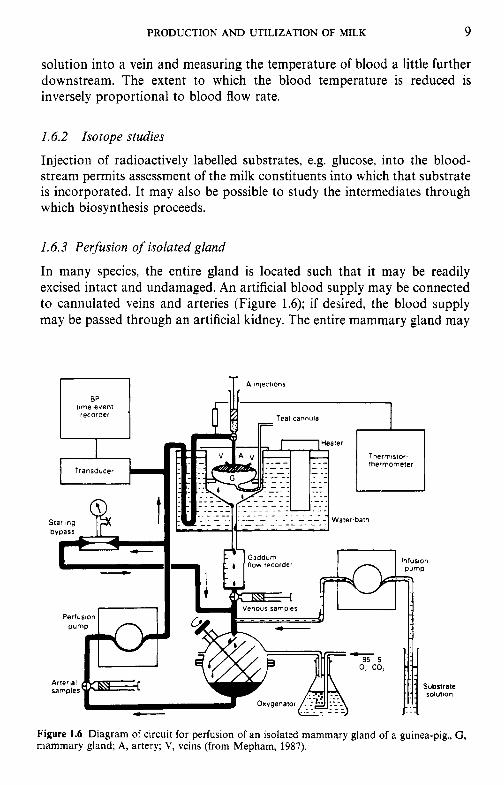
1.6.3 Perfusion of isolated gland
In many species, the entire gland is located such that it may be readily excised intact and undamaged. An artificial blood supply may be connected to cannulated veins and arteries (Figure 1.6); if desired, the blood supply may be passed through an artificial kidney. The entire mammary gland may
thermometer
Figure 1.6 Diagram of circuit for perfusion of an isolated mammary gland of a guinea-pig., G , mammary gland; A, artery; V, veins (from Mepham, 1987).
10 DAIRY CHEMISTRY AND BIOCHEMISTRY
be maintained active and secreting milk for several hours; substrates may readily be added to the blood supply for study.
1.6.4 Tissue slices
The use of tissue slices is a standard technique in all aspects of metabolic biochemistry. The tissue is cut into slices, sufficiently thin to allow adequate rates of diffusion in and out of the tissue. The slices are submerged in physiological saline to which substrates or other compounds may be added.
Changes in the composition of the slices and/or incubation medium give some indication of metabolic activity, but extensive damage may be caused to the cells on slicing; the system is so artificial that data obtained by the tissue slice technique may not pertain to the physiological situation. How- ever, the technique is widely used at least for introductory, exploratory experiments.
1.6.5 Cell homogenates
Cell homogenates are an extension of the tissue slice technique, in which the tissue is homogenized. As the tissue is completely disorganized, only individual biosynthetic reactions may be studied in such systems; useful preliminary work may be done with homogenates.
1.6.6 Tissue culture
Tissue cultures are useful for preliminary or specific work but are in- complete.
In general, the specific constituents of milk are synthesized from small molecules absorbed from the blood. These precursors are absorbed across the basal membrane but very little is known about the mechanism by which they are transported across the membrane. Since the membrane is rich in lipids, and precursors are mostly polar with poor solubility in lipid, it is unlikely that the precursors enter the cell by simple diffusion. It is likely, in common with other tissues, that there are specialized carrier systems to transport small molecules across the membrane; such carriers are probably proteins.
The mammary gland of the mature lactating female of many species is by far the most metabolically active organ of the body. For many small mammals, the energy input required for the milk secreted in a single day may exceed that required to develop a whole litter in utero. A cow at peak lactation yielding 45 kg milk day-' secretes approximately 2 kg lactose and 1.5 kg each of fat and protein per day. This compares with the daily weight gain for a beef animal of 1-1.5 kgday-', 60-70% of which is water. In large
PRODUCTION AND UTILIZATION OF MILK 11
measure, a high-yielding mammal is subservient to the needs of its mam- mary gland to which it must supply not only the precursors for the synthesis of milk constituents but also an adequate level of high-energy-yielding substrates (ATP, UTP, etc.) required to drive the necessary synthetic reactions. In addition, minor constituents (vitamins and minerals) must be supplied.
1.7 Biosynthesis of milk constituents
The constituents of milk can be grouped into four general classes according to their source:
0 organ-(mammary gland) and species-specific (e.g. most proteins and
0 organ- but not species-specific (lactose); 0 species- but not organ-specific (some proteins); 0 neither organ- nor species-specific (water, salts, vitamins).
The principal constituents (lactose, lipids and most proteins) of milk are synthesized in the mammary gland from constituents absorbed from blood. However, considerable modification of constituents occurs in the mammary gland; the constituents are absorbed from blood through the basal mem- brane, modified (if necessary) and synthesized into the finished molecule (lactose, triglycerides, proteins) within the mammocyte (mainly in the endoplasmic reticulum) and excreted from the mammocyte through the apical membrane into the lumen of the alveolus.
We believe that it is best and most convenient to describe the synthesis of the principal constituents in the appropriate chapter.
lipids);
1.8 Production and utilization of milk
Sheep and goats were domesticated early during the Agricultural Revo- lution, 8000-10000 years ago. Cattle were domesticated later but have become the principal dairying species in the most intense dairying areas, although sheep and goats are very important in arid regions, especially around the Mediterranean. Buffalo are important in some regions, especially in India and Egypt. Mare’s milk is used extensively in central Asia and is receiving attention in Europe for special dietary purposes since its compo- sition is closer to that of human milk than is bovine milk.
Some milk and dairy products are consumed in probably all regions of the world but they are major dietary items in Europe, North and South America, Australia, New Zealand and some Middle Eastern countries. Total milk production in 1996 was estimated to be 527 x lo6 tonnes, of which 130,

12 DAIRY CHEMISTRY AND BIOCHEMISTRY
Table 1.2 Consumption (kg caput-' annum-I) of liquid milk, 1993 (IDF, 1995)
Country Total Country Total
Russia" 252 Luxembourg" 86 Ireland" 182 Netherlands 84 Iceland 180 Hungary 81 Finland 170 Estonia" 81 Norway 147 Canada 77 Sweden 126 France 77 Denmark 115 Italy 75 United Kingdom 115 Germany 70 Spain 115 Greece" 67 Switzerland 101 Belgium 65 New Zealand 101 India 51 Australia 99 Lithuaniao 46 Czech and Slovak Reps" 97 Japan 42 USA 93 South Africa 38 Austria 92 Chile" 18
'Data for 1991, from I D F (1993).
Table 1.3 Consumption (kg caput-' annum-*) of cheese, 1993 (IDF, 1995)
Country Fresh Ripened Total
France Greece" Italy Belgium Germany Lithuania" Iceland Switzerland Sweden Luxembourg" Netherlands Denmark Finland Norway Canada USA Austria Czech and Slovak Reps" Estonia Australia United Kingdom New Zealand Hungary Russia" Spain Ireland" Chile" South Africa Japan India
7.5 0.2 6.7 4.7 8.0
11.6 5.2 2.8 0.9 5.0 1.7 0.9 2.3 0.2 0.9 1.3 3.9 4.0 5.6 - -
-
3.3 2.8 - -
2.0 0.1 0.2 0.2
15.5 21.8 13.4 15.1 10.5 6.8
11.9 13.6 15.5 11.3 14.1 14.5 12.0 14.0 12.4 11.9 7.5 6.6 4.4 - - -
4.6 4.9 -
-
2.0 1.5 1.2 -
22.8 22.0 20.1 19.8 18.5 18.4 17.1 16.4 16.4 16.3 15.8 15.4 14.3 14.2 13.3 13.2 11.4 10.6 10.0
8.8 8.3 8.1 7.9 7.7 7.0 5.6 4.0 1.6 1.4 0.2
"Data for 1991, from I D F (1993).
PRODUCTION AND UTILIZATION OF MILK 13
Table 1.4 Consumption (kg caput- ' annum-I) of butter, 1993 (IDF, 1995)
Country Butter
Lithuania" New Zealand Belgium France Germany Russia" Estonia Luxembourg" Finland Switzerland Czech and Slovak Reps" Austria Denmark United Kingdom Ireland" Netherlands Australia Canada Norway Sweden Iceland USA Italy Greece" India Hungary Japan Chile" South Africa Spain
18.8 9.3 7.0 6.8 6.8 6.5 5.9 5.8 5.3 5.3 5.0 4.3 4.1 3.5 3.4 3.3 3.3 3.0 2.3 2.3 2.2 2.1 1.8 1.1 0.1 0.9 0.7 0.6 0.5 0.2
"Data for 1991, from I D F (1993).
103, 78, 26 x lo6 tonnes were produced in western Europe, eastern Europe, North America and the Pacific region, respectively (IDF, 1996). The European Union and some other countries operate milk production quotas which are restricting growth in those areas. Data on the consumption of milk and dairy products in countries that are members of the International Dairy Federation (IDF) are summarized in Tables 1.2-1.6. Milk and dairy products are quite important in several countries that are not included in Tables 1.2-1.6 since they are not members of the IDF.
Because milk is perishable and its production was, traditionally, seasonal, milk surplus to immediate requirements was converted to more stable products, traditional examples being butter or ghee, fermented milk and cheese; smaller amounts of dried milk products were produced traditionally by sun-drying. These traditional products are still very important and many new variants thereof have been introduced. In addition, several new products have been developed during the past 130 years, e.g.
14 DAIRY CHEMISTRY A N D BIOCHEMISTRY
Table 1.5 Consumption (kg caput-' annum-') of cream (but- terfat equivalent), 1993 (IDF, 1995)
~
Country Total
Sweden Denmark Lithuania" Luxembourg" Iceland Norway Switzerland Russia" Finland Germany Estonia Hungary Belgium Austria New Zealand United Kingdom" Greece" France Czech and Slovak Reps" Ireland" Netherlands Canada USA Spain Italy South Africa Japan Chile"
3.0 2.9 2.9 2.6 2.4 2.4 2.3 2.1 2.0 1.8 1.7 1.6 1.5 1.3 1.3 1.1 1 .o 1 .o 0.9 0.9 0.7 0.6 0.6 0.4 0.3 0.3 0.2 0.2
"Data for 1991, from IDF (1993).
sweetened condensed milk, sterilized concentrated milk, a range of milk powders, UHT sterilized milk, ice-creams, infant foods and milk protein products.
One of the important developments in dairy technology in recent years has been the fractionation of milk into its principal constituents, e.g. lactose, milk fat fractions and milk protein products (caseins, caseinates, whey protein concentrates, whey protein isolates, mainly for use as functional proteins but more recently as 'nutraceuticals', i.e. proteins for specific physiological and/or nutritional functions, e.g. lactotransferrin, immuno- globulins).
As a raw material, milk has many attractive features:
1. Milk was designed for animal nutrition and hence contains the necessary nutrients in easily digestible forms (although the balance is designed for
PRODUCTION AND UTILIZATION OF MILK
Table 1.6 Consumption (kg caput-' annum-') of fermented milks, 1993 (IDF, 1995)
Country Total
Finland 37.0 Sweden 28.6 Iceland 25.9 Netherlands 20.7 France 17.3 Switzerland 17.0 India 16.1 Denmark 15.1 Lithuania" 14.6 Germany 12.2 Austria 11.1 Spain 9.8 Belgium 9.6 Estonia 8.8 Czech and Slovak Reps" 8.8 Japan 8.5 Luxembourg" 7.0 Greece" 6.8 Norway 6.3 Italy 5.0 Australia 4.8 United Kingdom" 4.8 Chile" 4.1 Hungary 3.6 South Africa 3.6 Ireland" 3.3 Canada 3.0 USA 2.1
15
aData for 1991, from I D F (1993).
the young of a particular species) and free of toxins. No other single food, except the whole carcass of an animal, including the bones, contains the complete range of nutrients at adequate concentrations.
2. The principal constituents of milk, i.e. lipids, proteins and carbohydrates, can be readily fractionated and purified by relatively simple methods, for use as food ingredients.
3. Milk itself is readily converted into products with highly desirable organoleptic and physical characteristics and its constituents have many very desirable and some unique physicochemical (functional) properties.
4. The modern dairy cow is a very efficient convertor of plant material; average national yields, e.g. in the USA and Israel, are about 8000 kg annum- ', with individual cows producing up to 20000 kg annum-'. In terms of kilograms of protein that can be produced per hectare, milk
16 DAIRY CHEMISTRY AND BIOCHEMISTRY
8 3 v; x d
Sclcc1cd I’nod products
Figure 1.7 Number of days of protein supply for a moderately active man produced per hectare yielding selected food products.
production, especially by modern cows, is much more efficient than meat production (Figure 1.7) but less efficient than some plants (e.g. cereals and soybeans). However, the functional and nutritional properties of milk proteins are superior to those of soy protein, and since cattle, and especially sheep and goats, can thrive under farming conditions not suitable for growing cereals or soybeans, dairy animals need not be competitors with humans for use of land, although high-yielding dairy cows are fed products that could be used for human foods. In any case, dairy products improve the ‘quality of life’, which is a desirable objective per se.
PRODUCTION AND UTILIZATION OF MILK 17
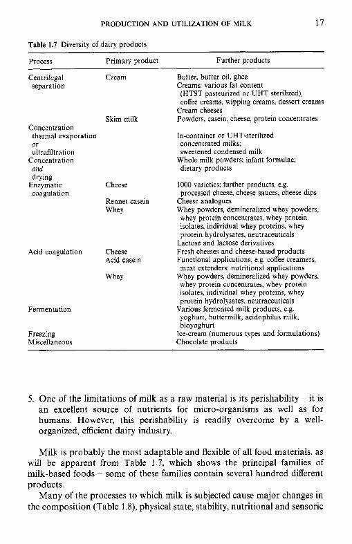
Table 1.7 Diversity of dairy products ~
Process Primary product Further products
Centrifugal separation
Concentration thermal evaporation or ultrafiltration
Concentration and drying
coagulation Enzymatic
Cream Butter, butter oil, ghee Creams: various fat content (HTST pasteurized or UHT sterilized), coffee creams, wipping creams, dessert creams
Cream cheeses Powders, casein, cheese, protein concentrates
In-container or UHT-sterilized
Skim milk
concentrated milks; sweetened condensed milk
dietary products Whole milk powders; infant formulae;
Cheese
Rennet casein Whey
Acid coagulation Cheese Acid casein
Whey
Fermentation
Freezing Miscellaneous
1000 varieties; further products, e.g. processed cheese, cheese sauces, cheese dips
Cheese analogues Whey powders, demineralized whey powders, whey protein concentrates, whey protein isolates, individual whey proteins, whey protein hydrolysates, neutraceuticals
Lactose and lactose derivatives Fresh cheeses and cheese-based products Functional applications, e.g. coffee creamers, meat extenders; nutritional applications
Whey powders, demineralized whey powders, whey protein concentrates, whey protein isolates, individual whey proteins, whey protein hydrolysates, neutraceuticals
Various fermented milk products, e.g. yoghurt, buttermilk, acidophilus milk, bioyoghurt
Ice-cream (numerous types and formulations) Chocolate products
5. One of the limitations of milk as a raw material is its perishability - it is an excellent source of nutrients for micro-organisms as well as for humans. However, this perishability is readily overcome by a well- organized, efficient dairy industry.
Milk is probably the most adaptable and flexible of all food materials, as will be apparent from Table 1.7, which shows the principal families of milk-based foods - some of these families contain several hundred different products.
Many of the processes to which milk is subjected cause major changes in the composition (Table 1.8), physical state, stability, nutritional and sensoric
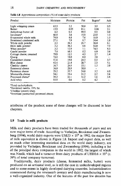
18 DAIRY CHEMISTRY AND BIOCHEMISTRY
Table 1.8 Approximate composition (%) of some dairy products
Product Moisture Protein Fat Sugars" Ash
Light whipping cream 63.5 2.2 30.9 3.0 0.5 Butter 15.9 0.85 81.1 0.06 2.1 Anhydrous butter oil 0.2 0.3 99.5 0.0 0.0 Ice-creamb 60.8 3.6 10.8 23.8 1 .o Evaporated whole milk 74.0 6.8 7.6 10.0 1.5 Sweetened condensed milk 27.1 7.9 8.7 54.4 1.8 Whole milk powder 2.5 26.3 26.7 38.4 6.1
Whey powder' 3.2 12.9 1.1 74.5 8.3 Casein powder 7.0 88.5 0.2 0.0 3.8 Cottage cheese, creamed 79.0 12.5 4.5 2.7 1.4 Qua% 72.0 18.0 8.0 3.0 -
Camembert cheese 51.8 19.8 24.3 0.5 3.7 Blue cheese 42.4 21.4 28.7 2.3 5.1 Cheddar cheese 36.7 24.9 33.1 1.3 3.9 Emmental cheese 36.0 28.9 30.0 - -
Parmesan cheese 29.2 35.7 24.8 3.2 6.0 Mozzarella cheese 54.1 19.4 31.2 2.2 2.6 Processed cheesed 39.2 22.1 31.2 1.6 5.8 Acid whey 93.9 0.6 0.2 4.2 -
"Total carbohydrate. bHardened vanilla, 19% fat. 'Cheddar (sweet) whey. dArnerican pasteurized processed cheese.
Skim milk powder 3.2 36.2 0.8 52.0 7.9
attributes of the product; some of these changes will be discussed in later chapters.
1.9 Trade in milk products
Milk and dairy products have been traded for thousands of years and are now major items of trade. According to Verheijen, Brockman and Zwanen- berg (1994), world dairy exports were U S 2 3 x lo9 in 1992; the major flow of milk equivalent is shown in Figure 1.8. Import and export data, as well as much other interesting statistical data on the world dairy industry, are provided by Verheijen, Brockman and Zwanenberg (1994), including a list of the principal dairy companies in the world in 1992, the largest of which was Nestle, which had a turnover from dairy products of US$10.6 x lo9 (c. 39% of total company turnover).
Traditionally, dairy products (cheese, fermented milks, butter) were produced on an artisanal level, as is still the case in underdeveloped regions and to some extent in highly developed dairying countries. Industrialization commenced during the nineteenth century and dairy manufacturing is now a well-organized industry. One of the features of the past few decades has
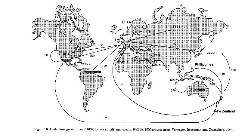
Figure 1.8 Trade flows greater than 250000tonnes in milk equivalents, 1992 (in 1000tonnes) (from Verheigen, Brockrnan and Zwaneberg, 1994).
20 DAIRY CHEMISTRY AND BIOCHEMISTRY
been the amalgamation of smaller dairy companies both within countries, and, recently, internationally. Such developments have obvious advantages in terms of efficiency and standardization of product quality but pose the risk of over-standardization with the loss of variety. Greatest diversity occurs with cheeses and, fortunately in this case, diversity is being preserved and even extended.
References
I D F (1993) Consumption Statistics f o r Milk and Milk Products. Bulletin 282, International
I D F (1995) Consumption Statistics f o r Milk and Milk Products. Bulletin 301, International
I D F (1996) The World Dairy Situation, 1996. Bulletin 314, International Dairy Federation,
Jensen, R.G. (ed.) (1995) Handbook of Milk Composition, Academic Press, San Diego. Linzell, J.L. (1972) Milk yield, energy loss, and mammary gland weight in different species.
Mepham, T.B. (1987) Physiology of Lactation, Open University Press, Milton Keynes, UK. Patton, S. (1969) Milk. Sci. Am., 221, 58-68. Tucker, H.A. (1969) Factors affecting mammary gland cell numbers. J . Dairy Sci., 52, 720-9. Verheigen, J.A.G., Brockman, J.E. and Zwanenberg, A.C.M. (1994) The World Dairy Industry:
Dairy Federation, Brussels.
Dairy Federation, Brussels.
Brussels.
Dairy Sci. Abstr., 34, 351-60.
Deselopments and Strategy, Rabobank Nederland, Amsterdam.
Suggested reading
Cowie, A.T. and Tindal, J.S. (1972) T h e Physiology of Lactation, Edward Arnold, London. Jensen, R.G. (ed.) (1995) Handbook of Milk Composition, Academic Press, San Diego. Larson, B.L. and Smith, V.R. (1 974- 1979) Lactation: A Comprehensive Treatise, Academic
Mepham, T.B. (1975) The Secretion of Milk , Studies in Biology Series No. 60, Edward Arnold,
Mepham, T.B. (ed.) (1983) Biochemistry of Lactation, Elsevier, Amsterdam. Mepham, T.B. (1987) Physiology of Lactation, Open University Press, Milton Keynes, UK.
Press, New York, Vols 1-4.
London.
2 Lactose
2.1 Introduction
Lactose is the principal carbohydrate in the milks of all mammals; non- mammalian sources are very rare. Milk contains only trace amounts of other sugars, including glucose (50 mg l-’), fructose, glucosamine, galac- tosamine, neuraminic acid and neutral and acidic oligosaccharides.
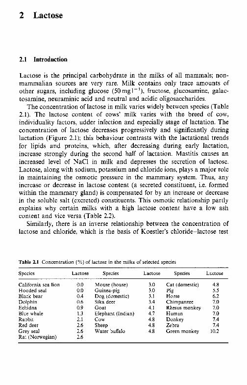
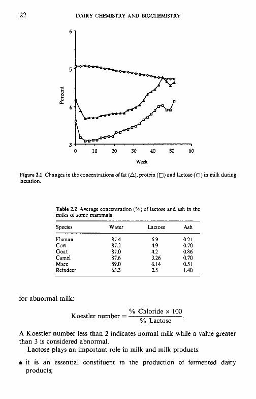
The concentration of lactose in milk varies widely between species (Table 2.1). The lactose content of cows’ milk varies with the breed of cow, individuality factors, udder infection and especially stage of lactation. The concentration of lactose decreases progressively and significantly during lactation (Figure 2.1); this behaviour contrasts with the lactational trends for lipids and proteins, which, after decreasing during early lactation, increase strongly during the second half of lactation. Mastitis causes an increased level of NaCl in milk and depresses the secretion of lactose. Lactose, along with sodium, potassium and chloride ions, plays a major role in maintaining the osmotic pressure in the mammary system. Thus, any increase or decrease in lactose content (a secreted constituent, i.e. formed within the mammary gland) is compensated for by an increase or decrease in the soluble salt (excreted) constituents. This osmotic relationship partly explains why certain milks with a high lactose content have a low ash content and vice versa (Table 2.2) .
Similarly, there is an inverse relationship between the concentration of lactose and chloride, which is the basis of Koestler’s chloride-lactose test
Table 2.1 Concentration (%) of lactose in the milks of selected species
Species Lactose Species Lactose Species Lactose
California sea lion Hooded seal Black bear Dolphin Echidna Blue whale Rabbit Red deer Grey seal Rat (Norwegian)
0.0 0.0 0.4 0.6 0.9 1.3 2.1 2.6 2.6 2.6
Mouse (house) Guinea-pig Dog (domestic) Sika deer Goat Elephant (Indian) c o w Sheep Water buffalo
3.0 3.0 3.1 3.4 4.1 4.7 4.8 4.8 4.8
Cat (domestic) Pig Horse Chimpanzee Rhesus monkey Human Donkey Zebra Green monkey
4.8 5.5 6.2 7.0 7.0 7.0 7.4 7.4
10.2
22 DAIRY CHEMISTRY AND BIOCHEMISTRY
5
3 0 10 20 30 40 50 60
Week
Figure 2.1 Changes in the concentrations of fat (A), protein (0) and lactose (0) in milk during lactation.
Table 2.2 Average concentration (%) of lactose and ash in the milks of some mammals
Species Water Lactose Ash
Human 87.4 6.9 0.21 cow 87.2 4.9 0.70 Goat 87.0 4.2 0.86 Camel 87.6 3.26 0.70 Mare 89.0 6.14 0.51 Reindeer 63.3 2.5 1.40
for abnormal milk:
YO Chloride x 100 Koestler number = YO Lactose
A Koestler number less than 2 indicates normal milk while a value greater than 3 is considered abnormal.
Lactose plays an important role in milk and milk products:
products; 0 it is an essential constituent in the production of fermented dairy
LACTOSE 23
0 it contributes to the nutritive value of milk and its products; however, many non-Europeans have limited or zero ability to digest lactose in adulthood, leading to a syndrome known as lactose intolerance;
0 it affects the texture of certain concentrated and frozen products; 0 it is involved in heat-induced changes in the colour and flavour of highly
heated milk products.
2.2 Chemical and physical properties of lactose
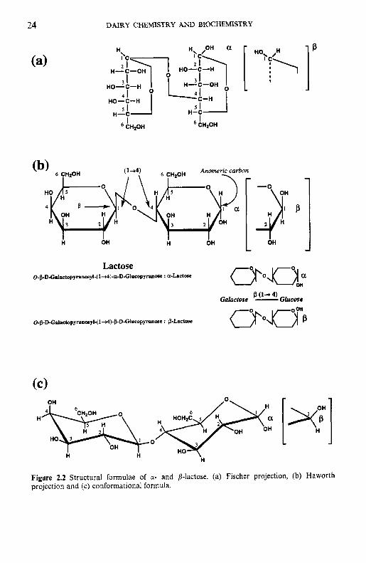
2.2.1 Structure of lactose
Lactose is a disaccharide consisting of galactose and glucose, linked by a pl-4 glycosidic bond (Figure 2.2). Its systematic name is j3-0-D-galac- topyranosyl-( 1 -4)-ol-~-glucopyranose (a-lactose) or P-0-D-galactopyranosyl- (1-4)-P-~-glucopyranose (p-lactose). The hemiacetal group of the glucose moiety is potentially free (i.e. lactose is a reducing sugar) and may exist as an a- or p-anomer. In the structural formula of the a-form, the hydroxyl group on the C , of glucose is cis to the hydroxyl group at C, (oriented downward).
2.2.2 Biosynrhesis of lactose
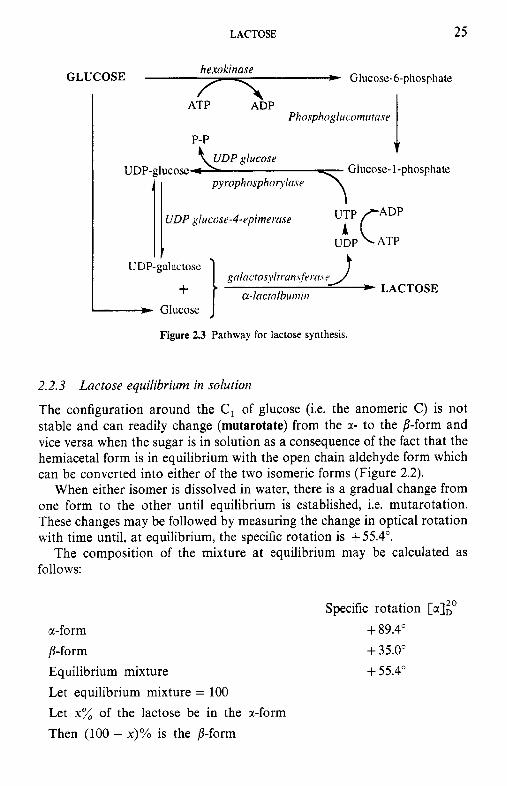
Lactose is essentially unique to mammary secretions. It is synthesized from glucose absorbed from blood. One molecule of glucose is isomerized to UDP-galactose via the four-enzyme Leloir pathway (Figure 2.3). UDP-Gal is then linked to another molecule of glucose in a reaction catalysed by the enzyme, lactose synthetase, a two-component enzyme. Component A is a non-specific galactosyl transferase which transfers the galactose from UDP- Gal to a number of acceptors. In the presence of the B component, which is the whey protein, a-lactalbumin, the transferase becomes highly specific for glucose (its K , decreases 1000-fold), leading to the synthesis of lactose. Thus, r-lactalbumin is an enzyme modifier and its concentration in the milk of several species is directly related to the concentration of lactose in those milks; the milks of some marine mammals contain neither a-lactalbumin nor lactose.
The presumed significance of this control mechanism is to enable mammals to terminate the synthesis of lactose when necessary, i.e. to regulate and control osmotic pressure when there is an influx of NaC1, e.g. during mastitis or in late lactation (lactose and NaCl are major determi- nants of the osmotic pressure of milk, which is isotonic with blood, the osmotic pressure of which is essentially constant). The ability to control osmotic pressure is sufficiently important to justify an elaborate control mechanism and the ‘wastage’ of the enzyme modifier.
DAIRY CHEMISTRY AND BIOCHEMISTRY
H B
H-C-OH
HO-C-H
HO-C-H
H-C I H-C
' CHzOH I CHzOH
-$ OH
O-&D-CPLPetopyrPnaPyl~i~)-@-D-Glucopy~naPe : @.Lactose
4 OH OH [xy n
n 2
3 0 HO 3
H HO
OH
H
Figure 2.2 Structural formulae of a- and p-lactose. (a) Fischer projection, (b) Haworth projection and (c) conformational formula.
LACTOSE 25
Glucose- 1 -phoSPhE
UDP gliiccisr-4-rpinier.osr
gnlncros~llrr~~l~?.\:fr,.cl.vr *LACTOSE cr-I~/ctnlDu/ttil?
Glucose
Figure 2.3 Pathway for lactose synthesis.
2.2.3 Lactose equilibrium in solution
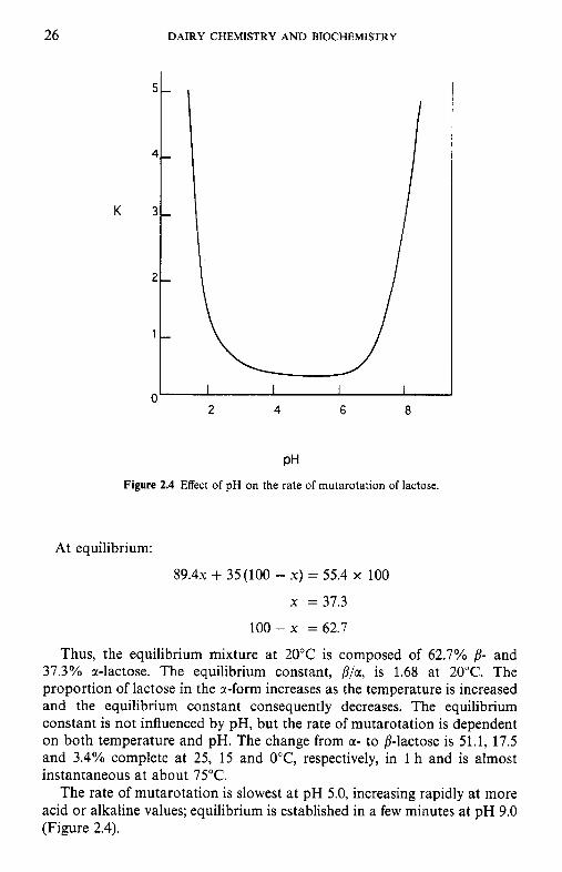
The configuration around the C , of glucose (i.e. the anomeric C) is not stable and can readily change (mutarotate) from the x - to the /?-form and vice versa when the sugar is in solution as a consequence of the fact that the hemiacetal form is in equilibrium with the open chain aldehyde form which can be converted into either of the two isomeric forms (Figure 2.2).
When either isomer is dissolved in water, there is a gradual change from one form to the other until equilibrium is established, i.e. mutarotation. These changes may be followed by measuring the change in optical rotation with time until, at equilibrium, the specific rotation is + 55.4".
The composition of the mixture at equilibrium may be calculated as follows:
Specific rotation [NIP a-form + 89.4" p-form + 35.0" Equilibrium mixture + 55.4" Let equilibrium mixture = 100 Let x% of the lactose be in the cr-form Then (100 - x)% is the p-form
26 DAIRY CHEMISTRY AND BIOCHEMISTRY
oL I I I I
2 4 6 8
PH Figure 2.4 Effect of pH on the rate of mutarotation of lactose.
At equilibrium:
8 9 . 4 ~ + 35(100 - X) = 55.4 x 100
x = 37.3
100-x = 62.7
Thus, the equilibrium mixture at 20°C is composed of 62.7% 8- and 37.3% a-lactose. The equilibrium constant, P/a, is 1.68 at 20°C. The proportion of lactose in the @-form increases as the temperature is increased and the equilibrium constant consequently decreases. The equilibrium constant is not influenced by pH, but the rate of mutarotation is dependent on both temperature and pH. The change from m- to p-lactose is 51.1, 17.5 and 3.4% complete at 25, 15 and O"C, respectively, in 1 h and is almost instantaneous at about 75°C.
The rate of mutarotation is slowest at pH 5.0, increasing rapidly at more acid or alkaline values; equilibrium is established in a few minutes at pH 9.0 (Figure 2.4).
LACTOSE 27
2.2.4 Signgcance of mutarotation
The a- and 8-forms of lactose differ with respect to:
0 solubility; 0 crystal shape and size; 0 hydration of crystal form - hygroscopicity; 0 specific rotation; 0 sweetness.
Many of these characteristics are discussed in the following sections.
2.2.5 Solubility of lactose
The solubility characteristics of the a- and /?-isomers are distinctly different. When a-lactose is added in excess to water at 20°C, about 7 g per 100 g water dissolve immediately. Some a-lactose mutarotates to the 8 anomer to establish the equilibrium ratio 62.78 : 37.3~; therefore, the solution becomes unsaturated with respect to a and more a-lactose dissolves. These two processes (mutarotation and solubilization of a-lactose) continue until two criteria are met: - 7 g a-lactose in solution and a P/a ratio of 1.6 : 1.0. Since the P/sc ratio at equilibrium is about 1.6 at 20"C, the final solubility is 7 g + (1.6 x 7) g = 18.2 g per 100 g water.
When /-lactose is dissolved in water, the initial solubility is -50g per 100 g water at 20°C. Some /?-lactose mutarotates to a to establish a ratio of 1.6: 1. At equilibrium, the solution would contain 30.8 g /? and 19.2 g a/100 ml; therefore, the solution is supersaturated with a-lactose, some of which crystallizes, upsetting the equilibrium and leading to further mutaro- tation of /? -+ a. These two events, i.e. crystallization of a-lactose and mutarotation of 8, continue until the same two criteria are met, i.e. -7g a-lactose in solution and a P/a ratio of 1.6: 1. Again, the final solubility is - 18.2 g lactose per 100 g water. Since 8-lactose is much more soluble than a and mutarotation is slow, it is possible to form more highly concentrated solutions by dissolving /?- rather than a-lactose. In either case, the final solubility is the same.
The solubility of lactose as a function of temperature is summarized in Figure 2.5. The solubility of a-lactose is more temperature dependent than that of /?-lactose and the solubility curves intersect at 93.5"C. A solution at 60°C contains approximately 59g lactose per lOOg water. Suppose that a 50% solution of lactose (- 30 g p- and 20 g a-) at 60°C is cooled to 15°C. At this temperature, the solution can contain only 7 g a-lactose or a total of 18.2 g per 100 g water at equilibrium. Therefore, lactose will crystallize very slowly out of solution as irregularly sized crystals which may give rise to a sandy, gritty texture.
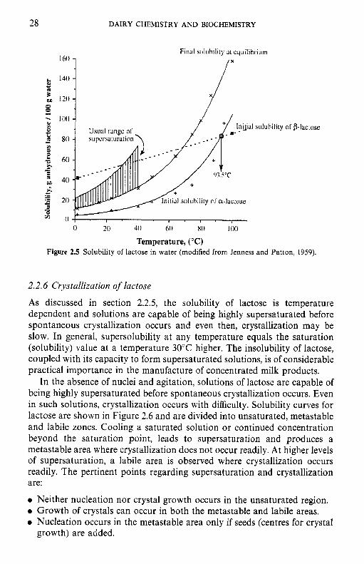
Solu
bilit
y, g
anh
ydro
us la
ctos
e I1
00 g
wat
er
--
*-
s
gg
gg
sg
gg
LACTOSE
200
100
29
-
2.1 - 1
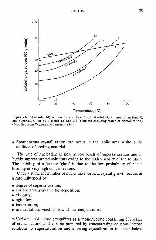
Figure 2.6 Initial solubility of a-lactose and b-lactose, final solubility a t equilibrium (line l), and supersaturation by a factor 1.6 and 2.1 (r-lactose excluding water of crystallization). (Modified from Walstra and Jenness, 1984.)
Spontaneous crystallization can occur in the labile area without the addition of seeding material.
The rate of nucleation is slow at low levels of supersaturation and in highly supersaturated solutions owing to the high viscosity of the solution. The stability of a lactose 'glass' is due to the low probability of nuclei forming at very high concentrations.
Once a sufficient number of nuclei have formed, crystal growth occurs at rate influenced by:
degree of supersaturation; surface area available for deposition; viscosity ; agitation; temperature; mutarotation, which is slow at low temperatures.
?-Hydrate. cc-Lactose crystallizes as a monohydrate containing 5% water of crystallization and can be prepared by concentrating aqueous lactose solutions to supersaturation and allowing crystallization to occur below
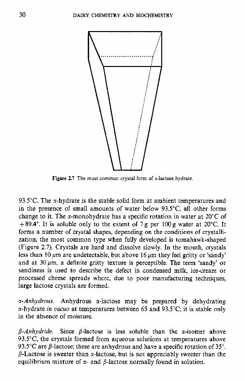
30 DAIRY CHEMISTRY A N D BIOCHEMISTRY
Figure 2.7 The most common crystal form of a-lactose hydrate.
93.5"C. The a-hydrate is the stable solid form at ambient temperatures and in the presence of small amounts of water below 93.5"C, all other forms change to it. The a-monohydrate has a specific rotation in water at 20°C of +89.4". It is soluble only to the extent of 7g per 1OOg water at 20°C. It forms a number of crystal shapes, depending on the conditions of crystalli- zation; the most common type when fully developed is tomahawk-shaped (Figure 2.7). Crystals are hard and dissolve slowly. In the mouth, crystals less than 10 pm are undetectable, but above 16 pm they feel gritty or 'sandy' and at 30pm, a definite gritty texture is perceptible. The term 'sandy' or sandiness is used to describe the defect in condensed milk, ice-cream or processed cheese spreads where, due to poor manufacturing techniques, large lactose crystals are formed.
a-Anhydrous. Anhydrous a-lactose may be prepared by dehydrating a-hydrate in V ~ C U O at temperatures between 65 and 93.5"C; it is stable only in the absence of moisture.
B-Anhydride. Since /%lactose is less soluble than the a-isomer above 93.5"C, the crystals formed from aqueous solutions at temperatures above 93.5"C are p-lactose; these are anhydrous and have a specific rotation of 35". /%Lactose is sweeter than a-lactose, but is not appreciably sweeter than the equilibrium mixture of a- and p-lactose normally found in solution.
LACTOSE 31
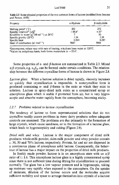
Table 2.3 Some physical properties of the two common forms of lactose (modified from Jenness and Patton, 1959)
Property a-H ydrate 8- Anhydride
Melting point" ("C) 202 252
Solubility in water (g 100 rn1-l) at 20°C I 50 Specific gravity (20°C) 1.54 1.59 Specific heat 0.299 0.285 Heat of combustion (kJ mol-') 5687 5946
"Decomposes; values vary with rate of heating, ti-hydrate loses water at 120°C. bValues on anhydrous basis, both forms mutarotate to f55.4".
Specific rotaltionb [a]:' + 89.4" +35"
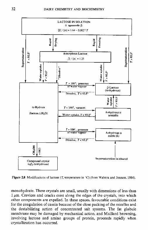
Some properties of c(- and !-lactose are summarized in Table 2.3. Mixed a/! crystals, e.g. asp3, can be formed under certain conditions. The relation- ship between the different crystalline forms of lactose is shown in Figure 2.8.
Lactose glass. When a lactose solution is dried rapidly, viscosity increases so quickly that crystallization is impossible. A noncrystalline form is produced containing a- and !-forms in the ratio at which they exist in solution. Lactose in spray-dried milk exists as a concentrated syrup or amorphous glass which is stable if protected from air, but is very hygro- scopic and absorbs water rapidly from the atmosphere, becoming sticky.
2.2.7 Problems related to lactose crystallization
The tendency of lactose to form supersaturated solutions that do not crystallize readily causes problems in many dairy products unless adequate controls are exercised. The problems are due primarily to the formation of large crystals, which cause sandiness, or to the formation of a lactose glass, which leads to hygroscopicity and caking (Figure 2.9).
Dried milk and whey. Lactose is the major component of dried milk products: whole-milk powder, skim-milk powder and whey powder contain c. 30, 50 and 70% lactose, respectively. Protein, fat and air are dispersed in a continuous phase of amorphous solid lactose. Consequently, the behav- iour of lactose has a major impact on the properties of dried milk products.
In freshly made powder, lactose is in an amorphous state with an a/! ratio of 1 : 1.6. This amorphous lactose glass is a highly concentrated syrup since there is not sufficient time during drying for crystallization to proceed normally. The glass has a low vapour pressure and is hygroscopic, taking up moisture very rapidly when exposed to the atmosphere. On the uptake of moisture, dilution of the lactose occurs and the molecules acquire sufficient mobility and space to arrange themselves into crystals of a-lactose
32
LACTOSE IN SOLUTION a-b
[Pl/[al= 1.64-0 .0027T
DAIRY CHEMISTRY AND BIOCHEMISTRY
L .-
Amorphous Lactose
V
a-Hydrate
(lactose. I.H,O)
Conipound crystal asp3 (anhydrous)
[ p] / [a] = 1.25 I
T = IOW, presence 1
01 \ v a y
L I I Anhydrous a
unstable - 1 Water uptake, T < !USo I stable (S)
T I 150", presence 01 water vapour
Dissolve, T c 93.5' . . . . . . . . . . . . . . . . . . . . . . .
Silpersatiiration in ethanol
Figure 2.8 Modifications of lactose (T temperature in 'C) (from Walstra and Jenness, 1984).
monohydrate. These crystals are small, usually with dimensions of less than 1 pm. Crevices and cracks exist along the edges of the crystals, into which other components are expelled. In these spaces, favourable conditions exist for the coagulation of casein because of the close packing of the micelles and the destabilizing action of concentrated salt systems. The fat globule membrane may be damaged by mechanical action, and Maillard browning, involving lactose and amino groups of protein, proceeds rapidly when crystallization has occurred.
LACTOSE 33
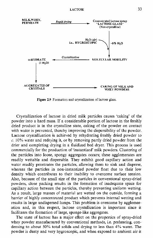
Rapid drying Concentrated lactose syrup MILK,WHEY, PERMEATE * “LACTOSE GLASS”
(Non-crystalline)
a-HYDRATE Cryslallizatioir
4 MOLECULAR MOBILITY
CAKING OF MILK AND WHEY POWDERS
AGGREGATES OF CRYSTALS
Figure 2.9 Formation and crystallization of lactose glass.
Crystallization of lactose in dried milk particles causes ‘caking’ of the powder into a hard mass. If a considerable portion of lactose in the freshly dried product is in the crystalline state, caking of the powder on contact with water is prevented, thereby improving the dispersibility of the powder. Lactose crystallization is achieved by rehydrating freshly dried powder to c. 10% water and redrying it, or by removing partly dried powder from the drier and completing drying in a fluidized bed dryer. This process is used commercially for the production of ‘instantized’ milk powders. Clustering of the particles into loose, spongy aggregates occurs; these agglomerates are readily wettable and dispersible. They exhibit good capillary action and water readily penetrates the particles, allowing them to sink and disperse, whereas the particles in non-instantized powder float due to their low density which contributes to their inability to overcome surface tension. Also, because of the small size of the particles in conventional spray-dried powders, close packing results in the formation of inadequate space for capillary action between the particles, thereby preventing uniform wetting. As a result, large masses of material are wetted on the outside, forming a barrier of highly concentrated product which prevents internal wetting and results in large undispersed lumps. This problem is overcome by agglomer- ation and, in this respect, lactose crystallization is important since it facilitates the formation of large, sponge-like aggregates.
The state of lactose has a major effect on the properties of spray-dried whey powder manufactured by conventional methods, i.e. preheating, con- densing to about 50% total solids and drying to less than 4% water. The powder is dusty and very hygroscopic, and when exposed to ambient air it
34 DAIRY CHEMISTRY AND BIOCHEMISTRY
has a pronounced tendency to cake owing to its very high lactose content ( - 70%).
Problems arising from the crystallization of lactose in milk and whey powders may also be avoided or controlled by pre-crystallizing the lactose. Essentially, this involves adding finely divided lactose powder which acts as nuclei on which the supersaturated lactose crystallizes. Addition of 0.5 kg of finely ground lactose to the amount of concentrated product (whole milk, skim milk or whey) containing 1 tonne of lactose will induce the formation of c. lo6 crystals ml- l , about 95% of which will have dimensions less than 10pm and 100% less than 15 pm, i.e. too small to cause textural defects.
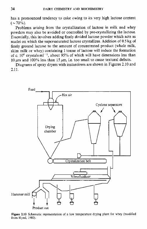
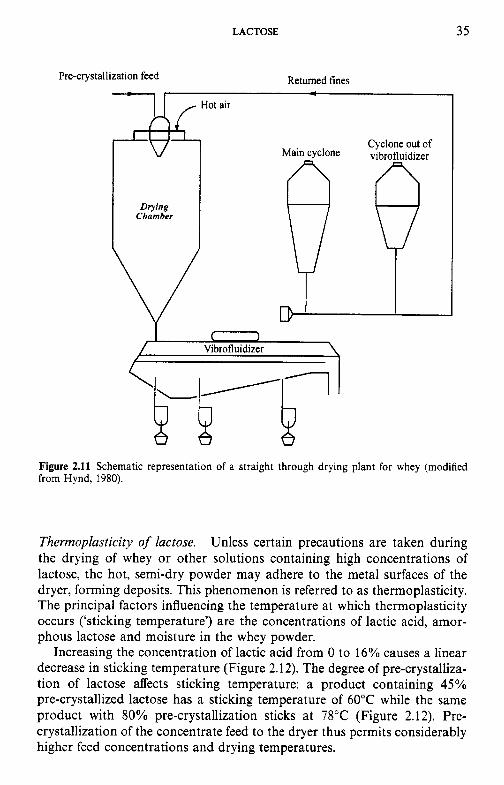
Diagrams of spray dryers with instantizers are shown in Figures 2.10 and 2.11.
Feed-[ I /Hot air
Cyclone separators (?/
I I I 1
Crystalization belt
b Vibrofluidizer
Hammer mill
Product out
Figure 2.10 Schematic representation of a low temperature drying plant for whey (modified from Hynd, 1980).
LACTOSE 35
Figure 2.11 Schematic representation of a straight through drying plant for whey (modified from Hynd, 1980).
Thermoplasticity of lactose. Unless certain precautions are taken during the drying of whey or other solutions containing high concentrations of lactose, the hot, semi-dry powder may adhere to the metal surfaces of the dryer, forming deposits. This phenomenon is referred to as thermoplasticity. The principal factors influencing the temperature at which thermoplasticity occurs (‘sticking temperature’) are the concentrations of lactic acid, amor- phous lactose and moisture in the whey powder.
Increasing the concentration of lactic acid from 0 to 16% causes a linear decrease in sticking temperature (Figure 2.12). The degree of pre-crystalliza- tion of lactose affects sticking temperature: a product containing 45% pre-crystallized lactose has a sticking temperature of 60°C while the same product with 80% pre-crystallization sticks at 78°C (Figure 2.12). Pre- crystallization of the concentrate feed to the dryer thus permits considerably higher feed concentrations and drying temperatures.
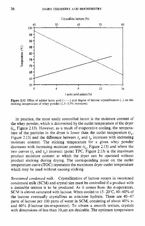
36 DAIRY CHEMISTRY AND BIOCHEMISTRY
Crystalline lactose (%)
45 55 65 75 1 I I
3
0 4 8 12 16
Lactic acid added (%)
Figure 2.12 Effect of added lactic acid ( - - - - ) and degree of lactose crystallization (-) on the sticking temperature of whey powder (1.5-3.5% moisture).
In practice, the most easily controlled factor is the moisture content of the whey powder, which is determined by the outlet temperature of the dryer (to, Figure 2.13). However, as a result of evaporative cooling, the tempera- ture of the particles in the dryer is lower than the outlet temperature (tp, Figure 2.13) and the difference between to and t , increases with increasing moisture content. The sticking temperature for a given whey powder decreases with increasing moisture content ( t s , Figure 2.13) and where the two curves ( t , and t,) intersect (point TPC, Figure 2.13) is the maximum product moisture content at which the dryer can be operated without product sticking during drying. The corresponding point on the outlet temperature curve (TOC) represents the maximum dryer outlet temperature which may be used without causing sticking.
Sweetened condensed milk. Crystallization of lactose occurs in sweetened condensed milk (SCM) and crystal size must be controlled if a product with a desirable texture is to be produced. As it comes from the evaporators, SCM is almost saturated with lactose. When cooled to 15-20°C, 40-60% of the lactose eventually crystallizes as a-lactose hydrate. There are 40-47 parts of lactose per 100 parts of water in SCM, consisting of about 40% a- and 60% /?-lactose (ex-evaporator). To obtain a smooth texture, crystals with dimensions of less than 10 pm are desirable. The optimum temperature
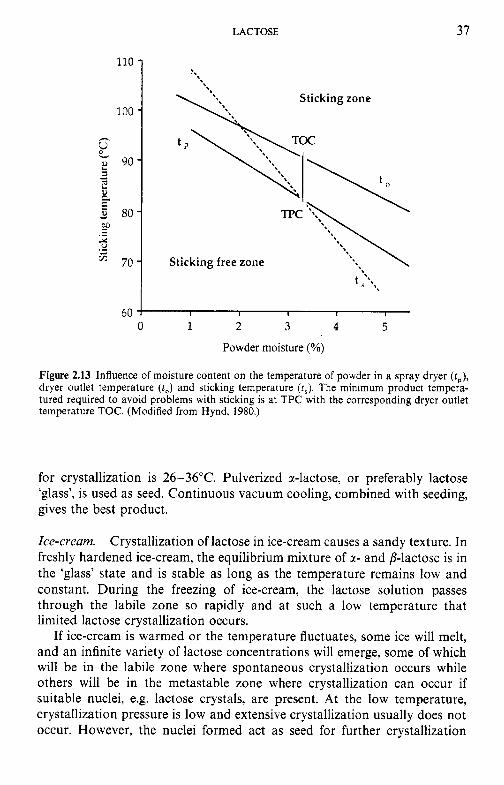
LACTOSE 37
110 -
100 - h
9 0 - * : * E 80-
E ?L
2 3 .- ‘ 70-
60 0 1 2 3 4 5
Powder moisture (%)
Figure 2.13 Influence of moisture content on the temperature of powder in a spray dryer (t,), dryer outlet temperature ( t o ) and sticking temperature (fJ The minimum product tempera- tured required to avoid problems with sticking is at TPC with the corresponding dryer outlet temperature TOC. (Modified from Hynd, 1980.)
for crystallization is 26-36°C. Pulverized @-lactose, or preferably lactose ‘glass’, is used as seed. Continuous vacuum cooling, combined with seeding, gives the best product.
Ice-cream. Crystallization of lactose in ice-cream causes a sandy texture. In freshly hardened ice-cream, the equilibrium mixture of a- and p-lactose is in the ‘glass’ state and is stable as long as the temperature remains low and constant. During the freezing of ice-cream, the lactose solution passes through the labile zone so rapidly and at such a low temperature that limited lactose crystallization occurs.
If ice-cream is warmed or the temperature fluctuates, some ice will melt, and an infinite variety of lactose concentrations will emerge, some of which will be in the labile zone where spontaneous crystallization occurs while others will be in the metastable zone where crystallization can occur if suitable nuclei, e.g. lactose crystals, are present. At the low temperature, crystallization pressure is low and extensive crystallization usually does not occur. However, the nuclei formed act as seed for further crystallization
38 DAIRY CHEMISTRY AND BIOCHEMISTRY
when the opportunity arises and they tend to grow slowly with time, eventually causing a sandy texture. The defect is controlled by limiting the milk solids content or by using /?-galactosidase to hydrolyse lactose.
Other frozen dairy products. Although milk may become frozen inadver- tently, freezing is not a common commercial practice. However, concen- trated or unconcentrated milks are sometimes frozen commercially, e.g. to supply remote locations (as an alternative to dried or UHT milk), to store sheep's or goats' milk, production of which is seasonal, or human milk for infant feeding in emergencies (milk banks).
As will be discussed in Chapter 3, freezing damages the milk fat globule membrane, resulting in the release of 'free fat'. The casein system is also destabilized due to a decrease in pH and an increase in Ca2+ concentration, both caused by the precipitation of soluble CaH2P0, and/or Ca,HPO, as Ca,(PO,),, with the release of H + (Chapter 5); precipitation of Ca,(PO,), occurs on freezing because pure water crystallizes, causing an increase in soluble calcium phosphate with which milk is already saturated. Crystalli- zation of lactose as a-hydrate during frozen storage aggravates the problem by reducing the amount of solvent water available.
In frozen milk products, lactose crystallization causes instability of the casein system. On freezing, supersaturated solutions of lactose are formed: e.g. in concentrated milk at -8"C, 25% of the water is unfrozen and contains 80 g lactose per 100 g, whereas the solubility of lactose at - 8°C is only about 7%. During storage at low temperatures, lactose crystallizes slowly as a monohydrate and consequently the amount of free water in the product is reduced.
The formation of supersaturated lactose solutions inhibits freezing, and consequently stabilizes the concentration of solutes in solution. However, when lactose crystallizes, water freezes and the concentration of other solutes increases markedly (Table 2.4).
Table 2.4 Comparison of ultrafiltrate from liquid and frozen skim milk
Ultrafiltrate of liquid portion of Constituent Ultrafiltrate of skim milk frozen concentrated milk
PH 6.1 5.8 Chloride (mM) 34.9 459 Citrate (mM) 8.0 89 Phosphate (mM) 10.5 84 Sodium (mM) 19.7 218 Potassium (mM) 38.5 393 Calcium (mM) 9.1 59
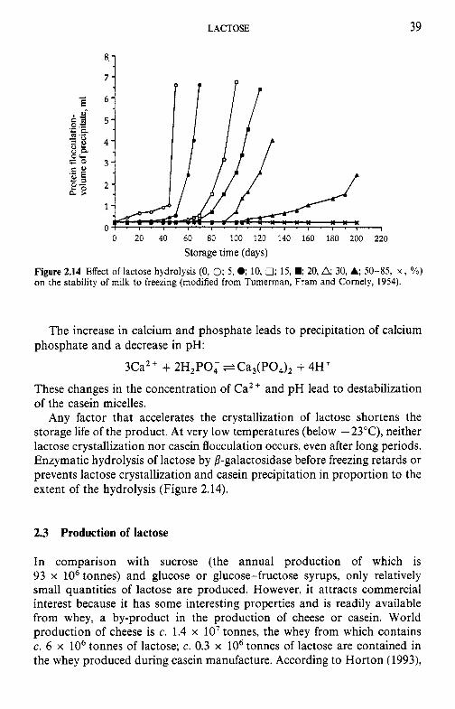
Po
8.
3
C.
1
Prot
ein
flocc
ulat
ion-
%
P
EL
vo
lum
e of
pre
cipi
tate
, ml
Ern
f
ur
W
W
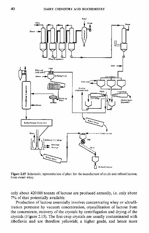
40 DAIRY CHEMISTRY AND BIOCHEMISTRY
I t / W"
1
ii
7 Srrd
,
Figure 2.15 Schematic representation of plant for the manufacture of crude and refined lactose, from sweet whey.
only about 420000 tonnes of lactose are produced annually, i.e. only about 7 % of that potentially available.
Production of lactose essentially involves concentrating whey or ultrafil- tration permeate by vacuum concentration, crystallization of lactose from the concentrate, recovery of the crystals by centrifugation and drying of the crystals (Figure 2.15). The first-crop crystals are usually contaminated with riboflavin and are therefore yellowish; a higher grade, and hence more
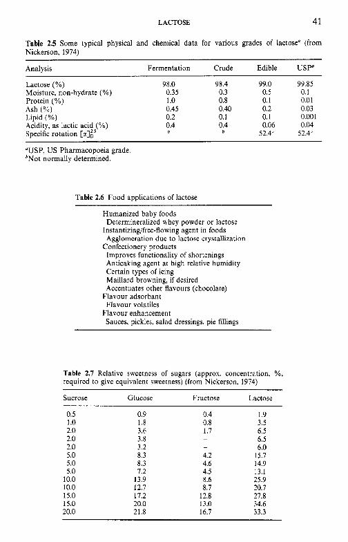
LACTOSE 41
Table 2.5 Some typical physical and chemical data for various grades of lactose" (from Nickerson, 1974)
Analysis Fermentation Crude Edible U S P
Lactose (YO) 98.0 98.4 99.0 99.85 Moisture, non-hydrate (YO) 0.35 0.3 0.5 0.1 Protein (YO) 1 .o 0.8 0.1 0.01 Ash (%) 0.45 0.40 0.2 0.03 Lipid (YO) 0.2 0.1 0.1 0.001 Acidity, as lactic acid (YO) 0.4 0.4 0.06 0.04 Specific rotation [%]i5 b b 52.4" 52.4"
"USP, US Pharmacopoeia grade. bNot normally determined.
Table 2.6 Food applications of lactose
Humanized baby foods
Instantizingifree-flowing agent in foods
Confectionery products
Determineralized whey powder or lactose
Agglomeration due to lactose crystallization
Improves functionality of shortenings Anticaking agent at high relative humidity Certain types of icing Maillard browning, if desired Accentuates other flavours (chocolate)
Flavour adsorbant Flavour volatiles
Flavour enhancement Sauces, pickles, salad dressings, pie fillings
Table 2.7 Relative sweetness of sugars (approx. concentration, YO, required to give equivalent sweetness) (from Nickerson, 1974)
Sucrose Glucose Fructose Lactose
0.5 1 .o 2.0 2.0 2.0 5.0 5.0 5.0
10.0 10.0 15.0 15.0 20.0
0.9 1.8 3.6 3.8 3.2 8.3 8.3 7.2
13.9 12.7 17.2 20.0 21.8
0.4 0.8 1.7
-
4.2 4.6 4.5 8.6 8.7
12.8 13.0 16.7
1.9 3.5 6.5 6.5 6.0
15.7 14.9 13.1 25.9 20.7 27.8 34.6 33.3
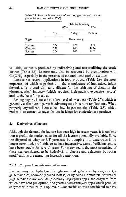
42 DAIRY CHEMISTRY AND BIOCHEMISTRY
Table 2.8 Relative humectancy of sucrose, glucose and lactose (% moisture absorbed at 20°C)
Relative humidity 60% 100%
l h 9 days 25 days
Sugar Humectancy
Lactose 0.54 1.23 1.38 Glucose 0.29 9.00 47.14 Sucrose 0.04 0.03 18.35
valuable, lactose is produced by redissolving and recrystallizing the crude lactose (Table 2.5). Lactose may also be recovered by precipitation with Ca(OH),, especially in the presence of ethanol, methanol or acetone.
Lactose has several applications in food products (Table 2.6), the most important of which is probably in the manufacture of humanized infant formulae. It is used also as a diluent for the tableting of drugs in the pharmaceutical industry (which requires high-quality, expensive lactose) and as the base for plastics.
Among sugars, lactose has a low level of sweetness (Table 2.7), which is generally a disadvantage but is advantageous in certain applications. When properly crystallized, lactose has low hygroscopicity (Table 2 . Q which makes it an attractive sugar for use in icings for confectionary products.
2.4 Derivatives of lactose
Although the demand for lactose has been high in recent years, it is unlikely that a profitable market exists for all the lactose potentially available. Since the disposal of whey or UF permeate by dumping into waterways is no longer permitted, profitable, or at least inexpensive, ways of utilizing lactose have been sought for several years. For many years, the most promising of these was considered to be hydrolysis to glucose and galactose, but other modifications are attracting increasing attention.
2.4.1 Enzymatic mod$cation of lactose
Lactose may be hydrolysed to glucose and galactose by enzymes (p- galactosidases, commonly called lactase) or by acids. Commercial sources of 8-galactosidase are moulds (especially Aspergillus spp.), the enzymes from which have acid pH optima, and yeasts (Kluyveromyces spp.) which produce enzymes with neutral pH optima. P-Galactosidases were considered to have
LACTOSE 43
considerable commercial potential as a solution to the ‘whey problem’ and for the treatment of lactose intolerance (section 2.6.1). The very extensive literature on various aspects of P-galactosidases and on their application in free or immobilized form has been reviewed by Mahoney (1997). Techno- logical problems in the production of glucose-galactose syrups have been overcome but the process is not commercially successful. Glucose-galactose syrups are not economically competitive with glucose or glucose-fructose syrups produced by hydrolysis of maize starch, unless the latter are heavily taxed. As discussed in section 2.6.1, an estimated 70% of the adult human population have inadequate intestinal P-galactosidase activity and are therefore lactose intolerant; the problem is particularly acute among Asians and Africans. Pre-hydrolysis of lactose was considered to offer the potential to develop new markets for dairy products in those countries. Various protocols are available: addition of P-galactosidase to milk in the home, pre-treatment at the factory with free or immobilized enzyme or aseptic addition of sterilized free P-galactosidase to UHT milk, which appears to be particularly successful. However, the method is not used widely and it is now considered that the treatment of milk with P-galactosidase will be commer- cially successful only in niche markets.
Glucose-galactose syrups are about three times sweeter than lactose (70% as sweet as sucrose) and hence lactose-hydrolysed milk could be used in the production of ice-cream, yoghurt or other sweetened dairy products, permitting the use of less sucrose and reducing caloric content. However, such applications have not been commercially successful.
The glucose moiety can be isomerized to fructose by the well-established glucose isomerization process to yield a galactose-glucose-fructose syrup with increased sweetness. Another possible variation would involve the isomerization of lactose to lactulose (galactose-fructose) which can be hydrolysed to galactose and fructose by some P-galactosidases.
8-Galactosidase has transferase as well as hydrolase activity and pro- duces oligosaccharides (galacto-oligosaccharides, Figure 2.16) which are later hydrolysed (Figure 2.17). This property may be a disadvantage since the oligosaccharides are not digestible by humans and reach the large intestine where they are fermented by bacteria, leading to the same problem caused by lactose. However, they stimulate the growth of BiJidobacteriurn spp. in the lower intestine; a product (oligonate, 6’-galactosyl lactose) is produced commercially by the Yokult Company in Japan for addition to infant formulae. Some galacto-oligosaccharides have interesting functional properties and may find commercial applications.
2.4.2 Chemical modifications
Lactulose. Lactulose is an epimer of lactose in which the glucose moiety is isomerized to fructose (Figure 2.18). The sugar does not occur naturally and
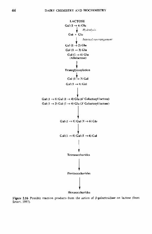
44 DAIRY CHEMISTRY AND BIOCHEMISTRY
Gal (1 4 2) Glu
Gal (1 + 3) Glu Gal (1 + 6) Glu
(Allolactose)
I Transglgcosylation
1 Gal (1 + 3) Gal
t Tetrasaccliaricles
Hexasaccharides
Figure 2.16 Possible reaction products from the action of 8-galactosidase on lactose (from Smart, 1993).
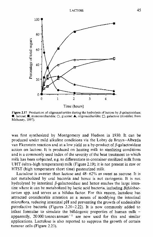
LACTOSE 45
so Lo
8
- 60 2 Lo
cd 0 cr 0 0
Y
Y
40 * 8 E fi 20
4
0 0 1 2 3 4
Time (hours) Figure 2.17 Production of oligosaccharides during the hydrolysis of lactose by 8-galactosidase; 0, lactose; ., monosaccharides; 0, glucose: A, oligosaccharides; 0, galactose (modified from Mahoney, 1997).
was first synthesized by Montgomery and Hudson in 1930. It can be produced under mild alkaline conditions via the Lobry de Bruyn-Alberda van Ekenstein reaction and at a low yield as a by-product of p-galactosidase action on lactose. It is produced on heating milk to sterilizing conditions and is a commonly used index of the severity of the heat treatment to which milk has been subjected, e.g. to differentiate in-container sterilized milk from UHT (ultra-high temperature) milk (Figure 2.19); it is not present in raw or HTST (high temperature short time) pasteurized milk.
Lactulose is sweeter than lactose and 48-62% as sweet as sucrose. It is not metabolized by oral bacteria and hence is not cariogenic. It is not hydrolysed by intestinal 8-galactosidase and hence reaches the large intes- tine where it can be metabolized by lactic acid bacteria, including Bifidobac- terium spp. and serves as a bifidus factor. For this reason, lactulose has attracted considerable attention as a means of modifying the intestinal microflora, reducing intestinal pH and preventing the growth of undesirable putrefactive bacteria (Figures 2.20-2.22). It is now commonly added to infant formulae to simulate the bifidogenic properties of human milk - apparently, 20000 tonnes annum-' are now used for this and similar applications. Lactulose is also reported to suppress the growth of certain tumour cells (Figure 2.23).
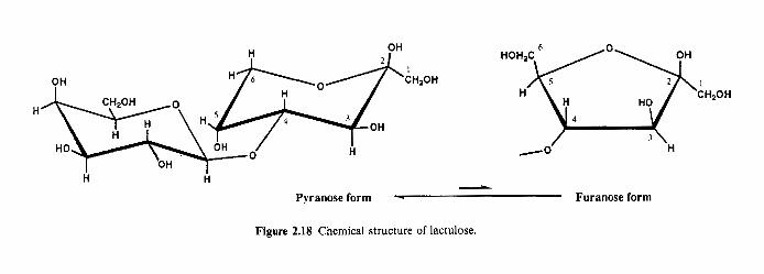
H % w A H 2 0 H CHzOH 5 H o ~ ~ o ~ , HO CH,OH
7 OH 4 4
H 7
HO OH 0 H -0 H OH
H H L
Pyranose form - Furanose form
Figure 2.18 Chemical structure of lactulose.

LACTOSE 47
0 I0 2LI 30 40 SO 60 7 0 X O 90 1 ( X ) 1 1 0 120
Lactulose concentration (mg 100 m1-l)
Figure 2.19 Concentration of lactulose in heated milk products (modified from Andrews, 1989).
LACTULOSE
Oral intake
- Non-absorption and migration Not carogenic to large intestine
Utilization by Bi@iobacterium and increme In bifldobacteria
Favourable change of intestinal microflora ,
n Production of organic acids I 1 I and lowering of intestinal pH 1
Ensuring intes- Lessening burdens tinal function to hepatic function
Suppression of inlestinal putrefactive bacteria
Suppression of production of harmful subsun&
Viumin synthesis
Stimulation of immune response
Figure 2.20 Significance of lactulose in health (modified from Tamura et al., 1993).
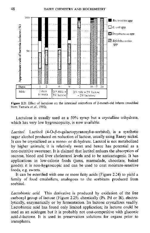
48 DAIRY CHEMISTRY AND BIOCHEMISTRY
Figure 2.21 Effect of lactulose on the intestinal microflora of 2-month-old infants (modified from Tamura et al., 1993).
Lactulose is usually used as a 50% syrup but a crystalline trihydrate, which has very low hygroscopicity, is now available.
Lactitol. Lactitol (4-O-~-~-galactopyranosyl-~-sorbitol), is a synthetic sugar alcohol produced on reduction of lactose, usually using Raney nickel. It can be crystallized as a mono- or di-hydrate. Lactitol is not metabolized by higher animals; it is relatively sweet and hence has potential as a non-nutritive sweetener. It is claimed that lactitol reduces the absorption of sucrose, blood and liver cholesterol levels and to be anticariogenic. It has applications in low-calorie foods ('jams, marmalade, chocolate, baked goods); it is non-hygroscopic and can be used to coat moisture-sensitive foods, e.g. sweets.
It can be esterified with one or more fatty acids (Figure 2.24) to yield a family of food emulsifiers, analogous to the sorbitans produced from sorbitol.
Lactobionic acid. This derivative is produced by oxidation of the free carbonyl group of lactose (Figure 2.25), chemically (Pt, Pd or Bi), electro- lytically, enzymatically or by fermentation. Its lactone crystallizes readily. Lactobionic acid has found only limited application; its lactone could be used as an acidogen but it is probably not cost-competitive with gluconic acid-h-lactone. It is used in preservation solutions for organs prior to transplants.
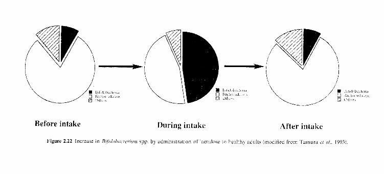
Refore intake During intake After intake
Figure 2.22 Increase in Ui/irlo/~ric,rc,,iir,,i spp. by adminis t ra t ion of lactillox to healthy :idults (modified from Tamtila ('I ( I / . . 1993)
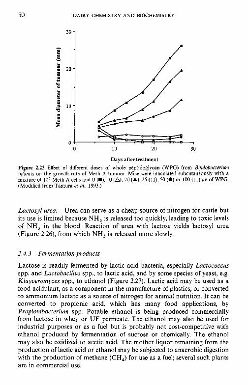
50 DAIRY CHEMISTRY AND BIOCHEMISTRY
30
h
E E v
1 L x 20
5 c) h 0 L 4 c) 4
E .o 10 W
0 10 20 30
Days after treatment Figure 2.23 Effect of different doses of whole peptidoglycan (WPG) from Bifdobacterium infantis on the growth rate of Meth A tumour. Mice were inoculated subcutaneously with a mixture of lo5 Meth A cells and 0 (U), 10 (A), 20 (A), 25 (O), 50 (0) or 100 (0) pg of WPG. (Modified from Tamura et al., 1993.)
Lactosyl urea. Urea can serve as a cheap source of nitrogen for cattle but its use is limited because NH, is released too quickly, leading to toxic levels of NH, in the blood. Reaction of urea with lactose yields lactosyl urea (Figure 2.26), from which NH, is released more slowly.
2.4.3 Fermentation products
Lactose is readily fermented by lactic acid bacteria, especially Lactococcus spp. and Lactobacillus spp., to lactic acid, and by some species of yeast, e.g. Kluyveromyces spp., to ethanol (Figure 2.27). Lactic acid may be used as a food acidulant, as a component in the manufacture of plastics, or converted to ammonium lactate as a source of nitrogen for animal nutrition. It can be converted to propionic acid, which has many food applications, by Propionibacterium spp. Potable ethanol is being produced commercially from lactose in whey or U F permeate. The ethanol may also be used for industrial purposes or as a fuel but is probably not cost-competitive with ethanol produced by fermentation of sucrose or chemically. The ethanol may also be oxidized to acetic acid. The mother liquor remaining from the production of lactic acid or ethanol may be subjected to anaerobic digestion with the production of methane (CH,) for use as a fuel; several such plants are in commercial use.
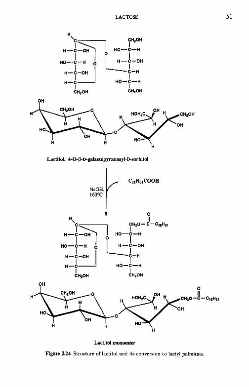
LACTOSE 51
H CHIOH
no-c-H
I 0 H-C-OH
I HO-C-H
I I
H-C-OH
n-c no-c-H
CHzOH
I I CHzOH
PH Hme HO 0
HO OH
H H H
Lactitol, 4-O-~-D-galactopyranosyl-D-sorbitol
,600c 1 C1SH31COOH NaOH,
0
CH20- C-CISHJI H II
no-c-H I 0
H-C-OH
L-II,"" H-Lon 1 HO-C-H '7 I '
I I CHZOH
HO-C-H I I CHzOH
n-c
OH 0 Hm e y - C - c l & l I1
0 no
OH HO H H
H
Lactitol monoester
Figure 2.24 Structure of lactitol and its conversion to lactyl palmitate.
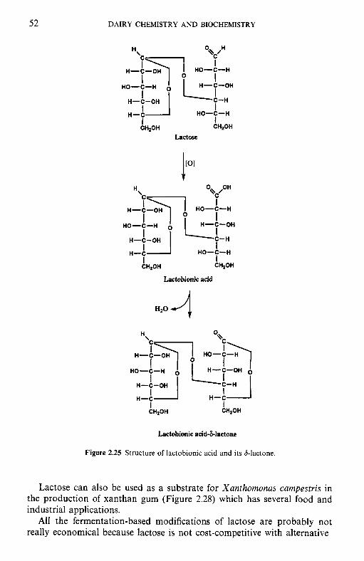
52 DAIRY CHEMISTRY AND BIOCHEMISTRY
CH,OH
Lactose
t
H-C-OH I
HO-C-H I I I H-C
CHzOH CH20H
Lactobionic acid
~ H ~ O H CH,OH
Lactobionic acid-&lactone
Figure 2.25 Structure of lactobionic acid and its &lactone.
Lactose can also be used as a substrate for Xanthomonas campestris in the production of xanthan gum (Figure 2.28) which has several food and industrial applications.
All the fermentation-based modifications of lactose are probably not really economical because lactose is not cost-competitive with alternative
LACTOSE 53
FH20H CH2OH
Lactosyl urea
Figure 2.26 Structure of lactosyl urea.
LACTOSE
0 II //”
c\ OH
H3C-C-
Pyruvic acid
J
2CH3CH2COOH + CHACOOII propionic acid arctic acid
+ CO2 + H 2 0 OH
H ‘ “ONH,
I //” H3C-C-
Animoniuni lar ta te (animal feed)
H H I I
H-C-C-OH I I
H H
Ethanol (potable or industrial)
t
7 0 H-C-C’
‘OH
Acetic acid (vinegar)
Figure 2.27 Fermentation products from lactose.
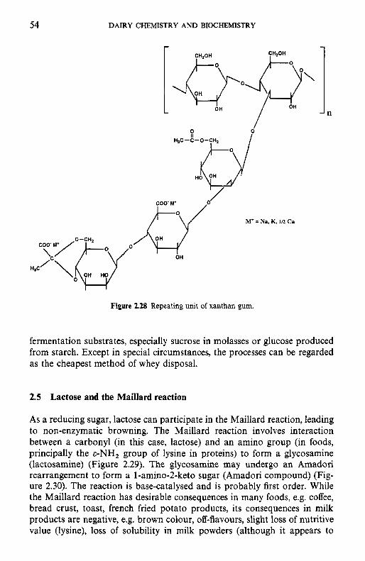
54 DAIRY CHEMISTRY AND BIOCHEMISTRY
Figure 2.28 Repeating unit of xanthan gum.
fermentation substrates, especially sucrose in molasses or glucose produced from starch. Except in special circumstances, the processes can be regarded as the cheapest method of whey disposal.
2.5 Lactose and the Maillard reaction
As a reducing sugar, lactose can participate in the Maillard reaction, leading to non-enzymatic browning. The Maillard reaction involves interaction between a carbonyl (in this case, lactose) and an amino group (in foods, principally the E-NH, group of lysine in proteins) to form a glycosamine (lactosamine) (Figure 2.29). The glycosamine may undergo an Amadori rearrangement to form a l-amino-2-keto sugar (Amadori compound) (Fig- ure 2.30). The reaction is base-catalysed and is probably first order. While the Maillard reaction has desirable consequences in many foods, e.g. coffee, bread crust, toast, french fried potato products, its consequences in milk products are negative, e.g. brown colour, off-flavours, slight loss of nutritive value (lysine), loss of solubility in milk powders (although it appears to
LACTOSE 55
D-Glucopyranose
HO 0 OH HR
OH
Glycosylaiiiine
Figure 2.29 Formation of glycosylamine, the initial step in Maillard browning.
prevent or retard age-gelation in UHT milk products). Maillard reaction products (MRPs) have antioxidant properties; the production of MRPs may be a small-volume outlet for lactose.
The Amadori compound may be degraded via either of two pathways, depending on pH, to a variety of active alcohol, carbonyl and dicarbonyl compounds and ultimately to brown-coloured polymers called melanoidins (Figure 2.31). Many of the intermediates are (off-) flavoured. The dicar- bonyls can react with amino acids via the Strecker degradation pathway (Figure 2.32) to yield another family of highly flavoured compounds.
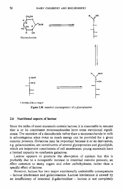
56
1
DAIRY CHEMISTRY A N D BIOCHEMISTRY
I
CHPOH
HO H p H R - L
Glycosylamine
C-NHR
c=o I
L .
HvNR C I C-
I- t-
\ JNHR
II C-OH
1-Amiiio-2-kcto sugar
Figure 2.30 Arnadori rearrangement of a glycosylamine.
2.6 Nutritional aspects of lactose
Since the milks of most mammals contain lactose, it is reasonable to assume that it or its constituent monosaccharides have some nutritional signifi- cance. The secretion of a disaccharide rather than a monosaccharide in milk is advantageous since twice as much energy can be provided for a given osmotic pressure. Galactose may be important because it or its derivatives, e.g. galactosamine, are constituents of several glycoproteins and glycolipids, which are important constituents of cell membranes; young mammals have a limited capacity to synthesize galactose.
Lactose appears to promote the absorption of calcium but this is probably due to a nonspecific increase in intestinal osmotic pressure, an effect common to many sugars and other carbohydrates, rather than a specific effect of lactose.
However, lactose has two major nutritionally undesirable consequences - lactose intolerance and galactosaemia. Lactose intolerance is caused by an insufficiency of intestinal P-galactosidase - lactose is not completely
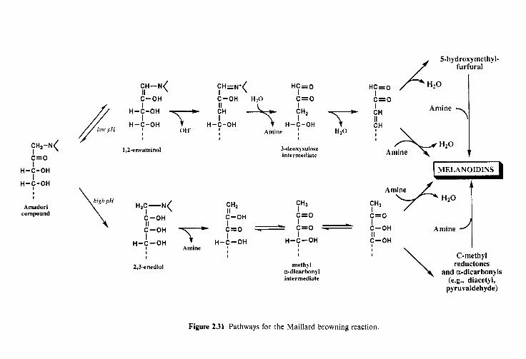
I I
HO-T-H \
1 I
HO-3-H
.lW aiqiuy HO-3-H
‘H3 I II I
o=j O’H HO-5 HO-5 I I II
0=3H )*N=H3 )N-H~
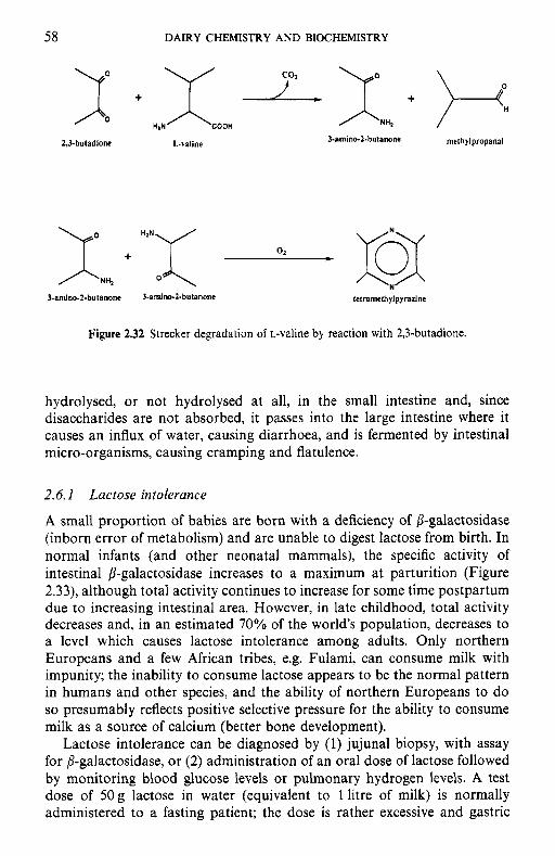
58 DAIRY CHEMISTRY A N D BIOCHEMISTRY
2,3-butadione L-valine 3-amino-2-hutanone methjlpropanal
J-anuno-2.butnnone 3-amino-2.butanone letramethylpyrazine
Figure 2.32 Strecker degradation of L-valine by reaction with 2,3-butadione.
hydrolysed, or not hydrolysed at all, in the small intestine and, since disaccharides are not absorbed, it passes into the large intestine where it causes an influx of water, causing diarrhoea, and is fermented by intestinal micro-organisms, causing cramping and flatulence.
2.6.1 Lactose intolerance
A small proportion of babies are born with a deficiency of P-galactosidase (inborn error of metabolism) and are unable to digest lactose from birth. In normal infants (and other neonatal mammals), the specific activity of intestinal P-galactosidase increases to a maximum at parturition (Figure 2.33), although total activity continues to increase for some time postpartum due to increasing intestinal area. However, in late childhood, total activity decreases and, in an estimated 70% of the world's population, decreases to a level which causes lactose intolerance among adults. Only northern Europeans and a few African tribes, e.g. Fulami, can consume milk with impunity; the inability to consume lactose appears to be the normal pattern in humans and other species, and the ability of northern Europeans to do so presumably reflects positive selective pressure for the ability to consume milk as a source of calcium (better bone development).
Lactose intolerance can be diagnosed by (1) jujunal biopsy, with assay for P-galactosidase, or (2) administration of an oral dose of lactose followed by monitoring blood glucose levels or pulmonary hydrogen levels. A test dose of 50 g lactose in water (equivalent to 1 litre of milk) is normally administered to a fasting patient; the dose is rather excessive and gastric
8 c < E 5 El 5'
L
cn
P,
tl a
f.
i; &.
"2 pl 3 a
5.
p-G
alad
osid
ase a
ctiv
ity
(pm
ol g
luco
se r
elea
sedl
mg
prot
einl
lo m
in)
2
N
r b 8 !4
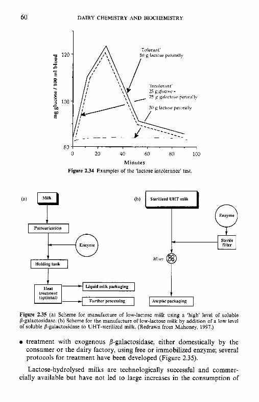
60
0 0 r( .
DAIRY CHEMISTRY AND BIOCHEMISTRY
'Tolerant' 50 f i lactiise perorally
' ln tolerdnt '
0 20 4 0 60 80 100 Minutes
Figure 2.34 Examples of the 'lactose intolerance' test.
Enzyme
Enzyme
filter
M I ver
Liquid milk packaging
Further processing 1-1 Figure 2.35 (a) Scheme for manufacture of low-lactose milk using a 'high' level of soluble 8-galactosidase. (b) Scheme for the manufacture of low-lactose milk by addition of a low level of soluble P-galactosidase to UHT-sterilized milk. (Redrawn from Mahoney, 1997.)
0 treatment with exogenous P-galactosidase, either domestically by the consumer or the dairy factory, using free or immobilized enzyme; several protocols for treatment have been developed (Figure 2.35).
Lactose-hydrolysed milks are technologically successful and commer- cially available but have not led to large increases in the consumption of
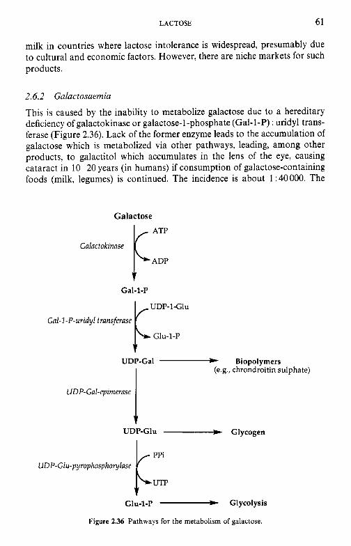
LACTOSE 61
milk in countries where lactose intolerance is widespread, presumably due to cultural and economic factors. However, there are niche markets for such products.
2.6.2 Galactosaemia
This is caused by the inability to metabolize galactose due to a hereditary deficiency of galactokinase or galactose-1-phosphate (Gal-1-P) : uridyl trans- ferase (Figure 2.36). Lack of the former enzyme leads to the accumulation of galactose which is metabolized via other pathways, leading, among other products, to galactitol which accumulates in the lens of the eye, causing cataract in 10-20 years (in humans) if consumption of galactose-containing foods (milk, legumes) is continued. The incidence is about 1 : 40000. The
Galactose
f Gal-1-P
UDP-1Clu
Glu-1-P
(e.g., chrondroitin sulphate)
1 I
G d - 1 -P-uridyl fransferase
UDP-Gal - Biopolymers
UDFGal-rpirnernse
UDP-Glu - Glycogen
Glu-1-p - Glycolysis
Figure 2.36 Pathways for the metabolism of galactose.
62 DAIRY CHEMISTRY AND BIOCHEMISTRY
lack of Gal-1-P : uridyl transferase leads to the accumulation of Gal and Gal-1-P. The latter interferes with the synthesis of glycoproteins and glycolipids (important for membranes, e.g. in the brain) and results in irreversible mental retardation within 2-3 months if the consumption of galactose-containing foods is continued. The incidence of this disease, often called 'classical galactosaemia', is about 1 in 60 000.
The ability to metabolize galactose decreases on ageing (after 70 years), leading to cataract; perhaps this, together with the fact that mammals normally encounter lactose only while suckling, explains why many people lose the ability to utilize lactose at the end of childhood.
2.7 Determination of lactose concentration
Lactose may be quantified by methods based on one of five principles:
1. polarimetry; 2. oxidation-reduction titration; 3. colorimetry; 4. chromatography; 5. enzymatically.
2.7.1 Polarimetry
The specific rotation, [a]?, of lactose in solution at equilibrium is + 55.4" expressed on an anhydrous basis (+52.6" on a monohydrate basis). The specific rotation is defined as the optical rotation of a solution containing 1 gml- ' in a 1 dm polarimeter tube; it is affected by temperature (20°C is usually used; indicated by superscript) and wavelength (usually the sodium D line (589.3 nm) is used; indicated by subscript).
where a is the measured optical rotation; 1, the light path in dm; and c, the concentration as g m1-I. It is usually expressed as:
where c is in g perlOOml. The milk sample must first be defatted and deproteinated, usually by
treatment with mercuric nitrate (Hg(NO,),). In calculating the concentra- tion of lactose, a correction should be used for the concentration of fat and protein in the precipitate.
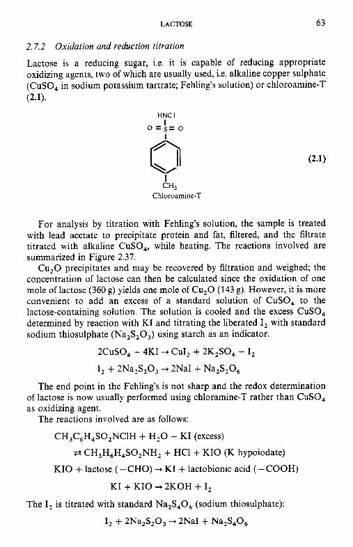
LACTOSE 63
2.7.2 Oxidation and reduction titration
Lactose is a reducing sugar, i.e. it is capable of reducing appropriate oxidizing agents, two of which are usually used, i.e. alkaline copper sulphate (CuSO, in sodium potassium tartrate; Fehling’s solution) or chloroamine-T (2.1).
HNC I I o = s = o I 0 I
kH, Chloroamine-T
For analysis by titration with Fehling’s solution, the sample is treated with lead acetate to precipitate protein and fat, filtered, and the filtrate titrated with alkaline CuSO,, while heating. The reactions involved are summarized in Figure 2.37.
Cu,O precipitates and may be recovered by filtration and weighed; the concentration of lactose can then be calculated since the oxidation of one mole of lactose (360 g) yields one mole of Cu,O (143 g). However, it is more convenient to add an excess of a standard solution of CuSO, to the lactose-containing solution. The solution is cooled and the excess CuSO, determined by reaction with KI and titrating the liberated I, with standard sodium thiosulphate (Na,S,O,) using starch as an indicator.
2CuS0, + 4KI -, CuI, + 2K,SO, + I, 1, + 2Na,S,03 --+ 2NaI + Na2S,06
The end point in the Fehling’s is not sharp and the redox determination of lactose is now usually performed using chloramine-T rather than CuSO, as oxidizing agent.
The reactions involved are as follows:
CH,C,H,SO,NClH + H,O + KI (excess)
* CH,H,H,SO,NH, + HCl + KIO (K hypoiodate)
KIO + lactose (- CHO) -+ KI + lactobionic acid ( - COOH)
KI + KIO -, 2KOH + I, The I, is titrated with standard Na,S,O, (sodium thiosulphate):
I, + 2Na,S,O, --* 2NaI + Na,S,O,
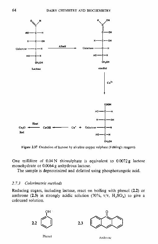
64 DAIRY CHEMISTRY AND BIOCHEMISTRY
CU”
0 \c/H
I HO-C-H
H
II I C-OH
H-C-OH
I I I
H-LOH - Galactose -C-H Alkali
Galactose -C-H
I HO-C-H HO-C-H
CH2OH CHaOH I L a C t W enediol
COOH
I I I
Red I I
HO-C-H
H-C-OH
Heat CuzO - CuOH Cu’ + Galactose -C-H
HO-C-H
CHIOH
Figure 2.37 Oxidation of lactose by alkaline copper sulphate (Fehling’s reagent).
One millilitre of 0.04 N thiosulphate is equivalent to 0.0072 g lactose monohydrate or 0.0064 g anhydrous lactose.
The sample is deproteinized and defatted using phosphotungstic acid.
2.7.3 Colorimetric methods
Reducing sugars, including lactose, react on boiling with phenol (2.2) or anthrone (2.3) in strongly acidic solution (70%, v/v, H,SO,) to give a coloured solution.
0 P” 2.2 0 2.3
LACTOSE 65
The complex with anthrone absorbs maximally at 625 nm. The concen- tration of lactose is determined from a standard curve prepared using a range of lactose concentrations.
The method is very sensitive but must be performed under precisely controlled conditions.
2.7.4 Chromaiographic methods
While lactose may be determined by gas liquid chromatography, high performance liquid chromatography (HPLC), using a refractive index de- tector, is now usually used.
2.7.5 Enzymatic methods
Enzymatic methods are very sensitive but are rather expensive, especially for a small number of samples.
Lactose is first hydrolysed by 8-galactosidase to glucose and galactose. The glucose may be quantified using:
1. glucose oxidase using a platinum electrode, or the H,O, generated may
2. glucose-6-phosphate dehydrogenase (G-6-P-DH) be quantified by using a peroxidase and a suitable dye acceptor; or
Hexokinase D-Glucose + ATP - Glucose-6-P + ADP
G-6-P-DH, NADP' Gluconate-6-P + NADPH + H t
The concentration of NADPH produced may be quantified by measuring the increase in absorbance at 334, 340 or 365 nm.
Alternatively, the galactose produced may be quantified using galactose dehydrogenase (Gal-DH):
D-galactose + NAD' - Galactonic acid + NADH + H + Gal-DH
The NADH produced may be quantified by measuring the increase in absorbance at 334, 340 or 365 nm.
References
Andrews, G. (1989) Lactulose in heated milk, in Heat-Induced Changes in Milk, (ed. P.F. Fox),
Horton, B.S. (1993) Economics of marketing lactose and lactose by-products in a global trading
Hynd, J. (1980) Drying of whey. J . Soc. Dairy Technol., 33, 52-4.
Bulletin 238, International Dairy Federation, Brussels, pp. 45-52.
environment, in Bulletin 289, International Dairy Federation, Brussels, pp. 7-9.
66 DAIRY CHEMISTRY AND BIOCHEMISTRY
Jenness, R. and Patton, S. (1959) Lactose, in Principles of Dairy Chemistry, John Wiley and Sons, NY, pp. 73-100.
Mahoney, R.R. (1997) Lactose: enzymatic modification, in AdFanced Dairy Chemistry, Vol. 3: Lactose, Water, Salts and Wtamins, 2nd edn (ed. P.F. Fox), Chapman & Hall, London, pp.
Nickerson, T.A. (1974) Lactose, in Fundamentals of Dairy Chemistry, (eds B.H. Webb, A.H. Johnson and J.A. Alford), AVI Publishing, Westport, CT, pp. 273-324.
Smart, J.B. (1993) Transferase reactions of P-galactosidases - New product opportunities, in Lactose Hydrolysis, Bulletin 239. International Dairy Federation, Brussels, pp. 16-22.
Tamura, Y., Mizota, T., Shimamura, S. and Tomita, M. (1993) Lactulose and its application to food and pharmaceutical industries, in Lactose Hydrolysis, Bulletin 239, International Dairy Federation, Brussels, pp. 43-53.
Tumerman, L.. Fram, H. and Comely, K.W. (1954) The effect of lactose crystallization on protein stability in frozen concentrated milk. J . Dairy Sci., 37, 830-9.
Walstra, P. and Jenness, R. (1984) Dairy Chemistry and Physics, John Wiley and Sons, New York.
77-125.
Suggested reading
Fox, P.F. (ed.) (1985) Developments in Dairy Chemistry, Vol. 3: Lactose and Minor Constituents,
Fox, P.F. (ed.) (1997) Advanced Dairy Chemistry, Vol. 2 Lactose, Water, Salts and Vitamins,
Holsinger, V.H. (1988) Lactose, in Fundamentals of Dairy Chemistry, (ed. N.P. Wong), Van
I D F (1989) Monograph on heat-induced changes in milk, Bulletin 238, International Dairy
I D F (1993) Proceedings o f the IDF Workshop on Lactose Hydrolysis, Bulletin 289, International
Jenness, R. and Patton, S. (1959) Lactose, in Principles of Dairy Chemistry, John Wiley and
Labuza, T.P., Reineccius, G.A., Monnier, V.M. et a/ . (eds) (1994) Maillard Reactions in
Nickerson, T.A. (1965) Lactose, in Fundamentals of Dairy Chemistry, (eds B.H. Webb and A.H.
Nickerson, T.A. (1974) Lactose, in Fundamentals of Dairy Chemistry, (eds B.H. Webb, A.H.
Walstra, P. and Jenness, R. (1984) Dairy Chemistry and Physics, John Wiley and Sons, New
Yang, S.T. and Silva, E.M. (1995) Novel products and new technologies for use of a familiar
Elsevier Applied Science Publishers, London.
Chapman & Hall, London.
Nostrand Reinhold, New York, pp. 279-342.
Federation, Brussels.
Dairy Federation, Brussels.
Son, New York, pp. 73-100.
Chemistry, Food and Health, Royal Society of Chemistry, Cambridge.
Johnson), AVI Publishing, Westport, CT, pp. 224-60.
Johnson and J.A. Alford), AVI Publishing, Westport, CT, pp. 273-324.
York.
carbohydrate, milk lactose. J . Dairy Sci., 78, 2541-62.
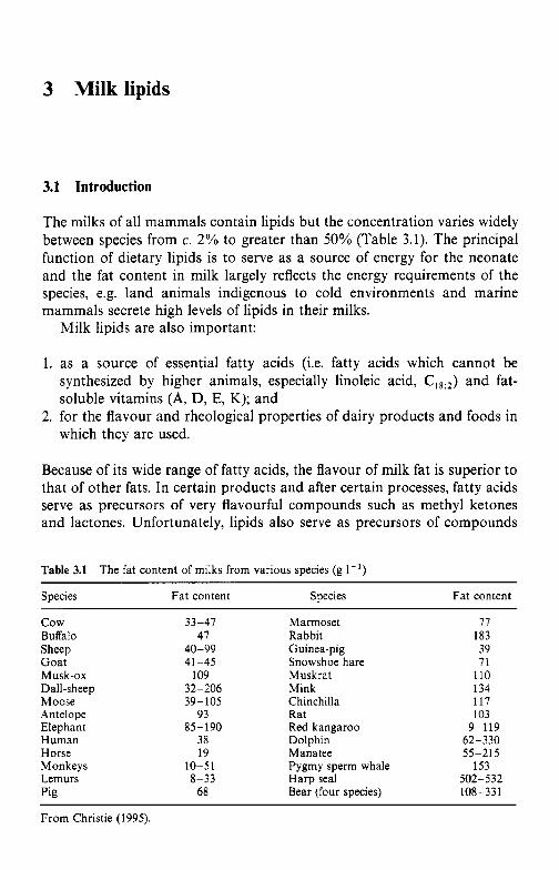
3 Milk lipids
3.1 Introduction
The milks of all mammals contain lipids but the concentration varies widely between species from c. 2 % to greater than 50% (Table 3.1). The principal function of dietary lipids is to serve as a source of energy for the neonate and the fat content in milk largely reflects the energy requirements of the species, e.g. land animals indigenous to cold environments and marine mammals secrete high levels of lipids in their milks.
Milk lipids are also important:
1. as a source of essential fatty acids (i.e. fatty acids which cannot be synthesized by higher animals, especially linoleic acid, &) and fat- soluble vitamins (A, D, E, K); and
2. for the flavour and rheological properties of dairy products and foods in which they are used.
Because of its wide range of fatty acids, the flavour of milk fat is superior to that of other fats. In certain products and after certain processes, fatty acids serve as precursors of very flavourful compounds such as methyl ketones and lactones. Unfortunately, lipids also serve as precursors of compounds
Table 3.1 The fat content of milks from various species (g I - ' )
Species Fat content Species Fat content
c o w Buffalo Sheep Goat
Dall-sheep Moose Antelope Elephant Human Horse Monkeys Lemurs Pig
Musk-ox
33-47 47
40-99 41 -45
109 32-206 39-105
93 85-190
38 19
10-51 8-33
68
Marmoset Rabbit Guinea-pig Snowshoe hare Muskrat Mink Chinchilla Rat Red kangaroo Dolphin Manatee Pygmy sperm whale Harp seal Bear (four species)
77 183 39 71
110 134 117 103
9-119 62-330 55-215
502- 5 32 108-331
153
From Christie (1995).
68 DAIRY CHEMISTRY AND BIOCHEMISTRY
that cause off-flavour defects (hydrolytic and oxidative rancidity) and as solvents for compounds in the environment which may cause off-flavours.
For many years, the economic value of milk was based mainly or totally on its fat content, which is still true in some cases. This practice was satisfactory when milk was used mainly or solely for butter production. Possibly, the origin of paying for milk on the basis of its fat content, apart from its value for butter production, lies in the fact that relatively simple quantitative analytical methods were developed for fat earlier than for protein or lactose. Because of its economic value, there has long been commercial pressure to increase the yield of milk fat per cow by nutritional or genetic means.
To facilitate the reader, the nomenclature, structure and properties of the principal fatty acids and of the principal lipid classes are summarized in Appendices 3A, 3B and 3C. The structure and properties of the fat-soluble vitamins, A, D, E and K, are discussed in Chapter 6.
3.2 Factors that affect the fat content of bovine milk
Bovine milk typically contains c. 3.5% fat but the level varies widely, depending on several factors. including: breed, individuality of the animal, stage of lactation, season, nutritional status, type of feed, health and age of the animal, interval between milkings and the point during milking when the sample is taken.
Of the common European breeds, milk from Jersey cows contains the highest level of fat and that from Holstein/Friesians the lowest (Figure 3.1). The data in Figure 3.1 also show the very wide range of fat content in individual-cow samples.
The fat content of milk decreases during the first 4-6 weeks after parturition and then increases steadily throughout the remainder of lacta- tion, especially toward the end (Figure 3.2). For any particular population, fat content is highest in winter and lowest in summer, due partly to the effect of environmental temperature. Production of creamery (manufacturing) milk in Ireland, New Zealand and parts of Australia is very seasonal; lactational, seasonal and possibly nutritional effects coincide, leading to large seasonal changes in the fat content of milk (Figure 3.3), and also in the levels of protein and lactose.
For any individual animal, fat content decreases slightly during success- ive lactations, by c. 0.2% over a typical productive lifetime (about five lactations). In practice, this factor usually has no overall effect on the fat content of a bulk milk supply because herds normally include cows of various ages. The concentration of fat (and of all other milk-specific constituents) decreases markedly on mastitic infection due to impaired
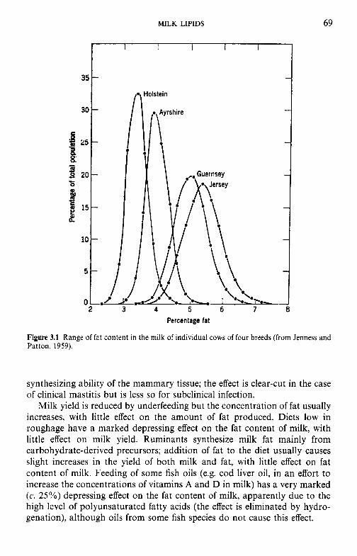
MILK LIPIDS
I 35 -
30 -
B f - 25- 0
%
3
- S s 20- r 0
15- 2
10 -
5 -
69
Percentage fat
Figure 3.1 Range of fat content in the milk of individual cows of four breeds (from Jenness and Patton, 1959).
synthesizing ability of the mammary tissue; the effect is clear-cut in the case of clinical mastitis but is less so for subclinical infection.
Milk yield is reduced by underfeeding but the concentration of fat usually increases, with little effect on the amount of fat produced. Diets low in roughage have a marked depressing effect on the fat content of milk, with little effect on milk yield. Ruminants synthesize milk fat mainly from carbohydrate-derived precursors; addition of fat to the diet usually causes slight increases in the yield of both milk and fat, with little effect on fat content of milk. Feeding of some fish oils (e.g. cod liver oil, in an effort to increase the concentrations of vitamins A and D in milk) has a very marked (c. 25%) depressing effect on the fat content of milk, apparently due to the high level of polyunsaturated fatty acids (the effect is eliminated by hydro- genation), although oils from some fish species do not cause this effect.
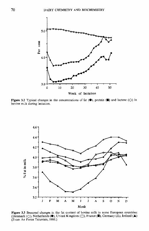
70 DAIRY CHEMISTRY AND BIOCHEMISTRY
5.0
- 0
L.
2 4.0
3.0 0 10 20 30 40 50
Week of lactation
Figure 3.2 Typical changes in the concentrations of fat (O), protein (m) and bovine milk during lactation.
4.6 -
4.4 -
4.2 - 4.0 -
3.8 -
3.6 -
3.4 -
lactose (0) in
J F M A M J J A S O N D
Month
Figure 3.3 Seasonal changes in the fat content of bovine milk in some European countries: (Denmark (O), Netherlands (O), United Kingdom (O), France (U), Germany (A), Ireland (A) (From An Foras Taluntais, 1981.)
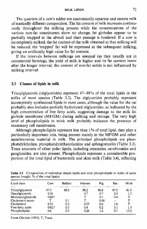
MILK LIPIDS 71
The quarters of a cow’s udder are anatomically separate and secrete milk of markedly different composition. The fat content of milk increases continu- ously throughout the milking process while the concentrations of the various non-fat constituents show no change; fat globules appear to be partially trapped in the alveoli and their passage is hindered. If a cow is incompletely milked, the fat content of the milk obtained at that milking will be reduced; the ‘trapped’ fat will be expressed at the subsequent milking, giving an artificially high value for fat content.
If the intervals between milkings are unequal (as they usually are in commercial farming), the yield of milk is higher and its fat content lower after the longer interval; the content of non-fat solids is not influenced by milking interval.
3.3 Classes of lipids in milk
Triacylglycerols (triglycerides) represent 97-98% of the total lipids in the milks of most species (Table 3.2). The diglycerides probably represent incompletely synthesized lipids in most cases, although the value for the rat probably also includes partially hydrolysed triglycerides, as indicated by the high concentration of free fatty acids, suggesting damage to the milk fat globule membrane (MFGM) during milking and storage. The very high level of phospholipids in mink milk probably indicates the presence of mammary cell membranes.
Although phospholipids represent less than 1% of total lipid, they play a particularly important role, being present mainly in the MFGM and other membraneous material in milk. The principal phospholipids are phos- phatidylcholine, phosphatidylethanolamine and sphingomyelin (Table 3.3). Trace amounts of other polar lipids, including ceramides, cerobrosides and gangliosides, are also present. Phospholipids represent a considerable pro- portion of the total lipid of buttermilk and skim milk (Table 3.4), reflecting
Table 3.2 species (weight YO of the total lipids)
Composition of individual simple lipids and total phospholipids in milks of some
Lipid class
Triacylgl ycerols Diacylglycerols Monoacylgl ycerols Cholesteryl esters Cholesterol Free fatty acids Phospholipids
cow Buffalo Human Pig Rat Mink
97.5 0.36 0.027 T
0.31 0.027 0.6
98.6 98.2 0.7 T
0.1 T 0.3 0.25 0.5 0.4 0.5 0.26
96.8 0.7 0.1 0.06 0.6 0.2 1.6
87.5 2.9 0.4
1.6 3.1 0.7
-
81.3 1.7 T T T 1.3
15.3
From Christie (1995). T, Trace.
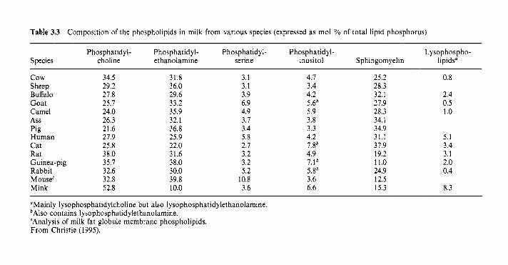
Table 3.3 Composition of the phospholipids in milk from various species (expressed as mol YO of total lipid phosphorus)
Phosphatidyl- Phosphatidyl- Phosphatidyl- Phosphatidyl- Lysophospho- Species choline ethanolamine serine inositol Sphingomyelin lipids“
cow Sheep Buffalo Goat Camel Ass Pig Human Cat Rat Guinea-pig Rabbit Mouse‘ Mink
34.5 29.2 27.8 25.7 24.0 26.3 21.6 27.9 25.8 38.0 35.7 32.6 32.8 52.8
31.8 36.0 29.6 33.2 35.9 32.1 36.8 25.9 22.0 31.6 38.0 30.0 39.8 10.0
3.1 3.1 3.9 6.9 4.9 3.1 3.4 5.8 2.7 3.2 3.2 5.2
10.8 3.6
4.1 3.4 4.2 5Ab 5.9 3.8 3.3 4.2 7.8b 4.9 7.1b 5.8’ 3.6 6.6
25.2 28.3 32.1 21.9 28.3 34.1 34.9 31.1 31.9 19.2 11.0 24.9 12.5 15.3
0.8
2.4 0.5 1 .o
5.1 3.4 3.1 2.0 0.4
8.3
“Mainly lysophosphatidylcholine but also lysophosphatidylethanolamine. bAlso contains lysophosphatidylethanolamine. ‘Analysis of milk fat globule membrane phospholipids. From Christie (1995).
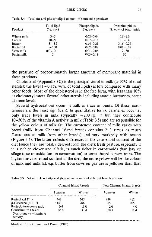
MILK LIPIDS 73
Table 3.4 Total fat and phospholipid content of some milk products
Total lipid Phospholipids Phospholipid as Product (%. WIV) (%, WIV) YO, w/w, of total lipids
Whole milk 3-5 Cream 10-50 Butter 81-82 Butter oil - 100 Skim milk 0.03-0.1 Buttermilk 2
0.02-0.04 0.6- 1 .O 0.07-0. I8 0.3-0.4 0.14-0.25 0.16-0.29 0.02-0.08 0.02-0.08 0.01-0.06 17-30 0.03 -0.18 10
the presence of proportionately larger amounts of membrane material in these products.
Cholesterol (Appendix 3C) is the principal sterol in milk (> 95% of total sterols); the level (-O.3%, w/w, of total lipids) is low compared with many other foods. Most of the cholesterol is in the free form, with less than 10% as cholesteryl esters. Several other sterols, including steroid hormones, occur at trace levels.
Several hydrocarbons occur in milk in trace amounts. Of these, caro- tenoids are the most significant. In quantitative terms, carotenes occur at only trace levels in milk (typically -2OOpg1-') but they contribute 10-50% of the vitamin A activity in milk (Table 3.5) and are responsible for the yellow colour of milk fat. The carotenoid content of milk varies with breed (milk from Channel Island breeds contains 2-3 times as much p-carotene as milk from other breeds) and very markedly with season (Figure 3.4). The latter reflects differences in the carotenoid content of the diet (since they are totally derived from the diet); fresh pasture, especially if it is rich in clover and alfalfa, is much richer in carotenoids than hay or silage (due to oxidation on conservation) or cereal-based concentrates. The higher the carotenoid content of the diet, the more yellow will be the colour of milk and milk fat, e.g. butter from cows on pasture is yellower than that
Table 3.5 Vitamin A activity and P-carotene in milk of different breeds of cows ~ ~ ~ ~~ ~~~~~
Channel Island breeds Non-Channel Island breeds
Summer Winter Summer Winter
Retinol (pl 1- ') 649 265 619 412 j-Carotene (pl I - ' ) 1143 266 315 105 Retinollb-carotene ratio 0.6 11.0 2.0 4.0 Contribution (%) of 46.8 33.4 20.3 11.4 p-carotene to vitamin A activity
Modified from Cremin and Power (1985).
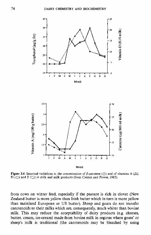
4
P
e,w
gi
h rn
E
Ob
-. "I
- V
itam
in A
(mg/
100
g bu
tter)
-3
Y
Toco
pher
ol (&
g fa
t)
'<
-. P 2.
2
Car
oten
e (pg/100 m
l milk
) V
itam
in D
(IU
A m
ilk)
P
MILK LIPIDS 75
peroxides, e.g. H,O, or benzoyl peroxide, or masked, e.g. with chlorophyll or titanium oxide).
Milk contains significant concentrations of fat-soluble vitamins (Table 3.5, Figure 3.4) and milk and dairy products make a significant contribution to the dietary requirements for these vitamins in Western countries. The actual form of the fat-soluble vitamins in milk appears to be uncertain and their concentration varies widely with breed of animal, feed and stage of lactation, e.g. the vitamin A activity of colostrum is c. 30 times higher than that of mature milk.
Several prostaglandins occur in milk but it is not known whether they play a physiological role; they may not survive storage and processing in a biologically active form. Human milk contains prostaglandins E and F at concentrations 100-fold higher than human plasma and these may have a physiological function, e.g. gut motility.
3.4 Fatty acid profile of milk lipids
Milk fats, especially ruminant fats, contain a very wide range of fatty acids: more than 400 and 184 distinct acids have been detected in bovine and human milk fats, respectively (Christie, 1995). However, the vast majority of these occur at only trace concentrations. The concentrations of the principal fatty acids in milk fats from a range of species are shown in Table 3.6.
Notable features of the fatty acid profiles of milk lipids include:
1. Ruminant milk fats contain a high level of butanoic acid (C4:o) and other short-chain fatty acids. The method of expressing the results in Table 3.6 (Yo, w/w) under-represents the proportion of short-chain acids - if ex- pressed as mol %, butanoic acid represents c. 10% of all fatty acids (up to 15% in some samples), i.e. there could be a butyrate residue in c. 30% of all triglyceride molecules. The high concentration of butyric (butanoic) acid in ruminant milk fats arises from the direct incorporation of P-hydroxybutyrate (which is produced by micro-organisms in the rumen from carbohydrate and transported via the blood to the mammary gland where it is reduced to butanoic acid). Non-ruminant milk fats contain no butanoic or other short-chain acids; the low concentrations of butyrate in milk fats of some monkeys and the brown bear require confirmation.
The concentration of butanoic acid in milk fat is the principle of the widely used criterion for the detection and quantitation of adulteration of butter with other fats, i.e. Reichert Meissl and Polenski numbers, which are measures of the volatile water-soluble and volatile water- insoluble fatty acids, respectively.
Short-chain fatty acids have strong, characteristic flavours and aromas. When these acids are released by the action of lipases in milk or
Table 3.6 Principal fatty acids (wt YO of total) in milk triacylglycerols or total lipids from various species
Species 4:O 6:O 8 : O 1O:O 12:O 14:O 16:O 16.1 18:O 18.1 18:2 18:3 C,,-C2,
c o w Buffalo Sheep Goat
Dall-sheep Moose Blackbuck antelope Elephant Human Monkey (mean of six species
Baboon Lemur macaco Horse Pig Rat Guinea-pig Marmoset Rabbit Cottontail rabbit European hare Mink Chinchilla Red kangaroo Platypus Numbat Bottle-nosed dolphin Manatee Pygmy sperm whale Harp seal Northern elephant seal Polar bear Grizzly bear
Musk-ox
1.6 1.6 2.8 2.9 0.9 0.3 T 6.0
T 0.6
0.4
T
-
-
- - T
T
T
-
- - - - - - - - - - - T T
1.3 1.1 2.7 2.7 1.9 0.2 8.4 2.7 0.3 T 5.9
5.1 0.2 1.8
1.1 -
- -
22.4 9.6
10.9 - - - ~
- - 0.6 - - - - -
3 .O 1.9 9.0 8.4 4.7 4.9 5.5 6.5
29.4 1.3
11.0
7.9 1.9 5.1 0.7 7.0
8.0 20.1 14.3 17.7 - - - - - -
3.5 - - -
T -
3.1 2.0 5.4 3.3 2.3 1.8 0.6 3.5
18.3 3.1 4.4
2.3 10.5 6.2 0.5 7.5
8.5 2.9 3.8 5 . 5 0.5 T 0.1
0.1 0.3 4.0
-
-
- - - 0.5 0.1
9.5 8.7
11.8 10.3 6.2
10.6 2.0
11.5 5.3 5.1 2.8
1.3 15.0 5.7 4.0 8.2 2.6 7.7 1.7 2.0 5.3 3.3 3.0 2.7 1.6 0.9 3.2 6.3 3.6 5.3 2.6 3.9 2.1
26.3 2.3 30.4 3.4 25.4 3.4 24.6 2.2 19.5 1.7 23.0 2.4 28.4 4.3 39.3 5.7 12.6 3 .O 20.2 5.7 21.4 6.7
16.5 1.2 27.1 9.6 23.8 7.8 32.9 11.3 22.6 1.9 31.3 2.4 18.1 5.5 14.2 2.0 18.7 1 .o 24.8 5.0 26.1 5.2 30.0 -
31.2 6.8 19.8 13.9 14.1 3.4 21.1 13.3 20.2 11.6 27.6 9.1 13.6 17.4 14.2 5.7 18.5 16.8 16.4 3.2
14.6 10.1 9.0
12.5 23.0 15.5 4.5 5.5 0.5 5.9 4.9
4.2 1 .o 2.3 3.5 6.5 2.9 3.4 3.8 3.0 2.9
10.9
6.3 3.9 7.0 3.3 0.5 7.4 4.9 3.6
13.9 20.4
-
29.8 28.7 20.0 28.5 27.2 23.1 21.2 19.2 17.3 46.4 26.0
22.7 25.7 20.9 35.2 26.7 33.6 29.6 13.6 12.7 14.4 36.1 35.2 37.2 22.7 57.7 23.1 47.0 46.6 21.5 41.6 30.1 30.2
2.4 2.5 2.1 2.2 2.1 4.0
20.2 3.3 3.0
13.0 14.5
37.6 6.6
14.9 11.9 16.3 18.4 10.9 14.0 24.7 10.6 14.9 26.8 10.4 5.4 7.9 1.2 1.8 0.6 1.2 1.9 1.2 5.6
0.8 2.5 1.4
3.0 4.1 3.7
0.7 1.4 1.3
0.6 0.5
12.6 0.7 0.8 5.7 0.9 4.4 9.8 1.7 1.5 2.9 2.1 7.6 0.1 0.2 2.2 0.6 0.9
0.4 2.3
-
-
-
T T - -
0.4 2.6 - - - T -
- - - -
1.1 T 7.0 T
0.4 T - -
0.1 12.2 0.2
17.3 0.4 4.5
31.2 29.3 11.3 9.5
From Christie (1995).
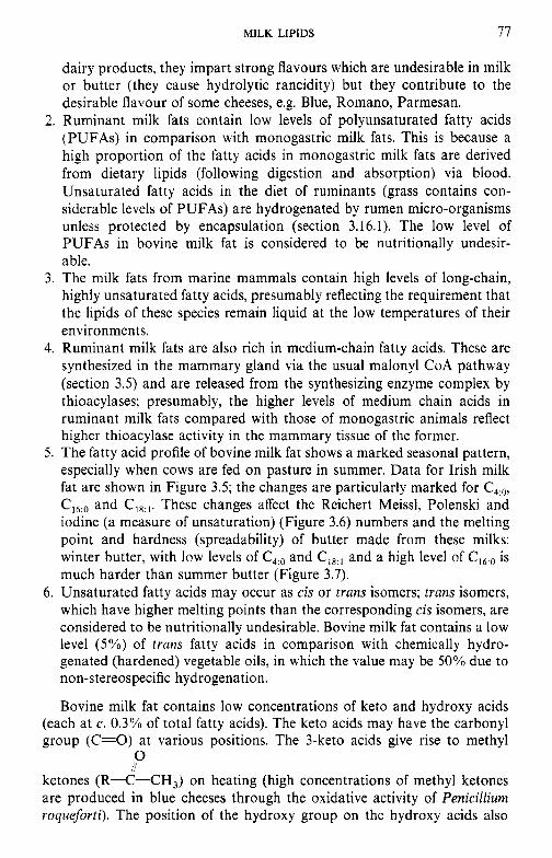
MILK LIPIDS 77
dairy products, they impart strong flavours which are undesirable in milk or butter (they cause hydrolytic rancidity) but they contribute to the desirable flavour of some cheeses, e.g. Blue, Romano, Parmesan.
2. Ruminant milk fats contain low levels of polyunsaturated fatty acids (PUFAs) in comparison with monogastric milk fats. This is because a high proportion of the fatty acids in monogastric milk fats are derived from dietary lipids (following digestion and absorption) via blood. Unsaturated fatty acids in the diet of ruminants (grass contains con- siderable levels of PUFAs) are hydrogenated by rumen micro-organisms unless protected by encapsulation (section 3.16.1). The low level of PUFAs in bovine milk fat is considered to be nutritionally undesir- able.
3. The milk fats from marine mammals contain high levels of long-chain, highly unsaturated fatty acids, presumably reflecting the requirement that the lipids of these species remain liquid at the low temperatures of their environments.
4. Ruminant milk fats are also rich in medium-chain fatty acids. These are synthesized in the mammary gland via the usual malonyl CoA pathway (section 3.5) and are released from the synthesizing enzyme complex by thioacylases; presumably, the higher levels of medium chain acids in ruminant milk fats compared with those of monogastric animals reflect higher thioacylase activity in the mammary tissue of the former.
5. The fatty acid profile of bovine milk fat shows a marked seasonal pattern, especially when cows are fed on pasture in summer. Data for Irish milk fat are shown in Figure 3.5; the changes are particularly marked for C,:,, C,,:, and c18:I. These changes affect the Reichert Meissl, Polenski and iodine (a measure of unsaturation) (Figure 3.6) numbers and the melting point and hardness (spreadability) of butter made from these milks: winter butter, with low levels of C4:o and c18:] and a high level of C,,:, is much harder than summer butter (Figure 3.7).
6. Unsaturated fatty acids may occur as cis or trans isomers; trans isomers, which have higher melting points than the corresponding cis isomers, are considered to be nutritionally undesirable. Bovine milk fat contains a low level (5%) of trans fatty acids in comparison with chemically hydro- genated (hardened) vegetable oils, in which the value may be 50% due to non-stereospecific hydrogenation.
Bovine milk fat contains low concentrations of keto and hydroxy acids (each at c. 0.3% of total fatty acids). The keto acids may have the carbonyl group (C=O) at various positions. The 3-keto acids give rise to methyl
ketones (R-C-CH,) on heating (high concentrations of methyl ketones are produced in blue cheeses through the oxidative activity of Penicilliurn roqueforti). The position of the hydroxy group on the hydroxy acids also
0 //
78 DAIRY CHEMISTRY AND BIOCHEMISTRY
4 -
3 -
2 -
. . . I I . I - , . , . , . , .
M J J A S O N D J F M A
13 i
1 2 - 0 0
8
3 1 1 -
g l o -
W .-
cd LL
9 -
M J J A S O N D J F M A M J J A Month (b)
MILK LIPIDS 79
42 - h Y
d? 4 0 -
2 M 0
!l 38- - on v
8 3 6 - $
2 5 0 34 -
M J J A S O N D J F
Month
Figure 3.6 Seasonal changes in the iodine number of Irish bovine milk fat (from Cullinane et al., 1984a).
M J J A S O N D J F M A M J J A S O
Month Figure 3.7 Seasonal variations in the mean firmness of Irish butter at 4°C (@) or 15°C (0) (from Cullinane et al., 1984b).
Table 3.7 The fatty acid composition of cholesteryl esters, phosphatidylcholine and phosphatidylethanolamine in the milks of some species
Fatty acid composition (wt% of the total)
Fatty acid
12:o 14:O 16:O 16: 1 1 8 : O 18: 1 18:2 18:3 20: 3 20:4 22:6
cow Human Pig
CE PC PE CE PC PE PC PE
0.2 0.3 0.1 3.2 - 2.3 7.1 1 .o 4.8 4.5 1.1 1.8 0.4
23.1 32.2 11.4 23.8 33.7 8.5 39.9 12.4 8.8 3.4 2.7 1.5 1.7 2.4 6.3 7.3
10.6 7.5 10.3 8.0 23.1 29.1 10.3 12.3 17.1 30.1 47.0 45.7 14.0 15.8 21.8 36.2 27.1 8.9 13.5 12.4 15.6 17.7 15.9 17.8 4.2 1.4 2.3 T 1.3 4.1 1.5 1.9 0.7 1 .o 1.7 2.1 3.4 0.3 0.7 1.4 1.2 2.7 T 3.3 12.5 1.3 6.6
0.1 0.4 2.6 0.2 1.6
~
-
- - -
Mink Mouse
CE PC PE PC PE
0.3 1.1 1.3 0.8
25.4 26.4 20.6 4.4 1.1 1.2
14.7 20.8 29.3 35.7 31.7 27.8 13.5 17.4 19.1 2.6 2.2 0.5
- -
- - -
- -
- 4.5 20.3 8.9 - 2.7
30.0 18.0 13.9 19.8 22.8 17.2
- - 8.9 20.0 1.8 6.3
Abbreviations: CE, cholesteryl esters; PC, phosphatidylcholine; PE, phosphatidylethanolamine; T, trace amount. From Christie (1995).
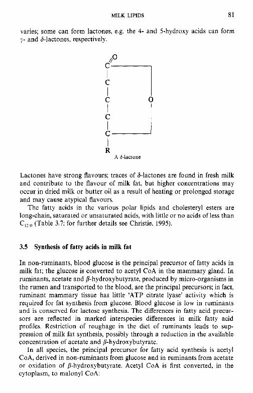
MILK LIPIDS 81
varies; some can form lactones, e.g. the 4- and 5-hydroxy acids can form y- and 8-lactones, respectively.
C ‘2 I R
A 6-lactone
Lactones have strong flavours; traces of S-lactones are found in fresh milk and contribute to the flavour of milk fat, but higher concentrations may occur in dried milk or butter oil as a result of heating or prolonged storage and may cause atypical flavours.
The fatty acids in the various polar lipids and cholesteryl esters are long-chain, saturated or unsaturated acids, with little or no acids of less than C,,:, (Table 3.7; for further details see Christie, 1995).
3.5 Synthesis of fatty acids in milk fat
In non-ruminants, blood glucose is the principal precursor of fatty acids in milk fat; the glucose is converted to acetyl CoA in the mammary gland. In ruminants, acetate and P-hydroxybutyrate, produced by micro-organisms in the rumen and transported to the blood, are the principal precursors; in fact, ruminant mammary tissue has little ‘ATP citrate lyase’ activity which is required for fat synthesis from glucose. Blood glucose is low in ruminants and is conserved for lactose synthesis. The differences in fatty acid precur- sors are reflected in marked interspecies differences in milk fatty acid profiles. Restriction of roughage in the diet of ruminants leads to sup- pression of milk fat synthesis, possibly through a reduction in the available concentration of acetate and P-hydroxybutyrate.
In all species, the principal precursor for fatty acid synthesis is acetyl CoA, derived in non-ruminants from glucose and in ruminants from acetate or oxidation of 8-hydroxybutyrate. Acetyl CoA is first converted, in the cytoplasm, to malonyl CoA:
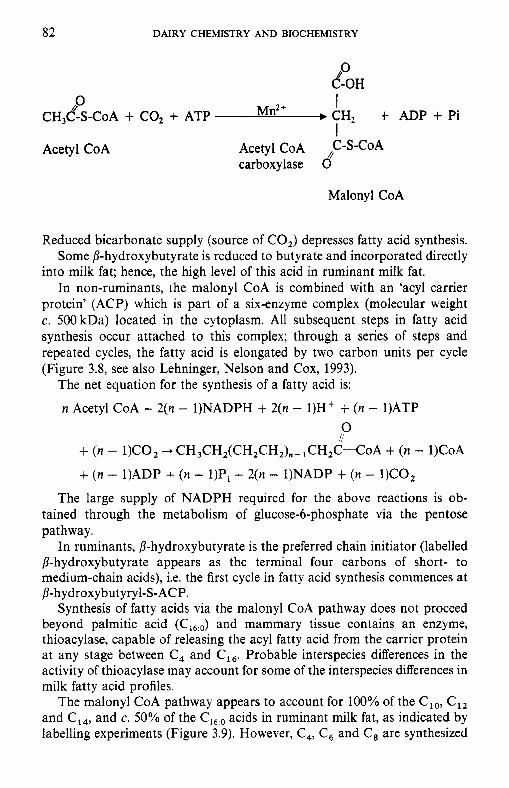
82 DAIRY CHEMISTRY AND BIOCHEMISTRY
0 &-OH
0 I I
b CH2 t ADP + Pi CH,&-S-C~A + co2 + ATP Mn2+
Acetyl CoA Acetyl CoA C-S-CoA carboxylase d
Malonyl CoA
Reduced bicarbonate supply (source of CO,) depresses fatty acid synthesis. Some P-hydroxybutyrate is reduced to butyrate and incorporated directly
into milk fat; hence, the high level of this acid in ruminant milk fat. In non-ruminants, the malonyl CoA is combined with an ‘acyl carrier
protein’ (ACP) which is part of a six-enzyme complex (molecular weight c. 500 kDa) located in the cytoplasm. All subsequent steps in fatty acid synthesis occur attached to this complex; through a series of steps and repeated cycles, the fatty acid is elongated by two carbon units per cycle (Figure 3.8, see also Lehninger, Nelson and Cox, 1993).
The net equation for the synthesis of a fatty acid is:
n Acetyl CoA + 2(n - 1)NADPH + 2(n - 1)H’ + (n - 1)ATP 0 il + ( n - 1)C02 -+ CH,CH,(CH~CH,),-~CH,C-COA + (n - 1)CoA
+ ( n - 1)ADP + ( n - 1)Pi + 2(n - 1)NADP + ( n - 1)COl
The large supply of NADPH required for the above reactions is ob- tained through the metabolism of glucose-6-phosphate via the pentose pathway.
In ruminants, P-hydroxybutyrate is the preferred chain initiator (labelled P-hydroxybutyrate appears as the terminal four carbons of short- to medium-chain acids), i.e. the first cycle in fatty acid synthesis commences at P-hydroxybut yryl-S- ACP.
Synthesis of fatty acids via the malonyl CoA pathway does not proceed beyond palmitic acid (C,,,,) and mammary tissue contains an enzyme, thioacylase, capable of releasing the acyl fatty acid from the carrier protein at any stage between C, and c16. Probable interspecies differences in the activity of thioacylase may account for some of the interspecies differences in milk fatty acid profiles.
The malonyl CoA pathway appears to account for 100% of the C,,, C,, and C14, and c. 50% of the C,,:, acids in ruminant milk fat, as indicated by labelling experiments (Figure 3.9). However, C,, c6 and C, are synthesized
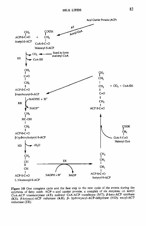
MILK LIPIDS
Acyl Carrier Protein (ACP)
83
SH3 I
Acetyl-S-ACP I ACP-SC=O + CH,
CoA-S-C=O Malonyl-SACP
y 3
c=o
ACP-S-C=O CH2 I I / + CO, + CoA-SH
bketobutyryl-S-ACP c=o
KR r""' NADP' + H+ 7H3
HC-OH I
I CH2
I ACP-SC=O
CH2 I
ACP-S-C=O khy droxybutyryl-S-ACP
, 7 3 3 7% CH2 I
HD kHZ0 CH II ** 'HZ I CH
ACP-S-C=O I ACP-S-C=O NADPH + H+ NADP
2, J-butenoyl-S-ACP butyryl-S-ACP
CoA-S-C=O Malonyl CoA
Figure 3.8 One complete cycle and the first step in the next cycle of the events during the synthesis of fatty acids. ACP = acyl carrier protein, a complex of six enzymes: i.e. acetyl CoA-ACP transacetylase (AT); malonyl CoA-ACP transferase (MT); B-keto-ACP synthase (KS); 8-ketoacyl-ACP reductase (KR); p- hydroxyacyl-ACP-dehydrase (HD); enoyl-ACP reductase (ER).
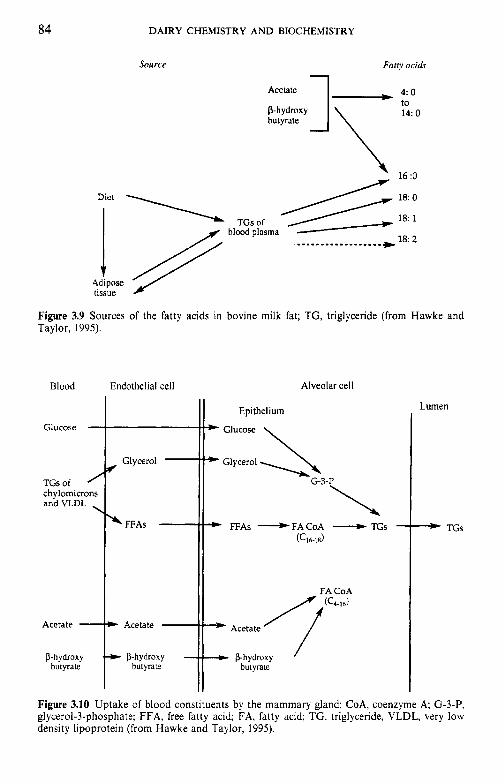
84 DAIRY CHEMISTRY AND BIOCHEMISTRY
Source Fatry acids
1 Acetate ., 4: 0
to 14: 0
hutyrate
\ d
TGs of hlood plasma
16 :O
18: 0
18: 1
18: 2
Figure 3.9 Sources of the fatty acids in bovine milk fat; TG, triglyceride (from Hawke and Taylor, 1995).
Blood Endothelial cell
D-hvdroxv
..
Alveolar cell
Lumen
- TGs
Figure 3.10 Uptake of blood constituents by the mammary gland; CoA, coenzyme A; G-3-P, glycerol-3-phosphate; FFA, free fatty acid; FA, fatty acid; TG, triglyceride, VLDL, very low density lipoprotein (from Hawke and Taylor, 1995).
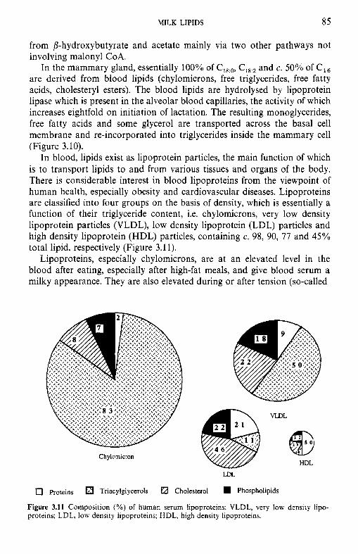
MILK LIPIDS 85
from P-hydroxybutyrate and acetate mainly via two other pathways not involving malonyl CoA.
In the mammary gland, essentially 100% of C,8:o, C,,:, and c. 50% of C,, are derived from blood lipids (chylomicrons, free triglycerides, free fatty acids, cholesteryl esters). The blood lipids are hydrolysed by lipoprotein lipase which is present in the alveolar blood capillaries, the activity of which increases eightfold on initiation of lactation. The resulting monoglycerides, free fatty acids and some glycerol are transported across the basal cell membrane and re-incorporated into triglycerides inside the mammary cell (Figure 3.10).
In blood, lipids exist as lipoprotein particles, the main function of which is to transport lipids to and from various tissues and organs of the body. There is considerable interest in blood lipoproteins from the viewpoint of human health, especially obesity and cardiovascular diseases. Lipoproteins are classified into four groups on the basis of density, which is essentially a function of their triglyceride content, i.e. chylomicrons, very low density lipoprotein particles (VLDL), low density lipoprotein (LDL) particles and high density lipoprotein (HDL) particles, containing c. 98, 90, 77 and 45% total lipid, respectively (Figure 3.1 1).
Lipoproteins, especially chylomicrons, are at an elevated level in the blood after eating, especially after high-fat meals, and give blood serum a milky appearance. They are also elevated during or after tension (so-called
Chylomicron
VIDL
HDL
LDL
0 Proteins Triacylglycerols Cholesterol Phospholipids
Figure 3.11 Composition (%) of human serum lipoproteins; VLDL, very low density lipo- proteins; LDL, low density lipoproteins; HDL, high density lipoproteins.
86 DAIRY CHEMISTRY AND BIOCHEMISTRY
Palmitic Acid
'almitoleic acid Stcaric acid 9 -Cm1
Vaccenic acid
11 - CI,:,
Oleic acid ClOl
J y
5,8,11- C,, 15 - C,,,
Eicosatrienoic Nervonic acid acid
up Series
9.12 - c,,;, 9.12.15 - c,,;, Linoleic acid Linolenic acid
K 6.9,12,15 - C,,;,
11.14 - C,, #q Eicosadienoic acid 6,9,12-cl,! Linolenic acid
+cll 8,11,14 - c,,
-2Hl 5,s. 11,14 - Cm, Arachidonic acid
w series (b)
8,11,14,17 - Cm.
5,8,11,14,17 - C ,
1.c'
7,10,13,16,19 - Ca5 1 - 2 H
P.7.10,13,16,19 - c,,:, Docosahexenoic acid
0, series
Figure 3.12 Elongation and/or desaturation of fatty acids in the mammary gland.
MILK LIPIDS 87
racing driver syndrome). Chylomicrons, which are formed in the intestinal mucosa, are secreted into the lymph and enter the blood via the thoracic duct. VLDL lipoproteins are synthesized in intestinal mucosa and liver. LDL lipoproteins are formed at various sites, including mammary gland, by removing of triglycerides from VLDL.
Since about 50% of c16:o and 100% of C,,:,, C,,:, and C18:2 are derived from blood lipids, about 50% of the total fatty acids in ruminant milk fat originate from the blood via diet or other organs.
In liver mitochondria, palmitic acid, as its CoA ester, is lengthened by successive additions of acetyl CoA. There is also a liver microsomal enzyme capable of elongating saturated and unsaturated fatty acids by addition of acetyl CoA or malonyl CoA.
The principal monoenoic acids, oleic (C18:J and palmitoleic (Cl6:1), are derived from blood lipids but about 30% of these acids are produced by microsomal enzymes (in the endoplasmic reticulum) in the secretory cells by desaturation of stearic and palmitic acids, respectively:
Stearyl CoA + NADPH + 0, - oleoyl CoA + NADP' + 2H,O
Shorter chain unsaturated acids (Clo:l to C14: ,) are probably also produced by the same enzyme.
Linoleic (&) and linolenic (c1@3) acids cannot be synthesized by mammals and must be supplied in the diet, i.e. they are essential fatty acids (linoleic is the only true essential acid). These two polyenoic acids may then be elongated and/or further desaturated by mechanisms similar to stearic +
oleic, to provide a full range of polyenoic acids. A summary of these reactions is given in Figure 3.12a, b.
b-Hydroxy acids are produced by &oxidation of fatty acids and p-keto acids may arise from incomplete syntheses or via P-oxidation.
desaturase
3.6 Structure of milk lipids
Glycerol for milk lipid synthesis is obtained in part from hydrolysed blood lipids (free glycerol and monoglycerides), partly from glucose and a little from free blood glycerol. Synthesis of triglycerides within the cell is catalysed by enzymes located on the endoplasmic reticulum, as shown in Figure 3.13.
Esterification of fatty acids is not random: c,,-c16 are esterified principally at the sn-2 position while C, and (26 are esterified principally at the sn-3 position (Table 3.8). The concentrations of C, and C,, appear to be rate-limiting because of the need to keep the lipid liquid at body temperature. Some features of the structures are notable:
0 Butanoic and hexanoic acids are esterified almost entirely, and octanoic and decanoic acids predominantly, at the sn-3 position.
CH2OH c--. Glucose CH20H I 1
CHOH
CH,O -Pe OH I 0
I
I c = o I 0
CH,-0 -(Lo"
Glycerol t ATP ___t Glycerol-3-P 0 t ADP 4 Dihydroxy 6- acetone P glycerokinase
Phosphatidic acid *P Phosphatase H , C - O - C - R 4-
I *P H C - 0 - C - R
I H2C - OH
Diglyceride H,C -0 -C 1 R
I .P I s .0
H C - 0 - C - R
H 2 C - 0 - C - R
RC-S-CoA Triglyceride
NADPH + H,
0.0 2 RC-S-CoA acyl hansferase
H,C-o -c-R I 9 I ..o
H C - 0 - C - R
HZC-0 - P - O H '0
Phosphatidic acid
Figure 3.13 Biosynthesis of triglycerides in the mammary gland
Table 3.8 Composition of fatty acids (mol% of the total) esterified to each position of the triacyl-sn-glycerols in the milks of various species
c o w Human Rat Pig Rabbit Seal Echidna Fatty acid sn-1 sn-2 SIT-3 sn-l sn-2 sn-3 sn-l sn-2 sn-3 sn-l sn-2 sn-3 sn-l sn-2 sn-3 sn-l sn-2 sn-3 sn-1 sn-2 sn-3
~~
4:O - 6:O - 0.9 8:O 1.4 0.7
l o : o 1.9 3.0 12:o 4.9 6.2 14:O 9.7 17.5 16:O 34.0 32.3 16: 1 2.8 3.6 18:O 10.3 9.5 18: 1 30.0 18.9 18:2 1.7 3.5 18:3 - -
-
c 2 0 - c 2 2 - -
35.4 - - 12.9 - -
- - - - - - - - -
- - - - - - - - -
3.6 - - 3.7 5.7 10.0 - - 19.2 6.2 0.2 0.2 1.1 10.1 20.0 26.0 - - - - 22.5 0.6 1.3 2.1 5.6 10.4 15.9 15.1 - - 3.5 6.4 3.2 7.3 6.9 9.6 17.8 8.9 2.4 6.8 3.7 2.7 2.1 5.4 16.1 58.2 5.5 20.2 28.7 12.6 21.8 57.6 15.4 24.1 12.7 1.4 3.6 4.7 7.6 1.8 2.1 1.8 6.6 11.2 10.4 4.1 1.3 1.2 15.0 3.3 1.8 4.9 0.8 1.5 6.9 1.1 5.5 6.9 3.5
23.1 46.1 12.7 50.4 24.2 3.3 11.8 49.6 13.9 51.7 40.8 16.6 2.3 11.0 7.3 15.0 14.1 5.2 11.6 11.3 8.4 11.5 15.6 15.1
- - -
- -
- 0.4 0.6 1.7 1.2 0.5 0.7 1.4 1.0 1.8 3.4 3.5 - - - - - - - - - - - -
- - - - - - - - - - - - -
- - - - 33.7 38.9 - 22.5 26.1 - - - - -
- - 2.8 1.8 0.3 0.2 - 2.6 0.7 23.6 3.8 1.7 0.9 0.4
23.8 6.1 31.0 1.0 31.5 9.0 27.9
0.9 1.9 0.7 1.0 16.8 2.1 14.3 3.8 11.4 19.4 45.4 33.1 57.6 39.8 6.4 9.7 2.3 2.8 4.1 18.3 4.9 2.0 2.3 0.5 0.7 1.0 2.9 2.0
- - 1.5 1.1 16.8 14.1 -
- - - -- 0.8 28.7 -
From Christie (1995).
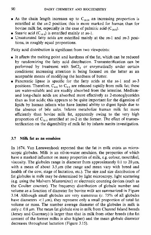
90 DAIRY CHEMISTRY AND BIOCHEMISTRY
0 As the chain length increases up to c,6,0, an increasing proportion is esterified at the sn-2 position; this is more marked for human than for bovine milk fat, especially in the case of palmitic acid (CI6:,J,
0 Stearic acid (C,8:o) is esterified mainly at sn-1. 0 Unsaturated fatty acids are esterified mainly at the sn-1 and sn-3 posi-
Fatty acid distribution is significant from two viewpoints:
0 It affects the melting point and hardness of the fat, which can be reduced by randomizing the fatty acid distribution. Transesterification can be performed by treatment with SnCl, or enzymatically under certain conditions; increasing attention is being focused on the latter as an acceptable means of modifying the hardness of butter.
0 Pancreatic lipase is specific for the fatty acids at the sn-1 and sn-3 positions. Therefore, C4:o to C8:o are released rapidly from milk fat; these are water-soluble and are readily absorbed from the intestine. Medium- and long-chain acids are absorbed more effectively as 2-monoglycerides than as free acids; this appears to be quite important for the digestion of lipids by human infants who have limited ability to digest lipids due to the absence of bile salts. Infants metabolize human milk fat more efficiently than bovine milk fat, apparently owing to the very high proportion of C,6:o esterified at sn-2 in the former. The effect of transes- terification on the digestibility of milk fat by infants merits investigation.
tions, in roughly equal proportions.
3.7 Milk fat as an emulsion
In 1674, Van Leeuwenhoek reported that the fat in milk exists as micro- scopic globules. Milk is an oil-in-water emulsion, the properties of which have a marked influence on many properties of milk, e.g. colour, mouthfeel, viscosity. The globules range in diameter from approximately 0.1 to 20 pm, with a mean of about 3.5pm (the range and mean vary with breed and health of the cow, stage of lactation, etc.). The size and size distribution of fat globules in milk may be determined by light microscopy, light scattering (e.g. using the Malvern Mastersizer) or electronic counting devices (such as the Coulter counter). The frequency distribution of globule number and volume as a function of diameter for bovine milk are summarized in Figure 3.14. Although small globules are very numerous (c. 75% of all globules have diameters < 1 pm), they represent only a small proportion of total fat volume or mass. The number average diameter of the globules in milk is only c. 0.8 pm. The mean fat globule size in milk from Channel Island breeds (Jersey and Guernsey) is larger than that in milk from other breeds (the fat content of the former milks is also higher) and the mean globule diameter decreases throughout lactation (Figure 3.1 5).
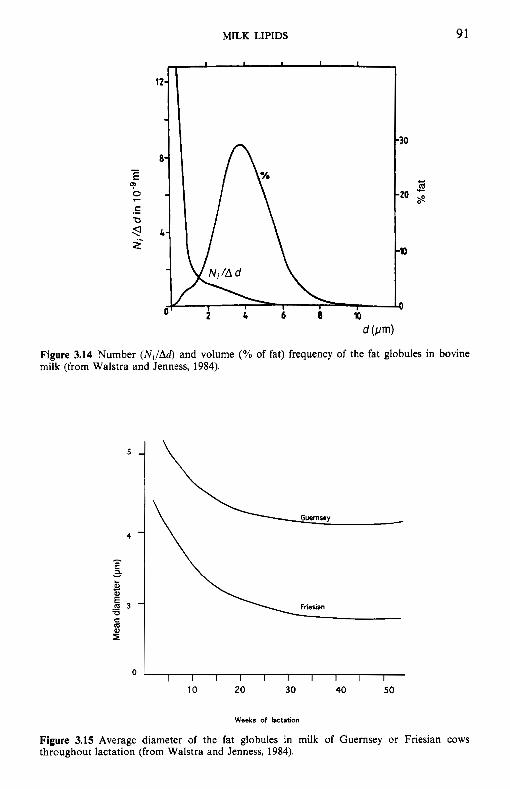
MILK LIPIDS 91
I I I I
v N ; / A d \
-30
- -20
-lo
Figure 3.14 Number (NJAd) and volume (% of fat) frequency of the fat globules in bovine milk (from Walstra and Jenness, 1984).
0 1 I I I I I I I I I
10 20 30 40 50
Weeks of lactation
Figure 3.15 Average diameter of the fat globules in milk of Guernsey or Friesian cows throughout lactation (from Walstra and Jenness, 1984).
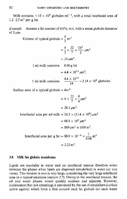
92 DAIRY CHEMISTRY AND BIOCHEMISTRY
Milk contains - 15 x lo9 globules ml-', with a total interfacial area of 1.2-2.5 m2 per g fat.
Example. of 3 pm.
Assume a fat content Of 4.O%, w/v, with a mean globule diameter
4 3
Volume of typical globule = - 71r3
4 22 (3)3 = - x - x - pm3 3 7 2
- 14pm3.
1 ml milk contains: 0.04g fat
= 4.4 x 10" pm3.
1 ml milk contains: 4'4 1010-3.14 x lo9 globules. 14
Surface area of a typical globule = 471r2
22 9 7 4
= 4 x - x -pmZ
= 28.3 pm2.
Interfacial area per ml milk = 28.3 x (3.14 x 10') pm2
= 88.9 x 10⁹pm2
= 889 cm2 % 0.09 rn2.
1 x - m2
0.04 Interfacial area per g fat = 88.9 x
= 2.22mZ.
3.8 Milk fat globule membrane
Lipids are insoluble in water and an interfacial tension therefore exists between the phases when lipids are dispersed (emulsified) in water (or vice versa). This tension in toto is very large, considering the very large interfacial area in a typical emulsion (section 3.7). Owing to the interfacial tension, the oil and water phases would quickly coalesce and separate. However, coalescence (but not creaming) is prevented by the use of emulsifiers (surface active agents) which form a film around each fat globule (or each water
MILK LIPIDS 93
droplet in the case of water-in-oil emulsions) and reduce interfacial tension. In the case of unprocessed milk, the emulsifying film is much more complex than that in ‘artificial’ emulsions, and is referred to as the milk fat globule membrane (MFGM).
In 1840, Ascherson observed an emulsion-stabilizing membrane sur- rounding the fat globules in milk and suggested that the membrane was ‘condensed’ albumin (from the skim-milk phase) aggregated at the fat/ plasma interface. Babcock, in the 188Os, also felt that the milk fat emulsifier was adsorbed serum protein. Histological staining and light microscopy were employed around the turn of the century to identify the nature of the membrane material but it was early recognized that contamination of fat globules by skim-milk components presented a major problem. By analysing washed globules, it was shown that the MFGM contained phospholipids and protein which differed from the skim-milk proteins (see Brunner (1974) for historical review).
3.8. I Isolation of the f a t globule membrane
The definition of what precisely constitutes the membrane leads to consider- able difficulty and uncertainty. The outer boundary is assumed to constitute everything that travels with the fat globule when it moves slowly through milk; however, the outer regions of the membrane are loosely attached and some or all may be lost, depending on the extent of mechanical damage the globule suffers. The inner boundary is ill-defined and depends on the method of preparation; there is considerable discussion as to whether a layer of high melting point triglyceride, immediately inside the membrane, is part of the membrane or not. Some hydrophobic constituents of the membrane prob- ably diffuse into the core of the globules while components of the plasma may adsorb at the outer surface. Since the membrane contains numerous enzymes, enzymatic changes may occur.
Several methods are available for isolating all or part of the membrane. The usual initial step involves separating a cream from milk by mechanical centrifugation (which may cause some damage) or by gravity. The cream is washed repeatedly (3-6 times) with water or dilute buffer by dilution and gravity separation; soluble salts and other small molecules are probably lost into the serum. Mechanical damage may remove the loosely bound outer layers and may even cause some homogenization and adsorption of serum constituents; small globules are lost during each washing cycle.
The washed cream is destabilized by churning or freezing; then the fat (mainly triglycerides) is melted and separated from the membrane material by centrifugation. Cross-contamination of membrane with core material may be considerable, and methods must be carefully standardized. An elaborate scheme for the isolation and fractionation of the MFGM was developed by Brunner and co-workers (Brunner, 1974).
94 DAIRY CHEMISTRY AND BIOCHEMISTRY
Treatment of washed cream with surfactants, usually sodium de- oxycholate, releases part of the membrane, assumed to represent only the outer layer. Unless the treatment is carefully controlled, some inner material will be released also.
3.8.2
Yields of 0.5-1.5g MFGM per lOOg fat have been reported; the range reflects variations in temperature history, washing technique, age, agitation, etc. The gross chemical composition of the membrane is reasonably well established and the relatively small differences reported are normally at- tributed to different methods used to isolate and fractionate the membrane material. The data in Table 3.9, from Mulder and Walstra (1974) and based on the investigations of many workers, give a reasonable estimate of the gross composition of the MFGM. A more detailed compositional analysis is provided by Keenan et al. (1983) (Table 3.10). Brunner (1965, 1974), Mulder and Walstra (1974), Patton and Keenan (1975), Keenan et al. (1983) and Keenan and Dylewski (1995) should be consulted for more detailed compositional data.
Gross chemical composition of MFGM
3.8.3 The protein fraction
Depending on the preparative method used, the membrane may or may not contain skim-milk proteins (i.e. caseins and whey proteins); if the membrane has been damaged prior to isolation, it may contain considerable amounts of these proteins. The membrane contains unique proteins which do not occur in the skim-milk phase. Many of the proteins are glycoproteins and contain a considerable amount of carbohydrate (hexose, 2.8-4.1 5%; hexosamine, 2.5-4.2%; and sialic acid, 1.3-1.8%).
Sodium dodecyl sulphate-polyacrylamide gel electrophoresis (SDS- PAGE), with silver staining of the gels, resolves MFGM proteins into as many as 60 discrete bands, ranging in molecular mass from 11 to 250 kDa (Keenan and Dylewski, 1995). Most of these proteins are present at very low concentrations (many are detectable only when gels are stained with silver but not with Coomassie blue). Some of these proteins may be genetic variants and, since the MFGM contains a plasmin-like proteinase, some of the smaller polypeptides may be fragments of larger proteins. The three principal proteins, with molecular masses (by SDS-PAGE) of 155, 67 and 48 kDa, are xanthine oxidase, butyrophilin and glycoprotein B, respectively; five or six glycoproteins have been detected by staining with SchifYs reagent.
Xanthine oxidase, which requires Fe, Mo and flavin adenine dinucleotide (FAD) as co-factors, is capable of oxidizing lipids via the production of superoxide radicals. It represents about 20% of the MFGM protein and part is readily lost from the membrane, e.g. on cooling; isoelectric focusing
MILK LIPIDS 95
indicates at least four variants with isoelectric points (pl) in the range
Butyrophilin, the principal MFGM protein and so named because of its high affinity for milk lipids, is a very hydrophobic, difficult to solubilize (insoluble or only sparingly soluble in most protein solvents, including detergents) glycoprotein. Isoelectric focusing indicates at least four variants (pls 5.2-5.3). The amino acid sequence of butyrophilin has been determined and its gene has been cloned, which indicates that butyrophilin is syn- thesized with a leader sequence; it consists of 526 amino acids and has a molecular mass, without carbohydrate, of 56 460 Da. It binds phospholipids tenaciously and perhaps even contains covalently bound fatty acids. It is located only at the apical cell surface of the mammary epithelial cells, suggesting a role in membrane envelopment of fat globules.
Several of the minor proteins of the MFGM have been isolated and partially characterized (Keenan and Dylewski, 1995). A systematic nomen- clature has not been developed for the MFGM proteins and most are referred to by their relative electrophoretic mobility on SDS-PAGE and whether or not they are glycoproteins. The proteins of the MFGM represent approximately 1% of the total proteins in milk.
7.0-7.5.
3.8.4 The lipidfvaction
The membrane contains 0.5-1.0% of the total lipid in milk and is composed principally of phospholipids and neutral lipids in the approximate ratio 2 : 1, with lesser amounts of other lipids (Tables 3.9 and 3.10); contamination with core lipid is a major problem. The phospholipids are principally phos- phatidylcholine, phosphatidylethanolamine and sphingomyelin in the ap- proximate ratio 2 : 2 : 1. The principal fatty acids and their approximate percentages in the phospholipids are C14:o (5%) , C,,:, (25%), c,&o (14%), CI,:, (25%) C,,:, YO), C,,:, (3%) and C24:o (3%). Thus, the membrane contains a significantly higher level of polyunsaturated fatty acids than milk
Table 3.9 Gross composition of the milk fat globule membrane ~ ~~~~
mg 100 g-' mg m-' fat Yo (w/w) of Component fat globule globule surface total membrane
Protein 900 4.5 41 Phospholipid 600 3.0 27 Cerebrosides 80 0.4 3 Cholesterol 40 0.2 2 Neutral glycerides 300 1.5 14 Water 280 1.4 13
Total 2200 11.0 100
From Mulder and Walstra (1974).
96 DAIRY CHEMISTRY AND BIOCHEMISTRY
Table 3.10 Composition of bovine milk fat globule membranes
Constituent class Amount
Protein Total lipid Phospholipid Phosphatidyl choline Phosphatidylethanolamine Sphingomyelin Phosphatidylinositol Phosphatidylserine
Hydrocarbons Sterols Sterol esters Glycerides Free fatty acids
Cerebrosides Gangliosides Total sialic acids Hexoses Hexosamines Cytochrome b , + P420 Uronic acids RNA
Neutral lipid
25-60% of dry weight 0.5- 1.2 mg per mg protein 0.13-0.34 mg per mg protein
34% of total lipid phosphorus 28% of total lipid phosphorus 22% of total lipid phosphorus 10% of total lipid phosphorus 6% of total lipid phosphorus
56-80% of total lipid 1.2% of total lipid 0.2-5.2% of total lipid 0.1-0.8% of total lipid 53-74% of total lipid 0.6-6.3% of total lipid
3.5 nmoles per mg protein 6-7.4 nmoles sialic acid per mg protein 63 nmoles per mg protein 0.6 pmoles per mg protein 0.3 pmoles per mg protein 30 pmoles per mg protein 99 ng per mg protein 20 pg per mg protein
From Keenan et a/. (1983).
Table 3.1 1 Structures of glycosphingolipids of bovine milk fat globule membrane
GI ycosphingolipid Structure
Glucosyl cerarnide Lactosyl ceramide GM, (hematoside) G M ,
G M ,
GD, (disialohematoside)
GD,
GD,,
p-Glucosyl-(1 -+ 1)-ceramide fl-Glucosyl-( 1 -+ 4)-/3-glucosyl-( 1 -+ 1)-ceramide NeuraminosyL(2 + 3)-galactosyl-glucosyl-ceramide N- Acetylgalactosamin yl-( neuraminosy1)-galactosyl-glucosyl-
Galactosyl-N-acetylgalactosaminyl-(neuraminosyl)-galactosyl-
Neuraminosyl-(2 -+ 8)-neuraminosyl-(2 -+ 3)-galactosyl-
N-Acetylgalactosaminyl-(neuraminosyl-neuraminosyl)-
Galactosyl-N-acetylgalactosaminyl-(neuraminosyl-
ceramide
glucosyl-ceramide
glucosyl-ceramide
galactosyl-glucosyl-ceramide
neuraminosy1)-galactosyl-glucosyl-ceramide
From Keenan et al. (1983).
MILK LIPIDS 97
fat generally and is, therefore, more susceptible to oxidation. The cerebro- sides are rich in very long chain fatty acids which possibly contribute to membrane stability. The membrane contains several glycolipids (Table 3.11).
The amount and nature of the neutral lipid present in the MFGM is uncertain because of the difficulty in defining precisely the inner limits of the membrane. It is generally considered to consist of 83-88% triglyceride, 5 1 4 % diglyceride and 1-5% free fatty acids. The level of diglyceride is considerably higher than in milk fat as a whole; diglycerides are relatively polar and are, therefore, surface-active. The fatty acids of the neutral lipid fraction are longer-chained than in milk fat as a whole and in order of proportion present are palmitic, stearic, myristic, oleic and lauric.
Most of the sterols and sterol esters, vitamin A, carotenoids and squalene in milk are dissolved in the core of the fat globules but some are probably present in the membrane.
3.8.5 Other membrane components
Trace metals. The membrane contains 5 2 5 % of the indigenous Cu and 30-60% of the indigenous Fe of milk as well as several other elements, e.g. Co, Ca, Na, K, Mg, Mn, Mo, Zn, at trace levels; Mo is a constituent of xanthine oxidase.
Enzymes. The MFGM contains many enzymes (Table 3.12). These en- zymes originate from the cytoplasm and membranes of the secretory cell and are present in the MFGM due to the mechanism of globule excretion from the cells.
3.8.6 Membrane structure
Several early attempts to describe the structure of the MFGM included King (1955), Hayashi and Smith (1965), Peereboom (1969), Prentice (1969) and Wooding (1971). Although the structures proposed by these workers were inaccurate, they stimulated thinking on the subject. Keenan and Dylewski (1995) and Keenan and Patton (1995) should be consulted for recent reviews.
Understanding of the structure of the MFGM requires understanding three processes: the formation of lipid droplets from triglycerides synthesized in or on the endoplasmic reticulum at the base of the cell, movement of the droplets (globules) through the cell and excretion of the globules from the cell into the lumen of the alveolus.
The MFGM originates from regions of apical plasma membrane, and also from endoplasmic reticulum (ER) and perhaps other intracellular compartments. That portion of the MFGM derived from apical plasma
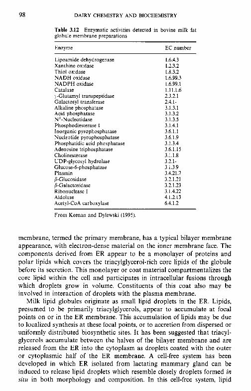
98 DAIRY CHEMISTRY AND BIOCHEMISTRY
Table 3.12 Enzymatic activities detected in bovine milk fat globule membrane preparations
Enzyme EC number
Lipoamide dehydrogenase Xanthine oxidase Thiol oxidase NADH oxidase NADPH oxidase Catalase y-Glutamyl transpeptidase Galactosyl transferase Alkaline phosphatase Acid phosphatase N -Nucleotidase Phosphodiesterase I Inorganic pyrophosphatase Nucleotide pyrophosphatase Phosphatidic acid phosphatase Adenosine triphosphatase Cholinesterase UDP-glycosyl hydrolase Glucose-6-phosphatase Plasmin P-Glucosidase P-Galactosidase Ribonuclease I Aldolase Acetyl-CoA carboxylase
1.6.4.3 1.2.3.2 1.8.3.2 1.6.99.3 1.6.99.1 1.1 1.1.6 2.3.2.1 2.4.1 - 3.1.3.1 3.1.3.2 3.1.3.5 3.1.4.1 3.6.1.1 3.6.1.9 3.1.3.4 3.6.1.15 3.1.1.8
3.1.3.9 3.4.21.7 3.2.1.21
3.2.1-
3.2.1.23 3.1.4.22 4.1.2.1 3 6.4.1.2
From Keenan and Dylewski (1995).
membrane, termed the primary membrane, has a typical bilayer membrane appearance, with electron-dense material on the inner membrane face. The components derived from ER appear to be a monolayer of proteins and polar lipids which covers the triacylglycerol-rich core lipids of the globule before its secretion. This monolayer or coat material compartmentalizes the core lipid within the cell and participates in intracellular fusions through which droplets grow in volume. Constituents of this coat also may be involved in interaction of droplets with the plasma membrane.
Milk lipid globules originate as small lipid droplets in the ER. Lipids, presumed to be primarily triacylglycerols, appear to accumulate at focal points on or in the ER membrane. This accumulation of lipids may be due to localized synthesis at these focal points, or to accretion from dispersed or uniformly distributed biosynthetic sites. It has been suggested that triacyl- glycerols accumulate between the halves of the bilayer membrane and are released from the ER into the cytoplasm as droplets coated with the outer or cytoplasmic half of the ER membrane. A cell-free system has been developed in which ER isolated from lactating mammary gland can be induced to release lipid droplets which resemble closely droplets formed in situ in both morphology and composition. In this cell-free system, lipid
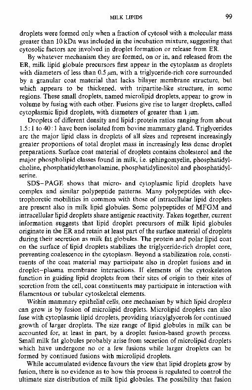
MILK LIPIDS 99
droplets were formed only when a fraction of cytosol with a molecular mass greater than 10 kDa was included in the incubation mixture, suggesting that cytosolic factors are involved in droplet formation or release from ER.
By whatever mechanism they are formed, on or in, and released from the ER, milk lipid globule precursors first appear in the cytoplasm as droplets with diameters of less than 0.5 pm, with a triglyceride-rich core surrounded by a granular coat material that lacks bilayer membrane structure, but which appears to be thickened, with tripartite-like structure, in some regions. These small droplets, named microlipid droplets, appear to grow in volume by fusing with each other. Fusions give rise to larger droplets, called cytoplasmic lipid droplets, with diameters of greater than 1 pm.
Droplets of different density and lipid : protein ratios ranging from about 1.5 : 1 to 40 : 1 have been isolated from bovine mammary gland. Triglycerides are the major lipid class in droplets of all sizes and represent increasingly greater proportions of total droplet mass in increasingly less dense droplet preparations. Surface coat material of droplets contains cholesterol and the major phospholipid classes found in milk, i.e. sphingomyelin, phosphatidyl- choline, phosphatidylethanolamine, phosphatidylinositol and phosphatidyl- serine.
SDS-PAGE shows that micro- and cytoplasmic lipid droplets have complex and similar polypeptide patterns. Many polypeptides with elec- trophoretic mobilities in common with those of intracellular lipid droplets are present also in milk lipid globules. Some polypeptides of MFGM and intracellular lipid droplets share antigenic reactivity. Taken together, current information suggests that lipid droplet precursors of milk lipid globules originate in the ER and retain at least part of the surface material of droplets during their secretion as milk fat globules. The protein and polar lipid coat on the surface of lipid droplets stabilizes the triglyceride-rich droplet core, preventing coalescence in the cytoplasm. Beyond a stabilization role, consti- tuents of the coat material may participate also in droplet fusions and in droplet-plasma membrane interactions. If elements of the cytoskeleton function in guiding lipid droplets from their sites of origin to their sites of secretion from the cell, coat constituents may participate in interaction with filamentous or tubular cytoskeletal elements.
Within mammary epithelial cells, one mechanism by which lipid droplets can grow is by fusion of microlipid droplets. Microlipid droplets can also fuse with cytoplasmic lipid droplets, providing triacylglycerols for continued growth of larger droplets. The size range of lipid globules in milk can be accounted for, at least in part, by a droplet fusion-based growth process. Small milk fat globules probably arise from secretion of microlipid droplets which have undergone no or a few fusions while larger droplets can be formed by continued fusions with microlipid droplets.
While accumulated evidence favours the view that lipid droplets grow by fusion, there is no evidence as to how this process is regulated to control the ultimate size distribution of milk lipid globules. The possibility that fusion
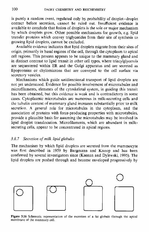
100 DAIRY CHEMISTRY AND BIOCHEMISTRY
is purely a random event, regulated only by probability of droplet-droplet contact before secretion, cannot be ruled out. Insufficient evidence is available to conclude that fusion of droplets is the sole or major mechanism by which droplets grow. Other possible mechanisms for growth, e.g. lipid transfer proteins which convey triglycerides from their site of synthesis to growing lipid droplets, cannot be excluded.
Available evidence indicates that lipid droplets migrate from their sites of origin, primarily in basal regions of the cell, through the cytoplasm to apical cell regions. This process appears to be unique to the mammary gland and in distinct contrast to lipid transit in other cell types, where triacylglycerols are sequestered within ER and the Golgi apparatus and are secreted as lipoproteins or chylomicrons that are conveyed to the cell surface via secretory vesicles.
Mechanisms which guide unidirectional transport of lipid droplets are not yet understood. Evidence for possible involvement of microtubules and microfilaments, elements of the cytoskeletal system, in guiding this transit has been obtained, but this evidence is weak and is contradictory in some cases. Cytoplasmic microtubules are numerous in milk-secreting cells and the tubulin content of mammary gland increases substantially prior to milk secretion. A general role for microtubules in the cytoplasm, and the association of proteins with force-producing properties with microtubules, provide a plausible basis for assuming the microtubules may be involved in lipid droplet translocation. Microfilaments, which are abundant in milk- secreting cells, appear to be concentrated in apical regions.
3.8.7 Secretion of milk lipid globules
The mechanism by which lipid droplets are secreted from the mammocyte was first described in 1959 by Bargmann and Knoop and has been confirmed by several investigators since (Keenan and Dylewski, 1995). The lipid droplets are pushed through and become enveloped progressively by
Figure 3.16 Schematic representation of the excretion of a fat globule through the apical membrane of the mammary cell.
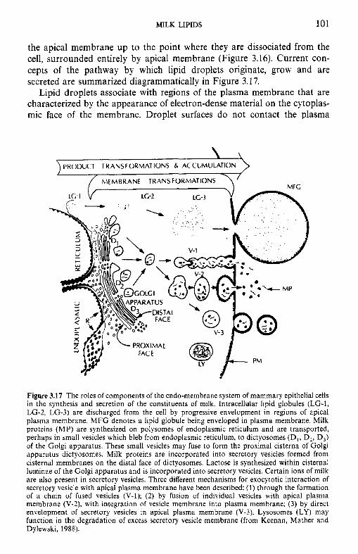
MILK LIPIDS 101
the apical membrane up to the point where they are dissociated from the cell, surrounded entirely by apical membrane (Figure 3.16). Current con- cepts of the pathway by which lipid droplets originate, grow and are secreted are summarized diagrammatically in Figure 3.17.
Lipid droplets associate with regions of the plasma membrane that are characterized by the appearance of electron-dense material on the cytoplas- mic face of the membrane. Droplet surfaces do not contact the plasma
Figure 3.17 The roles of components of the endo-membrane system of mammary epithelial cells in the synthesis and secretion of the constituents of milk. Intracellular lipid globules (LG-1, LG-2, LG-3) are discharged from the cell by progressive envelopment in regions of apical plasma membrane. M F G denotes a lipid globule being enveloped in plasma membrane. Milk proteins (MP) are synthesized on polysomes of endoplasmic reticulum and are transported, perhaps in small vesicles which bleb from endoplasmic reticulum, to dictyosomes (D1, D,, D3) of the Golgi apparatus. These small vesicles may fuse to form the proximal cisterna of Golgi apparatus dictyosomes. Milk proteins are incorporated into secretory vesicles formed from cisternal membranes on the distal face of dictyosomes. Lactose is synthesized within cisternal luminae of the Golgi apparatus and is incorporated into secretory vesicles. Certain ions of milk are also present in secretory vesicles. Three different mechanisms for exocytotic interaction of secretory vesicle with apical plasma membrane have been described: (1) through the formation of a chain of fused vesicles (V-I); (2) by fusion of individual vesicles with apical plasma membrane (V-2), with integration of vesicle membrane into plasma membrane; (3) by direct envelopment of secretory vesicles in apical plasma membrane (V-3). Lysosomes (LY) may function in the degradation of excess secretory vesicle membrane (from Keenan, Mather and Dylewski, 1988).
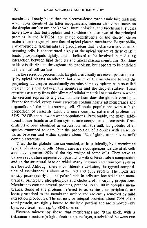
102 DAIRY CHEMISTRY AND BIOCHEMISTRY
membrane directly but rather the electron-dense cytoplasmic face material; which constituents of the latter recognize and interact with constituents on the droplet surface are not known. Immunological and biochemical studies have shown that butyrophilin and xanthine oxidase, two of the principal proteins in the MFGM, are major constituents of the electron-dense material on the cytoplasmic face of apical plasma membrane. Butyrophilin, a hydrophobic, transmembrane glycoprotein that is characteristic of milk- secreting cells, is concentrated highly at the apical surface of these cells; it binds phospholipids tightly, and is believed to be involved in mediating interaction between lipid droplets and apical plasma membrane. Xanthine oxidase is distributed throughout the cytoplasm, but appears to be enriched at the apical cell surface.
In the secretion process, milk fat globules usually are enveloped compact- ly by apical plasma membrane, but closure of the membrane behind the projecting fat droplet occasionally entrains some cytoplasm as a so-called crescent or signet between the membrane and the droplet surface. These crescents can vary from thin slivers of cellular material to situations in which the crescent represents a greater volume than does the globule core lipid. Except for nuclei, cytoplasmic crescents contain nearly all membranes and organelles of the milk-secreting cell. Globule populations with a high proportion of crescents exhibit a more complex pattern of proteins by SDS-PAGE than low-crescent populations. Presumably, the many addi- tional minor bands arise from cytoplasmic components in crescents. Cres- cents have been identified in association with the milk fat globules of all species examined to date, but the proportion of globules with crescents varies between and within species; about 1% of globules in bovine milk contain crescents.
Thus, the fat globules are surrounded, at least initially, by a membrane typical of eukaryotic cells. Membranes are a conspicuous feature of all cells and may represent 80% of the dry weight of some cells. They serve as barriers separating aqueous compartments with different solute composition and as the structural base on which many enzymes and transport systems are located. Although there is considerable variation, the typical composi- tion of membranes is about 40% lipid and 60% protein. The lipids are mostly polar (nearly all the polar lipids in cells are located in the mem- branes), principally phospholipids and cholesterol in varying proportions. Membranes contain several proteins, perhaps up to 100 in complex mem- branes. Some of the proteins, referred to as extrinsic or peripheral, are loosely attached to the membrane surface and are easily removed by mild extraction procedures. The intrinsic or integral proteins, about 70% of the total protein, are tightly bound to the lipid portion and are removed only by severe treatment, e.g. by SDS or urea.
Electron microscopy shows that membranes are 79 nm thick, with a trilaminar structure (a light, electron-sparse layer, sandwiched between two
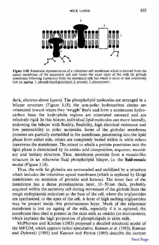
MILK LIPIDS 103
Figure 3.18 Schematic representation of a trilaminar cell membrane which is derived from the apical membrane of the mammary cell and forms the outer layer of the milk fat globule membrane following expression from the mammary cell, but which is more or less extensively lost on ageing. 1, phospholipid/glycolipid; 2, protein; 3, glycoprotein.
dark, electron-dense layers). The phospholipid molecules are arranged in a bilayer structure (Figure 3.1 8); the non-polar hydrocarbon chains are orientated inward where they ‘wriggle’ freely and form a continuous hydro- carbon base; the hydrophilic regions are orientated outward and are relatively rigid. In this bilayer, individual lipid molecules can move laterally, endowing the bilayer with fluidity, flexibility, high electrical resistance and low permeability to polar molecules. Some of the globular membrane proteins are partially embedded in the membrane, penetrating into the lipid phase from either side, others are completely buried within it, while others transverse the membrane. The extent to which a protein penetrates into the lipid phase is determined by its amino acid composition, sequence, second- ary and tertiary structure. Thus, membrane proteins form a mosaic-like structure in an otherwise fluid phospholipid bilayer, i.e. the fluid-mosaic model (Figure 3.18).
Thus, the milk fat globules are surrounded and stabilized by a structure which includes the trilaminar apical membrane (which is replaced by Golgi membranes on secretion of proteins and lactose). The inner face of the membrane has a dense proteinaceous layer, 10-50 nm thick, probably acquired within the secretory cell during movement of the globule from the rough endoplasmic reticulum at the base of the cell, where the triglycerides are synthesized, to the apex of the cell. A layer of high melting triglycerides may be present inside this proteinaceous layer. Much of the trilaminar membrane is lost on ageing of the milk, especially if it is agitated; the membrane thus shed is present in the skim milk as vesicles (or microsomes), which explains the high proportion of phospholipids in skim milk.
McPherson and Kitchen (1983) proposed a detailed structural model of the MFGM, which appears rather speculative. Keenan et aI. (1983), Keenan and Dylewski (1995) and Keenan and Patton (1995) describe the current
Next Page
104 DAIRY CHEMISTRY AND BIOCHEMISTRY
views on the structure of the MFGM and note that complete information on the structure is still not available. Since the MFGM is a dynamic, unstable structure, it is probably not possible to describe a structure which is applicable in all situations and conditions.
3.9 Stability of the milk fat emulsion
The stability, or instability, of the milk fat emulsion is very significant with respect to many physical and chemical characteristics of milk and dairy products. The stability of the emulsion depends strongly on the integrity of the MFGM and, as discussed in section 3.8.7, this membrane is quite fragile and is more or less extensively changed during dairy processing operations.
In the following, some of the principal aspects and problems related to or arising from the stability of the milk fat emulsion are discussed. Some of these relate to the inherent instability of emulsions in general, others are specifically related to the milk system.
3.9.1 Emulsion stability in general
Lipid emulsions are inherently unstable systems due to:
1. The difference in density between the lipid and aqueous phases (c. 0.9 and 1.036 g cm-3, respectively, for milk), which causes the fat globules to float or cream according to Stokes’ equation:
where V is the rate of creaming; Y, the radius of fat globules; p1 and p 2 , the densities of the continuous and dispersed phases, respectively; g, acceleration due to gravity; and rl, viscosity of the system. If creaming is not accompanied by other changes, it is readily reversible by gentle agitation.
2. The interfacial tension between the oil and aqueous phases. Although interfacial tension is reduced by the use of an emulsifier, the interfacial film may be imperfect. When two globules collide, they may adhere (flocculate), e.g. by sharing emulsifier, or they may coalesce due to the Laplace principle which states that the pressure is greater inside small globules than inside large globules and hence there is a tendency for large fat globules (or gas bubbles) to grow at the expense of smaller ones. Taken to the extreme, this will lead to the formation of a continuous mass of fat.
Destabilization processes in emulsions are summarized schematically in Figure 3.19. The rate of destabilization is influenced by the fat content, shear rate (motion), liquid: solid fat ratio, inclusion of air and globule size.
Previous Page
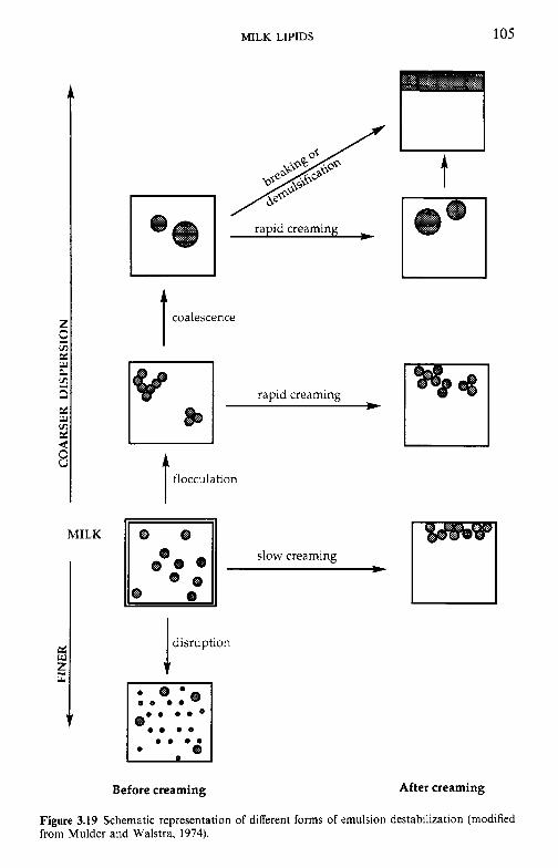
MILK LIPIDS 105
coalescence I rapid creaming *
flocculation I slow creaming *
disruption I Before creaming After creaming
Figure 3.19 Schematic representation of different forms of emulsion destabilization (modified from Mulder and Walstra, 1974).
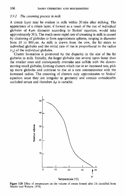
106 DAIRY CHEMISTRY AND BIOCHEMISTRY
3.9.2
A cream layer may be evident in milk within 20min after milking. The appearance of a cream layer, if formed as a result of the rise of individual globules of 4 pm diameter according to Stokes' equation, would take approximately 50 h. The much more rapid rate of creaming in milk is caused by clustering of globules to form approximate spheres, ranging in diameter from 10 to 800pm. As milk is drawn from the cow, the fat exists as individual globules and the initial rate of rise is proportional to the radius ( rJ of the individual globules.
Cluster formation is promoted by the disparity in the size of the fat globules in milk. Initially, the larger globules rise several times faster than the smaller ones and consequently overtake and collide with the slower- moving small globules, forming clusters which rise at an increased rate, pick up more globules and continue to rise at a rate commensurate with the increased radius. The creaming of clusters only approximates to Stokes' equation since they are irregular in geometry and contain considerable occluded serum and therefore A p is variable.
The creaming process in milk
30 t 40 0 10 20
37 Temperature ("C)
Figure 3.20 Effect of temperature on the volume of cream formed after 2 h (modified from Mulder and Walstra, 1974).
MILK LIPIDS 107
In 1889, Babcock postulated that creaming of cows’ milk resulted from an agglutination-type reaction, similar to the agglutination of red blood cells; this hypothesis has been confirmed. Creaming is enhanced by adding blood serum or colostrum to milk; the responsible agents are immunog- lobulins (Ig, which are present at high levels in colostrum), especially IgM. Because these Igs aggregate and precipitate at low temperature ( c 37°C) and redisperse on warming, they are often referred to as cryoglobulins. Aggregation is also dependent on ionic strength and pH. When aggregation of the cryoglobulins occurs in the cold they may precipitate on to the surfaces of large particles, e.g. fat globules, causing them to agglutinate, probably through a reduction in surface (electrokinetic) potential. The cryoprecipitated globulins may also form a network in which the fat globules are entrapped. The clusters can be dispersed by gentle stirring and are completely dispersed on warming to 37°C or higher. Creaming is strongly dependent on temperature and does not occur above 37°C (Figure 3.20). The milks of buffalo, sheep and goat do not exhibit flocculation and the milks of some cows exhibit little or none, apparently a genetic trait.
The rate of creaming and the depth of the cream layer show considerable variation. The concentration of cryoglobulin might be expected to influence the rate of creaming and although colostrum (rich in Ig) creams well and late lactation milk (deficient in Ig) creams poorly, there is no correlation in mid-lactation milks between Ig concentration and the rate of creaming. An uncharacterized lipoprotein appears to act synergistically with cryoglobulin in promoting clustering. The rate of creaming is increased by increasing the ionic strength and retarded by acidification. High-fat milks, which also tend to have a higher proportion of larger fat globules, cream quickly, probably because the probability of collisions between globules is greater and because large globules tend to form larger aggregates. The depth of the cream layer in high-fat milks is also greater than might be expected, possibly because of greater ‘dead space’ in the interstices of aggregates formed from large globules.
The rate of creaming and the depth of the cream layer are very markedly influenced by processing operations. Creaming is faster and more complete at low temperatures (c 20°C; Figure 3.20), probably because of the tempera- ture-dependent precipitation of the cryoglobulins. Gentle (but not pro- longed) agitation during the initial stages of creaming promotes and enhances cluster formation and creaming, possibly because of an increased probability of collisions. It would be expected that stirring cold milk would lead to the deposition of all the cryoglobulin on to the fat globule surfaces, and rapid creaming, without a time lag, would be expected when stirring ceased. However, milk so treated does not cream at all or only slightly after a prolonged lag period. If cold, creamed milk is agitated gently, the clusters are dispersed and do not reform unless the milk is rewarmed to c. 40°C and then recooled, i.e. the whole cycle repeated. Violent agitation is detrimental
108 DAIRY CHEMISTRY AND BIOCHEMISTRY
to creaming, possibly due to denaturation of the cryoglobulins and/or alteration to the fat globule surface. If milk is separated at 40°C or above, the cryoglobulins are present predominantly in the serum, whereas they are in the cream produced at lower temperatures. Agglutination and creaming are impaired or prevented by heating ( e g 70°C x 30min or 77°C x 20s) owing to denaturation of the cryoglobulins; addition of Igs to heated milk restores creaming (except after very severe heat treatment, e.g. 2 min at 95°C or equivalent). Homogenization prevents creaming, not only due to the reduction of fat globule size but also to some other factor since a blend of raw cream and homogenized skim milk does not cream well. In fact two types of euglobulin appear to be involved in agglutination, one of which is denatured by heating, the other by homogenization. Thus, a variety of factors which involve temperature changes, agitation or homogenization influence the rate and extent of creaming.
3.10 Influence of processing operations on the fat globule membrane
As discussed in section 3.8.7, the milk fat globule membrane (MFGM) is relatively fragile and susceptible to damage during a range of processing operations; consequently, emulsion stability is reduced by dislodging inter- facial material by agitation, homogenization, heat treatment, concentration, drying and freezing. Rearrangement of the membrane increases the suscep- tibility of the fat to hydrolytic rancidity, light-activated flavours and ‘oiling-off of the fat, but reduces susceptibility to metal-catalysed oxidation. The influence of the principal dairy processing operations on MFGM and concomitant defects are discussed below.
3. IO. I Milk supply: hydrolytic rancidity
The production of milk on the farm and transportation to the processing plant are potentially major causes of damage to the MFGM. Damage to the membrane may occur at several stages of the milking operation: foaming due to air sucked in at teat-cups, agitation due to vertical sections (risers) in milk pipelines, constrictions and/or expansion in pipelines, pumps, especially if not operating at full capacity, surface coolers, agitators in bulk tanks and freezing of milk on the walls of bulk tanks. While some oiling-off and perhaps other physical damage to the milk fat emulsion may accrue from such damage, by far the most serious consequence is the development of hydrolytic rancidity. The extent of lipolysis is commonly expressed as ‘acid degree value’ (ADV) of the fat as millimoles of free fatty acids per 100 g fat; A D V s greater than 1 are undesirable and are probably perceptible by taste to most people.
MILK LIPIDS 109
The principal lipase in bovine milk is a lipoprotein lipase (LPL; Chapter 8) which is associated predominantly with the casein micelles and is isolated from its substrate, milk fat, by the MFGM, i.e. the enzyme and its substrate are compartmentalized. However, even slight damage to the membrane permits contact between enzyme and substrate, resulting in hydrolytic rancidity. The enzyme is optimally active at around 37°C and pH 8.5 and is stimulated by divalent cations, e.g. Ca2+ (CaZ+ complex free fatty acids, which are strongly inhibitory). The initial turnover of milk LPL is c. 3000 s-', i.e. 3000 fatty acid molecules are liberated per second per mole of enzyme (milk usually contains 1-2 mg lipase l-', i.e. 10-20 nM) which, if fully active, is sufficient to induce rancidity in about 10s. This never happens in milk due to a variety of factors, e.g. the pH, ionic strength and, usually, the temperature are not optimal; the lipase is bound to the casein micelles; the substrate is not readily available; milk probably contains lipase inhibitors, including caseins. The activity of lipase in milk is not correlated with its concentration due to the various inhibitory and adverse factors.
Machine milking, especially pipe-line milking systems, markedly increases the incidence of hydrolytic rancidity unless adequate precautions are taken. The effectors are the clawpiece and the tube taking the milk from the clawpiece to the pipeline; damage at the clawpiece may be minimized by proper regulation of air intake, and low-line milking installations cause less damage than high-line systems but the former are more expensive and less convenient for operators. Larger-diameter pipelines (e.g. 5 cm) reduce the incidence of rancidity but may cause cleaning problems and high milk losses. The receiving jar, pump (diaphragm or centrifugal, provided they are operated properly) and type of bulk tank, including agitator, transportation in bulk tankers or preliminary processing operations (e.g. pumping and refrigerated storage) at the factory, make little if any contribution to hydrolytic rancidity.
The frequency and severity of lipolysis increases in late lactation, possibly owing to a weak MFGM and the low level of milk produced (which may aggravate agitation); this problem is particularly acute when milk produc- tion is seasonal, e.g. as in Ireland or New Zealand.
The lipase system can also be activated by cooling freshly drawn milk to 5"C, rewarming to 30°C and recooling to 5°C. Such a temperature cycle may occur under farm conditions, e.g. addition of a large quantity of warm milk to a small volume of cold milk. It is important that bulk tanks be emptied completely at each collection (this practice is also essential for the mainten- ance of good hygiene). No satisfactory explanation for temperature activa- tion is available but changes in the physical state of fat (liquid/solid ratio) have been suggested; damage/alteration of the globule surface and binding of lipoprotein co-factor may also be involved.
Some cows produce milk which is susceptible to a defect known as 'spontaneous rancidity' - no activation treatment, other than cooling of the milk, is required; the frequency of such milks may be as high as 30% of the
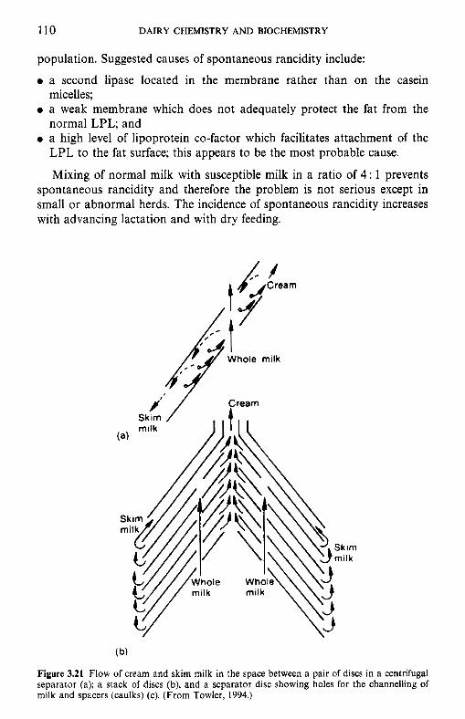
110 DAIRY CHEMISTRY AND BIOCHEMISTRY
population. Suggested causes of spontaneous rancidity include:
0 a second lipase located in the membrane rather than on the casein micelles;
0 a weak membrane which does not adequately protect the fat from the normal LPL; and
0 a high level of lipoprotein co-factor which facilitates attachment of the LPL to the fat surface; this appears to be the most probable cause.
Mixing of normal milk with susceptible milk in a ratio of 4: 1 prevents spontaneous rancidity and therefore the problem is not serious except in small or abnormal herds. The incidence of spontaneous rancidity increases with advancing lactation and with dry feeding.
r”’ / Who‘e mi’k
( b)
Figure 3.21 Flow of cream and skim milk in the space between a pair of discs in a centrifugal separator (a); a stack of discs (b); and a separator disc showing holes for the channelling of milk and spacers (caulks) (c). (From Towler, 1994.)
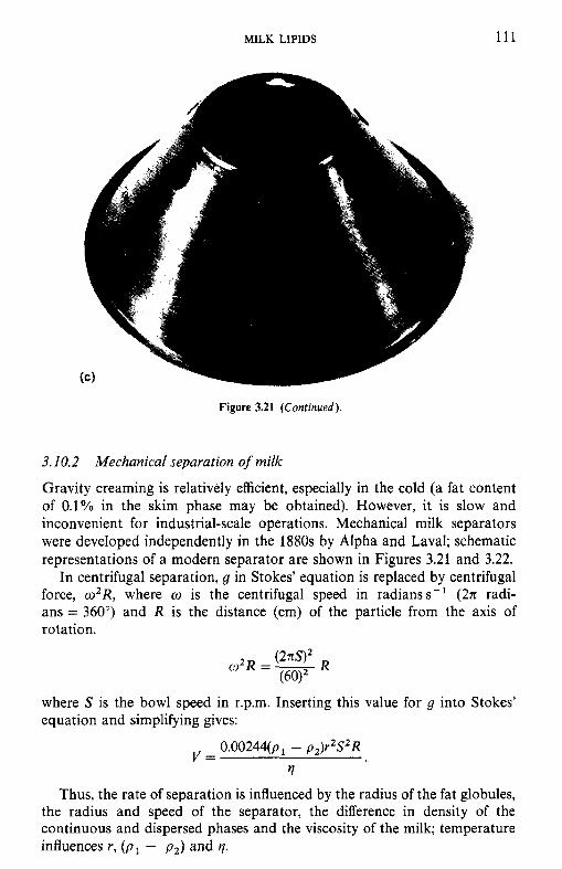
MILK LIPIDS 111
Figure 3.21 (Continued),
3.10.2 Mechanical separation of milk
Gravity creaming is relatively efficient, especially in the cold (a fat content of 0.1% in the skim phase may be obtained). However, it is slow and inconvenient for industrial-scale operations. Mechanical milk separators were developed independently in the 1880s by Alpha and Laval; schematic representations of a modern separator are shown in Figures 3.21 and 3.22.
In centrifugal separation, g in Stokes' equation is replaced by centrifugal force, wZR, where w is the centrifugal speed in radianss-' (2n radi- ans = 360") and R is the distance (cm) of the particle from the axis of rotation.
where S is the bowl speed in r.p.m. Inserting this value for g into Stokes' equation and simplifying gives:
O.O0244(p - p z ) r Z S 2 R
rl
Thus, the rate of separation is influenced by the radius of the fat globules, the radius and speed of the separator, the difference in density of the continuous and dispersed phases and the viscosity of the milk; temperature influences r, (pi - p 2 ) and q.
V =
112 DAIRY CHEMISTRY AND BIOCHEMISTRY
Figure 3.22 Cutaway diagram of a modern milk separator (from Towler, 1994).
Fat globules of less than 2pm diameter are incompletely removed by cream separators and since the average size of fat globules decreases with advancing lactation (Figure 3.19, the efficiency of separation decreases concomitantly. The percentage fat in cream is regulated by manipulating the ratio of cream to skim-milk streams from the separator, which in effect regulates back-pressure. With any particular separator operating under more or less fixed conditions, temperature is the most important variable affecting the efficiency of separation via its effects on r, q and ( p l - pz) . The
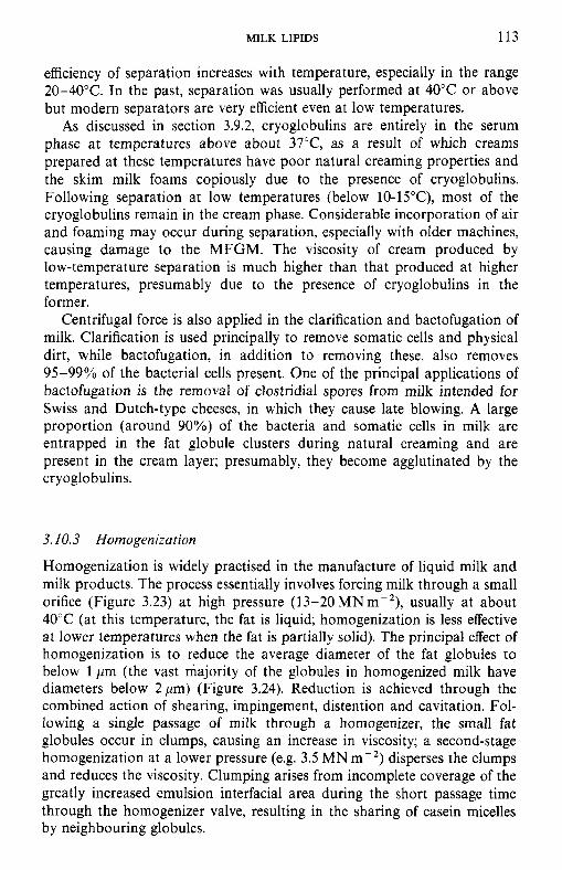
MILK LIPIDS 113
efficiency of separation increases with temperature, especially in the range 20-40°C. In the past, separation was usually performed at 40°C or above but modern separators are very efficient even at low temperatures.
As discussed in section 3.9.2, cryoglobulins are entirely in the serum phase at temperatures above about 37"C, as a result of which creams prepared at these temperatures have poor natural creaming properties and the skim milk foams copiously due to the presence of cryoglobulins. Following separation at low temperatures (below lO-l5"C), most of the cryoglobulins remain in the cream phase. Considerable incorporation of air and foaming may occur during separation, especially with older machines, causing damage to the MFGM. The viscosity of cream produced by low-temperature separation is much higher than that produced at higher temperatures, presumably due to the presence of cryoglobulins in the former.
Centrifugal force is also applied in the clarification and bactofugation of milk. Clarification is used principally to remove somatic cells and physical dirt, while bactofugation, in addition to removing these, also removes 95-99% of the bacterial cells present. One of the principal applications of bactofugation is the removal of clostridial spores from milk intended for Swiss and Dutch-type cheeses, in which they cause late blowing. A large proportion (around goo/,) of the bacteria and somatic cells in milk are entrapped in the fat globule clusters during natural creaming and are present in the cream layer; presumably, they become agglutinated by the cryoglo bulins.
3.10.3 Homogenization
Homogenization is widely practised in the manufacture of liquid milk and milk products. The process essentially involves forcing milk through a small orifice (Figure 3.23) at high pressure (13-20 MNmP2), usually at about 40°C (at this temperature, the fat is liquid; homogenization is less effective at lower temperatures when the fat is partially solid). The principal effect of homogenization is to reduce the average diameter of the fat globules to below 1 pm (the vast majority of the globules in homogenized milk have diameters below 2 pm) (Figure 3.24). Reduction is achieved through the combined action of shearing, impingement, distention and cavitation. Fol- lowing a single passage of milk through a homogenizer, the small fat globules occur in clumps, causing an increase in viscosity; a second-stage homogenization at a lower pressure (e.g. 3.5 MN m-2) disperses the clumps and reduces the viscosity. Clumping arises from incomplete coverage of the greatly increased emulsion interfacial area during the short passage time through the homogenizer valve, resulting in the sharing of casein micelles by neighbouring globules.
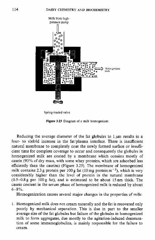
114 DAIRY CHEMISTRY AND BIOCHEMISTRY
Milk from high- pressure pump
n
Spring-loaded valve
Figure 3.23 Diagram of a milk homogenizer.
Reducing the average diameter of the fat globules to 1 pm results in a four- to sixfold increase in the fat/plasma interface. There is insufficient natural membrane to completely coat the newly formed surface or insuffi- cient time for complete coverage to occur and consequently the globules in homogenized milk are coated by a membrane which consists mostly of casein (93% of dry mass, with some whey proteins, which are adsorbed less efficiently than the caseins) (Figure 3.25). The membrane of homogenized milk contains 2.3 g protein per lOOg fat (10mg proteinm-'), which is very considerably higher than the level of protein in the natural membrane (0.5-0.8g per 1OOg fat), and is estimated to be about 15nm thick. The casein content in the serum phase of homogenized milk is reduced by about 6-8%.
Homogenization causes several major changes in the properties of milk:
1. Homogenized milk does not cream naturally and the fat is recovered only poorly by mechanical separation. This is due in part to the smaller average size of the fat globules but failure of the globules in homogenized milk to form aggregates, due mostly to the agitation-induced denatura- tion of some immunoglobulins, is mainly responsible for the failure to cream.
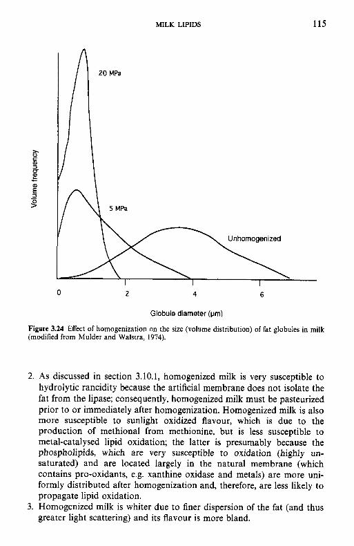
MILK LIPIDS 115
2 4 6
Globule diameter (um)
Figure 3.24 Effect of homogenization on the size (volume distribution) of fat globules in milk (modified from Mulder and Walstra, 1974).
2. As discussed in section 3.10.1, homogenized milk is very susceptible to hydrolytic rancidity because the artificial membrane does not isolate the fat from the lipase; consequently, homogenized milk must be pasteurized prior to or immediately after homogenization. Homogenized milk is also more susceptible to sunlight oxidized flavour, which is due to the production of methional from methionine, but is less susceptible to metal-catalysed lipid oxidation; the latter is presumably because the phospholipids, which are very susceptible to oxidation (highly un- saturated) and are located largely in the natural membrane (which contains pro-oxidants, e.g. xanthine oxidase and metals) are more uni- formly distributed after homogenization and, therefore, are less likely to propagate lipid oxidation.
3. Homogenized milk is whiter due to finer dispersion of the fat (and thus greater light scattering) and its flavour is more bland.
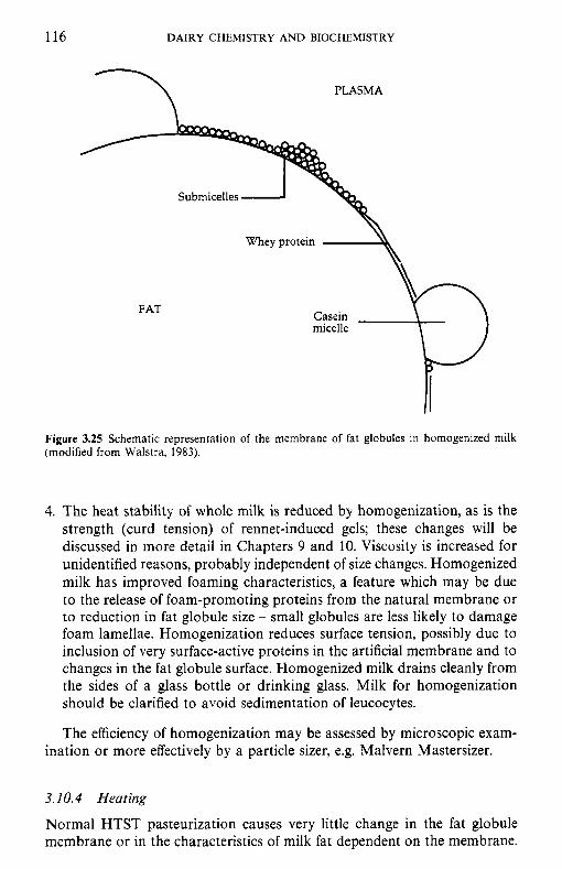
116 DAIRY CHEMISTRY AND BIOCHEMISTRY
PLASMA 7
\\ Whey protein
FAT
Figure 3.25 Schematic representation of the membrane of fat globules in homogenized milk (modified from Walstra, 1983).
4. The heat stability of whole milk is reduced by homogenization, as is the strength (curd tension) of rennet-induced gels; these changes will be discussed in more detail in Chapters 9 and 10. Viscosity is increased for unidentified reasons, probably independent of size changes. Homogenized milk has improved foaming characteristics, a feature which may be due to the release of foam-promoting proteins from the natural membrane or to reduction in fat globule size - small globules are less likely to damage foam lamellae. Homogenization reduces surface tension, possibly due to inclusion of very surface-active proteins in the artificial membrane and to changes in the fat globule surface. Homogenized milk drains cleanly from the sides of a glass bottle or drinking glass. Milk for homogenization should be clarified to avoid sedimentation of leucocytes.
The efficiency of homogenization may be assessed by microscopic exam- ination or more effectively by a particle sizer, e.g. Malvern Mastersizer.
3.10.4 Heating
Normal HTST pasteurization causes very little change in the fat globule membrane or in the characteristics of milk fat dependent on the membrane.
MILK LIPIDS 117
However, excessively high pasteurization temperatures denature the cryog- lobulins and aggregation of the fat globules and creaming are impaired or prevented. Severe treatments, e.g. 80°C x 15 min, remove lipid and protein material from the membrane, the fat globules are partially denuded and may coalesce, forming large clumps of fat and resulting in defects such as cream plug in milk or cream (section 3.11).
Processes such as thermal concentration also cause membrane damage, especially since many of these treatments also involve vigorous agitation in high velocity heating systems. Since milk for concentrated and dehydrated milk products is normally homogenized, damage to the natural membrane is of little significance.
3.11 Physical defects in milk and cream
In addition to the flavour defects initiated or influenced by damage to the fat globule membrane, such damage also results in a variety of physical defects in milk and especially in cream. The more important of these are ‘oiling-off, ‘cream plug’ and ‘age thickening’.
‘Oiling-off, characterized by the appearance of globules of oil or fat on the surface of coffee or tea when milk, and especially cream, is added, is due to membrane damage during processing, resulting in ‘free fat’; low pressure homogenization re-emulsifies the free fat and eliminates the defect.
‘Cream plug’ is characterized by the formation of a layer of solid fat on the surface of cream or milk in bottles; the defect is due to a high level of ‘free fat’ which forms interlocking crystals on cooling and is most common in high-fat creams. Cream plug is common in unhomogenized, pasteurized, late lactation milk, presumably due to a weak MFGM.
‘Age thickening’ is due essentially to a high level of free fat, especially in high-fat creams; the product becomes very viscous due to interlocking of crystals of free fat.
Two somewhat related instability problems are ‘feathering’ and ‘bitty’ cream. ‘Feathering’ is characterized by the appearance of white flecks when milk or cream is poured on hot coffee and is a form of heat-induced coagulation; the white ‘flecks are mainly destabilized protein. The heat stability of cream and its resistance to feathering are reduced by:
0 single-stage homogenization; 0 high homogenization pressure at low temperature; 0 high concentrations of Caz+ in the cream or water; 0 a high ratio of fat to serum solids, i.e. high-fat creams; 0 high temperature and low pH of the coffee.
Protein-lipid interaction is enhanced by homogenization, while high tem- peratures, low pH and high divalent cation concentration induce aggrega- tion of the casein-coated fat globules into large visible particles. Stability
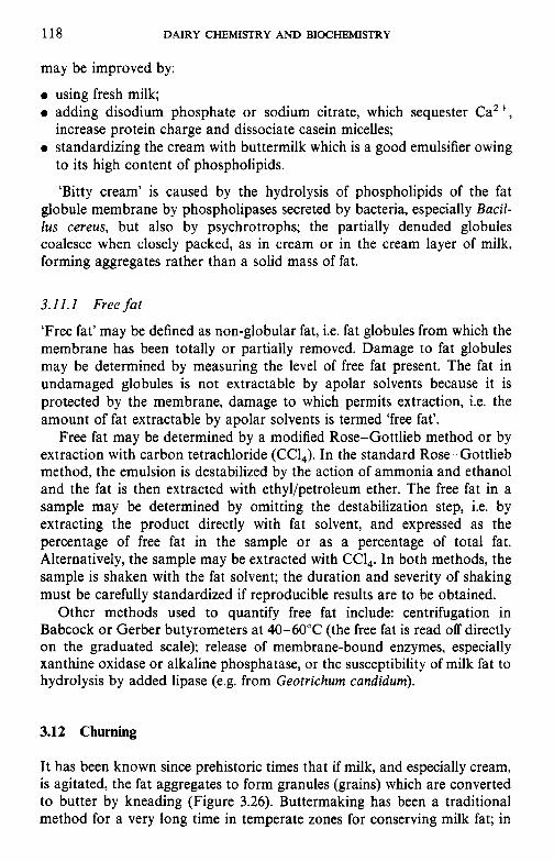
118 DAIRY CHEMISTRY AND BIOCHEMISTRY
may be improved by:
0 using fresh milk; 0 adding disodium phosphate or sodium citrate, which sequester Ca2+,
increase protein charge and dissociate casein micelles; 0 standardizing the cream with buttermilk which is a good emulsifier owing
to its high content of phospholipids.
‘Bitty cream’ is caused by the hydrolysis of phospholipids of the fat globule membrane by phospholipases secreted by bacteria, especially Bacil- lus cereus, but also by psychrotrophs; the partially denuded globules coalesce when closely packed, as in cream or in the cream layer of milk, forming aggregates rather than a solid mass of fat.
3.11.1 Free fat
‘Free fat’ may be defined as non-globular fat, i.e. fat globules from which the membrane has been totally or partially removed. Damage to fat globules may be determined by measuring the level of free fat present. The fat in undamaged globules is not extractable by apolar solvents because it is protected by the membrane, damage to which permits extraction, i.e. the amount of fat extractable by apolar solvents is termed ‘free fat’.
Free fat may be determined by a modified Rose-Gottlieb method or by extraction with carbon tetrachloride (CCl,). In the standard Rose-Gottlieb method, the emulsion is destabilized by the action of ammonia and ethanol and the fat is then extracted with ethyl/petroleum ether. The free fat in a sample may be determined by omitting the destabilization step, i.e. by extracting the product directly with fat solvent, and expressed as the percentage of free fat in the sample or as a percentage of total fat. Alternatively, the sample may be extracted with CCl,. In both methods, the sample is shaken with the fat solvent; the duration and severity of shaking must be carefully standardized if reproducible results are to be obtained.
Other methods used to quantify free fat include: centrifugation in Babcock or Gerber butyrometers at 40-60°C (the free fat is read off directly on the graduated scale); release of membrane-bound enzymes, especially xanthine oxidase or alkaline phosphatase, or the susceptibility of milk fat to hydrolysis by added lipase (e.g. from Geotrichum candidum).
3.12 Churning
It has been known since prehistoric times that if milk, and especially cream, is agitated, the fat aggregates to form granules (grains) which are converted to butter by kneading (Figure 3.26). Buttermaking has been a traditional method for a very long time in temperate zones for conserving milk fat; in
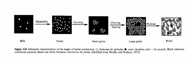
Churning Working Separation Churning - - - H Milk Cream Small grains Large grains Butter
Figure 3.26 Schematic representation of the stages of butter production. 0, Indicates fat globules; @, water droplets; and -, fat crystals. Black indicates continuous aqueous phase and white indicates continuous fat phase. (Modified from Mulder and Walstra, 1974.)
120 DAIRY CHEMISTRY AND BIOCHEMISTRY
tropical regions, butter grains or cream are heated to remove all the water; the resulting product is called ‘ghee’, a crude form of butter oil.
The cream used for butter may be fresh ( - pH 6.6) or ripened (fermented; - pH 4.6), yielding ‘sweet-cream’ and ‘ripened cream (lactic)’ butter, respect- ively. Sweet-cream butter is most common in English-speaking countries but ripened cream butter is more popular elsewhere. Traditionally, the cream for ripened cream butter was fermented by the natural microflora, which was variable. Product quality and consistency were improved by the introduc- tion in the 1880s of cultures (starters) of selected lactic acid bacteria, which produce lactic acid from lactose and diacetyl (the principal flavour compo- nent in ripened cream butter) from citric acid. A flavour concentrate, containing lactic acid and diacetyl, is now frequently used in the manufac- ture of ripened cream butter, to facilitate production schedules and improve consistency.
Butter manufacture or churning essentially involves phase inversion, i.e. the conversion of the oil-in-water emulsion of cream to a water-in-oil emulsion. Inversion is achieved by some form of mechanical agitation which denudes some of the globules of their stabilizing membrane; the denuded globules coalesce to form butter grains, entrapping some globular fat. The butter grains are then kneaded (‘worked’) which releases fat liquid at room temperature. Depending on temperature and on the method and extent of
Figure 3.27 Schematic representation of the structure of butter. 1, fat globule; 2, membrane; 3, aqueous droplet; 4, fat crystals; 5 , air cell. (Modified from Mulder and Walstra, 1974.)
MILK LIPIDS 121
Table 3.13 Structural elements of conventional butter
Approximate Proportion number of butter Dimensions
Element (mi-') (%, VIV) (Pm) Remarks
Fat globules 1 O ' O 10-50 2-8 Differ in composition; with complete or partial membrane
temperature; at higher temperature occur mainly in globules; a t low temperature, form solid networks
Fat crystals 1013 10-40 0.01-2 Amount depends on
Moisture 1 O ' O 16 1-25 Differ in composition
Air cells 107 5 > 20 droplets
Modified from Mulder and Walstra (1974).
working, liquid fat may represent 50-95% of total fat. The liquid fat forms the continuous phase in which fat globules, fat crystals, membrane material, water droplets and small air bubbles are dispersed (Figure 3.27, Table 3.13). NaCl may be added (to c. 2%) to modify flavour but more importantly as a preservative: added salt dissolves in the water droplets (to give c. 12% salt in moisture) which also contain contaminating bacteria. Usually, ripened cream butter is not salted.
The process of phase inversion has received considerable attention (see McDowall (1953) and Wilbey (1994) for a detailed discussion). Briefly, churning methods can be divided into (1) traditional batch methods and (2) continuous methods.
1. The traditional method involves placing 3O-4O0/o fat cream in a churn (of various shapes and design, Figure 3.28) which is rotated gently. During rotation, air is incorporated and numerous small air bubbles are formed; fat globules are trapped between the lamellae of the bubbles. As the bubbles grow, the lamellae become thinner and exert a shearing effect on the fat globules. Some globules become denuded of membrane and coalesce; the aggregated globules are cemented by liquid fat expressed from the globules. A portion of the liquid fat spreads over the surface of the air bubbles, causing them to collapse, releasing butter grains and buttermilk (representing the serum phase of cream plus the fat globule membrane).
When a certain degree of globular destabilization has occurred, the foam collapses rather abruptly and when the grains have grown to the requisite size, the buttermilk is drained off and the grains worked to a continuous mass. Proper working of the butter is essential for good
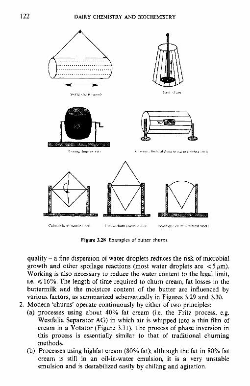
122 DAIRY CHEMISTRY AND BIOCHEMISTRY
Figure 3.28 Examples of butter churns.
quality - a fine dispersion of water droplets reduces the risk of microbial growth and other spoilage reactions (most water droplets are < 5 pm). Working is also necessary to reduce the water content to the legal limit, i.e. < 16%. The length of time required to churn cream, fat losses in the buttermilk and the moisture content of the butter are influenced by various factors, as summarized schematically in Figures 3.29 and 3.30.
2. Modern 'churns' operate continuously by either of two principles: ~- processes using about 40% fat cream (i.e. the F k z piocess, e.g. Westfalia Separator AG) in which air is whipped into a thin film of cream in a Votator (Figure 3.31). The process of phase inversion in this process is essentially similar to that of traditional churning methods. Processes using highfat cream (80% fat); although the fat in 80% fat cream is still in an oil-in-water emulsion, it is a very unstable emulsion and is destabilized easily by chilling and agitation.
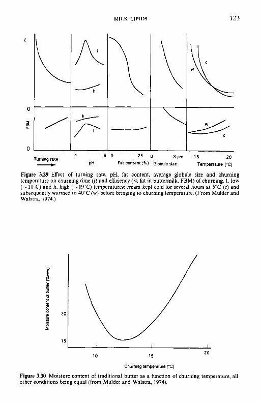
MILK LIPIDS 123
t
0
2 U
0
/
n
Turning rate v
15 20 Temperature (“C)
Figure 3.29 Effect of turning rate, pH, fat content, average globule size and churning temperature on churning time ( t ) and efficiency (% fat in buttermilk, FBM) of churning. 1, low ( - 11°C) and h, high ( - 19°C) temperatures; cream kept cold for several hours a t 5°C (c) and subsequently warmed to 40°C (w) before bringing to churning temperature. (From Mulder and Walstra, 1974.)
20 10 15
Churning temperature (“C)
Figure 3.30 Moisture content of traditional butter as a function of churning temperature, all other conditions being equal (from Mulder and Walstra, 1974).
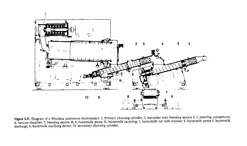
Figure 3.31 Diagram of a Westfalia continuous buttermaker. 1, Primary churning cylinder; 2, texturizer with blending section I; 3, metering connections; 4, vacuum chamber; 5, blending section 11; 6, buttermilk pump 11, buttermilk recycling; 7, buttermilk vat with strainer; 8, buttermilk pump I , buttermilk discharge; 9, buttermilk clarifying device; 10, secondary churning cylinder.
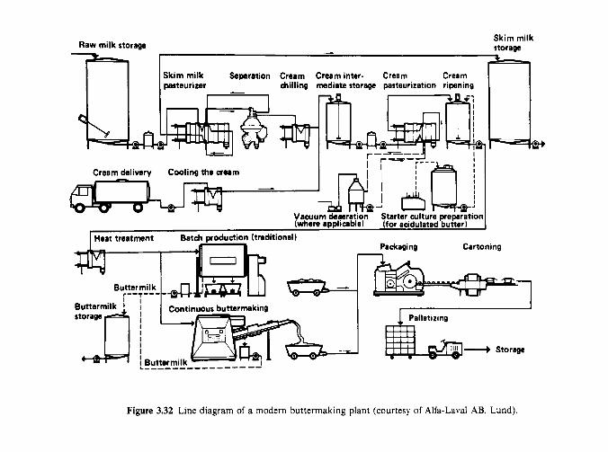
Raw milk storage Skim milk storage - .-
Skim milk Separation Cream Cream inter. Cream Cream parteuri~er billing mdiate storage pasteurization ripening
Packaging Canoning
Figure 3.32 Line diagram of a modern buttermaking plant (courtesy of Alfa-Lava1 AB, Lund).
126 DAIRY CHEMISTRY AND BIOCHEMISTRY
The line diagram for a modern buttermaking plant is shown in Figure 3.32.
All the methods of butter manufacture involve complete or partial removal of the fat globule membrane which is largely lost in the buttermilk, which is, consequently, a good source of phospholipids and other emulsi- fiers.
3.13 Freezing
Freezing and dehydration tend to destabilize all lipoprotein complexes, both natural and artificial. Thus, freezing of milk, and especially cream, results in damage to the membrane which causes destabilization when the product is thawed. Most of the destabilizing effect is due to physicochemical changes induced by dehydration of the lipoprotein complexes but some physical damage is also caused by ice crystals. The damage is manifest as oiling-off and free fat formation. The extent of damage is proportional to fat concentration and moderately high-fat creams (50%) are completely de- stabilized by freezing.
Frozen cream is produced commercially and is used mainly for the production of soups, butter-oil, butter, etc., where emulsion stability is not important. Damage may be reduced by:
0 rapid freezing as thin blocks or continuously on refrigerated drums; 0 homogenization and pasteurization before freezing; 0 storage at very low temperature (c. -30°C) and avoiding temperature
fluctuations during storage.
3.14 Dehydration
The physicochemical state of fat in milk powder particles, which markedly influences the wettability and dispersibility of the powder on reconstitution, depends on the manufacturing process. The fat occurs either in a finely emulsified or in a partly coalesced, de-emulsified state. In the latter case, the membrane has been ruptured or completely removed, causing the globules to run together to form pools of free fat. The amount of de-emulsified ‘free fat’ depends on the manufacturing method and storage conditions. Typical values for ‘free fat’ (as a percentage of total fat) in milk powders are: spray-dried powders, 3.3-20%; roller-dried powders, 91.6-95.8%; freeze- dried powders, 43-75%; foam-dried powders, less than 10%.
The high level of ‘free fat’ in roller-dried powder is due to the effects of the high temperature to which milk is exposed on the roller surfaces and to the mechanical effect of the scraping knives. In properly made and stored
MILK LIPIDS 127
spray-dried powder, the fat globules are distributed throughout the powder particles. The amount of free fat depends on the total fat content, and may be about 25% of total fat. Homogenization pre-drying reduces the level of free fat formed.
Further liberation of ‘free fat’ may occur under adverse storage condi- tions. If powder absorbs water it becomes ‘clammy’ and lactose crystallizes, resulting in the expulsion of other milk components from the lactose crystals into the spaces between the crystals. De-emulsification of the fat may occur due to the mechanical action of sharp edges of lactose crystals on the fat globule membrane. If the fat is liquid at the time of membrane rupture, or if it becomes liquid during storage, it will adsorb on to the powder particles, forming a water-repellant film around the particles.
The state of fat in powder has a major influence on wettability, i.e. the ease with which the powder particles make contact with water. Adequate wettability is a prerequisite for good dispersibility. Free fat has a water- repelling effect on the particles during dissolution, making the powder difficult to reconstitute. Clumps of fat and oily patches appear on the surface of the reconstituted powder, as well as greasy films on the walls of containers. The presence of ‘free fat’ on the surface of the particles tends to increase the susceptibility of fat to oxidation. A scum of fat-protein complexes may appear on the surface of reconstituted milk; the propensity to scum formation is increased by high storage temperatures.
3.15 Lipid oxidation
Lipid oxidation, leading to oxidative rancidity, is a major cause of deterio- ration in milk and dairy products. The subject has been reviewed by Richardson and Korycka-Dahl(l983) and O’Connor and O’Brien (1995).
Lipid oxidation is an autocatalysed free-radical chain reaction which is normally divided into three phases: initiation, propagation and termination (Figure 3.33).
The initial step involves abstracting a hydrogen atom from a fatty acid, forming a fatty acid (FA) free radical, e.g.
CH 3 - - - - CH ,-CH=CH-CH-CH=CH-CH 2 - - - - - COOH
Although saturated fatty acids may lose a H’ and undergo oxidation, the reaction principally involves unsaturated fatty acids, especially polyunsatu- rated fatty acids (PUFA), the methylene, -CH,--, group between double bonds being particularly sensitive:
c18:3 >> cIE:2 ” c I E : I ’ c l S : O
The polar lipids in milk fat are richer in PUFA than neutral lipids and are
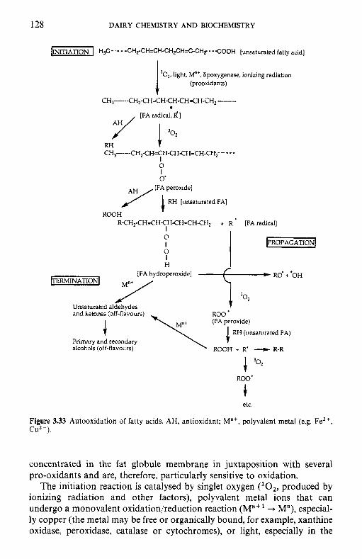
128 DAIRY CHEMISTRY A N D BIOCHEMISTRY
'O,, light, M"', lipoxygenase, ionizing radiation (prooxidants) 1
CH,-------CH,-CH=CH-CH-CH=CH-CH-CH~ ~ [FA t a r a d ]
3 0 2
RH CH3-----CH,CH=CH-CHCH=CH-CH2- ----
I 0 I 0'
~ [ F i peroxide]
RH [unsaturated FA]
ROOH RCH,-CH=CHCHCH=CH-CH, + R [FA radical]
I 0 I 0 I H _ _
[FA hydroperoxide]
~ERMINATION~ M"' / /
Unsaturated aldehvdes and ketones (off-flavours)
(FA peroxide)
RH (unsaturated FA) Primary and secondary alcohols (off-flavours) ROOH + R' 4 R-R
etc.
Figure 3.33 Autooxidation of fatty acids. AH, antioxidant; M"', polyvalent metal (e.g. Fez', C U Z + ) .
concentrated in the fat globule membrane in juxtaposition with several pro-oxidants and are, therefore, particularly sensitive to oxidation.
The initiation reaction is catalysed by singlet oxygen (lo2, produced by ionizing radiation and other factors), polyvalent metal ions that can undergo a monovalent oxidation/reduction reaction (M"" + M"), especial- ly copper (the metal may be free or organically bound, for example, xanthine oxidase, peroxidase, catalase or cytochromes), or light, especially in the
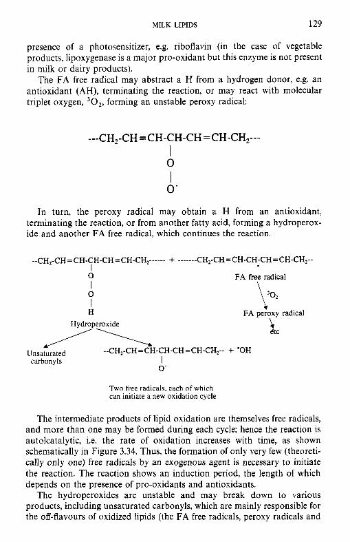
MILK LIPIDS 129
presence of a photosensitizer, e.g. riboflavin (in the case of vegetable products, lipoxygenase is a major pro-oxidant but this enzyme is not present in milk or dairy products).
The FA free radical may abstract a H from a hydrogen donor, e.g. an antioxidant (AH), terminating the reaction, or may react with molecular triplet oxygen, 30,, forming an unstable peroxy radical:
---CH,-CH = CH-CH-CH =CH-CH,--- I 0
0 ' I
In turn, the peroxy radical may obtain a H from an antioxidant, terminating the reaction, or from another fatty acid, forming a hydroperox- ide and another FA free radical, which continues the reaction.
0
0 I
FA free radical \ \ Q
I !v H FA peroxy radical
Hydroperoxide i etc
--CHI-CH=CH-CH-CH=CH-CH,-- + 'OH I
/\ Unsaturated carbonyls
0'
Two free radicals, each of which can initiate a new oxidation cycle
The intermediate products of lipid oxidation are themselves free radicals, and more than one may be formed during each cycle; hence the reaction is autolcatalytic, i.e. the rate of oxidation increases with time, as shown schematically in Figure 3.34. Thus, the formation of only very few (theoreti- cally only one) free radicals by an exogenous agent is necessary to initiate the reaction. The reaction shows an induction period, the length of which depends on the presence of pro-oxidants and antioxidants.
The hydroperoxides are unstable and may break down to various products, including unsaturated carbonyls, which are mainly responsible for the off-flavours of oxidized lipids (the FA free radicals, peroxy radicals and
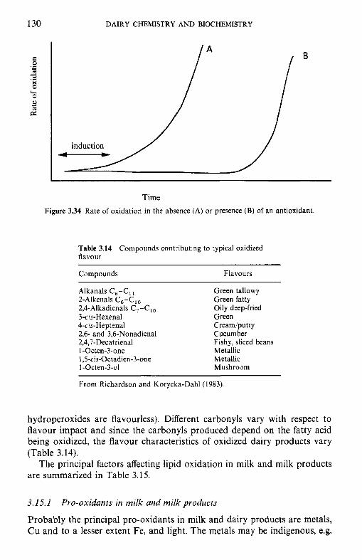
130 DAIRY CHEMISTRY A N D BIOCHEMISTRY
induction - Time
Figure 3.34 Rate of oxidation in the absence (A) or presence (B) of an antioxidant,
Table 3.14 Compounds contributing to typical oxidized flavour
Compounds Flavours
Alkanals C,-C, Green tallowy 2-Alkenals C,-C,, Green fatty 2,4-Alkadienals C,-C,, Oily deep-fried 3-cis-Hexenal Green 4-cis-Heptenal Cream/putt y 2,6- and 3,6-Nonadienal Cucumber 2,4,7-Decatrienal Fishy, sliced beans 1 -0cten-3-one Metallic 1 ,S-cis-Octadien-3-one Metallic 1 -0cten-3-01 Mushroom
From Richardson and Korycka-Dahl (1983).
hydroperoxides are flavourless). Different carbonyls vary with respect to flavour impact and since the carbonyls produced depend on the fatty acid being oxidized, the flavour characteristics of oxidized dairy products vary (Table 3.14).
The principal factors affecting lipid oxidation in milk and milk products are summarized in Table 3.15.
3.15.1
Probably the principal pro-oxidants in milk and dairy products are metals, Cu and to a lesser extent Fe, and light. The metals may be indigenous, e.g.
Pro-oxidants in milk and milk products
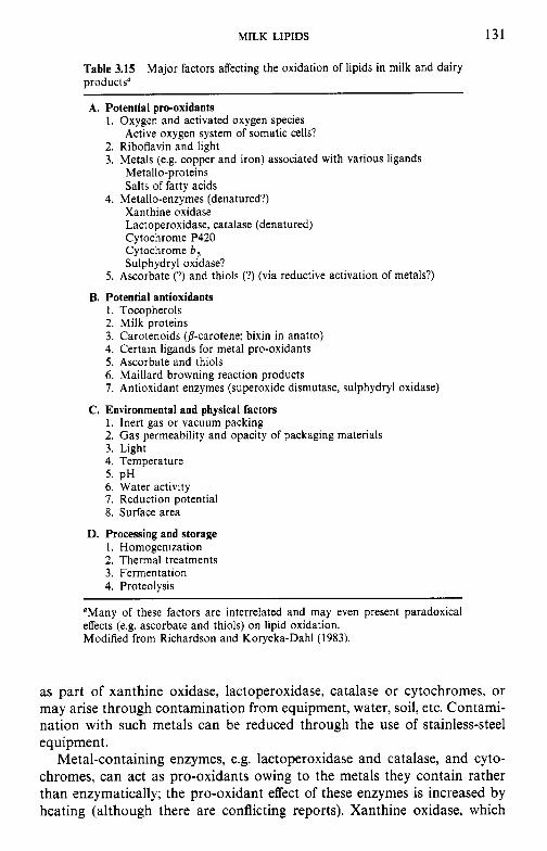
MILK LIPIDS 131
Table 3.15 Major factors affecting the oxidation of lipids in milk and dairy products"
A. Potential pro-oxidants 1. Oxygen and activated oxygen species
2. Riboflavin and light 3. Metals (e.g. copper and iron) associated with various ligands
Active oxygen system of somatic cells?
Metallo-proteins Salts of fatty acids
Xanthine oxidase Lactoperoxidase, catalase (denatured) Cytochrome P420 Cytochrome b , Sulphydryl oxidase?
4. Metallo-enzymes (denatured?)
5 . Ascorbate (?) and thiols (?) (via reductive activation of metals?)
1 . Tocopherols 2. Milk proteins 3. Carotenoids @-carotene; bixin in anatto) 4. Certain ligands for metal pro-oxidants 5 . Ascorbate and thiols 6. Maillard browning reaction products 7. Antioxidant enzymes (superoxide dismutase, sulphydryl oxidase)
C. Environmental and physical factors 1. Inert gas or vacuum packing 2. Gas permeability and opacity of packaging materials 3. Light 4. Temperature 5. pH 6. Water activity 7. Reduction potential 8. Surface area
D. Processing and storage 1. Homogenization 2. Thermal treatments 3. Fermentation 4. Proteolysis
B. Potential antioxidants
"Many of these factors are interrelated and may even present paradoxical effects (e.g. ascorbate and thiols) on lipid oxidation. Modified from Richardson and Korycka-Dahl (1983).
as part of xanthine oxidase, lactoperoxidase, catalase or cytochromes, or may arise through contamination from equipment, water, soil, etc. Contami- nation with such metals can be reduced through the use of stainless-steel equipment.
Metal-containing enzymes, e.g. lactoperoxidase and catalase, and cyto- chromes, can act as pro-oxidants owing to the metals they contain rather than enzymatically; the pro-oxidant effect of these enzymes is increased by heating (although there are conflicting reports). Xanthine oxidase, which

132 DAIRY CHEMISTRY A N D BIOCHEMISTRY
contains Fe and Mo, can act enzymatically and as a source of pro-oxidant metals.
Riboflavin is a potent photosensitizer and catalyses a number of oxidative reactions in milk, e.g. fatty acids, proteins (with the formation of 3-methyl thiopropanal from methionine which is responsible for light- induced off-flavour) and ascorbic acid. Milk and dairy products should be protected from light by suitable packaging and exposure to UV light should be minimized.
Ascorbic acid is a very effective anti-oxidant but combinations of ascor- bate and copper can be pro-oxidant depending on their relative concentra- tions. Apparently, ascorbate reduces Cuz+ to Cu'.
3.15.2 Antioxidants in milk
Antioxidants are molecules with an easily detachable H atom which they donate to fatty acid free radicals or fatty acid peroxy radicals, which would otherwise abstract a H from another fatty acid, forming another free radical. The residual antioxidant molecule (less its donatable H) is stable and antioxidants thus break the autocatalytic chain reaction.
Milk and dairy products contain several antioxidants, of which the following are probably the most important:
0 Tocopherols (vitamin E), which are discussed more fully in Chapter 6. The principal function of tocopherols in uiuo is probably to serve as antioxidants. The concentration of tocopherols in milk and meat products can be increased by supplementing the animal's diet.
0 Ascorbic acid (vitamin C): at low concentrations, as in milk, ascorbic acid is an effective antioxidant, but acts as a pro-oxidant at higher concentra- tions.
0 Superoxidase dismutase (SOD). This enzyme, which occurs in various body tissues and fluids, including milk, scavenges superoxide radicals (0;) which are powerful pro-oxidants. SOD is discussed more fully in Chapter 8.
0 Carotenoids can act as scavengers of free radicals but whether or not they act as antioxidants in milk is controversial.
0 The thiol groups of P-lactoglobulin and proteins of the fat globule membrane are activated by heating. Most evidence indicates that thiol groups have antioxidant properties but they may also produce active oxygen species which could act as pro-oxidants under certain circumstan- ces. The caseins are also effective antioxidants, possibly via chelation of cu.
0 Some products of the Maillard reaction are effective antioxidants.
The addition of synthetic antioxidants, e.g. P-hydroxyanisole or butylated hydroxytoluene, to dairy products is prohibited in most countries.
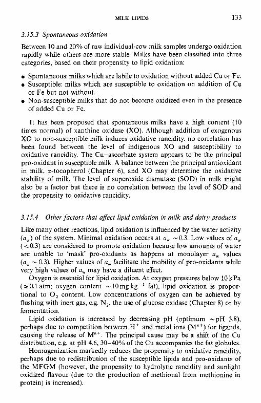
MILK LIPIDS 133
3.15.3 Spontaneous oxidation
Between 10 and 20% of raw individual-cow milk samples undergo oxidation rapidly while others are more stable. Milks have been classified into three categories, based on their propensity to lipid oxidation:
Spontaneous: milks which are labile to oxidation without added Cu or Fe. Susceptible: milks which are susceptible to oxidation on addition of Cu
Non-susceptible milks that do not become oxidized even in the presence or Fe but not without.
of added Cu or Fe.
It has been proposed that spontaneous milks have a high content (10 times normal) of xanthine oxidase (XO). Although addition of exogenous XO to non-susceptible milk induces oxidative rancidity, no correlation has been found between the level of indigenous XO and susceptibility to oxidative rancidity. The Cu-ascorbate system appears to be the principal pro-oxidant in susceptible milk. A balance between the principal antioxidant in milk, r-tocopherol (Chapter 6 ) , and XO may determine the oxidative stability of milk. The level of superoxide dismutase (SOD) in milk might also be a factor but there is no correlation between the level of SOD and the propensity to oxidative rancidity.
3.1.5.4
Like many other reactions, lipid oxidation is influenced by the water activity (a,) of the system. Minimal oxidation occurs at a, -0.3. Low values of a, (< 0.3) are considered to promote oxidation because low amounts of water are unable to 'mask' pro-oxidants as happens at monolayer a, values (a , - 0.3). Higher values of a, facilitate the mobility of pro-oxidants while very high values of a, may have a diluent effect.
Oxygen is essential for lipid oxidation. At oxygen pressures below 10 kPa (z 0.1 atm; oxygen content - 10 mg kg- fat), lipid oxidation is propor- tional to 0, content. Low concentrations of oxygen can be achieved by flushing with inert gas, e.g. N,, the use of glucose oxidase (Chapter 8) or by fermentation.
Lipid oxidation is increased by decreasing pH (optimum -pH 3.8), perhaps due to competition between H f and metal ions (M"') for ligands, causing the release of M"'. The principal cause may be a shift of the Cu distribution, e.g. at pH 4.6, 30-40% of the Cu accompanies the fat globules.
Homogenization markedly reduces the propensity to oxidative rancidity, perhaps due to redistribution of the susceptible lipids and pro-oxidants of the MFGM (however, the propensity to hydrolytic rancidity and sunlight oxidized flavour (due to the production of methional from methionine in protein) is increased).
Other factors that afect lipid oxidation in milk and dairy products
134 DAIRY CHEMISTRY AND BIOCHEMISTRY
NaCl reduces the rate of auto-oxidation in sweet-cream butter but increases it in ripened cream butter (c. pH 5 ) ; the mechanism in unknown.
In addition to influencing the rate of lipid oxidation via activation of thiol groups and metallo-enzymes, heating milk may also affect oxidation via redistribution of Cu (which migrates to the FGM on heating) and possibly by the formation of Maillard browning products, some of which have metal chelating and antioxidant properties.
The rate of auto-oxidation increases with increasing temperature (Qlo - 2) but oxidation in raw and HTST-pasteurized milk is promoted by low temperatures whereas the reverse is true for UHT-sterilized products (i.e. the effect of temperature is normal). The reason(s) for this anomalous behaviour is unknown.
3.1.5.5 Measurement of lipid oxidation
In addition to organoleptic assessment, several chemical/physical methods have been developed to measure lipid oxidation. These include: peroxide value, thiobarbituric acid (TBA) value, ultraviolet absorption (at 233 nm), ferric thiocyanate, Kreis test, chemiluminescence, oxygen uptake and analy- sis of carbonyls by HPLC (see Rossell, 1986).
3.16 Rheology of milk fat
The rheological properties of many dairy products are strongly influenced by the amount and melting point of the fat present. The sensory properties of cheese are strongly influenced by fat content but the effect is even greater in butter in which hardness/spreadability is of major concern. The hardness of fats is determined by the ratio of solid to liquid fat which is influenced by: fatty acid profile, fatty acid distribution and processing treatments.
3.16.1
The fatty acid profile of ruminant fats (milk and adipose tissue) is relatively constant due to the 'buffering' action of the rumen microflora that modify ingested lipids. However, the proportions of various fatty acids in milk lipids show seasonal/nutritional/lactational variations (Figure 3.5) which are re- flected in seasonal variations in the hardness of milk fat (Figure 3.7).
The fatty acid profile can be modified substantially by feeding encap- sulated (protected) polyunsaturated oils to cows. The oil is encapsulated in a film of polymerized protein or in crushed oil-rich seeds. The encapsul- ating protein is digested in the abomasum, resulting in the release of the unsaturated lipid, a high proportion of the fatty acids of which are then incorporated into the milk (and adipose tissue) lipids. The technical
Fatty acid pro$le and distribution
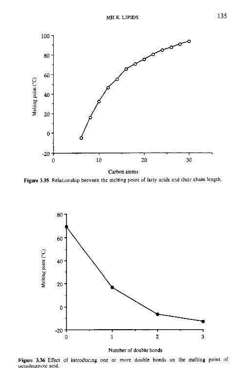
MILK LIPIDS 135
-20 0 10 20 30
Carhon atoms
Figure 3.35 Relationship between the melting point of fatty acids and their chain length.
80
60
40
20
0
-20 0 1 2 3
Numher of double bonds
Figure 3.36 Effect of introducing one or more double bonds on the melting point of octadecanoic acid.
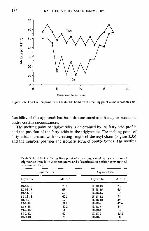
136 DAIRY CHEMISTRY AND BIOCHEMISTRY
60 - 50 -
* ,s 40- 8
30 -
20 - 10 -
s v
M e .- - * z"
Cis
0 1 I 1
0 5 10 15 20
Position of double bond
Figure 3.37 Effect of the position of the double bond on the melting point of octadecenoic acid.
feasibility of this approach has been demonstrated and it may be economic under certain circumstances.
The melting point of triglycerides is determined by the fatty acid profile and the position of the fatty acids in the triglyceride. The melting point of fatty acids increases with increasing length of the acyl chain (Figure 3.35) and the number, position and isomeric form of double bonds. The melting
Table 3.16 Effect on the melting point of shortening a single fatty acid chain of triglyceride from 18 to 0 carbon atoms and of esterification position (symmetrical or asymmetrical)
Symmetrical Asymmetrical
Glyceride M P "C Glyceride M P "C
18-18-18 18-16-18 1 8- 14-1 8 18-1 2-18 18- 10-1 8 18-8-18 18-6- 18 18-4-1 8 18-2- 18 18-0-18
73.1 68 62.5 60.5 57 51.8 47.2 51 62 78
18-18-1 8 18-18-16 18-18-14 18-18-1 2 18-18-10 18-18-8 18-18-6 18-1 8-4 18-1 8-2 18-18-0
73.1 65 62 54 49 47.6 44
55.2 68
-
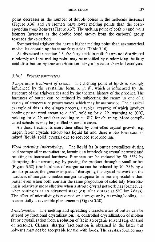
MILK LIPIDS 137
point decreases as the number of double bonds in the molecule increases (Figure 3.36) and cis isomers have lower melting points than the corre- sponding trans isomers (Figure 3.37). The melting point of both cis and trans isomers increases as the double bond moves from the carboxyl group towards the o-carbon.
Symmetrical triglycerides have a higher melting point than asymmetrical molecules containing the same fatty acids (Table 3.16).
As discussed in section 3.6, the fatty acids in milk fat are not distributed randomly and the melting point may be modified by randomizing the fatty acid distribution by transesterification using a lipase or chemical catalysts.
3.16.2 Process parameters
Temperature treatment of cream. The melting point of lipids is strongly influenced by the crystalline form, ct, f l , f l ' , which is influenced by the structure of the triglycerides and by the thermal history of the product. The hardness of butter can be reduced by subjecting the cream to one of a variety of temperature programmes, which may be automated. The classical example of this is the Alnarp process, a typical example of which involves cooling pasteurized cream to c. 8"C, holding for c. 2 h, warming to 20°C, holding for c. 2 h and then cooling to c. 10°C for churning. More compli- cated schedules may be justified in certain cases.
All these treatments exert their effect by controlled crystal growth, e.g. larger, fewer crystals adsorb less liquid fat and there is less formation of mixed (liquid-solid) crystals due to reduced supercooling.
Work softening (microfixing). The liquid fat in butter crystallizes during cold storage after manufacture, forming an interlocking crystal network and resulting in increased hardness. Firmness can be reduced by 50-55% by disrupting this network, e.g. by passing the product through a small orifice (Figure 3.38) (the hardness of margarine can be reduced by 70-75% by a similar process; the greater impact of disrupting the crystal network on the hardness of margarine makes margarine appear to be more spreadable than butter even when both contain the same proportion of solid fat). Microfix- ing is relatively more effective when a strong crystal network has formed, i.e. when setting is at an advanced stage (e.g. after storage at 5°C for 7days). The effect of microfixing is reversed on storage or by warming/cooling, i.e. is essentially a reversible phenomenon (Figure 3.38).
Fractionation. The melting and spreading characteristics of butter can be altered by fractional crystallization, i.e. controlled crystallization of molten fat or crystallization from a solution of fat in an organic solvent (e.g. ethanol or acetone). Cleaner, sharper fractionation is obtained in the latter but solvents may not be acceptable for use with foods. The crystals formed may

138 DAIRY CHEMISTRY AND BIOCHEMISTRY
1 - 1 margarine
I before working
l o a butter 1 c F .- LA
O' 0 2 4 6 a
Days after working
Figure 3.38 Effect of microfixing on the hardness of butter and conventional margarine (from Mulder and Walstra, 1974).
be removed by centrifugation (special centrifuges have been developed) or filtration. Early studies on fractional crystallization involved removing the high-melting point fraction for use in other applications, the mother liquor being used as a modified butter spread. This approach shifts the melting point-temperature curve to lower temperatures without significantly chang- ing its shape (Figure 3.39). While the resulting butter has acceptable spreadability at low temperatures, its 'stand-up' properties are unsatisfac- tory, i.e. it becomes totally liquid at too low a temperature. A better approach is to blend low and high melting point fractions, by which an ideal melting curve can be approached. The problem of finding economic uses for the middle melting point fraction remains.
Blending. Blends of vegetable oils and milk fat offer an obvious solution to the problem of butter hardness - any desired hardness values can be obtained. Such products were introduced in the 1960's and are now used widely in many countries. These products may be produced by blending an
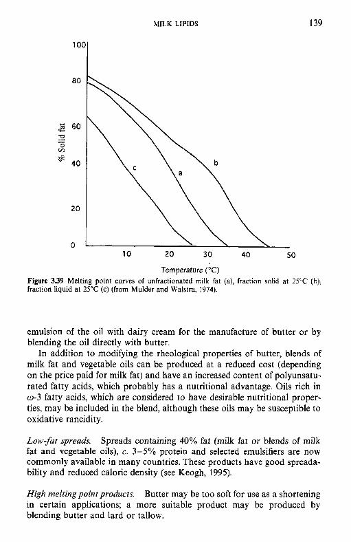
MILK LIPIDS 139
1001 80
20
0 10 20 30 40 50
Temperature ("C) Figure 3.39 Melting point curves of unfractionated milk fat (a), fraction solid at 25°C (b), fraction liquid at 25°C (c) (from Mulder and Walstra, 1974).
emulsion of the oil with dairy cream for the manufacture of butter or by blending the oil directly with butter.
In addition to modifying the rheological properties of butter, blends of milk fat and vegetable oils can be produced at a reduced cost (depending on the price paid for milk fat) and have an increased content of polyunsatu- rated fatty acids, which probably has a nutritional advantage. Oils rich in 0-3 fatty acids, which are considered to have desirable nutritional proper- ties, may be included in the blend, although these oils may be susceptible to oxidative rancidity.
Low-fat spreads. Spreads containing 40% fat (milk fat or blends of milk fat and vegetable oils), c. 3-5% protein and selected emulsifiers are now commonly available in many countries. These products have good spreada- bility and reduced caloric density (see Keogh, 1995).
High meltingpointproducts. Butter may be too soft for use as a shortening in certain applications; a more suitable product may be produced by blending butter and lard or tallow.
140 DAIRY CHEMISTRY AND BIOCHEMISTRY
References
An Foras Taluntais (1981) Chemical Composition of Milk in Ireland, An Foras Taluntais, Dublin.
Brunner, J.R. (1965) Physical equilibria in milk: the lipid phase, in Fundamentals of Dairy Chemistry, (eds B.H. Webb and A.H. Johnson), AVI Publishing, CT, pp. 403-505.
Brunner, J.R. (1974) Physical equilibria in milk: the lipid phase, in Fundamentals of Dairy Chemistry, 2nd edn, (eds B.H. Webb, A.H. Johnson and J.A. Alford), AVI Publishing, CT,
Christie, W.W. (1995) Composition and structure of milk lipids, in Advanced Dairy Chemistry, Vol. 2 Lipids, 2nd edn, (ed. P.F. Fox), Chapman & Hall, London, pp. 136.
Cremin, F.H. and Power, P. (1985) Vitamins in bovine and human milks, in Developments in Dairy Chemistry, Vol. 3: Lactose and Minor Constituents, (ed. P.F. Fox), Elsevier Applied Science, London, pp. 337-98.
Cullinane, N., Aherne, S., Connolly, J.F. and Phelan, J.A. (1984a) Seasonal variation in the triglyceride and fatty acid composition of Irish butter. Irish J . Food Sci. Technol., 8, 1-12.
Cullinane, N., Condon, D., Eason, D. et a/ . (1984b) Influence of season and processing parameters on the physical properties of Irish butter. Irish J . Food Sci. Techno/. 8, 13-25.
Hawke, J.C. and Taylor, M.W. (1995) Influence of nutritional factors on the yield, composition and physical properties of milk fat, in Adcanced Dairy Chemistry, Vol. 2: Lipids, 2nd edn, (ed. P.F. Fox), Chapman & Hall, London, pp. 37-88.
Hayashi, S. and Smith, L.M. (1965) Membranous material of bovine milk fat globules. I . Comparison of membranous fractions released by deoxycholate and by churning. Biochem.
Jenness, R. and Patton, S. (1959) Principles of Dairy Chemistry, John Wiley and Sons, New York.
Keenan, T.W. and Dylewski, D.P. (1995) lntracellular origin of milk lipid globules and the nature and structure of the milk lipid globule membrane, in Aduanced Dairy Chemistry, Vol. 2: Lipids, 2nd edn, (ed. P.F. Fox), Chapman & Hall, London, pp. 89-130.
Keenan, T.W. and Patton, S. (1995) The structure of milk: implications for sampling and storage. A. The milk lipid globule membrane, in Handbook of M i k Composirion, (ed. R.G. Jensen), Academic Press, San Diego, pp. 5-50.
Keenan, T.W., Mather, I.H. and Dylewski, D.P. (1988) Physical equilibria: Lipid phase, in Fundamentals of Dairy Chemistry, 3rd edn, (ed. N.P. Wong), van Nostrand Reinhold, New York, pp. 511-82.
Keenan, T.W., Dylewski, D.P., Woodford, T.A. and Ford, R.H. (1983) Origin of milk fat globules and the nature of the milk fat globule membrane, in Decelopments in Dairy Chemistry, Vol. 2: Lipids, (ed. P.F. Fox), Applied Science Publishers, London, pp. 83-118.
Keogh, M.K. (1995) Chemistry and technology of milk fat spreads, in Adcanced Dairy Chemistry, Vol. 2 Lipids, 2nd edn, (ed. P.F. Fox), Chapman & Hall, London, pp. 213-45.
King, N. (1955) The Milk Fat Globule Membrane, Commonwealth Agricultural Bureau, Farnham Royal, Bucks, UK.
Lehninger, A.L.. Nelson, D.L. and Cox, M.M. (1993) Principles of Biochemistry, 2nd edn, Worth Publishers, New York.
McDowall, F.H. (1953). The Buttermakers Manual, Vols I and 11, New Zealand University Press, Wellington.
McPherson, A.V. and Kitchen, B.J. (1983) Reviews of the progress of dairy science: the bovine milk fat globule membrane - its formation, composition, structure and behaviour in milk and dairy products. J . Dairy Res. 50. 107-33.
Mulder, H. and Walstra, P. (1974) The Milk Fat Globule: Emulsion Science as Applied to Milk Products and Comparable Foods, Podoc, Wageningen.
O’Connor, T.P. and O’Brien, N.M. (1995) Lipid oxidation, in Advanced Dairy Chemistry, Vol. 2: Lipids, 2nd edn, (ed. P.F. Fox), Chapman & Hall, London, pp. 309-47.
Patton, S. and Keenan, T.W. (1975) The milk fat globule membrane. Biochim. Biophys. Acta,
Peereboom, J.W.C. (1969) Theory on the renaturation of alkaline milk phosphates from
pp. 474-602.
4. 2550-7.
415, 273-309.
pasteurized cream. Milchwissenschaf, 24, 266-9.
MILK LIPIDS 141
Prentice, J.H. (1969) The milk fat globule membrane 1955-1968. Dairy Sci. Abstr., 31, 353-6. Richardson, T. and Korycka-Dahl, M. (1983) Lipid oxidation, in Developments in Dairy
Chemistry, Vol. 2: Lipids, (ed. P.F. Fox), Applied Science Publishers, London, pp. 241 -363. Rossell, J.B. (1986) Classical analysis of oils and fats, in Analysis of Oils and Fats, (eds R.J.
Hamilton and J.B. Rossell), Elsevier Applied Science, London, pp. 1-90. Towler, C. (1994) Dewlopments in cream separation and processing, in Modern Dairy.
Technology, Vol. 1, 2nd edn, (ed. R.K. Robinson), Chapman & Hall, London, pp. 61-105. Walstra, P. (1983) Physical chemistry of milk fat globules, in Decelopments in Dairy Chemistry,
Vol. 2: Lipids, (ed. P.F. Fox), Applied Science Publishers, London, pp. 119-58. Walstra, P. and Jenness, R. (1984) Dairy Chemistry and Physics, John Wiley and Sons, New
York. Wilbey, R.A. (1994) Production of butter and dairy based spreads, in Modern Dairy Technology,
Vol. 1, 2nd edn, (ed. R.K. Robinson), Chapman & Hall, London, pp. 107-58. Wooding, F.B.P. (1971) The structure of the milk fat globule membrane. J . Ultrastructure Res.,
37. 388-400.
Suggested reading
Fox, P.F. (ed.) (1983) Developments in Dairy Chemistry. Vol. 2: Lipids, Applied Science
Fox, P.F. (ed.) (1995) Advanced Dairy Chemistry, Vol. 2: Lipids, 2nd edn, Chapman & Hall,
Mulder, H. and Walstra, P. (1974) The Milk Fat Globule, Podoc, Wageningen. Walstra, P. and Jenness. R. (1984) Dairy Chemistry and Physics, Wiley-Interscience, New York. Webb, B.H. and Johnson, A.H. (1965) Fundamentals of Dairy Chemistry, AVI Publishing,
Webb, B.H., Johnson, A.H. and Alford, J.A. (eds) (1974) Fundamentals ofDairy Chemistry, 2nd
Wong, N.P. (ed.) (1980) Fundamentals of Dairy Chemistry, Vol. 1, 3rd edn, AVI Publishing,
Publishers, London.
London.
Westport, CT.
edn, AVI Publishing, Westport, CT.
Westport, CT.
Appendices
Appendix 3 A Principal fat ty acids in milk fat
See table overleaf.
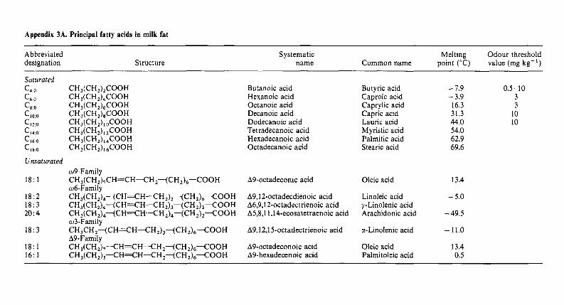
Appendix 3A. Principal fatty acids in milk fat
Abbreviated designation Structure
Systematic Melting Odour threshold name Common name point ("C) value (mg kg- ' )
Saturated c4 0 CH,(CH,),COOH C6 0 CH,(CH,),COOH c, 0 CH,(CH,),COOH CIO 0 CH,(CH,),COOH C,, 0 CH,(CH,), ,COOH '14 0 CH,(CH,),,COOH CI, 0 CH,(CH,),,COOH CI, 0 CH,(CH,), ,COOH
Unsaturated
18: 1 CH3(CH,),CH=CH-CH,-(CH,)6-COOH
18:2 CH3(CH,),-(CH%H-CH,)~~CH2)6-COOH 18:3 CH3(CH,),-(CH=CH-CHz)3~CH2)3<OOH 20:4 CH I(CH ,),-(CH%H--CH 2)4-(CH ,) ,-COOH
w3-Family
A9-Family
w9-Family
w6-Family
18:3 CH,CH,--(CH=CH<H,)j-(CHJ6<OOH
18: 1 CH,(CH,),--CH%H--CH ,<CH ,),--COOH 16: 1 CH,(CH,),--CH%H-CH,-(CH,)6--COOH
Butanoic acid Hexanoic acid Octanoic acid Decanoic acid Dodecanoic acid Tetradecanoic acid Hexadecanoic acid Octadecanoic acid
A9-octadeconic acid
A9J2-0ctadecdienoic acid A6,9,12-octadectrienoic acid A5,8,11,14-ecosatetraenoic acid
A9,12,15-octadectrienoic acid
A9-octadeconoic acid A9-hexadecenoic acid
Butyric acid Caproic acid Caprylic acid Capric acid Lauric acid Myristic acid Palmitic acid Stearic acid
Oleic acid
Linoleic acid y-Linolenic acid Arachidonic acid
a-Linolenic acid
Oleic acid Palmitoleic acid
- 7.9 0.5-10 - 3.9 3 16.3 3 31.3 10 44.0 10 54.0 62.9 69.6
13.4
- 5.0
-49.5
-11.0
13.4 0.5
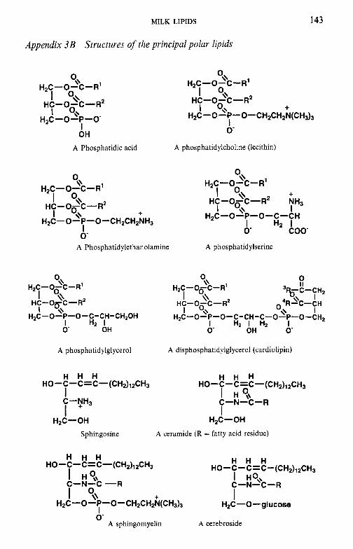
MILK LIPIDS 143
Appendix 3B Structures of the principal polar lipids
O\\
I o\\
HZC -0-P -0- I
O\\ H~C-0- C- R'
I o\\ HC-O-C-F?
H2C- 0 - C - R1
HC-O-C-F? I 0,. + I 0,. H,C-O-P-O-CH2CHZN(CH,),
0- I OH
A Phosphatidic acid A phosphatidylcholine (lecithin)
O\\ O\\ H~C-0- C- R'
H~C-0-C-R' I o\\ + I o\\ HC-O-C-R2 NH3 I o\\ I HC -0-c - R~
H2C-O-P-O-CH2CH2NH3 I H2 I I o\\ + HZC-0-P-0-C-CH
I 0 coo 0 A Phosphatidylethanolamine A phosphatidylserine
t 0 \\
H ~ C - o ~ c - R' '%-C-CHz
HC-O-C-R~ 1 % 'R3C-CH
H ~ C - O ~ C - R ' O\\
H C - O ~ C - R ~ I \ \ I
I \ \ I o,\ \\ I H2C-O-P-O-C-CH-CH20H HzC-0- P-0- C- CH- C-0- P- 0-CHp
I H2 I I H2 I Hz I 0' OH 0 OH 0
A phosphatidylglycerol A disphosphatidylglycerol (cardiolipin)
H H H H H H
I H?, C-N-C-R
HO-C-C=C-(CH2)12CH3 HO-C-C=C-(CH2)12CH3 I I C-yH3
I HZC-OH HzC-OH
Sphingosine A ceramide (R = fatty acid residue)
H H H H H H
I H?\ I Ho+ C-N-C --R C-N-C-R
HO-C-C=C-(CH2)12CH3 HO-C-C=C-(CH2)12CH3
I O,\ + I H2C-O-P-O-CH2CH2N(CH3)3 H2C - 0 - glucose
I
A sphingomyelin A cerebroside 0
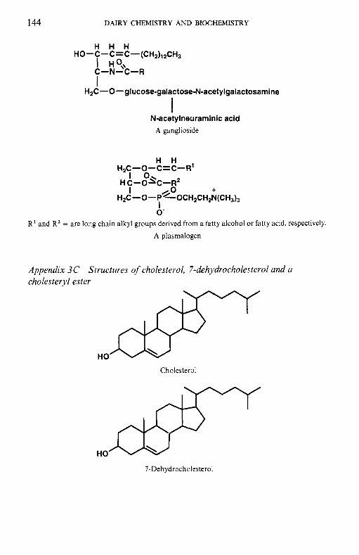
144 DAIRY CHEMISTRY AND BIOCHEMISTRY
H H H
I H O\\ C-N-C-R
HO-C-C=C-(CH2)12CH3
I H2C-0-glucose-galactose-N-acetylgalactosamine
I N-acetylneuraminic acid
A ganglioside
H H H ~ C -0 - c = C- R'
I 0 HC-OpC-OR2 I
H2C- 0 - P%OCHZCH~;(CH~)~ I 0-
R' and R2 = are long chain alkyl groups derived from a fatty alcohol or fatty acid, respectively. A plasmalogen
Appendix 3C Structures of cholesterol, 7-dehydrocholesterol and a cholesteryl ester
Cholesterol
HO
7-Dehydrocholesterol
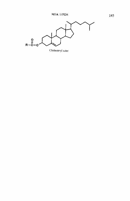
145 MILK LIPIDS
Cholesteryl ester
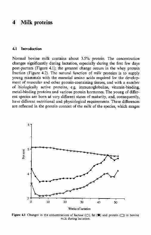
4 Milk proteins
4.1 Introduction
Normal bovine milk contains about 3.5% protein. The concentration changes significantly during lactation, especially during the first few days post-partum (Figure 4.1); the greatest change occurs in the whey protein fraction (Figure 4.2). The natural function of milk proteins is to supply young mammals with the essential amino acids required for the develop- ment of muscular and other protein-containing tissues, and with a number of biologically active proteins, e.g. immunoglobulins, vitamin-binding, metal-binding proteins and various protein hormones. The young of differ- ent species are born at very different states of maturity, and, consequently, have different nutritional and physiological requirements. These differences are reflected in the protein content of the milk of the species, which ranges
6
d 5
2 8
4
3 0 10 20 30 40 50
Weeks of lactation
Figure 4.1 Changes in the concentrations of lactose (O), fat (0) and protein (0) in bovine milk during lactation.
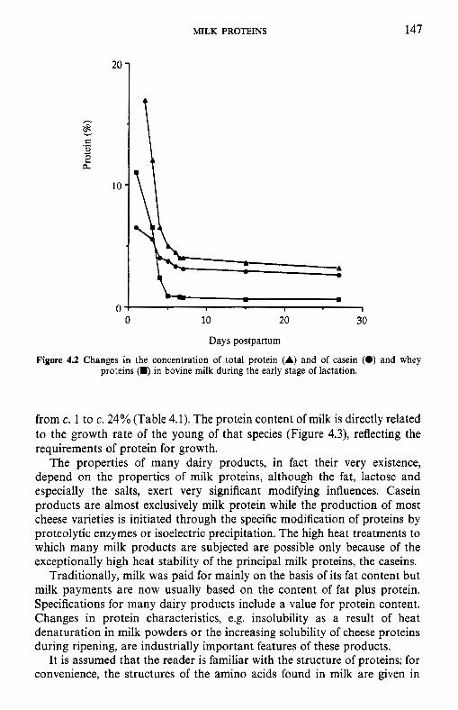
MILK PROTEINS 147
10 -
0 : I I I
0 10 20 30
Days postpartum
Figure 4.2 Changes in the concentration of total protein (A) and of casein (0) and whey proteins (W) in bovine milk during the early stage of lactation.
from c. 1 to c. 24% (Table 4.1). The protein content of milk is directly related to the growth rate of the young of that species (Figure 4.3), reflecting the requirements of protein for growth.
The properties of many dairy products, in fact their very existence, depend on the properties of milk proteins, although the fat, lactose and especially the salts, exert very significant modifying influences. Casein products are almost exclusively milk protein while the production of most cheese varieties is initiated through the specific modification of proteins by proteolytic enzymes or isoelectric precipitation. The high heat treatments to which many milk products are subjected are possible only because of the exceptionally high heat stability of the principal milk proteins, the caseins.
Traditionally, milk was paid for mainly on the basis of its fat content but milk payments are now usually based on the content of fat plus protein. Specifications for many dairy products include a value for protein content. Changes in protein characteristics, e.g. insolubility as a result of heat denaturation in milk powders or the increasing solubility of cheese proteins during ripening, are industrially important features of these products.
It is assumed that the reader is familiar with the structure of proteins; for convenience, the structures of the amino acids found in milk are given in
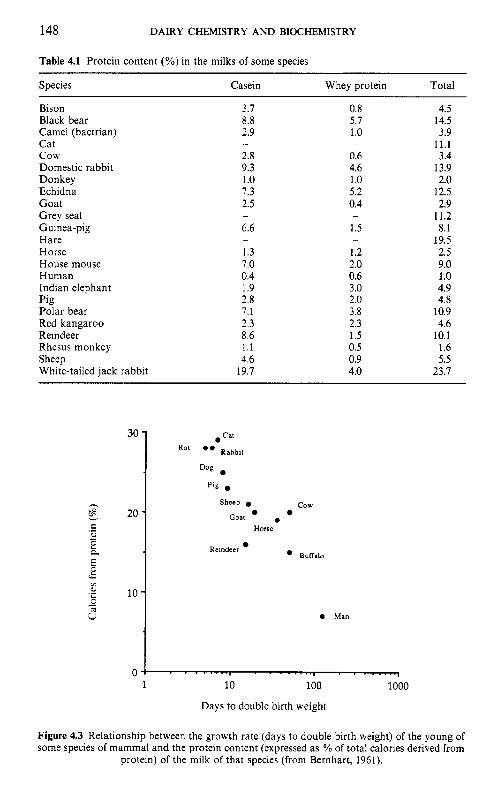
148 DAIRY CHEMISTRY AND BIOCHEMISTRY
30 -
h
20- E .- e,
K - E
b z
.z 10- 8 - s
0
Table 4.1 Protein content (YO) in the milks of some species
Cat
K a t ** Rabbit
Dog
P I S
cow Sheep
Goat . Horse
Reindeer Buffalo
0 Man
4
Species Casein Whey protein Total ~ ~~
Bison 3.7 0.8 4.5 Black bear 8.8 5.7 14.5 Camel (bactrian) 2.9 1 .o 3.9 Cat 11.1 c o w 2.8 0.6 3.4 Domestic rabbit 9.3 4.6 13.9 Donkey 1 .o 1.0 2.0 Echidna 7.3 5.2 12.5 Goat 2.5 0.4 2.9 Grey seal 11.2 Guinea-pig 6.6 1.5 8.1 Hare 19.5 Horse 1.3 1.2 2.5 House mouse 7.0 2.0 9.0 Human 0.4 0.6 1 .o Indian elephant 1.9 3.0 4.9 Pig 2.8 2.0 4.8 Polar bear 7.1 3.8 10.9 Red kangaroo 2.3 2.3 4.6 Reindeer 8.6 1.5 10.1 Rhesus monkey 1.1 0.5 1.6
White-tailed jack rabbit 19.7 4.0 23.7
- -
- -
- -
Sheep 4.6 0.9 5.5
Days to double birth weight
Figure 4.3 Relationship between the growth rate (days to double birth weight) of the young of some species of mammal and the protein content (expressed as YO of total calories derived from
protein) of the milk of that species (from Bernhart, 1961).
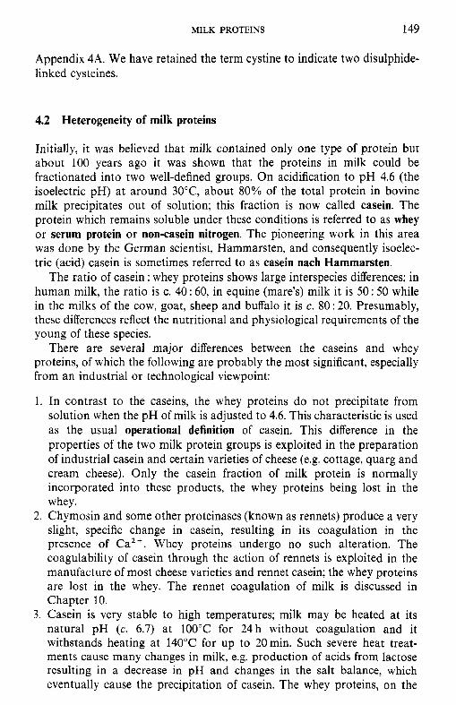
MILK PROTEINS 149
Appendix 4A. We have retained the term cystine to indicate two disulphide- linked cysteines.
4.2 Heterogeneity of milk proteins
Initially, it was believed that milk contained only one type of protein but about 100 years ago it was shown that the proteins in milk could be fractionated into two well-defined groups. On acidification to pH 4.6 (the isoelectric pH) at around 30°C, about 80% of the total protein in bovine milk precipitates out of solution; this fraction is now called casein. The protein which remains soluble under these conditions is referred to as whey or serum protein or non-casein nitrogen. The pioneering work in this area was done by the German scientist, Hammarsten, and consequently isoelec- tric (acid) casein is sometimes referred to as casein nach Hammarsten.
The ratio of casein : whey proteins shows large interspecies differences; in human milk, the ratio is c. 40 : 60, in equine (mare's) milk it is 50: 50 while in the milks of the cow, goat, sheep and buffalo it is c. 80 : 20. Presumably, these differences reflect the nutritional and physiological requirements of the young of these species.
There are several major differences between the caseins and whey proteins, of which the following are probably the most significant, especially from an industrial or technological viewpoint:
1. In contrast to the caseins, the whey proteins do not precipitate from solution when the pH of milk is adjusted to 4.6. This characteristic is used as the usual operational definition of casein. This difference in the properties of the two milk protein groups is exploited in the preparation of industrial casein and certain varieties of cheese (e.g. cottage, quarg and cream cheese). Only the casein fraction of milk protein is normally incorporated into these products, the whey proteins being lost in the whey.
2. Chymosin and some other proteinases (known as rennets) produce a very slight, specific change in casein, resulting in its coagulation in the presence of Ca2+. Whey proteins undergo no such alteration. The coagulability of casein through the action of rennets is exploited in the manufacture of most cheese varieties and rennet casein; the whey proteins are lost in the whey. The rennet coagulation of milk is discussed in Chapter 10.
3. Casein is very stable to high temperatures; milk may be heated at its natural pH (c. 6.7) at 100°C for 24h without coagulation and it withstands heating at 140°C for up to 20min. Such severe heat treat- ments cause many changes in milk, e.g. production of acids from lactose resulting in a decrease in pH and changes in the salt balance, which eventually cause the precipitation of casein. The whey proteins, on the
150 DAIRY CHEMISTRY AND BIOCHEMISTRY
other hand, are relatively heat labile, being completely denatured by heating at 90°C for 10min. Heat-induced changes in milk are discussed in Chapter 9.
4. Caseins are phosphoproteins, containing, on average, 0.85% phosphorus, while the whey proteins contain no phosphorus. The phosphate groups are responsible for many of the important characteristics of casein, especially its ability to bind relatively large amounts of calcium, making it a very nutritionally valuable protein, especially for young animals. The phosphate, which is esterified to the protein via the hydroxyl group of serine, is generally referred to as organic phosphate. Part of the inorganic phosphorus in milk is also associated with the casein in the form of colloidal calcium phosphate (c. 57% of the inorganic phosphorus) (Chap- ter 5).
The phosphate of casein is an important contributor to its remarkably high heat stability and to the calcium-induced coagulation of rennet- altered casein (although many other factors are involved in both cases).
5. Casein is low in sulphur (0.8%) while the whey proteins are relatively rich (1.7%). Differences in sulphur content become more apparent if one considers the levels of individual sulphur-containing amino acids. The sulphur of casein is present mainly in methionine, with low concentra- tions of cysteine and cystine; in fact the principal caseins contain only methionine. The whey proteins contain significant amounts of both cysteine and cystine in addition to methionine and these amino acids are responsible, in part, for many of the changes which occur in milk on heating, e.g. cooked flavour, increased rennet coagulation time (due to interaction between P-lactoglobulin and K-casein) and improved heat stability of milk pre-heated prior to sterilization.
6 . Casein is synthesized in the mammary gland and is found nowhere else in nature. Some of the whey proteins (P-lactoglobulin and cr-lactalbumin) are also synthesized in the mammary gland, while others (e.g. bovine serum albumin and the immunoglobulins) are derived from the blood.
7. The whey proteins are molecularly dispersed in solution or have simple quaternary structures, whereas the caseins have a complicated quaternary structure and exist in milk as large colloidal aggregates, referred to as micelles, with particle masses of 106-109 Da.
8. Both the casein and whey protein groups are heterogeneous, each containing several different proteins.
4.2.1 Other protein fractions
In addition to the caseins and whey proteins, milk contains two other groups of proteins or protein-like material, i.e. the proteose-peptone frac- tion and the non-protein nitrogen (NPN) fraction. These fractions were recognized as early as 1938 by Rowland but until recently very little was
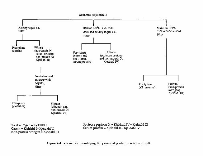
Skimmilk (Kjeldahl I)
I I Heat at loO°C x 20 min. Make to 12% cool and acidify to pH 4.6, filter
trichloroacetic acid, filter
I Acidify to pH 4.6, filter
Precipitate Filtrate (casein) (non-casein N:
serum proteins non-protein N; Kjeldahl 11)
Neutralize and saturate with
filter MgS04
Precipitate (globulins)
Filtrate (albumin and non-protein N; Kjeldahl V)
Total nitrogen = Kjeldahl I Casein = Kjeldahl I - Kjeldahl II Non-protein nitrogen = Kjeldahl 111
Precipitate Filtrate (casein and (proteose peptone heat-labile and non-protein N; semm proteins) Kjeldahl IV)
I Precipitate (all proteins)
1 Filtrate (non-protein nitmgen; Kjeldahl 111)
Proteose peptone N = Kjeldahl IV-Kjeldahl III Serum protein = Kjeldahl 11- Kjeldalil IV
Figure 4.4 Scheme for quantifying the principal protein fractions in milk.
152 DAIRY CHEMISTRY AND BIOCHEMISTRY
known about them. Rowland observed that when milk was heated to 95°C for 10 min, 80% of the nitrogenous compounds in whey were denatured and co-precipitated with the casein when the pH of the heated milk was adjusted subsequently to 4.6. He considered that the heat-denaturable whey proteins represented the lactoglobulin and lactalbumin fractions and designated the remaining 20% 'proteose-peptone'. The proteose-peptone fraction was precipitated by 12% trichloracetic acid (TCA) but some nitrogenous com- pounds remained soluble in 12% TCA and were designated as nonprotein nitrogen.
A scheme for the fractionation of the principal groups of milk proteins, based on that of Rowland, is shown in Figure 4.4.
4.3 Preparation of casein and whey proteins
Skim milk prepared by mechanical separation (see Chapter 3) is used as the starting material for the preparation of casein and whey proteins.
4.3.1 Acid (isoelectric) precipitation
Acidification of milk to about pH 4.6 induces coagulation of the casein. Aggregation occurs at all temperatures, but below about 6°C the aggregates are very fine and remain in suspension, although they can be sedimented by low-speed centrifugation. At higher temperatures (3O-4OcC), the aggregates are quite coarse and precipitate readily from solution. At temperatures above about 5OCC, the precipitate tends to be stringy and difficult to handle.
For laboratory-scale production of casein, HCl is usually used for acidification; acetic or lactic acids are used less frequently. Industrially, HC1 is also usually used; H,SO, is used occasionally but the resulting whey is not suitable for animal feeding (MgSO, is a laxative). Lactic acid produced in situ by a culture of lactic acid bacteria is also widely used, especially in New Zealand, the principal producer of casein.
The inorganic colloidal calcium phosphate associated with casein in normal milk dissolves on acidification of milk to pH 4.6 so that if sufficient time is allowed for solution, isoelectric casein is essentially free of calcium phosphate. In the laboratory, best results are obtained by acidifying skim milk to pH 4.6 at 2"C, holding for about 30min and then warming to 30-35°C. The fine precipitate formed at 2°C allows time for the colloidal calcium phosphate to dissolve (Chapter 5) . A moderately dilute acid (1 M) is preferred, since concentrated acid may cause localized coagulation. Acid production by a bacterial culture occurs slowly and allows time for colloidal calcium phosphate to dissolve. The casein is recovered by filtration or centrifugation and washed repeatedly with water to free the casein of lactose and salts. Thorough removal of lactose is essential since even traces of
MILK PROTEINS 153
lactose will interact with casein on heating via the Maillard browning reaction, with undesirable consequences.
The procedure used for the industrial production of acid (isoelectric) casein is essentially the same as that used on a laboratory scale, except for many technological differences (section 4.15.1).The whey proteins may be recovered from the whey by salting out, dialysis or ultrafiltration.
4.3.2
Because they occur as large aggregates, micelles, most (90-95%) of the casein in milk is sedimented by centrifugation at 100 000 g for 1 h. Sedimen- tation is more complete at higher (30-37°C) than at low (2°C) temperature, at which some of the casein components dissociate from the micelles and are non-sedimentable. Casein prepared by centrifugation contains its original level of colloidal calcium phosphate and can be redispersed as micelles with properties essentially similar to the original micelles.
Cen tr ifuga t ion
4.3.3 Centrifugation of calcium-supplemented milk
Addition of CaCI, to about 0.2 M causes aggregation of the casein such that it can be readily removed by low-speed centrifugation. If calcium is added at 90"C, the casein forms coarse aggregates which precipitate readily. This principle is used in the commercial production of some 'casein co-precipi- tates' in which the whey proteins, denatured on heating milk at 90°C for lOmin, co-precipitate with the casein. Such products have a very high ash content.
4.3.4 Salting-out methods
Casein can be precipitated from solution by any of several salts. Addition of (NH,),SO, to milk to a concentration of 260 g 1- causes complete precipi- tation of the casein together with some whey proteins (immunoglobulins, Ig). MgSO, may also be used. Saturation of milk with NaCl at 37°C precipitates the casein and Igs while the major whey proteins remain soluble, provided they are undenatured. This characteristic is the basis of a commercial test used for the heat classification of milk powders which contain variable levels of denatured whey proteins.
4.3.5 Ultrajiltration
The casein micelles are retained by fine-pore filters. Filtration through large-pore ceramic membranes is used to purify and concentrate casein on a laboratory scale. Ultrafiltration (UF) membranes retain both the caseins
154 DAIRY CHEMISTRY AND BIOCHEMISTRY
and whey proteins while lactose and soluble salts are permeable; total milk protein may be produced by this method. The casein micelles permeate the membranes used in microfiltration (pore size - 0.05-10 pm) but bacteria are retained by membranes with pores of less than OSpm, thus providing a method for removing more than 99.9% of the bacteria in milk without heat treatment; microfiltration is being used increasingly in several sectors of the dairy industry.
Industrially, whey proteins are prepared by ultrafiltration or diafiltration of whey (to remove lactose and salts), followed by spray drying; these products, referred to as whey protein concentrates, contain 30-80% protein.
4.3.6 Geljltration (gel permeation chromatography)
Filtration through cross-linked dextrans (e.g. Sephadex, Pharmacia, Upp- sala, Sweden) makes it possible to fractionate molecules, including proteins, on a commercial scale. It is possible to separate the casein and whey proteins by gel filtration but the process is uneconomical on an industrial scale.
4.3.7 Precipitation with ethanol
The caseins may be precipitated from milk by c. 40% ethanol while the whey proteins remain soluble; lower concentrations of ethanol may be used at lower pH values.
4.3.8 Cryoprecipitation
Casein, in a mainly micellar form, is destabilized and precipitated by freezing milk or, preferably, concentrated milk, at about - 10°C; casein prepared by this method has some interesting properties but is not produced commer- cially at present.
4.3.9 Rennet coagulation
Casein may be coagulated and recovered as rennet casein by treatment of milk with selected proteinases (rennets). However, one of the caseins, K-casein, is hydrolysed during renneting and therefore the properties of rennet casein differ fundamentally from those of acid casein. Rennet casein, which contains the colloidal calcium phosphate of milk, is insoluble in water at pH 7 but can be dissolved by adding calcium sequestering agents, usually citrates or polyphosphates. It has desirable functional properties for certain food applications, e.g. in the production of cheese analogues.
MILK PROTEINS 155
4.3.10
Highly purified whey protein preparations, referred to as whey protein isolates (containing 90-95% protein), are prepared industrially from whey by ion exchange chromatography. Denatured (insoluble) whey proteins, referred to as lactalbumin, may be prepared by heating whey to 95°C for 10-20 min at about pH 6.0; the coagulated whey proteins are recovered by centrifugation. The whey proteins may also be precipitated using FeCl, or polyphosphates (section 4.15.6).
Other methods for the preparation of whey proteins
4.4 Heterogeneity and fractionation of casein
Initially, casein was considered to be a homogeneous protein. Heterogeneity was first demonstrated in the 1920s by Linderstrsm-Lang and co-workers, using fractionation with ethanol-HC1, and confirmed in 1936 by Pedersen, using analytical ultracentrifugation, and in 1939 by Mellander, using free boundary electrophoresis. Three components were demonstrated and named a-, ,!?- and y-casein in order of decreasing electrophoretic mobility and represented 75, 22 and 3%, respectively, of whole casein. These caseins were successfully fractionated in 1952 by Hipp and collaborators based on differential solubilities in urea at c. pH 4.6 or in ethanol/water mixtures; the former is widely used although the possibility of forming artefacts through interaction of casein with cyanate produced from urea is of concern.
In 1956, Waugh and von Hippel showed that the a-casein fraction of Hipp et al. contained two proteins, one of which was precipitated by low concentrations of Ca2+ and was called a,-casein (s = sensitive) while the other, which was insensitive to Ca2+, was called k--casein. a,-Casein was later shown to contain two proteins which are now called uSl- and a,,-caseins. Thus, bovine casein contains four distinct gene products, desig- nated ctsl-, rs2-, /I- and K-caseins which represent approximately 37, 10, 35 and 12% of whole casein, respectively.
Various chemical methods were developed to fractionate the caseins but none gives homogeneous preparations. Fractionation is now usually achieved by ion-exchange chromatography on, for example, DEAE-cellu- lose, using urea-containing buffers; quite large (e.g. 10 g) amounts of caseinate can be fractionated by this method, with excellent results (Figure 4Sa, b). Good results are also obtained by ion-exchange chromatography using urea-free buffers at 2-4°C. High performance ion-exchange chromatography (e.g. Pharmacia FPLCTM on Mono Q or Mono S ) gives excellent results for small amounts of sample (Figure 4.5c, d). Reversed- phase HPLC or hydrophobic interaction chromatography may also be used but are less effective than ion-exchange chromatography.
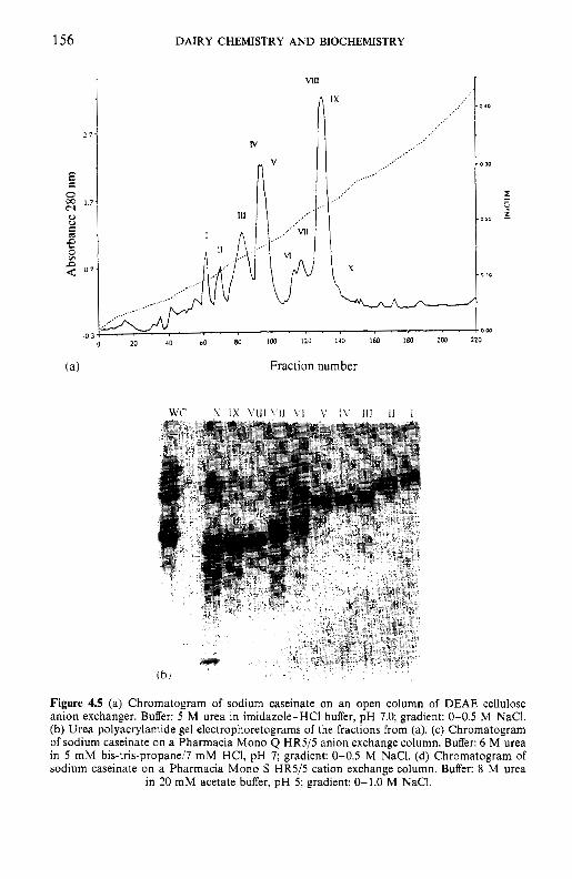
156 DAIRY CHEMISTRY A N D BIOCHEMISTRY
N
X
Y
L &-
20 40 00 80 LOO I20 I40 160 180 2w 220
Fraction number
Figure 4.5 (a) Chromatogram of sodium caseinate on an open column of DEAE cellulose anion exchanger. Buffer: S M urea in imidazole-HCI buffer, pH 7.0; gradient: 0-0.5 M NaC1. (b) Urea polyacrylamide gel electrophoretograms of the fractions from (a). (c) Chromatogram of sodium caseinate on a Pharmacia Mono Q HR5/S anion exchange column. Buffer: 6 M urea in 5 mM bis-tris-propane/7 m M HCI, pH 7; gradient: 0-0.5 M NaC1. (d) Chromatogram of sodium caseinate on a Pharmacia Mono S HRS/S cation exchange column. Buffer: 8 M urea
in 20 mM acetate buffer, pH 5; gradient: 0-1.0 M NaC1.
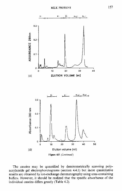
- v Q A
bsor
banc
e 280
nrn
0
?
2
R e
AB
SOR
BA
NC
E 28
0nm
h
v
0
P
0
0
d
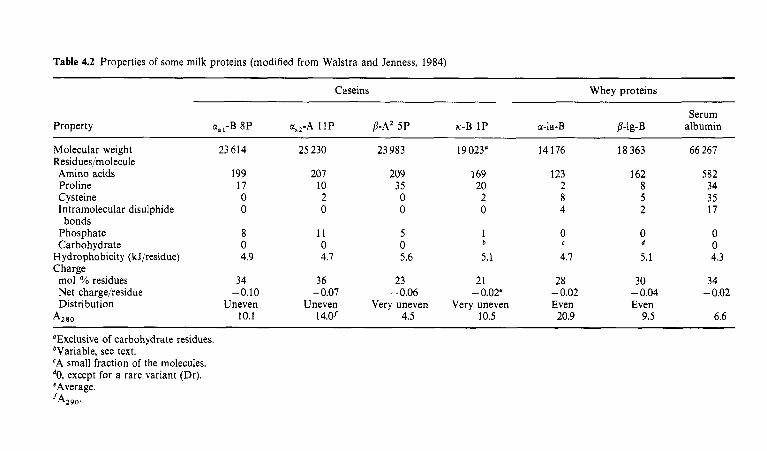
Table 4.2 Properties of some milk proteins (modified from Walstra and Jenness, 1984)
Property
Caseins Whey proteins
Serum u,,-B 8P GL.~-A 11P p-A’ 5 P K-B 1P u-la-B P-10-B albumin
Molecular weight 23 614 25 230 23 983 19 023” 14 176 18 363 66 267 Residues/molecule Amino acids 199 207 209 169 123 162 582 Proline 17 10 35 20 2 8 34 Cysteine 0 2 0 2 8 5 35 Intramolecular disulphide 0 0 0 0 4 2 17
0 0 0
Phosphate 8 11 5 1 0 Carbohydrate 0 0 0
Hydrophobicity (kJ/residue) 4.9 4.7 5.6 5.1 4.7 5.1 4.3
mol YO residues 34 36 23 21 28 30 34 Net charge/residue -0.10 - 0.07 -0.06 -0.02’ - 0.02 - 0.04 - 0.02 Distribution Uneven Uneven Very uneven Very uneven Even Even
bonds
b e d
Charge
A,,, 10.1 14.0’ 4.5 10.5 20.9 9.5 6.6
“Exclusive of carbohydrate residues. bVariable, see text. ‘A small fraction of the molecules.
eAverage. except for a rare variant (Dr).
’A29l.l.
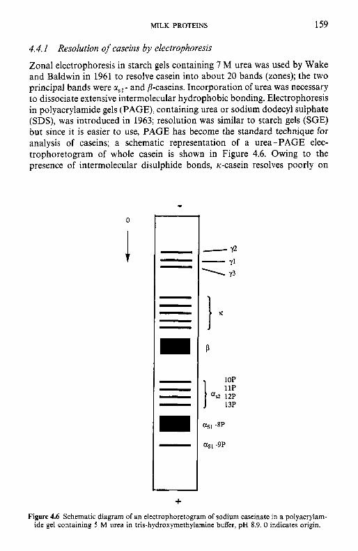
MILK PROTEINS 159
4.4. I Resolution of caseins by electrophoresis
Zonal electrophoresis in starch gels containing 7 M urea was used by Wake and Baldwin in 1961 to resolve casein into about 20 bands (zones); the two principal bands were 01,~ - and p-caseins. Incorporation of urea was necessary to dissociate extensive intermolecular hydrophobic bonding. Electrophoresis in polyacrylamide gels (PAGE), containing urea or sodium dodecyl sulphate (SDS), was introduced in 1963; resolution was similar to starch gels (SGE) but since it is easier to use, PAGE has become the standard technique for analysis of caseins; a schematic representation of a urea-PAGE elec- trophoretogram of whole casein is shown in Figure 4.6. Owing to the presence of intermolecular disulphide bonds, Ic-casein resolves poorly on
+
-12 Yl Y3
-
P
I or
asl -8P
asl -9P
Figure 4.6 Schematic diagram of an electrophoretogram of sodium caseinate in a polyacrylam- ide gel containing 5 M urea in tris-hydroxymethylamine buffer, pH 8.9. 0 indicates origin.
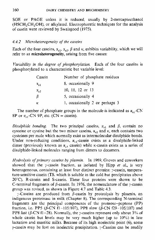
160 DAIRY CHEMISTRY AND BIOCHEMISTRY
SGE or PAGE unless it is reduced, usually by 2-mercaptoethanol (HSCH,CH,OH), or alkylated. Electrophoretic techniques for the analysis of casein were reviewed by Swaisgood (1975).
4.4.2 Microheterogeneity of the caseins
Each of the four caseins, as,, zs2, P and K , exhibits variability, which we will refer to as microheterogeneity, arising from five causes:
Variability in the degree of phosphorylation. phosphorylated to a characteristic but variable level:
Each of the four caseins is
Casein Number of phosphate residues a, 1 8, occasionally 9
zsz 10, 11, 12 or 13 P 5, occasionally 4 K 1, occasionally 2 or perhaps 3
The number of phosphate groups in the molecule is indicated as a,,-CN 8P or a,,-CN 9P, etc. (CN = casein).
Disulphide bonding. The two principal caseins, a,, and P, contain no cysteine or cystine but the two minor caseins, CI,, and K, each contains two cysteines per mole which normally exist as intermolecular disulphide bonds. Under non-reducing conditions, a,,-casein exists as a disulphide-linked dimer (previously known as as5 casein) while K-casein exists as a series of disulphide-linked molecules ranging from dimers to decamers.
Hydrolysis of primary caseins by plasmin. In 1969, Groves and coworkers showed that the y-casein fraction, as isolated by Hipp et al., is very heterogeneous, containing at least four distinct proteins: y-casein, tempera- ture-sensitive casein (TS, which is soluble in the cold but precipitates above 20”C), R-casein and S-casein. These four proteins were shown to be C-terminal fragments of j-casein. In 1976, the nomenclature of the y-casein group was revised, as shown in Figure 4.7 and Table 4.3.
?-Caseins are produced from P-casein by proteolysis by plasmin, an indigenous proteinase in milk (Chapter 8). The corresponding N-terminal fragments are the principal components of the proteose-peptone (PP) fraction, i.e. PP5 (P-CN fl-105/107), PP8 slow (P-CN f29-105/107) and PPS fast (P-CN fl-28). Normally, the ?-caseins represent only about 3% of whole casein but levels may be very much higher (up to 10%) in late lactation and mastitic milks. Because of its high isoelectric point (6), some y-casein may be lost on isoelectric precipitation. ?-Caseins can be readily
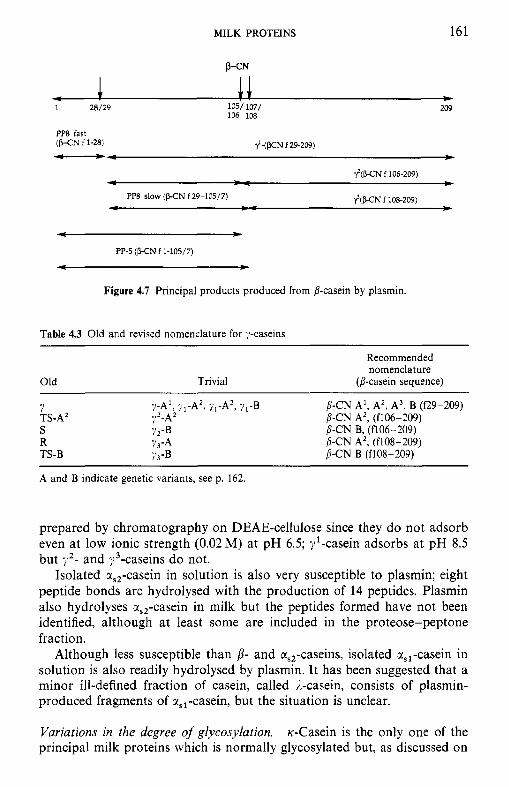
MILK PROTEINS 161
1 28/29 1051 107/ 209 106 108
PP8 fast (P-CN f 1-28) .I'-(WN f 29-209)
PP8 slow ( H N f 29-105/7) ?CWN f 108-209)
- b
PP-5 ( H N f 1-105/7)
- - Figure 4.7 Principal products produced from p-casein by plasmin.
Table 4.3 Old and revised nomenclature for y-caseins
Recommended nomenclature
Old Trivial @-casein sequence)
7 TS-A' ;.LA2 b-CN A', (f106-209) S ?2-B j-CN B, (f106-209) R ?3-A j - C N A2, (f108-209) TS-B ;J3-B j-CN B (f108-209)
?-A', yl-A2, yL-A3, 7'-B P-CN A', A2, A3, B (f29-209)
A and B indicate genetic variants, see p. 162.
prepared by chromatography on DEAE-cellulose since they do not adsorb even at low ionic strength (0.02 M) at pH 6.5; $-casein adsorbs at pH 8.5 but y 2 - and "/-caseins do not.
Isolated %,,-casein in solution is also very susceptible to plasmin; eight peptide bonds are hydrolysed with the production of 14 peptides. Plasmin also hydrolyses r,,-casein in milk but the peptides formed have not been identified, although at least some are included in the proteose-peptone fraction.
Although less susceptible than p- and &,,-caseins, isolated %,,-casein in solution is also readily hydrolysed by plasmin. It has been suggested that a minor ill-defined fraction of casein, called ,&casein, consists of plasmin- produced fragments of &,,-casein, but the situation is unclear.
Variations in the degree of glycosylation. ic-Casein is the only one of the principal milk proteins which is normally glycosylated but, as discussed on
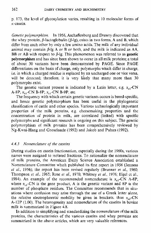
162 DAIRY CHEMISTRY A N D BIOCHEMISTRY
p. 173, the level of glycosylation varies, resulting in 10 molecular forms of ic-casein.
Genetic polymorphism. In 1956, Aschaffenburg and Drewry discovered that the whey protein, P-lactoglobulin (0-lg), exists in two forms, A and B, which differ from each other by only a few amino acids. The milk of any individual animal may contain p-lg A or B or both, and the milk is indicated as AA, BB or AB with respect to P-lg. This phenomenon was referred to as genetic polymorphism and has since been shown to occur in all milk proteins; a total of about 30 variants have been demonstrated by PAGE. Since PAGE differentiates on the basis of charge, only polymorphs which differ in charge, i.e. in which a charged residue is replaced by an uncharged one or vice versa, will be detected; therefore, it is very likely that many more than 30 polymorphs exist.
The genetic variant present is indicated by a Latin letter, e.g. a,,-CN A-8P, c(,,-CN B-8P, a,,-CN B-9P, etc.
The frequency with which certain genetic variants occurs is breed-specific, and hence genetic polymorphism has been useful in the phylogenetic classification of cattle and other species. Various technologically important properties of the milk proteins, e.g. cheesemaking properties and the concentration of protein in milk, are correlated (linked) with specific polymorphs and significant research is ongoing on this subject. The genetic polymorphism of milk proteins has been comprehensively reviewed by Ng-Kwai-Hang and Grosclaude (1992) and Jakob and Puhan (1992).
4.4.3 Nomenclature of the caseins
During studies on casein fractionation, especially during the 1960s, various names were assigned to isolated fractions. To rationalize the nomenclature of milk proteins, the American Dairy Science Association established a Nomenclature Committee which published its first report in 1956 (Jenness er al., 1956); the report has been revised regularly (Brunner et al., 1960; Thompson et al., 1965; Rose et al., 1970; Whitney et al., 1976; Eigel et al., 1984). An example of the recommended nomenclature is x,,-CN A-SP, where a,,-CN is the gene product, A is the genetic variant and 8P is the number of phosphate residues. The Committee recommends that in situ- ations where confusion may arise through the use of a Greek letter alone, the relative electrophoretic mobility be given in brackets, thus a,,-CN A-12P (1.00). The heterogeneity and nomenclature of the caseins in bovine milk is summarized in Figure 4.8.
In addition to simplifying and standardizing the nomenclature of the milk proteins, the characteristics of the various caseins and whey proteins are summarized in the above articles, which are very valuable references.
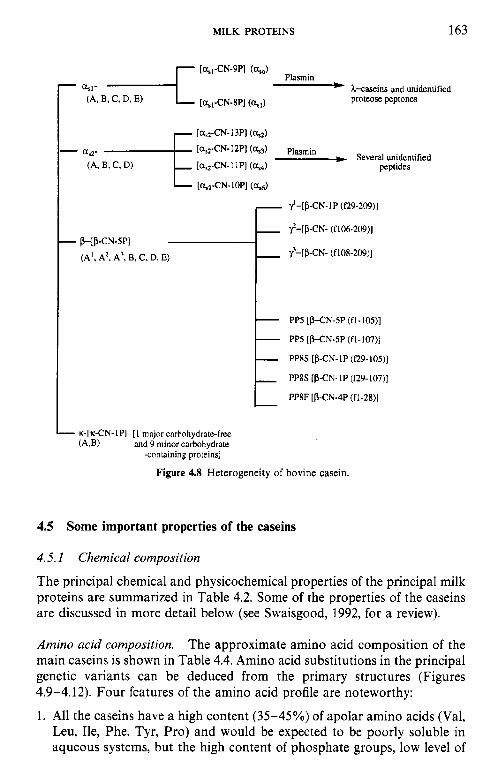
MILK PROTEINS 163
L K-[K-CN-IPI [ I major cnrhohydrate-free and 9 minor carbohydrate
-containing pro~eins]
Figure 4.8 Heterogeneity of bovine casein.
4.5 Some important properties of the caseins
4.5.1 Chemical composition
The principal chemical and physicochemical properties of the principal milk proteins are summarized in Table 4.2. Some of the properties of the caseins are discussed in more detail below (see Swaisgood, 1992, for a review).
Amino acid composition. The approximate amino acid composition of the main caseins is shown in Table 4.4. Amino acid substitutions in the principal genetic variants can be deduced from the primary structures (Figures 4.9-4.12). Four features of the amino acid profile are noteworthy:
1. All the caseins have a high content (35-45%) of apolar amino acids (Val, Leu, Ile, Phe, Tyr, Pro) and would be expected to be poorly soluble in aqueous systems, but the high content of phosphate groups, low level of
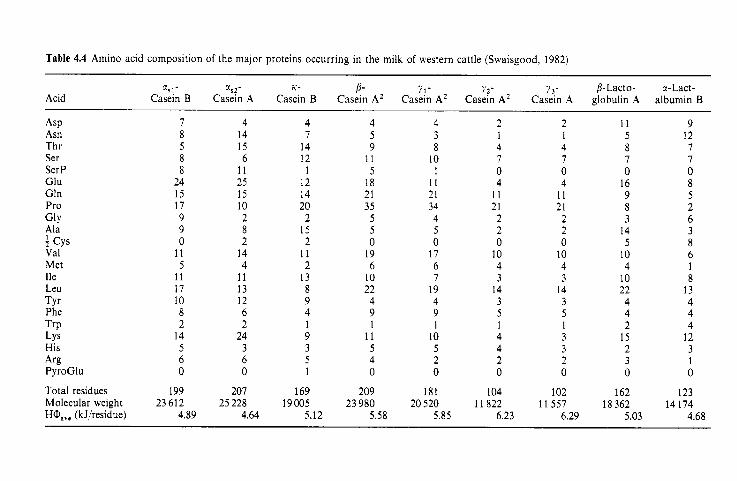
Table 4.4 Amino acid composition of the major proteins occurring in the milk of western cattle (Swaisgood, 1982)
Acid
~~
3 9 1 - X s 2 - K- B- Y 1 - ??- ? 3,- j- Lact 0- a- Lact - Casein B Casein A Casein B Casein A’ Casein A2 Casein A’ Casein A globulin A albumin B
ASP Asn Thr Ser Ser P Glu Gln Pro
Ala
Val Met Ile Leu TY r Phe TrP LYS His A rg PyroGlu
Total residues Molecular weight HO,,, (kJ/residue)
GlY
f Cys
8 24 15 17 9 9 0
11 5
11 17 10 8 2
14 5 6 0
199 23 612
4.89
4 14 15 6
11 25 15 10 2 8 2
14 4
11 13 12 6 2
24 3 6 0
207 25 228
4.64
4 7
14 12
1 12 14 20
2 15 2
11 2
13 8 9 4 1 9 3 5 1
169 19005
5.12
4 4 2 5 3 1 9 8 4
11 10 7 5 I 0
18 11 4 21 21 11 35 34 21
5 4 2 5 5 2 0 0 0
19 17 10 6 6 4
10 7 3 22 19 14 4 4 3 9 9 5 1 1 1
11 10 4 5 5 4 4 2 2 0 0 0
209 181 104 23 980 20 520 11 822
5.58 5.85 6.23
2 1 4 7 0 4
1 1 21 2 2 0
10 4 3
14 3 5 1 3 3 2 0
102 11 557
6.29
11 5 8 7 0
16 9 8 3
14 5
10 4
10 22 4 4 2
15 2 3 0
162 18362
5.03
9 12 7 7 0 8 5 2 6 3 8 6 1 8
13 4 4 4
12 3 1 0
123 14 174
4.68
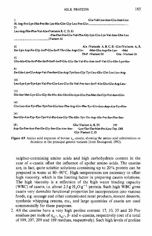
MILK PROTEINS 165
1 Glu-Val-Leu-Asn-Glu-Asn-Leu- H. Arg-Pro-Lys-His-Pro-Ile-Lys-His-Gln-Gly-Leu-P~-Gln-. ............................ 21 Leu-Arg-Phe-Phe-Val-Ala-(Variants B, C, D, E)
-Pro-Phe-Pro-Glu-Val-Phe-Gly-Lys.Glu-Lys-Val-Asn-Glu-Leu .............._.........lV ariant A)
41 Ser-Lys-Asp-Ile-Gly-SerPGlu-SerP-Thr.G1u-Asp-Gln- -Met-Glu-Asp-Ile-Lys- -Met
61 G1u-Ala-Glu-SerP-lle-SerP-SerP-SerP-Glu-G1u-lle-Val-Pro-Asn-SerP-Val-Glu-G1n-Lys-His-
81 Ile-Gln-Lys-Glu-Asp-Val-Pro-Ser-Glu-Arg-Tyr-Leu-Gly-Tyr-Leu-Glu-Gln-Leu-Leu-Ar~
101 Leu-Lys-Lys-Tyr-Lys-Val-ProCln-Leu-Glu-Ile-Val-Pro-Asn-S~P-Ala-Glu-Glu-Arg-Leu-
121 His-Ser-Met-Lys-Glu-Gly-Ile-His-Ala-Gln-Gln-Lys-Glu-Pro-Met-Ile-Gly-Val-Asn~ln-
Ala (Variants A, B, C, E) Gln (Variants A, B,
ThrP (Variant D) Glu (Variant E)
141 Glu-Leu-Ala-Tyr-Phe-TyrPro-Glu-Leu-Phe-Arg-GIn-Phe-Tyr-Gln-Leu-Asp-Ala-Tyr-Pro-
161 Ser-Gly-Ala-Trp-Tyr-Tyr-Val-Pro-Leu-Gly-Thr-GIn-Tyr-Thr-Asp-Ala-Pro-Ser-Phe-Ser-
181 Glu (Variant A, B, D) 199 Asp-Ile-Pro-Asn-Pro-Ile-Gly-Ser-Glu-Asn-Ser- -Lys-Thr-Thr-Met-Pro-Leu-Trp. OH
Gly (Variant C, E)
Figure 4.9 Amino acid sequence of bovine %,,-casein, showing the amino acid substitutions or deletions in the principal genetic variants (from Swaisgood, 1992).
sulphur-containing amino acids and high carbohydrate content in the case of K-casein offset the influence of apolar amino acids. The caseins are, in fact, quite soluble: solutions containing up to 20% protein can be prepared in water at 80-90°C. High temperatures are necessary to offset high viscosity, which is the limiting factor in preparing casein solutions. The high viscosity is a reflection of the high water binding capacity (WBC) of casein, i.e. about 2.5 g H,O g- protein. Such high WBC gives casein very desirable functional properties for incorporation into various foods, e.g. sausage and other comminuted meat products, instant desserts, synthetic whipping creams, etc., and large quantities of casein are used commercially for these purposes.
2. All the caseins have a very high proline content: 17, 10, 35 and 20 Pro residues per mole of asl-, c(,~-, p- and K-caseins, respectively (out of a total of 199,207,209 and 169 residues, respectively). Such high levels of proline
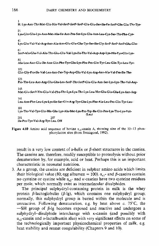
166 DAIRY CHEMISTRY AND BIOCHEMISTRY
I H. Lys-Asn-Thr-Met-Glu-His-Val-SerP-SerP-SerP-Glu-GIu-Ser-Ile-Ile-SerP-GIn-Glu-Thr-Tyr-
21 Lys-Gln-Glu-Lys-Asn-Me t-Ala-Ile-Asn-Pro-Ser-Lys-Glu-Asn-Leu-Cys-Ser-Thr-Phe-Cys-
41 Lys-Glu-Val-Val-Arg-Asn-Ala-Asn-Glu-Glu-Glu-Tyr-Ser-lle-Gly-SerP-SerP-SerP~P-Glu-GIu-
61 SerP-Ala-Glu-Val-Ala-Thr-Glu-G1u-Val-Lys-lle-Thr-Val-Asp-Asp-Lys-His-Tyr-Gln-Lys-
81 Ala-Leu-Asn-Glu-Ile-Asn-Gln-Phe-Tyr-Gln-Lys-Phe-Pro-Gln-Tyr-Leu-Gln-Tyr-Leu-Tyr-
101 Gln-Gly.Pro-Ile-Val-Leu-Asn-Pro-Trp-Asp-GIn-Val-Lys-Arg-Asn-Ala-Val-Pro-Ile-Thr-
121 Pro-Thr-Leu-Asn-Arg-GIu-G1n-Leu-SerP-Thr-SerP-Glu-Glu-Asn-Ser-Lys-Lys-Thr-Val-Asp-
141 Met-GIu-SerP-Thr-GIu-Val-Phe-Thr-Lys-Lys-Thr-Lys-Leu-Thr-Glu-Glu-Glu-Lys-Asn-Arg-
161 Leu-Asn-Phe-Leu-Lys-Lys-lle-Ser-GIn-Arg-Tyr-G1n-Lys-Phe-Ala-Leu-Pro-Gln-Tyr-Leu-
181 Lys-Thr-Val-Tyrr-GIn-His-Gln-Lys-Ala-Met-Lys-Pro-Trp-Ile-Gln-Pro-Lys-Thr-Lys-Val.
201 207 Ile-Pro-Tyr-Val-Arg-Tyr-Leu. OH
(Leu)
Figure 4.10 Amino acid sequence of bovine a,,-casein A, showing nine of the 10-13 phos- phorylation sites (from Swaisgood, 1992).
result in a very low content of cc-helix or P-sheet structures in the caseins. The caseins are, therefore, readily susceptible to proteolysis without prior denaturation by, for example, acid or heat. Perhaps this is an important characteristic in neonatal nutrition.
3. As a group, the caseins are deficient in sulphur amino acids which limits their biological value (SO; egg albumen = 100). us1- and p-caseins contain no cysteine or cystine while rs2- and K-caseins have two cysteine residues per mole, which normally exist as intermolecular disulphides.
The principal sulphydryl-containing protein in milk is the whey protein P-lactoglobulin (p-lg), which contains one sulphydryl group; normally, this sulphydryl group is buried within the molecule and is unreactive. Following denaturation, e.g. by heat above c. 75"C, the -SH group of p-lg becomes exposed and reactive and undergoes a sulphydryl-disulphide interchange with rc-casein (and possibly with cc,,-casein and cc-lactalbumin also) with very significant effects on some of the technologically important physicochemical properties of milk, e.g. heat stability and rennet coagulability (Chapters 9 and 10).
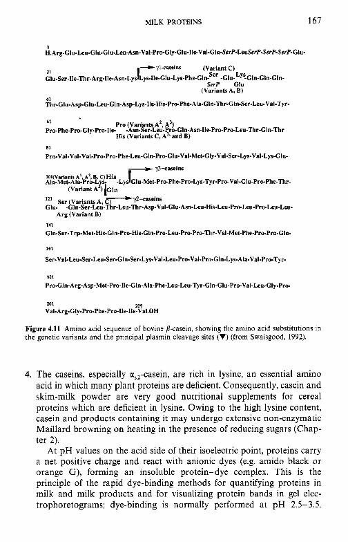
MILK PROTEINS 167
1 H.Arg-Clu-Leu-Glu-Glu-Leu-Asn.Val-Pro-Gly.Glu-1le-Val-Glu~SerP.LeuSerP-SerP-SerP-Glu-
SerP Glu (Variants A, B)
21 Clu-Ser-Ile-Thr.Arg-Ile- Asn-Lys
41 Thr-Clu-Asp-Glu-Leu-Gln-Asp-Lys~Ile-His-Pro-Phe-Ala-Gln-Thr-Gln-Ser-Leu-Val-Tyr~
Pro (Variants A’ A’) His (Variants C, A” and B)
61
Pro-Phe-Pro-Gly.Pro-Ile- -Asn-Ser-Le~.Pro-Gln-Asn-lle-Pro-Pro-Leu-Thr-Gln-Thr
81
Pro-Val-Val-Val-Pro-Pro~Phe-Leu-Gln-Pro-Glu-Val-Met-Gly.Val-Ser-Lys-Val-Lys-Glu-
121 Ser Variants A c’@~~~~~ Glu- .~ln-Ser-Le~-~r-Leu.Thr-Asp-Val-Glu-Asn-Leu.His-Leu-Pro-Leu.Pr~Leu-Le~-
Arg (Variant B) 1 I1 Cln-Ser-Trp-Met-His-Cln-Pro-His-GIn-Pro-Leu-Pro-Pro-~r-Val-Met-Phe-Pro-Pro-Gln.
161
Ser-Val-Leu-Ser~Leu-Ser-Gln-Ser-Lys-Val-Leu-Pro~Val-Pro-Gln~Lys-Ala-Val~Pro-Tyr-
181
Pro-Cln-Arg-Asp-Met-Pro-Ile-GIn-Ala-Phe-Leu-Leu-Tyr-CIn-GIu-Pro-Val-Leu-Gly-Pro-
209 201 Val-Arg-Cly-Pro-Phe-Pro-Ile-Ile-Val.OH
Figure 4.11 Amino acid sequence of bovine /?-casein, showing the amino acid substitutions in the genetic variants and the principal plasmin cleavage sites (V) (from Swaisgood, 1992).
4. The caseins, especially us,-casein, are rich in lysine, an essential amino acid in which many plant proteins are deficient. Consequently, casein and skim-milk powder are very good nutritional supplements for cereal proteins which are deficient in lysine. Owing to the high lysine content, casein and products containing it may undergo extensive non-enzymatic Maillard browning on heating in the presence of reducing sugars (Chap- ter 2).
At pH values on the acid side of their isoelectric point, proteins carry a net positive charge and react with anionic dyes (e.g. amido black or orange G), forming an insoluble protein-dye complex. This is the principle of the rapid dye-binding methods for quantifying proteins in milk and milk products and for visualizing protein bands in gel elec- trophoretograms; dye-binding is normally performed at pH 2.5-3.5.
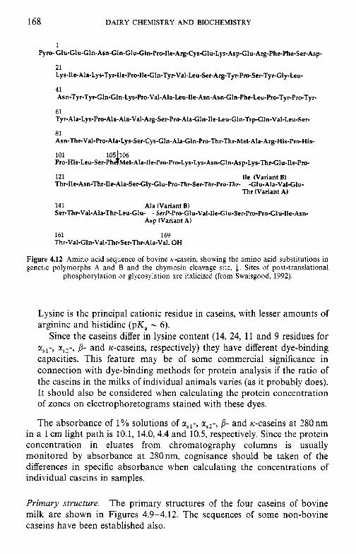
168 DAIRY CHEMISTRY AND BIOCHEMISTRY
I
Pyro- Glu-GluGln-Asn-Gln-GluG1n-Pro-Ile-Arg-Cys.G1u-Lys-AspGlu-Arg-Phe-Phe-Ser-Asp-
LI Lys-Ile-Ala-Lys-Tyr-Ile-Pro-lle-Gln-Tyr-Val-Leu-Ser-Arg-Tyr-Pro-Ser-Tyr-Gly-Leu-
41 Asn-Tyr-Tyr-GIn-Gln-Lys-Pro-Val-Ala-Leu-Ile-Asn-Asn-G1n-Phe-Leu-Pro-Tyr-Pro-Tyr-
61 Tyr-Ala-Lys-Pro-Ala-Ala-Val-Arg-Ser-Pr~Ala~ln-Ile-Leu-Gln-Trp-Gln-VaI-Leu-Ser-
81 Asn-Thr-Val-Pro-Ala-Lys-Ser-Cys-Gln-Ala-Gln-Pro-Thr-Thr-Met-Ala-Arg-His-Pro-His-
101 105)06 Pro-His-Leu-Ser-Ph Met-Ala-Ile-Pro-Pro-Lys-Lys-Asn-Gln-Asp-Lys-Thr-GIu-Ile-Pro-
121 Ile (Variant B) Thr-lle-Asn-Thr-Ile-Ala-Ser-Gly-Clu-Pro-~rSer-~~r-Pro-~~- -Glu-Ala-Val-Glu-
Thr (Variant A)
141 Ala (Variant B) Ser-Thr-Val-Ala-Thr-LeuGlu- - SerP-Pro-Glu-Val-Ile-Glu-Ser-Pro-ProGlu-IIe-Asn-
Asp (Variant A)
161 169 Thr-Val-Gln-Val-Thr-Ser-Thr-Ala-Val. OH
Figure 4.12 Amino acid sequence of bovine K-casein, showing the amino acid substitutions in genetic polymorphs A and B and the chymosin cleavage site, 1. Sites of post-translational
phosphorylation or glycosylation are italicized (from Swaisgood, 1992).
Lysine is the principal cationic residue in caseins, with lesser amounts of arginine and histidine (pK, - 6).
Since the caseins differ in lysine content (14, 24, 11 and 9 residues for xs1-, xS2- , j- and rc-caseins, respectively) they have different dye-binding capacities. This feature may be of some commercial significance in connection with dye-binding methods for protein analysis if the ratio of the caseins in the milks of individual animals varies (as it probably does). It should also be considered when calculating the protein concentration of zones on electrophoretograms stained with these dyes.
The absorbance of 1 YO solutions of rsl-, rS2-, j- and k--caseins at 280 nm in a 1 cm light path is 10.1, 14.0, 4.4 and 10.5, respectively. Since the protein concentration in eluates from chromatography columns is usually monitored by absorbance at 280 nm, cognisance should be taken of the differences in specific absorbance when calculating the concentrations of individual caseins in samples.
Primary structure. The primary structures of the four caseins of bovine milk are shown in Figures 4.9-4.12. The sequences of some non-bovine caseins have been established also.
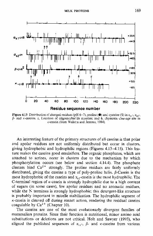
ffs YcnB
2 8-cnA
I I I I I I I I I I I
MILK PROTEINS 169
+155
-185 K-cnB
An interesting feature of the primary structures of all caseins is that polar and apolar residues are not uniformly distributed but occur in clusters, giving hydrophobic and hydrophilic regions (Figures 4.1 3-4.15). This fea- ture makes the caseins good emulsifiers. The organic phosphates, which are attached to serines, occur in clusters due to the mechanism by which phosphorylation occurs (see below and section 4.14.4). The phosphate clusters bind Ca2 + strongly. The proline residues are fairly uniformly distributed, giving the caseins a type of poly-proline helix. 0-Casein is the most hydrophobic of the caseins and a,,-casein is the most hydrophilic. The C-terminal region of K-casein is strongly hydrophilic due to a high content of sugars (in some cases), few apolar residues and no aromatic residues, while the N terminus is strongly hydrophobic; this detergent-like structure is probably important in micelle stabilization. The hydrophilic segment of K--casein is cleaved off during rennet action, rendering the residual caseins coagulable by Ca2+ (Chapter 10).
The caseins are one of the most evolutionarly divergent families of mammalian proteins. Since their function is nutritional, minor amino acid substitutions or deletions are not critical. Holt and Sawyer (1993), who aligned the published sequences of ciSl-, 0- and K--caseins from various
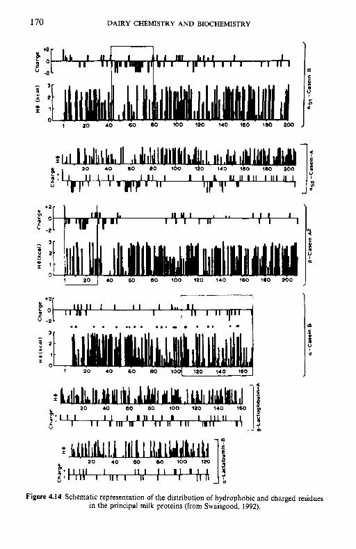
170 DAIRY CHEMISTRY AND BIOCHEMISTRY
- 3 - " 9 2
: 1
0 ~ . I ~
1 . i0 40 60 80 100 120 140 rb0 . 100 2 0 0
I I
Y P. 1 1 1
u
I. I1 Y I 1 .. 1 1 1 I 1 1 1 I l l I 1
- E 3 0 5
- - Y 2 2
z 1 m
0 1 20 I 40 60 80 100 120 140 160 180 200
7 ___- t2
U en
u -2
: o II I I I I 1 L a , I I I I 11 I II l~'" ~
.A . . . ... . ... .. . . .. . .)
1 20 40 60 80 loo( 120 140 1601
I e
Y
Figure 4.14 Schematic representation of the distribution of hydrophobic and charged residues in the principal milk proteins (from Swaisgood, 1992).
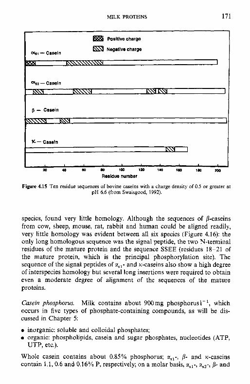
MILK PROTEINS 171
I Positive charge
%I - Casein I Negative charge
c- Casein L\Y 1
1 I I
(10 a, 100 120 140 1 0 110 200 P 40
Residue number
Figure 4.15 Ten residue sequences of bovine caseins with a charge density of 0.5 or greater at pH 6.6 (from Swaisgood, 1992).
species, found very little homology. Although the sequences of /3-caseins from cow, sheep, mouse, rat, rabbit and human could be aligned readily, very little homology was evident between all six species (Figure 4.16): the only long homologous sequence was the signal peptide, the two N-terminal residues of the mature protein and the sequence SSEE (residues 18-21 of the mature protein, which is the principal phosphorylation site). The sequence of the signal peptides of asl- and tc-caseins also show a high degree of interspecies homology but several long insertions were required to obtain even a moderate degree of alignment of the sequences of the mature proteins.
Casein phosphorus. Milk contains about 900mg phosphorus l- l , which occurs in five types of phosphate-containing compounds, as will be dis- cussed in Chapter 5:
0 inorganic: soluble and colloidal phosphates; 0 organic: phospholipids, casein and sugar phosphates, nucleotides (ATP,
Whole casein contains about 0.85% phosphorus; aSl-, /3- and Ic-caseins contain 1.1,0.6 and 0.16% P, respectively; on a molar basis, asl-, tls2-, /I- and
UTP, etc.).
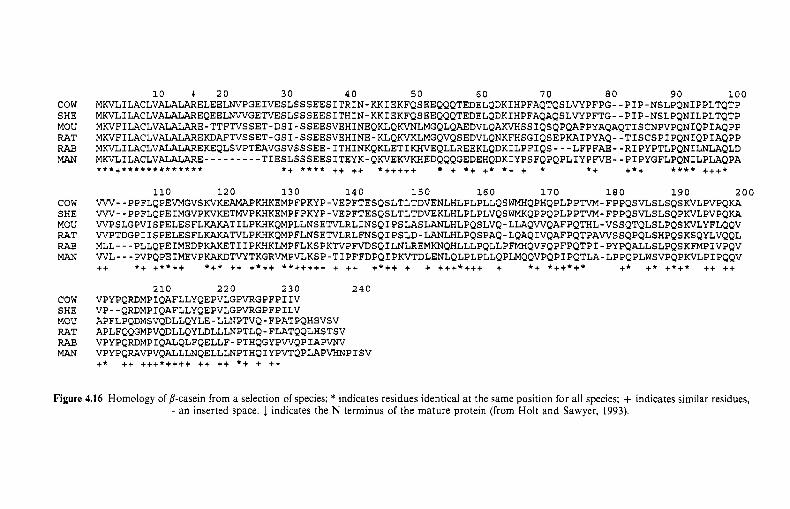
cow SHE MOW RAT RAE MAN
cow SHE MOW RAT RAE MAN
cow SHE MOW RAT RAE MAN
10 .1 2 0 30 40 5 0 60 70 80 90 100 MKVLILACLVALALARELEELNVPGEIVESLSSSEESITRIN-KKIEKFQSEEQQQTEDELQDKIHPFAQTQSLVYPFPG--PIP-NSLPQNIPPLTQTP MKVLILACLVALALAREQEELNWGETVESLSSSEESLSSSEESITHIN-KKIEKFQSEEQQQTEDELQDKIHPFAQAQSL~PFTG--PIP-NSLPQNILPLTQTP MKVFILACLVALALARE-TTFTVSSET-DSI-SSEESVEHINEQKLQKVNLMGQLQAEDVLQAKVHSSIQSQPQAFPYAQAQTISCNPVPQNIQPIAQPP MKVFILACLVALALAREKDAFTVSSET-GSI~SSEESVEHINE~KLQKVKLMGQVQSEDVLQNKFHSGIQSEP~IPYAQ--TISCSPIPQNIQPIAQPP MKVLILACLVALALAREKEQLSVPTEAVGSVSSSEE~ITHINKQKLETIKHVEQLLREEKLQDKILPFIQS---LFPFAE--RIPYPTLPQNILNLAQLD MKVLILACLVALALARE~~~~~~~~~TIESLSSSEESITEYK~QKEKVKHEDQQQGEDQQQGEDEHQDKIYPSFQPQPLIYP~E--PIPYGFLPQNILPLAQPA * * *+*** * * * * * * * * * * *+ * * * * ++ ++ *+++++ + *+ +* *+ + * *+ +*+ * * * * +++*
110 120 130 140 150 160 170 180 190 200 VW--PPFLQPEVMGVSKVKEAMAPKHKEMPFPKYP-VEPFTESQSLTLTDVENLHLPLPLLQSWQPHQPLPPTVM-FPPQSVLSLSQSKVLPVPQKA VW--PPFLQPEIMGVPKVKETMVPKHKEMPFPKYP-VEPFPKYP-VEPFTESQSLTLTDVEKLHLPLPLVQS~KQPPQPLPPTVM-FPPQSVLSLSQPKVLPVPQKA WPSLGPVISPELESFLKAKATILPKHKQMPLLNSETVLRLINSQIPSLASLANLHLPQSLVQ-LLAQWQAFPQTHL-VSSQTQLSLPQSKVLYFLQQV WPTDGPIISPELESFLKLP~KQMPFLNSETVLSQIPSLD-LANLHLPQSPAQ-LQAQIVQAFPQTPAWSSQPQLSHPQSKSQYLVQQL MLL---PLLQPEIMEDPKAKETIIPKHKLMPFLKSPK?lrPFVDSQILNLREMKNQHLLLPQLLPFQVFQPFPQTPI-PYPQALLSLPQSKFMPIVPQV WL---PVPQPEIMEVPKAKDTVYTKGRVMPVLKSP-TIPFFDPQIPKVTDLENLQLPLPLLQPLMQQVPQPIPQTLA-LP?QPLWSVPQPKVLPIPQQV ++ * + +**++ *+* ++ +*++ **+++++ + ++ +*++ + + +++*+++ + *+ *++*+* +* +* +*+* ++ ++
210 220 230 240 VPYPQRDMPIQAFLLYQEPVLGPVRGPFPIIV VP--QRDMPIQAFLLYQEPVLGPVRGPFPILV APFLPQDMSVQDLLQYLE-LLNPTVQ-FPATPQHSVSV APLFQQGMPVQDLLQYLDLLLNPTLQ-FLATQQLHSTSV VPYPQRDMPIQALQLFQELLF-PTHQGYPWQPIAPVNV VPYPQRAVPVQALLLNQELLLN?THQIYPVTQPL?.P”IPISV + * ++ +++*++++ ++ ++ *+ + ++
Figure 4.16 Homology of /3-casein from a selection of species; * indicates residues identical at the same position for all species; + indicates similar residues, - an inserted space. 1. indicates the N terminus of the mature protein (from Holt and Sawyer, 1993).
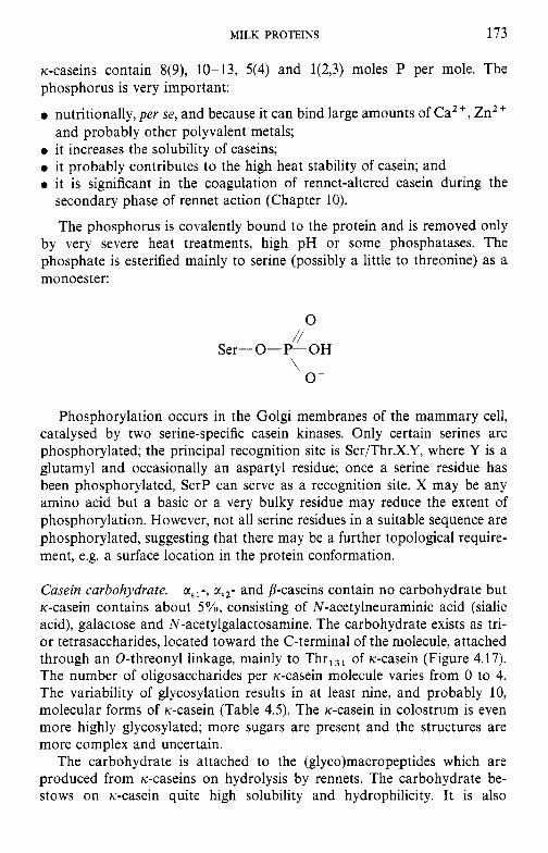
MILK PROTEINS 173
Ic-caseins contain 8(9), 10-13, 5(4) and 1(2,3) moles P per mole. The phosphorus is very important:
0 nutritionally, per se, and because it can bind large amounts of Ca2+, ZnZ+
0 it increases the solubility of caseins; 0 it probably contributes to the high heat stability of casein; and 0 it is significant in the coagulation of rennet-altered casein during the
secondary phase of rennet action (Chapter 10).
The phosphorus is covalently bound to the protein and is removed only by very severe heat treatments, high pH or some phosphatases. The phosphate is esterified mainly to serine (possibly a little to threonine) as a monoester:
and probably other polyvalent metals;
0 //
Ser-0-P-OH
0- \
Phosphorylation occurs in the Golgi membranes of the mammary cell, catalysed by two serine-specific casein kinases. Only certain serines are phosphorylated; the principal recognition site is Ser/Thr.X.Y, where Y is a glutamyl and occasionally an aspartyl residue; once a serine residue has been phosphorylated, SerP can serve as a recognition site. X may be any amino acid but a basic or a very bulky residue may reduce the extent of phosphorylation. However, not all serine residues in a suitable sequence are phosphorylated, suggesting that there may be a further topological require- ment, e.g. a surface location in the protein conformation.
Casein carbohydrate. asl-, aS2- and b-caseins contain no carbohydrate but K-casein contains about 5%, consisting of N-acetylneuraminic acid (sialic acid), galactose and N-acetylgalactosamine. The carbohydrate exists as tri- or tetrasaccharides, located toward the C-terminal of the molecule, attached through an 0-threonyl linkage, mainly to Thr,,, of K-casein (Figure 4.17). The number of oligosaccharides per K-casein molecule varies from 0 to 4. The variability of glycosylation results in at least nine, and probably 10, molecular forms of K-casein (Table 4.5). The K-casein in colostrum is even more highly glycosylated; more sugars are present and the structures are more complex and uncertain.
The carbohydrate is attached to the (g1yco)macropeptides which are produced from K-caseins on hydrolysis by rennets. The carbohydrate be- stows on x-casein quite high solubility and hydrophilicity. It is also
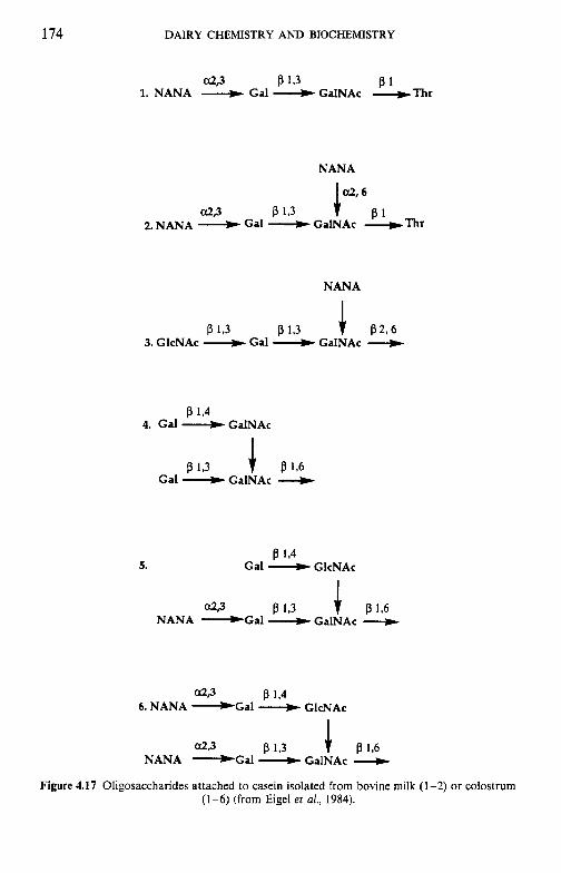
174 DAIRY CHEMISTRY A N D BIOCHEMISTRY
a29 P 1 3 81 1. NANA d Gal d GalNAc +Thr
NANA
P 1 3 P 1 3 + P296 3. GlcNAc d Gal GalNAc --.)
5. P 1,4
Gal __t GlcNAc
a23 P 1.3 + P 1.6 NANA -Gal d GalNAc
a.29 P 1.4 6. NANA -Gal __t GlcNAc
a29 P 13 + P 196 NANA --Gal GalNAc
Figure 4.17 Oligosaccharides attached to casein isolated from bovine milk (1-2) or colostrum (1-6) (from Eigel et a/., 1984).
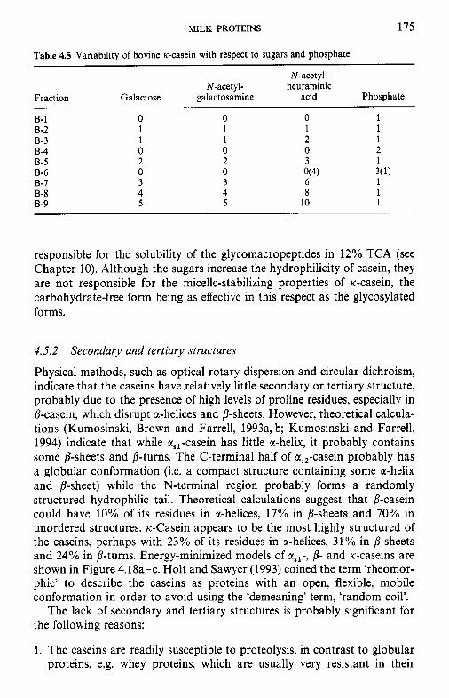
MILK PROTEINS 175
Table 4.5 Variability of bovine K-casein with respect to sugars and phosphate
N-acetyl- N-acetyl- neuraminic
Fraction Galactose galactosamine acid Phosphate
B- 1 0 0 0 1 B-2 1 1 1 1 B-3 1 1 2 1 B-4 0 0 0 2 B-5 2 2 3 1
1 B-6 0 0 O(4) B-7 3 3 6 B-8 4 4 8 1 B-9 5 5 10 1
3(1)
responsible for the solubility of the glycomacropeptides in 12% TCA (see Chapter 10). Although the sugars increase the hydrophilicity of casein, they are not responsible for the micelle-stabilizing properties of Ic-casein, the carbohydrate-free form being as effective in this respect as the glycosylated forms.
4.5.2 Secondary and tertiary structures
Physical methods, such as optical rotary dispersion and circular dichroism, indicate that the caseins have relatively little secondary or tertiary structure, probably due to the presence of high levels of proline residues, especially in p-casein, which disrupt r-helices and P-sheets. However, theoretical calcula- tions (Kumosinski, Brown and Farrell, 1993a, b; Kumosinski and Farrell, 1994) indicate that while a,,-casein has little or-helix, it probably contains some P-sheets and p-turns. The C-terminal half of a,,-casein probably has a globular conformation (i.e. a compact structure containing some or-helix and P-sheet) while the N-terminal region probably forms a randomly structured hydrophilic tail. Theoretical calculations suggest that p-casein could have 10% of its residues in a-helices, 17% in /?-sheets and 70% in unordered structures. Ic-Casein appears to be the most highly structured of the caseins, perhaps with 23% of its residues in or-helices, 31% in /3-sheets and 24% in p-turns. Energy-minimized models of asl-, p- and Ic-caseins are shown in Figure 4.18a-c. Holt and Sawyer (1993) coined the term ‘rheomor- phic’ to describe the caseins as proteins with an open, flexible, mobile conformation in order to avoid using the ‘demeaning’ term, ‘random coil’.
The lack of secondary and tertiary structures is probably significant for the following reasons:
1. The caseins are readily susceptible to proteolysis, in contrast to globular proteins, e.g. whey proteins, which are usually very resistant in their
176 DAIRY CHEMISTRY AND BIOCHEMISTRY
native state. This has obvious advantages for the digestibility of the caseins, the natural function of which is presumably nutritional and hence easy digestibility in the ‘native’ state is important. The caseins are also readily hydrolysed in cheese, which is important for the development of cheese flavour and texture (Chapter lo). However, casein hydrolysates may be bitter due to a high content of hydrophobic amino acids (small hydrophobic peptides tend to be bitter). The caseins are readily hy- drolysed by proteinases secreted by spoilage micro-organisms. The caseins adsorb readily at air-water and oil-water interfaces due to their open structure, relatively high content of apolar amino acid residues and the uneven distribution of amino acids. This gives the caseins very good emulsifying and foaming properties, which are widely exploited in the food industry. The lack of higher structures probably explains the high stability of the caseins to denaturing agents, including heat.
#
(a)
Figure 4.18 Energy-minimized models of the tertiary structures of bovine asi- (a), p- (b) and K- (c) caseins (from Kumosinski, Brown and Farrell, 1993a, b; Kumosinski and Farrell,
1994)
Figure 4.18 (Continued).
178 DAIRY CHEMISTRY AND BIOCHEMISTRY
Figure 4.18 (Continued).
4.5.3 Molecular size
All the caseins are relatively small molecules, ranging in molecular weight from about 20 to 25 kDa (Table 4.2).
4.5.4 Hydrophobicity
The caseins are often considered to be rather hydrophobic molecules. However, consideration of the amino acid composition indicates that they are not particularly so; in fact, some are more hydrophilic than the whey protein, P-lactoglobulin (Table 4.2). However, the caseins do have high
MILK PROTEINS 179
surface hydrophobicity, in contrast to the globular whey proteins. In globular proteins, the hydrophobic residues are buried, as far as possible, within the molecule, with most of the hydrophilic residues exposed on the surface. Owing to the relative lack of secondary and tertiary structures in the caseins, such an arrangement is not possible, and hence the hydrophobic residues are rather exposed.
Thus, the caseins are relatively small, relatively hydrophobic, am- phipathic, randomly or flexibly structured molecules, with relatively low levels of secondary and tertiary structures.
4.5.5 Influence of Caz + on caseins
At all temperatures, a,,-CN B and C are insoluble in calcium-containing solutions and form a coarse precipitate at CaZ + concentrations greater than about 4mM. as,-CN A, from which the very hydrophobic sequence, residues 13-26, is deleted, is soluble at [Ca"] up to 0.4 M in the temperature range 1-33°C. Above 33"C, it precipitates but redissolves on cooling to 28°C. The presence of a,,-CN A modifies the behaviour of a,,-CN B so that an equimolar mixture of the two is soluble in 0.4M Caz+ at 1°C; a,,-CN B precipitates from the mixture at 18°C and both r,,-CN A and B precipitate at 33°C. a,,-CN A does not form normal micelles with rc-casein. Since a,,-CN A occurs at very low frequency, these abnormalities are of little consequence in dairy processing but may become important if the frequency of cr,,-CN A increases as a result of breeding practices.
The a,,-caseins are also insoluble in Caz+ (above about 4mM) at all temperatures, but their behaviour has not been studied in detail.
p-Casein is soluble at high concentrations of Ca2+ (0.4M) at tempera- tures below 18"C, but above 18°C /?-casein is very insoluble, even in the presence of low concentrations of CaZ + (4 mM). Ca-precipitated p-casein redissolves readily on cooling to below 18°C. About 20°C is also the critical temperature for the temperature-dependent polymerization of p-casein and the two phenomena may be related.
ic-Casein is soluble in Caz+ at all concentrations up to those at which general salting-out occurs. Solubility is independent of temperature and pH (outside the pH range at which isoelectric precipitation occurs). Not only is rc-casein soluble in the presence of Ca2+ but it is capable of stabilizing xsl-, aSz- and p-caseins against precipitation by CaZ + (section 4.5.8).
4.5.6 Action of rennets on casein
This subject is dealt with in Chapter 10. Suffice it to say here that rc-casein is the only major casein hydrolysed by rennets during the primary phase of milk coagulation, which is the first step in the manufacture of most cheese varieties.
180 DAIRY CHEMISTRY A N D BIOCHEMISTRY
4.5.7 Casein association
All the major caseins associate with themselves and with each other. In unreduced form, K-casein is present largely as disulphide-linked polymers. K-Casein also forms hydrogen and hydrophobic bonds with itself and other caseins but these secondary associations have not been studied in detail.
At 4"C, /?-casein exists in solution as monomers of molecular mass 25 kDa. As the temperature is increased, the monomers polymerize to form long thread-like chains of about 20 units at 8.5"C and to still larger aggregates at higher temperatures. The degree of association is dependent on protein concentration. The ability to form thread-like polymers may be important in micelle structure. p-Casein also undergoes a temperature- dependent conformational change in which the content of poly-L-proline helix decreases with increasing temperature. The transition temperature is about 20°C, i.e. very close to the temperature at which 8-casein becomes insoluble in CaZ+.
x,,-Casein polymerizes to form tetramers of molecular mass 113 kDa; the degree of polymerization increases with increasing protein concentration and increasing temperature.
The major caseins interact with each other and, in the presence of Ca", these associations lead to the formation of casein micelles.
4.5.8 Casein micelle structure
Composition and general features. About 95% of the casein exists in milk as large colloidal particles, known as micelles. On a dry matter basis, casein micelles contain c. 94% protein and 6% low molecular weight species referred to as colloidal calcium phosphate, consisting of calcium, mag- nesium, phosphate and citrate. The micelles are highly hydrated, binding about 2.0g H,Og-' protein. Some of the principal properties of casein micelles are summarized in Table 4.6.
Electron microscopy shows that casein micelles are generally spherical in shape, with diameters ranging from 50 to 500nm (average c. 120nm) and masses ranging from lo6 to lo9 Da (average about lo8 Da). There are very many small micelles but these represent only a small proportion of the volume or mass (Figure 4.19). There are 10'4-10'6micellesml-1 milk; they are roughly two micelle diameters (240nm) apart, i.e. they are quite tightly packed. The surface (interfacial) area of the micelles is very large, 5 x lo4 cm2 ml- '; hence, the surface properties of the micelles are critical to their behaviour.
Since the micelles are of colloidal dimensions, they are capable of scattering light and the white colour of milk is due largely to light scatter- ing by the casein micelles; the white colour is lost if the micelles are disrupted, e.g. by removing colloidal calcium phosphate (by citrate, ethylene
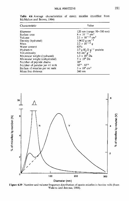
MILK PROTEINS 181
Table 4.6 Average characteristics of casein micelles (modified from McMahon and Brown, 1984)
Characteristic Value
Diameter Surface area Volume Density (hydrated) Mass Water content Hydration Voluminosity Molecular weight (hydrated) Molecular weight (dehydrated) Number of peptide chains Number of paticles per ml milk Surface of micelles per ml milk Mean free distance
36
- A 7-l-
120 nm (range: 50-500 nm) 8 x lo-'' cm2 2.1 x cm3 1.0632 g cm-3 2.2 x 10⁻¹⁵ g 63 Yo 3.7 g H,O g-' protein 4.4 cm3 g- ' 1.3 x lo9 Da 5 x 10' Da 104 1 0 ~ ~ - 1 0 ~ 6 5 x lo4 cm3 240 nm
100 200
Diameter (nm) 300
Figure 4.19 Number and volume frequency distribution of casein micelles in bovine milk (from Walstra and Jenness, 1984).
182 DAIRY CHEMISTRY AND BIOCHEMISTRY
diaminetetraacetic acid (EDTA) or oxalate), by increasing pH (to greater than 9), or by the addition of urea or SDS.
Stability
1. The micelles are stable to the principal processes to which milk is normally subjected (except those in which it is intended to destabilize the micelles, e.g. rennet- and acid-induced coagulation). They are very stable at high temperatures, coagulating only after heating at 140°C for 15- 20min at the normal pH of milk. Such coagulation is not due to denaturation in the narrow sense of the word but to major changes which occur in milk exposed to such high heat treatments, including a decrease in pH due to the pyrolysis of lactose to various acids, dephosphorylation of the casein, cleavage of u-casein, denaturation of the whey proteins and their attachment to the casein micelles, precipitation of soluble calcium phosphate on the micelles and a decrease in hydration (Chapter 9).
2. They are stable to compaction, e.g. they can be sedimented by ultracen- trifugation and redispersed readily by mild agitation.
3. They are stable to commercial homogenization but are changed slightly at very high pressures (500 MPa).
4. They are stable to high [Ca2+], up to at least 200mM at temperatures up to 50°C.
5. They aggregate and precipitate from solution when the pH is adjusted to the isoelectric point of caseins (c. pH4.6). Precipitation at this pH, which is temperature-dependent (i.e. does not occur at temperatures below 58°C and occurs over a wide pH range, perhaps 3.0-5.5, at higher temperatures, e.g. 70"C), occurs owing to the loss of net positive or negative charge as the pH approaches 4.6.
6. As the pH of milk is reduced, the colloidal calcium phosphate (CCP) dissolves and is completely soluble at pH 4.9 (Chapter 5). pH adjustment, followed by dialysis against bulk milk, is a convenient and widely used technique for varying the CCP content of milk. As the concentration of CCP is reduced, the properties of the micelles are altered but they retain some of their structure even after removing 70% of the CCP. Removal of more than 70% of the CCP results in disintegration of the micelles into smaller particles (aggregates).
7. Many proteinases catalyse the hydrolysis of a specific bond in K-casein, as a consequence of which the micelles aggregate or gel in the presence of Ca2+ or other divalent ions. This is the key step in the manufacture of most cheese varieties (Chapter 10).
8. The micelles are destabilized by c. 40% ethanol at pH 6.7 and by lower concentrations if the pH is reduced.
9. They are destabilized by freezing (cryodestabilization) due to a decrease in pH and an increase in the [CaZ+] in the unfrozen phase of milk (Chapters 2 and 5).
MILK PROTEINS 183
Principal micelle characteristics. The structure of the casein micelles has attracted the attention of scientists for a considerable time. Knowledge of micelle structure is important because the stability and behaviour of the micelles are central to many dairy processing operations, e.g. cheese manu- facture, stability of sterilized, sweetened-condensed and reconstituted milks and frozen products. Without knowledge of the structure and properties of the casein micelle, attempts to solve many technological problems faced by the dairy industry will be empirical and not generally applicable. From the academic viewpoint, the casein micelle presents an interesting and complex problem in protein quaternary structure.
Since the pioneering work of Waugh in 1958, a considerable amount of research effort has been devoted to elucidating the structure of the casein micelle, and several models have been proposed. This work has been reviewed in the references cited in the next section. The principal properties of the casein micelles are listed below and the models which best meet these requirements discussed briefly in the next section.
1. K-Casein, which represents about 15% of total casein, is a critical feature of micelle structure and stability and must be located so as to be able to stabilize the calcium-sensitive asl-, as2- and p-caseins, which represent about 85% of total casein.
2. The rc-casein content of casein micelles is inversely proportional to their
3,
4.
5.
6.
7.
size, while the content of colloidal calcium phosphate 1s directly related to size. Ultracentrifugally sedimented micelles have a hydration of 1.6-2.7 g H,Og-' protein but voluminosities of 3-7mlg-' have been found by viscosity measurements and calculation of specific hydrodynamic vol- umes. These values suggest that the micelle has a porous structure in which the protein occupies about 25% of the total volume. Chymosin and similar proteinases, which are relatively large molecules (c. 36 kDa), very rapidly and specifically hydrolyse most of the micellar rc-casein. When heated in the presence of whey proteins, as in normal milk, rc-casein and P-lactoglobulin interact to form a disulphide-linked com- plex which modifies many properties of the micelles, including rennet coagulability and heat stability. Removal of colloidal calcium phosphate (CCP) results in disintegration of the micelles into particles of mass - 3 x lo6 Da. The properties of the CCP-free system are very different from those of the normal milk system, e.g. it is sensitive to and precipitated by relatively low concen- trations of Ca2+, it is more stable to high temperatures, e.g. 140°C, and is not coagulable by rennets. Many of these properties can be restored, at least partially, by increased concentrations of calcium. The micelles can be dispersed (dissociated) by urea or SDS, suggesting the involvement of hydrogen and hydrophobic bonds in micelle integrity.
184 DAIRY CHEMISTRY AND BIOCHEMISTRY
8.
9.
10.
11.
The micelles can be destabilized by alcohols, acetone and similar solvents, suggesting an important role for electrostatic interactions in micelle structure. As the temperature is lowered, caseins, especially /?-casein, dissociate from the micelles; depending on the method of measurement, 10-50% of /?-casein is non-micellar at 4°C. Electron microscopy shows that the interior of the micelles are not uniformly electron dense. The micelles have a surface (zeta) potential of about - 20 mV at pH 6.7.
Micelle structure. Various models of casein micelle structure have been proposed and refined over the past 40years. Progress has been reviewed regularly, including Schmidt (1982), McMahon and Brown (1984), Farrell (1988), Holt (1992, 1994), Rollema (1992) and Visser (1992).
The proposed models fall into three general categories, although there is some overlap:
1. core-coat; 2. internal structure; 3. subunit (submicelles); in many of the models in this category, it is
proposed that the submicelles have a core-coat structure.
For many years there has been strong support for the view that the micelles are composed of submicelles of mass -106Da and diameter 10-15nm. This model was introduced in 1967 by Morr who proposed that the submicelles are linked together by CCP, giving the micelle an open porous structure. On removal of CCP, e.g. by acidification/dialysis, EDTA, citrate or oxalate, the micelles disintegrate. Disintegration may also be achieved by treatment with urea, SDS or at pH greater than 9; presumably,
Submicelle
F'rF'r;uding 0 - Calcium
phosphate
Figure 4.20 Submicelle model of the casein micelle (from Walstra and Jenness, 1984).
MILK PROTEINS 185
these treatments do not solubilize CCP, suggesting that other forces, e.g. hydrophobic and hydrogen bonds, contribute to micelle structure.
The submicellar model has undergone several refinements (see Schmidt, 1982; Walstra and Jenness, 1984; Ono and Obata, 1989). The current view is that the K-casein content of the submicelles varies and that the K--casein- deficient submicelles are located in the interior of the micelles with the K-casein-rich submicelles concentrated at the surface, giving the micelles a K--casein-rich layer but with some xsl-, xs2- and b-caseins also exposed on the surface. It is proposed that the hydrophilic C-terminal region of K-casein protrudes from the surface, forming a layer 5-10nm thick and giving the micelles a hairy appearance (Figure 4.20). This hairy layer is responsible for micelle stability through a major contribution to zeta potential ( - 20mV) and steric stabilization. If the hairy layer is removed, e.g. specific hydrolysis of x-casein, or collapsed, e.g. by ethanol, the colloidal stability of the micelles is destroyed and they coagulate or precipitate.
Although the submicellar model of the casein micelle readily explains many of the principal features and physicochemical reactions undergone by the micelles and has been widely supported, it has never enjoyed unanimous support and two alternative models have been proposed recently. Visser (1992) proposed that the micelles are spherical conglomerates of individual casein molecules randomly aggregated and held together partly by salt bridges in the form of amorphous calcium phosphate and partly by other forces, e.g. hydrophobic bonds, with a surface layer of K-casein. Holt (1992, 1994) depicted the casein micelle as a tangled web of flexible casein
Figure 4.21 Model of the casein micelle (modified from Holt, 1994).
186 DAIRY CHEMISTRY AND BIOCHEMISTRY
molecules forming a gel-like structure in which microgranules of colloidal calcium phosphate are an integral feature and from the surface of which the C-terminal region of K-casein extends, forming a hairy layer (Figure 4.21). These models retain two of the central features of the submicellar model, i.e. the cementing role of CCP and the predominantly surface location of K-casein.
Holt (1992, 1994) also proposed that, in addition to supplying amino acids, caseins should be considered to have a biological function, i.e. to enable a high concentration of calcium to be carried in stable form in milk; without the stabilizing effect of casein, calcium phosphate would precipitate in the mammary cells, resulting in ectopic mineralization, which might lead to the death of the mammary gland or of the whole animal. A similar situation occurs with kidney stones, gallstones and calcified synovial and salivary fluid.
Since the micelles are closely packed, intermicellar collisions are frequent; however, the micelles do not normally remain together after collisions. The micelles are stabilized by two principal factors: (1) a surface (zeta) potential of c. -20mV at pH 6.7, which, alone, is probably too small for colloidal stability, and (2) steric stabilization due to the protruding K-casein hairs.
4.6 Whey proteins
About 20% of the total protein of bovine milk belongs to a group of proteins generally referred to as whey or serum proteins or non-casein nitrogen. Acid and rennet wheys also contain casein-derived peptides; both contain proteose-peptones, produced by plasmin, mainly from p-casein, and the latter also contains (g1yco)macropeptides produced by rennets from K-casein. These peptides are excluded from the present discussion.
4.6.1 Preparation
The whey proteins, as a group, are readily prepared from milk by any of the methods described in section 4.3, i.e.
1. the proteins remaining soluble at pH 4.6; 2. soluble in saturated NaCI; 3. soluble after rennet coagulation of the caseins; 4. by gel permeation chromatography; 5. by ultracentrifugation, with or without added Ca2+.
The whey prepared by any of the above methods, except 4, contains lactose and soluble salts. Total whey proteins may be prepared from the
MILK PROTEINS 187
wheys by dialysis and drying the retentate. The products prepared by these various methods differ: acid whey contains some y-casein and proteose- peptones; immunoglobulins are co-precipitated with the caseins by saturated NaCl; rennet whey contains the ic-CN macropeptides produced by rennet action, plus, perhaps, very small amounts of other caseins; small casein micelles remain in the ultracentrifugal supernatant, especially if Ca is not added. The salt composition of the serum differs very considerably in wheys produced by various methods.
On a commercial scale, whey protein-rich products are prepared by:
1. Ultrafiltration/diafiltration of acid or rennet whey to remove varying amounts of lactose, and spray-drying to produce whey protein concen- trates (30-80% protein).
2. Ion-exchange chromatography: proteins are adsorbed on an ion ex- changer, washed free of lactose and salts and then eluted by pH adjustment. The eluate is freed of salts by ultrafiltration and spray-dried to yield whey protein isolate, containing about 95% protein.
3. Demineralization by electrodialysis and/or ion exchange, thermal evap- oration of water and crystallization of lactose.
4. Thermal denaturation, recovery of precipitated protein by filtration/ centrifugation and spray-drying, to yield lactalbumin which has very low solubility and limited functionality.
Several other methods are available for the removal of whey proteins from whey but are not used commercially. Several methods for the purifica- tion of the major and minor whey proteins on a commercial scale have also been developed and will be discussed briefly in sections 4.15.6 and 4.16.
4.6.2 Heterogeneity of whey proteins
It was recognized 60years ago that whey prepared by any of the above methods contained two well-defined groups of proteins which could be fractionated by saturated MgSO, or half saturated (NH,),SO,; the precipi- tate (roughly 20% of total N) was referred to as lactoglobulin and the soluble protein as lactalbumin.
The lactoglobulin fraction consists mainly of immunoglobulins (Ig), especially IgG, , with lesser amounts of IgG,, IgA and IgM (section 4.10). The lactalbumin fraction of bovine milk contains three main proteins, p- lactoglobulin (p-lg), a-lactalbumin (a-la) and blood serum albumin (BSA), which represent approximately 50, 20 and 10% of total whey protein, respectively, and trace amounts of several other proteins, notably lactotrans- ferrin, serotransferrin and several enzymes. The whey proteins of sheep, goat
Next Page
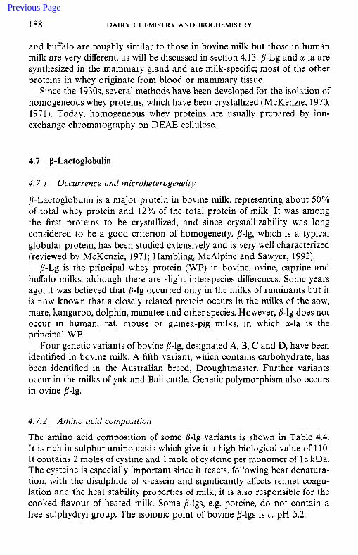
188 DAIRY CHEMISTRY A N D BIOCHEMISTRY
and buffalo are roughly similar to those in bovine milk but those in human milk are very different, as will be discussed in section 4.13. p-Lg and a-la are synthesized in the mammary gland and are milk-specific; most of the other proteins in whey originate from blood or mammary tissue.
Since the 1930s, several methods have been developed for the isolation of homogeneous whey proteins, which have been crystallized (McKenzie, 1970, 1971). Today, homogeneous whey proteins are usually prepared by ion- exchange chromatography on DEAE cellulose.
4.7 p-Lactoglobulin
4.7. I Occurrence and microheterogeneity
P-Lactoglobulin is a major protein in bovine milk, representing about 50% of total whey protein and 12% of the total protein of milk. It was among the first proteins to be crystallized, and since crystallizability was long considered to be a good criterion of homogeneity, p-lg, which is a typical globular protein, has been studied extensively and is very well characterized (reviewed by McKenzie, 1971; Hambling, McAlpine and Sawyer, 1992).
p-Lg is the principal whey protein (WP) in bovine, ovine, caprine and buffalo milks, although there are slight interspecies differences. Some years ago, it was believed that 13-lg occurred only in the milks of ruminants but it is now known that a closely related protein occurs in the milks of the sow, mare, kangaroo, dolphin, manatee and other species. However, p-lg does not occur in human, rat, mouse or guinea-pig milks, in which a-la is the principal WP.
Four genetic variants of bovine p-lg, designated A, B, C and D, have been identified in bovine milk. A fifth variant, which contains carbohydrate, has been identified in the Australian breed, Droughtmaster. Further variants occur in the milks of yak and Bali cattle. Genetic polymorphism also occurs in ovine p-lg.
4.7.2 Amino acid composition
The amino acid composition of some p-lg variants is shown in Table 4.4. It is rich in sulphur amino acids which give it a high biological value of 110. It contains 2 moles of cystine and 1 mole of cysteine per monomer of 18 kDa. The cysteine is especially important since it reacts, following heat denatura- tion, with the disulphide of Ic-casein and significantly affects rennet coagu- lation and the heat stability properties of milk; it is also responsible for the cooked flavour of heated milk. Some fl-lgs, e.g. porcine, do not contain a free sulphydryl group. The isoionic point of bovine p-lgs is c. pH 5.2.
Previous Page
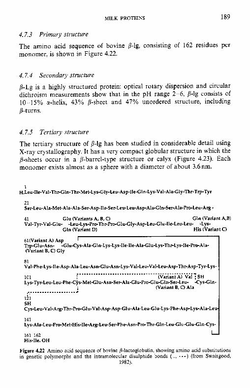
MILK PROTEINS 189
4.7.3 Primary structure
The amino acid sequence of bovine p-lg, consisting of 162 residues per monomer, is shown in Figure 4.22.
4.7.4 Secondary structure
p-Lg is a highly structured protein: optical rotary dispersion and circular dichroism measurements show that in the pH range 2-6, p-lg consists of 10- 15% a-helix, 43% P-sheet and 47% unordered structure, including p-turns.
4.7.5 Tertiary structure
The tertiary structure of p-lg has been studied in considerable detail using X-ray crystallography. It has a very compact globular structure in which the ,&sheets occur in a p-barrel-type structure or calyx (Figure 4.23). Each monomer exists almost as a sphere with a diameter of about 3.6 nm.
1 H.Leu-Ile-Val-Thr-G1n-Thr-Met-Lys-Gly-Leu-Asp-Ile-Gln-Lys-Val-Ala-Gly-Thr-Trp-Tyr
21 Ser-Leu-Ala-Met-AIa-Ala-Ser-Asp-Ile-Ser-Leu-Leu-Asp-Ala-Gln-Ser-Ala-Pro-Leu-Arg
41 Glu (Variants A, B, C) Gln (Variant A,BI Val-Tyr-Val-Glu- -Leu-Lys-Pro-Thr-Pro-Glu-GIy-Asp-Leu-Glu-Ile-Leu-Leu- -Lys-
Gln (Variant D) His (Variant C)
61Wariant A) Asp I Trp-Glu-Asn- -Glu-Cys-Ala-G1n-Lys-Lys-Ile-Ile-Ala-Glu-Lys-Thr-Lys-I1e-Pro-Ala- (Variant B, C) Gly
81 Val-Phe-Lys-Ile-Asp-Ala-Leu-Asn-Glu-Asn-Lys-Val-Leu-VaI-Leu-Asp-Thr-Asp-Tyr-Lys-
................................................... 101 Lys-Tyr-Leu-Leu-Phe-Cjrs-Met-Glu-Asn-Ser-Ala-Glu-Pro-Glu-Gln-Ser-Leu- -Cys-Gln-
(Variant A) Val ; SH
(Variant B, C) Ala ...................... 15; SH Cys-Leu-Val-Arg-Thr-Pro-GIu-Val-Asp-Asp-Glu-Ala-Leu-Glu-Lys-Phe-Asp-Lys-Ala-Leu
141 Lys-Ala-Leu-Pro-Met-His-11e-Arg-Leu-Ser-Phe-Asn-Pro-Thr-Gln-Leu-GI-Glu-Gln-Cys-
161 162 His-Ile. OH
L
Figure 4.22 Amino acid sequence of bovine B-lactoglobulin, showing amino acid substitutions in genetic polymorphs and the intramolecular disulphide bonds (-, - - - ) (from Swaisgood,
1982).
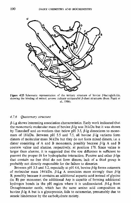
190 DAIRY CHEMISTRY AND BIOCHEMISTRY
Figure 4.23 Schematic representation of the tertiary structure of bovine /?-lactoglobulin, showing the binding of retinol; arrows indicate antiparallel 8-sheet structures (from Papiz et
al., 1986).
4.7.6 Quaternary structure
p-Lg shows interesting association characteristics. Early work indicated that the monomeric molecular mass of bovine 8-lg was 36 kDa but it was shown by Timasheff and co-workers that below pH 3.5, p-lg dissociates to mono- mers of 18kDa. Between pH 5.5 and 7.5, all bovine p-lg variants form dimers of molecular mass 36 kDa but they do not form mixed dimers, i.e. a dimer consisting of A and B monomers, possibly because p-lg A and B contain valine and alanine, respectively, at position 178. Since valine is larger than alanine, it is suggested that the size difference is sufficient to prevent the proper fit for hydrophobic interaction. Porcine and other p-lgs that contain no free thiol do not form dimers; lack of a thiol group is probably not directly responsible for the failure to dimerize.
Between pH 3.5 and 5.2, especially at pH 4.6, bovine p-lg forms octamers of molecular mass 144kDa. p-Lg A associates more strongly than p-lg B, possibly because it contains an additional aspartic acid instead of glycine (in B) per monomer; the additional Asp is capable of forming additional hydrogen bonds in the pH region where it is undissociated. p-Lg from Droughtmaster cattle, which has the same amino acid composition as bovine p-lg A but is a glycoprotein, fails to octamerize, presumably due to stearic hinderance by the carbohydrate moiety.
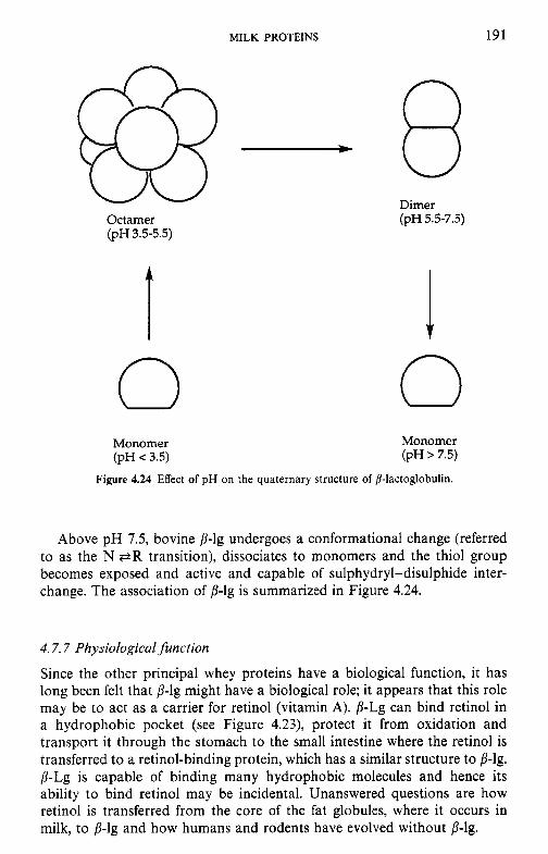
MILK PROTEINS 191
Octamer (pH3.5-5.5)
0 Monomer (pH < 3.5)
Dimer (PH 5.5-7.5)
I 0 Monomer (pH > 7.5)
Figure 4.24 Effect of pH on the quaternary structure of 8-lactoglobulin.
Above pH 7.5, bovine 13-lg undergoes a conformational change (referred to as the N P R transition), dissociates to monomers and the thiol group becomes exposed and active and capable of sulphydryl-disulphide inter- change. The association of p-lg is summarized in Figure 4.24.
4.7.7 Physiological function
Since the other principal whey proteins have a biological function, it has long been felt that p-lg might have a biological role; it appears that this role may be to act as a carrier for retinol (vitamin A). p-Lg can bind retinol in a hydrophobic pocket (see Figure 4.23), protect it from oxidation and transport it through the stomach to the small intestine where the retinol is transferred to a retinol-binding protein, which has a similar structure to p-lg. p-Lg is capable of binding many hydrophobic molecules and hence its ability to bind retinol may be incidental. Unanswered questions are how retinol is transferred from the core of the fat globules, where it occurs in milk, to p-lg and how humans and rodents have evolved without p-lg.
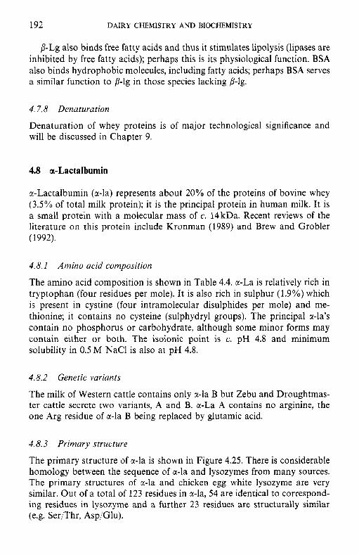
192 DAIRY CHEMISTRY A N D BIOCHEMISTRY
/I-Lg also binds free fatty acids and thus it stimulates lipolysis (lipases are inhibited by free fatty acids); perhaps this is its physiological function. BSA also binds hydrophobic molecules, including fatty acids; perhaps BSA serves a similar function to p-lg in those species lacking D-lg.
4.7.8 Denaturation
Denaturation of whey proteins is of major technological significance and will be discussed in Chapter 9.
4.8 or-Lactalburnin
a-Lactalbumin (a-la) represents about 20% of the proteins of bovine whey (3.5% of total milk protein); it is the principal protein in human milk. It is a small protein with a molecular mass of c. 14kDa. Recent reviews of the literature on this protein include Kronman (1989) and Brew and Grobler (1992).
4.8. I Amino acid composition
The amino acid composition is shown in Table 4.4. a-La is relatively rich in tryptophan (four residues per mole). It is also rich in sulphur (1.9?40) which is present in cystine (four intramolecular disulphides per mole) and me- thionine; it contains no cysteine (sulphydryl groups). The principal a-la’s contain no phosphorus or carbohydrate, although some minor forms may contain either or both. The isoionic point is c. pH 4.8 and minimum solubility in 0.5 M NaCl is also at pH 4.8.
4.8.2 Genetic variants
The milk of Western cattle contains only r-la B but Zebu and Droughtmas- ter cattle secrete two variants, A and B. a-La A contains no arginine, the one Arg residue of a-la B being replaced by glutamic acid.
4.8.3 Primary structure
The primary structure of a-la is shown in Figure 4.25. There is considerable homology between the sequence of a-la and lysozymes from many sources. The primary structures of r-la and chicken egg white lysozyme are very similar. Out of a total of 123 residues in r-la, 54 are identical to correspond- ing residues in lysozyme and a further 23 residues are structurally similar (e.g. Ser/Thr, Asp/Glu).
MILK PROTEINS 193
1 I Are (Variant B) Y
H. Glu-Gln-Leu-Thr-Lys-Cys-Glu-Val-F'he- -Glu-Leu-Lys- Asp-Leu-Lys-Gly-Tyr-Gl y-Gly- Gln (Variant A)
21 Val-Ser-Leu-Pro-Glu-Trp-Val-Cys-Thr-Thr-Phe-His-Thr-Ser-G1y-Tyr-Asp-Thr-Glu-Ala-
Ile-Val-G1n-Asn-Asn-Asp-Ser-Thr-Glu-Tyr-Gly-Leu-Phe-Gln-Ile-Asn-Asn-Lys-Ile-Trp-
Cy~-Lys-Asp-Asp-Gln-Asn-Pro-His-Ser-Asn-I~e-Cys-Asn-Ile-~er-Cys-Asp-Lys-P~e- 1
Leu-Asp-Asp-Asp-Leu-Thr-Asp-Asp-Ile-Met-
I -nrI Ile-Asn-Tyr-Trp-Leu-Ala-His-Lys-Ala-Leu-Cys-Ser-Glu-Lys-Leu-Asp-Gln-Trp-Leu-Cys-
121 123 Glu-Lys-Leu. OH
I-
Figure 4.25 Amino acid sequence of a-lactalbumin showing intramolecular disulphide bonds (-) and amino acid substitutions in genetic polymorphs (from Brew and Grobler, 1992).
4.8.4 Secondary and tertiary structure
cc-La is a compact globular protein, which exists in solution as a prolate ellipsoid with dimensions of 2.5 x 3.7 x 3.2nm. It consists of 26% cc-helix, 14% p-structure and 60% unordered structure. The metal binding and molecular conformational properties of r-la were discussed in detail by Kronman (1989). The tertiary structure of a-la is very similar to that of lysozyme. It has been difficult to crystallize bovine a-la in a form suitable for X-ray crystallography but work on the detailed structure is at an advanced stage (Brew and Grobler, 1992).
4.8.5 Quaternary structure
@-La associates under a variety of environmental conditions but the associ- ation process has not been well studied.
4.8.6 Other species
a-La has been isolated from several species, including the cow, sheep, goat, sow, human, buffalo, rat and guinea-pig. Some minor interspecies differences in the amino acid sequence and properties have been reported. The milks of sea mammals contain very little or no a-la.
194 DAIRY CHEMISTRY AND BIOCHEMISTRY
4.8.7 Biological function
One of the most interesting characteristics of cc-lactalbumin is its role in lactose synthesis:
UDP-D-Galactose + D-glucose ---+ lactose + UDP lactose
synthetase
Lactose synthetase, the enzyme which catalyses the final step in the biosynthesis of lactose, consists of two dissimilar protein subunits, A and B; the A protein is UDP-galactosyl transferase while the B protein is a-la. In the absence of B protein, the A protein acts as a non-specific galactosyl transferase, i.e. it transfers galactose from UDP-galactose to a range of acceptors, but in the presence of B protein it becomes highly specific and transfers galactose only to glucose to form lactose ( K , for glucose is reduced approximately 1000-fold). cc-Lactalbumin is, therefore, a ‘specifier protein’ and its action represents a unique form of molecular control in biological reactions. cc-La from the milks of many species are effective modifier proteins for the UDP-galactosyl transferase of bovine lactose synthetase. How it exercises its control is not understood, but it is suggested that the synthesis of lactose is controlled directly by a-lactalbumin which, in turn, is under hormonal control (Brew and Grobler, 1992). The concentration of lactose in milk is directly related to the concentration of a-la; milks of marine mammals, which contain no x-la, contain no lactose. Since lactose is the principal constituent in milk affecting osmotic pressure, its synthesis must be controlled rigorously and this is the presumed physiological role of a-la. Perhaps each molecule of x-la regulates lactose synthesis for a short period and is then discarded and replaced; while this is an expensive and wasteful use of an enzyme component, the rapid turnover affords a faster response should lactose synthesis need to be altered, as in mastitic infection, when the osmotic pressure of milk increases due to an influx of NaCl from the blood (Chapter 2).
4.8.8 Metal binding and heat stability
a-La is a metallo-protein; it binds one Ca2+ per mole in a pocket containing four Asp residues (Figure 4.26); these residues are highly conserved in all a-la’s and in lysozyme. The Ca-containing protein is quite heat stable (it is the most heat stable whey protein) or more correctly, the protein renatures following heat denaturation (denaturation does occur at relatively low temperatures, as indicated by differential scanning calorimetry). When the pH is reduced to below about 5, the Asp residues become protonated and lose their ability to bind Ca2+. The metal-free protein is denatured at quite low temperatures and does not renature on cooling; this characteristic has been exploited to isolate x-la from whey.
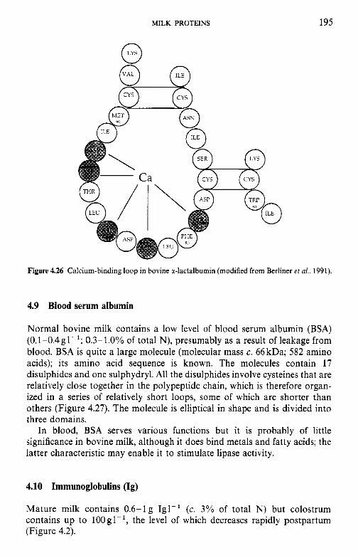
MILK PROTEINS 195
Figure 4.26 Calcium-binding loop in bovine a-lactalbumin (modified from Berliner e t a/., 1991).
4.9 Blood serum albumin
Normal bovine milk contains a low level of blood serum albumin (BSA) (0.1-0.4gl-'; 0.3-1.0% of total N), presumably as a result of leakage from blood. BSA is quite a large molecule (molecular mass c. 66 kDa; 582 amino acids); its amino acid sequence is known. The molecules contain 17 disulphides and one sulphydryl. All the disulphides involve cysteines that are relatively close together in the polypeptide chain, which is therefore organ- ized in a series of relatively short loops, some of which are shorter than others (Figure 4.27). The molecule is elliptical in shape and is divided into three domains.
In blood, BSA serves various functions but it is probably of little significance in bovine milk, although it does bind metals and fatty acids; the latter characteristic may enable it to stimulate lipase activity.
4.10 Immunoglobulins (Ig)
Mature milk contains 0.6-lg Igl-' (c. 3% of total N) but colostrum contains up to 1OOgl-', the level of which decreases rapidly postpartum (Figure 4.2).
196 DAIRY CHEMISTRY AND BIOCHEMISTRY
41
0
141 A
Net charge
-10
-8
0
Figure 4.27 Model of the bovine serum albumin molecule.
Igs are very complex proteins which will not be reviewed here. Essential- ly, there are five classes of Ig: IgA, IgG, IgD, IgE and IgM. IgA, IgG and IgM are present in milk. These occur as subclasses, e.g. IgG occurs as IgG, and IgG,. IgG consists of two long (heavy) and two shorter (light) polypeptide chains linked by disulphides (Figure 4.28). IgA consists of two such units (i.e. eight chains) linked together by secretory component (SC) and a junction (J) component, while IgM consists of five linked four-chain units (Figure 4.29). The heavy and light chains are specific to each type of Ig. For a review of immunoglobuins in milk, see Larson (1992).
The physiological function of Ig is to provide various types of immunity in the body. The principal Ig in bovine milk is IgG, while in human milk it is IgA. The calf (and the young of other ruminants) is born without Ig in its blood serum and hence is very susceptible to infection. However, the intestine of the calf is permeable to large molecules for about 3 days postpartum and therefore Ig is absorbed intact and active from its mother’s milk; Igs from colostrum appear in the calves blood within about 3 h of
MILK PROTEINS 197
Figure 4.28 Model of the basic 7s immunoglobulin (Ig) molecule showing two heavy and two light chains joined by disulphide bonds: V, variable region; C, constant region; L, light chain; H, heavy chain; 1, 2 and 3 subscripts refer to the three constant regions of the heavy chains; CHO, carbohydrate groups; Fab refers to the (top) antigen-specific portion of the Ig molecule;
Fc refers to the cell-binding effector portion of the Ig molecule (from Larson, 1992).
suckling and persist for about 3 months, although the calf is able to synthesize its own Ig within about 2 weeks. It is, therefore, essential that a calf should receive colostrum within a few hours of birth, otherwise it will probably die. The human baby obtains Ig in utero and hence, unlike the calf, is not as dependent on Ig from milk (in fact its intestine is impermeable to Ig). However, the Ig in human colostrum is beneficial to the baby, e.g. it reduces the risk of intestinal infections.
As regards the type and function of Ig in colostrum, mammals fall into three groups (Figure 4.30) - those like the cow (i.e. other ruminants), those like the human, and some, e.g. the horse, with features of the other two groups (Larson, 1992).
198 DAIRY CHEMISTRY A N D BIOCHEMISTRY
, H-chain
Figure 4.29 Models of IgG, IgA, IgD, IgE and IgM. (a) Structural model of IgG, before and after fragmentation by pepsin and papain and reduction with a sulphydryl reagent. Solid black chain portion = variable regions; light chain portion = constant regions. Small black lines represent disulphide and half-cystine (-SH) groups. Small black dots in Fc regions represent attached carbohydrate groups. The various parts of the model are labelled. (b) The structure of four classes of immunoglobulins are shown with monomeric IgA, dimeric IgA and secretory IgA. Location of the J-chain, secretory component (SC) and carbohydrate is approximate.
(From Larson, 1992.)
MILK PROTEINS 199
selective, 12-48h Probably Moderate, selective Extensive, selective Extensive, non-
none 19 days in rots, mice 12- 4 8 h
Figure 4.30 Transfer of maternal immunoglobulins to the foetus and neonate of representative mammalian species. Group I species transfer Ig in utero before birth. Group I1 species transfer Ig both in utero before birth and via colostrum after birth. Group 111 species transfer Ig only via colostrum after birth. The size of the immunoglobulin notation (IgA, IgM, IgG, IgG,) indicates the relative percentage composition of the immunoglobulins in colostrum. Species in group I1 may have IgG as the predominant Ig in colostrum. Significant IgG, also may be present in the colostrum of some Group I11 species. The relative absorption of immuno-
globulins in the gut of the neonate is also shown. (From Larson, 1992.)
4.11 Minor milk proteins
Milk contains numerous minor proteins, including perhaps 60 indigenous enzymes, some of which, e.g. lipase, proteinase, phosphatases and lac- toperoxidase, are technologically important (Chapter 8). Most of the minor proteins have biological functions and probably play very significant roles (section 4.16).
4.12 Non-protein nitrogen
Nitrogen soluble in 12% TCA is referred to as non-protein nitrogen (NPN), of which milk contains 250-300mgl-', i.e. 5-6% of total milk nitrogen. The NPN is a very heterogeneous fraction (Table 4.7).
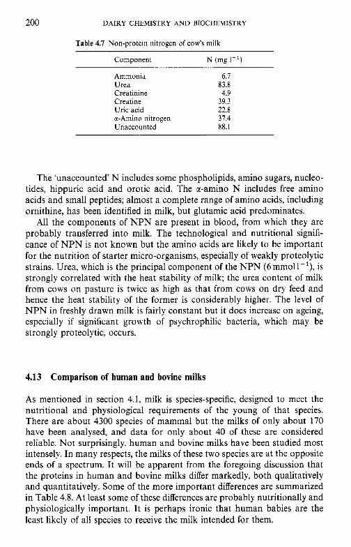
200 DAIRY CHEMISTRY AND BIOCHEMISTRY
Table 4.7 Non-protein nitrogen of cow's milk
Component N (mg I - ' )
Ammonia 6.7 Urea 83.8 Creatinine 4.9 Creatine 39.3 Uric acid 22.8 a-Amino nitrogen 37.4 Unaccounted 88.1
The 'unaccounted' N includes some phospholipids, amino sugars, nucleo- tides, hippuric acid and orotic acid. The x-amino N includes free amino acids and small peptides; almost a complete range of amino acids, including ornithine, has been identified in milk, but glutamic acid predominates.
All the components of NPN are present in blood, from which they are probably transferred into milk. The technological and nutritional signifi- cance of NPN is not known but the amino acids are likely to be important for the nutrition of starter micro-organisms, especially of weakly proteolytic strains. Urea, which is the principal component of the NPN (6mmoll-'), is strongly correlated with the heat stability of milk; the urea content of milk from cows on pasture is twice as high as that from cows on dry feed and hence the heat stability of the former is considerably higher. The level of NPN in freshly drawn milk is fairly constant but it does increase on ageing, especially if significant growth of psychrophilic bacteria, which may be strongly proteolytic, occurs.
4.13 Comparison of human and bovine milks
As mentioned in section 4.1, milk is species-specific, designed to meet the nutritional and physiological requirements of the young of that species. There are about 4300 species of mammal but the milks of only about 170 have been analysed, and data for only about 40 of these are considered reliable. Not surprisingly, human and bovine milks have been studied most intensely. In many respects, the milks of these two species are at the opposite ends of a spectrum. It will be apparent from the foregoing discussion that the proteins in human and bovine milks differ markedly, both qualitatively and quantitatively. Some of the more important differences are summarized in Table 4.8. At least some of these differences are probably nutritionally and physiologically important. It is perhaps ironic that human babies are the least likely of all species to receive the milk intended for them.
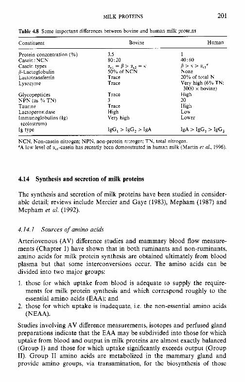
MILK PROTEINS 20 1
Table 4.8 Some important differences between bovine and human milk proteins
Constituent Bovine Human
Protein concentration (YO) Casein : NCN Casein types /?-Lactoglobulin Lactotransferrin Lysozyme
GI ycopeptides N P N (as YO TN) Taurine Lactoperoxidase Immunoglobulins (Ig)
Ig type (colostrum)
3.5 80: 20
50% of NCN Trace Trace
Trace 3 Trace High Very high
IgG, > IgG, > IgA
ZSl = p > a,, = K
1 40: 60
None 20% of total N Very high (6% TN;
High 20 High Low Lower
IgA > IgG, > IgG,
p > K > C i S l y
3000 x bovine)
NCN, Non-casein nitrogen; NPN, non-protein nitrogen; TN, total nitrogen. "A low level of ?,,-casein has recently been demonstrated in human milk (Martin et al., 1996).
4.14 Synthesis and secretion of milk proteins
The synthesis and secretion of milk proteins have been studied in consider- able detail; reviews include Mercier and Gaye (1983), Mepham (1987) and Mepham et al. (1992).
4.14.1 Sources of amino acids
Arteriovenous (AV) difference studies and mammary blood flow measure- ments (Chapter 1) have shown that in both ruminants and non-ruminants, amino acids for milk protein synthesis are obtained ultimately from blood plasma but that some interconversions occur. The amino acids can be divided into two major groups:
1. those for which uptake from blood is adequate to supply the require- ments for milk protein synthesis and which correspond roughly to the essential amino acids (EAA); and
2. those for which uptake is inadequate, i.e. the non-essential amino acids (NEAA).
Studies involving AV difference measurements, isotopes and perfused gland preparations indicate that the EAA may be subdivided into those for which uptake from blood and output in milk proteins are almost exactly balanced (Group I) and those for which uptake significantly exceeds output (Group 11). Group I1 amino acids are metabolized in the mammary gland and provide amino groups, via transamination, for the biosynthesis of those
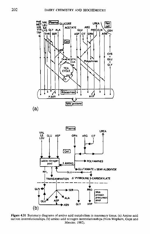
202 DAIRY CHEMISTRY AND BIOCHEMISTRY
PI M H
n r
ci
VAL ILE GLU LEU
$?
1 CYS + GLU + GLY
[Plrsmsl UREA
POLYAMINES
GLUTAMATE y SEMI ALDEHYDE
Figure 4.31 Summary diagrams of amino acid metabolism in mammary tissue. (a) Amino acid carbon interrelationships, (b) amino acid nitrogen interrelationships (from Mepham, Gaye and
Mercier, 1982).
MILK PROTEINS 203
amino acids for which uptake from blood is inadequate (Group 111), their carbon skeletons are oxidized to CO,. Considered as a whole, total uptake and output of amino acids from blood are the major, or sole, precursors of the milk-specific proteins (i.e. the caseins, P-lactoglobulin and a-lactal- bumin).
0 Group I amino acids: methionine, phenylalanine, tyrosine, histidine and tryptophan.
0 Group I1 amino acids: valine, leucine, isoleucine, lysine, arginine and threonine.
0 Group I11 amino acids: aspartic acid, glutamic acid, glycine, alanine, serine, cysteine/cystine, proline.
The interrelationships between the carbon and nitrogen of amino acids are summarized in Figure 4.31.
4.14.2 Amino acid transport into the mammary cell
Since the cell membranes are composed predominantly of lipids, amino acids (which are hydrophilic) cannot enter by diffusion and are transported by special carrier systems. In the case of mammary cells, the carrier system(s) has not yet been elucidated.
4.14.3 Synthesis of milk proteins
Synthesis of the major milk proteins occurs in the mammary gland; the principal exceptions are serum albumin and some of the immunoglobulins, which are transferred from the blood. Polymerization of the amino acids occurs on ribosomes fixed on the rough endoplasmic reticulum of the secretory cells, apparently by a method common to all cells.
The primary blueprint for the amino acid sequence of proteins is contained in deoxyribonucleic acid (DNA) within the cell nucleus. The requisite information is transcribed in the nucleus to ribonucleic acid (RNA) of which there are three types: messenger RNA (mRNA), transfer RNA (tRNA) and ribosomal RNA (rRNA). These are transferred to the cyto- plasm where each plays a specific role in protein synthesis.
Protein synthesis actually takes place in the ribosomes of the rough endoplasmic reticulum (RER) which contain rRNA. There is a specific tRNA for each amino acid, with which it forms an acyl complex:
Amino acid + tRNA + ATP Mg2: amino acyl-tRNA + AMP + PPi
amino acyl-tRNA synthetase
There is a specific amino acyl-tRNA synthetase for each amino acid; these enzymes have two specific binding sites, one for the amino acid and the
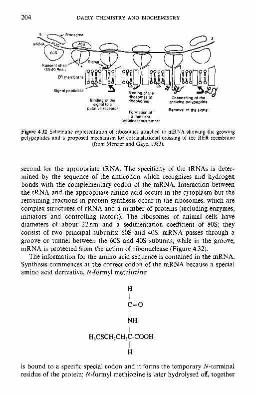
204 DAIRY CHEMISTRY AND BIOCHEMISTRY
(30-40 Res )
Binding Of the ribophorins growing polypeptide sianal to a of Removal of the signal putatrve receptor
a transient proteinaceous tunnel
Figure 4.32 Schematic representation of ribosomes attached to mRNA showing the growing polypeptides and a proposed mechanism for cotranslational crossing of the RER membrane
(from Mercier and Gaye, 1983).
second for the appropriate tRNA. The specificity of the tRNAs is deter- mined by the sequence of the anticodon which recognizes and hydrogen bonds with the complementary codon of the mRNA. Interaction between the tRNA and the appropriate amino acid occurs in the cytoplasm but the remaining reactions in protein synthesis occur in the ribosomes, which are complex structures of rRNA and a number of proteins (including enzymes, initiators and controlling factors). The ribosomes of animal cells have diameters of about 22nm and a sedimentation coefficient of 80s; they consist of two principal subunits: 60s and 40s. mRNA passes through a groove or tunnel between the 60s and 40s subunits; while in the groove, mRNA is protected from the action of ribonuclease (Figure 4.32).
The information for the amino acid sequence is contained in the mRNA. Synthesis commences at the correct codon of the mRNA because a special amino acid derivative, N-formyl methionine:
H
c=o NH
I I
I
I H,CSCH,CH$-COOH
H
is bound to a specific special codon and it forms the temporary N-terminal residue of the protein; N-formyl methionine is later hydrolysed off, together
MILK PROTEINS 205
with a short hydrophobic signal peptide, exposing the permanent N - terminal residue. The acyl amino acid-tRNA is bound to the mRNA just outside the ribosome by becoming attached to its corresponding codon; presumably, a full range of amino acid-tRNAs are available in the environment but only the tRNA with the appropriate anticodon is bound. GTP and a number of specific cytoplasmic protein factors are required for binding.
In the ribosome, the amino group of the newly bound amino acid reacts through nucleophilic substitution with the C-terminal carbonyl carbon of the existing peptide, and in the process the peptide is transferred to the newly bound tRNA, releasing the tRNA just vacated. Condensation is catalysed by peptidyl transferase, which is part of the ribosomal subunit.
For the next cycle, a new acyl amino acid-tRNA is bound to the mRNA, the ribosome tracks along the mRNA and the emptied tRNA is ejected. As the polypeptide is elongated it assumes its secondary and tertiary structure (Figure 4.32).
The factors controlling termination of synthesis are poorly understood; it is known that there is a specific ribosomally bound protein release factor which promotes the hydrolysis of the linkage between the tRNA and the newly formed protein.
A strand of mRNA is long enough to accommodate several ribosomes along its length, e.g. the mRNA for haemoglobin (150 amino acid residues/ molecule) contains 450 nucleotides and is c. 150nm long; since each ribosome is about 20nm in diameter, 5-6 ribosomes can be accommodated. The ribosomes are connected to each other by the mRNA strand, forming a polysome (polyribosome) which can be isolated intact if adequate care is taken. Each ribosome in a polysome is at a different stage in the synthesis of a protein molecule, thereby utilizing the mRNA more efficiently (Figure 4.32).
Milk proteins are destined to be exported from the cell. Like other exported proteins, translocation through cell membranes is facilitated by a signal sequence, a sequence of 15-29 amino acids at the amino terminal of the growing polypeptide chain. This sequence causes the ribosome to bind to the ER membrane, in which a ‘channel’ forms, allowing the growing chain to enter the ER lumen (Figure 4.32). Subsequently, the signal sequence is cleaved from the polypeptide by signal peptidase, an enzyme located on the luminal side of the ER membrane.
4.14.4
In addition to proteolytic processing (i.e. removal of the signal peptide sequence), the polypeptide is subject to other covalent modifications: N - and 0-glycosylation and 0-phosphorylation. After synthesis and transportation across the ER lumen, the proteins pass to the Golgi apparatus and thence,
Modfications of the polypeptide chain
206 DAIRY CHEMISTRY AND BIOCHEMISTRY
via secretory vesicles, to the apical membrane. Covalent modification must therefore occur at some point(s) along this route. Such modifications may be either co-translational (occurring when chain elongation is in progress) or post-translational. Proteolytic cleavage of the signal peptide is co-trans- lational and this seems to be the case also for N-glycosylation, in which dolichol-linked oligosaccharides are enzymatically transferred to aspara- ginyl residues of the chain when these are present in the sequence code, Asn-X-Thr/Ser (where X is any amino acid except proline). The large oligosaccharide component may be 'trimmed' as it traverses the secretory pathway. Formation of disulphide bonds between adjacent sections of the chain, or between adjacent chains (as in K-casein), may also be partly co-translational.
By contrast, O-glycosylation and O-phosphorylation appear to be post- translational events. Glycosylation of the principal milk-specific glyco- protein, casein, is believed to be effected by membrane-bound glycosyltrans- ferases (three such enzymes have been described) located in the Golgi apparatus. O-Phosphorylation involves transfer of the y-phosphate of ATP to serine (or, less frequently, threonine) residues, occurring in the sequence, Ser/Thr-X-A (where X is any amino acid residue and A is an acidic residue, such as aspartic or glutamic acid or a phosphorylated amino acid). Phos- phorylation is effected by casein kinases which are located chiefly in the Golgi membranes. In addition to the correct triplets, the local conformation of the protein is also important for phosphorylation of Ser since not all serines in caseins in the correct sequence are phosphorylated. Some serine residues in p-lg occur in a Ser-X-A sequence but are not phosphorylated, probably due to extensive folding of this protein.
The Golgi complex is also the locus of casein micelle formation. In association with calcium, which is actively accumulated by Golgi vesicles, the polypeptide chains associate to form submicelles, and then micelles, prior to secretion.
4.14.5 Structure and expression of milk protein genes
The structure, organization and expression of milk protein genes are now understood in considerable detail. This subject is considered to be outside the scope of this book and the interested reader is referred to Mepham et al. (1992). Such knowledge permits the genetic engineering of milk proteins with respect to the transfer of genes from one species to another, the overexpression of a particular desirable protein(s), the elimination of certain undesirable proteins, changing the amino acid sequence by point mutations to modify the functional properties of the protein or transfer of a milk protein gene to a plant or microbial host. This topic is also considered to be outside the scope of this text and the interested reader is referred to Richardson et al. (1992).
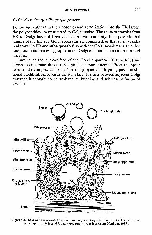
MILK PROTEINS 207
4.14.6 Secretion of milk-specific proteins
Following synthesis in the ribosomes and vectorization into the ER lumen, the polypeptides are transferred to Golgi lumina. The route of transfer from ER to Golgi has not been established with certainty. It is possible that lumina of the ER and Golgi apparatus are connected, or that small vesicles bud from the ER and subsequently fuse with the Golgi membranes. In either case, casein molecules aggregate in the Golgi cisternal lumina in the form of micelles.
Lumina at the nuclear face of the Golgi apparatus (Figure 4.33) are termed cis cisternae; those at the apical face trans cisternae. Proteins appear to enter the complex at the cis face and progress, undergoing post-transla- tional modification, towards the trans face. Transfer between adjacent Golgi cisternae is thought to be achieved by budding and subsequent fusion of vesicles.
Milk fat globule Signet
Figure 4.33 Schematic representation of a mammary secretory cell as interpreted from electron micrographs; c, cis face of Golgi apparatus; t , trans face (from Mepham, 1987).
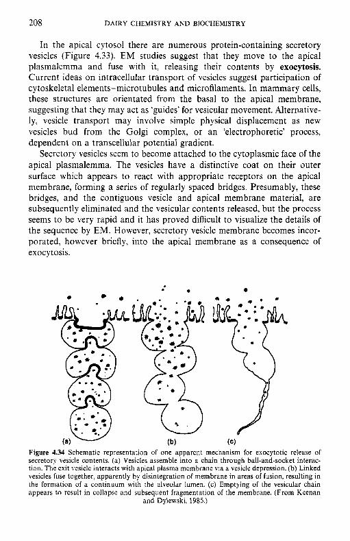
208 DAIRY CHEMISTRY AND BIOCHEMISTRY
In the apical cytosol there are numerous protein-containing secretory vesicles (Figure 4.33). EM studies suggest that they move to the apical plasmalemma and fuse with it, releasing their contents by exocytosis. Current ideas on intracellular transport of vesicles suggest participation of cytoskeletal elements-microtubules and microfilaments. In mammary cells, these structures are orientated from the basal to the apical membrane, suggesting that they may act as ‘guides’ for vesicular movement. Alternative- ly, vesicle transport may involve simple physical displacement as new vesicles bud from the Golgi complex, or an ‘electrophoretic’ process, dependent on a transcellular potential gradient.
Secretory vesicles seem to become attached to the cytoplasmic face of the apical plasmalemma. The vesicles have a distinctive coat on their outer surface which appears to react with appropriate receptors on the apical membrane, forming a series of regularly spaced bridges. Presumably, these bridges, and the contiguous vesicle and apical membrane material, are subsequently eliminated and the vesicular contents released, but the process seems to be very rapid and it has proved difficult to visualize the details of the sequence by EM. However, secretory vesicle membrane becomes incor- porated, however briefly, into the apical membrane as a consequence of exocytosis.
Figure 4.34 Schematic representation of one apparent mechanism for exocytotic release of secretory vesicle contents. (a) Vesicles assemble into a chain through ball-and-socket interac- tion. The exit vesicle interacts with apical plasma membrane via a vesicle depression. (b) Linked vesicles fuse together, apparently by disintegration of membrane in areas of fusion, resulting in the formation of a continuum with the alveolar lumen. (c) Emptying of the vesicular chain appears to result in collapse and subsequent fragmentation of the membrane. (From Keenan
and Dylewski, 1985.)
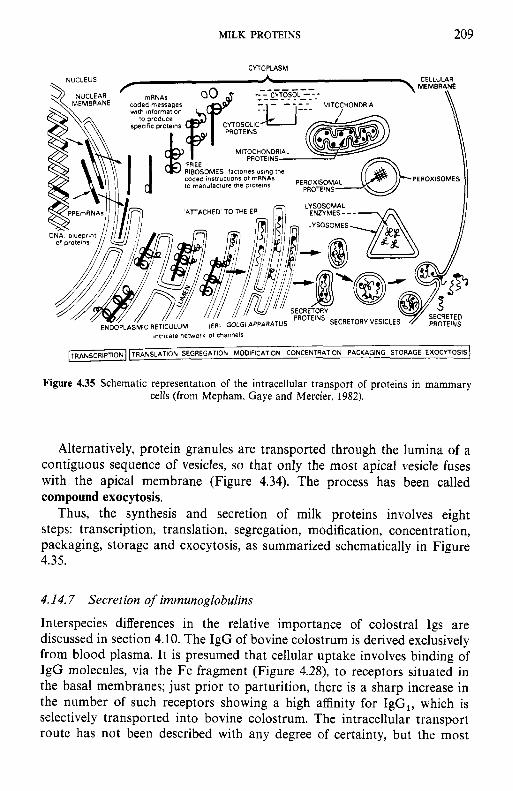
MILK PROTEINS 209
CYIOPLASM
lntrlcate network ol channels
ANS SLAT ION SEGREGATION MODIFICATION CONCENTRATION PACKAGING STORAGE EXOCUOSIS]
Figure 4.35 Schematic representation of the intracellular transport of proteins in mammary cells (from Mepham, Gaye and Mercier, 1982).
Alternatively, protein granules are transported through the lumina of a contiguous sequence of vesicles, so that only the most apical vesicle fuses with the apical membrane (Figure 4.34). The process has been called compound exocytosis.
Thus, the synthesis and secretion of milk proteins involves eight steps: transcription, translation, segregation, modification, concentration, packaging, storage and exocytosis, as summarized schematically in Figure 4.35.
4.14.7 Secretion of immunoglobulins
Interspecies differences in the relative importance of colostral Igs are discussed in section 4.10. The IgG of bovine colostrum is derived exclusively from blood plasma. It is presumed that cellular uptake involves binding of IgG molecules, via the Fc fragment (Figure 4.28), to receptors situated in the basal membranes; just prior to parturition, there is a sharp increase in the number of such receptors showing a high affinity for IgG,, which is selectively transported into bovine colostrum. The intracellular transport route has not been described with any degree of certainty, but the most
210 DAIRY CHEMISTRY AND BIOCHEMISTRY
likely scheme appears to involve vesicular transport, followed by exocytosis at the apical membrane.
IgA in colostrum is derived partly from intramammary synthesis and partly by accumulation in the gland after being transported in the blood from other sites of synthesis. In either case, IgA molecules are transported into the secretory cells across the basal membrane by means of a large, membrane-bound form of secretory component, which acts as a recognition site. It is presumed that, following endocytosis, the sIgA complex (Figure 4.29) is transported to the apical membrane of the secretory cell where, following cleavage of a portion of the complex, the mature sIgA complex is secreted by exocytosis.
4.15 Functional milk proteins
The term ‘Functional Properties of Proteins’ in relation to foods refers to those physicochemical properties of a protein which affect the functionality of the food, i.e. its texture (rheology), colour, flavour, water sorption/binding and stability. Probably the most important physicochemical properties are solubility, hydration, rheology, surface activity and gelation, the relative importance of which depends on the food in question; these properties are, at least to some extent, interdependent.
The physical properties of many foods, especially those of animal origin, are determined primarily by their constituent proteins, but those properties are not the subject of this section. Rather, we are concerned with isolated, more or less pure, proteins which are added to foods for specific purposes. The importance of such proteins has increased greatly in recent years, partly because suitable technology for the production of such proteins on a commercial scale has been developed and partly because a market for functional proteins has been created through the growth of fabricated foods, i.e. foods manufactured from more or less pure ingredients (proteins, fats/oils, sugars/polysaccharides, flavours, colours). Perhaps one should view the subject the other way round, i.e. fabricated foods developed because suitable functional proteins were available. Some functional proteins have been used in food applications for a very long time, e.g. egg white in various types of foamed products or gelatine in gelled products. The principal functional food proteins are derived from milk (caseins and whey proteins) or soybeans; other important sources are egg white, blood, connective tissue (gelatine) and wheat (gluten).
Probably because of the ease with which casein can be produced from skim milk, essentially free of lipids, lactose and salts, by rennet or isoelectric coagulation and washing of the curd, acid and rennet caseins have been produced commercially since the beginning of this century. However, until relatively recently, they were used for industrial applications, e.g. in glues,
MILK PROTEINS 21 1
plastics, fibres or dye-binders for paper glazing. Although some casein is still used for industrial applications, at least 80% of world production is now used in foods. This change has occurred partly because cheaper and possibly better materials have replaced casein for industrial applications while growth in the production of fabricated foods has created a demand for functional proteins at higher prices than those available for industrial-grade products. Obviously, the production of a food-grade protein requires better hygienic standards than industrial proteins; the pioneering work in this area was done mainly in Australia and to a lesser extent in New Zealand in the 1960s.
Although heat-denatured whey protein, referred to as lactalbumin, has been available for many years for food applications, it was of little signifi- cance, mainly because the product is insoluble and therefore has limited functionality. The commercial production of functional whey protein be- came possible with the development of ultrafiltration in the 1960s. Whey protein concentrates (WPCs) produced by ultrafiltration are now of major commercial importance, with many specific food applications. Superior whey protein products (whey protein isolates, WPI) are being produced on a limited scale by chromatography, although their substantially higher cost has limited their production.
As discussed in section 4.16, many of the whey proteins have interesting biological and physical properties. It is now possible to isolate individual whey proteins on a commercial scale in a relatively pure form; it is likely that in the immediate future such purified whey proteins will be readily available for specific applications.
4.15.1 Industrial production of caseins
There are two principal established methods for the production of casein on an industrial scale: isoelectric precipitation and enzymatic (rennet) coagu- lation. There are a number of comprehensive reviews on the subject (e.g. Muller, 1982; Fox, 1989; Mulvihill, 1992; Fox and Mulvihill, 1992) which should be consulted for references.
Acid casein is produced from skim milk by direct acidification, usually with HCI, or by fermentation with a Lactococcus culture, to c. pH 4.6. The curds/whey are cooked to about 50°C, separated using inclined perforated screens or decanting centrifuges, washed. thoroughly with water (usually in counter-flow mode), dewatered by pressing, dried (fluidized bed, attrition or ring dryers) and milled. A flow diagram of the process and a line diagram of the plant are shown in Figures 4.36 and 4.37.
Acid casein is insoluble in water but soluble caseinate can be formed by dispersing the casein in water and adjusting the pH to 6.5-7.0 with NaOH (usually), KOH, Ca(OH), or NH, to produce sodium, potassium, calcium or ammonium caseinate, respectively (Figure 4.38). The caseinates are

212 DAIRY CHEMISTRY AND BIOCHEMISTRY
I Proteolvtic Coanulatioa 4. Calf Rennet or
substitute 1. Mineral Acid 2. Ion Exchange 3. Lactic Starter
I t 1 t 1 1 1
1 Precipicdionl Coagulation
Cooking
Dewheying
Washing
Dewatering
Drying, Tempering, Grinding
Bennet Casein
< 1 0OC 22-26'C -31'C
Mineral L!ak Acid Case in Casein 2 5- 30'C
(B)
Skim milk at
J Mineral Ion Exchange Lactic Rennet
Resin Starter x 1 4 h X - 1 h
Acid
pH 4.6 pH 2.2 pH 4.6 pH 6.6
Cook at - 50°C
untreated milk
pH 4.6
- 5O0C - 55OC
Figure 4.36 (a) Line diagram of industrial processes for the manufacture of acid and rennet casein. The conditions (time, temperature and pH) of precipitation are shown in (b). (Modified
from Mulvihill, 1992.)
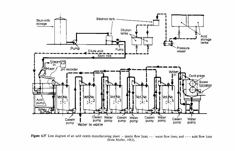
Skim-milk Balance tank storage -
vessel
Figure 4.37 Line diagram of an acid casein manufacturing plant: - casein flow lines; - - - water flow lines; and acid flow lines (from Muller, 1982).
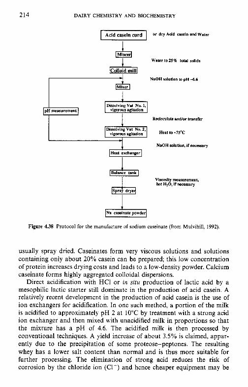
214
Dissolving Vat No. 2, vigorous agitation
DAIRY CHEMISTRY A N D BIOCHEMISTRY
Heat to - 7 5 ~
NnOH solution to pH -6.6
Dissolving Vat No. 1,
Recirculate andor transfer
pH measurement
I
NaOH solution, if necessary
Viscosity measurement, hot H20, if necessary
INa caseinate powder1
Figure 4.38 Protocol for the manufacture of sodium caseinate (from Mulvihill, 1992).
usually spray dried. Caseinates form very viscous solutions and solutions containing only about 20% casein can be prepared; this low concentration of protein increases drying costs and leads to a low-density powder. Calcium caseinate forms highly aggregated colloidal dispersions.
Direct acidification with HCl or in situ production of lactic acid by a mesophilic lactic starter still dominate in the production of acid casein. A relatively recent development in the production of acid casein is the use of ion exchangers for acidification. In one such method, a portion of the milk is acidified to approximately pH 2 at 10°C by treatment with a strong acid ion exchanger and then mixed with unacidified milk in proportions so that the mixture has a pH of 4.6. The acidified milk is then processed by conventional techniques. A yield increase of about 3.5% is claimed, appar- ently due to the precipitation of some proteose-peptones. The resulting whey has a lower salt content than normal and is thus more suitable for further processing. The elimination of strong acid reduces the risk of corrosion by the chloride ion (Cl-) and hence cheaper equipment may be
MILK PROTEINS 215
used. However, in spite of these advantages, this process has not been widely accepted and, as far as we are aware, is used only in France, where it was developed. In other proposed methods, deproteinated whey or milk ultrafil- tration permeate, acidified by ion exchangers, is used to acid-precipitate casein from skim milk or skim-milk concentrate. Apparently, these methods have not been commercialized.
Rennet casein is produced from skim milk by treatment with certain proteolytic enzymes, known as rennets. The rennet coagulation of milk and related aspects are discussed in Chapter 10. Apart from the coagulation mechanism, the protocol for the production of rennet casein is essentially similar to that for acid casein.
Rennet casein is insoluble in water or alkali but can be solubilized by treatment with polyphosphates. Most rennet casein is used in the manufac- ture of cheese analogues, the recipe for which includes calcium chelators, e.g. polyphosphates (Chapter 10).
4.15.2 Novel methods for casein production
Cryoprecipitation. When milk is frozen and stored at about -1O"C, the ionic strength of the liquid phase increases with a concomitant increase in [Ca' '1 and a decrease in pH (to approximately 5.8) due to precipitation of calcium phosphates with the release of hydrogen ions (H') (Chapter 5). These changes destabilize the casein micelles which precipitate when the milk is thawed.
Cryodestabilization of casein limits the commercial feasibility of frozen milk, which may be attractive in certain circumstances. However, cryode- stabilized casein might be commercially viable, especially if applied to milks concentrated by ultrafiltration, which are less stable than normal milk. Cryodestabilized casein may be processed in the usual way. The product is dispersible in water and can be reconstituted as micelles in water at 40°C. The heat stability and rennet coagulability of these micelles are generally similar to those of normal micelles and casein produced by cryodestabiliz- ation may be suitable for the production of fast-ripening cheeses, e.g. Mozzarella or Camembert, when the supply of fresh milk is inadequate. As far as we are aware, casein is not produced commercially by cryodestabiliz- ation.
Precipitation with ethanol. The casein in milk coagulates at pH 6.6 on addition of ethanol to about 40%; stability decreases sharply as the pH is reduced, and only 10-15% ethanol is required at pH 6 . Ethanol-precipi- tated casein may be dispersed in a micellar form and has very good emulsifying properties. The commercial production of ethanol-precipitated casein is probably economically viable but the process is not being used commercially.
216 DAIRY CHEMISTRY A N D BIOCHEMISTRY
Membraneprocessing. The use of ultrafiltration (UF) for the production of whey protein concentrates (WPCs) is now well established (p. 223). Obvi- ously, U F or diafiltration (DF) can be used to prepare products enriched in total milk protein. Products with protein concentrations up to 85% have been produced and assessed for a range of functionalities and applications (Fox and Mulvihill, 1992).
The development of large-pore membranes facilitates the separation of whey proteins from casein micelles by microfiltration (MF). Membranes used in M F have cut-offs in the range 0.01-10pm, and therefore casein micelles may be in the permeate or retentate streams, depending on the pore size of the M F membranes chosen. M F with large-pore membranes very effectively removes bacteria and somatic cells from milk and may also be used to remove lipoprotein complexes from whey prior to the production of WPCs with improved functionality. The preparation of micellar casein by M F is still at the exploratory stage.
High-speed centrifugation. The casein micelles may be sedimented by centrifugation at greater than lOOOOOg for 1 h, which is widely used on a laboratory scale. A combination of ultrafiltration and ultracentrifugation has been proposed for the industrial production of ‘native’ phos- phocaseinate. Almost all the casein in skim milk and UF retentates, containing 3.0- 17% protein, can be sedimented by centrifugation at greater than 75 OOOg for 1 h at 50°C.
‘Native’ casein. An exciting new development is the production of ‘native’ casein. Few details on the process are available at present but it involves electrodialysis of skim milk at 10°C against acidified whey to reduce the pH to about 5; the acidified milk is centrifuged and the sedimented casein dispersed in water, concentrated by U F and dried. The product disperses readily in water and is claimed to have properties approaching those of native casein micelles.
4.15.3 Fractionation of casein
As discussed in section 4.4, individual caseins may also be isolated on a laboratory scale by methods based on differences in solubility in urea solutions at around pH 4.6, by selective precipitation with CaCl, or by various forms of chromatography, especially ion-exchange or reverse-phase high performance liquid chromatography (RP-HPLC). Obviously, these methods are not amenable to scale-up for industrial application.
There is considerable interest in developing techniques for the fractiona- tion of caseins on an industrial scale for special applications. For example:
0 p-Casein has very high surface activity and may find special applications as an emulsifier or foaming agent.
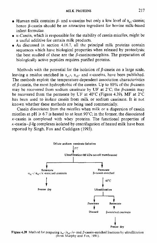
MILK PROTEINS 217
0 Human milk contains p - and K-caseins but only a low level of a,,-casein; hence ,!?-casein should be an attractive ingredient for bovine milk-based infant formulae.
0 K-Casein, which is responsible for the stability of casein micelles, might be a useful additive for certain milk products.
0 As discussed in section 4.16.7, all the principal milk proteins contain sequences which have biological properties when released by proteolysis; the best studied of these are the /I-caseinomorphins. The preparation of biologically active peptides requires purified proteins.
Methods with the potential for the isolation of p-casein on a large scale, leaving a residue enriched in rs l - , xs2- and K-caseins, have been published. The methods exploit the temperature-dependent association characteristics of /?-casein, the most hydrophobic of the caseins. Up to 80% of the /I-casein may be recovered from sodium caseinate by U F at 2°C; the ,!?-casein may be recovered from the permeate by U F at 40°C (Figure 4.39). M F at 2°C has been used to isolate casein from milk or sodium caseinate. It is not known whether these methods are being used commercially.
Casein dissociates from the micelles when milk or a dispersion of casein micelles at pH 2 6.7 is heated to at least 90°C; in the former, the dissociated K-casein is complexed with whey proteins. The functional properties of K-casein-/I-lg complexes isolated by centrifugation of heated milk have been reported by Singh, Fox and Cuddigan (1993).
Dilute sodium caseinate Solution
I 2 O C
Ultrafiltration (10 kDa cut-off membranes)
Retentate * Permeate
a,, -/ CQ/K -enriched caseinate pcasein enriched
Freeze dry 1 40"c
Ultrafiltration
Permeate t7 Retentate
t t Discard P-enriched caseinate
Freize dry
Figure 4.39 Method for preparing usl-/us2-/K- and P-casein-enriched fractions by ultrafiltration (from Murphy and Fox, 1991).
218 DAIRY CHEMISTRY AND BIOCHEMISTRY
4.15.4 Functional (physicochemical) properties of caseins
Solubility. Solubility is an important functional property per se, i.e. in fluid products, and is essential for other functionalities since insoluble proteins can not perform useful functions in foods. The caseins are, by definition, insoluble at their isoelectric points, i.e. in the pH range c. 3.5-5.5; the insolubility range becomes wider with increasing temperature. Insolubility in the region of the isoelectric point is clearly advantageous in the produc- tion of acid casein and is exploited in the production of two major families of dairy products, i.e. fermented milks and fresh cheeses. However, such insolubility precludes the use of casein in acid liquid foods, e.g. protein- enriched fruit juices or carbonated beverages. Acid-soluble casein can be prepared by limited proteolysis or by interaction with certain forms of pectin.
Rheological proper ties. Viscosity, an important physicochemical property of many foods, can be modified by proteins or polysaccharides. The caseins form rather viscous solutions, a reflection of their rather open structure and relatively high water-binding capacity. While the high viscosity of caseinate may be of some importance in casein-stabilized emulsions, it causes produc- tion problems; for example, due to very high viscosity, not more than about 20% protein can be dissolved even at a high temperature. The low protein content of caseinate solution increases the cost of drying and results in low-density powders which are difficult to handle.
Hydration. The ability of proteins to bind and hold water without syner- esis is critical in many foods, e.g. comminuted meat products. Although the caseins are relatively hydrophobic, they bind c. 2 g H,O g-' protein, which is typical of proteins. Hydration increases with increasing pH and is relatively independent of NaCl concentration, which is especially important in the efficacy of casein in meat-based products. The water-holding capacity of sodium caseinate is higher than that of calcium caseinate or micellar casein.
Gelation. One of the principal functional applications of proteins is the formation of gels. In milk, caseins undergo gelation when the environment is changed in one of several ways, but the most important are rennet- induced coagulation for cheese or rennet casein manufacture (which is discussed in Chapter 10) or on acidification to the isoelectric point (pH 4.6), which is exploited in the preparation of fermented milk products and isoelectric casein. In addition, casein may be gelled or coagulated by organic solvents, prolonged heat treatments or during storage of heat-sterilized products; these changes are usually negative. Heat-induced gelation is used in the preparation of many food products but, as discussed in Chapter 9,
MILK PROTEINS 219
the caseins are remarkably heat stable and do not undergo thermally- induced gelation except under extremely severe conditions; their stability is of course a major advantage in milk processing.
Surface activity. Probably the outstanding property of caseins, as far as their functionality in foods is concerned, is their surface activity, which makes them good foaming agents and especially good emulsifiers. Surface- active agents are molecules with hydrophilic and hydrophobic regions which can interact with the aqueous and nonaqueous (air or lipid) phases of emulsions and foams, thus reducing the interfacial or surface tension.
Caseins are among the most surface-active proteins available to food technologists, p-casein being particularly effective. To exhibit good surface activity, a protein must possess three structural features:
1. It should be relatively small, since the rate of migration to the interface is inversely proportional to the molecular mass. In actual food processing operations, the rate of diffusion is not particularly important since the production of emulsions and foams involves a large imput of work with vigorous agitation which moves the protein rapidly to the interface.
2. The molecule must be capable of adsorbing at the oil-water or air-water interface and hence must have relatively high surface hydrophobicity; the caseins, especially 8-casein, meet this requirement very well.
3. Once adsorbed, the molecule must open and spread over the interface; thus, an open, flexible structure is important. The caseins, which have relatively low levels of secondary and tertiary structures and have no intramolecular disulphide bonds, can open and spread readily.
In practice, while the caseins are very good emulsifiers and foam readily, the resultant foams are not very stable, possibly because the lamella of the foam bubbles are thin and drain rapidly in contrast to the thicker foams formed by egg albumin.
4.15.5 Applications of caseins
World production of casein and caseinates is about 250000tonnes per annum. While some casein is still used for industrial applications, the vast majority is used in foods, in which it has numerous applications, as summarized in Table 4.9.
4.15.6 Whey proteins
Many whey proteins possess interesting functional, nutritional, physiologi- cal or pharmaceutical properties. Unfortunately, all the proteins in whey are present at low concentrations and hence are relatively expensive to produce, although at least some of them are capable of carrying high production
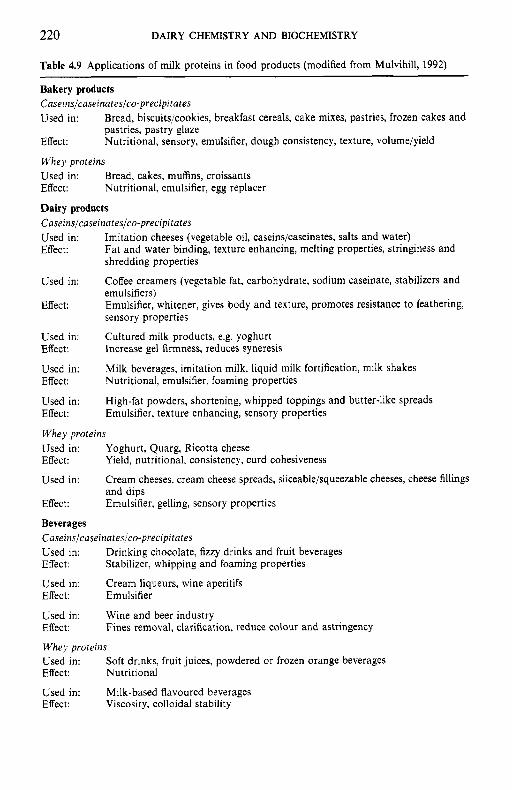
220 DAIRY CHEMISTRY AND BIOCHEMISTRY
Table 4.9 Applications of milk proteins in food products (modified from Mulvihill, 1992) ~~~~
Bakery products Caseinslcaseinateslco-precipitates Used in:
Effect:
Whey proteins Used in: Effect: Nutritional, emulsifier, egg replacer
Dairy products Caseinslcaseinateslco-precipitates Used in: Effect:
Bread, biscuits/cookies, breakfast cereals, cake mixes, pastries, frozen cakes and pastries, pastry glaze Nutritional, sensory, emulsifier, dough consistency, texture, volume/yield
Bread, cakes, muffins, croissants
Imitation cheeses (vegetable oil, caseins/caseinates, salts and water) Fat and water binding, texture enhancing, melting properties, stringiness and shredding properties
Coffee creamers (vegetable fat, carbohydrate, sodium caseinate, stabilizers and emulsifiers) Emulsifier, whitener, gives body and texture, promotes resistance to feathering, sensory properties
Cultured milk products, e.g. yoghurt Increase gel firmness, reduces syneresis
Milk beverages, imitation milk, liquid milk fortification, milk shakes
High-fat powders, shortening, whipped toppings and butter-like spreads Emulsifier, texture enhancing, sensory properties
Used in:
Effect:
Used in: Effect:
Used in: Effect: Nutritional, emulsifier, foaming properties
Used in: Effect:
Whey proteins Used in: Effect:
Used in:
Effect: Emulsifier, gelling, sensory properties
Beverages Caseinslcuseinateslco-precipitates Used in: Effect:
Used in: Effect: Emulsifier
Used in: Effect:
Whey proteins Used in: Effect: Nutritional
Used in: Milk-based flavoured beverages Effect: Viscosity, colloidal stability
Yoghurt, Quarg, Ricotta cheese Yield, nutritional, consistency, curd cohesiveness
Cream cheeses, cream cheese spreads, sliceable/squeezable cheeses, cheese fillings and dips
Drinking chocolate, fizzy drinks and fruit beverages Stabilizer, whipping and foaming properties
Cream liqueurs, wine aperitifs
Wine and beer industry Fines removal, clarification, reduce colour and astringency
Soft drinks, fruit juices, powdered or frozen orange beverages
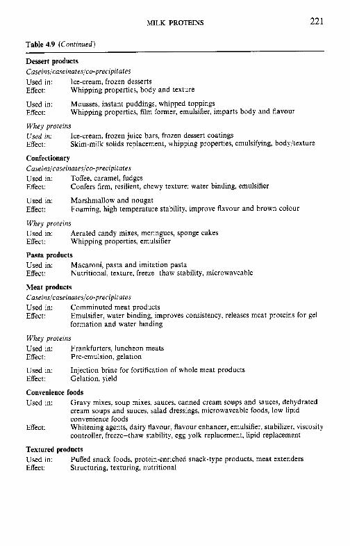
MILK PROTEINS 22 1
Table 4.9 (Continued)
Dessert products Caseinslcaseinateslco-precipitates Used in: Ice-cream, frozen desserts Effect:
Used in: Effect:
Whey proteins Used in: Effect:
Confectionary Caseinslcaseinateslco-precipitates Used in: Toffee, caramel, fudges Effect:
Used in: Marshmallow and nougat Effect:
Whey proteins Used in: Effect: Whipping properties, emulsifier
Pasta products Used in: Effect:
Meat products Caseinslcaseinateslco-precipitates Used in: Comminuted meat products Effect: Emulsifier, water binding, improves consistency, releases meat proteins for gel
formation and water binding
Whipping properties, body and texture
Mousses, instant puddings, whipped toppings Whipping properties, film former, emulsifier, imparts body and flavour
Ice-cream, frozen juice bars, frozen dessert coatings Skim-milk solids replacement, whipping properties, emulsifying, bodyltexture
Confers firm, resilient, chewy texture; water binding, emulsifier
Foaming, high temperature stability, improve flavour and brown colour
Aerated candy mixes, meringues, sponge cakes
Macaroni, pasta and imitation pasta Nutritional, texture, freeze-thaw stability, microwaveable
Whey proteins Used in: Frankfurters, luncheon meats Effect: Pre-emulsion, gelation
Used in: Effect: Gelation, yield
Convenience foods Used in:
Injection brine for fortification of whole meat products
Gravy mixes, soup mixes, sauces, canned cream soups and sauces, dehydrated cream soups and sauces, salad dressings, microwaveable foods, low lipid convenience foods Whitening agents, dairy flavour, flavour enhancer, emulsifier, stabilizer, viscosity controller, freeze-thaw stability, egg yolk replacement, lipid replacement
Effect:
Textured products Used in: Effect: Structuring, texturing, nutritional
Puffed snack foods, protein-enriched snack-type products, meat extenders
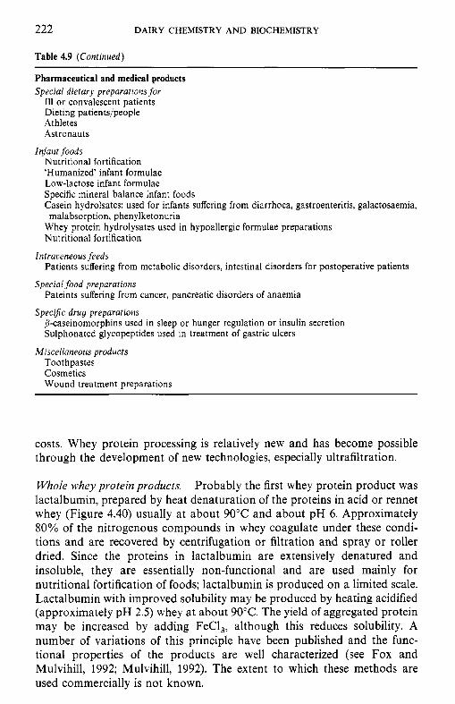
222 DAIRY CHEMISTRY AND BIOCHEMISTRY
Table 4.9 (Continued)
Pharmaceutical and medical products Special dietary preparations for
Ill or convalescent patients Dieting patients/people Athletes Astronauts
Nutritional fortification ‘Humanized’ infant formulae Low-lactose infant formulae Specific mineral balance infant foods Casein hydrolsates: used for infants suffering from diarrhoea, gastroenteritis, galactosaemia,
Whey protein hydrolysates used in hypoallergic formulae preparations Nutritional fortification
Patients suffering from metabolic disorders, intestinal disorders for postoperative patients
Pateints suffering from cancer, pancreatic disorders of anaemia
B-caseinomorphins used in sleep or hunger regulation or insulin secretion Sulphonated glycopeptides used in treatment of gastric ulcers
Toothpastes Cosmetics Wound treatment preparations
Infant foods
malabsorption, phenylketonuria
Intraoeneous feeds
Special food preparations
Specifk drug preparations
Miscellaneous products
costs. Whey protein processing is relatively new and has become possible through the development of new technologies, especially ultrafiltration.
Whole whey protein products. Probably the first whey protein product was lactalbumin, prepared by heat denaturation of the proteins in acid or rennet whey (Figure 4.40) usually at about 90°C and about pH 6 . Approximately 80% of the nitrogenous compounds in whey coagulate under these condi- tions and are recovered by centrifugation or filtration and spray or roller dried. Since the proteins in lactalbumin are extensively denatured and insoluble, they are essentially non-functional and are used mainly for nutritional fortification of foods; lactalbumin is produced on a limited scale. Lactalbumin with improved solubility may be produced by heating acidified (approximately pH 2.5) whey at about 90°C. The yield of aggregated protein may be increased by adding FeCl,, although this reduces solubility. A number of variations of this principle have been published and the func- tional properties of the products are well characterized (see Fox and Mulvihill, 1992; Mulvihill, 1992). The extent to which these methods are used commercially is not known.
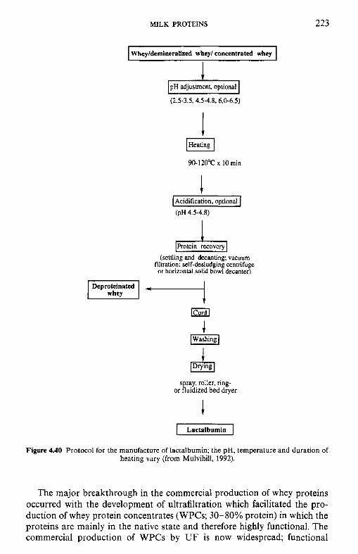
MILK PROTEINS 223
I Wheyldemineralized whey/ concentrated whey I
(2.5-3.5.4.5-4.8.6.0-6.5)
I Acidification, optional (PH 4.5-4.8)
4 Protein recovery (settling and decanting; vacuum
filtration; self-desludging centrifuge or horizontal solid bowl decanler)
Deproteinated
J [Washing]
J spray, roller, ring-
or fluidized bed dryer
i pZiGG-1
Figure 4.40 Protocol for the manufacture of lactalbumin; the pH, temperature and duration of heating vary (from Mulvihill, 1992).
The major breakthrough in the commercial production of whey proteins occurred with the development of ultrafiltration which facilitated the pro- duction of whey protein concentrates (WPCs; 3 0 4 0 % protein) in which the proteins are mainly in the native state and therefore highly functional. The commercial production of WPCs by UF is now widespread; functional
224 DAIRY CHEMISTRY AND BIOCHEMISTRY
properties and applications have been thoroughly described (e.g. Marshall, 1982; Morr, 1989; de Wit, 1989a, b).
The presence of even a low level of lipids impairs the functionality of WPCs and reduces flux rates during U F processing. Both problems are minimized by clarifying the whey prior to UF, e.g. by adding CaC1, to whey (to 1.2gI-'), adjusting the pH to 7.3 and heating to 50°C; the flocculent calcium phospholipoprotein complexes formed are allowed to settle and the clear supernatant siphoned off, or removed by centrifugation or microfiltra- tion.
Whey proteins complex with, and are precipitated by, several polyionic compounds which may be used to prepare WPCs (see Marshall, 1982). The most effective of these are polyphosphates which can be removed from the resolubilized protein by precipitation with Ca2 +, electrodialysis, ion-ex- change or gel filtration. Polyphosphate-precipitated WPCs are commercial- ly available.
The use of ion-exchange resins (Figure 4.41) offers an effective method for the preparation of high-quality whey protein products, referred to as whey protein isolates (WPI), containing 90-95% protein (see Marshall, 1982; Mulvihill, 1992). Although the functional properties of WPI are superior to those of WPCs on an equiprotein basis (due to lower levels of lipids, lactose and salts), their production is rather limited, due to higher production costs.
Fractionation of whey proteins. Techniques for the isolation of individual whey proteins on a laboratory scale by salting-out, ion-exchange chromatography and/or crystallization have been available for about 40 years. Owing to the unique functional, physiological or other biological properties of many of the whey proteins, there is an economic incentive for their isolation on an industrial scale. For example, p-lg, the principal whey protein in bovine milk, produces better thermoset gels than a-la. However, human milk does not contain p-lg, which is the most allergenic of the bovine milk proteins for the human infant; therefore, a-la would appear to be a more appropriate protein for the preparation of infant formulae than total whey protein.
A number of methods have been developed for the separation of x-la and B-lg (Figure 4.42). Probably the most commercially feasible of these exploits the low heat stability of calcium-free x-la to precipitate it from whey, leaving p-lg, BSA and Ig in solution. x-La loses its calcium on acidification to about pH 5.0, aggregates on heating to about 55°C and can be recovered by centrifugation, filtration or microfiltration.
a-La and B-lg are insoluble in pure water at their isoelectric points (around pH 5); p-lg requires a higher ionic strength for solubility than a-la, a characteristic which may be exploited to fractionate a-la and p-lg. When UF-concentrated whey is acidified to pH 4.65 and demineralized by elec-
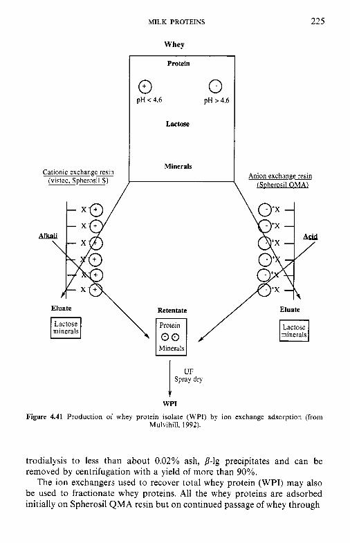
MILK PROTEINS 225
Whey
I Protein
I0 0 pH < 4.6 pH > 4.6
Lactose
Minerals Cationic exchangeresin
(vistec, Spherosil S ) Anion exchangeMn
[Spherosil OMA) \
Eluate \ Retentate / Eluate
minerals
Minerals
t WPI
Figure 4.41 Production of whey protein isolate (WPI) by ion exchange adsorption (from Mulvihill, 1992).
trodialysis to less than about 0.02% ash, 8-1s precipitates and can be removed by centrifugation with a yield of more than 90%.
The ion exchangers used to recover total whey protein (WPI) may also be used to fractionate whey proteins. All the whey proteins are adsorbed initially on Spherosil QMA resin but on continued passage of whey through
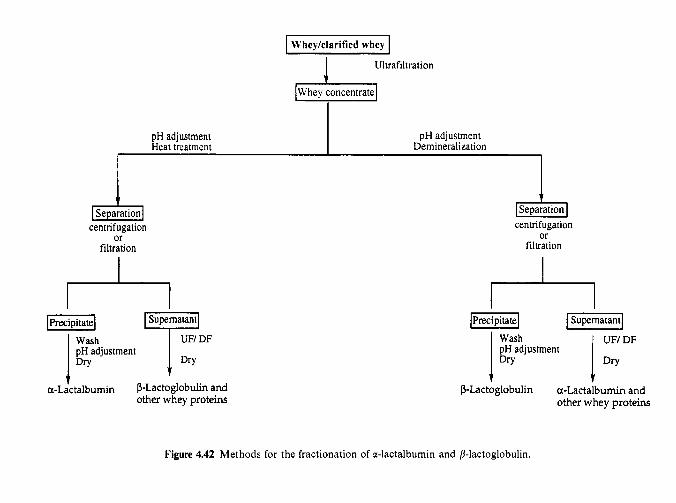
pH adjustment Heat treatment
Wheyklarified whey
i Whey concentrate
Ultrafiltration
pH adjustment Demineralization
I ISeparationl
centrifugation or
filtration
I
centrifugation Of
filtration
U N DF
a-lactalbumin P-Lactoglobulin and other whey proteins
[Precipitate/ FJ UF/ DF
pH adjustment 1 :;h 1 Dry P-Lactoglobulin a-Lactalbumin and
other whey proteins
Figure 4.42 Methods for the fractionation of a-lactalbumin and P-lactoglobulin.
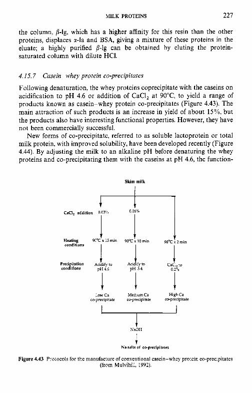
MILK PROTEINS 227
the column, p-lg, which has a higher affinity for this resin than the other proteins, displaces a-la and BSA, giving a mixture of these proteins in the eluate; a highly purified p-lg can be obtained by eluting the protein- saturated column with dilute HCl.
4.15.7 Casein-whey protein co-precipitates
Following denaturation, the whey proteins coprecipitate with the caseins on acidification to pH 4.6 or addition of CaCI, at 90°C, to yield a range of products known as casein-whey protein co-precipitates (Figure 4.43). The main attraction of such products is an increase in yield of about 15%, but the products also have interesting functional properties. However, they have not been commercially successful.
New forms of co-precipitate, referred to as soluble lactoprotein or total milk protein, with improved solubility, have been developed recently (Figure 4.44). By adjusting the milk to an alkaline pH before denaturing the whey proteins and co-precipitating them with the caseins at pH 4.6, the function-
Skim milk
CaCI, addition 0.03% I 1
Heating 9OoC x 15 rnin conditions
Precipitation Acidify to conditions p~ 4.6
I Low Ca
co-precipitate
0.06%
9OoC 10 rnin
Aci a ify to pH 5.4
Medium Ca cc-precipitate
1 90°C x 2 rnin
CaCI, to 0.20'"
High Ca co-precipita te
t NaOH
I Na salts of co-precipitates
Figure 4.43 Protocols for the manufacture of conventional casein-whey protein co-precipitates (from Mulvihill, 1992).
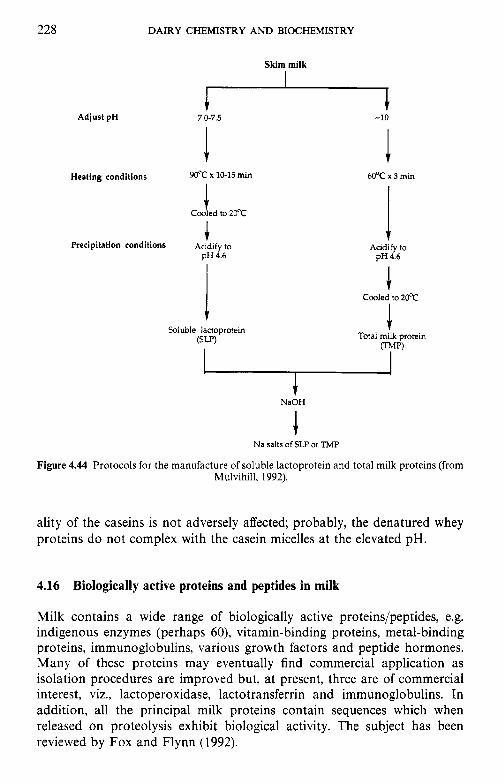
228
Adjust pH
DAIRY CHEMISTRY AND BIOCHEMISTRY
Skim milk
7.0-7.5 * -10
1 Heating conditions 90°C x 10-15 min
Coo t ed to 20°C
Precipitation conditions Acidify to pH 4.6
Soluble lactoprotein ( S W
I 60°C x 3 min
Acidify to pH4.6
Cooled to 20°C
Total milk protein TruP)
t NaOH
Na salts of SLP or TMP
Figure 4.44 Protocols for the manufacture of soluble lactoprotein and total milk proteins (from Mulvihill, 1992).
ality of the caseins is not adversely affected; probably, the denatured whey proteins do not complex with the casein micelles at the elevated pH.
4.16 Biologically active proteins and peptides in milk
Milk contains a wide range of biologically active proteins/peptides, e.g. indigenous enzymes (perhaps 60), vitamin-binding proteins, metal-binding proteins, immunoglobulins, various growth factors and peptide hormones. Many of these proteins may eventually find commercial application as isolation procedures are improved but, at present, three are of commercial interest, viz., lactoperoxidase, lactotransferrin and immunoglobulins. In addition, all the principal milk proteins contain sequences which when released on proteolysis exhibit biological activity. The subject has been reviewed by Fox and Flynn (1992).
MILK PROTEINS 229
4.16.1 Lactoperoxidase
Lactoperoxidase (LPO) is a broad-specificity peroxidase present at high concentrations in bovine milk but at low levels in human milk. LPO, which has been isolated and well characterized (Chapter 8), has attracted consider- able interest owing to its antibacterial activity in the presence of H,O, and thiocyanate (SCN-); the active species is hypothiocyanate (OSCN-) or other higher oxidation species. Milk normally contains no indigenous H,O,, which must be added or produced in situ, e.g. by the action of glucose oxidase or xanthine oxidase; it is usually necessary to supplement the indigenous SCN-. Commercial interest in LPO is focused on:
1. activation of the indigenous enzyme for cold pasteurization of milk or protection of the mammary gland against mastitis; and
2. addition of isolated LPO to calf or piglet milk replacers to protect against enteritis, especially when the use of antibiotics in animal feed is not permitted.
LPO, which is positively charged at neutral pH, can be isolated from milk or whey by ion-exchange chromatography which has been scaled up for industrial application. These methods isolate LPO together with lactotrans- ferrin (Lf) which is also cationic at neutral pH. LPO and Lf can be resolved by chromatography on CM-Toyopearl or by hydrophobic interaction chromatography on Butyl Toyopearl 650 M (see Fox and Mulvihill, 1992).
4.16.2 Lactotransferrin
The transferrins are a group of specific metal-binding proteins, the best characterized of which are serotransferrin (present in blood plasma, milk, spinal fluid and semen), ovotransferrin (conalbumin; present in avian and reptile egg white) and lactotransferrin (present in milk, pancreatic juice, tears and leucocytes).
Human colostrum and milk contain 6-8 mg ml- ' and 2-4 mg ml- ' lactotransferrin, respectively, representing about 20% of the total protein in the latter; bovine colostrum and milk contain about 1 and 0.02- 0.35 mg ml- ', respectively. The concentration of lactotransferrin in human milk decreases slightly during lactation but appears to increase slightly in bovine milk and very markedly during the dry period.
Lactotransferrin binds iron very strongly, which suggests two roles for this protein: iron absorption and protection against enteric infection in the neonate. Because the concentration of Lf in human milk is considerably higher than that in bovine milk, there is considerable interest in supplement- ing bovine milk-based infant formulae with Lf. The concentration of Lf in milk increases markedly during mastitic infections, suggesting that it may have a protective role in the mammary gland. The structure and function of
230 DAIRY CHEMISTRY AND BIOCHEMISTRY
Lf have been reviewed by Lonnerdal and Iyer (1995) and Hutchens and Lonnerdal (1996).
Lactotransferrins have been isolated from the milks of several species, including human and bovine, and some have been well characterized, including determination of their amino acid sequence. Some of the isolation procedures have industrial-scale potential; the preparations obtained from such procedures usually contain both Lf and LPO.
4.16.3 Immunoglobulins
The occurrence, significance and interspecies aspects of immunglobulin in milk were described in section 4.10. Classically, Ig is prepared by salting-out, usually with ammonium sulphate [(NH4)*S04]. This method is effective but expensive and current commercial products are usually prepared by ultra- filtration of colostrum or milk from hyperimmunized cows. Some recently developed methods for the isolation of Ig, sometimes with Lf, use mono- clonal antibodies, metal chelate or gel filtration chromatographies (see Fox and Mulvihill, 1992).
Ig-rich preparations are commercially available for the nutrition of calves and other neonatal animals. Although human infants do not absorb Ig from the intestine, Igs still play an important defensive role by reducing the incidence of intestinal infection. While breast feeding is best for healthy full-term infants, it is frequently impossible to breast-feed pre-term or very-low-birth-weight infants, who may be fed on banked human milk. Such infants have high protein and energy requirements which may not be met by human milk and consequently special formulae have been developed. A ‘milk immunological concentrate’, prepared by diafiltration of acid whey from colostrum and early lactation milk from immunized cows, for use in such formulae has been described; the product contains approximately 75% protein, 50% of which is Ig, mainly IgG, and not IgA, which is predominant in human milk.
The development of Ig in cows agains human pathogens, e.g. rotavirus, an important cause of illness in children, is considered to be an attractive approach in human medicine. The Ig could be administered in milk or as a concentrate prepared from milk.
4.16.4 Vitamin-binding proteins
Milk contains specific binding proteins for retinol (vitamin A), vitamin D, riboflavin (vitamin BJ, folate and cyanocobalamin (vitamin BIZ). Such proteins may improve the absorption of these vitamins by protecting and transferring them to receptor proteins in the intestine, or they may have antibacterial activity by rendering vitamins required by intestinal bacteria unavailable. The activity of these proteins is reduced or destroyed by heat treatments.
MILK PROTEINS 23 1
4.16.5 Growth factors
The term ‘growth factor’ is applied to a group of potent hormone-like polypeptides which play a critical role in the regulation and differentiation of a variety of cells acting through cell membrane receptors. The milk and, especially, colostrum from several species contain several growth factors, including insulin-like growth factors (IGF1, IFG2), transforming growth factors (TGF,,, TGF,,, TGF,), mammary-derived growth factors (MDGF I, MDGF II), fibroblast growth factors, platelet-derived growth factor (PDGF) and bombasin.
The source of these polypeptides may be blood plasma, mammary gland or both. The biological significance of these growth-promoting activities in colostrum and mature milks is not yet clear. In terms of possible physiologi- cal significance, two potential targets may be considered, i.e. the mammary gland or the neonate. In general, most attention has focused on the latter. It is not known whether the factors in milk that possess the capacity to promote cell proliferation (1) influence growth of mammary tissue, (2) promote the growth of cells within the intestines of the recipient neonate, or (3) are absorbed in a biologically active form and exert an effect on enteric or other target organs.
Methods using ultrafiltration and chromatography have been developed for the concentration of growth factors from whey. In addition to possible food (nutritceutical) applications for such growth factors, a major potential application is in tissue cultures, for which foetal bovine serum is used as a source of growth factors. However, the supply of foetal bovine serum is limited, unreliable, expensive and of variable quality. Whey-derived growth factors have the potential to have a major impact on the biotechnological and pharmaceutical industries for the production of vaccines, hormones, drugs, monoclonal antibodies, and the production of tissue, especially skin for treatment of burns, ulcers and lacerations.
4.16.6 BiJidus factors
Special types of growth factors are those that promote the growth of bifidobacteria. It has been recognized for many years that breast-fed babies are more resistant to gastroenteritis than bottle-fed babies. This is un- doubtedly a multifactorial phemonenon, including better hygiene, more appropriate milk composition, several antibacterial systems (especially im- munoglobulins, lysozyme, lactotransferrin, vitamin-binding proteins and lactoperoxidase, which are discussed above), and a lower intestinal pH. The mean pH of the faeces of breast-fed babies is 5.1 while that of bottle-fed babies is 6.4; the low pH of the former may be due partly to the lower buffering capacity of human milk compared to bovine milk, due to its lower content of protein and phosphate, and partly to differences in the intestinal microflora of breast-fed and bottle-fed infants. Bifidobacteria represent
232 DAIRY CHEMISTRY A N D BIOCHEMISTRY
about 99% of the faecal microflora of breast-fed infants. These bacteria also represent a high proportion of the microflora of bottle-fed infants but several other genera, e.g. Bacteroides, Clostridium and coliforms, also occur at high numbers. Furthermore, the predominant species of Bifidobacterium in breast-fed infants is B. bifidum, with lesser numbers of B. longum; the faecal microflora of bottle-fed infants is dominated by B. longum, with lower numbers of B. bifidum, B. infantis, B. adolescentis and B. breve.
The preponderance of B. biJdum in the faeces of breast-fed infants is due to the presence of stimulatory factors in human milk. The most important of these are N-acetylglucosamine-containing saccharides, referred to as bifidus factor I, which is present at high levels in human milk and colostrum and bovine colostrum but at very low concentrations in the milk of cows, goats and sheep. Human milk also contains several non-dialysable bifidus- promoting factors which are glycoproteins, referred to as bifidus factor 11. Many of the glycoproteins have been isolated and characterized (see Fox and Flynn, 1992).
Bifidobacterium spp. are also stimulated by lactulose, a derivative of lactose (Chapter 2) which is not related to bifidus factors I and 11.
4.16.7 Milk protein hydrolysates
Several methods have been described for the production, characterization and evaluation of milk protein hydrolysates tailored for specific applications in the health-care, pharmaceutical, baby food and consumer product areas (see Fox and Mulvihill, 1992, for references).
Several peptides with specific properties may be prepared from milk proteins, either in vivo or in vitro; some may have commercial potential and can be produced on a relatively large scale by preparative ion-exchange chromatography.
Macropeptides from rc-casein. These peptides represent the C-terminal region of rc-casein (residues 106-169) which is released by rennet during the manufacture of cheese or rennet casein (Chapter 10). The (g1yco)macropep- tides are released into the whey which contains 1.2-1.5gl-l, and from which they can be readily recovered, e.g. by anion exchange using Spherosil QMA resin. The peptides contain no Phe, Tyr, Trp or Cys; the absence of aromatic amino acids makes the macropeptides suitable for the nutrition of patients suffering from phenylketonuria.
Phosphopeptides. It is claimed that phosphopeptides prepared from casein hydrolysates stimulate the absorption of Ca in the intestine, but views on this are not unanimous. Such peptides are resistant to proteolysis due to the high density of negative charges; they have been detected in the small intestine of the rat and may pass intact through the intestinal wall. Since

MILK PROTEINS 233
phosphopeptides also bind iron it has been proposed that casein phos- phopeptide-Fe complexes are useful supplements for dietary iron but their influence on the bioavailability of iron is ambiguous.
Caseinomorphins. Several peptides with opioid activity have been isolated from enzymatic digests of milk proteins (see Fox and Flynn, 1992). Such peptides were first isolated from enzymatic digests of casein and character- ized as a family of peptides containing 4-7 amino acids with a common N-terminal sequence, H.Tyr.Pro.Phe.Pro-, and 0-3 additional residues (Gly, Pro, Ile), i.e. residues 60-63/6 of /?-casein, and hence were called caseinomorphins (P-CM) 4 to 7, respectively. P-CM-5 is the most effective of these peptides, which are 300-4000 times less effective than morphine. j3-CMs are very resistant to enzymes of the gastrointestinal tract (GIT) and appear in the contents of the small intestine following ingestion of milk. P-CN f60-70 also has weak opiate activity but may be hydrolysed to smaller, more active j3-CMs by peptidases in the brush border of the GIT.
The sequence 5 1-57 of human j-casein, Tyr.Pro.Phe.Val.Glu.Pro.Ile, corresponds to bovine /3-CN f60-66 (i.e. Tyr.Pro.Phe.Pro.Gly.Pro,Ile) and has weak opioid activity. Peptides corresponding to human P-casein resi- dues 41-44 and 59-63 also have weak opioid activity. Exorphines have also been isolated from hydrolysates of a,,-casein (f90-95 and f90-96; Arg.Tyr.Leu.Gly.Tyr.Leu (Glu)), K-casein (f35-41, 57-60 and 25-34), a- lactalbumin (f50-53, Tyr.Gly.Leu.Phe), j3-lactoglobulin (f102- 105, Tyr.Leu. Leu.Phe) and lactotransferrin (Tyr.Leu.Gly.Ser.Gly.Tyr, Arg.Tyr.Tyr.Gly. Tyr and Lys.Tyr.Leu.Gly.Pro.Gln.Tyr).
Thus, all the major milk proteins contain sequences which, when liber- ated by gastrointestinal proteinases, possess opioid activity. These peptides are very resistant to proteolysis by gastrointestinal proteinases and, because of their high hydrophobicity, can be absorbed intact from the intestine. They possess physiological activity in uitro but their activity in uivo is as yet uncertain.
Immunomodulating peptides. Enzymatic digests of human caseins contain immunomodulating peptides which stimulate the phagocytic activity of human macrophages in uitro and exert a protective effect in uivo in mice against Klebsiella pneumoniae infection. Two of the peptides were character- ized as H.Val.Glu.Pro.1le.Pro.Tyr (/?-CN f54-59) and H.Gly.Leu.Phe (origin not identified).
Platelet-modifying peptide. The undecapeptide, H.Met.Ala.Ile.Pro.Pro.Lys. Lys.Asn.Gln.Asp.Lys (residues 106-1 16 of bovine K-casein) inhibits the aggregation of ADP-treated blood platelets; its behaviour is similar to that of the structurally related C-terminal dodecapeptide (residues 400-41 1) of human fibrinogen y-chain. K-CN f106-116 is produced from the (g1yco)-
234 DAIRY CHEMISTRY A N D BIOCHEMISTRY
macropeptide, K-CN f106-169, formed by the action of chymosin. Shorter peptides, K-CN f106-112 and 113-116, have similar but weaker effects on platelet aggregation. A peptide with similar properties has been isolated from a hydrolysate of lactotransferrin.
Angiotensin converting enzyme (ACE) inhibitor. ACE is a dipep- tidylaminopeptidase (EC 3.4.15.1) which cleaves dipeptides from the C- terminus of peptides. It converts angiotensin I to the potent vasoconstrictor, angiotensin 11, and inactivates the vasodilator, bradykinin. The dodecapep- tide, H.Phe.Phe.Val.Ala.Pro.Phe.Pro.Glu.Val.Phe.Gly.Lys, i.e. r,,-CN f23-34, from tryptic hydrolysates of casein inhibits ACE. The C-terminal sequence of cr,,-casein, H.Thr.Thr.Met.Pro.Leu.Tyr, sr,,-CN f194- 199, also has ACE inhibitory activity. Peptides from the sequence 39-52 of human p-casein, especially H.Ser.Phe.Gln.Pro.Gln.Pro.Leu.1le.Tyr.Pro (b-CN f43-52), also have ACE inhibitory activity.
Calmodulin-binding peptides. Peptides that inhibit calmodulin-dependent cyclic nucleotide phosphodiesterase have been isolated from peptic digests of a,,-casein (as1 plus x,,) and identified as cr,,-CN f164-179, cr,,-CN f183-206 and a,,-CN f183-207. The physiological significance of these peptides is unknown.
Bacteriocidal peptides from lactotransferrin (Lf). The bactericidal proper- ties of Lf, presumed to be due to iron-binding, were discussed in section 4.16.2. It has been reported that a number of bactericidal peptides are formed when Lf is heated at 120°C for 15 min, especially at pH 2, at which the degree of hydrolysis is about 10%. The effectiveness of these peptides is not related to iron-binding properties, i.e. their bactericidal properties are retained in Fe-rich media in which Lf is ineffective.
Potent antibacterial peptides can also be produced by hydrolysis of Lf by pepsins and some other acid proteinases. The low molecular weight peptides in the peptic hydrolysates were at least eight times more potent than Lf, were effective against a wider range of bacteria than Lf and retained their potency in the presence of added iron, unlike native Lf.
References
Berliner, L.J., Meinholtz, D.C,. Hirai, Y. et al. (1991) Functional implications resulting from disruption of the calcium binding loop in bovine a-lactalbumin. J . Dairy Sci., 74, 2394-402.
Bernhart, F.W. (1961) Correlation between growth-rate of the suckling of various species and the percentage of total calories from protein in the milk. Nature, 191, 358-60.
Brew, K. and Grobler, J.A. (1992) a-Lactalbumin, in Adt'anced Dairy Chemistry, Vol. 1: Proteins, (ed. P.F. Fox), Elsevier Applied Science, London, pp. 191-229.
MILK PROTEINS 235
Brunner, J.R., Ernstrom, C.A., Hollis, R.A. et al. (1960) Nomenclature of the proteins of bovine milk - first revision. J . Dairy Sci., 43, 901-1 1.
de Wit, J.N. (1989a) Functional properties of whey proteins, in Developments in Dairy Chemistry, Vol. 4 Functional Milk Proteins, (ed. P.F. Fox), Elsevier Applied Science, London,
de Wit, J.N. (1989b) The use of whey protein products, in Developments in Dairy Chemistry Vol. 4 Functional Milk Proteins, (ed. P.F. Fox), Elsevier Applied Science, London, pp. 323-45.
Eigel, W.N., Butler, J.E., Ernstrom, C.A. et al. (1984) Nomenclature of proteins of cow’s milk: fifth revision. J . Dairy Sci., 67, 1599-631.
Farrell, H.M. Jr (1988) Physical equilibria: proteins, in Fundamentals of Dairy Chemistry, 3rd edn (ed. N.P. Wong), van Nostrand Reinhold, New York, pp. 461-510.
Fox, P.F. (ed.) (1989) Deuelopments in Dairy Chemistry, Vol. 1: Proteins, Applied Science Publishers, London.
Fox, P.F. and Flynn, A. (1992) Biological properties of milk proteins, in Advanced Dairy Chemistry, Vol. 1: Proteins, (ed. P.F. Fox), Elsevier Applied Science, London, pp. 255-84.
Fox, P.F. and Mulvihill, D.M. (1992) Developments in milk protein processing. Food Sci. Technol. Today, 7, 152-61.
Hambling, S.G., McAlpine, A S . and Sawyer, L. (1992) /?-Lactoglobulin, in Aduanced Dairy Chemistry, Vol. 1: Proteins, (ed. P.F. Fox), Elsevier Applied Science, London, pp. 141-90.
Holt, C. (1992) Structure and stability of bovine casein micelles. Adv. Prof . Chem., 43, 63-151. Holt, C. (1994) The biological function of casein, in Yearbook 1994, The Hannah Institute, Ayr,
Scotland, pp. 60-8. Holt, C. and Sawyer, L. (1993) Caseins as rheomorphic proteins: Interpretation of primary and
secondary structures of a,,-, /?- and K-caseins. J . Chem. Soc. Faraday Trans., 89, 2683-92. Hutchens, T.W. and Lonnerdal, B. (1996) Lactoferrin: Structure and Function, Chapman &
Hall, London. Jakob, E. and Puhan, Z. (1992) Technological properties of milk as influenced by genetic
polymorphism of milk proteins - a review. Int. Dairy J., 2, 157-78. Jenness, R., Larson, B.L., McMeekin, T.L. et al. (1956) Nomenclature of the proteins of bovine
milk. J. Dairy Sci., 39, 536-41. Keenan, T.W. and Dylewski, D.P. (1985) Aspects of intracellular transit of serum and lipid
phases of milk. J . Dairy Sci., 68, 1025-40. Kronman, M.J. (1989) Metal-ion binding and the molecular conformational properties of
a-lactalbumin. Crit . Rev. Biochem. Mol. Biol., 24, 565-667. Kumosinski, T.F. and Farrell, H.M. Jr. (1994) Solubility of proteins: salt-water interactions, in
Protein Functionality in Food Systems, (eds N.S. Hettiarachchy and G.R. Ziegler), Marcel Dekker, New York. pp. 39-77.
Kumosinski, T.F., Brown, E.M. and Farrell, H.M. Jr (1993a) Three-dimensional molecular modeling of bovine caseins: An energy-minimized /?-casein structure. J . Dairy Sci., 76,
Kumosinski, T.F., Brown, E.M. and Farrell, H.M. J r (1993b). Three-dimensional molecular modeling of bovine caseins: a refined, energy-minimized K-casein structure. J. Dairy Sci., 76,
Larson, B.L. (1992). Immunoglobulins of the mammary secretions, in Advanced Dairy Chemis- try, Vol. 1: Proteins, (ed. P.F. Fox), Elsevier Applied Science, London, pp. 231-54.
Lonnerdal, B. and Iyer, S. (1995) Lactoferrin: Molecular structure and biological function. Ann. Rev. Nutr. . 15, 93-110.
McKenzie, H.A. (ed.) (1970) Milk Proteins: Chemistry and Molecular Biology, Vol. 1, Academic Press, New York.
McKenzie, H.A. (1971) /3-Lactoglobulin, in Milk Proteins, Chemistry and Molecular Biology, Vol. 11, (ed. H.A. McKenzie), Academic Press, New York, pp. 257-330.
McMahon, D.J. and Brown, R.J. (1984) Composition, structure and integrity of casein micelles: A review. J . Dairy Sci., 67, 499-512.
Marshall, K.R. (1982) Industrial isolation of milk proteins: whey proteins, in Developments in Dairy Chemistry, Vol. 1: Proteins, (ed. P.F. Fox), Applied Science Publishers, London,. pp. 339-73.
Martin, P., Brignon, G., Furet, J.P. and Leroux, C. (1996) The gene encoding a,,-casein is expressed in human mammary epithelial cells during lactation. Lait, 76, 523-35.
pp. 285-321.
931-45.
2507-20.
236 DAIRY CHEMISTRY AND BIOCHEMISTRY
Mepham, T.B. (1987) Physiology of Lactation, Open University Press, Milton, Keynes, UK. Mepham, T.B., Gaye, P. and Mercier, J.C. (1982) Biosynthesis of milk proteins, in Dairy
Chemistry, Vol. 1: Proteins, (ed. P.F. Fox), Elsevier Applied Science, London, pp. 115-56. Mepham, T.B., Gaye, P., Martin, P. and Mercier, J.-C. (1992) Biosynthesis of milk proteins, in
Adcanced Dairy Chemistry, Vol. 1: Proteins, (ed. P.F. Fox), Elsevier Applied Science, London,
Mercier, J.C. and Gaye, P.C. (1983) Milk protein syntheses, in Biochemistry of Lactation, (ed. T.B. Mepham), Elsevier, Amsterdam, pp. 177-227.
Morr, C.V. (1989) Whey proteins: manufacture, in Developments in Dairy Chemistry, Vol. 4: Functional Milk Proteins, (ed. P.F. Fox), Elsevier Applied Science, London, pp. 245-84.
Muller, L.L. ( I 982) Manufacture of casein. caseinates and casein co-precipitates, in Develop- ments in Dairy Chemistry, Vol. 1: Proteins, (ed. P.F. Fox), Applied Science Publishers, London, pp. 315-37.
Mulvihill, D.M. (1992) Production, functional properties and utilization of milk proteins, in Adiwnced Dairy Chemistry, Vol. 1: Proteins, (ed. P.F. Fox), Elsevier Applied Science, London,
Murphy, J.F. and Fox, P.F. (1991) Fractionation of sodium caseinate by ultrafiltration. Food Chem., 39, 27-38.
Ng-Kwai-Hang, K.F. and Grosclaude, F. (1992) Genetic polymorphism of milk proteins, in Advanced Dairy Chemistry, Vol. 1: Proteins, (ed. P.F. Fox), Elsevier Applied Science, London,
Ono, T. and Obata, T. (1989) A model for the assembly of bovine casein micelles from F2 and F3 subunits. J . Dairy Res., 56, 453-61.
Papiz, M.Z., Sawyer, L., Eliopoulos, E.E. et al. (1986) The structure of P-lactoglobulin and its similarity to plasma retinol-binding protein. Nature, 324, 383-5.
Richardson, T., Oh, S., Jimenez-Flores, R. et a/ . (1992) Molecular modeling and genetic engineering of milk proteins, in Advanced Dairy Chemistry, Vol. 1: Proteins, (ed. P.F. Fox), Elsevier Applied Science, London, pp. 545-77.
Rollema, H.S. (1 992) Casein association and micelle formation, in Adcanced Dairy Chemistry, Vol. 1: Proteins, (ed. P.F. Fox), Elsevier Applied Science, London, pp. 111-40.
Rose, D., Brunner, J.R.; Kalan, E.B. et al. (1970) Nomenclature of the proteins of cow’s milk: third revision. J . Dairy Sci., 53, 1-17.
Schmidt, D.G. (1982) Association of caseins and casein micelle structure, in Decelopments in Dairy Chemistry, Vol. 1: Proteins, (ed. P.F. Fox), Applied Science Publishers, London, pp.
Singh, H., Fox, P.F. and Cuddigan, M. (1993) Emulsifying properties of protein fractions prepared from heated milk. Food Chem., 47, 1-6.
Swaisgood, H.E. (ed.) (1975) Methods of Gel Electrophoresis of Milk Proteins, American Dairy Science Association, Champaign, IL, 33 pp.
Swaisgood, H.E. (1982) Chemistry of milk proteins, in Decelopments in Dairy Chemistry, Vol. 1: Proteins, (ed. P.F. Fox), Applied Science Publishers, London, pp. 1-59.
Swaisgood, H.E. (1992) Chemistry of the caseins, in Advanced Dairy Chemistry, Vol. 1: Proteins, (ed. P.F. Fox), Elsevier Applied Science, London, pp. 63-110.
Thompson, M.P., Tarassuk, N.P., Jenness, R. er al. (1965) Nomenclature of the proteins of cow’s milk-second revision. J . Dairg Sci., 48, 159-69.
Visser, H. (1992) A new casein micelle model and its consequences for pH and temperature effects on the properties of milk. in Protein Interactions, (ed. H. Visser), VCH, Weinheim, pp.
Walstra, P. and Jenness, R. (1984) Dairy Chemistry and Physics, John Wiley & Sons, New York. Whitney, R. McL., Brunner, J.R., Ebner, K.E. et al. (1976) Nomenclature of cow’s milk: Fourth
pp. 491-543.
pp. 369-404.
pp. 405-55.
61-86.
135-65.
revision. J . Dairy Sci., 59, 795-815.
Suggested reading
Fox, P.F. (1982) Developments in Dairy Chemistry, Vol. 1: Proteins, Applied Science Publishers,
Fox, P.F. (ed.) (1989) Developments in Dairy Chemistry, Vol. 4: Functional Proteins, Elsevier London.
Applied Science Publishers, London.
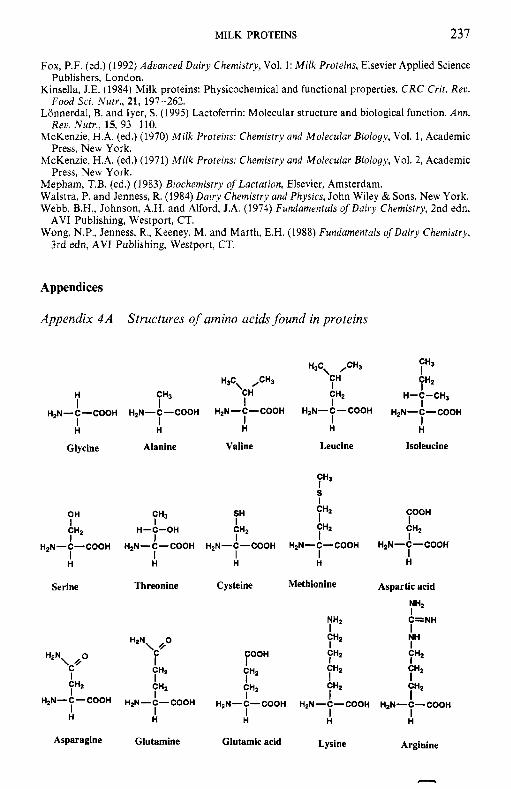
MILK PROTEINS 237
Fox, P.F. (ed.) (1992) Adcanced Dairy Chemistry, Vol. 1: Milk Proteins, Elsevier Applied Science
Kinsella, J.E. (1984) Milk proteins: Physicochemical and functional properties. C R C Crit. Rec.
Lonnerdal, B. and Iver. S. (1995) Lactoferrin: Molecular structure and biological function. Ann.
Publishers, London.
Food Sci. Nutr., 21, 197-262.
Reti. Nurr., IS, 93-110.
Press, New York.
-
McKenzie, H.A. (ed.) (1970) Milk Proteins: Chemistry and Molecular Biology, Vol. 1, Academic
McKenzie, H.A. (ed.) (1971) Milk Proteins: Chemistry and Molecular Biology, Vol. 2, Academic
Mepham, T.B. (ed.) (1983) Biochemisrry of Lactation, Elsevier, Amsterdam. Walstra, P. and Jenness, R. (1984) Dairy Chemistry and Physics, John Wiley & Sons, New York. Webb, B.H., Johnson, A.H. and Alford, J.A. (1974) Fundamentals of Dairy Chemistry, 2nd edn,
Wong, N.P., Jenness, R., Keeney, M. and Marth, E.H. (1988) Fundamentals ofDairy Chemistry,
Press, New York.
AVI Publishing, Westport, CT.
3rd edn, AVI Publishing, Westport, CT.
Appendices
Appendix 4A Structures of amino acids found in proteins
Glycine Alanine Valine Leucine Isoleucine
7H3
CH2 y 2 y 2 I
s I CH2 COOH I I
I
SH y 3 I
OH I CH2 I
I H
H-C-OH I
I H
HpN-C-COOH H2N-C-COOH H2N-C-COOH H2N-C-COOH H2N-C-COOH
H I H
I H
Serine Threonine Cysteine Methionine Aspartic acid
y 2
y 2 I ' 7 2 y 2
C=NH
NH I
NH2 I
CH2 I CH2 I I
CH2 I CHI I I
FooH CH2 I CH2 I
I H2N-C-COOH H2N-C-COOH HaN-C-COOH
H H H
Lysine Arginine Glutamic acid Asparagine Glutamine
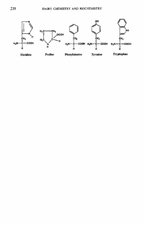
238 DAIRY CHEMISTRY AND BIOCHEMISTRY
HaC-Cb
I I,c00H cHz Hzc\ /'\H I
HzN - C -COOH I
N H
H
CHz I I H
HzN- C- COOH
Hitidine Proline Phenylalanine Tyrosine Tryptophan
5 Salts of milk
5.1 Introduction
The salts of milk are mainly the phosphates, citrates, chlorides, sulphates, carbonates and bicarbonates of sodium, potassium, calcium and mag- nesium. Approximately 20 other elements are found in milk in trace amounts, including copper, iron, silicon, zinc and iodine. Strictly speaking, the proteins of milk should be included as part of the salt system since they carry positively and negatively charged groups and can form salts with counter-ions; however, they are not normally treated as such. There is no lactate in freshly drawn milk but it may be present in stored milk and in milk products. The major elements are of importance in nutrition, in the preparation, processing and storage of milk products due to their marked influence on the conformation and stability of milk proteins, especially caseins, and to a lesser extent the stability of lipids and the activity of some indigenous enzymes.
5.2 Method of analysis
The mineral content of foods is usually determined from the ash prepared by heating a sample at 500-600°C in a muffle furnace for about 4 h to oxidize organic matter. The ash does not represent the salts as present in the food because:
1. the ash is a mixture, not of the original salts, but of the carbonates and
2. phosphorus and sulphur from proteins and lipids are present in the ash,
3. the temperature usually employed in ashing may vaporize certain volatile
oxides of the elements present in the food;
while organic ions, such as citrate, are lost during incineration; and
elements, e.g. sodium and potassium.
Therefore, it is difficult or impossible to relate the ash obtained from a food with its salts system, and low values are obtained for certain mineral elements by analysis of the ash compared to direct analysis of the intact food. Titrimetric, colorimetric, polarographic, flame photometric and atomic absorption spectrophotometric techniques are frequently used to analyse for the various mineral constituents; however, the quantitative estimation of
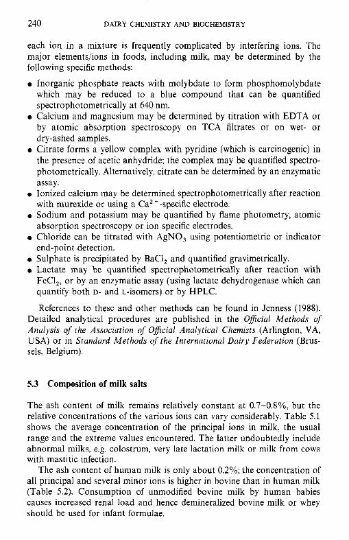
240 DAIRY CHEMISTRY A N D BIOCHEMISTRY
each ion in a mixture is frequently complicated by interfering ions. The major elements/ions in foods, including milk, may be determined by the following specific methods:
0 Inorganic phosphate reacts with molybdate to form phosphomolybdate which may be reduced to a blue compound that can be quantified spectrophotometrically at 640 nm.
0 Calcium and magnesium may be determined by titration with EDTA or by atomic absorption spectroscopy on TCA filtrates or on wet- or dry-ashed samples.
0 Citrate forms a yellow complex with pyridine (which is carcinogenic) in the presence of acetic anhydride; the complex may be quantified spectro- photometrically. Alternatively, citrate can be determined by an enzymatic assay.
0 Ionized calcium may be determined spectrophotometrically after reaction with murexide or using a Ca*'-specific electrode. Sodium and potassium may be quantified by flame photometry, atomic absorption spectroscopy or ion specific electrodes.
0 Chloride can be titrated with AgNO, using potentiometric or indicator end-point detection.
0 Sulphate is precipitated by BaCl, and quantified gravimetrically. 0 Lactate may be quantified spectrophotometrically after reaction with
FeCl,, or by an enzymatic assay (using lactate dehydrogenase which can quantify both D- and L-isomers) or by HPLC.
References to these and other methods can be found in Jenness (1988). Detailed analytical procedures are published in the Oflcial Methods of Analysis of the Association of Oficial Analytical Chemists (Arlington, VA, USA) or in Standard Methods of the International Dairy Federation (Brus- sels, Belgium).
5.3 Composition of milk salts
The ash content of milk remains relatively constant at 0.7-0.8%, but the relative concentrations of the various ions can vary considerably. Table 5.1 shows the average concentration of the principal ions in milk, the usual range and the extreme values encountered. The latter undoubtedly include abnormal milks, e.g. colostrum, very late lactation milk or milk from cows with mastitic infection.
The ash content of human milk is only about 0.2%; the concentration of all principal and several minor ions is higher in bovine than in human milk (Table 5.2). Consumption of unmodified bovine milk by human babies causes increased renal load and hence demineralized bovine milk or whey should be used for infant formulae.
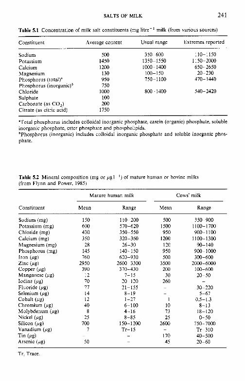
SALTS OF MILK 24 1
Table 5.1 Concentration of milk salt constituents (mg litre-' milk (from various sources)
Constituent Average content Usual range Extremes reported
Sodium 500 350-600 1 10- 1 150 Potassium 1450 1350-1550 11 50-2000 Calcium 1200 1000-1400 650-2650 Magnesium 130 100-150 20-230 Phosphorus (total)" 950 750-1100 470- 1440
Chloride 1000 800-1400 540-2420 Phosphorus (inorganic)b 750
Sulphate 100 Carbonate (as CO,) 200 Citrate (as citric acid) 1750
"Total phosphorus includes colloidal inorganic phosphate, casein (organic) phosphate, soluble inorganic phosphate, ester phosphate and phospholipids. bPhosphorus (inorganic) includes colloidal inorganic phosphate and soluble inorganic phos- phate.
Table 5.2 Mineral composition (mg or pgl- ' ) of mature human or bovine milks (from Flynn and Power, 1985)
Mature human milk Cows' milk
Constituent Mean Range Mean Range
Sodium (mg) Potassium (mg) Chloride (mg) Calcium (mg) Magnesium (mg) Phosphorus (mg) Iron (pg) Zinc (pg) Copper (pg) Manganese (pg) Iodine (pg) Fluoride (pg) Selenium (pg) Cobalt (pg) Chromium (pg) Molybdenum (pg) Nickel (pg) Silicon (pg) Vanadium (pg)
Arsenic (pg) Tin (Peg)
150 600 430 350 28
145 760
2950 390
12 70 77 14 12 40
8 25
700 7
50 -
110-200 570-620 350-550 320-360 26-30
140-150 620-930
2600-3300 370-430
7-15 20- 120 21-155
8-19 1-27 6-100 4-16 8-85
150- 1200 Tr-15
-
-
500 1500 950
1200 120 950 500
3500 200 30
260 -
-
1 10 73 25
2600
170 45
-
350-900 1100-1700 900- 1100
1100- 13OO 90-140
900-1000 300-600
2000-6000 100-600 20-50 -
30-220 5-67
0.5-1.3 8-13
18-120 0-50
750-7000 Tr-310 40-500 20-60
Tr. Trace.
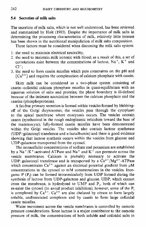
242 DAIRY CHEMISTRY A N D BIOCHEMISTRY
5.4 Secretion of milk salts
The secretion of milk salts, which is not well understood, has been reviewed and summarized by Holt (1985). Despite the importance of milk salts in determining the processing characteristics of milk, relatively little interest has been shown in the nutritional manipulation of milk salts composition.
Three factors must be considered when discussing the milk salts system:
1. the need to maintain electrical neutrality; 2. the need to maintain milk isotonic with blood; as a result of this, a set of
correlations exist between the concentrations of lactose, Na', K + and c1-;
3. the need to form casein micelles which puts constraints on the pH and [Ca"] and requires the complexation of calcium phosphate with casein.
Skim milk can be considered as a two-phase system consisting of casein-colloidal calcium phosphate micelles in quasi-equilibrium with an aqueous solution of salts and proteins; the phase boundary is ill-defined because of the intimate association between the calcium phosphate and the caseins (phosphoproteins).
A fat-free primary secretion is formed within vesicles formed by blebbing- off of the Golgi dicytosomes; the vesicles pass through the cytoplasm to the apical membrane where exocytosis occurs. The vesicles contain casein (synthesized in the rough endoplasmic reticulum toward the base of the mammocyte); fully-formed casein micelles have been demonstrated within the Golgi vesicles. The vesicles also contain lactose synthetase (UDP : galactosyl transferase and sr-lactalbumin) and there is good evidence showing that lactose synthesis occurs within the vesicles from glucose and UDP-galactose transported from the cytosol.
The intracellular concentrations of sodium and potassium are established by a Na+/K+-activated ATPase and Na+ and K + can permeate across the vesicle membranes. Calcium is probably necessary to activate the UDP : galactosyl transferase and is transported by a CaZ +/Mg2 +-ATPase which concentrates Ca2 + against an electrical potential gradient from pM concentrations in the cytosol to mM concentrations in the vesicles. Inor- ganic P (Pi) can be formed intravesicularly from UDP formed during the synthesis of lactose from UDP-galactose and glucose. UDP, which cannot cross the membrane, is hydrolysed to UMP and Pi, both of which can re-enter the cytosol (to avoid product inhibition); however, some of the Pi is complexed by Ca2+. Caz+ are also chelated by citrate to form largely soluble, undissociated complexes and by casein to form large colloidal casein micelles.
Water movement across the vesicle membranes is controlled by osmotic pressure considerations. Since lactose is a major contributor to the osmotic pressure of milk, the concentrations of both soluble and colloidal salts in
SALTS OF MILK 243
/
GO1 GOLGI VESICLE C a s e i n
u D P-G a1 ac t ose
,GI VESICLE C a s e i n
-----=2 CanPO, \ C l l
D
’ CYTOSOL
ci I Figure 5.1 Summary of some transport mechanisms for calcium, phosphate and citrate from
the cytosol of the secretory cell to the inside of Golgi vesicles (from Holt, 1981).
milk are strongly influenced by lactose concentration and the mechanism by which it is synthesized.
Inter-relationships in the biosynthesis of the principal milk salts are summarized in Figure 5.1. Transport of several ionic species via the junctions between cells (paracellular) occurs during early and late lactation and during mastitic infection when the junctions between cells are more open.
5.5 Factors influencing variation in salt composition
The composition of milk salts is influenced by a number of factors, including breed, individuality of the cow, stage of lactation, feed, mastitic infection and season of the year. The more important factors are discussed below.
5.5.1 Breed of cow
Milk from Jersey cows usually contains more calcium and phosphorus than milk from other breeds, including Holstein, but the concentrations of sodium and chloride are usually lower.
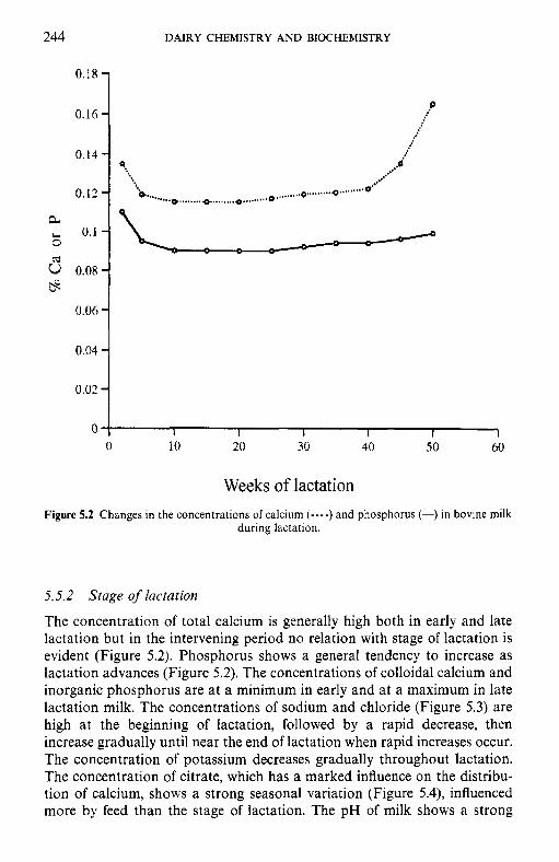
244 DAIRY CHEMISTRY AND BIOCHEMISTRY
0.18 -
0.16-
0.14 - 0.12 - 0.1-
a 6
& G 0.08-
0.06 -
p
...d ...*.. , .o,. ... . .. * .... ....*O"'
9..
o"..*....* ,... * ... *... , , . , , .*. .... I.1.0 .I..*..
= = - ~ -
Weeks of lactation Figure 5.2 Changes in the concentrations of calcium (----) and phosphorus (-) in bovine milk
during lactation.
5.5.2 Stage of lactation
The concentration of total calcium is generally high both in early and late lactation but in the intervening period no relation with stage of lactation is evident (Figure 5.2). Phosphorus shows a general tendency to increase as lactation advances (Figure 5.2). The concentrations of colloidal calcium and inorganic phosphorus are at a minimum in early and at a maximum in late lactation milk. The concentrations of sodium and chloride (Figure 5.3) are high at the beginning of lactation, followed by a rapid decrease, then increase gradually until near the end of lactation when rapid increases occur. The concentration of potassium decreases gradually throughout lactation. The concentration of citrate, which has a marked influence on the distribu- tion of calcium, shows a strong seasonal variation (Figure 5.4), influenced more by feed than the stage of lactation. The pH of milk shows a strong
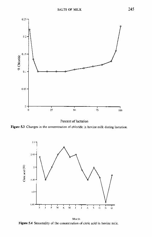
SALTS OF MILK 245
0.25
0.2
0.15
3 2 u ct
0.1
0.05
Percent of lactation Figure 5.3 Changes in the concentration of chloride in bovine milk during lactation.
Monih Figure 5.4 Seasonality of the concentration of citric acid in bovine milk.
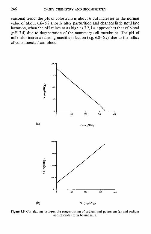
246 DAIRY CHEMISTRY AND BIOCHEMISTRY
seasonal trend; the pH of colostrum is about 6 but increases to the normal value of about 6.6-6.7 shortly after parturition and changes little until late lactation, when the pH raises to as high as 7.2, i.e. approaches that of blood (pH 7.4) due to degeneration of the mammary cell membrane. The pH of milk also increases during mastitic infection (e.g. 6.8-6.9), due to the influx of constituents from blood.
X
Figure 5.5 Correlations between the concentration of sodium and potassium (a) and sodium and chloride (b) in bovine milk.
SALTS OF MILK 247
5.5.3 Infection of the udder
Milk from cows with mastitic infections contains a low level of total solids, especially lactose, and high levels of sodium and chloride, the concentration of which are directly related (Figure 5.5). The sodium and chloride ions come from the blood to compensate osmotically for the depressed lactose synthesis or vice versa.
These are related by the Koestler number:
100 x %C1 %lactose
Koestler number =
which is normally 1.5-3.0 but increases on mastitic infection and has been used as an index of such (better methods are now available, e.g. somatic cell count, activity of certain enzymes, especially catalase and N-acetyl- glucosamidase). The pH of milk increases to approach that of blood during mastitic infection.
5.5.4 Feed
Feed has relatively little effect on the concentration of most elements in milk because the skeleton acts as a reservoir of minerals. The level of citrate in milk decreases on diets very deficient in roughage and results in the ‘Utrecht phenomenon’, i.e. milk of very low heat stability. Relatively small changes in the concentrations of milk salts, especially of Ca, Pi and citrate, can have very significant effects on the processing characteristics of milk and hence these can be altered by the level and type of feed, but definitive studies on this are lacking.
5.6 Interrelations of milk salt constituents
Various milk salts are interrelated and the interrelationships are affected by pH (Table 5.3). Those constituents, the concentrations of which are related to pH in the same way, are also directly related to each other (e.g. the concentrations of total soluble calcium and ionized calcium), while those related to pH in opposite ways are inversely related (e.g. the concentrations of potassium and sodium).
Relationships between some of the more important ions/molecules are shown in Figure 5.6. Three correlations are noteworthy:
1. The concentration of lactose is inversely related to the concentration of soluble salts expressed as osmolarity. This results from the requirement that milk be isotonic with blood.
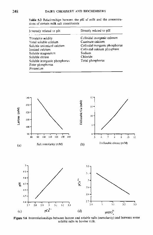
248
7 -
6 8 6.7
6.6 - 6.S - 6.4
DAIRY CHEMISTRY AND BIOCHEMISTRY
3.2 -
6 9 : / ; l \ 2 9 -
2.n -
I I I I I 1 2 7 I I I I
2 9 3 3.1 3.2 3.3
Table 5.3 Relationships between the pH of milk and the concentra- tions of certain milk salt constituents
Inversely related to pH
Titratable acidity Colloidal inorganic calcium Total soluble calcium Caseinate calcium Soluble unionized calcium Ionized calcium Colloidal calcium phosphate Soluble magnesium Sodium Soluble citrate Soluble inorganic phosphorus Total phosphorus Ester phosphorus Potassium
Directly related to pH
Colloidal inorganic phosphorus
Chloride
140 - 130-
120-
110-
loo -
RO DO 100 110 120 130 140
Salt osmoluity (mM)
Figure 5.6 Interrelationships between lactose and soluble salts (osmolarity) and between some soluble salts in bovine milk.
SALTS OF MILK 249
2. There is a direct correlation between the concentration of diffusible Ca (and diffusible Mg) and the concentration of diffusible citrate (Figure 5.6b); this correlation, which is very good at constant pH, exists because citrate chelates Ca2 + more strongly than phosphate to form soluble unionized salts.
3. The ratio HPOa-/H,PO; is strongly pH dependent, as is the solubility of Ca,(PO,), (section 5.8.1). As the pH is reduced, colloidal Ca,(PO,), dissolves but HP0;- -, H,PO, as the pH is reduced and hence both [Ca”] and soluble Pi are directly related to pH (Figure 5.6~). The [HPO:-] is inversely related to [Ca”] (Figure 5.6d).
5.7 Partition of milk salts between colloidal and soluble phases
Certain of the milk salts (e.g. chlorides, and the salts of sodium and potassium) are sufficiently soluble to be present almost entirely in the dissolved phase. The concentration of others, in particular calcium phos- phate, is higher than can be maintained in solution at the normal pH of milk. Consequently, these exist partly in soluble form and partly in an insoluble or colloidal form associated with casein. The state and distribution of these salts has been extensively reviewed by Pyne (1962) and Holt (1985).
The dividing line between soluble and colloidal is somewhat arbitrary, its exact position depending very much on the method used to achieve separation. However, a fairly sharp separation between the two phases is not difficult since the insoluble salts occur mainly associated with the colloidal casein micelles.
5.7.1
The methods used include dialysis, ultrafiltration, high-speed centrifugation and rennet coagulation. The method used must not cause changes in equilibrium between the two phases. The two most important precautions are to avoid changes in pH (lowering the pH dissolves colloidal calcium phosphate, see Figure 5.1 1) and temperature (reducing the temperature dissolves colloidal calcium phosphate and vice versa). Since milk comes from the cow at about 40”C, working at 20°C and especially at 4°C will cause significant shifts in calcium phosphate equilibrium.
Ultrafiltrates obtained using cellophane or polysulphone membranes at 20°C and a transmembrane pressure of c. 100 kPa are satisfactory, but the concentrations of citrate and calcium are slightly low due to sieving effects which are accentuated by high pressures. Dialysis of a small volume of water against at least 50 times its volume of milk (to which a little chloroform or azide has been added as preservative) at 20°C for 48h is the most satisfactory separation procedure and agrees closely with results obtained
Methods used to separate the colloidal and soluble phases
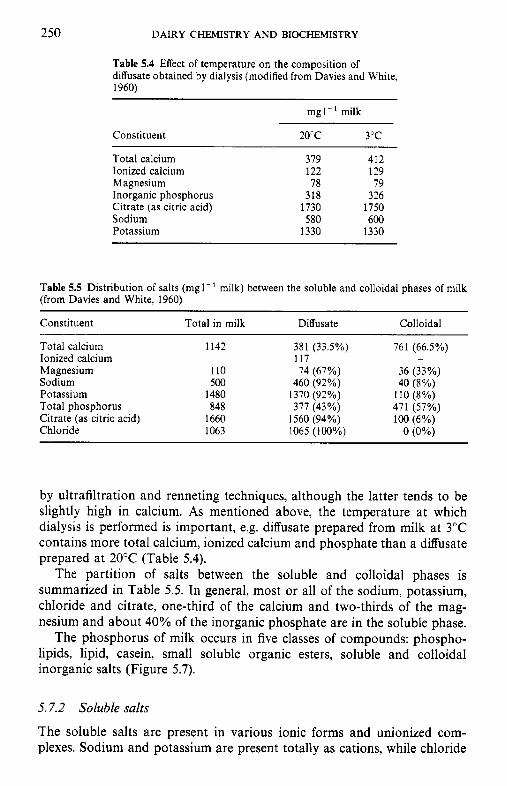
250 DAIRY CHEMISTRY A N D BIOCHEMISTRY
Table 5.4 Effect of temperature on the composition of diffusate obtained by dialysis (modified from Davies and White, 1960)
mg1-' milk
Constituent 20°C 3°C
Total calcium 379 412 Ionized calcium 122 129 Magnesium 78 79 Inorganic phosphorus 318 326 Citrate (as citric acid) 1730 1750 Sodium 580 600 Potassium 1330 1330
Table 5.5 Distribution of salts (mgl-' milk) between the soluble and colloidal phases of milk (from Davies and White, 1960)
Constituent Total in milk Diffusate Colloidal
Total calcium Ionized calcium Magnesium Sodium Potassium Total phosphorus Citrate (as citric acid) Chloride
1142 381 (33.5%) 117
110 74 (67%)
1480 1370 (92%) SO0 460 (92%)
848 377 (43%) 1660 1560 (94%) 1063 1065 (100%)
761 (66.5%)
36 (33%) 40 (8%)
110 (So/,)
100 (6%)
-
471 (57%)
0 (0%)
by ultrafiltration and renneting techniques, although the latter tends to be slightly high in calcium. As mentioned above, the temperature at which dialysis is performed is important, e.g. diffusate prepared from milk at 3°C contains more total calcium, ionized calcium and phosphate than a diffusate prepared at 20°C (Table 5.4).
The partition of salts between the soluble and colloidal phases is summarized in Table 5.5. In general, most or all of the sodium, potassium, chloride and citrate, one-third of the calcium and two-thirds of the mag- nesium and about 40% of the inorganic phosphate are in the soluble phase.
The phosphorus of milk occurs in five classes of compounds: phospho- lipids, lipid, casein, small soluble organic esters, soluble and colloidal inorganic salts (Figure 5.7).
5.7.2 Soluble salts
The soluble salts are present in various ionic forms and unionized com- plexes. Sodium and potassium are present totally as cations, while chloride
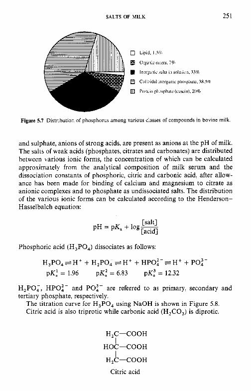
SALTS OF MILK 25 1
Figure 5.7 Distribution of phosphorus among various classes of compounds in bovine milk.
and sulphate, anions of strong acids, are present as anions at the pH of milk. The salts of weak acids (phosphates, citrates and carbonates) are distributed between various ionic forms, the concentration of which can be calculated approximately from the analytical composition of milk serum and the dissociation constants of phosphoric, citric and carbonic acid, after allow- ance has been made for binding of calcium and magnesium to citrate as anionic complexes and to phosphate as undissociated salts. The distribution of the various ionic forms can be calculated according to the Henderson- Hasselbalch equation:
[salt] [acid]
pH = pK, + log-
Phosphoric acid (H,P04) dissociates as follows:
H 3 P 0 , e H + + H,PO, e H + + HPOt- e H + + PO:- pK,' = 1.96 pK: = 6.83 pK2 = 12.32
H,PO,, HPOZ- and PO:- are referred to as primary, secondary and tertiary phosphate, respectively.
The titration curve for H3PO4 using NaOH is shown in Figure 5.8. Citric acid is also triprotic while carbonic acid (H,C03) is diprotic.
H,C-COOH
HOC-COOH
H,C-COOH
Citric acid
I I
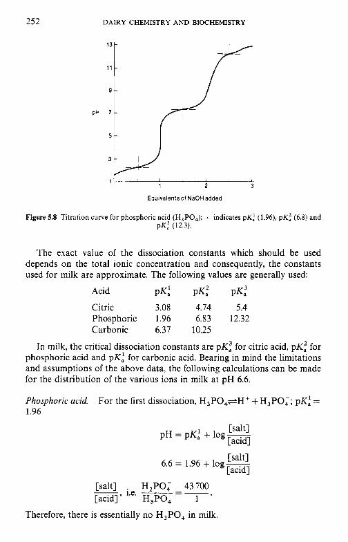
252 DAIRY CHEMISTRY AND BIOCHEMISTRY
I I I I I I
1 2 3
Eauivalentsof NaOH added
Figure 5.8 Titration curve for phosphoric acid (H,PO,); + indicates p K : (1.96), p K i (6.8) and pK: (12.3).
The exact value of the dissociation constants which should be used depends on the total ionic concentration and consequently, the constants used for milk are approximate. The following values are generally used:
Acid PK,' PK,Z PK,3 Citric 3.08 4.74 5.4 Phosphoric 1.96 6.83 12.32 Carbonic 6.37 10.25
In milk, the critical dissociation constants are pK5 for citric acid, pK: for phosphoric acid and pK,' for carbonic acid. Bearing in mind the limitations and assumptions of the above data, the following calculations can be made for the distribution of the various ions in milk at pH 6.6.
Phosphoric acid. 1.96
For the first dissociation, H3P04+H+ + H,PO;; pKi =
[salt] [acid]
[salt] [acid]
PH = p ~ , ' + log-
6.6 = 1.96 + log-
[salt] . H,PO; 43700 1.e. ___ -
[acid]' H,PO, 1 '
--
Therefore, there is essentially no H,PO, in milk.
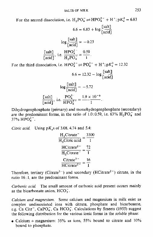
SALTS OF MILK 253
For the second dissociation, i.e. H,PO, e HPOi- + H’; pK: = 6.83
[salt] [acid]
6.6 = 6.83 + log-
[salt] [acid]
log- = -0.23
[salt] HPOi- - 0.59 i.e. ~ - - [acid]’ H,PO, 1 ’
For the third dissociation, i.e. HPOi- *PO:- + H’; pKi = 12.32
[salt] [acid]
6.6 = 12.32 + log-
[salt] [acid]
log- = -5.72
PO:- 1.9 x - - [salt] [acid]’ HPOi- 1 ’
Dihydrogenphosphate (primary) and monohydrogenphosphate (secondary) are the predominant forms, in the ratio of 1.0:0.59, i.e. 63% H,PO, and 37% HPOi- .
Citric acid. Using pK,s of 3.08, 4.74 and 5.4:
H,Citrate- 3300 H,Citric acid - 1
HCitrate2- 72 H,Citrate- 1
Citrate3- 16 HCitrate2- 1
Therefore, tertiary (Citrate, -) and secondary (HCitrate’ -) citrate, in the ratio 16: 1, are the predominant forms.
Carbonic acid. as the bicarbonate anion, HCO;.
--
- _ -
- _ -
The small amount of carbonic acid present occurs mainly
Calcium and magnesium. Some calcium and magnesium in milk exist as complex undissociated ions with citrate, phosphate and bicarboante, e.g. Ca Citr-, CaPO,, Ca HCO;. Calculations by Smeets (1955) suggest the following distribution for the various ionic forms in the soluble phase:
0 Calcium + magnesium: 35% as ions, 55% bound to citrate and 10% bound to phosphate.
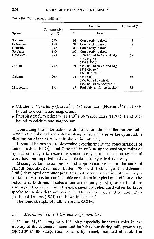
254 DAIRY CHEMISTRY AND BIOCHEMISTRY
Table 5.6 Distribution of milk salts ~~
Soluble Colloidal (%) Concentration
Species (mg1-l) YO form
Sodium Potassium Chloride Sulphate Phosphate
Citrate
Calcium
Magnesium
500 1450 1200 100 750
1750
1200
130
92 92
100 100 43
94
34
67
Completely ionized 8 Completely ionized 8 Completely ionized - Completely ionized -
10% bound to Ca and Mg 57 - 51% H,PO- 39% HPOf- 85% b o u d to Ca and Mg 14% Citrate3- 1% HCitrate2- 35% Ca2+ 66 55% bound to citrate 10% bound to phosphate Probably similar to calcium 33
0 Citrates: 14% tertiary (Citrate3-), 1 % secondary (HCitrate2-) and 85%
0 Phosphates: 51% primary (H2P0,), 39% secondary (HPO:-) and 10% bound to calcium and magnesium.
bound to calcium and magnesium.
Combining this information with the distribution of the various salts between the colloidal and soluble phases (Table 5.5), gives the quantitative distribution of the salts in milk shown in Table 5.6.
It should be possible to determine experimentally the concentrations of anions such as HPOi- and Citrate3- in milk using ion-exchange resins or by nuclear magnetic resonance spectroscopy, but no such experimental work has been reported and available data are by calculation only.
Making certain assumptions and approximations as to the state of various ionic species in milk, Lyster (1981) and Holt, Dalgleish and Jenness (1981) developed computer programs that permit calculation of the concen- trations of various ions and soluble complexes in typical milk diffusate. The outcome of both sets of calculations are in fairly good agreement and are also in good agreement with the experimentally determined values for those species for which data are available. The values calculated by Holt, Dal- gleish and Jenness (1981) are shown in Table 5.7.
The ionic strength of milk is around 0.08 M.
5.7.3
Ca2+ and Mg2+, along with H', play especially important roles in the stability of the caseinate system and its behaviour during milk processing, especially in the coagulation of milk by rennet, heat and ethanol. The
Measurement of calcium and magnesium ions
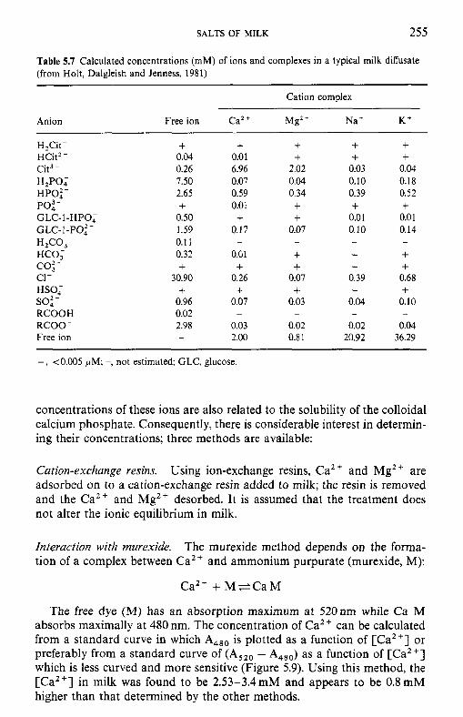
SALTS OF MILK 255
Table 5.7 Calculated concentrations (mM) of ions and complexes in a typical milk diffusate (from Holt, Dalgleish and Jenness, 1981)
Cation complex
Anion Free ion Ca2 + Mg2 + Na' K'
H,Cit- HCit2- Cit3- H2PO; HP0:- Po: -
GLC- 1 -PO: - HZCO,
co: -
so: -
GLC- 1 -HPO;
HCO;
c1- HSO;
RCOOH RCOO- Free ion
+ 0.04 0.26 7.50 2.65 +
0.50 1.59 0.1 1 0.32 +
30.90 +
0.96 0.02 2.98
+ 0.0 1 6.96 0.07 0.59 0.01 +
0.17
0.Gl +
0.26 +
0.07
0.03 2.00
-
+ +
2.02 0.04 0.34 + +
0.07
+ +
0.07 +
0.03
0.02 0.8 1
-
+ +
0.03 0.10 0.39 + 0.01 0.10
+ + 0.39 +
0.04
0.02 20.92
-
+ +
0.04 0.18 0.52 + 0.01 0.14
+ + 0.68 +
0.10
0.04 36.29
-
+, <0.005 pM; -, not estimated; GLC, glucose.
concentrations of these ions are also related to the solubility of the colloidal calcium phosphate. Consequently, there is considerable interest in determin- ing their concentrations; three methods are available:
Cation-exchange resins. Using ion-exchange resins, Ca2 + and Mg2 + are adsorbed on to a cation-exchange resin added to milk; the resin is removed and the Ca2+ and Mg2+ desorbed. It is assumed that the treatment does not alter the ionic equilibrium in milk.
Interaction with murexide. The murexide method depends on the forma- tion of a complex between Ca2+ and ammonium purpurate (murexide, M):
Ca2+ + M e C a M
The free dye (M) has an absorption maximum at 520nm while Ca M absorbs maximally at 480 nm. The concentration of Ca2+ can be calculated from a standard curve in which A,,, is plotted as a function of [Ca2+] or preferably from a standard curve of (A,,, - A,,,) as a function of [Ca2+] which is less curved and more sensitive (Figure 5.9). Using this method, the [Ca"] in milk was found to be 2.53-3.4 mM and appears to be 0.8 mM higher than that determined by the other methods.
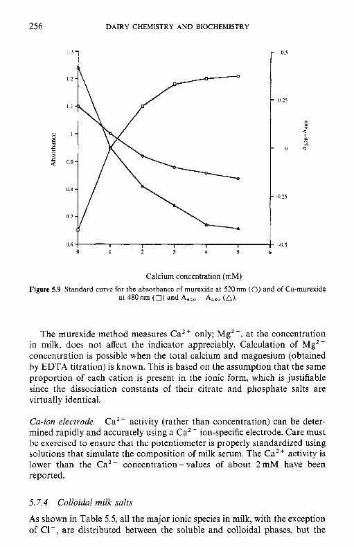
256 DAIRY CHEMISTRY AND BIOCHEMISTRY
"1
0.6 I I I I I
0 1 2 3 4 5 6
Calcium concentration (mM) Figure 5.9 Standard curve for the absorbance of murexide at 520nm (0) and of Ca-murexide
at 480 nrn (0) and A,,, - A480 (A).
The murexide method measures Ca2+ only; Mg2+, at the concentration in milk, does not affect the indicator appreciably. Calculation of Mgz+ concentration is possible when the total calcium and magnesium (obtained by EDTA titration) is known. This is based on the assumption that the same proportion of each cation is present in the ionic form, which is justifiable since the dissociation constants of their citrate and phosphate salts are virtually identical.
Ca-ion electrode. Ca2 + activity (rather than concentration) can be deter- mined rapidly and accurately using a Ca2 + ion-specific electrode. Care must be exercised to ensure that the potentiometer is properly standardized using solutions that simulate the composition of milk serum. The Ca2 i. activity is lower than the Ca2+ concentration - values of about 2 mM have been reported.
5.7.4 Colloidal milk salts
As shown in Table 5.5, all the major ionic species in milk, with the exception of C1-, are distributed between the soluble and colloidal phases, but the
SALTS OF MILK 257
principal colloidal salt is calcium phosphate; about 67% and 57%, respec- tively, of the total calcium and phosphate are in the colloidal phase. The colloidal inorganic salts are, therefore, frequently referred to as colloidal calcium phosphate (CCP), although some sodium, potassium, magnesium, zinc and citrate are also present in the colloidal phase. CCP is closely associated with the casein micelles and there are two principal questions as to its nature:
0 its composition and structure; 0 the nature of its association with casein.
Composition and structure. All the colloidal sodium (40 mg 1-I), potassium ( l lomgl- ' ) and most of the magnesium (30mg 1-') are probably asso- ciated with the casein as counter-ions to the negatively charged organic phosphate and carboxylic acid groups of the protein. It has been calculated that approximately 30% of the colloidal calcium (c. 250 mg I - ' ) is also directly attached to these groups. According to most authors (Pyne, 1962), casein is capable of binding 25-30 moles calcium per los g casein (i.e. about 116Og calcium per 10sg casein). Assuming that milk contains 25g casein 1-', the calcium-binding potential of the casein is about 300 mg 1-' of milk. Since the neutralizing potential of Na' and K + is half that of Ca2+ and Mg2+, the binding capacity of 300 mg I - ' is reasonably close to the sum of the values given above.
These calculations leave about 500 mg of calcium and about 350 mg of phosphate present in the colloidal phase per litre of milk to be accounted for. The available evidence suggests that the excess CCP is present largely as tricalcium phosphate, Ca,(PO,),, or some similar salt.
The so-called Ling oxalate titration indicates that CCP consists of 80% Ca,(PO,), and 20% CaHPO,, with an overall Ca: P ratio of 1.4: 1 (Pyne, 1962). However, the oxalate titration procedure has been criticized because many of the assumptions made are not reliable. Pyne and McGann (1960) developed a new technique to study the composition of CCP. Milk was acidified to about pH4.9 at 2"C, followed by exhaustive dialysis of the acidified milk against a large excess of bulk milk; this procedure restored the acidified milk to normality in all respects except that CCP was not reformed. Analysis of milk and CCP-free milk (assumed to differ from milk only in respect of CCP) showed that the ratio of C a : P in CCP was 1.7: 1. The difference between this value and that obtained by the oxalate titration (i.e. 1.4: 1) was attributed to the presence of citrate in the CCP complex, which is not measured by the oxalate method. Pyne and McGann (1960) suggested that CCP has an apatite structure with the formula:
3Ca,(P0,)2, CaHCitr- or 2.5Ca3(PO,),, CaHPO,, 0.5 Ca,Citr;.
Based on the assumption that the amount of Ca bound directly to casein is equivalent to the number of ester phosphate groups present, Schmidt
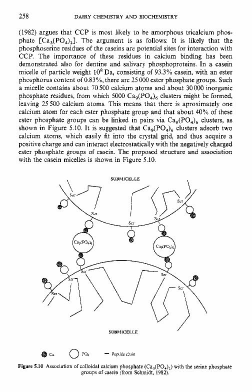
258 DAIRY CHEMISTRY AND BIOCHEMISTRY
(1982) argues that CCP is most likely to be amorphous tricalcium phos- phate [Ca,(PO,),]. The argument is as follows: It is likely that the phosphoserine residues of the caseins are potential sites for interaction with CCP. The importance of these residues in calcium binding has been demonstrated also for dentine and salivary phosphoproteins. In a casein micelle of particle weight 10' Da, consisting of 93.3% casein, with an ester phosphorus content of 0.83%, there are 25 000 ester phosphate groups. Such a micelle contains about 70 500 calcium atoms and about 30 000 inorganic phosphate residues, from which 5000 Ca,(PO,), clusters might be formed, leaving 25500 calcium atoms. This means that there is aproximately one calcium atom for each ester phosphate group and that about 40% of these ester phosphate groups can be linked in pairs via Ca,(PO,), clusters, as shown in Figure 5.10. It is suggested that Ca,(PO,), clusters adsorb two calcium atoms, which easily fit into the crystal grid, and thus acquire a positive charge and can interact electrostatically with the negatively charged ester phosphate groups of casein. The proposed structure and association with the casein micelles is shown in Figure 5.10.
\ SUBMICELLE \
/ SUBMICELLE
Ca 0 PO4 - Pcplidc chain
Figure 5.10 Association of colloidal calcium phosphate (Ca,(PO,),) with the serine phosphate groups of casein (from Schmidt, 1982).
SALTS OF MILK 259
The best physical studies, using various forms of X-ray spectroscopy, on the structure of CCP have been undertaken by Holt and colleagues (Holt, 1985). It was concluded that the most likely form of CCP is brushite (CaHP0,.2H20), which has also been identified in bone and other calcified tissues. He explains the difference between the Ca/P ratio found by analysis, i.e. 1.51-1.6 and the Ca/P ratio of CaHPO,, i.e. 1.0, as being due to the ability of the phosphate moiety of phosphoserine to substitute in surface sites of a brushite-type lattice.
Association with casein. The colloidal calcium phosphate is closely asso- ciated with the casein; it does not precipitate out of solution and is considered to be protected against precipitation by the casein. Two possible forms of protection are suggested:
0 physical protection; 0 chemical association between CCP and casein.
Experimental evidence strongly favours the idea of chemical associ- ation:
0 CCP remains attached to the casein following treatment with protein dissociating agents (e.g. urea) or following proteolysis.
0 Comparison of the potentiometric titration curves of milk and CCP-free milk shows more reactive organic phosphate groups in the latter, sugges- ting that CCP is attached to the organic casein phosphate groups, thereby rendering them less active.
0 The formol titration is not influenced by removal of CCP, suggesting that &NH,-groups of lysine are not involved.
The views of Schmidt and Holt on the association between CCP and casein, i.e. via a shared Ca2+ (Schmidt) or a shared phosphoserine, i.e. phosphoserine as part of the CCP crystal lattice (Holt), support the hypothesis of chemical association.
Although CCP represents only about 6% of the dry weight of the casein micelle, it plays an essential role in its structure and properties and hence has major effects on the properties of milk; it is the integrating factor in the casein micelle; without it, milk is not coagulable by rennet and its heat and calcium stability properties are significantly altered. In fact, milk would be a totally different fluid without colloidal calcium phosphate.
As discussed in Chapter 4 (p. 186), Holt (1994) has proposed that casein has evolved with the ability to bind high concentrations of calcium and phosphate so that milk can contain high levels of these ions, which are essential for neonatal growth, without precipitation in the ducts of the mammary glands.
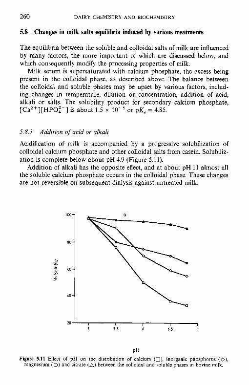
260 DAIRY CHEMISTRY AND BIOCHEMISTRY
100-
8.0 -
60-
40 -
5.8 Changes in milk salts equilibria induced by various treatments
The equilibria between the soluble and colloidal salts of milk are influenced by many factors, the more important of which are discussed below, and which consequently modify the processing properties of milk.
Milk serum is supersaturated with calcium phosphate, the excess being present in the colloidal phase, as described above. The balance between the colloidal and soluble phases may be upset by various factors, includ- ing changes in temperature, dilution or concentration, addition of acid, alkali or salts. The solubility product for secondary calcium phosphate, [Ca2+][HPO:-] is about 1.5 x or pK, = 4.85.
5.8.1
Acidification of milk is accompanied by a progressive solubilization of colloidal calcium phosphate and other colloidal salts from casein. Solubiliz- ation is complete below about pH 4.9 (Figure 5.11).
Addition of alkali has the opposite effect, and at about pH 11 almost all the soluble calcium phosphate occurs in the colloidal phase. These changes are not reversible on subsequent dialysis against untreated milk.
Addition of acid or alkali
b)
3 a - 0 v1
20 ' I I I I I 5 5.5 6 6.5 7
PH Figure 5.11 Effect of pH on the distribution of calcium (O), inorganic phosphorus (O),
magnesium (0) and citrate (A) between the colloidal and soluble phases in bovine milk.
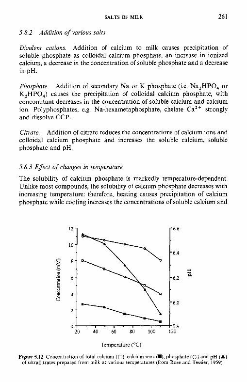
SALTS OF MILK 26 1
5.8.2 Addition of various salts
Divalent cations. Addition of calcium to milk causes precipitation of soluble phosphate as colloidal calcium phosphate, an increase in ionized calcium, a decrease in the concentration of soluble phosphate and a decrease in pH.
Phosphate. Addition of secondary Na or K phosphate (i.e. Na,HPO, or K,HPO,) causes the precipitation of colloidal calcium phosphate, with concomitant decreases in the concentration of soluble calcium and calcium ion. Polyphosphates, e.g. Na-hexametaphosphate, chelate Ca2 + strongly and dissolve CCP.
Citrate. Addition of citrate reduces the concentrations of calcium ions and colloidal calcium phosphate and increases the soluble calcium, soluble phosphate and pH.
5.8.3 Efect of changes in temperature
The solubility of calcium phosphate is markedly temperature-dependent. Unlike most compounds, the solubility of calcium phosphate decreases with increasing temperature; therefore, heating causes precipitation of calcium phosphate while cooling increases the concentrations of soluble calcium and
5.8 20 40 60 80 100 120
Temperature ("C)
Figure 5.12 Concentration of total calcium (O), calcium ions (M), phosphate (0) and pH (A) of ultrafiltrates prepared from milk at various temperatures (from Rose and Tessier, 1959).
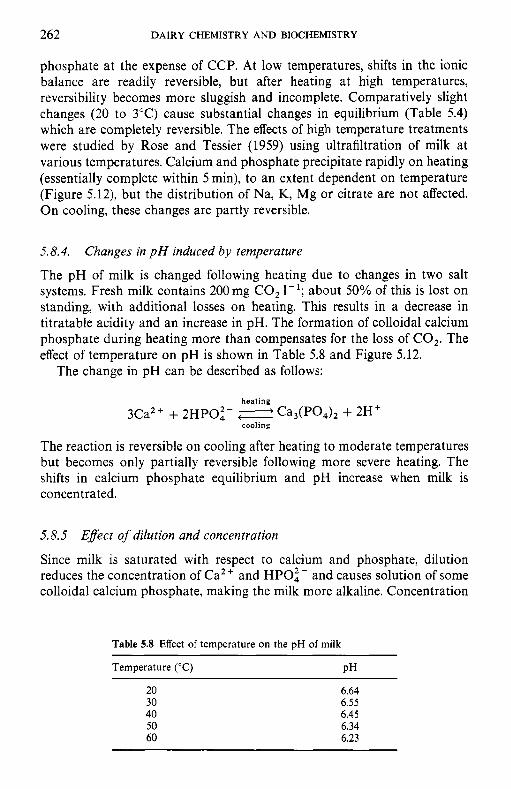
262 DAIRY CHEMISTRY AND BIOCHEMISTRY
phosphate at the expense of CCP. At low temperatures, shifts in the ionic balance are readily reversible, but after heating at high temperatures, reversibility becomes more sluggish and incomplete. Comparatively slight changes (20 to 3°C) cause substantial changes in equilibrium (Table 5.4) which are completely reversible. The effects of high temperature treatments were studied by Rose and Tessier (1959) using ultrafiltration of milk at various temperatures. Calcium and phosphate precipitate rapidly on heating (essentially complete within 5 min), to an extent dependent on temperature (Figure 5.12), but the distribution of Na, K, Mg or citrate are not affected. On cooling, these changes are partly reversible.
5.8.4.
The pH of milk is changed following heating due to changes in two salt systems. Fresh milk contains 200mg CO, 1-'; about 50% of this is lost on standing, with additional losses on heating. This results in a decrease in titratable acidity and an increase in pH. The formation of colloidal calcium phosphate during heating more than compensates for the loss of CO,. The effect of temperature on pH is shown in Table 5.8 and Figure 5.12.
Changes in p H induced by temperature
The change in pH can be described as follows:
heating
cooling 3Ca2+ + 2HPOi- < e C a d P O . d 2 + 2Ht
The reaction is reversible on cooling after heating to moderate temperatures but becomes only partially reversible following more severe heating. The shifts in calcium phosphate equilibrium and pH increase when milk is concentrated.
5.8.5 Effect of dilution and concentration
Since milk is saturated with respect to calcium and phosphate, dilution reduces the concentration of Ca2+ and HPOi - and causes solution of some colloidal calcium phosphate, making the milk more alkaline. Concentration
Table 5.8 Effect of temperature on the pH of milk
Temperature ('C) PH
20 30 40 50 60
6.64 6.55 6.45 6.34 6.23
SALTS OF MILK 263
of milk causes precipitation of colloidal phosphate and shifts the reaction of milk to the acid side, e.g. concentration by a factor of 2: 1 reduces the pH to 6.2.
H20 Dilution: Ca,(PO,), - 3Ca2+ + 2HPOi- + 2 0 H -
Concentration: 3Ca2+ + 2HPO:- + Ca,(PO,), + 2H+
5.8.6 Efect of freezing
Freezing milk causes crystallization of pure water and the unfrozen liquid becomes more saturated with respect to various salts. Some soluble calcium phosphate precipitates as Ca,(PO,),, with the release of H + and a decrease in pH (e.g. to 5.8 at -20°C).
As discussed in Chapter 2 (p. 38), crystallization of lactose as r-monohyd- rate exacerbates the situation. The combination of increased concentrations of Ca2+ and reduced pH causes destabilization of the casein micelles.
References
Davies, D.T. and White, J.C.D. (1960) The use of ultrafiltration and dialysis in isolating the aqueous phase of milk and in determining the partition of milk constituents between the aqueous and disperse phases. J . Dairy Res., 27, 171-90.
Flynn, A. and Power, P. (1985) Nutritional aspects of minerals in bovine and human milks, in Developments in Dairy Chemistry, Vol. 3: Lactose and Minor Constituents (ed. P.F. Fox), Elsevier Applied Science, London, pp. 183-215.
Holt, C. (1981) Some principles determining salt composition and partitioning of ions in milk. J . Dairy Sci., 64, 1958-64.
Holt, C. (1985) The milk salts: their secretion, concentrations and physical chemistry, in Developments in Dairy Chemistry, Vol. 3: Lactose and Minor Constituents (ed. P.F. Fox), Elsevier Applied Science, London, pp. 143-81.
Holt, C. (1994) The biological function of casein, in Yearbook 1994, The Hannah Research Institute, Ayr, Scotland, pp, 60-8.
Holt, C., Dalgleish, D.G. and Jenness, R. (1981) Calculation of the ion equilibria in milk diflusate and comparison with experiment. Anal. Biochem., 113, 154-63.
Jenness, R. (1988) Composition of milk, in Fundamentals of Dairy Chemistry, 3rd edn (ed. N.P. Wong), Van Norstrand Reinhold, New York, pp. 1-38.
Lyster, R.L.J. (1981) Calculation by computer of individual concentrations in simulated milk salt solution. 11. An extension to the previous model. J . Dairy Res., 48, 85-9.
Pyne, G.T. (1962) Some aspects of the physical chemistry of the salts of milk. J . Dairy Res., 29,
Pyne, G.T. and McGann T.C.A. (1960) The colloidal phosphate of milk. 11. Influence of citrate.
Rose, D. and Tessier, H. (1959) Composition of ultrafiltrates from milk heated at 80 to 230°F
Schmidt, D.G. (1982) Association of caseins and casein micelle structure, in Developments in
Smeets, W.J.G.M. (1955) The determination of the concentration of calcium ions in milk
101-30.
J . Dairy Res., 27, 9-17.
in relation to heat stability. J . Dairy Sci., 42, 969-80.
Dairy Chemistry, Vol. 1: Proteins (ed. P.F. Fox), Applied Science, London, pp. 61-86.
ultrafiltrate. Neth. Milk Dairy J., 9, 249-60.
264 DAIRY CHEMISTRY AND BIOCHEMISTRY
Suggested reading
Flynn, A. and Cashman, K. (1997) Nutritional aspects of minerals in bovine and human milks, in Advanced Dairy Chemistry, Vol. 3: Lactose, Water, Salts and Vitamins, 2nd edn (ed. P.F. Fox), Chapman & Hall, London, pp. 257-302.
Holt, C . (1985) The milk salts: their secretion, concentrations and physical chemistry, in Developments in Dairy Chemistry, Vol. 3: Lactose and Minor Constituents (ed. P.F. Fox), Elsevier .4pplied Science, London, pp. 143-8 1.
Holt, C. (1997) The milk salts and their interaction with casein, in Advanced Dairy Chemistry, Vol. 3, Lactose, Water, Salts and Vitamins, 2nd edn (ed. P.F. Fox), Chapman & Hall, London, pp. 233-56.
Jenness, R. and Patton, S. (1959) Principles ofDairy Chemistry, New York, John Wiley Sons. Jensen, R.G. (ed.) (1995) Handbook of Milk Composition, Academic Press, San Diego. Pyne, G.T. (1962) Some aspects of the physical chemistry of the salts of milk. J . Dairy Res., 29,
101-30.
6 Vitamins in milk and dairy products
6.1 Introduction
Vitamins are organic chemicals required by the body in trace amounts but which cannot be synthesized by the body. The vitamins required for growth and maintenance of health differ between species; compounds regarded as vitamins for one species may be synthesized at adequate rates by other species. For example, only primates and the guinea-pig require ascorbic acid (vitamin C; section 6.4) from their diet; other species possess the enzyme gluconolactone oxidase which is necessary for the synthesis of vitamin C from D-glucose or D-galactose. The chemical structures of the vitamins have no relationship with each other. The principal classification of vitamins is based on their solubility in water. Water-soluble vitamins are the B group (thiamin, riboflavin, niacin, biotin, panthothenate, folate, pyridoxine (and related substances, vitamin B6) and cobalamin (and its derivatives, vitamin BIZ)) and ascorbic acid (vitamin C) while the fat-soluble vitamins are retinol (vitamin A), calciferols (vitamin D), tocopherols (and related compounds, vitamin E) and phylloquinone (and related compounds, vitamin K). The water-soluble vitamins and vitamin K function as co-enzymes while vitamin A is important in the vision process, vitamin D functions like a hormone and vitamin E is primarily an antioxidant.
Milk is the only source of nutrients for the neonatal mammal during the early stage of life until weaning. Thus, in addition to providing macronutri- ents (protein, carbohydrate and lipid) and water, milk must also supply sufficient vitamins and minerals to support the growth of the neonate. Human beings continue to consume milk into adulthood and thus milk and dairy products continue to be important sources of nutrients in the diet of many peoples worldwide. The concentrations of macronutrients and min- erals in milk have been discussed in Chapters 1 and 5; vitamin levels in milk and dairy products will be considered here. Milk is normally processed to a lesser or greater extent before consumption. Thus it is important to consider the influence of processing on the vitamin status of milk and dairy products.
Recommended dietary allowances (RDA) for vitamins are recommended intake of various vitamin to ensure the good health of a high proportion of the human population. The RDA values quoted below refer to the United States population (Whitney and Rolfes, 1996). Reference nutrient intake
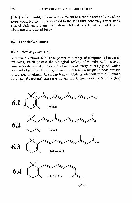
266 DAIRY CHEMISTRY AND BIOCHEMISTRY
(RNI) is the quantity of a nutrient sufficient to meet the needs of 97% of the population. Nutrient intakes equal to the RNI thus pose only a very small risk of deficiency. United Kingdom RNI values (Department of Health, 1991) are also quoted below.
6.2 Fat-soluble vitamins
6.2.1 Retinol (vitamin A )
Vitamin A (retinol, 6.1) is the parent of a range of compounds known as retinoids, which possess the biological activity of vitamin A. In general, animal foods provide preformed vitamin A as retinyl esters (e.g. 6.5, which are easily hydrolysed in the gastrointestinal tract) while plant foods provide precursors of vitamin A, i.e. carotenoids. Only carotenoids with a /3-ionone ring (e.g. p-carotene) can serve as vitamin A precursors. p-Carotene (6.6)
6.1 3
6.2
6.3
6.4
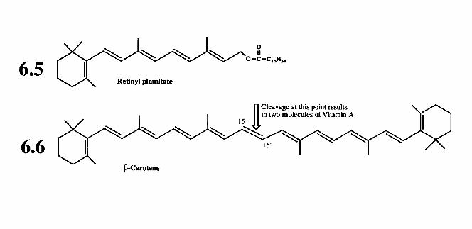
0
268 DAIRY CHEMISTRY AND BIOCHEMISTRY
may be cleavaged at its centre by the enzyme, p-carotene-1 5,15'-oxygenase (present in the intestinal mucosa), to yield 2 mol retinol per mol. However, cleavage of other bonds results in the formation of only 1 molecule of retinol per molecule of p-carotene. In practice, 6 pg 8-carotene will yield only 1 pg of retinol. Likewise, 12 pg other carotenes which are vitamin A precursors (i.e. which contain one p-ionone ring) are required to yield 1 pg of retinol. Thus, 1 retinol equivalent (RE) is defined as 1 pg retinol, 6 pg p-carotene or 12 pg of other precursor carotenes.
Retinol can be oxidized to retinal (6.2) and further to retinoic acid (6.3). Cis-trans isomerization can also occur, e.g. the conversion of all tvans- retinal to 11-cis-retinal (6.4), which is important for vision.
Vitamin A has a number of roles in the body: it is involved in the vision process, in cell differentiation, in growth and bone remodelling and in the immune system. US RDAs for vitamin A are 1000 pg RE day- for men and 800 pg RE day-' for women. UK RNI values for vitamin A are 700 and 600 pg RE day- ' for adult men and women, respectively. The body will tolerate a wide range of vitamin A intakes (500-15OOOpg REday-') but insufficient or excessive intakes result in illness. Vitamin A deficiency ( < 500 pg RE day- ') results in night blindness, xerophthalmia (progressive blindness caused by drying of the cornea of the eye), keratinization (accu- mulation of keratin in digestive, respiratory and urogenital tract tissues) and finally exhaustion and death. At excessive intake levels (> 15 000 pg REday-'), vitamin A is toxic. Symptoms of hypervitaminosis A include skin rashes, hair loss, haemorrhages, bone abnormalities and fractures, and in extreme cases, liver failure and death.
The major dietary sources of retinol are dairy products, eggs and liver, while important sources of p-carotene are spinach and other dark-green leafy vegetables, deep orange fruits (apricots, cantaloupe) and vegetables (squash, carrots, sweet potatoes, pumpkin). The richest natural sources of vitamin A are fish liver oils, particularly halibut and shark.
Vitamin A activity is present in milk as retinol, retinyl esters and as carotenes. Whole cows' milk contains an average of 52 pg retinol and 21 pg carotene per 1OOg. The concentration of retinol in raw sheep's and pas- teurized goats' milks is 83 and 44 pg per 100 g, respectively, although milks of these species are reported (Holland et al., 1991) to contain only trace amounts of carotenes. Human milk and colostrum contain an average of 58 and 155pg retinol per lOOg, respectively. In addition to their role as provitamin A, the carotenoids in milk are reponsible for the colour of milk fat (Chapter 11).
The concentration of vitamin A and carotenoids in milk is strongly influenced by the carotenoid content of the feed. Milk from animals fed on pasture contains higher levels of carotenes than that from animals fed on concentrate feeds. There is also a large seasonal variation in vitamin A concentration; summer milk contains an average of 62 pg retinol and 31 pg carotene per 100 g while the values for winter milk are 41 and 11 pg per
VITAMINS IN MILK A N D DAIRY PRODUCTS 269
lOOg, respectively. The breed of cow also has an influence on the concen- tration of vitamin A in milk: milk from Channel Islands breeds typically contains 65 pg and 27 pg retinol per 100 g in summer and winter, respect- ively, and 115 and 27pg carotene per lOOg in summer and winter, respectively.
Other dairy products are also important sources of vitamin A (Appendix 6A). Whipping cream (39% fat) contains about 565 pg retinol and 265 pg carotene per 1OOg. The level of vitamin A in cheese varies with the fat content (Appendix 6A). Camembert (23.7% fat) contains 230 pg retinol and 315 pg carotene per lOOg, while Cheddar (34.4% fat) contains 325 pg retinol and 225 pg carotene per 100 g. Whole-milk yogurt (3% fat; unflavoured) contains roughly 28pg retinol and 21 pg carotene per 1OOg while the corresponding values for ice-cream (9.8% fat) are 115 and 195 pg per 100 g, respectively.
Vitamin A is relatively stable to most dairy processing operations. In general, vitamin A activity is reduced by oxidation and exposure to light. Heating below 100°C (e.g. pasteurization) has little effect on the vitamin A content of milk, although some loss may occur at temperatures above 100°C (e.g. when frying using butter). Losses of vitamin A can occur in UHT milk during its long shelf-life at ambient temperatures. Vitamin A is stable in pasteurized milk at refrigeration temperatures provided the milk is pro- tected from light, but substantial losses can occur in milk packaged in translucent bottles. Low-fat milks are often fortified with vitamin A for nutritional reasons. Added vitamin A is less stable to light than the indigenous vitamin. The composition of the lipid used as a carrier for the exogenous vitamin influences its stability. Protective compounds (e.g. ascor- by1 palmitate or p-carotene) will reduce the rate at which exogenous vitamin A is lost during exposure to light. Yogurts containing fruit often contain higher concentrations of vitamin A precursor carotenoids than natural yogurts. The manufacture of dairy products which involves concentration of the milk fat (e.g. cheese, butter) results in a pro rata increase in the concentration of vitamin A. The increased surface area of dried milk products accelerates the loss of vitamin A; supplementation of milk powders with vitamin A and storage at low temperatures minimizes these losses.
6.2.2 Calciferols (vitamin D )
Unlike other vitamins, cholecalciferol (vitamin D,) can be formed from a steroid precursor, 7-dehydrocholesterol (6.7), by the skin when exposed to sunlight; with sufficient exposure to the sun, no preformed vitamin D is required from the diet.
UV light (280-320 nm) causes the photoconversion of 7-dehydrocholes- terol to pre-vitamin D,. This pre-vitamin can undergo further photoconver- sion to tachysterol and lumisterol or can undergo a temperature-dependent isomerization to cholecalciferol (vitamin D,, 6.8). At body temperature, this
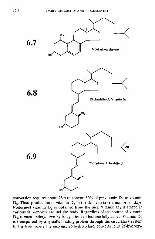
270 DAIRY CHEMISTRY AND BIOCHEMISTRY
6.7 HO
6.8
6.9
conversion requires about 28 h to convert 50% of previtamin D, to vitamin D,. Thus, production of vitamin D, in the skin can take a number of days. Preformed vitamin D, is obtained from the diet. Vitamin D, is stored in various fat deposits around the body. Regardless of the source of vitamin D,, it must undergo two hydroxylations to become fully active. Vitamin D, is transported by a specific binding protein through the circulatory system to the liver where the enzyme, 25-hydroxylase, converts it to 25-hydroxy-
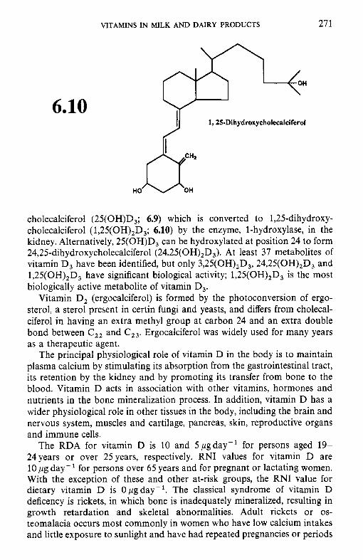
VITAMINS IN MILK A N D DAIRY PRODUCTS 271
6.10
cholecalciferol (25(OH)D,; 6.9) which is converted to 1,25-dihydroxy- cholecalciferol ( 1,25(OH),D,; 6.10) by the enzyme, 1-hydroxylase, in the kidney. Alternatively, 25(OH)D, can be hydroxylated at position 24 to form 24,25-dihydroxycholecalciferol (24,25(OH),D3). At least 37 metabolites of vitamin D, have been identified, but only 3,25(OH),D,, 24,25(OH),D, and 1,25(OH),D, have significant biological activity; 1,25(OH),D, is the most biologically active metabolite of vitamin D,.
Vitamin D, (ergocalciferol) is formed by the photoconversion of ergo- sterol, a sterol present in certin fungi and yeasts, and differs from cholecal- ciferol in having an extra methyl group at carbon 24 and an extra double bond between C,, and C23. Ergocalciferol was widely used for many years as a therapeutic agent.
The principal physiological role of vitamin D in the body is to maintain plasma calcium by stimulating its absorption from the gastrointestinal tract, its retention by the kidney and by promoting its transfer from bone to the blood. Vitamin D acts in association with other vitamins, hormones and nutrients in the bone mineralization process. In addition, vitamin D has a wider physiological role in other tissues in the body, including the brain and nervous system, muscles and cartilage, pancreas, skin, reproductive organs and immune cells.
The RDA for vitamin D is 10 and 5pgday- ' for persons aged 19- 24years or over 25 years, respectively. RNI values for vitamin D are 10 pg day- ' for persons over 65 years and for pregnant or lactating women. With the exception of these and other at-risk groups, the RNI value for dietary vitamin D is Opgday-'. The classical syndrome of vitamin D deficency is rickets, in which bone is inadequately mineralized, resulting in growth retardation and skeletal abnormalities. Adult rickets or os- teomalacia occurs most commonly in women who have low calcium intakes and little exposure to sunlight and have had repeated pregnancies or periods
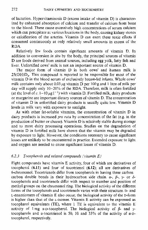
272 DAIRY CHEMISTRY AND BIOCHEMISTRY
of lactation. Hypervitaminosis D (excess intake of vitamin D) is character- ized by enhanced absorption of calcium and transfer of calcium from bone to the blood. These cause excessively high concentrations of serum calcium which can precipitate at various locations in the body, causing kidney stones or calcification of the arteries. Vitamin D can exert these toxic effects if consumed continuously at only relatively small amounts in excess of the RDA.
Relatively few foods contain significant amounts of vitamin D. In addition to conversion in situ by the body, the principal sources of vitamin D are foods derived from animal sources, including egg yolk, fatty fish and liver. Unfortified cows’ milk is not an important source of vitamin D.
The major form of vitamin D in both cows’ and human milk is 25(OH)D,. This compound is reported to be responsible for most of the vitamin D in the blood serum of exclusively breast-fed infants. Whole cows’ milk contains only about 0.03 p g vitamin D per 100 g and 1 litre of milk per day will supply only 10-20% of the RDA. Therefore, milk is often fortified (at the level of c. 1-10 pg 1-’) with vitamin D. Fortified milk, dairy products or margarine are important dietary sources of vitamin D. The concentration of vitamin D in unfortified dairy products is usually quite low. Vitamin D levels in milk vary with exposure to sunlight.
As with other fat-soluble vitamins, the concentration of vitamin D in dairy products is increased pro rata by concentration of the fat (e.g. in the production of butter or cheese). Vitamin D is relatively stable during storage and to most dairy processing operations. Studies on the degradation of vitamin D in fortified milk have shown that the vitamin may be degraded by exposure to light. However, the conditions necessary to cause significant losses are unlikely to be encountered in practice. Extended exposure to light and oxygen are needed to cause significant losses of vitamin D.
6.2.3
Eight compounds have vitamin E activity, four of which are derivatives of tocopherol (6.11) and four of tocotrienol (6.12); all are derivatives of 6-chromanol. Tocotrienols differ from tocopherols in having three carbon- carbon double bonds in their hydrocarbon side chain. a-, p-, y - or 6- tocopherols and tocotrienols differ with respect to number and position of methyl groups on the chromanol ring. The biological activity of the different forms of the tocopherols and tocotrienols varies with their structure. D- and L-enantiomers of vitamin E also occur; the biological activity of the D-form is higher than that of the L-isomer. Vitamin E activity can be expressed as tocopherol equivalents (TE), where 1 TE is equivalent to the vitamin E activity of 1 mg u-D-tocopherol. The biological activity of p- and y- tocopherols and u-tocotrienol is 50, 10 and 33% of the activity of a-D- tocopherol, respectively.
Tocopherols and related compounds (vitamin E )
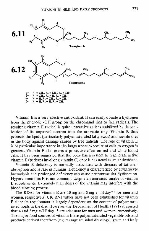
VITAMINS IN MILK AND DAIRY PRODUCTS 273
6.11
6.12
R,
I Tocotrienols I R3
Vitamin E is a very effective antioxidant. It can easily donate a hydrogen from the phenolic -OH group on the chromanol ring to free radicals. The resulting vitamin E radical is quite unreactive as it is stabilized by delocal- ization of its unpaired electron into the aromatic ring. Vitamin E thus protects the lipids (particularly polyunsaturated fatty acids) and membranes in the body against damage caused by free radicals. The role of vitamin E is of particular importance in the lungs where exposure of cells to oxygen is greatest. Vitamin E also exerts a protective effect on red and white blood cells. It has been suggested that the body has a system to regenerate active vitamin E (perhaps involving vitamin C ) once it has acted as an antioxidant.
Vitamin E deficiency is normally associated with diseases of fat mal- absorption and is rare in humans. Deficiency is characterized by erythrocyte haemolysis and prolonged deficiency can cause neuromuscular dysfunction. Hypervitaminosis E is not common, despite an increased intake of vitamin E supplements. Extremely high doses of the vitamin may interfere with the blood clotting process.
The RDAs for vitamin E are 10 mg and 8 mg c(-TE day- for men and women, respectively. UK RNI values have not been established for vitamin E since its requirement is largely dependent on the content of polyunsatu- rated lipids in the diet. However, the Department of Health (1991) suggested that 4 and 3 mg a-TE day- are adequate for men and women, respectively. The major food sources of vitamin E are polyunsaturated vegetable oils and products derived therefrom (e.g. maragrine, salad dressings), green and leafy
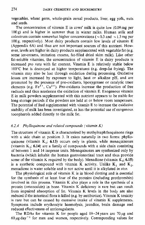
274 DAIRY CHEMISTRY A N D BIOCHEMISTRY
vegetables, wheat germ, whole-grain cereal products, liver, egg yolk, nuts and seeds.
The concentration of vitamin E in cows' milk is quite low (0.09mg per lOOg) and is higher in summer than in winter milks. Human milk and colostrum contain somewhat higher concentrations (-0.3 and - 1.3 mg per 100 g, respectively). Most dairy products contain low levels of vitamin E (Appendix 6A) and thus are not important sources of this nutrient. How- ever, levels are higher in dairy products supplemented with vegetable fat (e.g. some ice-creams, imitation creams, fat-filled dried skim milk). Like other fat-soluble vitamins, the concentration of vitamin E in dairy products is increased pro rafa with fat: content. Vitamin E is relatively stable below 100°C but is destroyed at higher temperatures (e.g. deep-fat frying). The vitamin may also be lost through oxidation during processing. Oxidative losses are increased by exposure to light, heat or alkaline pH, and are promoted by the presence of pro-oxidants, lipoxygenase or catalytic trace elements (e.g. Fe3+, Cu2+). Pro-oxidants increase the production of free radicals and thus accelerate the oxidation of vitamin E. Exogenous vitamin E in milk powders supplemented with this nutrient appears to be stable for long storage periods if the powders are held at or below room temperaure. The potential of feed supplemented with vitamin E to increase the oxidative stability of milk has been investigated, as has the potential use of exogenous tocopherols added directly to the milk fat.
6.2.4 Phylloquinone and related compounds (vitamin K )
The structure of vitamin K is characterized by methylnaphthoquinone rings with a side chain at position 3. It exists naturally in two forms: phyllo- quinone (vitamin K,; 6.13) occurs only in plants, while menaquinones (vitamin K,; 6.14) are a family of compounds with a side chain consisting of between 1 and 14 isoprene units. Menaquinones are synthesized only by bacteria (which inhabit the human gastrointestinal tract and thus provide some of the vitamin K required by the body). Menadione (vitamin K,; 6.15) is a synthetic compound with vitamin K activity. Unlike K, and K,, menadione is water soluble and is not active until it is alkylated in uiuo.
The physiological role of vitamin K is in blood clotting and is essential for the synthesis of at least four of the proteins (including prothrombin) involved in this process. Vitamin K also plays a role in the synthesis of a protein (osteocalcin) in bone. Vitamin K deficiency is rare but can result from impaired absorption of fat. Vitamin K levels in the body are also reduced if the intestinal flora is killed (e.g. by antibiotics). Vitamin K toxicity is rare but can be caused by excessive intake of vitamin K supplements. Symptoms include erythrocyte haemolysis, jaundice, brain damage and reduced effectiveness of anticoagulants.
The RDAs for vitamin K for people aged 19-24 years are 70pg and 60 pg day- for men and women, respectively. Corresponding values for
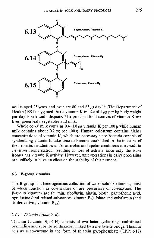
VITAMINS IN MILK AND DAIRY PRODUCTS 275
0
a
adults aged 25 years and over are 80 and 65 pgday-’. The Department of Health (1991) suggested that a vitamin K intake of 1 pg per kg body weight per day is safe and adequate. The principal food sources of vitamin K are liver, green leafy vegetables and milk.
Whole cows’ milk contains 0.4-1.8pg vitamin K per 1OOg while human milk contains about 0.2pg per 1OOg. Human colostrum contains higher concentrations of vitamin K, which are necessary since bacteria capable of synthesizing vitamin K take time to become established in the intestine of the neonate. Irradiation under anerobic and apolar conditions can result in cis-trans isomerization, resulting in loss of activity since only the trans isomer has vitamin K activity. However, unit operations in dairy processing are unlikely to have an effect on the stability of this nutrient.
6.3 B-group vitamins
The B-group is a heterogeneous collection of water-soluble vitamins, most of which function as co-enzymes or are precursors of co-enzymes. The B-group vitamins are thiamin, riboflavin, niacin, biotin, pantothenic acid, pyridoxine (and related substances, vitamin B6), folate and cobalamin (and its derivatives, vitamin B,J.
6.3. I Thiamin (vitamin B,)
Thiamin (vitamin B,; 6.16) consists of two heterocyclic rings (substitued pyrimidine and substituted thiazole), linked by a methylene bridge. Thiamin acts as a co-enzyme in the form of thiamin pyrophosphate (TPP; 6.17)
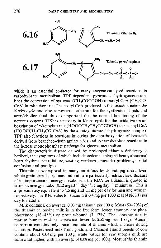
276 DAIRY CHEMISTRY AND BIOCHEMISTRY
Thiamin (Vitamin B1) 6.16 H3C
Thiamin pyrophosphate
0 0 II It
OH OH
CH2-CH2-O-T-O-T-OH
which is an essential co-factor for many enzyme-catalysed reactions in carbohydrate metabolism. TPP-dependent pyruvate dehydrogenase cata- lyses the conversion of pyruvate (CH,COCOOH) to acetyl CoA (CH,CO- CoA) in mitochondria. The acetyl CoA produced in this reaction enters the Krebs cycle and also serves as a substrate for the synthesis of lipids and acetylcholine (and thus is important for the normal functioning of the nervous system). TPP is necessary in Krebs cycle for the oxidative decar- boxylation of r-ketoglutarate (HOOCCH,CH,COCOOH) to succinyl CoA (HOOCCH,CH,CO-CoA) by the a-ketoglutarate dehydrogenase complex. TPP also functions in reactions involving the decarboxylation of ketoacids derived from branched-chain amino acids and in transketolase reactions in the hexose monophosphate pathway for glucose metabolism.
The characteristic disease caused by prolonged thiamin deficiency is beriberi, the symptoms of which include oedema, enlarged heart, abnormal heart rhythms, heart failure, wasting, weakness, muscular problems, mental confusion and paralysis.
Thiamin is widespread in many nutritious foods but pig meat, liver, whole-grain cereals, legumes and nuts are particularly rich sources. Because of its importance in energy metabolism, the RDA for thiamin is quoted in terms of energy intake (0.12mgkJ-'day-'; 1 mgday-' minimum). This is approximately equivalent to 1.5 mg and 1.1 mg per day for men and women, respectively. The RNI value for thiamin is 0.4 mg per 1000 kcal(4186 kJ) per day for adults.
Milk contains, on average, 0.03 mg thiamin per 100 g. Most (50-70%) of the thiamin in bovine milk is in the free form; lesser amounts are phos- phorylated (18-45%) or protein-bound (7- 17%). The concentration in mature human milk is somewhat lower (c.0.02mg per 100s). Human colostrum contains only trace amounts of thiamin which increase during lactation. Pasteurized milk from goats and Channel Island breeds of cow contain about 0.04mg per lOOg, while values for raw sheep's milk are somewhat higher, with an average of 0.08 mg per 100 g. Most of the thiamin
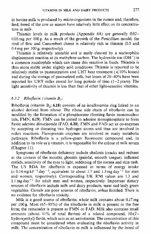
VITAMINS IN MILK AND DAIRY PRODUCTS 277
in bovine milk is produced by micro-organisms in the rumen and, therefore, feed, breed of the cow or season have relatively little effect on its concentra- tion in milk.
Thiamin levels in milk products (Appendix 6A) are generally 0.02- 0.05mg per 1OOg. As a result of the growth of the Penicillium mould, the rind of Brie and Camembert cheese is relatively rich in thiamin (0.5 and 0.4 mg per 100 g, respectively).
Thiamin is relatively unstable and is easily cleaved by a nucleophilic displacement reaction at its methylene carbon. The hydroxide ion (OH -) is a common nucelophile which can cause this reaction in foods. Thiamin is thus more stable under slightly acid conditions. Thiamin is reported to be relatively stable to pasteurization and UHT heat treatment ( < 10% losses) and during the storage of pasteurized milk, but losses of 20-40% have been reported for UHT milks stored for long periods of time (1-2years). The light sensitivity of thiamin is less than that of other light-sensitive vitamins.
6.3.2 Riboflavin (vitamin B,)
Riboflavin (vitamin B,; 6.18) consists of an isoalloxazine ring linked to an alcohol derived from ribose. The ribose side chain of riboflavin can be modified by the formation of a phosphoester (forming flavin mononucleo- tide, FMN, 6.19). FMN can be joined to adenine monophosphate to form flavin adenine dinucleotide (FAD, 6.20). FMN and FAD act as co-enzymes by accepting or donating two hydrogen atoms and thus are involved in redox reactions. Flavoprotein enzymes are involved in many metabolic pathways. Riboflavin is a yellow-green fluorescent compound and, in addition to its role as a vitamin, it is responsible for the colour of milk serum (Chapter 11).
Symptoms of riboflavin deficiency include cheilosis (cracks and redness at the corners of the mouth), glossitis (painful, smooth tongue), inflamed eyelids, sensitivity of the eyes to light, reddening of the cornea and skin rash. The US RDA for riboflavin is expessed in terms of energy intake (c. 0,14mgkJ-'day-', equivalent to about 1.7 and 1.3mgday-' for men and women, respectively). Corresponding UK RNI values are 1.3 and 1.1 mgday- ' for adult men and women, respectively. Important dietary sources of riboflavin include milk and dairy products, meat and leafy green vegetables. Cereals are poor sources of riboflavin, unless fortified. There is no evidence for riboflavin toxicity.
Milk is a good source of riboflavin; whole milk contains about 0.17mg per lOOg. Most (65-95%) of the riboflavin in milk is present in the free form; the remainder is present as FMN or FAD. Milk also contains small amounts (about 11% of total flavins) of a related compound, 1042'- hydroxyethyl) flavin, which acts as an antivitamin. The concentration of this compound must be considered when evaluating the riboflavin activity in milk. The concentration of riboflavin in milk is influenced by the breed of
278 DAIRY CHEMISTRY AND BIOCHEMISTRY
Riboflavin
Flavin mononucleotide
H H H 0 0 I l l I1 I1
HO-CH
6.20 0
Flavin adenine dinucleotide
NH2
cow (milk from Jersey and Guernsey cows contains more riboflavin than Holstein milk). Summer milk generally contains slighly higher levels of riboflavin than winter milk. Interspecies variations in concentration are also apparent. Raw sheep’s milk contains about 0.32 mg per 100 g while the mean value for pasteurized goats’ milk (0.13 mg per 100 g) is lower; human milk contains 0.03 mg per 100 g. Dairy products also contain significant amounts
VITAMINS IN MILK AND DAIRY PRODUCTS 279
0
Lumitlavin
of riboflavin (Appendix 6A). Cheese contains 0.3-0.5 mg per OOg anc yogurt about 0.3 mg per 1OOg. The whey protein fraction of milk contains a riboflavin-binding protein (RfBP) which probably originates from blood plasma, although its function in milk is unclear.
Riboflavin is stable in the presence of oxygen, heat and at acid pH. However, it is labile to thermal decomposition under alkaline conditions. The concentration of riboflavin in milk is unaffected by pasteurization and little loss is reported for UHT-treated milks. The most important parameter affecting the stability of riboflavin in dairy products is exposure to light (particularly wavelengths in the range 415-455 nm). At alkaline pH, irradi- ation cleaves the ribitol portion of the molecule, leaving a strong oxidizing agent, lumiflavin (6.21). Irradiation under acidic conditions results in the formation of lumiflavin and a blue fluorescent compound, lumichrome. Lumiflavin is capable of oxidizing other vitamins, particularly ascorbate (section 6.4 and Chapter 11). Loss of riboflavin in milk packaged in materials that do not protect against light can be caused by either sunlight or by lights in retail outlets. Packaging in paperboard containers is the most efficient method for minimizing this loss, although glass containing a suitable pigment has also been used. Riboflavin is more stable in high-fat than in low-fat or skim milk, presumably as a result of the presence of antioxidants (e.g. vitamin E) in the milk fat which protect riboflavin against photo-oxidation.
6.3.3 Niacin
Niacin is a generic term which refers to two related chemical compounds, nicotinic acid (6.22) and its amide, nicotinamide (6.23); both are derivatives of pyridine. Nicotinic acid is synthesized chemically and can be easily converted to the amide in which form it is found in the body. Niacin is obtained from food or can be synthesized from tryptophan (60 mg of dietary tryptophan has the same metabolic effect as 1 mg niacin). Niacin forms part of two important co-enzymes, nicotinamide adenine dinucleotide (NAD) and nicotinamide adenine dinucleotide phosphate (NADP), which are co-factors for many enzymes that participate in various metabolic pathways and function in electron transport.
280 DAIRY CHEMISTRY AND BIOCHEMISTRY
6e23
The classical niacin deficiency disease is pellagra, which is characterized by symptoms including diarrhoea, dermatitis, dementia and eventually death. High-protein diets are rarely deficient in niacin since, in addition to the preformed vitamin, such diets supply sufficient tryptophan to meet dietary requirements. Large doses of niacin can cause the dilation of capillaries, resulting in a painful tingling sensation.
The RDA for niacin is expressed in terms of energy intake; 6.6 mg niacin equivalent (NE, 1 mg niacin or 60 mg tryptophan) per 1000 kcal (4186 kJ) per day is recommended (13 NE day-’ minimum). This is approximately equivalent to 19 and 15 mg NE day- for men and women, respectively. The UK RNI value for niacin is 6.6 mg NE per 1000 kcal (4186 kJ) per day for adults. The richest dietary sources of niacin are meat, poultry, fish and whole-grain cereals.
Milk contains about 0.1 mg niacin per lOOg and thus is not a rich source of the preformed vitamin. Tryptophan contributes roughly 0.7 mg NE per 100 g milk. In milk, niacin exists primarily as nicotinamide and its concen- tration does not appear to be affected greatly by breed of cow, feed, season or stage of lactation. Pasteurized goats’ (0.3 mg niacin and 0.7 mg NE from tryptophan per 100 g) and raw sheep’s (0.4 mg niacin and 1.3 mg NE from tryptophan per lOOg) milk are somewhat richer than cows’ milk. Niacin levels in human milk are 0.2 mg niacin and 0.5 mg NE from tryptophan per 100 g. The concentration of niacin in most dairy products is low (Appendix 6A) but is compensated somewhat by tryptophan released on hydrolysis of the proteins.
Niacin is relatively stable to most food-processing operations. It is stable to exposure to air and resistant to autoclaving (and is therefore stable to pasteurization and UHT treatments). The amide linkage of nicotinamide can be hydrolysed to the free carboxylic acid (nicotinic acid) by treament with acid but the vitamin activity is unaffected. Like other water-soluble vitamins, niacin can be lost by leaching.
VITAMINS IN MILK AND DAIRY PRODUCTS 28 1
6.3.4 Biotin
Biotin (6.24) consists of an imidazole ring fused to a tetrahydrothiophene ring with a valeric acid side chain. Biotin acts as a co-enzyme for car- boxylases involved in the synthesis and catabolism of fatty acids and for branched-chain amino acids and gluconeogenesis.
Biotin
Biotin deficiency is rare but under laboratory conditions it can be induced by feeding subjects with large amounts of raw egg white which contains the protein, avidin, which has a binding site for the imidazole moiety of biotin, thus making it unavailable. Avidin is denatured by heat and, therefore, biotin binding occurs only in raw egg albumen. Symptoms of biotin deficiency include scaly dermatitis, hair loss, loss of appetite, nausea, hallucinations and depression.
Biotin is widespread in foods, although its availability is affected some- what by the presence of binding proteins. Biotin is required in only small amounts. Although US RDA values have not been established, the esti- mated safe and adequate intake of biotin is 30-100 pg day-' for adults. The Department of Health (1991) suggested that biotin intakes between 10 and 200 p g day- are safe and adequate. Biotin is reported to be non-toxic in amounts up to at least 10 mg day-'.
Milk contains about 1.9 pg biotin per 100 g, apparently in the free form. Pasteurized caprine, raw ovine and human milks contain 3.0, 2.5 and 0.7 pg per 100 g, respectively. The concentration of biotin in cheese ranges from 1.4 (Gouda) to 7.6 (Camembert) pg per l o g (Appendix 6A). Skim-milk powder contains high levels of biotin (c.2Opg per lOOg) owing to the concentration of the aqueous phase of milk during its manufacture. Biotin is stable during food processing and storage and is unaffected by pasteuriz- ation.
6.3.5 Panthothenic acid
Pantothenic acid (6.25) is a dimethyl derivative of butyric acid linked to p-alanine. Pantothenate is part of the structure of co-enzyme A (CoA), and
282 DAIRY CHEMISTRY AND BIOCHEMISTRY
6.25 Pantothenic acid
as such is vital as a co-factor for numerous enzyme-catalysed reactions in lipid and carbohydrate metabolism.
Pantothenate deficiency is rare, occurring only in cases of severe mal- nutrition; characteristic symptoms include vomiting, intestinal distress, in- somnia, fatigue and occasional diarrhoea. Pantothenate is widespread in foods; meat, fish, poulty, whole-grain cereals and legumes are particularly good sources. Although no RDA or RNI value has been established for panthothenate, safe and adequate intake of this vitamin for adults is estimated to be 3-7mgday-'. Pantothenate is non-toxic at doses up to 10 g day -
Milk contains, on average, 0.35 mg panthothenate per 100 g. Panto- thenate exists partly free and partly bound in milk and its concentration is influenced by breed, feed and season. Raw ovine and pasteurized caprine milks contain slightly higher concentrations of this nutrient (averaging 0.45 and 0.41 mg per 100 g, respectively). The values for pantothenate in human milk vary widely; values ranging from 0.2 to 0.7mg per lOOg have been reported. Mean concentrations of pantothenate in cheese vary from about 0.3 (cream cheese, Gouda) to 0.7 (Stilton) mg per 1OOg (Appendix 6A). Pantothenate is stable at neutral pH but is easily hydrolysed by acid or alkali at high temperatures. Pantothenate is reported to be stable to pasteurization.
6.3.6 Pyridoxine and related compounds (vitamin B6) Vitamin B, occurs naturally in three related forms: pyridoxine (6.26; the alcohol form), pyridoxal (6.27; aldehyde) and pyridoxamine (6.28; amine). All are structurally related to pyridine. The active co-enzyme form of this vitamin is pyridoxal phosphate (PLP; 6.29), which is a co-factor for transaminases which catalyse the transfer of amino groups (6.29). PLP is also important for amino acid decarboxylases and functions in the metab- olism of glycogen and the synthesis of sphingolipids in the nervous system. In addition, PLP is involved in the formation of niacin from tryptophan (section 6.3.3) and in the initial synthesis of haem.
Deficiency of vitamin B, is characterized by weakness, irritability and insomnia and later by convulsions and impairment of growth, motor
VITAMINS IN MILK AND DAIRY PRODUCTS 283
CHaOH I
HO CHpOH
HsC
Pyridoxine
CHaOH
6.27 "0 H3C
Pyridoxal
CHz-NHz
CHaOH
6.28 "0 H3C
Pyridoxamine
o* .H 0 CHrNH2 0 "0 -f:o $l_ Hoe HaO-P-0 II I
0 6.29 I H3C / 7 H3C /
Pyridoxamine phosphate Pyridoxal phosphate NH3
functions and immune response. High doses of vitamin B,, often associated with excessive intake of supplements, are toxic and can cause bloating, depression, fatigue, irritability, headaches and nerve damage.
Since vitamin B, is essential for amino acid (and hence protein) metab- olism, its RDA is quoted in terms of protein intake (0.016 mg per g protein per day, equivalent to about 2.0 and 1.6mgday-' for men and women,
284 DAIRY CHEMISTRY AND BIOCHEMISTRY
respectively). The corresponding UK RNI value for B, is 15 pg g-’ protein for adults. Important sources of B, include green, leafy vegetables, meat, fish and poultry, shellfish, legumes, fruits and whole grains.
Whole milk contains, on average, 0.06mg B, per lOOg, mainly in the form of pyridoxal (80%); the balance is mainly pyridoxamine (20%), with trace amounts of pyridoxamine phosphate. Concentrations in raw ovine and pasteurized caprine milks are similar to those in cows’ milk (0.08 and 0.06 mg per 100 g, respectively). The concentration of B, varies during lactation; colostum contains lower levels than mature milk. Seasonal vari- ation in the concentration of vitamin B, has been reported in Finnish milk; levels were higher (14%) when cattle were fed outdoors than when they were fed indoors. Mature human milk contains about 0.01 mg B, per 100 g.
In general, dairy products are not major sources of B, in the diet. Concentrations in cheeses and related products vary from about 0.04 (fromage frais, cream cheese) to 0.22 (Camembert) mg per 100 g (Appendix 6A). Whole-milk yogurt contains roughly 0.1 mg per 100 g and the concen- tration in skim-milk powder is c. 0.6 mg per 100 g.
All forms of B, are sensitive to UV light and may be decomposed to biologically inactive compounds. Vitamin B, may also be decomposed by heat. Losses of 45% and 20-30% can occur on cooking meat and veg- etables, respectively. The aldehyde group of pyridoxal and the amine group of pyridoxamine show some reactivity under conditions that may be encountered during milk processing. An outbreak of B, deficiency in 1952 was attributed to the consumption of heated milk products. Pyridoxal and/or its phosphate can react directly with the sulphydryl group of cysteine residues in proteins, forming an inactive thiazolidine derivative (6.30). Losses during pasteurization and UHT treatments are relatively small, although losses of up to 50% can occur in UHT milk during its shelf-life.
Thiazolidine derivative of pyridoxal
VITAMINS IN MILK AND DAIRY PRODUCTS 285
6.3,7 Folate
Folate consists of a substituted pteridine ring linked through a methylene bridge to p-aminobenzoic acid and glutamic acid (6.31). Up to seven glutamic acid residues can be attached by y-carboxyl linkages, producing polyglutamyl folate (6.31) which is the major dietary and intracellular form of the vitamin. Reductions and substitutions on the pteridine ring result in tetrahydrofolate (H4 folate; 6.32) and 5-methyl tetrahydrofolate (5-methyl- H, folate; 6.33). Folate is a co-factor in the enzyme-catalysed transfer of single carbon atoms in many metabolic pathways, including the biosynthesis of purines and pyramidines (essential for DNA and RNA) and interconver- sions of amino acids. Folate interacts with vitamin B,, (section 6.3.8) in the enzyme-catalysed synthesis of methionine and in the activation of 5-methyl- H, folate to H, folate. H, Folate is involved in a complex and inter-linked series of metabolic reactions (Garrow and James, 1993).
n
Folate
A Tetrahydrofolate
H
5-methyl tetrahydrofolate
286 DAIRY CHEMISTRY AND BIOCHEMISTRY
Folate deficiency impairs cell division and protein synthesis; symptoms include megaloblastic anaemia, digestive system problems (heartburn, diar- rhoea, constipation), suppression of the immune system, glossitis and problems with the nervous system (depression, fainting, fatigue, mental confusion). The RDA for folate is 3pg per kg body weight per day (equivalent to c. 200 and 180 pg day-' for men and women, respectively). The RNI value for adults is 200 pg day-'. Higher intakes of folate have been suggested for women of child-bearing age to prevent the development of neural tube defects in the developing foetus.
Rich dietary sources of folate include leafy green vegetables, legumes, seeds and liver. Milk contains about 6pg folate per 1OOg. The dominant form of folate in milk is 5-methyl-H, folate. Folate in milk is mainly bound to folate-binding proteins and about 40% occurs as conjugated polygluta- mate forms. The folate binding proteins of milks of various species have been characterized (Fox and Flynn, 1992). It has been suggested that protein binding increases the bioavailability of folate. Winter milk is reported to contain higher concentrations of folate than summer milk (7 and 4 pg per lOOg, respectively). Raw sheep's milk contains, on average, 5 pg per 1OOg while the value for pasteurized goats' milk is 1 pg per 100 g. Folate levels in human milk increase from 2 to 5pg per 1OOg as colostrum changes to mature milk. Folate levels in some dairy products are shown in Appendix 6A. Whipping cream contains about 7pg per lOOg while the value for cheese varies widely from 30-40 pg per 100 g (Edam, Cheddar) to greater than 100 pg per 100 g (Camembert); the high concentration found in mould- ripened varieties presumably reflects biosynthesis of folate by the mould. The concentration of folate in yogurt is about 18 pg per 100 g, principally in the form of formyl folate. The higher level of folate in yogurt is due to biosynthesis, particularly by Streptococcus salivarius subsp. thermophilus, and perhaps to some added ingredients.
Folate is a relatively unstable nutrient; processing and storage conditions that promote oxidation are of particular concern since some of the forms of folate found in foods are easily oxidized. The reduced forms of folate (dihydro- and tetrahydrofolate) are oxidized to p-aminobenzoylglutamic acid and pterin-6-carboxylic acid, with a concomitant loss in vitamin activity. 5-Methyl-H, folate can also be oxidized. Antioxidants (particularly ascorbic acid in the context of milk) can protect folate against destruction. The rate of the oxidative degradation of folate in foods depends on the derivative present and the food itself, particularly its pH, buffering capacity and concentration of catalytic trace elements and antioxidants.
Folate is sensitive to light and may be subject to photodecomposition. Heat treatment influences folate levels in milk. Pasteurization and the storage of pasteurized milks have relatively little effect on the stability of folate but UHT treatments can cause substantial losses. The concentration of oxygen in UHT milk (from the headspace above the milk or by diffusion
VITAMINS IN MILK AND DAIRY PRODUCTS 287
through the packaging material) has an important influence on the stability of folate during the storage of UHT milk, as have the concentrations of ascorbate in the milk and of 0, in the milk prior to heat treatment. Folate and ascorbic acid (section 6.4) are the least stable vitamins in powdered milks.
The heat stability of folate-binding proteins in milk should also be considered in the context of folate in dairy foods. Breast-fed babies require less dietary folate ( 5 5 pg folate day-' to maintain their folate status) than bottle-fed infants (78 pg day-,). The difference has been attributed to the presence of active folate-binding proteins in breast milk; folate-binding proteins originally present in milk formulae are heat-denatured during processing. However, a study involving feeding radiolabelled folate to rats together with dried milks prepared using different heat treatments showed no differences in folate bioavailability (Oste, Jagerstad and Anderson, 1997).
6.3.8
Vitamin B,, consists of a porphyrin-like ring structure, with an atom of Co chelated at its centre, linked to a nucleotide base, ribose and phosphoric acid (6.34). A number of different groups can be attached to the free ligand site on the cobalt. Cyanocobalamin has -CN at this position and is the commercial and therapeutic form of the vitamin, although the principal dietary forms of B , , are 5'-deoxyadenosylcobalamin (with 5'-deoxyadeno- sine at the R position), methylcobalamin (-CH,) and hydroxocobalamin (-OH). Vitamin B,, acts as a co-factor for methionine synthetase and methylmalonyl CoA mutase. The former enzyme catalyses the transfer of the methyl group of 5-methyl-H, folate to cobalamin and thence to homocys- teine, forming methionine. Methylmalonyl CoA mutase catalyses the con- version of methylmalonyl CoA to succinyl CoA in the mitochondrion.
Vitamin B, , deficiency normally results from indequate absorption rather than inadequate dietary intake. Pernicious anaemia is caused by vitamin B , , deficiency; symptoms include anaemia, glossitis, fatigue and degeneration of the peripheral nervous system and hypersensitivity of the skin. The adult RDA and RNI for B,, are 2 and lSpgday- ' , respectively. Unlike other vitamins, B,, is obtained exclusively from animal food sources, such as meat, fish, poultry, eggs, shellfish, milk, cheese and eggs. Vitamin B,, in these foods is protein-bound and released by the action of HCl and pepsin in the stomach.
Bovine milk contains, on average, 0.4 p g B,, per 100 g. The predominant form is hydroxycobalamin and more than 95% of this nutrient is protein bound. The concentration of B,, in milk is influenced by the Co intake of the cow. The predominant source of B,, for the cow, and hence the ultimate origin of B,, in milk, is biosynthesis in the rumen. Therefore, its concentra-
Cobalamin and its derivatives (vitamin B,2)
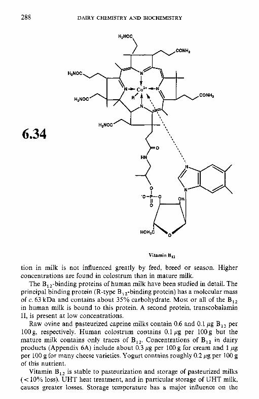
288 DAIRY CHEMISTRY AND BIOCHEMISTRY
6.34
Vitamin B,,
tion in milk is not influenced greatly by feed, breed or season. Higher concentrations are found in colostrum than in mature milk.
The BIZ-binding proteins of human milk have been studied in detail. The principal binding protein (R-type B , ,-binding protein) has a molecular mass of c. 63 kDa and contains about 35% carbohydrate. Most or all of the B,, in human milk is bound to this protein. A second protein, transcobalamin 11, is present at low concentrations.
Raw ovine and pasteurized caprine milks contain 0.6 and 0.1 pg B,, per 100 g, respectively. Human colostrum contains 0.1 pg per 1OOg but the mature milk contains only traces of B12, Concentrations of B,, in dairy products (Appendix 6A) include about 0.3 pg per 100 g for cream and 1 pg per 100 g for many cheese varieties. Yogurt contains roughly 0.2 pg per 100 g of this nutrient.
Vitamin B,, is stable to pasteurization and storage of pasteurized milks (c 10% loss). UHT heat treatment, and in particular storage of UHT milk, causes greater losses. Storage temperature has a major influence on the
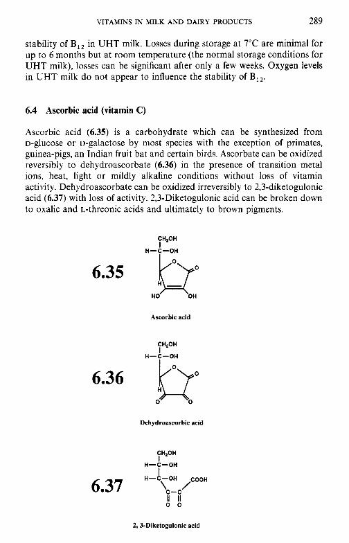
VITAMINS IN MILK AND DAIRY PRODUCTS 289
stability of B,, in UHT milk. Losses during storage at 7°C are minimal for up to 6 months but at room temperature (the normal storage conditions for UHT milk), losses can be significant after only a few weeks. Oxygen levels in UHT milk do not appear to influence the stability of B12.
6.4 Ascorbic acid (vitamin C)
Ascorbic acid (6.35) is a carbohydrate which can be synthesized from D-glucose or D-galactose by most species with the exception of primates, guinea-pigs, an Indian fruit bat and certain birds. Ascorbate can be oxidized reversibly to dehydroascorbate (6.36) in the presence of transition metal ions, heat, light or mildly alkaline conditions without loss of vitamin activity. Dehydroascorbate can be oxidized irreversibly to 2,3-diketogulonic acid (6.37) with loss of activity. 2,3-Diketogulonic acid can be broken down to oxalic and L-threonic acids and ultimately to brown pigments.
CH,OH I
H-$-OH
6.36
Ascorbic acid
CHzOH I
H-C-OH
0 H Lr Dehydroascorbic acid
CH20H
H-C-OH I I
H - y H ,COOH
c-c 0 0 II II
6.37
2,3-Diketogulonic acid
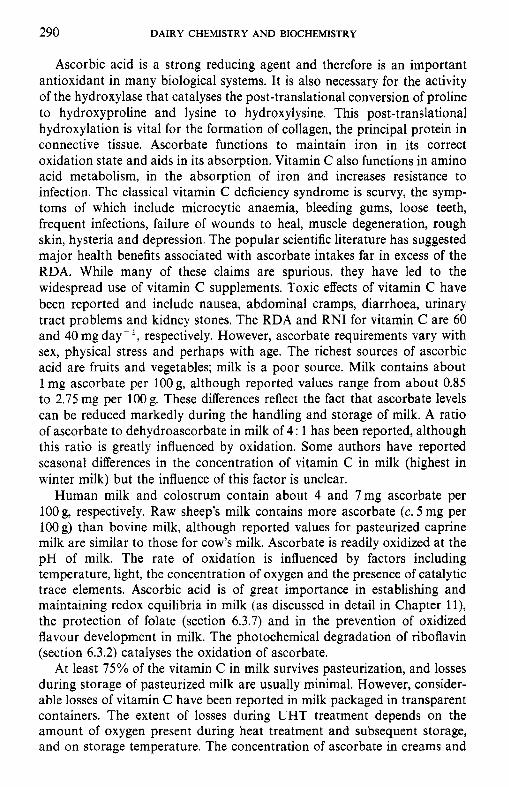
290 DAIRY CHEMISTRY A N D BIOCHEMISTRY
Ascorbic acid is a strong reducing agent and therefore is an important antioxidant in many biological systems. It is also necessary for the activity of the hydroxylase that catalyses the post-translational conversion of proline to hydroxyproline and lysine to hydroxylysine. This post-translational hydroxylation is vital for the formation of collagen, the principal protein in connective tissue. Ascorbate functions to maintain iron in its correct oxidation state and aids in its absorption. Vitamin C also functions in amino acid metabolism, in the absorption of iron and increases resistance to infection. The classical vitamin C deficiency syndrome is scurvy, the symp- toms of which include microcytic anaemia, bleeding gums, loose teeth, frequent infections, failure of wounds to heal, muscle degeneration, rough skin, hysteria and depression. The popular scientific literature has suggested major health benefits associated with ascorbate intakes far in excess of the RDA. While many of these claims are spurious, they have led to the widespread use of vitamin C supplements. Toxic effects of vitamin C have been reported and include nausea, abdominal cramps, diarrhoea, urinary tract problems and kidney stones. The RDA and RNI for vitamin C are 60 and 40 mg day- ’, respectively. However, ascorbate requirements vary with sex, physical stress and perhaps with age. The richest sources of ascorbic acid are fruits and vegetables; milk is a poor source. Milk contains about 1 mg ascorbate per lOOg, although reported values range from about 0.85 to 2.75 mg per 100 g. These differences reflect the fact that ascorbate levels can be reduced markedly during the handling and storage of milk. A ratio of ascorbate to dehydroascorbate in milk of 4 : 1 has been reported, although this ratio is greatly influenced by oxidation. Some authors have reported seasonal differences in the concentration of vitamin C in milk (highest in winter milk) but the influence of this factor is unclear.
Human milk and colostrum contain about 4 and 7mg ascorbate per 100 g, respectively. Raw sheep’s milk contains more ascorbate (c. 5 mg per 100 g) than bovine milk, although reported values for pasteurized caprine milk are similar to those for cow’s milk. Ascorbate is readily oxidized at the pH of milk. The rate of oxidation is influenced by factors including temperature, light, the concentration of oxygen and the presence of catalytic trace elements. Ascorbic acid is of great importance in establishing and maintaining redox equilibria in milk (as discussed in detail in Chapter ll), the protection of folate (section 6.3.7) and in the prevention of oxidized flavour development in milk. The photochemical degradation of riboflavin (section 6.3.2) catalyses the oxidation of ascorbate.
At least 75% of the vitamin C in milk survives pasteurization, and losses during storage of pasteurized milk are usually minimal. However, consider- able losses of vitamin C have been reported in milk packaged in transparent containers. The extent of losses during UHT treatment depends on the amount of oxygen present during heat treatment and subsequent storage, and on storage temperature. The concentration of ascorbate in creams and
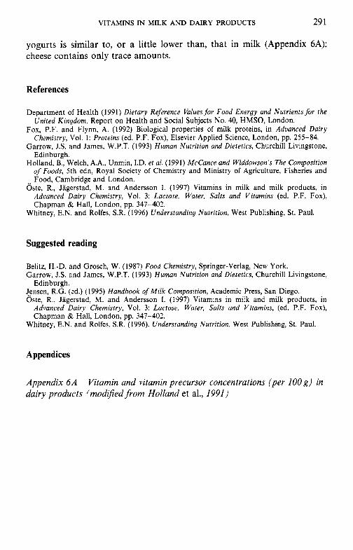
VITAMINS IN MILK A N D DAIRY PRODUCTS 29 1
yogurts is similar to, or a little lower than, that in milk (Appendix 6A); cheese contains only trace amounts.
References
Department of Health (1991) Dietary Reference Values for Food Energy and Nutrients for the United Kingdom, Report on Health and Social Subjects No. 40, HMSO, London.
Fox, P.F. and Flynn, A. (1992) Biological properties of milk proteins, in Adoanced Dairy Chemistry, Vol. 1: Proteins (ed. P.F. Fox), Elsevier Applied Science, London, pp. 255-84.
Garrow, J.S. and James, W.P.T. (1993) Human Nutrition and Dietetics, Churchill Livingstone, Edinburgh.
Holland, B., Welch, A.A., Unmin, I.D. et al. (1991) McCance and Widdowson’s The Composition of Foods, 5th edn, Royal Society of Chemistry and Ministry of Agriculture, Fisheries and Food, Cambridge and London.
Oste, R., Jagerstad, M. and Andersson I . (1997) Vitamins in milk and milk products, in Adcanced Dairy Chemistry, Vol. 3: Lactose, Water, Salts and Vitamins (ed. P.F. Fox), Chapman & Hall, London, pp. 347-402.
Whitney, E.N. and Rolfes, S.R. (1996) Understanding Nutrition, West Publishing, St. Paul.
Suggested reading
Belitz, H.-D. and Grosch, W. (1987) Food Chemistry, Springer-Verlag, New York. Garrow, J.S. and James, W.P.T. (1993) Human Nutrition and Dietetics, Churchill Livingstone,
Jensen, R.G. (ed.) (1995) Handbook of Milk Composition, Academic Press, San Diego. Oste, R., Jagerstad, M. and Andersson I. (1997) Vitamins in milk and milk products, in
Advanced Dairy Chemislry, Vol. 3: Lactose, Water, Salts and Vitamins, (ed. P.F. Fox), Chapman & Hall, London, pp. 347-402.
Edinburgh.
Whitney, E.N. and Rolfes, S.R. (1996). Understanding Nutrition, West Publishing, St. Paul.
Appendices
Appendix 6 A dairy products (mod@ed from Holland et al., 1991)
Vitamin and vitamin precursor concentrations (per 100 g ) in
Vitamin Vitamin Ribo- Vitamin Vitamin Panto- Retinol Carotone D E Thiamin flavin Niacin Trp-60 B, B,, Folate thenate Biotin
Product (fez) (m) (rg) (mg) (mg) (mg) (mg) (mg) (mg) (m) (m) (mg) ( p g )
Skimmed milk pasteurized UHT, fortified
Whole milk pasteurized summer winter sterilized. in container
Channcl Island milk whole,
summer winter semi-skimmed, UHT
Dried skimmed milk” (fortified) with vegetable fat (fortified)
Evaporated milk, whole
Goat’s milk, pasteurized
Human milk, colostrum transitional mature
pasteurized
Sheep’s milk, raw Fresh whipping cream, pasteurized (39.3% fat)
Cheeses Brie Camembert Cheddar, average
1 61
52 62 41
52
46 65 27
14
350
395
105
44
I55 85 58 83
565
285 230
325
Tr 18
21 31 I 1
21
71 115 27
22
5
15
100
Tr
(135)
(24) (37)
Tr
265
210 315
225
Tr 0. I
0.03 0.03 0.03
0.03
0.03 0.04 0.03
0.01
2.10
10.50
3.95‘
0.1 1
N N
0.04 0.18
0.22
0.20 (0.18)
0.26
Tr 0.02
0.09 0.10 0.07
0.09
0.1 I 0.13 0.09
0.04
0.27
1.32
0.19
0.03
1.3 0.48 0.34 0.11
0.86
0.84 0.65
0.53
0.04 0.04
0.04 0.04 0.04
0.03
0.04 0.04 0.04
0.04
0.38
0.23
0.07
0.04
Tr 0.01 0.02 0.08
0.02
0.04d 0.05’
0.03
0.18 0.18
0.17 0.17 0.17
0.14
0.19 0.19 0.19
0.19
I .63
1.20
0.42
0.13
0.03 0.03 0.03 0.32
0.17
0.43 0.52
0.40
0.1 0.1
0.1 0.1 0.1
0.1
0. I 0.1 0.1
0. I
1 .o 0.6
0.2
0.3
0. I 0.1 0.2 0.4
Tr
0.4 1.0
0.1
0.8 0.8
0.7 0.7 0.7
0.8
0.9 0.9 0.9
0.9
8.5
5.5
2.0
0.7
0.7 0.5 0.5 1.3
0.5
4.5 4.9
6.0
0.06 0.05
0.06 0.06 0.06
0.04
0.06 0.06 0.06
0.05
0.60
0.35
0.07
0.06
Tr Tr
0.01 0.08
0.04
0.15 0.22
0.10
0.4 Tr
0.4 0.4 0.4
0.1
0.4 0.4 0.4
0.2
2.6
2.3
0. I
0. I
0.1 Tr Tr 0.6
0.2
1.2 1.1
1.1
6 4
6 4 7
Tr
6 5 7
1
51
36
I I
1
2 3 5 5
7
58 102
33
0.32 0.33
0.35 0.35 0.35
0.28
0.36 0.36 0.36
0.34
3.28
2.15
0.75
0.4 1
0.12 0.20 0.25 0.45
0.22
0.35 0.36
0.36
2.0 1.5
1.9 1.9 I .9
1.8
1.9 1.9 1.9
1.5
20.1
15.0
4.0
3.0
Tr 0.2 0.7 2.5
1.4
5.6 7.6
3.0
Vitamin C
(mg)
1 35”
1 I I
Tr
1 I I
Tr
13
I I
1
I
7 6 4 5
1
TI Tr
Tr
Cheddar-type
Cheese spread, (15% fat)
plain Cottage cheese
plain reduced fat (1.4% fat)
Cream cheese Danish blue
Feta Fromage frais fruit plain very low fat (0.2% fat)
Edam
Gouda Parmesan Processed
Stilton. blue cheese, plain
Drinking yogurt, UHT
Low-fat yogurt,
Whole-milk plain
yogurt plain fruit
dairy, vanilla non-dairy, vanilla
Ice-cream
165 275
44
16 385 280 175 220
82 100
3 245 345
270 355
Tr
8
28 39
115
1
100 105
10
4 220 250 150 33
N Tr
N 145 210
95 I85
Tr
5
21 16
I95
6
0.11 0.17
0.03
0.01 0.27
(0.23) (0.19) 0.50
0.04 0.05
Tr (0.24) (0.25)
0.21 0.27
Tr
0.01
0.04 (0.04)
0.12
Tr
0.39 0.24
0.08
0.03 1 .00 0.76 0.48 0.37
(0.01) 0.02
Tr 0.53 0.70
0.55 0.61
Tr
0.0 I
0.05 (0.05)
0.21
0.84
0.03 0.05
0.03
(0.03) 0.03 0.03 0.03 0.04
0.02 0.04
(0.03) 0.03 0.03
0.03 0.03
0.03
0.05
0.06 0.06
0.04
0.04
0.53 0.36
0.26
(0.26) 0.13 0.41 0.35 0.21
0.35 0.40
(0.37) 0.30 0.44
0.28 0.43
0.16
0.25
0.27 0.30
0.25
0.24
0.1 0.1
0. I
(0.1 ) 0.1 0.5 0. I 0.2
0.1 0.1
(0.1 ) 0.1 0.1
0. I 0.5
0.1
0.1
0.2 0.1
0.1
0.1
7.4 3.2
3.2
3.1 0.7 4.7 6.1 3.5
1.6 1.6
1.8 5.6 9.3
4.9 5.3
0.7
1.2
1.3 1.3
0.8
0.7
0.13 0.08
0.08
(0.08) 0.04 0.12 0.09 0.07
0.04 0.10
(0.07) 0.08 0.13
0.08 0.16
0.05
0.09
0.10 0.07
0.08
0.07
1.3 0.6
0.7
(0.7) 0.3 1 .o 2.1 1.1
I .4 1.4
(1.4) 1.7 1.9
0.9 1 .0
0.2
0.2
0.2 0. I
0.4
0.5
56 19
27
(27) I 1 50 40 23
15 15
(15) 43 12
18 77
12
17
18 10
7
8
0.5 I 0.51
0.40
(0.40) 0.27 0.53 0.38 0.36
N N
N 0.32 0.43
0.31 0.71
0.19
0.45
0.50 0.30
0.44
0.43
3.8 3.6
3.0
(3.0) 1.6 2.7 1.8 2.4
N N
N 1.4 3.3
2.3 3.6
0.9
2.9
2.6 2.0
2.5
3.0
Tr Tr
Tr
Tr Tr Tr Tr Tr
Tr Tr
Tr Tr Tr
Tr Tr
0
I
1 I
1
1
Tr, Trace; N, nutrient present in significant quantities but thcrc is no reliable information on amount; ( ), estimated value. 'Unfortified milk would contain only traces of vitamin C. bunfortified skimmed milk powder contains approximately 8 pg retinol, 3 pg carotene, Tr vitamin D and 0.01 mg vitamin E per 100 g. Some brands contain as much as 755 pg retinol 10 pg carotene and 4.6 pg vitamin D per 100 g. This is for fortified product. Unfortified evaported milk contains approximately 0.09 pg vitamin D per 100 g. dThe rind alone contains 0.5 mg thiamin per 100 g. T h e rind alone contains 0.4 mg thiamin per 100 g.
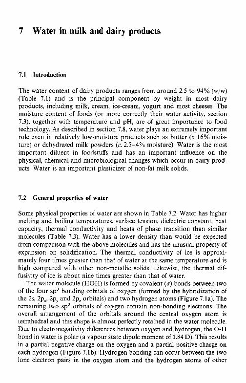
7 Water in milk and dairy products
7.1 Introduction
The water content of dairy products ranges from around 2.5 to 94% (w/w) (Table 7.1) and is the principal component by weight in most dairy products, including milk, cream, ice-cream, yogurt and most cheeses. The moisture content of foods (or more correctly their water activity, section 7.3), together with temperature and pH, are of great importance to food technology. As described in section 7.8, water plays an extremely important role even in relatively low-moisture products such as butter (c. 16% mois- ture) or dehydrated milk powders (c. 2 . 5 4 % moisture). Water is the most important diluent in foodstuffs and has an important influence on the physical, chemical and microbiological changes which occur in dairy prod- ucts. Water is an important plasticizer of non-fat milk solids.
7.2 General properties of water
Some physical properties of water are shown in Table 7.2. Water has higher melting and boiling temperatures, surface tension, dielectric constant, heat capacity, thermal conductivity and heats of phase transition than similar molecules (Table 7.3). Water has a lower density than would be expected from comparison with the above molecules and has the unusual property of expansion on solidification. The thermal conductivity of ice is approxi- mately four times greater than that of water at the same temperature and is high compared with other non-metallic solids. Likewise, the thermal dif- fusivity of ice is about nine times greater than that of water.
The water molecule (HOH) is formed by covalent (6) bonds between two of the four sp3 bonding orbitals of oxygen (formed by the hybridization of the 2s, 2p,, 2py and 2p, orbitals) and two hydrogen atoms (Figure 7.la). The remaining two sp3 orbitals of oxygen contain non-bonding electrons. The overall arrangement of the orbitals around the central oxygen atom is tetrahedral and this shape is almost perfectly retained in the water molecule. Due to electronegativity differences between oxygen and hydrogen, the O-H bond in water is polar (a vapour state dipole moment of 1.84 D). This results in a partial negative charge on the oxygen and a partial positive charge on each hydrogen (Figure 7.lb). Hydrogen bonding can occur between the two lone electron pairs in the oxygen atom and the hydrogen atoms of other
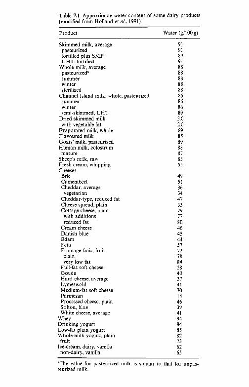
Table 7.1 Approximate water content of some dairy products (modified from Holland et a/., 1991)
Product Water (g/lOO g)
Skimmed milk, average pasteurized fortified plus SMP UHT, fortified
Whole milk, average pasteurized” summer winter sterilized
summer winter semi-skimmed, UHT
Dried skimmed milk with vegetable fat
Evaporated milk, whole Flavoured milk Goats’ milk, pasteurized Human milk, colostrum
Sheep’s milk, raw Fresh cream, whipping Cheeses
Brie Camembert Cheddar, average vegetarian
Cheddar-type, reduced fat Cheese spread, plain Cottage cheese, plain
Channel Island milk, whole, pasteurized
mature
with additions reduced fat
Cream cheese Danish blue Edam Feta Fromage frais, fruit plain very low fat
Full-fat soft cheese Gouda Hard cheese, average Lymeswold Medium-fat soft cheese Parmesan Processed cheese, plain Stilton, blue White cheese, average
Whey Drinking yogurt Low-fat plain yogurt Whole-milk yogurt, plain
Ice-cream, dairy, vanilla fruit
non-dairy, vanilla
91 91 89 91 88 88 88 88 88 86 86 86 89 3.0 2.0 69 85 89 88 87 83 55
49 51 36 34 41 53 79 17 80 46 45 44 51 72 18 84 58 40 31 41 10 18 46 39 41 94 84 85 82 13 62 65
“The value for pasteurized milk is similar to that for unpas- teurized milk.
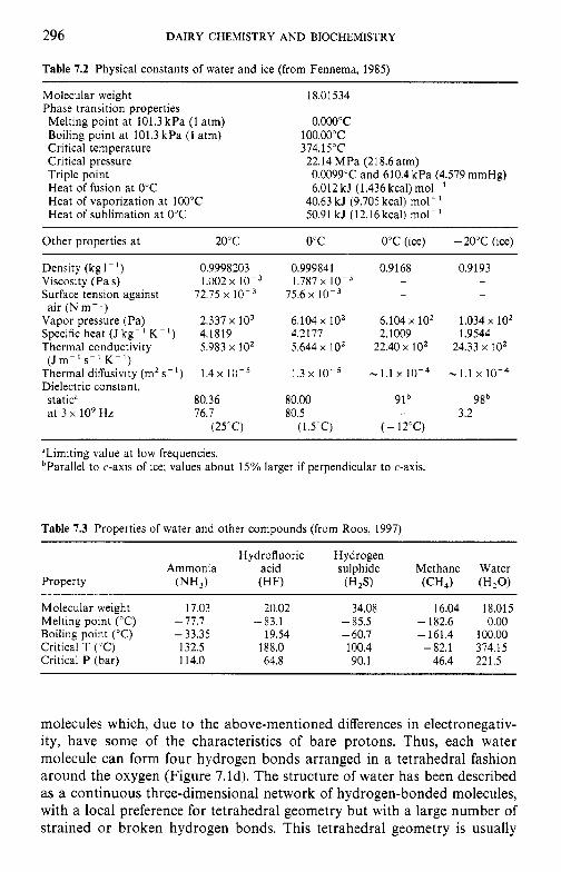
296 DAIRY CHEMISTRY AND BIOCHEMISTRY
Table 7.2 Physical constants of water and ice (from Fennema, 1985)
Molecular weight Phase transition properties Melting point at 101.3 kPa (1 atm) Boiling point at 101.3 kPa (1 atm) Critical temperature Critical pressure Triple point Heat of fusion at 0°C Heat of vaporization at 100°C Heat of sublimation at 0°C
18.01 534
0.ooo"c 100.00"C 374.15"C 22.14 MPa (218.6 atm) 0.0099'C and 610.4 kPa (4.579 mmHg) 6.012kJ (1.436kcal)mol-'
40.63 kJ (9.705 kcal) mol- 50.91 kJ (12.16kcal) mol-'
Other properties a t 20°C 0°C 0°C (ice) - 20°C (ice)
Density (kg I - ' ) 0.9998203
Surface tension against 72.75 x
Vapor pressure (Pa) 2.337 x lo3 Specific heat (J kg-' K - I ) 4.1819 Thermal conductivity 5.983 x 10'
Thermal diffusivity (m2 s-I) Dielectric constant,
Viscosity (Pa s) 1.002 x 10⁻³
air (N m - I )
(J m - ' s - ' K - ' 1 1.4 x
static" 80.36 at 3 x lo9 Hz 76.7
(25'C)
0.999841 1.787 x
75.6 x 10-3
6.104 x 10' 4.2177 5.644 x 10'
1.3 10-5
80.00 80.5
(1 .5"C)
0.9168 - -
6.104 x 10' 2.1009
22.40 x lo2
- 1.1 x 10⁻⁴
91b -
( - 12°C)
0.9193 - -
1.034 x 10' 1.9544
24.33 x 10'
- 1.1 x 10⁻⁴
98b 3.2
-
"Limiting value at low frequencies. bParallel to c-axis of ice; values about 15% larger if perpendicular to c-axis.
Table 7.3 Properties of water and other compounds (from Roos, 1997)
Hydrofluoric Hydrogen A m m o n i a acid sulphide Methane Water
Property (NH,) (HF) W 2 . T ( C H J (HZO)
Molecular weight 17.03 20.02 34.08 16.04 18.015 Melting point ('C) - 77.7 -83.1 - 85.5 - 182.6 0.00 Boiling point ("C) - 33.35 19.54 - 60.7 -161.4 100.00 Critical T ("C) 132.5 188.0 100.4 -82.1 374.15 Critical P (bar) 114.0 64.8 90.1 46.4 221.5
molecules which, due to the above-mentioned differences in electronegativ- ity, have some of the characteristics of bare protons. Thus, each water molecule can form four hydrogen bonds arranged in a tetrahedral fashion around the oxygen (Figure 7.ld). The structure of water has been described as a continuous three-dimensional network of hydrogen-bonded molecules, with a local preference for tetrahedral geometry but with a large number of strained or broken hydrogen bonds. This tetrahedral geometry is usually
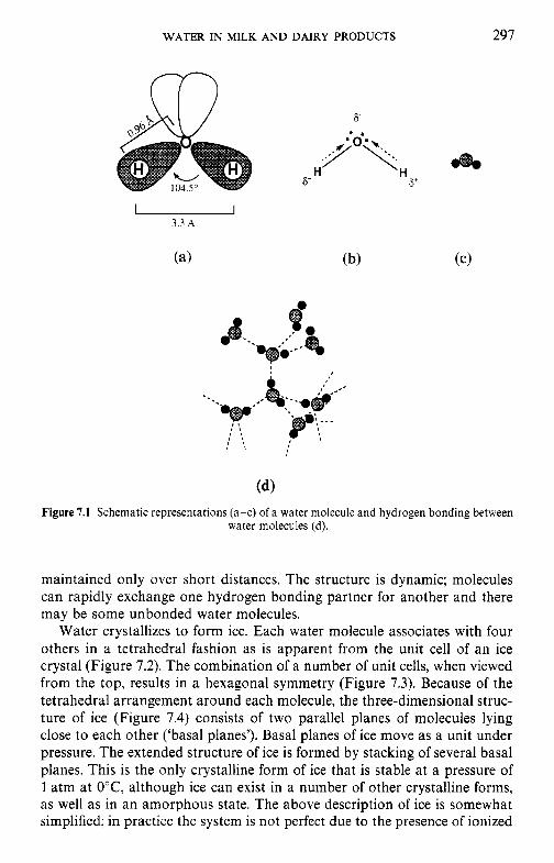
WATER IN MILK AND DAIRY PRODUCTS 297
Figure 7.1 Schematic representations (a-c) of a water molecule and hydrogen bonding between water molecules (d).
maintained only over short distances. The structure is dynamic; molecules can rapidly exchange one hydrogen bonding partner for another and there may be some unbonded water molecules.
Water crystallizes to form ice. Each water molecule associates with four others in a tetrahedral fashion as is apparent from the unit cell of an ice crystal (Figure 7.2). The combination of a number of unit cells, when viewed from the top, results in a hexagonal symmetry (Figure 7.3). Because of the tetrahedral arrangement around each molecule, the three-dimensional struc- ture of ice (Figure 7.4) consists of two parallel planes of molecules lying close to each other ('basal planes'). Basal planes of ice move as a unit under pressure. The extended structure of ice is formed by stacking of several basal planes. This is the only crystalline form of ice that is stable at a pressure of 1 atm at O'C, although ice can exist in a number of other crystalline forms, as well as in an amorphous state. The above description of ice is somewhat simplified; in practice the system is not perfect due to the presence of ionized
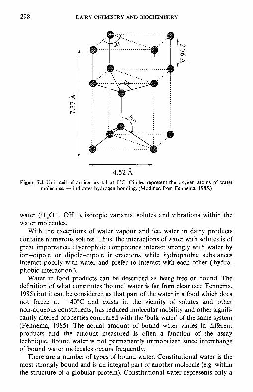
298 DAIRY CHEMISTRY AND BIOCHEMISTRY
4.52 A Figure 7.2 Unit cell of an ice crystal at 0°C. Circles represent the oxygen atoms of water
molecules, - indicates hydrogen bonding. (Modified from Fennema, 1985.)
water ( H 3 0 f , OH -), isotopic variants, solutes and vibrations within the water molecules.
With the exceptions of water vapour and ice, water in dairy products contains numerous solutes. Thus, the interactions of water with solutes is of great importance. Hydrophilic compounds interact strongly with water by ion-dipole or dipole-dipole interactions while hydrophobic substances interact poorly with water and prefer to interact with each other (‘hydro- phobic interaction’).
Water in food products can be described as being free or bound. The definition of what consitiutes ‘bound’ water is far from clear (see Fennema, 1985) but it can be considered as that part of the water in a food which does not freeze at -40°C and exists in the vicinity of solutes and other non-aqueous constituents, has reduced molecular mobility and other signifi- cantly altered properties compared with the ‘bulk water’ of the same system (Fennema, 1985). The actual amount of bound water varies in different products and the amount measured is often a function of the assay technique. Bound water is not permanently immobilized since interchange of bound water molecules occurs frequently.
There are a number of types of bound water. Constitutional water is the most strongly bound and is an integral part of another molecule (e.g. within the structure of a globular protein). Constitutional water represents only a
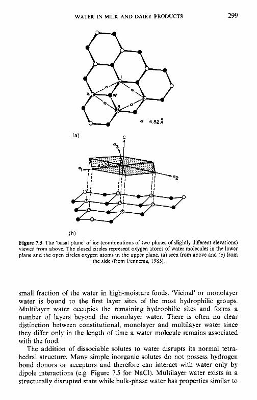
WATER IN MILK AND DAIRY PRODUCTS 299
(b) Figure 7.3 The ‘basal plane’ of ice (combinations of two planes of slightly different elevations) viewed from above. The closed circles represent oxygen atoms of water molecules in the lower plane and the open circles oxygen atoms in the upper plane, (a) seen from above and (b) from
the side (from Fennema, 1985).
small fraction of the water in high-moisture foods. ‘Vicinal’ or monolayer water is bound to the first layer sites of the most hydrophilic groups. Multilayer water occupies the remaining hydrophilic sites and forms a number of layers beyond the monolayer water. There is often no clear distinction between constitutional, monolayer and multilayer water since they differ only in the length of time a water molecule remains associated with the food.
The addition of dissociable solutes to water disrupts its normal tetra- hedral structure. Many simple inorganic solutes do not possess hydrogen bond donors or acceptors and therefore can interact with water only by dipole interactions (e.g. Figure 7.5 for NaCl). Multilayer water exists in a structurally disrupted state while bulk-phase water has properties similar to

300 DAIRY CHEMISTRY A N D BIOCHEMISTRY
C
4
Figure 7.4 The extended structure of ice. Open and shaded circles represent oxygen atoms of water molecules in the upper and lower layers, respectively, of a basal plane (from Fennema,
1985).
Figure 7.5 Arrangement of water molecules in the vicinity of sodium and chloride ions (modified from Fennema. 1985).
those of water in a dilute aqueous salt solution. Ions in solution impose structure on the water but disrupt its normal tetrahedral structure. Concen- trated solutions probably do not contain much bulk-phase water and structures caused by the ions predominate. The ability of an ion to influence the structure of water is influenced by its electric field. Some ions (princi- pally small and/or multivalent) have strong electric fields and loss of the inherent structure of the water is more than compensated for by the new structure resulting from the presence of the ions. However, large, mono- valent ions have weak electric fields and thus have a net disruptive effect on the structure of water.
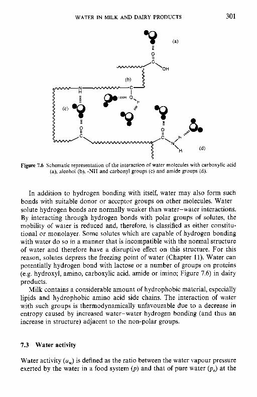
WATER IN MILK AND DAIRY PRODUCTS 301
0 I I
Figure 7.6 Schematic representation of the interaction of water molecules with carboxylic acid (a), alcohol (b), -NH and carbonyl groups (c) and amide groups (d).
In addition to hydrogen bonding with itself, water may also form such bonds with suitable donor or acceptor groups on other molecules. Water- solute hydrogen bonds are normally weaker than water-water interactions. By interacting through hydrogen bonds with polar groups of solutes, the mobility of water is reduced and, therefore, is classified as either constitu- tional or monolayer. Some solutes which are capable of hydrogen bonding with water do so in a manner that is incompatible with the normal structure of water and therefore have a disruptive effect on this structure. For this reason, solutes depress the freezing point of water (Chapter 11). Water can potentially hydrogen bond with lactose or a number of groups on proteins (e.g. hydroxyl, amino, carboxylic acid, amide or imino; Figure 7.6) in dairy products.
Milk contains a considerable amount of hydrophobic material, especially lipids and hydrophobic amino acid side chains. The interaction of water with such groups is thermodynamically unfavourable due to a decrease in entropy caused by increased water-water hydrogen bonding (and thus an increase in structure) adjacent to the non-polar groups.
7.3 Water activity
Water activity (a,) is defined as the ratio between the water vapour pressure exerted by the water in a food system ( p ) and that of pure water ( p , ) at the
302
same temperature:
DAIRY CHEMISTRY AND BIOCHEMISTRY
P Po
a =-. W
Due to the presence of various solutes, the vapour pressure exerted by water in a food system is always less than that of pure water (unity). Water activity is a temperature-dependent property of water which may be used to characterize the equilibrium or steady state of water in a food system (Roos, 1997).
For a food system in equilibrium with a gaseous atmosphere (i.e. no net gain or loss of moisture to or from the system caused by differences in the vapour pressure of water), the equilibrium relative humidity (ERH) is related to a, by:
ERH(%) = a, x 100. (7.2)
Thus, under ideal conditions, ERH is the % relative humidity of an atmosphere in which a foodstuff may be stored without a net loss or gain of moisture. Water activity, together with temperature and pH, is one of the most important parameters which determine the rates of chemical, bio- chemical and microbiological changes which occur in foods. However, since a, presupposes equilibrium conditions, its usefulness is limited to foods in which these conditions exist.
Water activity is influenced by temperature and therefore the assay tem- perature must be specified. The temperature dependence of a, is described by the Clausius-Clapeyron equation in modified form:
(7.3)
where T is temperature (K), R is the universal gas constant and AH is the change in enthalpy. Thus, at a constant water content, there is a linear relationship between log a, and 1/T (Figure 7.7). This linear relationship is not obeyed at extremes of temperature or at the onset of ice formation.
The concept of a, can be extended to cover sub-freezing temperatures. In these cases, a, is defined (Fennema, 1985) relative to the vapour pressure of supercooled water (poCscw,) rather than to that of ice:
where pfr is the vapour pressure of water in the partially frozen food and pice that of pure ice. There is a linear relationship between loga, and 1/T at sub-freezing temperatures (Figure 7.8). The influence of temperature on a, is greater below the freezing point of the sample and there is normally a pronounced break at the freezing point. Unlike the situation above freezing
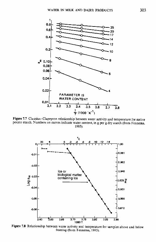
WATER IN MILK AND DAIRY PRODUCTS
PARAMETER IS WATER CONTENT
303
0 -2 -4 6 -8 .lo -12 -14 20 5
a-4 - 0 1 . . , I 1 I I 1 I I I I I I I I I I I
- t--- ----___ -0.01 - -
- -0.02 - -
- - % .0.03 - m -
-I - -
:: -0.04 -
- -0.05 - -
- -
-0.06 - - I I I I I I I
3.40 9.60 3.65 3.70 3.75 3.80 3.85 3.90
0.2 "i
1.00
0.981
0.962
0.940
0.925
0.907
0.890
0.872
m3 0.10 o . 0 8 ~ 0.06
I
0.02 " O 4 I
304 DAIRY CHEMISTRY AND BIOCHEMISTRY
(where a, is a function of composition and temperature), a, below freezing is independent of sample composition and is influenced only by temperature. Thus, a, values of foods at sub-freezing temperatures cannot be used to predict the a, of foods above freezing. Sub-freezing a, values are far less useful indicators of potential changes in foods than a, values determined above the freezing point.
Water activity may be measured by a number of techniques (Marcos, 1993). Comparison of manometric readings taken simultaneously on a food system and on pure water is the most direct technique. a, can also be measured in dilute solutions and liquid foods with low solute concentrations by cryoscopy, since under certain conditions a, can be considered as a colligative property. In these cases, the Clausius-Clapeyron equation is valid:
where n, and n, are the number of moles of solute and water, respectively, and y is the activity coefficient (approximately one for dilute solutions); n2 can be determined by measuring the freezing point from the relation:
GAT, n, = -
1000K,
where G is the grams of solvent in the sample, AT, is the freezing point depression ("C) and K , is the molal freezing point depression constant for water, i.e. 1.86.
Water activity may also be measured by determining the ERH for a food sample, using equation 7.2.
ERH may be estimated by measuring the relative humidity of the headspace over a food in a small, sealed container hygrometrically, psychro- metrically or directly by measuring the moisture content of the air by gas chromatography. ERH can be estimated by moisture-related colour changes in paper impregnated with cobalt thiocyanate (Co(SCN),) and compared to standards of known a,.
Differences in the hygroscopicity of various salts may also be used to estimate a,. Samples of the food are exposed to a range of crystals of known a,; if the a, of the sample is greater than that of a given crystal, the crystal will absorb water from the food.
Alternatively, a, may be measured by isopiestic equilibration. In this method, a dehydrated sorbent (e.g. microcrystalline cellulose) with a known moisture sorption isotherm (section 7.4) is exposed to the atmosphere in contact with the sample in an enclosed vessel. After the sample and sorbent have reached equilibrium, the moisture content of the sorbent can be measured gravimetrically and related to the a, of the sample.
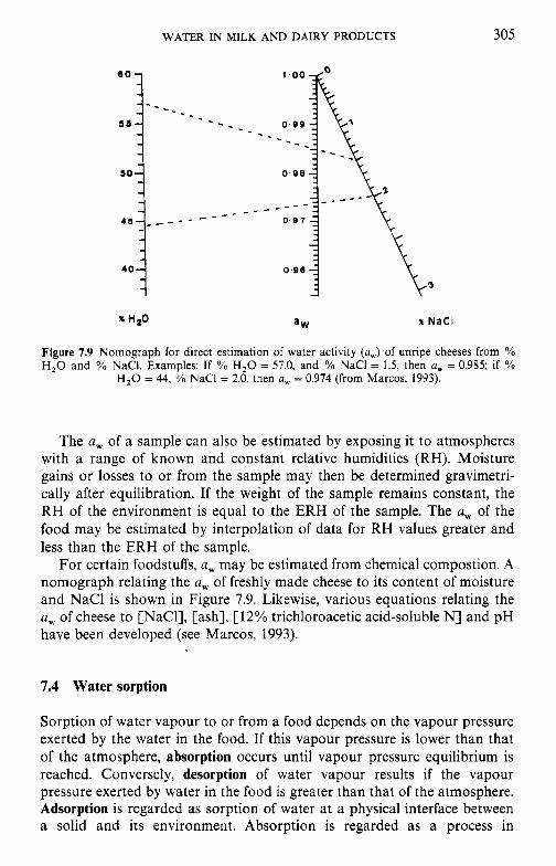
WATER IN MILK AND DAIRY PRODUCTS 305
X HZO % N a C l
Figure 7.9 Nomograph for direct estimation of water activity (a,) of unripe cheeses from % H,O and YO NaCI. Examples: If % H,O = 57.0, and % NaCl = 1.5, then a, = 0.985; if %
H,O = 44, YO NaCl = 2.0, then a, = 0.974 (from Marcos, 1993).
The a, of a sample can also be estimated by exposing it to atmospheres with a range of known and constant relative humidities (RH). Moisture gains or losses to or from the sample may then be determined gravimetri- cally after equilibration. If the weight of the sample remains constant, the RH of the environment is equal to the ERH of the sample. The a, of the food may be estimated by interpolation of data for RH values greater and less than the ERH of the sample.
For certain foodstuffs, a, may be estimated from chemical compostion. A nomograph relating the a, of freshly made cheese to its content of moisture and NaCl is shown in Figure 7.9. Likewise, various equations relating the a, of cheese to [NaCI], [ash], [12% trichloroacetic acid-soluble N] and pH have been developed (see Marcos, 1993).
7.4 Water sorption
Sorption of water vapour to or from a food depends on the vapour pressure exerted by the water in the food. If this vapour pressure is lower than that of the atmosphere, absorption occurs until vapour pressure equilibrium is reached. Conversely, desorption of water vapour results if the vapour pressure exerted by water in the food is greater than that of the atmosphere. Adsorption is regarded as sorption of water at a physical interface between a solid and its environment. Absorption is regarded as a process in
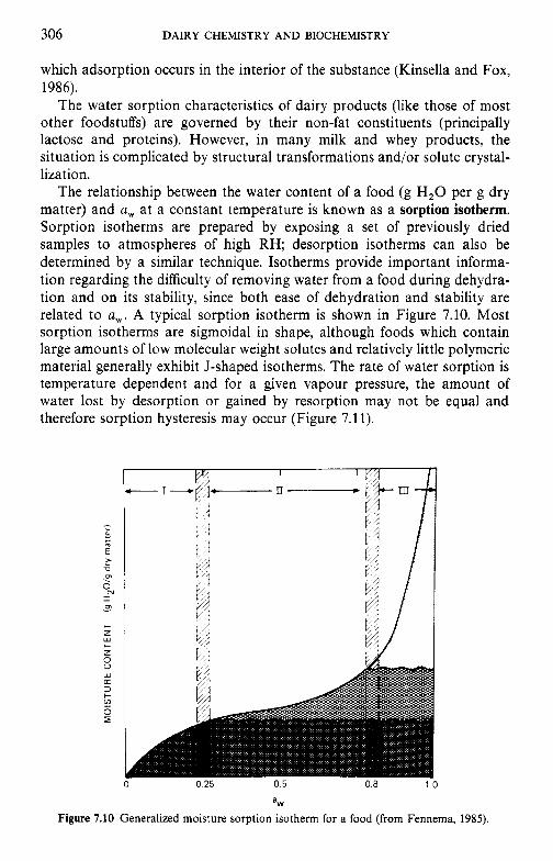
306 DAIRY CHEMISTRY AND BIOCHEMISTRY
which adsorption occurs in the interior of the substance (Kinsella and Fox, 1986).
The water sorption characteristics of dairy products (like those of most other foodstuffs) are governed by their non-fat constituents (principally lactose and proteins). However, in many milk and whey products, the situation is complicated by structural transformations and/or solute crystal- lization.
The relationship between the water content of a food (g H,O per g dry matter) and a, at a constant temperature is known as a sorption isotherm. Sorption isotherms are prepared by exposing a set of previously dried samples to atmospheres of high RH; desorption isotherms can also be determined by a similar technique. Isotherms provide important informa- tion regarding the difficulty of removing water from a food during dehydra- tion and on its stability, since both ease of dehydration and stability are related to a,. A typical sorption isotherm is shown in Figure 7.10. Most sorption isotherms are sigmoidal in shape, although foods which contain large amounts of low molecular weight solutes and relatively little polymeric material generally exhibit J-shaped isotherms. The rate of water sorption is temperature dependent and for a given vapour pressure, the amount of water lost by desorption or gained by resorption may not be equal and therefore sorption hysteresis may occur (Figure 7.1 1).
a,
Figure 7.10 Generalized moisture sorption isotherm for a food (from Fennema, 1985).
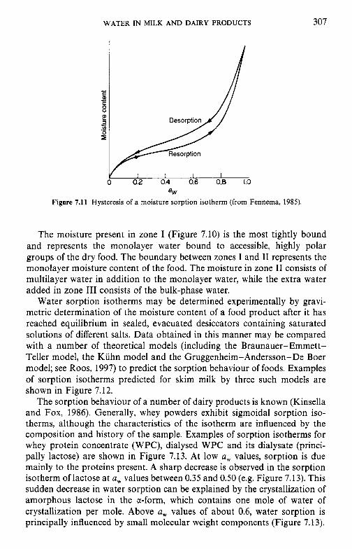
WATER IN MILK AND DAIRY PRODUCTS 307
- L E 8
r"
C
2 - .-
0 0.2 0.4 0.6 0.8 1.0
Figure 7.11 Hysteresis of a moisture sorption isotherm (from Fennema, 1985). a,
The moisture present in zone I (Figure 7.10) is the most tightly bound and represents the monolayer water bound to accessible, highly polar groups of the dry food. The boundary between zones I and I1 represents the monolayer moisture content of the food. The moisture in zone I1 consists of multilayer water in addition to the monolayer water, while the extra water added in zone I11 consists of the bulk-phase water.
Water sorption isotherms may be determined experimentally by gravi- metric determination of the moisture content of a food product after it has reached equilibrium in sealed, evacuated desiccators containing saturated solutions of different salts. Data obtained in this manner may be compared with a number of theoretical models (including the Braunauer-Emmett- Teller model, the Kuhn model and the Gruggenheim-Anderson-De Boer model; see Roos, 1997) to predict the sorption behaviour of foods. Examples of sorption isotherms predicted for skim milk by three such models are shown in Figure 7.12.
The sorption behaviour of a number of dairy products is known (Kinsella and Fox, 1986). Generally, whey powders exhibit sigmoidal sorption iso- therms, although the characteristics of the isotherm are influenced by the composition and history of the sample. Examples of sorption isotherms for whey protein concentrate (WPC), dialysed WPC and its dialysate (princi- pally lactose) are shown in Figure 7.13. At low a, values, sorption is due mainly to the proteins present. A sharp decrease is observed in the sorption isotherm of lactose at a, values between 0.35 and 0.50 (e.g. Figure 7.13). This sudden decrease in water sorption can be explained by the crystallization of amorphous lactose in the a-form, which contains one mole of water of crystallization per mole. Above a, values of about 0.6, water sorption is principally influenced by small molecular weight components (Figure 7.13).
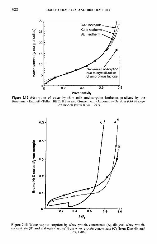
308 DAIRY CHEMISTRY AND BIOCHEMISTRY
Kuhn isotherm
of amorphous lactose
Water activity 8
Figure 7.12 Adsorption of water by skim milk and sorption isotherms predicted by the Braunauer-Emmett-Teller (BET), Kuhn and Guggenheim-Anderson-De Boer (GAB) sorp-
tion models (from Roos, 1997).
P/P,
Figure 7.13 Water vapour sorption by whey protein concentrate (A), dialysed whey protein concentrate (B) and dialysate (lactose) from whey protein concentrate (C) (from Kinsella and
Fox, 1986).
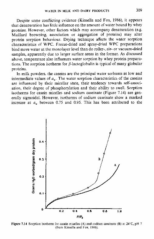
WATER IN MILK AND DAIRY PRODUCTS 309
Despite some conflicting evidence (Kinsella and Fox, 1986), it appears that denaturation has little influence on the amount of water bound by whey proteins. However, other factors which may accompany denaturation (e.g. Maillard browning, association or aggregation of proteins) may alter protein sorption behaviour. Drying technique affects the water sorption characteristics of WPC. Freeze-dried and spray-dried WPC preparations bind more water at the monolayer level than do roller-, air- or vacuum-dried samples, apparently due to larger surface areas in the former. As discussed above, temperature also influences water sorption by whey protein prepara- tions. The sorption isotherm for P-lactoglobulin is typical of many globular proteins.
In milk powders, the caseins are the principal water sorbants at low and intermediate values of a,. The water sorption characteristics of the caseins are influenced by their micellar state, their tendency towards self-associ- ation, their degree of phosphorylation and their ability to swell. Sorption isotherms for casein micelles and sodium caseinate (Figure 7.14) are gen- erally sigmoidal. However, isotherms of sodium caseinate show a marked increase at a, between 0.75 and 0.95. This has been attributed to the
0 0.2 0.4 0.6 0.8 1 .o
P/Po
Figure 7.14 Sorption isotherm for casein micelles (A) and sodium caseinate (B) at 2 4 T , pH 7 (from Kinsella and Fox, 1986).
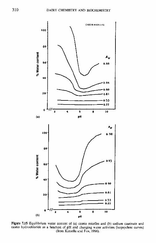
3 10 DAIRY CHEMISTRY AND BIOCHEMISTRY
1 oa
8C
CI C al c 8 bC
w
L
2
E c
40
2 0
0
(a)
100
60
60
40
2 0
0
0.53 0.22
2 4 6 8 LO
PH
0.95
0.90
0.53 0 . 2 1
f/ 2 4 b 6 10
PH
Figure 7.15 Equilibrium water content of (a) casein micelles and (b) sodium caseinate and casein hydrochloride as a function of pH and changing water activities (isopsychric curves)
(from Kinsella and Fox, 1986).
WATER IN MILK AND DAIRY PRODUCTS 311
presence of certain ionic groups, bound Na' or the increased ability of sodium caseinate to swell.
Heating of casein influences its water sorption characteristics, as does pH. With some exceptions at low pH, the hydration of sodium caseinate increases with pH (Figure 7.15b). Minimum water sorption occurs around the isoelectric pH (4.6). At low and intermediate values of a,, increasing pH, and thus [Na'], has little influence on water sorption. At low a, values, water is bound strongly to binding sites on the protein while at higher a, both protein and NaCl sorb available water in multilayer form. Water sorption by casein micelles (Figure 7.15a) has a minimum at about pH 6-7 at high a,. This difference in sorption minima between caseinate and casein micelles is because hydration of caseinate is due mainly to ion effects (Na' being more effective in this respect than C1-). Hydration behaviour of casein micelles, on the other hand, reflects effects of pH on micelle integrity. Hydrolysis of ic-casein by rennet appears to have only a small influence on its ability to bind water, although the chemical modification of amino groups has a greater effect. Genetic variation in the amino acid sequences of the caseins caused by genetic polymorphism also influences water sorption. The addition of NaCl to isoelectric casein greatly increases water sorption.
The greatest consequences of water sorption are in the context of dehydrated dairy products. In addition to being influenced by relative humidity, temperature and the relative amounts and intrinsic sorption properties of its constituents, the amount of water sorbed by milk powders is influenced by the method of preparation, the state of lactose, induced changes in protein conformation and swelling and dissolution of solutes such as salts. As discussed in Chapter 2, amorphous lactose is hygroscopic and may absorb large amounts of water at low relative humidities, while water sorption by crystalline lactose is significant only at higher relative humidities and thus water sorption by milk products containing crystallized lactose is due mainly to their protein fraction.
7.5 Glass transition and the role of water in plasticization
The non-fat solids in low-moisture dairy products (e.g. milk powders) or frozen milk products (since dehydration occurs on freezing) are amorphous in most dairy products (except those containing pre-crystallized lactose). The non-fat solids exist in a metastable, non-equilibrium state as a solid glass or a supercooled liquid. Phase changes can occur between these states with a phase transition temperature range called the glass transition (q; Roos, 1997). Changes in heat capacity, dielectric properties, volume, mol- ecular mobility and various mechanical properties occur on glass transition. The temperature of onset of the glass transition of amorphous water (i.e. the transformation of a solid, amorphous glass into a supercooled liquid and
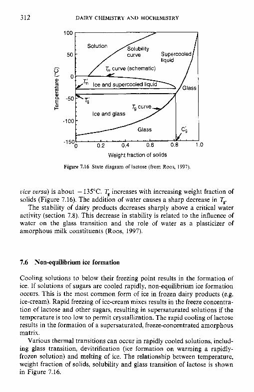
312 DAIRY CHEMISTRY AND BIOCHEMISTRY
100
- . .. A
0 0.2 0.4 0.6 0.8 1 .o ' . . ' ' . ' ' , ' ' .
Weight fraction of solids
Figure 7.16 State diagram of lactose (from Roos, 1997).
vice versa) is about - 135°C. T, increases with increasing weight fraction of solids (Figure 7.16). The addition of water causes a sharp decrease in T,.
The stability of dairy products decreases sharply above a critical water activity (section 7.8). This decrease in stability is related to the influence of water on the glass transition and the role of water as a plasticizer of amorphous milk constituents (Roos, 1997).
7.6 Non-equilibrium ice formation
Cooling solutions to below their freezing point results in the formation of ice. If solutions of sugars are cooled rapidly, non-equilibrium ice formation occurs. This is the most common form of ice in frozen dairy products (e.g. ice-cream). Rapid freezing of ice-cream mixes results in the freeze concentra- tion of lactose and other sugars, resulting in supersaturated solutions if the temperature is too low to permit crystallization. The rapid cooling of lactose results in the formation of a supersaturated, freeze-concentrated amorphous matrix.
Various thermal transitions can occur in rapidly cooled solutions, includ- ing glass transition, devitrification (ice formation on warming a rapidly- frozen solution) and melting of ice. The relationship between temperature, weight fraction of solids, solubility and glass transition of lactose is shown in Figure 7.16.
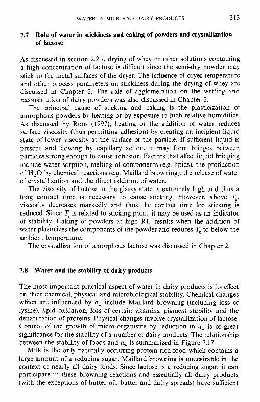
WATER IN MILK AND DAIRY PRODUCTS 313
7.7 Role of water in stickiness and caking of powders and crystallization of lactose
As discussed in section 2.2.7, drying of whey or other solutions containing a high concentration of lactose is difficult since the semi-dry powder may stick to the metal surfaces of the dryer. The influence of dryer temperature and other process parameters on stickiness during the drying of whey are discussed in Chapter 2. The role of agglomeration on the wetting and reconsitiution of dairy powders was also discussed in Chapter 2.
The principal cause of sticking and caking is the plasticization of amorphous powders by heating or by exposure to high relative humidities. As discussed by Roos (1997), heating or the addition of water reduces surface viscosity (thus permitting adhesion) by creating an incipient liquid state of lower viscosity at the surface of the particle. If sufficient liquid is present and flowing by capillary action, it may form bridges between particles strong enough to cause adhesion. Factors that affect liquid bridging include water sorption, melting of components (e.g. lipids), the production of H,O by chemical reactions (e.g. Maillard browning), the release of water of crystallization and the direct addition of water.
The viscosity of lactose in the glassy state is extremely high and thus a long contact time is necessary to cause sticking. However, above q, viscosity decreases markedly and thus the contact time for sticking is reduced. Since T, is related to sticking point, it may be used as an indicator of stability. Caking of powders at high RH results when the addition of water plasticizes the components of the powder and reduces < to below the ambient temperature.
The crystallization of amorphous lactose was discussed in Chapter 2.
7.8 Water and the stability of dairy products
The most important practical aspect of water in dairy products is its effect on their chemical, physical and microbiological stability. Chemical changes which are influenced by a, include Maillard browning (including loss of lysine), lipid oxidation, loss of certain vitamins, pigment stability and the denaturation of proteins. Physical changes involve crystallization of lactose. Control of the growth of micro-organisms by reduction in a, is of great significance for the stability of a number of dairy products. The relationship between the stability of foods and a, is summarized in Figure 7.17.
Milk is the only naturally occurring protein-rich food which contains a large amount of a reducing sugar. Maillard browning is undesirable in the context of nearly all dairy foods. Since lactose is a reducing sugar, it can participate in these browning reactions and essentially all dairy products (with the exceptions of butter oil, butter and dairy spreads) have sufficient
314 DAIRY CHEMISTRY AND BIOCHEMISTRY
Enzyme activity Loss of lysine
0 0.2 0.4 0.6 0.8 1 .o Water activity
Figure 7.17 Stability map for non-fat milk solids showing schematic rates of various deterio- rative changes and growth of micro-organisms as a function of water activity (from Roos,
1997).
protein to supply the necessary amino groups. Many of the stages of Maillard browning (Chapter 2) have high activation energies and thus the process is accelerated at high temperatures. The combination of the presence of lactose and high temperatures occurs during the production of many milk and whey powders, processed cheese and when dairy products are heated during cooking (e.g. the browning of Mozzarella cheese during baking of pizzas). The loss of lysine accompanies the early stages of the Maillard reaction in which its &-amino group participates. Loss of lysine is significant from a nutritional standpoint since it is an essential amino acid. Loss of lysine may occur without visible browning.
For a given product composition and temperature, the rate of browning is affected by a,. The influence of water on the rate of Maillard browning depends on the relative importance of a number of factors. Water imparts mobility to reacting species (thus increasing the rate of browning) but may also dilute reactants (thus reducing the rate of browning). At low values of a,, the increase in molecular mobility is most significant, while at higher values of a,.,, the dilution effect predominates. At lower a, values, water can also dissolve new reacting species. The presence of water can retard certain steps in browning in which water is released as a product (product inhibition, e.g. the initial glycosylamine reaction) or enhance other reactions (e.g. deamination). For many foods, the rate of Maillard browning usually
WATER IN MILK AND DAIRY PRODUCTS 315
reaches a maximum at intermediate moisture levels (a , x 0.40-0.80). However, the maximum rate is greatly influenced by the presence of other constituents in the food, such as glycerol or other liquid humectants which can shift the maximum to lower a, values. The rate of browning of milk powders is also accelerated by the crystallization of lactose.
Lipid oxidation can cause defects in high-fat dairy products. The mech- anism of lipid oxidation is discussed in Chapter 3. At low a,, the rate of oxidation decreases with increasing a, and reaches a minimum around the monolayer value and then increases at higher a,. The antioxidant effect of water at low values of a, has been attributed to bonding of hydroperoxide intermediates and the hydration of metal ions, which act as catalysts. The increased rate of oxidation at higher a, is a consequence of increased mobility of reactants. In general, water may influence the rate of lipid oxidation by affecting the concentration of initiating radicals, the degree of contact, the mobility of reacting species and the relative importance of radical transfer versus recombination events. Side reactions associated with lipid oxidation (e.g. cross-linking of proteins, enzyme inactivation by peroxi- dation products, degradation of amino acids) are also influenced by a,.
The stability of some vitamins is influenced by a,. In general, the stability of retinol (vitamin A), thiamin (vitamin B,) and riboflavin (vitamin B2) decreases with increasing a,. At low a, (below 0.40), metal ions do not have a catalytic effect on the destruction of ascorbic acid. The rate of loss of ascorbic acid increases exponentially as a, increases. The photodegradation of riboflavin (Chapter 6) is also accelerated by increasing a,.
Water activity influences the rate of thermal denaturation of proteins, including enzymes. Generally, the denaturation temperature increases with decreasing a,. The rate of nearly all enzyme-catalyzed reactions increases with increasing a,, as a consequence of increased molecular mobility.
The emulsification state of water in butter (i.e. the water droplet size) is very important for the quality of the product. Bacteria in butter can grow only in the aqueous emulsified phase. A finely divided aqueous phase restricts bacterial growth since the nutrients available in small droplets will quickly become limiting. Also, unless bacterial contamination is high, it is likely that most small water droplets in butter are sterile.
Together with pH and temperature, a, has a major influence on the rate of growth of micro-organisms. Indeed, reduction of a, by drying or the addition of salt or sugars is one of the principal traditional techniques used to preserve food. The minimum a, required for microbial growth is about 0.62, which permits the growth of xerophilic yeasts. As a, increases, moulds and other yeasts can grow and, finally, bacteria (above about 0.80). a, also controls the growth of pathogenic micro-organisms; StaphyIococcus aureus will not grow below a, - 0.86 while the growth of Listeria monocytogenes does not occur below a, - 0.92.
316 DAIRY CHEMISTRY AND BIOCHEMISTRY
References
Fennema, O.R. (ed.) (1985) Food Chemistry, 2nd edn, Marcel Dekker, New York. Holland, B., Welch, A.A., Unwin, I.D. et a/. (1991) McCance and Widdowson’s The Composition
of Foods, 5th edn, Royal Society of Chemistry and Ministery of Agriculture, Fisheries and Food, Cambridge, London.
Kinsella, J.E. and Fox P.F. (1986) Water sorption by proteins: Milk and whey proteins. CRC Crit. Rev. Food Sci. Nutr., 24, 91-139.
Marcos, A. (1993) Water activity in cheese in relation to composition, stability and safety, in Cheese: Chemistrj, Physics and Microbiology, Vol. 1, 2nd edn (ed. P.F. Fox), Chapman & Hall, London, pp. 439-69.
Roos, Y . (1997) Water in milk products, in Advanced Dairy Chemistry, Vol. 3: Lactose, Water, Salts and Vitamins (ed. P.F. Fox), Chapman & Hall, London, pp. 306-46.
Suggested reading
Fennerna, O.R. (ed.) (1985) Food Chemistry, 2nd edn, Marcel Dekker, New York. Rockland, L.B. and Beuchat, L.R. (eds) (1987) Water Activify: Theory and Applicarions to Food,
Roos. Y. (1997) Water in milk products, in Advanced Dairy Chemistry, Vol. 3: Lactose, Wafer, Marcel Dekker, New York.
Salts and Vitamins (ed. P.F. Fox), Chapman & Hall, London, pp. 306-46.
8 Enzymology of milk and milk products
8.1 Introduction
Like all other foods of plant or animal origin, milk contains several indigenous enzymes which are constituents of the milk as secreted. The principal constituents of milk (lactose, lipids and proteins) can be modified by exogenous enzymes, added to induce specific changes. Exogenous en- zymes may also be used to analyse for certain constituents in milk. In addition, milk and most dairy products contain viable micro-organisms which secrete extracellular enzymes or release intracellular enzymes after the cells have died and lysed. Some of these enzymes may cause undesirable changes, e.g. hydrolytic rancidity of milk and dairy products, bitterness and/or age gelation of UHT milks, bittiness in cream, malty flavours or bitterness in fluid milk, or they may cause desirable flavours, e.g. in ripened cheese.
This chapter is devoted mainly to the significance of indigenous enzymes in milk. The principal applications of exogenous enzymes have been dealt with in other chapters, e.g. rennets and lipases in cheese production (Chapter lo), P-galactosidase to modify lactose (Chapter 2). Some minor or potential applications of exogenous enzymes are presented here. Enzymes derived from contaminating bacteria, which may be significant in milk and some dairy products, will not be discussed. The interested reader is referred to McKellar (1989) for a comprehensive review of enzymes produced by psychrotrophs which are the principal spoilage microorganisms in refrig- erated milk and milk products. The significance of enzymes from microbial cultures in cheese ripening is discussed in Chapter 10.
8.2 Indigenous enzymes of bovine milk
8.2.1 Introduction
As many as 60 indigenous enzymes have been reported in normal bovine milk. With the exception of cr-lactalbumin, which is an enzyme modifier in lactose synthesis (Chapter 2) most, if not all, of the indigenous enzymes in milk have no obvious physiological role. They arise from three principal sources:
0 the blood via defective mammary cell membranes;
318 DAIRY CHEMISTRY AND BIOCHEMISTRY
0 secretory cell cytoplasm, some of which is occasionally entrapped within fat globules by the encircling fat globule membrane (MFGM) (Chapter 3);
0 the MFGM itself, the outer layers of which are derived from the apical membrane of the secretory cell, which, in turn, originates from the Golgi membranes (Chapter 3); this is probably the principal source of indigen- ous enzymes.
Thus, most enzymes enter milk due to peculiarities of the mechanism by which milk constituents, especially the fat globules, are excreted from the secretory cells. Milk does not contain substrates for many of the enzymes present, while others are inactive in milk owing to unsuitable environmental conditions, e.g. pH.
Many indigenous milk enzymes are technologically significant from five viewpoints:
1. Deterioration (lipase (commercially, probably the most significant en- zyme in milk), proteinase, acid phosphatase and xanthine oxidase) or preservation (sulphydryl oxidase, superoxide dismutase) of milk quality.
2. As indices of the thermal history of milk: alkaline phosphatase, y-glutamyl transpeptidase, lactoperoxidase.
3. As indices of mastitic infection: catalase, N-acetyl-P-D-glucosaminidase, acid phosphatase; the concentration of several other enzymes increases on mastitic infection.
4. Antimicrobial activity: lysozyme, lactoperoxidase (which is exploited as a component of the lactoperoxidase - H,O, - thiocyanate system for the cold pasteurization of milk).
5. As commercial source of enzymes: ribonuclease, lactoperoxidase.
With a few exceptions (e.g. lysozyme and lactoperoxidase), the indigenous milk enzymes do not have a beneficial effect on the nutritional or organo- leptic attributes of milk, and hence their destruction by heat is one of the objectives of many dairy processes.
The distribution of the principal indigenous enzymes in milk and their catalytic activity are listed in Table 8.1. In this chapter, the occurrence, distribution, isolation and characterization of the principal indigenous enzymes will be discussed, with an emphasis on their commercial signifi- cance in milk.
8.2.2 Proteinases (EC 3.4.-.-)
The presence of an indigenous proteinase in milk was suggested by Babcock and Russel in 1897 but because it occurs at a low concentration or has low activity in milk, it was felt until the 1960s that the proteinase in milk may be of microbial origin. Recent changes in the dairy industry, e.g. improved hygiene in milk production, extended storage of milk at a low temperature
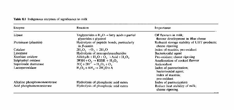
Table 8.1 Indigenous enzymes of significance to milk
Enzyme Reaction Importance
Lipase
Proteinase (plasmin)
Catalase Lysozyme Xanthine oxidase Sulphydryl oxidase Superoxide dismutase Lactoperoxidase
Alkaline phosphomonoesterase Acid phosphomonoesterase
Triglycerides + H,O 4 fatty acids +partial
Hydrolysis of peptide bonds, particularly
2H,O, + 0, + 2H,O Hydrolysis of mucopolysaccharides Aldehyde+H,O+O, + Acid+H,O, 2RSH + 0, + RSSR + H,O, 20;+2H+ 4 H,O,+O, H,O,+AH, +2H,O+A
glycerides +glycerol
in bcasein
Hydrolysis of phosphoric acid esters Hydrolysis of phosphoric acid esters
Off flavours in milk;
Reduced storage stability of UHT products;
Index of mastitis; pro-oxidant Bacteriocidal agent Pro-oxidant; cheese ripening Amelioration of cooked flavour Antioxidant Index of pasteurization;
flavour development in Blue cheese
chccse ripening
bacteriocidal agent; index of mastitis; pro-oxidant
Index of pasteurization Reduce heat stability of milk; cheese ripening
320 DAIRY CHEMISTRY AND BIOCHEMISTRY
at the farm and/or factory and altered product profile, e.g. UHT processing of milk, have increased the significance of indigenous milk proteinase which has, consequently, been the focus of considerable research.
Milk contains at least two proteinases, plasmin (alkaline milk proteinase) and cathepsin D (acid milk proteinase) and possibly several others, i.e. two thiol proteinases, thrombin and an aminopeptidase. In terms of activity and technological significance, plasmin is the most important of the indigenous proteinases and has been the subject of most attention. The relevant literature has been reviewed by Grufferty and Fox (1988) and Bastian and Brown (1996).
Plasmin (EC 3.4.21.7)
The physiological function of plasmin (fibrinolysin) is to dissolve blood clots. It is part of a complex system consisting of plasmin, its zymogen (plasminogen), plasminogen activators, plasmin inhibitors and inhibitors of plasminogen activators (Figure 8.1). In milk, there is about four times as much plasminogen as plasmin and both, as well as plasminogen activators, are associated with the casein micelles, from which they dissociate when the pH is reduced to 4.6. The inhibitors of plasmin and of plasminogen activators are in the milk serum. The concentration of plasmin and plas- minogen in milk increase with advancing lactation, mastitic infection and number of lactations.
Plasmin is usually extracted from casein at pH 3.5 and purified by precipitation with (NH,),SO, and various forms of chromatography, in- cluding affinity chromatography. Plasmin is optimally active at about pH 7.5 and 35°C; it exhibits c. 20% of maximum activity at 5°C and is stable over the pH range 4 to 9. Plasmin is quite heat stable: it is partially inactivated by heating at 72°C x 15s but its activity in milk increases following HTST pasteurization, probably through inactivation of the indig- enous inhibitors of plasmin or, more likely, inhibitors of plasminogen activators. It partly survives UHT sterilization and is inactivated by heating at 80°C x 10 min at pH 6.8; its stability decreases with increasing pH in the range 3.5-9.2.
Plasminogen activator(s) - Inhibitors of plasminogen
(milk serum) (casein micelles) activators
I
Plasminogen - Plasmin Plasmin inhibitors (casein micellesl (casein micelles) (milk serum)
Figure 8.1 Schematic representation of the plasmin system in milk.
ENZYMOLOGY OF MILK AND MILK PRODUCTS 321
Plasmin is a serine proteinase (inhibited by diisopropylfluorophosphate, phenylmethyl sulphonyl fluoride and trypsin inhibitor) with a high specific- ity for peptide bonds to which lysine or arginine supplies the carboxyl group. Its molecular weight is about 81 Da and its structure contains five intramolecular disulphide-linked loops (kringles) which are essential for its activity.
Activity of plasmin on milk proteins. 8-Casein is the most susceptible milk protein to plasmin action; it is hydrolysed rapidly at LyS28-Lys,g, Lys,,,-His,,, and Lys,,7-Glulo8, to yield y1 (8-CN f29-209), y z (P-CN f106-209) and y 3 (P-CN f 108-209) caseins and proteose-peptone (PP)5 (P-CN fl-105/7), PP8 slow (P-CN f29-105/7) and PP8 fast (8-CN fl-29) (Chapter 4). In solution, p-casein is also hydrolysed at Lys, 13-Tyr1 14 and Lys,,3-Asp,84, but it is not known if these bonds are hydrolysed in milk. ?-Caseins normally represent about 3% of total N in milk but can be as high as 10% in late lactation milk; the concentration of proteose peptones is about half that of the y-caseins.
a,,-Casein in solution is also hydrolysed very rapidly by plasmin at bonds Lys,,-Gln,,, Lys,,-Asn,,, Arg, 14-ASn1159 L Y ~ ~ ~ , - L Y ~ ~ , , , L Y ~ , , ~ - Thr,,,, LyS18,-Thr182, Lys187-Thr188 and Lys188-Ala1,g (See Bastian and Brown, 1996) but it is not known if it is hydrolysed in milk. Although less susceptible than z,,- or ,&caseins, a,,-casein in solution is also readily hydrolysed by plasmin (see Bastian and Brown, 1996) but it does not appear to be hydrolysed to a significant extent in milk although it has been suggested that /.-casein is produced from us,-casein by plasmin. Although K-casein contains several Lys and Arg residues, it appears to be quite resistant to plasmin, presumably due to a relatively high level of secondary and tertiary structure. P-Lactoglobulin, especially when denatured, inhibits plasmin, presumably via sulphydryl-disulphide interactions which rupture the structurally important kringles.
Signijicance of plasmin activity in milk. Plasmin and plasminogen accom- pany the casein micelles on the rennet coagulation of milk and are concentrated in cheese in which plasmin contributes to primary proteolysis of the caseins, especially in cheeses with a high-cook temperature, e.g. Swiss and some Italian varieties, in which the coagulant is totally or largely inactivated (Chapter 10). Plasmin activity may contribute to age gelation in UHT milk produced from high-quality raw milk (which contains a low level of Pseudomonas proteinase). It has been suggested that plasmin activity contributes to the poor cheesemaking properties of late-lactation milk but proof is lacking. The acid precipitability of casein from late lactation milk is also poor but evidence for the involvement of plasmin is lacking. Reduced yields of cheese and casein can be expected to result from plasmin action since the proteose peptones are, by definition, soluble at pH4.6.
322 DAIRY CHEMISTRY AND BIOCHEMISTRY
Cathepsin D (EC3.4.23.5). It has been known for more than 20 years that milk also contains an acid proteinase, (optimum pH x 4.0) which is now known to be cathepsin D, a lysozomal enzyme. It is relatively heat labile (inactivated by 70°C x 10min). Its activity in milk has not been studied extensively and its significance is unknown. At least some of the indigenous acid proteinase is incorporated into cheese curd; its specificity on zsl- and p-caseins is quite similar to that of chymosin but it has very poor milk-clotting activity (McSweeney, Fox and Olson, 1995). It may contribute to proteolysis in cheese but its activity is probably normally overshadowed by chymosin, which is present at a much higher level.
Other proteinases. The presence of low levels of other proteolytic enzymes in milk has been reported (see Fox and McSweeney, 1996). Most of these originate from somatic cells, and their level increases during mastitic infection. The presence of cathepsin D, a lysozomal enzyme, in milk suggests that all the lysozomal proteinases are present in milk although they may not be active. These minor proteinases are considered to be much less significant than plasmin, but more work on the subject is necessary.
8.2.3
Lipases catalyse the development of hydrolytic rancidity in milk, and, consequently, lipases and lipolysis in milk have been studied extensively.
Lipases and esterases (EC 3.1.1.-)
Milk contains three types of esterase:
1. A-type carboxylic ester hydrolases (arylesterases; EC 3.1.1.2), which hydrolyse aromatic esters, e.g. phenylacetate; they show little activity on tributyrin, and are not inhibited by organophosphates.
2. B-type esterases (glycerol tricarboxyl esterases, aliphatic esterases, lipases; EC 3.1.1.3): they are most active on aliphatic esters although they show some activity on aromatic esters; they are inhibited by organophosphates.
3. C-type esterases (cholinesterase; EC 3.1.1.7; EC 3.1.1.8): they are most active on choline esters but hydrolyse some aromatic and aliphatic esters slowly; they are inhibited by organophosphates.
In normal milk, the ratio of A : B : C esterase activity is about 3 : 10: 1 but the level of A-esterase activity increases considerably on mastitic infection. A and C esterases are considered to be of little technological significance in milk.
Classically, lipases hydrolyse ester bonds in emulsified esters, i.e. at a water/oil interface, although some may have limited activity on soluble esters; they are usually activated by blood serum albumin and Ca2+ which bind free fatty acids, which are inhibitory. Little lipolysis normally occurs in
ENZYMOLOGY OF MILK AND MILK PRODUCTS 323
milk because more than 90% of the lipase is associated with the casein micelles while the triglyceride substrates are in fat globules surrounded, and protected, by the fat globule membrane (MFGM). When the MFGM is damaged, lipolysis occurs rapidly, giving rise to hydrolytic rancidity.
Lipase was first isolated from skim milk and characterized by Fox and Tarassuk in 1967. The enzyme was optimally active at pH 9.2 and 37°C and found to be a serine enzyme (inactivated by organophosphates). A lipo- protein lipase (LPL; activated by lipoprotein co-factors) was demonstrated in milk by Korn in 1962 and was isolated by Egelrud and Olivecrona in 1972. LPL is, in fact, the principal indigenous lipase in milk and most recent work has been focused accordingly. The molecule has been characterized at the molecular, genetic, enzymatic and physiological levels (see Olivecrona et al., 1992).
In addition to LPL, human milk contains a bile salts-activated lipase, which probably contributes to the metabolism of lipids by breast-fed babies who have limited pancreatic lipase activity. Bovine milk and milks from other dairy animals do not contain this enzyme.
The lipolytic system in most milks becomes active only when the milk MFGM is damaged by agitation, homogenization or temperature fluctu- ations. However, some individual cows produce milk which becomes rancid spontaneously, i.e. without apparent activation. Spontaneous rancidity was considered to be due to a second lipase, termed membrane lipase, which was believed to be associated with the MFGM, but recent evidence suggests that LPL is responsible for spontaneous rancidity following activation by a lipoprotein (co-lipase) from blood serum; normal milk will become sponta- neously rancid if blood serum is added, suggesting that ‘spontaneous milks’ contain a higher than normal level of blood serum. Dilution of ‘spontaneous milk’ with normal milk prevents spontaneous rancidity, which consequently is not normally a problem with bulk herd milks; presumably, dilution with normal milk reduces the lipoprotein content of the mixture to below the threshold necessary for lipase adsorption.
Natural variations in the levels of free fatty acids in normal milk and the susceptibility of normal milks to lipolysis may be due to variations in the level of blood serum in milk.
Sign8cance of lipase. Technologically, lipase is arguably the most signi- ficant indigenous enzyme in milk. Although indigenous milk lipase may play a positive role in cheese ripening, undoubtedly the most industrially impor- tant aspect of milk lipase is its role in hydrolytic rancidity which renders liquid milk and dairy products unpalatable and eventually unsaleable. Lipolysis in milk has been reviewed extensively (Deeth and Fitz-Gerald, 1995). As discussed in Chapter 3, all milks contain an adequate level of lipase for rapid lipolysis, but become rancid only after the fat globule membrane has been damaged.
3 24 DAIRY CHEMISTRY AND BIOCHEMISTRY
8.2.4 Phosphatases
Milk contains several phosphatases, the principal ones being alkaline and acid phosphomonoesterases, which are of technological significance, and ribonuclease, which has no known function or significance in milk. The alkaline and acid phosphomonoesterases have been studied extensively (see Andrews (1993) for references).
Alkaline phosphomonoesterase (EC 3.1.3.1). The existence of a phospha- tase in milk was first recognized in 1925. Subsequently characterized as an alkaline phosphatase, it became significant when it was shown that the time-temperature combinations required for the thermal inactivation of alkaline phosphatase were slightly more severe than those required to destroy Mycobacteriurn tuberculosis, then the target micro-organism for pasteurization. The enzyme is readily assayed, and a test procedure based on alkaline phosphatase inactivation was developed for routine quality control of milk pasteurization. Several major modifications of the test have been developed. The usual substrates are phenyl phosphate, p-nitrophenyl- phosphate or phenolphthalein phosphate which are hydrolysed to inorganic phosphate and phenol, p-nitrophenol or phenolphthalein, respectively:
where XOH = phenol, p-nitrophenol or phenolphthalein. The release of inorganic phosphate may be assayed but the other product
is usually determined. Phenol is colourless but forms a coloured complex on reaction with one of several reagents, e.g. 2,6-dichloroquinonechloroimide, with which it forms a blue complex. p-Nitrophenol is yellow while phenol- phthalein is red at the alkaline pH of the assay (10) and hence the concentration of either of these may be determined easily.
Isolation and characterization. Alkaline phosphatase is concentrated in the fat globule membrane and hence in cream. It is released into the buttermilk on phase inversion; consequently, buttermilk is the starting material for most published methods for the purification of alkaline phos- phatase. Later methods have used chromatography on various media to give a homogeneous preparation with 7440-fold purification and 28% yield. The characteristics of milk alkaline phosphatase are summarized in Table 8.2. The enzyme appears to be similar to the alkaline phosphatase of mammary tissue.
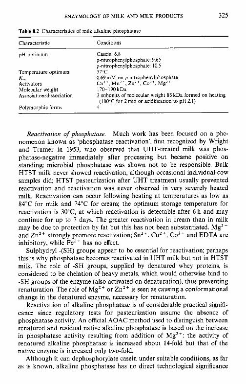
ENZYMOLOGY OF MILK AND MILK PRODUCTS 325
Table 8.2 Characteristics of milk alkaline phosphatase
Characteristic Conditions
pH optimum Casein: 6.8 p-nitrophenylphosphate: 9.65 p-nitrophenylphosphate: 10.5
0.69 mM on p-nitrophenylphosphate Ca2+, Mn2', Zn2+, Co2+ 3 g M 2 +
2 subunits of molecular weight 85 kDa formed on heating
Temperature optimum 37°C K m Activators Molecular weight 170- 190 kDa Association/dissociation
Polymorphic forms 4 (100°C for 2min or acidification to pH2.1)
Reactivation of phosphatase. Much work has been focused on a phe- nomenon known as 'phosphatase reactivation', first recognized by Wright and Tramer in 1953, who observed that UHT-treated milk was phos- phatase-negative immediately after processing but became positive on standing; microbial phosphatase was shown not to be responsible. Bulk HTST milk never showed reactivation, although occasional individual-cow samples did; HTST pasteurization after UHT treatment usually prevented reactivation and reactivation was never observed in very severely heated milk. Reactivation can occur following heating at temperatures as low as 84°C for milk and 74°C for cream; the optimum storage temperature for reactivation is 30°C, at which reactivation is detectable after 6 h and may continue for up to 7 days. The greater reactivation in cream than in milk may be due to protection by fat but this has not been substantiated. Mg2+ and Zn2+ strongly promote reactivation; Sn2+, CuZ+, Coz+ and EDTA are inhibitory, while Fe2+ has no effect.
Sulphydryl -(SH) groups appear to be essential for reactivation; perhaps this is why phosphatase becomes reactivated in UHT milk but not in HTST milk. The role of -SH groups, supplied by denatured whey proteins, is considered to be chelation of heavy metals, which would otherwise bind to -SH groups of the enzyme (also activated on denaturation), thus preventing renaturation. The role of Mg2+ or Zn2+ is seen as causing a conformational change in the denatured enzyme, necessary for renaturation.
Reactivation of alkaline phosphatase is of considerable practical signifi- cance since regulatory tests for pasteurization assume the absence of phosphatase activity. An official AOAC method used to distinguish between renatured and residual native alkaline phosphatase is based on the increase in phosphatase activity resulting from addition of Mg2+: the activity of renatured alkaline phosphatase is increased about 14-fold but that of the native enzyme is increased only two-fold.
Although it can dephosphorylate casein under suitable conditions, as far as is known, alkaline phosphatase has no direct technological significance
326 DAIRY CHEMISTRY AND BIOCHEMISTRY
in milk and milk products; perhaps its pH optimum is too far removed from that of milk; it is also inhibited by inorganic phosphate.
Acid phosphomonoesterase (EC 3.1.3.2). Milk contains an acid phospha- tase which has a pH optimum at 4.0 and is very heat stable (LTLT pasteurization causes only 10-20% inactivation and 30min at 88°C is required for full inactivation). Denaturation of acid phosphatase under UHT conditions follows first-order kinetics. When heated in milk at pH 6.7, the enzyme retains significant activity following HTST pasteurization but does not survive in-bottle sterilization or UHT treatment. The enzyme is not activated by Mg2+ (as is alkaline phosphatase), but it is slightly activated by Mn2+ and is very effectively inhibited by fluoride. The level of acid phosphatase activity in milk is only about 2% that of alkaline phosphatase; activity reaches a sharp maximum 5-6 days post-partum, then decreases and remains at a low level to the end of lactation.
Milk acid phosphatase has been purified to homogeneity by various forms of chromaotgraphy, including affinity chromatography; purification up to 40 000-fold has been claimed. The enzyme shows broad specificity on phosphate esters, including the phosphoseryl residues of casein. It has a molecular mass of about 42 kDa and an isoelectric point of 7.9. Many forms of inorganic phosphate are competitive inhibitors, while fluoride is a powerful non-competitive inhibitor. The enzyme is a glycoprotein and its amino acid composition is known. Milk acid phosphatase shows some similarity to the phosphoprotein phosphatase of spleen but differs from it in a number of characteristics.
Although casein is a substrate for milk acid phosphatase, the major caseins, in the order cts(ctsl + ~ 1 , ~ ) > p > K, also act as competitive inhibitors of the enzyme when assayed on p-nitrophenylphosphate, probably due to binding of the enzyme to the casein phosphate groups (the effectiveness of the caseins as inhibitors is related to their phosphate content).
Signijicance. Although acid phosphatase is present in milk at a much lower level than alkaline phosphatase, its greater heat stability and lower pH optimum may make it technologically significant. Dephosporylation of casein reduces its ability to bind Caz+, to react with K-casein, to form micelles and its heat stability. Several small partially dephosphorylated peptides have been isolated from Cheddar and Parmesan cheese. However, it is not known whether indigenous or bacterial acid phosphatases are mainly responsible for dephosphorylation in cheese. Dephosphorylation may be rate-limiting for proteolysis in cheese ripening since most pro- teinases and peptidases are inactive on phosphoproteins or peptides. It has been suggested that phosphatase activity should be included in the criteria for starter selection.
ENZYMOLOGY OF MILK A N D MILK PRODUCTS 327
The acid phosphatase activity in milk increases by a factor of 4-10 during mastitic infection; three isoenzymes are then present, only one of which is indigenous milk acid phosphatase, the other two being of leucocyte origin; these latter isoenzymes are more thermolabile and are inactivated by HTST pasteurization.
8.2.5 Lysozyme (EC 3.2.1.17)
Lysozyme (muramidase, mucopeptide N-acetylmuramylhydrolase) is a widely distributed enzyme which lyses certain bacteria by hydrolysing the P(1-4)-linkage between muramic acid and N-acetylglucosamine of mu- copolysaccharides of the bacterial cell wall.
Lysozyme was isolated from human milk in 1961 by Jolles and Jolles, who believed that bovine milk was devoid of lysozyme. Milks of many species have since been shown to contain lysozyme and several have been isolated and characterized. Human and equine milks are an exceptionally rich source, containing 130 mg 1-' (3000 times the level of bovine milk) and about 800 mg l-', respectively (see Farkye, 1992).
The pH optima of human milk lysozyme (HML), bovine milk lysozyme (BML) and egg-white lysozyme (EWL) are 7.9, 6.35 and 6.2, respectively. BML has a molecular weight of 18 kDa compared with 15 kDa for HML and EWL. The amino acid composition of BML is reported to be consider- ably different from that of HML or EWL. All lysozymes are relatively stable to heat at acid pH values (3-4) but are relatively labile at pH greater than 7 . Low concentrations of reducing agents increase the activity of BML and HML by about 330%.
SigniJicance. Presumably, the physiological role of lysozyme is to act as a bactericidal agent; in the case of milk it may simply be a 'spill-over' enzyme or it may have a definite protective role. If the latter is true, then the exceptionally high level of lysozyme in human and equine milk may be nutritionally significant. Breast-fed babies generally suffer less enteric prob- lems than bottle-fed babies. While there are many major compositional and physicochemical differences between bovine and human milks which may be responsible for the observed nutritional characteristics (Chapter 4), it has been suggested that the disparity in lysozyme content may be significant. A number of investigators have recommended fortification of bovine milk- based infant formulae with EWL, especially for premature babies. Feeding studies are equivocal on the benefits of this practice and recent trials failed to demonstrate any beneficial effect due to inactivation of EWL in the human stomach.
No beneficial effects from lysozyme on the shelf-life of milk have been reported. Addition of lysozyme to milk reduces its heat stability but the level
328 DAIRY CHEMISTRY AND BIOCHEMISTRY
of indigenous lysozyme is probably too low to contribute to the natural variations in the heat stability of milk.
8.2.6 N-Acetyl-P-D-glucosaminidase (EC 3.2.1.30)
N-Acet yl-P-D-glucosaminidase (NAGase) h ydrolyses terminal, non-reducing N-acetyl-P-D-glucosamine residues from glycoproteins. It is a lysosomal enzyme and originates mainly from somatic cells and mammary gland epithelial cells. Consequently, NAGase activity increases markedly and correlates highly with the intensity of mastitis. A field test for mastitis based on NAGase activity has been developed, using chromogenic N-acetyl-P-D- glucosamine-p-nitrophenol as substrate; hydrolysis yields yellow p-nitro- phenol. NAGase is optimally active at 50°C and pH 4.2 and is inactivated by HTST pasteurization (70-71°C x 15-18 s) (see Farkye, 1992).
8.2.7 y-Glutamyl transpeptidase (transferase) (EC 2.3.2.2)
y-Glutamyl transpeptidase (GGT) catalyses the transfer of y-glutamyl resi- dues from y-glutamyl-containing peptides:
y-glutamyl-peptide + X + peptide + y-glutamyl- X,
where X is an amino acid. GGT, which has been isolated from the fat globule membrane, has a
molecular mass of about 80 kDa and consists of two subunits of 57 and 26 kDa. It is optimally active at pH 8-9, has a pi of 3.85 and is inhibited by iodoacetate, diisopropylfluorophosphate and metal ions, e.g. Cu2+ and Fe3+
It plays a role in amino acid transport in the mammary gland. y-Glutamyl peptides have been isolated from cheese but since y-glutamyl bonds do not occur in milk proteins, their synthesis may be catalysed by GGT. The enzyme is relatively heat stable and has been proposed as a marker enzyme for milks pasteurized in the range 72-80°C x 15 s. GGT is absorbed from the gastrointestinal tract, resulting in high levels of GGT activity in the blood serum of newborn animals fed colostrum or early breast milk. Since GGT is inactivated by the heat treatment to which infant formulae are subjected, the level of GGTase activity in infants can be used to distinguish breast-fed from formula-fed infants (see Farkye, 1992).
8.2.8 Xanthine oxidase (EC 1.2.3.2)
It has been recognized for about 80years that milk contains an enzyme capable of oxidizing aldehydes and purines. The enzyme is now generally referred to as xanthine oxidase (XO); milk is a very good source of XO, at
ENZYMOLOGY OF MILK AND MILK PRODUCTS 329
least part of which is transported to the mammary gland via the blood- stream. A similar enzyme is found in various animal tissues and several bacterial species (Farkye, 1993).
Isolation. Numerous methods have been developed for the purification of XO from milk; since the enzyme is concentrated in the MFGM, in which it is one of the principal proteins, all methods employ cream as starting material, use a dissociating agent to liberate XO from membrane lipo- proteins and some form of chromatography for further purification.
Milk XO has a molecular weight of c.300kDa and consists of two subunits. The pH optimum is about 8.5 and the enzyme requires flavin adenine dinucleotide (FAD), Fe, Mo and an acid-labile compound as co-factors; cows deficient in Mo have low XO activity. The amino acid composition of XO has been determined by a number of workers; at least five genetic polymorphic forms have been reported.
Activity in milk. Various processing treatments affect the XO activity of milk. Activity is increased by about 100% on storage at 4°C for 24 h, by 50-100% on heating at 70°C for 5 min and by 60-90% on homogenization. These treatments cause the transfer to XO from the fat phase to the aqueous phase, rendering the enzyme more active. The heat stability of XO is very dependent on whether it is a component of the fat globules or is dissolved in the aqueous phase; ageing and homogenization increase susceptibility and explain the inconsistency of early work in which the history of the sample was unknown or unrecorded. XO is most heat stable in cream and least in skim milk. Homogenization of concentrated milk prepared from heated milk (90.5"C for 15 s) partially reactivates XO, which persists on drying the concentrate, but no reactivation occurs following more severe heating (93°C for 15 s); apparently, homogenization releases potentially active, unde- natured XO from the MFGM. All the major milk proteins can act as either activators or inhibitors of XO, depending on their concentration, and may have some significance in the activation, inactivation and reactivation of the enzyme.
Sign$cance of xanthine oxidase
Lipid oxidation XO, which can excite stable triplet oxygen (302), is a pro-oxidant. Milk which undergoes spontaneous rancidity contains about 10 times the normal level of XO, and spontaneous oxidation can be induced in normal milk by the addition of XO to about four times normal levels. Heat-denatured or flavin-free enzyme is ineffective and the susceptibility of unsaturated fatty acids to oxidation increases with the degree of unsaturation.
330 DAIRY CHEMISTRY AND BIOCHEMISTRY
At herosclerosis It has been suggested that XO from homogenized milk enters the vascular system and may be involved in atherosclerosis via oxidation of plasmalo- gens (Appendix 3B) in cell membranes. However, the experimental evidence in support of this view is very weak and the hypothesis has been disclaimed (see Farkye, 1992).
8.2.9 Sulphydryl oxidase (EC 1.8.3.-)
Milk contains an enzyme, sulphydryl oxidase (SO), capable of oxidizing sulphydryl groups of cysteine, glutathione and proteins to the corresponding disulphide (reviewed by Farkye, 1992). The enzyme is an aerobic oxidase which catalyses the following reaction:
2RSH + 0, RSSR + H,O,
It undergoes marked self-association and can be purified readily by chromatography on porous glass. The enzyme has a molecular weight of about 89 kDa, a pH optimum of 6.8-7.0, and a temperature optimum of 35°C. Its amino acid composition, its requirement for iron but not for molybdenum and FAD, and the catalytic properties of the enzyme, indicate that sulphydryl oxidase is a distinct enzyme from xanthine oxidase and thiol oxidase (EC 1.8.3.2).
SO is capable of oxidizing reduced ribonuclease and restoring enzymic activity, suggesting that its physiological role may be the non-random formation of protein disulphide bonds, e.g. during protein biosynthesis.
SO immobilized on glass beads has the potential to ameliorate the cooked flavour arising from sulphydryl groups exposed upon protein denaturation, but the commercial viability of this system is not known.
The production of sulphur compounds is believed to be very important in the development of Cheddar cheese flavour. Residual sulphydryl oxidase activity may play a role in initially reoxidizing sulphydryl groups exposed upon heating cheesemilk; the sulphydryl groups thus protected may be reformed during the ripening process.
8.2.10 Superoxide dismutase (EC 1.1.5.1.1)
Superoxide dismutase (SOD) scavenges superoxide radicals, 0; according to the reaction:
20; + 2Hf + H,Oz + 0,
The H,O, formed may be reduced by catalase, peroxidase or suitable reducing agents. SOD has been identified in many animal and bacterial cells; its biological function is to protect tissue against oxygen free radicals in anaerobic systems (reviewed by Farkye, 1992).
ENZYMOLOGY OF MILK AND MILK PRODUCTS 331
SOD, isolated from bovine erythrocytes, is a blue-green protein due to the presence of copper, removal of which by treatment with EDTA results in loss of activity, which is restored by adding Cu2+; it also contains Zn2+, which does not appear to be at the active site. The enzyme, which is very stable in 9 M urea at neutral pH, consists of two identical subunits of molecular weight 16 kDa held together by one or more disulphide bonds. The amino acid sequence has been established.
Milk contains trace amounts of SOD which has been isolated and characterized; it appears to be identical to the bovine erythrocyte enzyme. SOD inhibits lipid oxidation in model systems. The level of SOD in milk parallels that of XO (but at a lower level), suggesting that SOD may be excreted in milk in an attempt to offset the pro-oxidant effect of XO. However, the level of SOD in milk is probably insufficient to explain observed differences in the oxidative stability of milk. The possibility of using exogenous SOD to retard or inhibit lipid oxidation in dairy products has been considered.
SOD is more heat stable in milk than in purified preparations; in milk it is stable at 71°C for 30 min but loses activity rapidly at even slightly higher temperatures. Slight variations in pasteurization temperature are therefore critical to the survival of SOD in heated milk products and may contribute to variations in the stability of milk to oxidative rancidity.
8.2. I I Catalase (EC 1. I I . 1.6)
Indigenous milk catalase was first recognized in 1907. Although about 70% of the catalase activity of whole milk is in the skim-milk phase, cream has a higher specific activity; the pellet obtained from buttermilk on centrifu- gation at 10 000 g is a particularly rich source, from which catalase has been highly purified (reviewed by Farkye, 1992).
Milk catalase is a haem protein with a molecular weight of 200 kDa, and an isoelectric pH of 5.5; it is stable between pH 5 and 10 but rapidly loses activity outside this range. Heating at 70°C for 1 h causes complete inactiva- tion. Like other catalases, it is strongly inhibited by Hg2+, Fez+, Cu2+, Sn2+, CN- and NO,.
Catalase activity in milk varies with feed, stage of lactation and especially with mastitic infection, of which it may be used as an index. It may act as a lipid pro-oxidant via its haem iron.
8.2.12 Lactoperoxidase (EC 1.11.1.7)
The occurrence of a peroxidase, lactoperoxidase (LPO), in milk was recognized as early as 1881. It is one of the most heat-stable enzymes in milk; its destruction was used as an index of flash pasteurization (now very rarely used) and is now used as an index of super-HTST pasteurization.
332 DAIRY CHEMISTRY AND BIOCHEMISTRY
LPO was first isolated in 1943; several isolation procedures have since been published (reviewed by Bjorck, 1993).
LPO is a haem protein containing about 0.07% Fe, with an absorbance peak (Soret band) at 412 nm (A41z/A280 - 0.9); the pH optimum is around 8.0; its molecular weight is 77.5 kDa and it consists of two identical subunits. Two principal forms (A and B) occur, each of which exhibits micro- heterogeneity with regard to amide groups (glutamine and/or asparagine) and carbohydrate content, giving a total of 10 variants.
SigniJcance. Apart from its exploitation as an index of flash or super- HTST pasteurization, LPO is also technologically significant for a number of other reasons:
1. It is a possible index of mastitic infection; although the level of LPO in milk increases on mastitic infection, it is not well correlated with somatic cell count.
2. LPO causes non-enzymic oxidation of unsaturated lipids, probably acting through its haem group; the heat-denatured enzyme is more active than the native enzyme.
3. Milk contains bacteriostatic or bactericidal substances referred to as lactenins. One of these is LPO, which requires H,O, and thiocyanate (SCN-) to cause inhibition. The nature, mode of action and specificity of the LPO-H,O,-SCN- system has been widely studied. LPO and thiocyanate, which is produced in the rumen by enzymic hydrolysis of thioglycosides from Brassica plants, occur naturally in milk, but H,O, does not. However, H,O, can be generated metabolically by catalase- negative bacteria, or produced in situ through the action of exogenous glucose oxidase on glucose, or it may be added directly.
The peroxidase system has been found to have good bactericidal efficiency for the cold pasteurization of fluids or sanitization of immobi- lized enzyme columns. The generation of H,O, in situ through the action of immobilized glucose oxidase on glucose is effective against Gram- negative bacteria in thiocyanate and glucose-enriched milk and whey. A self-contained LPO-H,O,-SCN- system using coupled P-galactosidase and glucose oxidase, immobilized on porous glass beads, to generate H,O, in situ from lactose in milk containing 0.25mM thiocyanate has been developed. Indigenous xanthine oxidase, acting on added hypo- xanthine, may also be exploited to produce H,O, for the LPO-H,O,- SCN- system. The bactericidal effects of the LPO-H,O,-SCN- system may be used to cold pasteurize milk in situations where refrigeration and/or thermal pasteurization is lacking. LPO is cationic at the pH of milk and may be readily isolated on cation-exchange resins. Addition of isolated LPO to milk replacers for calves or piglets reduces the incidence of enteritis.
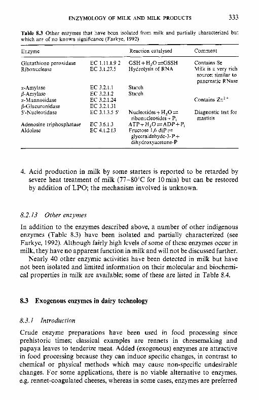
ENZYMOLOGY OF MILK AND MILK PRODUCTS 333
Table 8.3 Other enzymes that have been isolated from milk and partially characterized but which are of no known significance (Farkye, 1992) ~~
Enzyme Reaction catalysed Comment
Glutathione peroxidase EC 1.11.1.9 2 G S H + H , O e G S S H Contains Se Ri bonuclease EC 3.1.27.5 Hydrolysis of RNA Milk is a very rich
source; similar to pancreatic RNase
rl-Amylase EC 3.2.1.1 Starch P- Am ylase EC 3.2.1.2 Starch a-Mannosidase EC 3.2.1.24 Contains Zn2 +
/?-Glucuronidase EC 3.2.1.31 5'-Nucleotidase EC 3.1.3.5 5' Nucleotides+H,O+ Diagnostic test for
Adenosine triphosphatase EC 3.6.1.3 A T P + H , O = A D P + Pi Aldolase EC 4.1.2.13 Fructose 1,6 dip$
ribonucleosides + P, mastitis
glyceraldehyde-3-P + dihydroxyacetone-P
4. Acid production in milk by some starters is reported to be retarded by severe heat treatment of milk (77430°C for 10min) but can be restored by addition of LPO; the mechanism involved is unknown.
8.2.13 Other enzymes
In addition to the enzymes described above, a number of other indigenous enzymes (Table 8.3) have been isolated and partially characterized (see Farkye, 1992). Although fairly high levels of some of these enzymes occur in milk, they have no apparent function in milk and will not be discussed further.
Nearly 40 other enzymic activities have been detected in milk but have not been isolated and limited information on their molecular and biochemi- cal properties in milk are available; some of these are listed in Table 8.4.
8.3 Exogenous enzymes in dairy technology
8.3. I Introduction
Crude enzyme preparations have been used in food processing since prehistoric times; classical examples are rennets in cheesemaking and papaya leaves to tenderize meat. Added (exogenous) enzymes are attractive in food processing because they can induce specific changes, in contrast to chemical or physical methods which may cause non-specific undesirable changes. For some applications, there is no viable alternative to enzymes, e.g. rennet-coagulated cheeses, whereas in some cases, enzymes are preferred
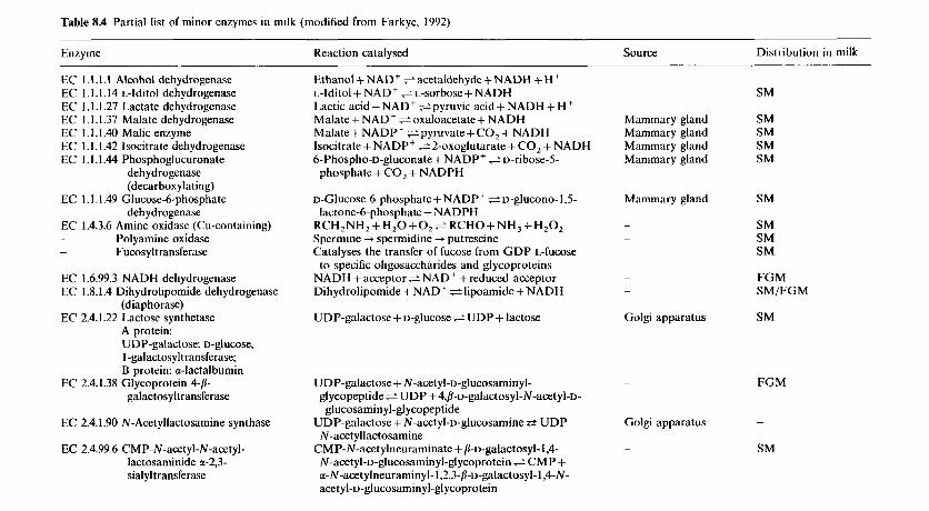
Table 8.4 Partial list of minor enzymes in milk (modified from Farkye, 1992)
Enzyme Reaction catalysed Source Distribution in milk
EC 1 .I .I .I Alcohol dehydrogenase EC 1.1.1.14 L-lditol dehydrogenase EC 1.1.1.27 Lactate dehydrogenase EC 1.1.1.37 Malate dehydrogenase EC 1. I. I .40 Malic enzyme EC 1 . I . I .42 lsocitrate dehydrogenase EC 1. I . 1.44 Phosphoglucuronate
deh ydrogenase (decarboxylating)
EC 1.1.1.49 Glucose-6-phosphate deh ydrogenase
EC 1.4.3.6 Amine oxidase (Cu-containing) - Polyamine oxidase - Fucosyltransferase
EC 1.6.99.3 NADH dehydrogenase EC 1.8.1.4 Dihydrolipomide dehydrogenase
(diaphorase) EC 2.4.1.22 Lactose synthetase
A protein: UDP-galactose: D-glUcoSe, 1 -galactosyltransferase; B protein: a-lactalbumin
EC 2.4.1.38 Glycoprotein 4-8- galactosyltransferase
EC 2.4.1.90 N-Acetyllactosamine synthase
EC 2.4.99.6 CMP-N-acetyl-N-acetyl- lactosaminide c(-2,3- sialyltransferase
Ethanol + NAD' =acetaldehyde + NADH + H ' L-lditol+ NAD' e~ - so rbose+NADH Lactic acid + NAD+ s pyruvic acid + NADH + H +
Malate + NAD+ e oxaloacetate+ NADH Malate + NADP ' e pyruvate+ CO, + NADH lsocitrate+NADP+ ~2-oxoglutarate+C02 + NADH 6-Phospho-~-gluconate + NADP+ e v-ribose-5-
phosphate+CO, + NADPH
~-Glucose-6-phosphate + NADPt e v-glucono-l,5-
RCH,NH, + H , O + O , e RCHO+NH, +H,O, Spermine + spermidine + putrescine Catalyses the transfer of fucose from G D P L-Cucose to specific oligosaccharides and glycoproteins
NADH +acceptor$ NAD+ +reduced acceptor Dihydrolipomide + NAD' e lipoamide+NADH
UDP-galactose+D-gtucosee UDP + lactose
lactone-6-phosphatc + NADPH
UDP-galactose + N-acety~-D-glucosaminyl- gl ycopeptide = U DP + 4,B-D-galaCt0Syl-N-acetyl-D- glucosamin yl-gl ycopeptide
UDP-galactose + N-acetyl-v-glucosamine e UDP
CMP-N-acetylneuraminate + ~-D-galactosyl- 1,4- N-acetyllactosamine
N-acetyl-D-glucosaminyl-glycoprotein e CMP + a-N-acetylneuraminyl- I ,2,3-~-u-galactosyl-1,4-N- acetyl-v-glucosaminyl-glycoprotein
Mammary gland Mammary gland Mammary gland Mammary gland
Mammary gland
-
Golgi apparatus
SM
SM SM SM SM
SM
SM SM SM
FG M SM/FG M
SM
FGM
Golgi apparatus -
~ SM
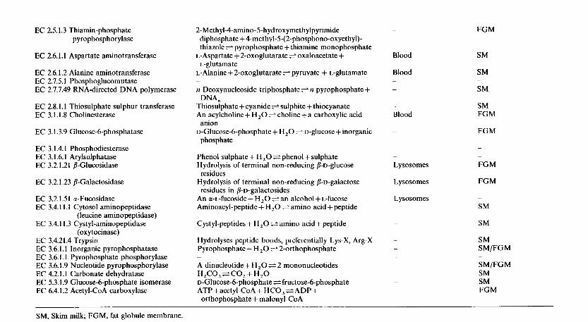
EC 2.5.1.3 Thiamin-phosphate pyrophosphorylase
EC 2.6.1.1 Aspartate aminotransferase
EC 2.6.1.2 Alanine aminotransferase EC 2.7.5.1 Phosphoglucomutase EC 2.7.7.49 RNA-directed DNA polymerase
EC 2.8.1.1 Thiosulphate sulphur transferase EC 3.1.1.8 Cholinesterase
EC 3.1.3.9 Glucose-6-phosphatase
EC 3.1.4.1 Phosphodiesterase EC 3.1.6.1 Arylsulphatase EC 3.2.1.21 b-Glucosidase
EC 3.2.1.23 p-Galactosidase
EC 3.2.1.5 I a-Fucosidase EC 3.4.1 1.1 Cytosol aminopeptidase
(leucine aminopeptidase) EC 3.4.1 1.3 Cystyl-aminopeptidase
(oxytocinase) EC 3.4.21.4 Trypsin EC 3.6.1.1 Inorganic pyrophosphatase EC 3.6.1.1 Pyrophosphate phosphorylase EC 3.6.1.9 Nucleotide pyrophosphorylase EC 4.2.1.1 Carbonate dehydratase EC 5.3.1.9 Glucose-6-phosphate isomerase EC 6.4.1.2 Acetyl-CoA carboxylase
2-Methyl-4-amino-5-hydroxymethylpyrimide diphosphate + 4-methyl-5-(2-phosphono-oxyethyl)- thiazole $ pyrophosphate + thiamine monophosphate
L-glutamate L-Aspartate + 2-oxoglutarate 5 oxaloacetate + L-Alanine + 2-oxoglutarate + pyruvate + L-glutamate
n Deoxynucleoside triphosphate e n pyrophosphate+
Thiosulphate + cyanide e sulphite + thiocyanate An acylcholine + H,O e choline +a carboxylic acid
~-Glucose-6-phosphate + H,O e D-glucose + inorganic
DNA,
anion
phosphate
Phenol sulphate + H,O e phenol + sulphate Hydrolysis of terminal non-reducing ~-D-glUCoSe
Hydrolysis of terminal non-reducing /I-D-galactose
An a-L-fucoside + H,O e an alcohol+ L-fucose Aminoacyl-peptide + H,O e amino acid + peptide
Cystyl-peptides + H,O =amino acid + peptide
Hydrolyses peptide bonds, preferentially Lys-X, Arg-X Pyrophosphate+ H,O + 2-orthophosphate
residues
residues in /I-1)-galactosides
A dinucleotide + H,O e 2 mononucleotides H,CO,eCO,+H,O ~-Glucose-6-phosphate e fructose-6-phosphate ATP +acetyl CoA + HCO, e ADP+ orthophosphate + malonyl CoA
-
Blood
Blood -
-
-
Blood
-
- -
Lysosomes
Lysosomes
Lysosomes ~
~
-
~
~
~
~
FGM
SM
SM
SM
SM FGM
FG M
-
~
~
FG M
FG M
~
SM
SM
SM SM/FGM
SM/FGM SM SM FGM
~
SM, Skim milk; FGM, fat globule membrane
336 DAIRY CHEMISTRY A N D BIOCHEMISTRY
over chemical methods because they cause fewer side-reactions and conse- quently give superior products, e.g. hydrolysis of starch.
Although relatively few enzymes are used in the dairy industry on a significant scale, the use of rennets in cheesemaking is one of the principal of all industrial applications of enzymes.
The applications of exogenous enzymes in dairy technology can be divided into two groups:
1. Technological, in which an enzyme is used to modify a milk constituent or to improve its microbiological, chemical or physical stability.
2. Enzymes as analytical reagents. Although the technological applications are quantitatively the more important, many of the analytical applica- tions of enzymes are unique and are becoming increasingly important.
Since the principal constituents of milk are proteins, lipids and lactose, proteinases, lipases and P-galactosidase (lactase) are the principal exogenous enzymes used in dairy technology. Apart from these, there are, at present, only minor applications for glucose oxidase, catalase, superoxide dismutase and lysozyme. Lactoperoxidase, xanthine oxidase and sulphydryl oxidase might also be included, although at present the indigenous form of these enzymes is exploited.
The application of enzymes in food technology has been widely reviewed (Fox, 1991; Nagodawithana and Reed, 1993). Reviews on applications of exogenous enzymes in dairy technology include Fox and Grufferty (1991), Fox (1993) and Brown (1993).
8.3.2 Proteinases
There is one major (rennet) and several minor applications of proteinases in dairy technology.
Rennets. The use of rennets in cheesemaking is the principal application of proteinases in food processing and is second only to amylases among industrial applications of enzymes. The sources of rennets and their role in milk coagulation and cheese ripening are discussed in Chapter 10 and will not be considered here.
Accelerated cheese ripening. Cheese ripening is a slow, expensive and partially uncontrolled process; consequently, there is increasing interest, at both the research and industrial levels, in accelerating ripening. Various approaches have been investigated to accelerate ripening, including a higher ripening temperature (especially for Cheddar-type cheese which is usually ripened at 6-8"C), exogenous proteinases and peptidases, modified starters (e.g. heat-shocked or lactose-negative) and genetically engineered starters or
ENZYMOLOGY OF MILK AND MILK PRODUCTS 337
starter adjuncts (Fox et al., 1996). The possible use of exogenous proteinases and peptidases attracted considerable attention for a period but uniform distribution of the enzymes in the cheese curd is a problem. Microencap- sulation of enzymes offers a possible solution but is not commercially viable at present. Exogenous proteinases/peptidases are not used commercially in natural cheeses but are being used to produce ‘enzyme modified cheese’ for use in processed cheese, cheese dips and sauces. Selected genetically modi- fied and adjunct cultures appear to be more promising.
Protein hydrolysates. Protein hydrolysates are used as flavourings in soups and gravies and in dietetic foods. They are generally prepared from soy, gluten, milk, meat or fish proteins by acid hydrolysis. Neutralization results in a high salt content which is acceptable for certain applications but may be unsuitable for dietetic foods and food supplements. Furthermore, acid hydrolysis causes total or partial destruction of some amino acids. Partial enzymatic hydrolysis is a viable alternative for some applications but bitterness due to hydrophobic peptides is frequently encountered. Bitterness may be eliminated or at least reduced to an acceptable level by treatment with activated carbon, carboxypeptidase, aminopeptidase, ultrafiltration, hydrophobic chromatography or by the plastein reaction. Caseins yield very bitter hydrolysates but the problem may be minimized by the judicious selection of the proteinase(s) (so as to avoid the production of very bitter peptides) and by using exopeptidases (especially aminopeptidases) together with the proteinase.
A novel, potentially very significant, application of proteinases in milk protein technology is the production of biologically active peptides (Chapter 4). Carefully selected proteinases of known specificity are required for such applications, but the resulting products have high added value.
Modification of protein functionality. The functional properties of milk proteins may be improved by limited proteolysis. Acid-soluble casein, free of off-flavour and suitable for incorporation into beverages and other acid foods (in which casein is insoluble) has been produced by limited proteo- lysis. The antigenicity of casein is destroyed by proteolysis and the hydroly- sate is suitable for use in milk protein-based foods for infants allergic to cows’ milk formulations. Controlled proteolysis improves the meltability of directly acidified cheese but excessive proteolysis causes bitterness. Partial proteolysis of lactalbumin (heat-coagulated whey proteins), which is insol- uble and has very poor functional properties, yields a product that is almost completely soluble above pH 6; although the product is slightly bitter, it appears promising as a food ingredient. Limited proteolysis of whey protein concentrate reduces its emulsifying capacity, increases its specific foam volume but reduces foam stability and increases heat stability.
338 DAIRY CHEMISTRY A N D BIOCHEMISTRY
8.3.3 /I- Galactosidase
P-Galactosidases (commonly referred to as lactase), which hydrolyse lactose to glucose and galactose, are probably the second most significant enzyme in dairy technology. Twenty years ago, P-galactosidase was considered to have very considerable potential but this has not materialized although there are a number of significant technological or nutritional applications. The various aspects of lactose and applications of P-galactosidase are considered in Chapter 2.
8.3.4 Lipases
The principal application of lipases in dairy technology is in cheese manufacture, particularly hard Italian varieties. The characteristic ‘piccante’ flavour of these cheeses is due primarily to short-chain fatty acids resulting from the action of lipase(s) in the rennet paste traditionally used in their manufacture. Rennet paste is prepared from the stomachs of calves, kids or lambs slaughtered after suckling; the stomachs and contents are held for about 60 days and then macerated. The product, which has proteolytic (rennet) and lipolytic activities, is considered to be unhygienic and its use is not permitted in some countries. The lipase in rennet paste, generally referred to as pregastric esterase (PGE), is secreted by a gland at the base of the tongue, which is stimulated by suckling; the secreted lipase is washed into the stomach with the ingested milk. The physiological significance of PGE, which is secreted by several species, is to assist in lipid digestion in the neonate which has limited pancreatic function. The considerable litera- ture has been reviewed by Nelson, Jensen and Pitas (1977) and Fox and Stepaniak (1993). PGE shows a high specificity for short-chain fatty acids, especially butanoic acid, esterified on the sn-3 position of glycerol, although some interspecies differences in specificity have been reported.
Semi-purified preparations of PGE from calf, kid and lamb are commer- cially available and give satisfactory results; slight differences in specificity renders one or other more suitable for particular applications. Connoisseurs of Italian cheese claim that rennet paste gives superior results to semipuri- fied PGE, and it is cheaper.
Rhizomucor miehei secretes a lipase that is reported to give satisfactory results in Italian cheese manufacture. This enzyme has been characterized and is commercially available as ‘Piccantase’. Lipases secreted by selected strains of Penicillium roqueforti and P. candidum are considered to be potentially useful for the manufacture of Italian and other cheese varieties.
Extensive lipolysis also occurs in Blue cheese varieties in which the principal lipase is secreted by P. roqueforti (Chapter 10). It is claimed that treatment of Blue cheese curd with PGE improves and intensifies its flavour but this practice is not widespread. Several techniques have been developed
ENZYMOLOGY OF MILK AND MILK PRODUCTS 339
for the production of fast-ripened Blue cheese-type products suitable for use in salad dressings, cheese dips, etc. Lipases, usually of fungal origin, are used in the manufacture of these products or to pre-hydrolyse fats/oils used as ingredients in their production.
Although Cheddar cheese undergoes relatively little lipolysis during ripening, it is claimed that addition of PGE, gastric lipase or selected microbial lipases improves the flavour of Cheddar, especially that made from pasteurized milk, and accelerates ripening. It is also claimed that the flavour and texture of Feta and Egyptian Ras cheese can be improved by adding kid or lamb PGE or low levels of selected microbial lipases to the cheese milk, especially if milk concentrated by ultrafiltration is used.
Lipases are used to hydrolyse milk fat for a variety of uses in the confectionary, sweet, chocolate, sauce and snack food industries and there is interest in using immobilized lipases to modify fat flavours for such applications (Kilara, 1985). Enzymatic interesterification of milk lipids to modify rheological properties is also feasible.
8.3.5 Lysozyme
As discussed in section 8.2.5, lysozyme has been isolated from the milk of a number of species; human and equine milks are especially rich sources. In view of its antibacterial activity, the large difference in the lysozyme content of human and bovine milks may have significance in infant nutrition. It is claimed that supplementation of baby food formulae based on cows’ milk with egg-white lysozyme gives beneficial results, especially with premature babies, but views on this are not unanimous.
Nitrate is added to many cheese varieties to prevent the growth of Clostridium tyrobutyricum which causes off-flavours and late gas blowing. However, the use of nitrate in foods is considered to be undesirable because of its involvement in nitrosamine formation, and many countries have reduced permitted levels or prohibited its use. Lysozyme, which inhibits the growth of vegetative cells of Cl. tyrobutyricum and hinders the germination of its spores, is an alternative to nitrate for the control of late gas blowing in cheese but is not widely used at present. Lysozyme also kills Listeria spp. Lysozyme addition permits the use of lower temperatures in food steriliz- ation. Co-immobilized lysozyme has been proposed for self-sanitizing immo- bilized enzyme columns; although the technique may be uneconomical for large-scale operations, it was considered feasible for pilot-scale studies, especially on expensive enzymes.
8.3.6 Catalase
Hydrogen peroxide is a very effective chemical sterilant and although it causes some damage to the physicochemical properties and nutritional value
340 DAIRY CHEMISTRY AND BIOCHEMISTRY
of milk protein, principally by oxidizing methionine, it is used as a milk preservative, especially in warm countries lacking refrigeration, and is permitted in the US for treatment of cheesemilk. Excess H , 0 2 may be reduced following treatment by soluble exogenous catalase (from beef liver, Aspergillus niger or Micrococcus lysodeiktieus). Immobilized catalase has been investigated for this purpose but the immobilized enzyme is rather unstable.
As discussed in section 8.3.7, catalase is frequently used together with glucose oxidase in many of the food applications of the latter; however, the principal potential application of glucose oxidase in dairy technology is for the in situ production of H,O,, for which the presence of catalase is obviously undesirable.
8.3.7 Glucose oxidase
Glucose oxidase (GO) catalyses the oxidation of glucose to gluconic acid (via gluconic acid-&lactone) according to the following reaction:
+ Gluconic acid-d-lactone + FADH, Glucose oxidase (GO) Glucose FAD, 0 2 / I
"iO/ Lactonase
or spontaneous
/ rl
Gluconic acid
O2 1 Go FAD + H,Oz
Catalase I H20 + %02
The H ,02 formed is normally reduced by catalase present as a contaminant in commercial GO preparations (from P. notatum, P. glaucurn or A. niger) or added separately. Glucose oxidase, which has a pH optimum of about 5.5, is highly specific for D-glucose and may be used to assay specifically for D-glucose in the presence of other sugars.
In the food industry, glucose oxidase has four principal applications:
1. Removal of residual trace levels of glucose. This application, which is particularly useful for the treatment of egg white prior to dehydration (although alternative procedures using yeast fermentation are used more commonly), is of little, if any, significance in dairy technology.
2. Removal of trace levels of oxygen. Traces of oxygen in wines and fruit juices cause discolouration and/or oxidation of ascorbic acid. Chemical
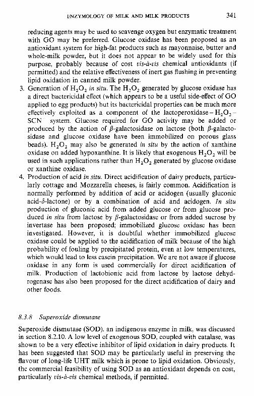
ENZYMOLOGY OF MILK AND MILK PRODUCTS 341
reducing agents may be used to scavenge oxygen but enzymatic treatment with G O may be preferred. Glucose oxidase has been proposed as an antioxidant system for high-fat products such as mayonnaise, butter and whole-milk powder, but it does not appear to be widely used for this purpose, probably because of cost vis-a-vis chemical antioxidants (if permitted) and the relative effectiveness of inert gas flushing in preventing lipid oxidation in canned milk powder.
3. Generation of H 2 0 2 in situ. The H ,02 generated by glucose oxidase has a direct bactericidal effect (which appears to be a useful side-effect of GO applied to egg products) but its bactericidal properties can be much more effectively exploited as a component of the lactoperoxidase - H 2 0 2 - SCN- system. Glucose required for G O activity may be added or produced by the action of P-galactosidase on lactose (both P-galacto- sidase and glucose oxidase have been immobilized on porous glass beads). H,02 may also be generated in situ by the action of xanthine oxidase on added hypoxanthine. It is likely that exogenous H 2 0 2 will be used in such applications rather than H,O, generated by glucose oxidase or xanthine oxidase.
4. Production of acid in situ. Direct acidification of dairy products, particu- larly cottage and Mozzarella cheeses, is fairly common. Acidification is normally performed by addition of acid or acidogen (usually gluconic acid-&lactone) or by a combination of acid and acidogen. I n situ production of gluconic acid from added glucose or from glucose pro- duced in siru from lactose by P-galactosidase or from added sucrose by invertase has been proposed; immobilized glucose oxidase has been investigated. However, it is doubtful whether immobilized glucose oxidase could be applied to the acidification of milk because of the high probability of fouling by precipitated protein, even at low temperatures, which would lead to less casein precipitation. We are not aware if glucose oxidase in any form is used commercially for direct acidification of milk. Production of lactobionic acid from lactose by lactose dehyd- rogenase has also been proposed for the direct acidification of dairy and other foods.
8.3.8 Superoxide dismutase
Superoxide dismutase (SOD), an indigenous enzyme in milk, was discussed in section 8.2.10. A low level of exogenous SOD, coupled with catalase, was shown to be a very effective inhibitor of lipid oxidation in dairy products. It has been suggested that SOD may be particularly useful in preserving the flavour of long-life UHT milk which is prone to lipid oxidation. Obviously, the commercial feasibility of using SOD as an antioxidant depends on cost, particularly vis-a-vis chemical methods, if permitted.
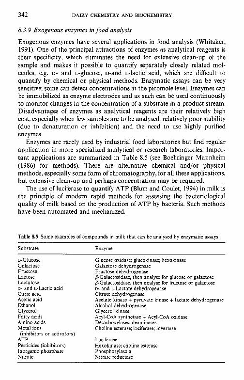
342 DAIRY CHEMISTRY A N D BIOCHEMISTRY
8.3.9 Exogenous enzymes in food analysis
Exogenous enzymes have several applications in food analysis (Whitaker, 1991). One of the principal attractions of enzymes as analytical reagents is their specificity, which eliminates the need for extensive clean-up of the sample and makes it possible to quantify separately closely related mol- ecules, e.g. D- and L-glucose, D-and L-lactic acid, which are difficult to quantify by chemical or physical methods. Enzymatic assays can be very sensitive; some can detect concentrations at the picomole level. Enzymes can be immobilized as enzyme electrodes and as such can be used continuously to monitor changes in the concentration of a substrate in a product stream. Disadvantages of enzymes as analytical reagents are their relatively high cost, especially when few samples are to be analysed, relatively poor stability (due to denaturation or inhibition) and the need to use highly purified enzymes.
Enzymes are rarely used by industrial food laboratories but find regular application in more specialized analytical or research laboratories. Impor- tant applications are summarized in Table 8.5 (see Boehringer Mannheim (1986) for methods). There are alternative chemical and/or physical methods, especially some form of chromatography, for all these applications, but extensive clean-up and perhaps concentration may be required.
The use of luciferase to quantify ATP (Blum and Coulet, 1994) in milk is the principle of modern rapid methods for assessing the bacteriological quality of milk based on the production of ATP by bacteria. Such methods have been automated and mechanized.
Table 8.5 Some examples of compounds in milk that can be analysed by enzymatic assays
Substrate Enzyme
D-Glucose Glucose oxidase; glucokinase; hexokinase Galactose Galactose dehydrogenase Fructose Fructose dehydrogenase Lactose Lactulose D- and L-Lactic acid Citric acid Citrate dehydrogenase Acetic acid Ethanol Alcohol dehydrogenase Glycerol Glycerol kinase Fatty acids Amino acids Decarboxylases; dearninases Metal ions
ATP Luciferase Pesticides (inhibitors) Hexokinase; choline esterase Inorganic phosphate Phosphorylase a Nitrate Nitrate reductase
b-Galactosidase, then analyse for glucose or galactose b-Galactosidase, then analyse for fructose or galactose D- and L-Lactate dehydrogenase
Acetate kinase + pyruvate kinase + lactate dehydrogenase
Acyl-CoA synthetase + Acyl-CoA oxidase
Choline esterase; luciferase; invertase (inhibitors or activators)
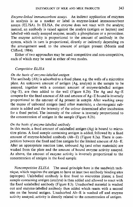
ENZYMOLOGY OF MILK AND MILK PRODUCTS 343
Enzyme-linked immunosorbent assays. An indirect application of enzymes in analysis is as a marker or label in enzyme-linked immunosorbent assays (ELISA). In ELISA, the enzyme does not react with the analyte; instead, an antibody is raised against the analyte (antigen or hapten) and labelled with easily assayed enzyme, usually a phosphatase or a peroxidase. The enzyme activity is proportional to the amount of antibody in the system, which in turn is proportional, directly or indirectly depending on the arrangement used, to the amount of antigen present (Morris and Clifford, 1984).
Either of two approaches may be used: competitive and non-competitive, each of which may be used in either of two modes.
Competitive ELISA
On the basis of enzyme-labelled antigen The antibody (Ab) is adsorbed to a fixed phase, e.g. the wells of a microtitre plate. An unknown amount of antigen (Ag, analyte) in the sample to be assayed, together with a constant amount of enzyme-labelled antigen (Ag-E), are then added to the well (Figure 8.2b). The Ag and Ag-E compete for the fixed amount of Ab and amount of Ag-E bound is inversely proportional to the amount of Ag present in sample. After washing away the excess of unbound antigen (and other materials), a chromogenic sub- strate is added and the intensity of the colour determined after incubation for a fixed period. The intensity of the colour is inversely proportional to the concentration of antigen in the sample (Figure 8.2b).
On the basis of enzyme-labelled antibody In this mode, a fixed amount of unlabelled antigen (Ag) is bound to micro- titre plates. A food sample containing antigen is added, followed by a fixed amount of enzyme-labelled antibody (Ab-E) (Figure 8.2a). There is com- petition between the fixed and free antigen for the limited amount of Ab-E. After an appropriate reaction time, unbound Ag (and other materials) are washed from the plate and the amount of bound enzyme activity assayed. As above, the amount of enzyme activity is inversely proportional to the concentration of antigen in the food sample.
Noncompetitive ELISA. The usual principle here is the sandwich tech- nique, which requires the antigen to have at least two antibody binding sites (epitopes). Unlabelled antibody is first fixed to microtitre plates; a food sample containing antigen (analyte) is then added and allowed to react with the fixed unlabelled antibody (Figure 8.3). Unadsorbed material is washed out and enzyme-labelled antibody then added which reacts with a second site on the bound antigen. Unadsorbed Ab-E is washed off and enzyme activity assayed; activity is directly related to the concentration of antigen.
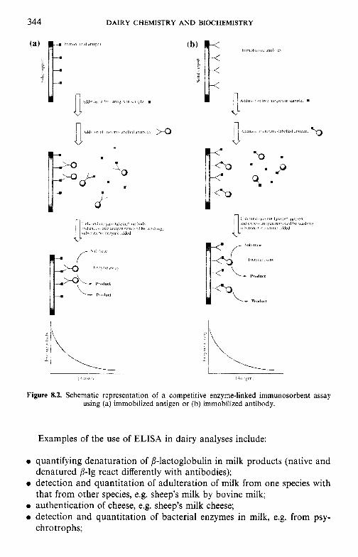
344 DAIRY CHEMISTRY A N D BIOCHEMISTRY
\,,i,ll' I
Figure 8.2. Schematic representation of a competitive enzyme-linked immunosorbent assay using (a) immobilized antigen or (b) immobilized antibody.
Examples of the use of ELISA in dairy analyses include:
quantifying denaturation of fl-lactoglobulin in milk products (native and denatured fl-lg react differently with antibodies); detection and quantitation of adulteration of milk from one species with that from other species, e.g. sheep's milk by bovine milk; authentication of cheese, e.g. sheep's milk cheese; detection and quantitation of bacterial enzymes in milk, e.g. from psy- chrotrophs;
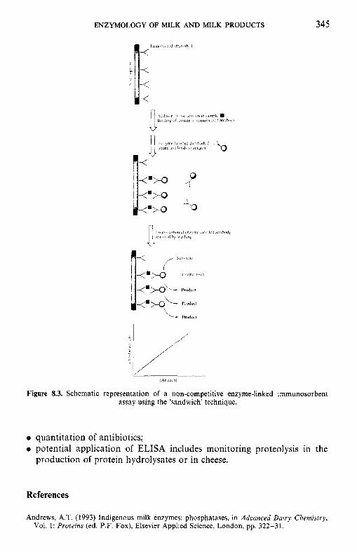
ENZYMOLOGY OF MILK AND MILK PRODUCTS
3
345
,411 /.<"I
Figure 8.3. Schematic representation of a non-competitive enzyme-linked immunosorbent assay using the 'sandwich' technique.
0 quantitation of antibiotics; 0 potential application of ELISA includes monitoring proteolysis in the
production of protein hydrolysates or in cheese.
References
Andrews, A.T. (1 993) Indigenous milk enzymes: phosphatases, in Adcanced Dairy Chemistry, Vol. 1: Proteins (ed. P.F. Fox), Elsevier Applied Science, London, pp. 322-31.
346 DAIRY CHEMISTRY AND BIOCHEMISTRY
Bastian, E.D. and Brown, R.J. (1996) Plasmin in milk and dairy products: an update. Int. Dairy
Bjorck, L. (1993) Indigenous enzymes in milk: Lactoperoxidase, in Advanced Dairy Chemistry,
Blum, L.J. and Coulet, P.R. (1994) Making light work. Chemistry in Britain, 30, 300-2. Boehringer Mannheim (1986) Methods of Biochemical Analysis and Food Analysis using
Test-Combinations, Boehringer Mannheim GmbH, D-6800 Mannheim 3 1, Germany. Brown, R.J. (1993) Dairy products, in Enzymes in Food Processing, 3rd edn (eds T.
Nagodawithana and G. Reed), Academic Press, San Diego, pp. 347-61. Deeth, H.C. and Fitz-Gerald, C.H. (1995) Lipolytic enzymes and hydrolytic rancidity in milk
and milk products, in Advanced Dairy Chemistry, Vol. 2: Lipids, 2nd edn (ed. P.F. Fox), Chapman & Hall, London, pp. 247-308.
Farkye, N.Y. (1992) Indigenous enzymes in milk: other enzymes, in Advanced Dairy Chemistry, Vol. 1: Proteins (ed. P.F. Fox), Elsevier Applied Science, London, pp. 339-67.
Fox, P.F. (ed.) (1991) Food Enzymology, Vols 1 and 2, Elsevier Applied Science, London. Fox, P.F. (1993) Exogenous enzymes in dairy technology-a review. J . Food Biochem., 17,
Fox, P.F. and Grufferty, M.B. (1991) Exogenous enzymes in dairy technology, in Food
Fox, P.F. and McSweeney, P.L.H. (1996) Proteolysis in cheese during ripening. Food Rev. Int.,
Fox, P.F. and Stepaniak, L. (1993) Enzymes in cheese technology. Int. Dairy J . , 3, 509-30. Fox, P. F., Wallace, J.M., Morgan, S. et a/ . (1996) Acceleration of cheese ripening. Antonie van
Grufferty, M.B. and Fox, P.F. (1988) Milk alkaline proteinase: A review. 1. Dairy Res., 55,
Kilara, A. (1985) Enzyme-modified lipid food ingredients. Process Biochem., 20 (2), 35-45. McKellar, R.C. (ed.) (1989) Enzymes ofpsychrotrophs in Raw Foods. CRC Press, Boca Raton,
FL. McSweeney, P.L.H., Fox, P.F. and Olson, N.F. (1995) Proteolysis of bovine caseins by
cathepsin D: preliminary observations and comparison with chymosin. Int. Dairy J . , 5,
Morris, B.A. and Clifford, M.N. (1984) Immunoassays in Food Analysis. Elsevier Applied Science Publishers, London.
Nagodawithana, T. and Reed, G. (eds.) (1993) Enzymes in Food Processing, 3rd edn, Academic Press, San Diego.
Nelson, J.H., Jensen, R.G. and Pitas, R.E. (1977) Pregastric esterase and other oral lipases: A review. J . Dairy Sci., 60, 327-62.
Olivecrona, T., Vilaro, S. and Bengtsson-Olivecrona, G. (1992) Indigenous enzymes in milk Lipases, in Aduanced Dairy Chemistry, Vol. 1: Proteins, 2nd edn (ed. P.F. Fox), Elsevier Applied Science. London, pp. 292-310.
Whitaker, J.R. (1991) Enzymes in analytical chemistry, in Food Enzymology, Vol. 2 (ed. P.F. Fox), Elsevier Applied Science, London, pp. 287-308.
J., 6, 435-57.
Vol. 1: Proteins, (ed. P.F. Fox), Elsevier Applied Science, London, pp. 332-9.
173-99.
Enzymology, Vol. 1 (ed. P.F. Fox), Elsevier Applied Science, London, pp. 219-69.
12, 457-509.
Leeuwenkoek, 70, 271-97.
609-30.
321-36.
Suggested reading
Fox, P.F. (ed.) (1991) Food Enzymology, Vols 1 and 2, Elsevier Applied Science, London. Nagodawithana, T. and Reed, G. (eds.) (1993) Enzymes in Food Processing, 3rd edn, Academic
Press, San Diego.
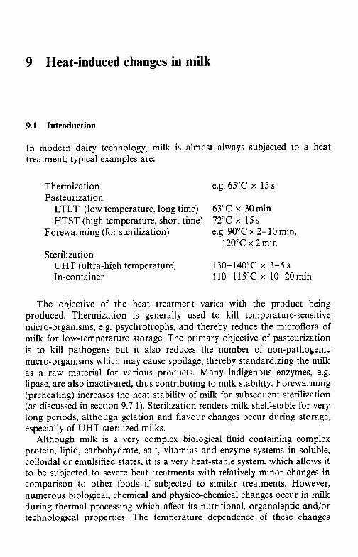
9 Heat-induced changes in milk
9.1 Introduction
In modern dairy technology, milk is almost always subjected to a heat treatment; typical examples are:
Thermization Pasteurization
e.g. 65°C x 15 s
LTLT (low temperature, long time) 63°C x 30 min HTST (high temperature, short time) 72°C x 15 s
e.g. 90°C x 2-10 min, Forewarming (for sterilization)
Sterilization 120°C x 2 min
UHT (ultra-high temperature) 130-140°C x 3-5s In-container 110-115°C x 10-20min
The objective of the heat treatment varies with the product being produced. Thermization is generally used to kill temperature-sensitive micro-organisms, e.g. psychrotrophs, and thereby reduce the microflora of milk for low-temperature storage. The primary objective of pasteurization is to kill pathogens but it also reduces the number of non-pathogenic micro-organisms which may cause spoilage, thereby standardizing the milk as a raw material for various products. Many indigenous enzymes, e.g. lipase, are also inactivated, thus contributing to milk stability. Forewarming (preheating) increases the heat stability of milk for subsequent sterilization (as discussed in section 9.7.1). Sterilization renders milk shelf-stable for very long periods, although gelation and flavour changes occur during storage, especially of UHT-sterilized milks.
Although milk is a very complex biological fluid containing complex protein, lipid, carbohydrate, salt, vitamins and enzyme systems in soluble, colloidal or emulsified states, it is a very heat-stable system, which allows it to be subjected to severe heat treatments with relatively minor changes in comparison to other foods if subjected to similar treatments. However, numerous biological, chemical and physico-chemical changes occur in milk during thermal processing which affect its nutritional, organoleptic and/or technological properties. The temperature dependence of these changes
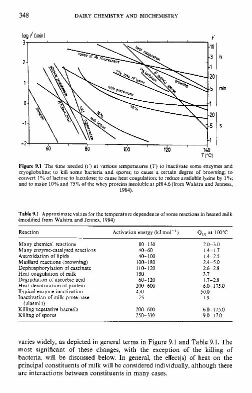
348 DAIRY CHEMISTRY AND BIOCHEMISTRY
h
min.
T ( " C )
Figure 9.1 The time needed ( 1 ' ) at various temperatures (T) to inactivate some enzymes and cryoglobulins; to kill some bacteria and spores; to cause a certain degree of browning; to convert 1% of lactose to lactulose; to cause heat coagulation; to reduce available lysine by 1%; and to make 10% and 75% of the whey proteins insoluble at pH 4.6 (from Walstra and Jenness,
1984).
Table 9.1 Approximate values for the temperature dependence of some reactions in heated milk (modified from Walstra and Jennes, 1984)
Reaction Activation energy (kJ mol- ') Qlo at 100°C
Many chemical reactions Many enzyme-catalysed reactions Autoxidation of lipids Maillard reactions (browning) Dephosphorylation of caseinate Heat coagulation of milk Degradation of ascorbic acid Heat denaturation of protein Typical enzyme inactivation Inactivation of milk proteinase
Killing vegetative bacteria Killing of spores
(plasmin)
80-130 40 - 60 40-100
100-180 110-120 150 60-120
200-600 450
75
200-600 250-330
2.0-3.0 1.4-1.7 1.4-2.5 2.4-5.0 2.6-2.8 3.7 1.7-2.8 6.0-175.0
50.0 1.9
6.0-175.0 9.0- 17.0
varies widely, as depicted in general terms in Figure 9.1 and Table 9.1. The most significant of these changes, with the exception of the killing of bacteria, will be discussed below. In general, the effect(s) of heat on the principal constituents of milk will be considered individually, although there are interactions between constituents in many cases.
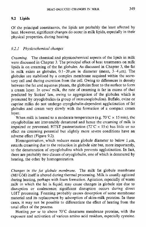
HEAT-INDUCED CHANGES IN MILK 349
9.2 Lipids
Of the principal constituents, the lipids are probably the least affected by heat. However, significant changes do occur in milk lipids, especially in their physical properties, during heating.
9.2.1 Physicochemical changes
Creaming. The chemical and physicochemical aspects of the lipids in milk were discussed in Chapter 3. The principal effect of heat treatments on milk lipids is on creaming of the fat globules. As discussed in Chapter 3, the fat in milk exists as globules, 0.1-20pm in diameter (mean, 3-4pm). The globules are stabilized by a complex membrane acquired within the secre- tory cell and during excretion from the cell. Owing to differences in density between the fat and aqueous phases, the globules float to the surface to form a cream layer. In cows’ milk, the rate of creaming is far in excess of that predicted by Stokes’ law, owing to aggregation of the globules which is promoted by cryoglobulins (a group of immunoglobulins). Buffalo, ovine or caprine milks do not undergo cryoglobulin-dependent agglutination of fat globules and cream very slowly with the formation of a compact cream layer.
When milk is heated to a moderate temperature (e.g. 70°C x 15 min), the cryoglobulins are irreversibly denatured and hence the creaming of milk is impaired or prevented; HTST pasteurization (72°C x 15 s) has little or no effect on creaming potential but slightly more severe conditions have an adverse effect (Figure 9.2).
Homogenization,, which reduces mean globule diameter to below 1 pm, retards creaming due to the reduction in globule size but, more importantly, to the denaturation of cryoglobulins which prevents agglutination. In fact, there are probably two classes of cryoglobulin, one of which is denatured by heating, the other by homogenization.
Changes in the fat globule membrane. The milk fat globule membrane (MFGM) itself is altered during thermal processing. Milk is usually agitated during heating, perhaps with foam formation. Agitation, especially of warm milk in which the fat is liquid, may cause changes in globule size due to disruption or coalescence; significant disruption occurs during direct UHT processing. Foaming probably causes desorption of some membrane material and its replacement by adsorption of skim-milk proteins. In these cases, it may not be possible to differentiate the effect of heating from the total effect of the process.
Heating per se to above 70°C denatures membrane proteins, with the exposure and activation of various amino acid residues, especially cysteine.
3 50 DAIRY CHEMISTRY AND BIOCHEMISTRY
50 60 70 80
Temperature ("C)
Figure 9.2 Time-temperature curves for the destruction of M . tuberculosis (. . .), inactivation of alkaline phosphatase (-) and creaming ability of milk (---) (from Webb and Johnson,
1965).
This may cause the release of H,S (which can result in the development of an off-flavour) and disulphide interchange reactions with whey proteins, leading to the formation of a layer of denatured whey proteins on the fat globules at high temperatures (> l0OT). The membrane and/or whey proteins may participate in Maillard browning with lactose and the cysteine may undergo p-elimination to dehydroalanine, which may then react with lysine to form lysinoalanine or with cysteine residues to form lanthionine, leading to covalent cross-linking of protein molecules (section 9.6.3). Mem- brane constituents, both proteins and phospholipids, are lost from the membrane to the aqueous phase at high temperatures. Much of the indigenous copper in milk is associated with the MFGM and some of it is transferred to the serum on heat processing. Thus, severe heat treatment of cream improves the oxidative stability of butter made from it as a result of the reduced concentration of pro-oxidant Cu in the fat phase and the antioxidant effect of exposed sulphydryl groups.
The consequences of these changes in the MFGM have been the subject of little study, possibly because severely heated milk products are usually homogenized and an artificial membrane, consisting mainly of casein and some whey proteins, is formed; consequently, changes in the natural mem- brane are not important. Damage to the membrane of unhomogenized products leads to the formation of free (non-globular) fat and consequently to 'oiling-off and the formation of a 'cream plug' (Chapter 3).
HEAT-INDUCED CHANGES IN MILK 351
Severe heat treatment, as is encountered during roller drying and to a lesser extent spray drying, results in at least some demulsification of milk fat, with the formation of free fat, which causes (Chapter 3):
the appearance of fat droplets when such products are used in tea or coffee; increased susceptibility of the fat to oxidation, since it is not protected by a membrane; reduced wettability/dispersibility of the powder; a tendency of powders to clump.
9.2.2 Chemical changes
Severe heat treatments, e.g. frying, may convert hydroxyacids to lactones, which have strong, desirable flavours and contribute to the desirable attributes of milk fat in cooking.
Release of fatty acids and some interesterification may also occur, but such changes are unlikely during the normal processing of milk.
Naturally occurring polyunsaturated fatty acids are methylene-interrup- ted but may be converted to conjugated isomers at high temperatures. Four
& R1
12 10 9
R2 12 in - Rl
R2
9.c- 11.1 9.1-1 1, t
13 12 10 9 Linoleic acid
- - - Rl water
9,C-12.C Liirolric acid
R l R1
12 10 R2 11
R2
I(l,t - 12.c 111,t - 12, t
Figure 9.3 Isomers of conjugated linoleic acid.
352 DAIRY CHEMISTRY AND BIOCHEMISTRY
Table 9.2 Concentration of conjugated linoleic acid (CLA) isomers in selected foods (modified from Ha, Grimm and Parka, 1989)
Sample mg CLA/kg Fat content CLA in fat
food (YO.) (mg kg-')
Parmesan cheese Cheddar cheese Romano cheese Blue cheese Processed cheese Cream cheese Blue spread Cheese whiz Milk pasteurized whole non-pasteurized whole
Ground beef grilled uncooked
622.3 f 15.0 440.6 f 14.5 356.9 f 6.3 169.3 f 8.9 574.1 f 24.8 334.5 f 13.3 202.6 & 6.1
1815.0 90.3
28.3 f 1.9 34.0 f 1.0
994.0 f 30.9 561.7 f 22.0
32.3 f 0.9 32.5 f 1.7 32.1 f 0.8 30.8 f 1.5 31.8 f 1.1 35.5 f 1.0 20.2 0.8 20.6 & 1.1
4.0 f 0.3 4.1 i 0.1
10.7 f 0.3 27.4 f 0.2
1926.7 1355.7 1111.9 549.8
1805.3 942.3
1003.0 8810.7
707.5 829.3
9289.7 2050.0
isomers of conjugated linoleic acid (CLA) are shown in Figure 9.3. It is claimed that CLA has anticarcinogenic properties. The mechanism of CLA formation in foods in general is not clear but heat treatment, free radical- type oxidation and microbial enzymatic reactions involving linoleic and linolenic acids in the rumen are thought to be major contributors. Rather high concentrations of CLA have been found in heated dairy products, especially processed cheese (Table 9.2). It has been suggested that whey proteins catalyse isomerization.
9.3 Lactose
The chemistry and physicochemical properties of lactose, a reducing disac- charide containing galactose and glucose linked by a p( l-4)-bond, were described in Chapter 2.
When severely heated in the solid or molten state, lactose, like other sugars, undergoes numerous changes, including mutarotation, various isomerizations and the formation of numerous volatile compounds, includ- ing acids, furfural, hydroxymethylfurfural, CO, and CO. In solution under strongly acidic conditions, lactose is degraded on heating to monosacchar- ides and other products, including acids. These changes do not normally occur during the thermal processing of milk. However, lactose is relatively unstable under mild alkaline conditions at moderate temperatures where it undergoes the Lobry de Bruyn- Alberda van Ekenstein rearrangement of aldoses to ketoses (Figure 9.4).
I I +
I m
s 0 P
0
0
c
.- I
3 54 DAIRY CHEMISTRY AND BIOCHEMISTRY
Lactose undergoes at least three heat-induced changes during the pro- cessing and storage of milk and milk products.
9.3. I Formation of lactulose
On heating at low temperatures under slightly alkaline conditions, the glucose moiety of lactose is epimerized to fructose with the formation of lactulose, which does not occur in nature. The significance of lactulose has been discussed in Chapter 2. Lactulose is not formed during HTST process- ing but is formed during UHT sterilization (more during indirect than direct heating) and especially during in-container sterilization; therefore, the con- centration of lactulose in milk is a useful index of the severity of the heat treatment to which the milk has been subjected (see Figure 2.19). The concentration of lactulose is probably the best index available at present for differentiating between UHT and in-container sterilized milks and a number of assay procedures have been developed, using HPLC or enzymatic/ spectrophotometric principles.
9.3.2 Formation of acids
Milk as secreted by the cow contains about 200 mg CO, 1-'. Owing to its low concentration in air, CO, is rapidly and, in effect, irreyersibly lost from milk on standing after milking; its loss is accelerated by heating, agitation
2 m u u .r - 9
0 1 2 3
Heating period at 116°C (h)
Figure 9.5 Changes in titratable acidity (O), lactic acid (0) and lactose (0) on heating homogenized milk in sealed cans at 116°C. Titratable acidity expressed as mg lactic acid/100 g
milk (from Gould, 1945.)
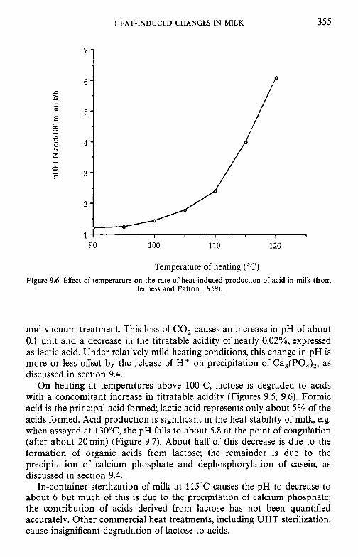
HEAT-INDUCED CHANGES IN MILK 355
7 -
6 - c 2 .d
3 8
5 -
2 4 -
5 3 -
u CJ
z - E
2 -
I I 1
90 100 110 120
Temperature of heating ("C) Figure 9.6 Effect of temperature on the rate of heat-induced production of acid in milk (from
Jenness and Patton, 1959).
and vacuum treatment. This loss of CO, causes an increase in pH of about 0.1 unit and a decrease in the titratable acidity of nearly 0.02%, expressed as lactic acid. Under relatively mild heating conditions, this change in pH is more or less offset by the release of H + on precipitation of Ca,(PO,),, as discussed in section 9.4.
On heating at temperatures above lOO"C, lactose is degraded to acids with a concomitant increase in titratable acidity (Figures 9.5, 9.6). Formic acid is the principal acid formed; lactic acid represents only about 5% of the acids formed. Acid production is significant in the heat stability of milk, e.g. when assayed at 130"C, the pH falls to about 5.8 at the point of coagulation (after about 20min) (Figure 9.7). About half of this decrease is due to the formation of organic acids from lactose; the remainder is due to the precipitation of calcium phosphate and dephosphorylation of casein, as discussed in section 9.4.
In-container sterilization of milk at 115°C causes the pH to decrease to about 6 but much of this is due to the precipitation of calcium phosphate; the contribution of acids derived from lactose has not been quantified accurately. Other commercial heat treatments, including UHT sterilization, cause insignificant degradation of lactose to acids.
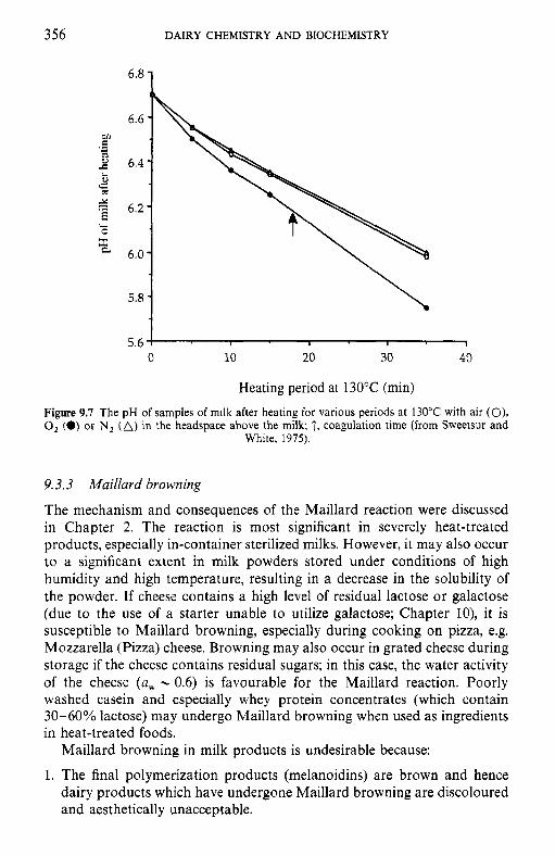
356 DAIRY CHEMISTRY AND BIOCHEMISTRY
r E .- L 0
5.6 0 10 20 30 40
Heating period at 130°C (min)
Figure 9.7 The pH of samples of milk after heating for various periods at 130°C with air (O), 0, (0) or N, (A) in the headspace above the milk; T, coagulation time (from Sweetsur and
White, 1975).
9.3.3 Maillard browning
The mechanism and consequences of the Maillard reaction were discussed in Chapter 2. The reaction is most significant in severely heat-treated products, especially in-container sterilized milks. However, it may also occur to a significant extent in milk powders stored under conditions of high humidity and high temperature, resulting in a decrease in the solubility of the powder. If cheese contains a high level of residual lactose or galactose (due to the use of a starter unable to utilize galactose; Chapter lo), it is susceptible to Maillard browning, especially during cooking on pizza, e.g. Mozzarella (Pizza) cheese. Browning may also occur in grated cheese during storage if the cheese contains residual sugars; in this case, the water activity of the cheese (a, - 0.6) is favourable for the Maillard reaction. Poorly washed casein and especially whey protein concentrates (which contain 30-60% lactose) may undergo Maillard browning when used as ingredients in heat-treated foods.
Maillard browning in milk products is undesirable because:
1. The final polymerization products (melanoidins) are brown and hence dairy products which have undergone Maillard browning are discoloured and aesthetically unacceptable.
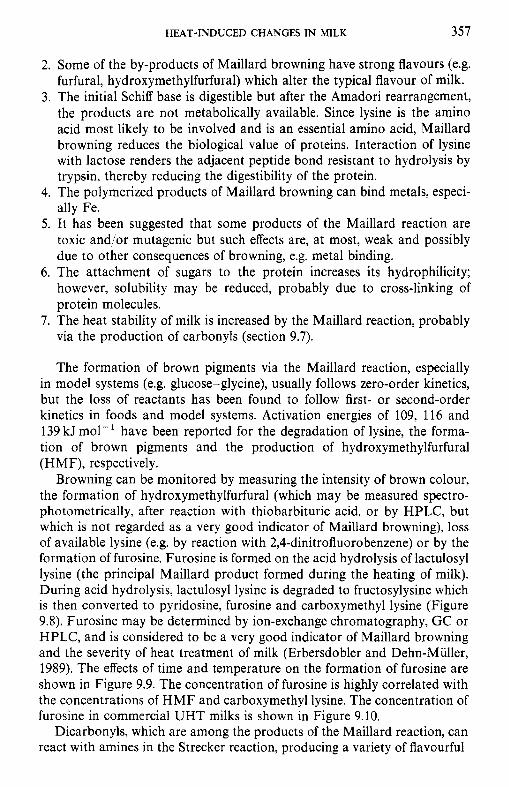
HEAT-INDUCED CHANGES IN MILK 357
2. Some of the by-products of Maillard browning have strong flavours (e.g. furfural, hydroxymethylfurfural) which alter the typical flavour of milk.
3. The initial Schiff base is digestible but after the Amadori rearrangement, the products are not metabolically available. Since lysine is the amino acid most likely to be involved and is an essential amino acid, Maillard browning reduces the biological value of proteins. Interaction of lysine with lactose renders the adjacent peptide bond resistant to hydrolysis by trypsin, thereby reducing the digestibility of the protein.
4. The polymerized products of Maillard browning can bind metals, especi- ally Fe.
5. It has been suggested that some products of the Maillard reaction are toxic and/or mutagenic but such effects are, at most, weak and possibly due to other consequences of browning, e.g. metal binding.
6. The attachment of sugars to the protein increases its hydrophilicity; however, solubility may be reduced, probably due to cross-linking of protein molecules.
7. The heat stability of milk is increased by the Maillard reaction, probably via the production of carbonyls (section 9.7).
The formation of brown pigments via the Maillard reaction, especially in model systems (e.g. glucose-glycine), usually follows zero-order kinetics, but the loss of reactants has been found to follow first- or second-order kinetics in foods and model systems. Activation energies of 109, 116 and 139 kJ mo1-l have been reported for the degradation of lysine, the forma- tion of brown pigments and the production of hydroxymethylfurfural (HMF), respectively.
Browning can be monitored by measuring the intensity of brown colour, the formation of hydroxymethylfurfural (which may be measured spectro- photometrically, after reaction with thiobarbituric acid, or by HPLC, but which is not regarded as a very good indicator of Maillard browning), loss of available lysine (e.g. by reaction with 2,4-dinitrofluorobenzene) or by the formation of furosine. Furosine is formed on the acid hydrolysis of lactulosyl lysine (the principal Maillard product formed during the heating of milk). During acid hydrolysis, lactulosyl lysine is degraded to fructosylysine which is then converted to pyridosine, furosine and carboxymethyl lysine (Figure 9.8). Furosine may be determined by ion-exchange chromatography, GC or HPLC, and is considered to be a very good indicator of Maillard browning and the severity of heat treatment of milk (Erbersdobler and Dehn-Miiller, 1989). The effects of time and temperature on the formation of furosine are shown in Figure 9.9. The concentration of furosine is highly correlated with the concentrations of H M F and carboxymethyl lysine. The concentration of furosine in commercial UHT milks is shown in Figure 9.10.
Dicarbonyls, which are among the products of the Maillard reaction, can react with amines in the Strecker reaction, producing a variety of flavourful
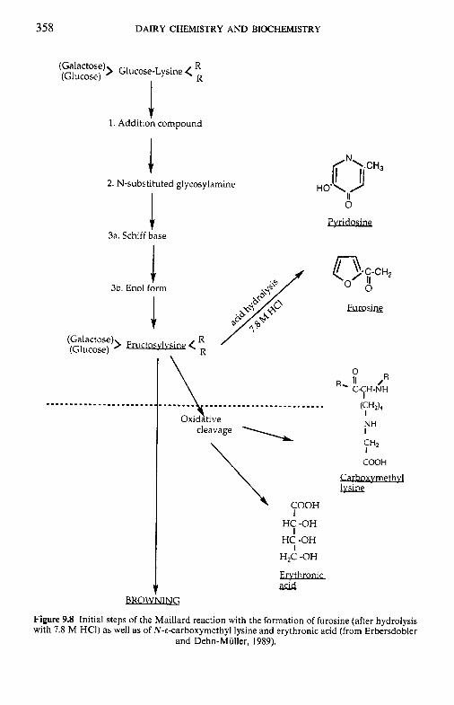
358
.~.~..~...~..... .~~...~........~~~~....~.. / Oxidative
cleavage
rCOOH I
DAIRY CHEMISTRY AND BIOCHEMISTRY
t 1. Addition compound
I 2. N-substituted glycosylamine
3a. Schiff base
3b. Enol form
> Fructosylysh < (Galactose) (Glucose)
H 0’ OCH3 II
0
Pvrldoslne
J
HC -OH
HC -OH
HZC -OH
I
I
Ervthroluc &id
O R RI C-CH-NH
I
11 /
W 2 ) 4 I NH I
y + 2
CGQH
Carboxymethvl
BROWNING
Figure 9.8 Initial steps of the Maillard reaction with the formation of furosine (after hydrolysis with 7.8 M HCI) as well as of N-E-carboxymethyl lysine and erythronic acid (from Erbersdobler
and Dehn-Muller, 1989).
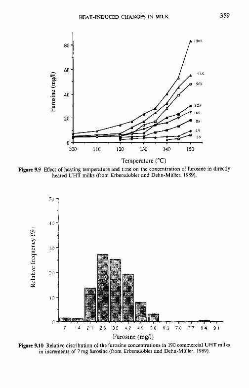
HEAT-INDUCED CHANGES IN MILK 3 59
Figure
80
60
40
20
0
I2HS
93s
56s
32s
16s
4s
2s - 100 110 120 130 140 150
Temperature ("C) 9.9 Effect of heating temperature and time on the concentration of furosine
heated UHT milks (from Erbersdobler and Dehn-Muller, 1989). in directly
7 1 4 7 1 2 8 3 5 4 2 4 9 5 6 6 3 7 0 7 7 8 4 97
Furosine (mg/l) Figure 9.10 Relative distribution of the furosine concentrations in 190 commercial UHT milks
in increments of 7 mg furosine (from Erbersdobler and Dehn-Muller, 1989).
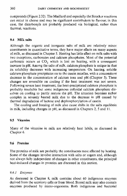
360 DAIRY CHEMISTRY AND BIOCHEMISTRY
compounds (Figure 2.32). The Maillard and especially the Strecker reactions can occur in cheese and may be significant contributors to flavour; in this case, the dicarbonyls are probably produced via biological, rather than thermal, reactions.
9.4 Milk salts
Although the organic and inorganic salts of milk are relatively minor constituents in quantitative terms, they have major effects on many aspects of milk, as discussed in Chapter 5. Heating has little effect on milk salts with two exceptions, carbonates and calcium phosphates. Most of the potential carbonate occurs as CO, which is lost on heating, with a consequent increase in pH. Among the salts of milk, calcium phosphate is unique in that its solubility decreases with increasing temperature. On heating, soluble calcium phosphate precipitates on to the casein micelles, with a concomitant decrease in the concentration of calcium ions and pH (Chapter 5). These changes are reversible on cooling if the heat treatment was not severe. Following severe heat treatment, the heat-precipitated calcium phosphate is probably insoluble but some indigenous colloidal calcium phosphate dis- solves on cooling to partly restore the pH. The situation becomes rather complex in severely heated milk due to the decrease in pH caused by thermal degradation of lactose and dephosphorylation of casein.
The cooling and freezing of milk also cause shifts in the salts equilibria in milk, including changes in pH, as discussed in Chapters 2, 5 and 11.
9.5 Vitamins
Many of the vitamins in milk are relatively heat labile, as discussed in Chapter 6.
9.6 Proteins
The proteins of milk are probably the constituents most affected by heating. Some of the changes involve interaction with salts or sugars and, although not always fully independent of changes in other constituents, the principal heat-induced changes in proteins are discussed in this section.
9.6.1 Enzymes
As discussed in Chapter 8, milk contains about 60 indigenous enzymes derived from the secretory cells or from blood. Stored milk may also contain enzymes produced by micro-organisms. Both indigenous and bacterial
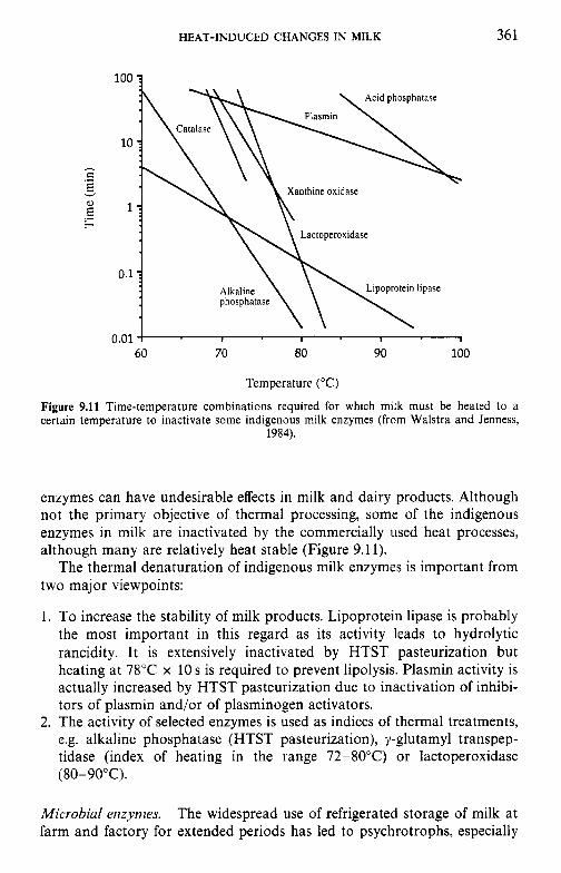
HEAT-INDUCED CHANGES IN MILK 361
h c .- E v
E F
Xanthine oxidase
0.1 :
0.01 ! I 1 I 1
60 70 80 90 100
Temperature ("C)
Figure 9.11 Time-temperature combinations required for which milk must be heated to a certain temperature to inactivate some indigenous milk enzymes (from Walstra and Jenness,
1984).
enzymes can have undesirable effects in milk and dairy products. Although not the primary objective of thermal processing, some of the indigenous enzymes in milk are inactivated by the commercially used heat processes, although many are relatively heat stable (Figure 9.1 1).
The thermal denaturation of indigenous milk enzymes is important from two major viewpoints:
1. To increase the stability of milk products. Lipoprotein lipase is probably the most important in this regard as its activity leads to hydrolytic rancidity. It is extensively inactivated by HTST pasteurization but heating at 78°C x 10 s is required to prevent lipolysis. Plasmin activity is actually increased by HTST pasteurization due to inactivation of inhibi- tors of plasmin and/or of plasminogen activators.
2. The activity of selected enzymes is used as indices of thermal treatments, e.g. alkaline phosphatase (HTST pasteurization), y-glutamyl transpep- tidase (index of heating in the range 72-80°C) or lactoperoxidase (80-90°C).
Microbial enzymes. The widespread use of refrigerated storage of milk at farm and factory for extended periods has led to psychrotrophs, especially
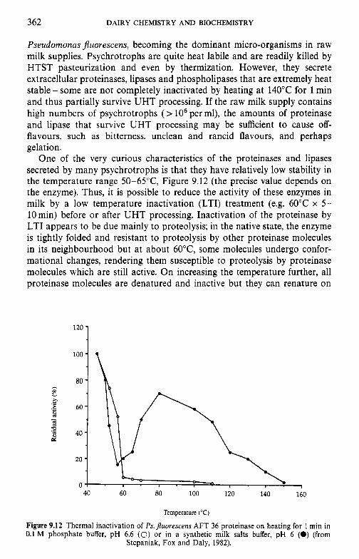
362 DAIRY CHEMISTRY AND BIOCHEMISTRY
Pseudomonas Juorescens, becoming the dominant micro-organisms in raw milk supplies. Psychrotrophs are quite heat labile and are readily killed by HTST pasteurization and even by thermization. However, they secrete extracellular proteinases, lipases and phospholipases that are extremely heat stable - some are not completely inactivated by heating at 140°C for 1 min and thus partially survive UHT processing. If the raw milk supply contains high numbers of psychrotrophs (> lo6 per ml), the amounts of proteinase and lipase that survive UHT processing may be sufficient to cause off- flavours, such as bitterness, unclean and rancid flavours, and perhaps gelation.
One of the very curious characteristics of the proteinases and lipases secreted by many psychrotrophs is that they have relatively low stability in the temperature range 50-65"C, Figure 9.12 (the precise value depends on the enzyme). Thus, it is possible to reduce the activity of these enzymes in milk by a low temperature inactivation (LTI) treatment (e.g. 60°C x 5- 10 min) before or after UHT processing. Inactivation of the proteinase by LTI appears to be due mainly to proteolysis; in the native state, the enzyme is tightly folded and resistant to proteolysis by other proteinase molecules in its neighbourhood but at about 60"C, some molecules undergo confor- mational changes, rendering them susceptible to proteolysis by proteinase molecules which are still active. On increasing the temperature further, all proteinase molecules are denatured and inactive but they can renature on
40 60 80 100 120 140 160
Temperature ("C)
Figure 9.12 Thermal inactivation of Ps. jluorescens AFT 36 proteinase on heating for 1 min in 0.1 M phosphate buffer, pH 6.6 (0) or in a synthetic milk salts buffer, pH 6 (0) (from
Stepaniak, Fox and Daly, 1982).
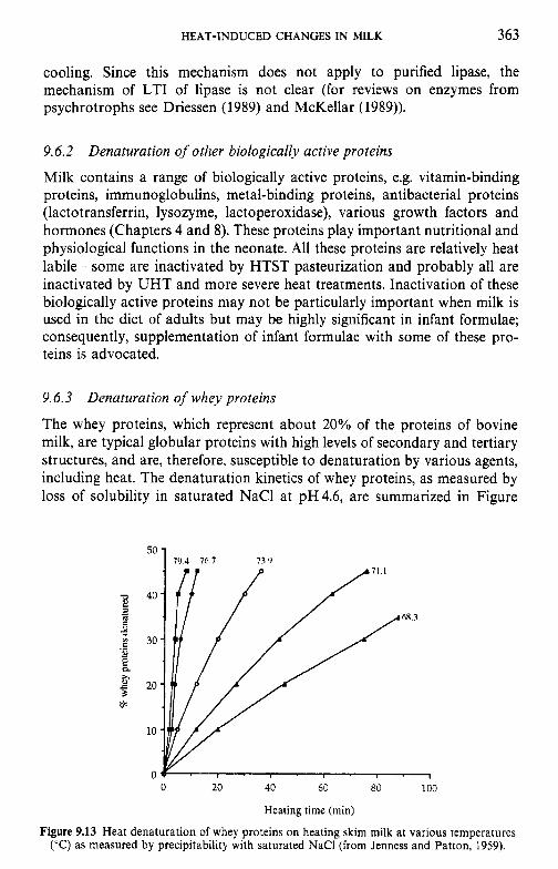
HEAT-INDUCED CHANGES IN MILK 363
cooling. Since this mechanism does not apply to purified lipase, the mechanism of LTI of lipase is not clear (for reviews on enzymes from psychrotrophs see Driessen (1989) and McKellar (1989)).
9.6.2 Denaturation of other biologically active proteins
Milk contains a range of biologically active proteins, e.g. vitamin-binding proteins, immunoglobulins, metal-binding proteins, antibacterial proteins (lactotransferrin, lysozyme, lactoperoxidase), various growth factors and hormones (Chapters 4 and 8). These proteins play important nutritional and physiological functions in the neonate. All these proteins are relatively heat labile - some are inactivated by HTST pasteurization and probably all are inactivated by UHT and more severe heat treatments. Inactivation of these biologically active proteins may not be particularly important when milk is used in the diet of adults but may be highly significant in infant formulae; consequently, supplementation of infant formulae with some of these pro- teins is advocated.
9.6.3 Denaturation of whey proteins
The whey proteins, which represent about 20% of the proteins of bovine milk, are typical globular proteins with high levels of secondary and tertiary structures, and are, therefore, susceptible to denaturation by various agents, including heat. The denaturation kinetics of whey proteins, as measured by loss of solubility in saturated NaCl at pH4.6, are summarized in Figure
50 i
0 20 40 60 80 100
Heating time (rnin)
Figure 9.13 Heat denaturation of whey proteins on heating skim milk at various temperatures ("C) as measured by precipitability with saturated NaCl (from Jenness and Patton, 1959).
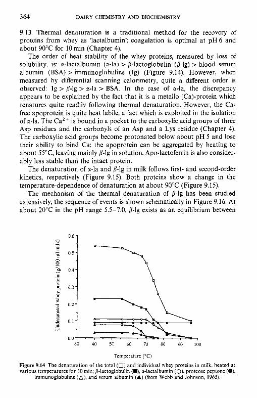
364 DAIRY CHEMISTRY A N D BIOCHEMISTRY
9.13. Thermal denaturation is a traditional method for the recovery of proteins from whey as 'lactalbumin'; coagulation is optimal at pH 6 and about 90°C for 10 min (Chapter 4).
The order of heat stability of the whey proteins, measured by loss of solubility, is: a-lactalbumin (a-la) > P-lactoglobulin (p-lg) > blood serum albumin (BSA) > immunoglobulins (Ig) (Figure 9.14). However, when measured by differential scanning calorimetry, quite a different order is observed: Ig > p-lg > r-la > BSA. In the case of a-la, the discrepancy appears to be explained by the fact that it is a metallo (Ca)-protein which renatures quite readily following thermal denaturation. However, the Ca- free apoprotein is quite heat labile, a fact which is exploited in the isolation of a-la. The Ca2+ is bound in a pocket to the carboxylic acid groups of three Asp residues and the carbonyls of an Asp and a Lys residue (Chapter 4). The carboxylic acid groups become protonated below about pH 5 and lose their ability to bind Ca; the apoprotein can be aggregated by heating to about 55"C, leaving mainly p-lg in solution. Apo-lactoferrin is also consider- ably less stable than the intact protein.
The denaturation of &-la and p-lg in milk follows first- and second-order kinetics, respectively (Figure 9.15). Both proteins show a change in the temperature-dependence of denaturation at about 90°C (Figure 9.15).
The mechanism of the thermal denaturation of p-lg has been studied extensively; the sequence of events is shown schematically in Figure 9.16. At about 20°C in the pH range 5.5-7.0, p-lg exists as an equilibrium between
- Y E .-
e, c
0.3 x c
rn 0.2 2
0.0 I 30 40 50 60 70 80 90 100
Temperature ("C)
Figure 9.14 The denaturation of the total (0) and individual whey proteins in milk, heated at various temperatures for 30 min; /?-lactoglobulin (m), r-lactalbumin (O), proteose peptone (O),
immunoglobulins (A), and serum albumin (A) (from Webb and Johnson, 1965).
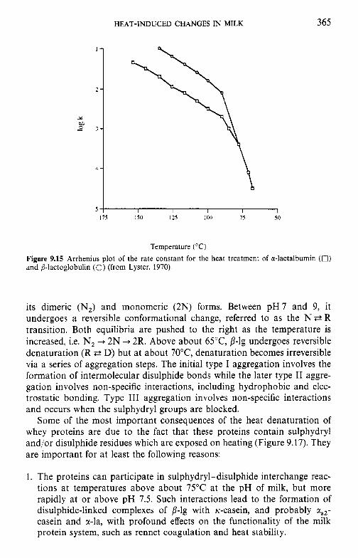
HEAT-INDUCED CHANGES IN MILK
1 -
2 -
3-
4 -
365
s ; I I 1 I 1 175 150 12s I00 7s SO
Temperature ("C) Figure 9.15 Arrhenius plot of the rate constant for the heat treatment of a-lactalbumin (0) and P-lactoglobulin (0) (from Lyster, 1970).
its dimeric (NJ and monomeric (2N) forms. Between p H 7 and 9, it undergoes a reversible conformational change, referred to as the N P R transition. Both equilibria are pushed to the right as the temperature is increased, i.e. N, + 2N + 2R. Above about 65"C, p-lg undergoes reversible denaturation (R P D) but at about 70"C, denaturation becomes irreversible via a series of aggregation steps. The initial type I aggregation involves the formation of intermolecular disulphide bonds while the later type I1 aggre- gation involves non-specific interactions, including hydrophobic and elec- trostatic bonding. Type I11 aggregation involves non-specific interactions and occurs when the sulphydryl groups are blocked.
Some of the most important consequences of the heat denaturation of whey proteins are due to the fact that these proteins contain sulphydryl and/or disulphide residues which are exposed on heating (Figure 9.17). They are important for at least the following reasons:
1. The proteins can participate in sulphydryl-disulphide interchange reac- tions at temperatures above about 75°C at the pH of milk, but more rapidly at or above pH 7.5. Such interactions lead to the formation of disulphide-linked complexes of p-lg with K-casein, and probably cc,,- casein and cc-la, with profound effects on the functionality of the milk protein system, such as rennet coagulation and heat stability.
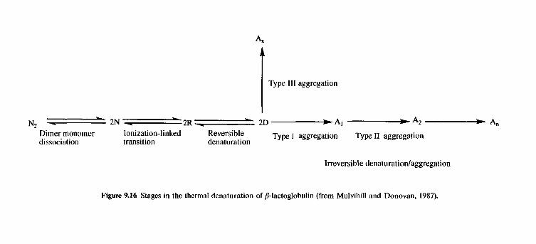
N2 - 2N - -2R - - 2D-Al -A2 - A,,
Dimer monomer Ionization-linked Reversible Type I aggregation Type I1 aggregation dissociation transition denaturation
Irreversible denaturationlaggregation
Figure 9.16 Stages in the thermal denaturation of /I-lactoglobulin (from Mulvihill and Donovan, 1987).
HEAT-INDUCED CHANGES IN MILK 367
0 20 40 60 80 100 120 140
Time of heating (min) Figure 9.17 Exposure of sulphydryl groups by heating milk at 75 (O), 80 (O), 85 (A) or 95
(A) "C; de-aerated milk heated at 85°C (a) (from Jenness and Patton, 1959).
Products unknown
y 2 Histidine
Tryptophan
Lysinoalanine CH2 0 CH3 0 [HI II I/
H~N-C-LLOH I 7 H2N-C-C-OH - y 3
c=o I H
Alanine / 3 1 :\ k - H Ornithine pyruvic acid
THz
CH2 0 I I/ H O
H
I 1. H2N- C - C- OH I I/ H2N-C-C-OH I H2N- C- C-OH
I 7H2 I p 2 S
p-aminoalnnine I CH2 0 I //
I H
H2N-C-C-OH
Lanthionine
NH
Figure 9.18 Interaction of dehydroalanine with amino acids.
368 DAIRY CHEMISTRY AND BIOCHEMISTRY
2. The activated sulphydryls may decompose with the formation of H,S and H,C-S-CH,, which are responsible for the cooked flavour of severely heated milk, including UHT milk. These compounds are volatile and unstable and disappear within about 1 week after processing so that the flavour of UHT milk improves during the first few weeks after processing.
3. Serine, serine phosphate, glycosylated serine, cysteine and cystine residues can undergo 8-elimination with the formation of dehydroalanine. Dehy- droalanine is very reactive and can react with various amino acid residues, especially lysine, leading to the formation of lysinoalanine, and to a lesser extent with cysteine with the formation of lanthionine (Figure 9.18). These reactions lead to intra- or intermolecular cross-linking which reduce protein solubility, digestibility and nutritive value (because the bonds formed are not hydrolysed in the intestinal tract and lysine is an essential amino acid). Although there are reports to the contrary, lysino- alanine is not normally found in UHT milk or cream.
9.6.4 Efect of heat on caseins
As discussed in Chapter 4, the caseins are rather unique proteins. They are rather small (20-25 kDa), relatively hydrophobic molecules, with little higher structure, few disulphide bonds (present only in the two minor caseins, tls2 and K) and no sulphydryl groups. All the caseins are phos- phorylated (8-9, 10-13,4-5 and 1 mole P per mole protein for uSl-, tlS2-, p- and K-casein, respectively); due to their high levels of phosphorylation, ctsl-, usz- and 8-caseins bind calcium strongly, causing them to aggregate and precipitate, and affecting their general stability, including heat stability.
Within the strict sense of the term, the caseins are not susceptible to thermal denaturation, e.g. sodium caseinate (pH 6.5-7.0) may be heated at 140°C for more than 1 h without any visible physicochemical changes. However, severe heat treatments do cause substantial changes, e.g. dephos- phorylation (about 100% in 1 h at 140"C), aggregation (as indicated by changes in urea-PAGE or gel permeation chromatography), possibly due to the formation of intermolecular disulphide and intermolecular isopeptide bonds, cleavage of peptide bonds (formation of peptides soluble at pH 4.6 or in 12% TCA). 8-Elimination of serine, serine phosphate and cysteine residues may also occur, especially at pH values above 7. Such heat-induced changes are evident in commercial sodium caseinate.
The remarkably high heat stability of the caseins allows heat-sterilized dairy products to be produced without major changes in physical properties (reviewed by Fox, 1982; Singh and Creamer, 1992). The heat stability of unconcentrated milk is almost always adequate to withstand the tempera- ture treatments to which it is normally subjected; only rarely is a defect known as the 'Utrecht phenomenon' encountered, when milk coagulates on HTST heating. This defect is due to a very high Caz+ concentration owing
HEAT-INDUCED CHANGES IN MILK 369
to a low concentration of citrate, arising from poor feed. However, the heat stability of milk decreases sharply on concentration and is usually inad- equate to withstand in-container or UHT processing unless certain adjust- ments and/or treatments are made. Although the heat stability of concentrated milk is poorly correlated with that of the original milk, most of the research on the heat stability of milk has been done on unconcen- trated milk.
9.7 Heat stability of milk
Studies on the heat stability of milk date from the pioneering work of Sommer and Hart, which commenced in 1919. Much of the early work concentrated on attempts to relate heat stability to variations in milk composition, especially the concentrations of milk salts. Although the heat coagulation time (HCT) of milk is inversely related to the concentrations of divalent cations (Ca2+ and Mg2+) and positively with the concentrations of polyvalent anions (i.e. phosphate and citrate), the correlations are poor and unable to explain the natural variations in HCT. This failure was largely explained in 1961 by Rose who showed that the HCT of most milks is extremely sensitive to small changes in pH in the neighbourhood of 6.7. In effect, the influence of all other factors on the HCT of milk must be considered against the background of the effect of pH.
For the majority of individual-cow and all bulk milks, the HCT increases with increasing pH from 6.4 to about 6.7, then decreases abruptly to a minimum at around pH 6.9 but increases continuously with further in- creases in pH (Figure 9.19). The HCT decreases sharply below pH 6.4. Milks which show a strong dependence of heat stability on pH are referred to as type A milks. Occasionally, the HCT of individual-cow milks increases continuously with increasing pH, which is as would be expected due to increasing protein charge with increasing pH; these are referred to as type B milks.
The maximum HCT and the shape of the HCT-pH profile are influenced by several compositional factors, of which the following are the most significant:
1. Ca2+ reduces HCT throughout the pH range 6.4-7.4. 2. Ca-chelators, e.g. citrate, polyphosphate, increase stability. 3. p-Lg, and probably a-la, increase the stability of casein micelles at
pH6.4-6.7 but reduce it at pH6.7-7.0; in fact, the occurrence of a maximum-minimum in the HCT-pH profile depends on the presence of
4. Addition of K-casein to milk increases stability in the pH range of the B-k.
HCT minimum.
3 70 DAIRY CHEMISTRY AND BIOCHEMISTRY
4 0 1 P
o ! . 8 . I . I . I - 1
6.2 6.4 6.6 6.8 7.0 7.2
PH
Figure 9.19 Effect of pH on the heat stability of type A milk (A), type B milk (0) and whey protein-free casein micelle dispersions (0) (from Fox, 1982).
5. Reducing the level of colloidal calcium phosphate increases stability in the region of the HCT maximum.
6. Natural variations in HCT are due mainly to variations in the concen- tration of indigenous urea due to changes in the animals’ feed.
The current explanation for the maximum-minimum in the HCT-pH profile is that on heating, x-casein dissociates from the micelles; at pH values below about 6.7, 8-1s reduces the dissociation of Ic-casein, but at pH values above 6.7, it accentuates dissociation. In effect, coagulation in the pH range of minimum stability involves aggregation of Ic-casein-depleted micelles, in a manner somewhat analogous to rennet coagulation, although the mech- anism by which the altered micelles are produced is very different.
As would be expected, heating milk at 140°C for an extended period causes very significant chemical and physical changes in milk, of which the following are probably the most significant:
1. Decrease in pH. After heating at 140°C for 20 min, the pH of milk has decreased to about 5.8 due to acid production from pyrolysis of lactose, precipitation of soluble calcium phosphate as Ca,(PO,),, with the release of H +, and dephosphorylation of casein with subsequent precipi- tation of the liberated phosphate as Ca,(PO,), with the release of H + . The heat-induced precipitation of Ca,(PO,), is partially reversible on cooling so that the actual pH of milk at 140°C at the point of coagulation is much lower than the measured value and is probably below 5.0.
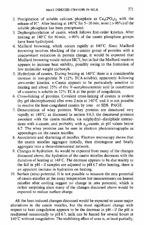
HEAT-INDUCED CHANGES IN MILK 371
2. Precipitation of soluble calcium phosphate as Ca,(PO,), with the release of H'. After heating at 140°C for 5-10min, most (>90%) of the soluble phosphate has been precipitated.
3. Dephosphorylation of casein, which follows first-order kinetics. After heating at 140°C for 60min, >90% of the casein phosphate groups have been hydrolysed.
4. Maillard browning, which occurs rapidly at 140°C. Since Maillard browning involves blocking of the &-amino group of proteins with a concomitant reduction in protein charge, it would be expected that Maillard browning would reduce HCT, but in fact the Maillard reaction appears to increase heat stability, possibly owing to the formation of low molecular weight carbonyls.
5 . Hydrolysis of caseins. During heating at 140°C there is a considerable increase in non-protein N (12% TCA-soluble), apparently following zero-order kinetics. K-Casein appears to be particularly sensitive to heating and about 25% of the N-acetylneuraminic acid (a constituent of K-casein) is soluble in 12% TCA at the point of coagulation.
6. Cross-linking of proteins. Covalent cross-linking of caseins is evident (by gel electrophoresis) after even 2 min at 140°C and it is not possible to resolve the heat-coagulated caseins by urea- or SDS-PAGE.
7. Denaturation of whey proteins. Whey proteins are denatured very rapidly at 140°C; as discussed in section 9.6.3, the denatured proteins associate with the casein micelles, via sulphydryl-disulphide interac- tions with K-casein, and probably with a,,-casein, at pH values below 6.7. The whey proteins can be seen in electron photomicrographs as appendages on the casein micelles.
8. Association and shattering of micelles. Electron microscopy shows that the casein micelles aggregate initially, then disintegrate and finally aggregate into a three-dimensional network.
9. Changes in hydration. As would be expected from many of the changes discussed above, the hydration of the casein micelles decreases with the duration of heating at 140°C. The decrease appears to be due mainly to the fall in pH - if samples are adjusted to pH 6.7 after heating, there is an apparent increase in hydration on heating.
10. Surface (zeta) potential. It is not possible to measure the zeta potential of casein micelles at the assay temperature but measurements on heated micelles after cooling suggest no change in zeta potential, which is rather surprising since many of the changes discussed above would be expected to reduce surface charge.
All the heat-induced changes discussed would be expected to cause major alterations in the casein micelles, but the most significant change with respect to heat coagulation appears to be the decrease in pH - if the pH is readjusted occasionally to pH 6.7, milk can be heated for several hours at 140°C without coagulation. The stabilizing effect of urea is, at least partially,
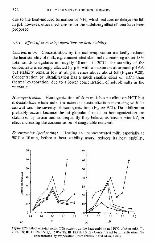
3 72 DAIRY CHEMISTRY AND BIOCHEMISTRY
due to the heat-induced formation of NH, which reduces or delays the fall in pH; however, other mechanisms for the stabilizing effect of urea have been proposed.
9.7.1 Efect of processing operations on heat stability
Concentration. Concentration by thermal evaporation markedly reduces the heat stability of milk, e.g. concentrated skim milk containing about 18% total solids coagulates in roughly 10min at 130°C. The stability of the concentrate is strongly affected by pH, with a maximum at around pH 6.6, but stability remains low at all pH values above about 6.8 (Figure 9.20). Concentration by ultrafiltration has a much smaller effect on HCT than thermal evaporation, due to a lower concentration of soluble salts in the retentate.
Homogenization. Homogenization of skim milk has no effect on HCT but it destabilizes whole milk, the extent of destabilization increasing with fat content and the severity of homogenization (Figure 9.21). Destabilization probably occurs because the fat globules formed on homogenization are stabilized by casein and consequently they behave as ‘casein micelles’, in effect increasing the concentration of coagulable material.
Forewarming (preheating). Heating an unconcentrated milk, especially at 90°C x 10 min, before a heat stability assay, reduces its heat stability,
T’ 50
6.4 6.6 6.8 7.0 7.2
PH ( 2 )
70 1 60 -
50 -
6.4 6.6 6.8 7.0 7.2
(h ) PH
Figure 9.20 Effect of total solids (TS) content on the heat stability at 130°C of skim milk 0, 9.3% TS; 0, 12.0% TS; 0, 15.0% TS; ., 18.4% TS. (a) Concentrated by ultrafiltration, (b)
concentrated by evaporation (from Sweetsur and Muir, 1980).
HEAT-INDUCED CHANGES IN MILK 373
40 1
6.6 6.8 7.0 7.2 7.4
PH Figure 9.21 Effect of pressure (Rannie homogenizer) on the heat coagulation time (at 140°C) of milk, unhomogenized (0) or homogenized at 3.5 MPa: (A); 10.4 MPa (W) or 20.7/3.5 MPa
(+) (from Sweetsur and Muir, 1983).
mainly by shifting its natural pH; maximum heat stability is affected only slightly or not at all. However, if milk is preheated before concentration, the heat stability of the concentrate is increased. Various preheating conditions are used, e.g. 90°C x lOmin, 120°C x 2min or 140°C x 5 s ; the last is particularly effective but is not widely used commercially. The stabilizing effect is probably due to the fact that the heat-induced changes discussed previously are less detrimental if they occur prior to concentration rather than in concentrated milk which is inherently less stable.
Additives. Orthophosphates, and less frequently citrates, have long been used commercially to increase the stability of concentrated milk. The mechanism was believed to involve Ca-chelation but pH adjustments may be the principal mechanism.
Numerous compounds increase heat stability (e.g. various carbonyls, including diacetyl, and ionic detergents) but few are permitted additives. Although added urea has a major effect on the stability of unconcentrated milk, it does not stabilize concentrated milks, although it does increase the effectiveness of carbonyls.
9.8 Effect of heat treatment on rennet coagulation of milk and related properties
The primary step in the manufacture of most cheese varieties and rennet casein involves coagulation of the casein micelles to form a gel. Coagulation
374 DAIRY CHEMISTRY AND BIOCHEMISTRY
involves two steps (phases), the first of which involves enzymatically hydrolysing the micelle-stabilizing protein, k--casein, by selected proteinases, referred to as rennets. The second step of coagulation involves coagulation of rennet-altered micelles by Ca2+ above 20°C (Chapter lo).
The rate of rennet coagulation is affected by many compositional factors, including the concentrations of Ca2 +, casein and colloidal calcium phos- phate and pH. Coagulation is adversely affected by heat treatment of the milk at temperatures above about 70°C due to interaction of denatured p-lg (and a-la) with K-casein. The primary and, especially, the secondary phases of rennet coagulation are adversely affected by the interaction and, if the heat treatment is sufficiently severe (e.g. 80°C x 5-10min), the milk does not coagulate on renneting. The effect on the primary phase is presumably due to blockage of the rennet-susceptible bond of k--casein following interaction with ,/?-lg. The adverse effect of heating on the second phase arises because the whey protein-coated micelles are unable to interact properly because the aggregation sites, which are unknown, are blocked.
The adverse effects of heat treatment on the rennetability of milk can be offset by acidifying or acidifying-reneutralizing the heated milk or supple- menting it with Ca2+. The mechanism by which acidification offsets the adverse effects of heating is not known but may involve changes in Ca2+ concentration.
The strength of the rennet-induced gel is also adversely affected by heat treatment of the milk, again presumably because the whey protein-coated micelles are unable to participate properly in the gel network. Gels from severely heat-treated milk have poor syneresis properties, resulting in high-moisture cheese which does not ripen properly. Syneresis is undesirable in fermented milks, e.g. yoghurt, the milk for which is severely heat-treated (e.g. 90°C x 10 min) to reduce the risk of syneresis.
9.9 Age gelation of sterilized milk
Two main problems limit the shelf-life of UHT sterilized milks: off-flavour development and gelation. Age gelation, which also occurs occasionally with in-container sterilized concentrated milks, is not related to the heat stability of the milk (provided that the product withstands the sterilization process) but the heat treatment does have a significant influence on gelation, e.g. indirectly heated UHT milk is more stable to age gelation than the directly heated product (the former is the more severe heat treatment). Plasmin may be responsible for the gelation of unconcentrated UHT milk produced from good-quality milk, while proteinases from psychrotrophs are probably responsible if the raw milk was of poor quality. It is possible that physico- chemical phenomena are also involved, e.g. interaction between whey proteins and casein micelles.
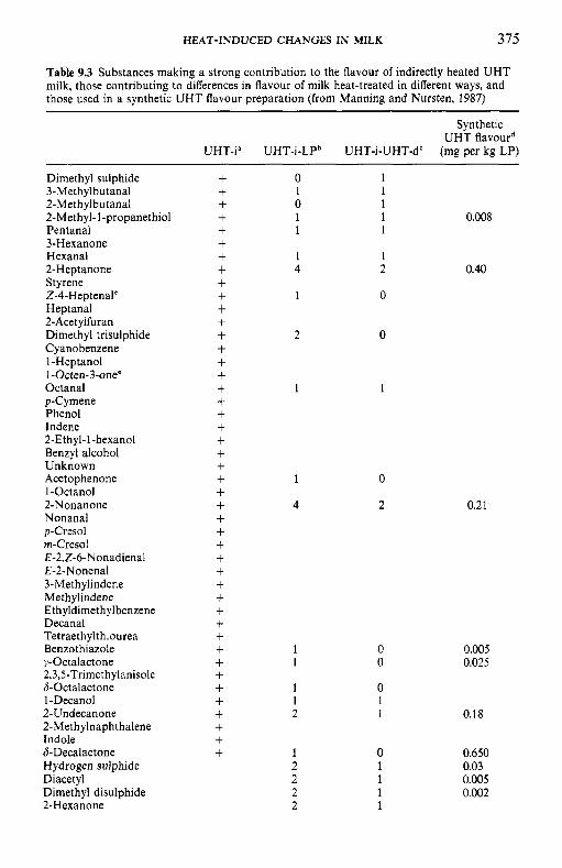
HEAT-INDUCED CHANGES IN MILK 375
Table 9.3 Substances making a strong contribution to the flavour of indirectly heated UHT milk, those contributing to differences in flavour of milk heat-treated in different ways, and those used in a synthetic UHT flavour preparation (from Manning and Nursten, 1987)
Synthetic UHT flavourd
UHT-ia UHT-i-LPb UHT-i-UHT-d' (mg per kg LP)
Dimethyl sulphide 3-Methylbutanal 2-Methylbutanal 2-Methyl-1-propanethiol Pen tanal 3-Hexanone Hexanal 2-Heptanone Styrene 2-4-Heptenale Heptanal 2-Acet ylfuran Dimethyl trisulphide Cyanobenzene I-Heptanol I-Octen-3-one' Octanal p-Cymene Phenol Indene 2-Ethyl-1-hexanol Benzyl alcohol Unknown Acetophenone I-Octanol 2-Nonanone Nonanal p-Cresol rn-Cresol E-Z,Z-&Nonadienal E-2-Nonenal 3-Methylindene Methylindene E thyldimethyl benzene Decanal Tetraethylthiourea Benzothiazole y-Octalactone 2,3,S-Trimethylanisole 6-Octalactone 1-Decanol 2-Undecanone 2-Methylnaphthalene Indole &Decalactone Hydrogen sulphide Diacetyl Dimethyl disulphide 2-Hexanone
+ + + + + + + + + + + + + + + + + + + + + + + + + + + + + + + + + + + + + + + + + + + + +
0 1 1 1 0 1 1 1 0.008 1 1
1 1 4 2 0.40
1 0
2 0
1 1
1
4
0
2 0.2 1
0.005 0.025
0.18
0.650 0.03 0.005 0.002
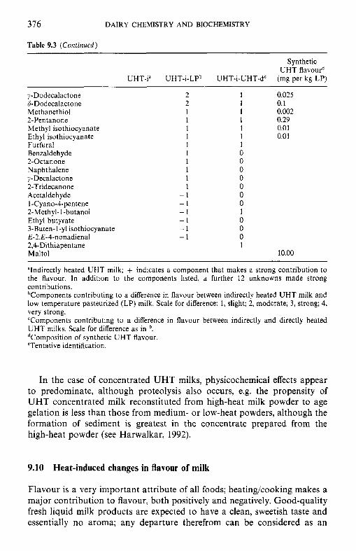
376 DAIRY CHEMISTRY AND BIOCHEMISTRY
Table 9.3 (Continued)
Synthetic U H T flavourd
UHT-i" UHT-i-LPb UHT-i-UHT-d' (mg per kg LP)
;$-Dodecalactone 6-Dodecalactone Methanethiol 2-Pentanone Methyl isothiocyanate Ethyl isothiocyanate Furfural Benzaldehyde 2-Octanone Naphthalene y-decalact one 2-Tridecanone Acetaldehyde I-Cyano-4-pentene 2-Methyl- 1-butanol Ethyl butyrate 3-Buten-1-yl isothiocyanate E-Z,E-4-nonadienal 2,CDithiapentane Maltol
2 2 1 1 1 1 1 1 1 1 1 1
- 1 - 1 - 1 -1 -1 -1
1 0.025 1 0.1 1 0.002 1 0.29 1 0.01 1 0.01 1 0 0 0 0 0 0 0 1 0 0 0 1
10.m
'Indirectly heated UHT milk; + indicates a component that makes a strong contribution to the flavour. In addition to the components listed, a further 12 unknowns made strong contributions. bComponents contributing to a difference in flavour between indirectly heated U H T milk and low temperature pasteurized (LP) milk. Scale for difference: 1, slight; 2, moderate; 3, strong; 4, very strong. 'Components contributing to a difference in flavour between indirectly and directly heated U H T milks. Scale for difference as in '. dComposition of synthetic UHT flavour. 'Tentative identification.
In the case of concentrated UHT milks, physicochemical effects appear to predominate, although proteolysis also occurs, e.g. the propensity of UHT concentrated milk reconstituted from high-heat milk powder to age gelation is less than those from medium- or low-heat powders, although the formation of sediment is greatest in the concentrate prepared from the high-heat powder (see Harwalkar, 1992).
9.10 Heat-induced changes in flavour of milk
Flavour is a very important attribute of all foods; heating/cooking makes a major contribution to flavour, both positively and negatively. Good-quality fresh liquid milk products are expected to have a clean, sweetish taste and essentially no aroma; any departure therefrom can be considered as an
HEAT-INDUCED CHANGES IN MILK 377
off-flavour. Heat treatments have a major impact on the flavour/aroma of dairy foods, either positively or negatively.
On the positive side, thermization and minimum pasteurization should not cause the formation of undesirable flavours and aromas and should, in fact, result in improved flavour by reducing bacterial growth and enzymatic activity, e.g. lipolysis. If accompanied by vacuum treatment (vacreation), pasteurization removes indigenous off-flavours, i.e. those arising from the cow’s metabolism or from feed, thereby improving the organoleptic qualities of milk.
Also on the positive side, severe heat treatment of cream improves the oxidative stability of butter produced therefrom due to the exposure of antioxidant sulphydryl groups. As discussed in section 9.2.2, lactones formed from hydroxyacids are major contributors to the desirable cooking quality of milk fats but contribute to off-flavours in other heated products, e.g. milk powders.
UHT processing causes substantial deterioration in the organoleptic quality of milk. Freshly processed UHT milk is described as ‘cooked and ‘cabbagy’, but the intensity of these flavours decreases during storage, giving maximum flavour acceptability after a few days. These off-flavours are due to the formation of sulphur compounds from the denatured whey proteins, as discussed in section 9.6.3. After this period of maximum acceptability, quality deteriorates, the milk being described as stale. At least 400 volatiles have been detected in UHT milk, about 50 of which (Table 9.3) are considered to make a significant contribution to flavour (Manning and Nursten, 1987). The shelf-life of UHT milk is usually limited by gelation and/or bitterness, both of which are due to proteolysis, as discussed in section 9.6.1.
Since sulphur compounds are important in the off-flavour of UHT milk, attempts to improve its flavour have focused on reducing the concentration of these, e.g. by adding thiosulphonates, thiosulphates or cystine (which react with mercaptans) or sulphydryl oxidase, an indigenous milk enzyme (which oxidizes sulphydryls to disulphides; Chapter 8).
.The products of Maillard browning have a significant negative impact on the flavour of heated milk products, especially in-container sterilized milks and milk powders.
References
Driessen, F.M. (1989) Inactivation of lipases and proteinases (indigenous and bacterial), in Heat-induced Changes in Milk (ed. P.F. Fox), Bulletin 238, International Dairy Federation, Brussels, pp. 71-93.
Erbersdobler, H.F. and Dehn-Miiller, B. (1989) Formation of early Maillard products during UHT treatment of milk, in Heat-induced Changes in Milk (ed. P.F. Fox), Bulletin 238, International Dairy Federation, Brussels, pp. 62-7.
378 DAIRY CHEMISTRY AND BIOCHEMISTRY
Fox, P.F. (1982) Heat-induced coagulation of milk, in Deuelopments in Dairy Chemistry, Vol. 1: Proteins (ed. P.F. Fox), Applied Science Publishers, London, pp. 189-228.
Gould, LA. (1945) Lactic acid in dairy products. 111. The effect of heat on total acid and lactic acid production and on lactose destruction. J . Dairy Sci., 28, 367-77.
Ha, Y.L., Grimm, N.K. and Pariza, M.W. (1989) Newly recognized anticarcinogenic fatty acids. Identification and quantification in natural and processed cheeses. J . Agric. Food Chem., 37,
Harwalkar, V.R. (1992) Age gelation of sterilized milks, in Advanced Dairy Chemistry, Vol. 1:
Jenness. R. and Patton, S. (1959) Principles ofDairy Chemistry, John Wiley & Sons, New York. Lyster, R.L.J. (1970) The denaturation of a-lactalbumin and fi-lactoglobulin in heated milk.
J . Dairy Res., 37, 233-43. McKellar, R.C. (ed.) (1989) Enzymes of Psychrotrophs in Raw Food, CRC Press, Boca Raton,
FL, USA. Manning, D.J. and Nursten, H.E. (1987) Flavour of milk and milk products, in Developments
in Dairy Chemistry, Vol. 3: Lactose and Minor Constituents (ed. P.F. Fox), Elsevier Applied Science, London, pp. 217-38.
Mulvihill, D.M. and Donovan, M. (1987) Whey proteins and their thermal denaturation: A review. Irish J . Food Sci. Techno/., 11, 43-75.
Singh, H. and Creamer, L.K. (1992) Heat stability of milk, in Advanced Dairy Chemistry, Vol. 1: Proteins, 2nd edn (ed. P.F. Fox), Elsevier Applied Science, London, pp. 621-56.
Stepaniak, L., Fox, P.F. and Daly, C. (1982) Isolation and general characterization of a heat-stable proteinase from Pseudomonas Juorescens AFT 36. Biochim. Biophys. Acta, 717,
Sweetsur, A.W.M. and Muir, D.D. (1980) Effect of concentration by ultrafiltration on the heat
Sweetsur, A.W.M. and Muir, D.D. (1983) Effect of homogenization on the heat stability ofmilk.
Sweetsur, A.W.M. and White, J.C.D. (1975) Studies on the heat stability of milk proteins. 111.
Walstra, P. and Jenness, R. (1984) Dairy Chemistry and Physics, John Wiley & Sons, New York. Webb, B.H. and Johnson, A.H. (1965) Fundamentals of Dairy Chemistry, AVI Publishing,
75-81.
Proteins, 2nd edn (ed. P.F. Fox), Elsevier Applied Science, London, pp. 691-734.
376-83.
stability of skim milk. J . Dairy Res. 47, 327-35.
J . Dairy Res., 50, 291-300.
Effect of heat-induced acidity in milk. J . Dairy Res., 42, 73-88.
Westport, CT.
Suggested reading
Fox, P.F. (ed.) (1982) Developments in Dairy Chemistry, Vol. 1: Proteins (ed. P.F. Fox), Applied
Fox, P.F. (ed.) (1989) Heat-induced Changes in Milk, Bulletin 238, International Dairy
Fox, P.F. (ed.) (1995) Heat-induced Changes in Milk, 2nd edn, Special Issue 9501, International
Walstra, P. and Jenness, R. (1984) Dairy Chemistry and Physics, John Wiley & Sons, New York. Wong, N.P. (ed.) (1980) Fundamentals of Dairy Chemistry, 3rd edn, AVI Publishing, Westport,
Science Publishers, London.
Federation, Brussels.
Dairy Federation, Brussels.
CT
10 Chemistry and biochemistry of cheese and fermented milks
10.1 Introduction
Cheese is a very varied group of dairy products, produced mainly in Europe, North and South America, Australia and New Zealand and to a lesser extent in North Africa and the Middle East, where it originated during the Agricultural Revolution, 6000-8000 years ago. Cheese production and con- sumption, which vary widely between countries and regions (Appendices 10A and lOB), is increasing in traditional producing countries (2-4% p.a. for several years) and is spreading to new areas. On a global scale, 30% of all milk is used for cheese; the proportion is about 40% in North America and about 50% in the European Union.
Although traditional cheeses have a rather high fat content, they are rich sources of protein and in most cases of calcium and phosphorus and have anticarigenic properties; some typical compositional data are presented in Table 10.1. Cheese is the classical example of a convenience food: it can be used as the main course in a meal, as a dessert or snack, as a sandwich filler, food ingredient or condiment.
There are at least 1000 named cheese varieties, most of which have very limited production. The principal families are Cheddar, Dutch, Swiss and Pasta filata (e.g. Mozzarella), which together account for about 80% of total cheese production. All varieties can be classified into three superfamilies based on the method used to coagulate the milk, i.e. rennet coagulation (representing about 75% of total production), isoelectric (acid) coagulation and a combination of heat and acid (which represents a very minor group).
Production of cheese curd is essentially a concentration process in which the milkfat and casein are concentrated about tenfold while the whey proteins, lactose and soluble salts are removed in the whey. The acid- coagulated and acid/heat-coagulated cheeses are normally consumed fresh but the vast majority of rennet-coagulated cheeses are ripened (matured) for a period ranging from 3 weeks to more than 2 years, during which numerous microbiological, biochemical, chemical and physical changes occur, resulting in characteristic flavour, aroma and texture. The biochemistry of cheese ripening is very complex and is not yet completely understood.
380 DAIRY CHEMISTRY AND BIOCHEMISTRY
Table 10.1 Composition of selected cheeses (per 100 g)
Water Protein Fat Cholesterol Energy Cheese type (9) (g) (g) (mg) (kJ)
Brie Caerphilly Camern bert Cheddar Cheshire Cottage Cream cheese Danish blue Edam Emmental Feta Fromage frais Gouda Gruyere Mozzarella Parmesan Ricotta Roquefort Stilton
48.6 41.8 50.7 36.0 40.6 79.1 45.5 45.3 43.8 35.7 56.5 77.9 40.1 35.0 49.8 18.4 72.1 41.3 38.6
19.3 23.2 20.9 25.5 24.0 13.8 3.1
20.1 26.0 28.7 15.6 6.8
24.0 21.2 25.1 39.4 9.4
19.7 22.7
26.9 31.3 23.1 34.4 31.4
3.9 47.4 29.6 25.4 29.7 20.2
7.1 31.0 33.3 21.0 32.1 11.0 32.9 35.5
100 90 15
100 90 13 95 75 80 90 70 25
100 100 65
100 50 90
105
1323 1554 1232 1708 1571 413
1807 1437 1382 1587 1037 469
1555 1695 1204 1880 599
1552 1701
10.2 Rennet-coagulated cheeses
The production of rennet-coagulated cheeses can, for convenience, be divided into two phases: (1) conversion of milk to curds and ( 2 ) ripening of the curds.
10.2.1 Preparation and treatment of cheesemilk
The milk for most cheese varieties is subjected to one or more pre- treatments (Table 10.2). The concentrations of fat and casein and the ratio of these components are two very important parameters affecting cheese quality. While the concentrations of these components in cheese are deter- mined and controlled by the manufacturing protocol, their ratio is regulated by adjusting the composition of the cheesemilk. This is usually done by adjusting the fat content by blending whole and skimmed milk in propor- tions needed to give the desired fat : casein ratio in the finished cheese, e.g. 1.0:0.7 for Cheddar or Gouda. It should be remembered that about 10% of the fat in milk is lost in the whey while only about 5% of the casein is lost (unavoidably, see section 10.2.2).
With the recent commercial availability of ultrafiltration, it has become possible to increase the concentration of casein, thus levelling out seasonal variations in milk composition and consequently in gel characteristics and
CHEMISTRY AND BIOCHEMISTRY OF CHEESE AND FERMENTED MILKS 381
Table 10.2 Pre-treatment of cheese milk
Standardization of fat: protein ratio Addition of skim milk Removal of some fat Addition of ultrafiltration retentate
Addition of CaCI, Adjustment of pH (e.g. by gluconic acid-6-lactone) Removal or killing of contaminating bacteria
Thermization (e.g. 65°C x 15 s) Pasteurization (e.g. 72°C x 15 s) Bactofugation Microfiltration
cheese quality. The capacity of a given plant is also increased by pre- concentrating milk by ultrafiltration.
The pH and the concentration of calcium in milk also vary, with consequential effects on the properties of renneted milk gels. The addition of CaCl, to cheesemilk (0.02%) is widely practised and adjustment and standardization of milk pH by using the acidogen, gluconic acid-d-lactone (GDL), is recommended and commercially practised on a limited scale.
Although raw milk is still widely used for cheese manufacture, e.g. Parmigiano-Reggiano (Italy), Emmental (Switzerland), Comte and Beaufort (France) and many less well known varieties, both on a factory and farmhouse scale, most Cheddar and Dutch-type cheeses are produced from pasteurized milk (HTST; c. 72°C x 15 s). Pasteurization is used primarily to kill pathogenic and spoilage bacteria. However, desirable indigenous bac- teria are also killed by pasteurization and it is generally agreed that cheese made from pasteurized milk ripens more slowly and develops a less intense flavour than raw milk cheese, apparently because certain, as yet unidentified, indigenous bacteria are absent. At present, some countries require that all cheese milk should be pasteurized or the cheese aged for at least 60days (during which time pathogenic bacteria die off). A global requirement for pasteurization of cheesemilk has been recommended but would create restrictions for international trade in cheese, especially for many of those with ‘Appellation d’Origine Protegee’ status. Research is under way to identify the important indigenous microorganisms in raw milk cheese for use as inoculants for pasteurized milk. While recognizing that pasteurization is very important in ensuring safe cheese, pH (below about 5.2) and water activity ( a w , which is controlled by addition of NaCl) are also critical safety hurdles.
Milk may be thermized (c. 65°C x 15s) on receipt at the factory to reduce bacterial load, especially psychrotrophs, which are heat labile. Since thermization does not kill pathogens, thermized milk is usually fully pasteurized before cheesemaking.
382 DAIRY CHEMISTRY AND BIOCHEMISTRY
Clostridium tyrobutyricum (an anaerobic spore-former) causes late gas blowing (through the production of H, and CO,) and off-flavours (butanoic acid) in many hard ripened cheeses; Cheddar-type cheeses are major exceptions. Contamination of cheese milk with clostridial spores can be avoided or kept to a very low level by good hygienic practices (soil and silage are the principal sources of clostridia) but they are usually prevented from growing through the use of sodium nitrate (NaNO,) or, less frequently, lysozyme, and/or removed by bactofugation (centrifugation) or microfiltra- tion.
10.2.2
Typically, five steps, or groups of steps, are involved in the conversion of milk to cheese curd: coagulation, acidification, syneresis (expulsion of whey), moulding/shaping and salting. These steps, which partly overlap, enable the cheesemaker to control the composition of cheese, which, in turn, has a major influence on cheese ripening and quality.
Conversion of milk to cheese curd
Enzymatic coagulation of milk. The enzymatic coagulation of milk involves modification of the casein micelles via limited proteolysis by selected proteinases, called rennets, followed by calcium-induced aggregation of the rennet-altered micelles:
Casein Rennet
____.* Para-casein + Macropeptides
Ca?', - 30°C
Gel
If present, the fat globules are occluded in the gel but do not participate in the formation of a gel matrix.
As discussed in Chapter 4, the casein micelles are stabilized by ti-casein, which represents 12-15% of the total casein and is located mainly on the surface of the micelles such that its hydrophobic N-terminal region reacts hydrophobically with the calcium-sensitive clsl-, cts2- and 0-caseins while its hydrophilic C-terminal region protrudes into the surrounding aqueous environment, stabilizing the micelles by a negative surface charge and steric stabilization.
Following its isolation in 1956, it was found that ti-casein is the only casein hydrolysed during the rennet coagulation of milk and that it was hydrolysed specifically at the Phe,,,-Met,,, bond, producing para-lc- casein (K-CN fl- 105) and macropeptides (f106- 169; also called glycomac- ropeptides since they contain most or all of the sugar groups attached to ti-casein) (Figure 10.1). The hydrophilic macropeptides diffuse into the surrounding medium while the para-#-casein remains attached to the
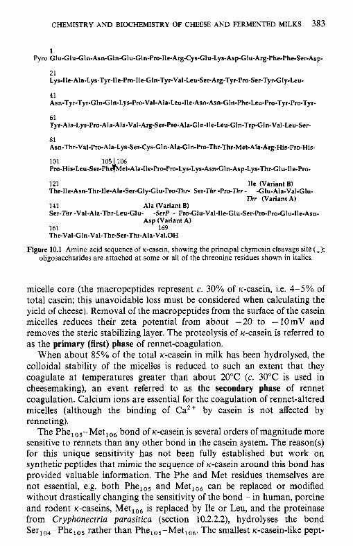
CHEMISTRY AND BIOCHEMISTRY OF CHEESE AND FERMENTED MILKS 383
1 Fyro Glu-Glu-Gln-Asn-Gln-Glu-GIn-Pro-Ile-Arg-Cys-GIu-Lys-Asp-GIu-Arg-Phe-Phe-Ser-Asp-
21 Lys-Ile-Ala-Lys-Tyr-lle-Pro-lle-GIn-Tyr-Val-Leu-Ser-Arg-Tyr-Pro-Ser-Tyr-Gly-Leu-
41 Asn-Tyr-Tyr-Gln-Gln-Lys-Pro-Val-Ala-Leu-Ile-Asn-Asn-Gln-Phe-Leu-Pro-Tyr-Pro-Tyr-
61 Tyr-AIa-Lys-Pro-Ala-Ala-Val-Arg-Ser-Pto-Ala-G1n-lle-Leu-Gln-Trp-GIn-Val-Leu-Ser-
81 Asn-Thr-Val-Pro-Ala-L ys-Ser-Cys-G1 n-Ala-Gln-Pro-Thr-Thr-Met-Ala-Arg-His-Pro-His-
101 105 106 Pro-His-Leu-Ser-Ph~et-Ala-lle-Pro-Pro-Lys-Ly~-Asn-Gln-As~-~ys-~r-Glu-IIe-Pro-
121 Ile (Variant B) Thr-He-Asn-Thr-Ile-Ala-Ser-Gly-Glu-Pro-Thr- Ser-Thr -Pro-Thr - -Glu-Ala-Val-Glu-
Thr (Variant A) 141 Ala (Variant 8) Ser-Thr -Val-Ala-Thr-Leu-Glu- -SerP - Pro-Glu-Val-lle-Glu-Ser-Pro-Pro-G1u-Ile-Asn-
Asp (Variant A) 161 169 Thr-Val-GIn-Val-Thr-Ser-Thr-Ala-Val.OH
Figure 10.1 Amino acid sequence of K-casein, showing the principal chymosin cleavage site (I); oligosaccharides are attached at some or all of the threonine residues shown in italics.
micelle core (the macropeptides represent c. 30% of Ic-casein, i.e. 4-5% of total casein; this unavoidable loss must be considered when calculating the yield of cheese). Removal of the macropeptides from the surface of the casein micelles reduces their zeta potential from about -20 to -1OmV and removes the steric stabilizing layer. The proteolysis of ic-casein is referred to as the primary (first) phase of rennet-coagulation.
When about 85% of the total ic-casein in milk has been hydrolysed, the collojdal stability of the micelles is reduced to such an extent that they coagulate at temperatures greater than about 20°C (c. 30°C is used in cheesemaking), an event referred to as the secondary phase of rennet coagulation. Calcium ions are essential for the coagulation of rennet-altered micelles (although the binding of Ca2+ by casein is not affected by renneting).
The Phe,,,-Met,,, bond of ic-casein is several orders of magnitude more sensitive to rennets than any other bond in the casein system. The reason(s) for this unique sensitivity has not been fully established but work on synthetic peptides that mimic the sequence of Ic-casein around this bond has provided valuable information. The Phe and Met residues themselves are not essential, e.g. both Phe,,, and Met,,, can be replaced or modified without drastically changing the sensitivity of the bond - in human, porcine and rodent Ic-caseins, Met,,, is replaced by Ile or Leu, and the proteinase from Cryphonectria parasitica (section 10.2.2.2), hydrolyses the bond Ser,,,-Phe,,, rather than Phe,,,-Met,,,. The smallest Ic-casein-like pept-
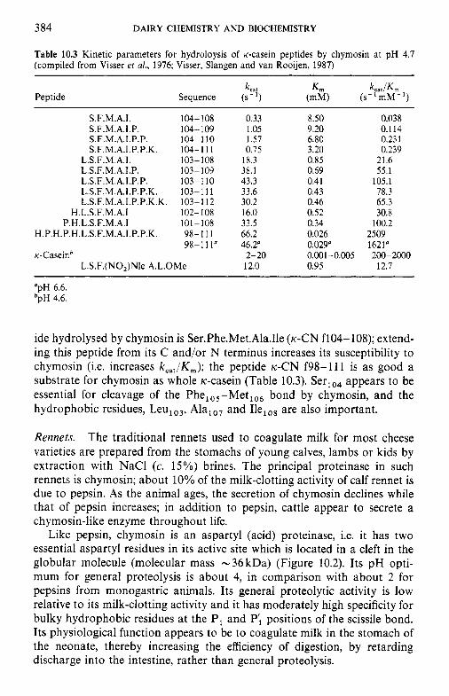
384 DAIRY CHEMISTRY AND BIOCHEMISTRY
Table 10.3 Kinetic parameters for hydroloysis of K-casein peptides by chymosin at pH 4.7 (compiled from Visser et al., 1976; Visser, Slangen and van Rooijen, 1987)
Peptide k,,,
Sequence (s- I )
S.F.M.A.I. 104-108 S.F.M.A.I.P. 104-109 S.F.M.A.I.P.P. 104-1 10 S.F.M.A.I.P.P.K. 104- I 1 1
L.S.F.M.A.I. 103-108 L.S.F.M.A.I.P. 103-109 L.S.F.M.A.I.P.P. 103-110 L.S.F.M.A.I.P.P.K. 103- 11 1 L.S.F.M.A.I.P.P.K.K. 103- 112
H.L.S.F.M.A.1 102-108 P.H.L.S.F.M.A.1 101 - 108
H.P.H.P.H.L.S.F.M.A.I.P.P.K. 98- 11 1 98-111"
k--Caseinb L.S.F.(NO,)Nle A.L.OMe
0.33 1.05 1.57 0.75
18.3 38.1 43.3 33.6 30.2 16.0 33.5 66.2 46.2"
2-20 12.0
8.50 9.20 6.80 3.20 0.85 0.69 0.41 0.43 0.46 0.52 0.34 0.026 0.029" 0.001-0.005 0.95
0.038 0.1 14 0.231 0.239
21.6 55.1
105.1 78.3 65.3 30.8
100.2 2509 1621"
12.7 200-2000
"pH 6.6. bpH 4.6.
ide hydrolysed by chymosin is Ser.Phe.Met.Ala.Ile (K-CN fl04- 108); extend- ing this peptide from its C and/or N terminus increases its susceptibility to chymosin (i.e. increases kcat/K,,,); the peptide K-CN f98-111 is as good a substrate for chymosin as whole K-casein (Table 10.3). Ser,,, appears to be essential for cleavage of the Phe,,,-Met,,, bond by chymosin, and the hydrophobic residues, Leu,,,, Ala,,, and Ilelo8 are also important.
Rennets. The traditional rennets used to coagulate milk for most cheese varieties are prepared from the stomachs of young calves, lambs or kids by extraction with NaCl (c. 15%) brines. The principal proteinase in such rennets is chymosin; about 10% of the milk-clotting activity of calf rennet is due to pepsin. As the animal ages, the secretion of chymosin declines while that of pepsin increases; in addition to pepsin, cattle appear to secrete a chymosin-like enzyme throughout life.
Like pepsin, chymosin is an aspartyl (acid) proteinase, i.e. it has two essential aspartyl residues in its active site which is located in a cleft in the globular molecule (molecular mass - 36 kDa) (Figure 10.2). Its pH opti- mum for general proteolysis is about 4, in comparison with about 2 for pepsins from monogastric animals. Its general proteolytic activity is low relative to its milk-clotting activity and it has moderately high specificity for bulky hydrophobic residues at the PI and Pi positions of the scissile bond. Its physiological function appears to be to coagulate milk in the stomach of the neonate, thereby increasing the efficiency of digestion, by retarding discharge into the intestine, rather than general proteolysis.
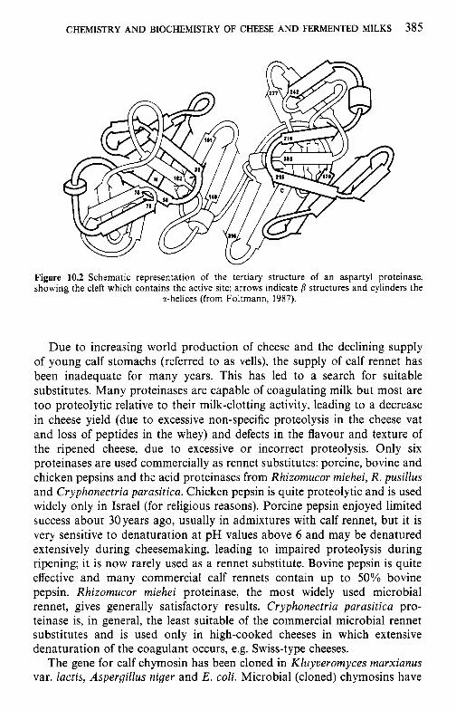
CHEMISTRY AND BIOCHEMISTRY OF CHEESE AND FERMENTED MILKS 385
Figure 10.2 Schematic representation of the tertiary structure of an aspartyl proteinase, showing the cleft which contains the active site; arrows indicate p structures and cylinders the
%-helices (from Foltmann, 1987).
Due to increasing world production of cheese and the declining supply of young calf stomachs (referred to as vells), the supply of calf rennet has been inadequate for many years. This has led to a search for suitable substitutes. Many proteinases are capable of coagulating milk but most are too proteolytic relative to their milk-clotting activity, leading to a decrease in cheese yield (due to excessive non-specific proteolysis in the cheese vat and loss of peptides in the whey) and defects in the flavour and texture of the ripened cheese, due to excessive or incorrect proteolysis. Only six proteinases are used commercially as rennet substitutes: porcine, bovine and chicken pepsins and the acid proteinases from Rhizomucor miehei, R. pusillus and Cryphonectria parasitica. Chicken pepsin is quite proteolytic and is used widely only in Israel (for religious reasons). Porcine pepsin enjoyed limited success about 30years ago, usually in admixtures with calf rennet, but it is very sensitive to denaturation at pH values above 6 and may be denatured extensively during cheesemaking, leading to impaired proteolysis during ripening; it is now rarely used as a rennet substitute. Bovine pepsin is quite effective and many commercial calf rennets contain up to 50% bovine pepsin. Rhizomucor miehei proteinase, the most widely used microbial rennet, gives generally satisfactory results. Cryphonectria parasitica pro- teinase is, in general, the least suitable of the commercial microbial rennet substitutes and is used only in high-cooked cheeses in which extensive denaturation of the coagulant occurs, e.g. Swiss-type cheeses.
The gene for calf chymosin has been cloned in Kluyveromyces marxianus var. lactis, Aspergillus niger and E. coli. Microbial (cloned) chymosins have
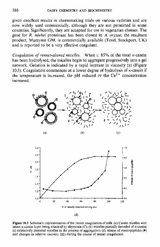
386 DAIRY CHEMISTRY AND BIOCHEMISTRY
given excellent results in cheesemaking trials on various varieties and are now widely used commercially, although they are not permitted in some countries. Significantly, they are accepted for use in vegetarian cheeses. The gene for R. miehei proteinase has been cloned in A . oryzae; the resultant product, Marzyme GM, is commercially available (Texel, Stockport, UK) and is reported to be a very effective coagulant.
Coagulation of rennet-altered micelles. When c. 85% of the total u-casein has been hydrolysed, the micelles begin to aggregate progressively into a gel network. Gelation is indicated by a rapid increase in viscosity ( q ) (Figure 10.3). Coagulation commences at a lower degree of hydrolysis of rc-casein if the temperature is increased, the pH reduced or the Ca2+ concentration increased.
0 20 40 M) R O
I of visunlly ohxrvcd dolling time
Figure 10.3 Schematic representation of the rennet coagulation of milk. (a) Casein micelles with intact ti-casein layer being attacked by chymosin (C); (b) rnicelles partially denuded of ti-casein; (c) extensively denuded micelles in the process of aggregation; (d) release of macropeptides (+) and changes in relative viscosity (B) during the course of rennet coagulation.
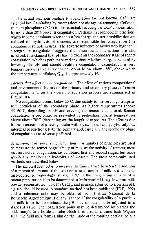
CHEMISTRY AND BIOCHEMISTRY OF CHEESE AND FERMENTED MILKS 387
The actual reactions leading to coagulation are not known. Ca2+ are essential but Ca-binding by caseins does not change on renneting. Colloidal calcium phosphate (CCP) is also essential: reducing the CCP concentration by more than 20% prevents coagulation. Perhaps, hydrophobic interactions, which become dominant when the surface charge and steric stabilization are reduced on hydrolysis of K-casein, are responsible for coagulation (the coagulum is soluble in urea). The adverse influence of moderately high ionic strength on coagulation suggests that electrostatic interactions are also involved. It is claimed that pH has no effect on the secondary stage of rennet coagulation, which is perhaps surprising since micellar charge is reduced by lowering the pH and should facilitate coagulation. Coagulation is very temperature-sensitive and does not occur below about 18"C, above which the temperature coefficient, Qlo, is approximately 16.
Factors that afect rennet coagulation. The effect of various compositional and environmental factors on the primary and secondary phases of rennet coagulation and on the overall coagulation process are summarized in Figure 10.4.
No coagulation occurs below 20"C, due mainly to the very high tempera- ture coefficient of the secondary phase. At higher temperatures (above 55-60"C, depending on pH and enzyme) the rennet is denatured. Rennet coagulation is prolonged or prevented by preheating milk at temperatures above about 70°C (depending on the length of exposure). The effect is due to the interaction of /3-lactoglobulin with K-casein via sulphydryl-disulphide interchange reactions; both the primary and, especially, the secondary phase of coagulation are adversely affected.
Measurement of rennet coagulation time. A number of principles are used to measure the rennet coagulability of milk or the activity of rennets; most measure actual coagulation, i.e. combined first and second stages, but some specifically monitor the hydrolysis of K-casein. The most commonly used methods are described below.
The simplest method is to measure the time elapsed between the addition of a measured amount of diluted rennet to a sample of milk in a tempera- ture-controlled water-bath at, e.g. 30°C. If the coagulating activity of a rennet preparation is to be determined, a 'reference' milk, e.g. low-heat milk powder reconstituted in 0.01% CaCl,, and perhaps adjusted to a certain pH, e.g. 6.5, should be used. A standard method has been published (IDF, 1992) and a reference milk may be obtained from Institut National de la Recherche Agronomique, Poligny, France. If the coagulability of a particu- lar milk is to be determined, the pH may or may not be adjusted to a standard value. The coagulation point may be determined by placing the milk sample in a bottle or tube which is rotated in a water-bath (Figure 10.5); the fluid milk forms a film on the inside of the rotating bottle/tube but
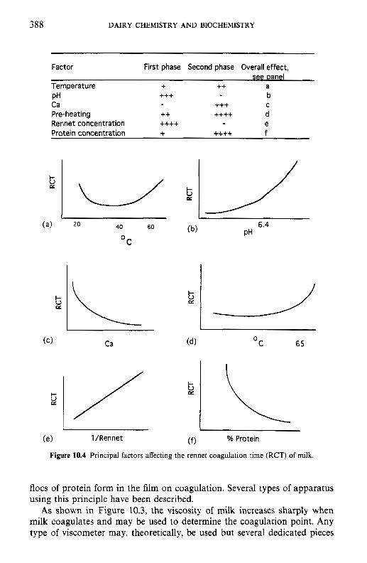
388 DAIRY CHEMISTRY AND BIOCHEMISTRY
Factor First phase Second phase Overall effect,
Temperature + ++ a +++ b
+++ C PH Ca Pre-heating ++ ++++ d Rennet concentration ++++ e Protein concentration + ++++ f
20 40 60
C 0
Ca
1 /Rennet
t ez
6.4 PH
C 65 0
% Protein
Figure 10.4 Principal factors affecting the rennet coagulation time (RCT) of milk.
flocs of protein form in the film on coagulation. Several types of apparatus using this principle have been described.
As shown in Figure 10.3, the viscosity of milk increases sharply when milk coagulates and may be used to determine the coagulation point. Any type of viscometer may, theoretically, be used but several dedicated pieces
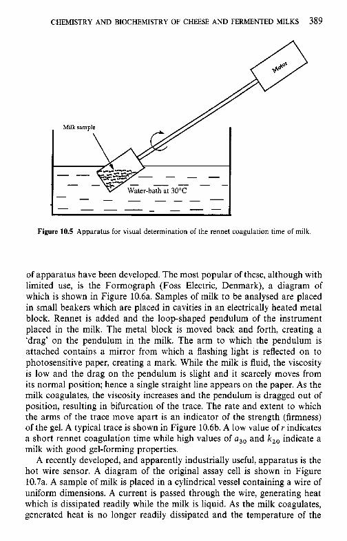
CHEMISTRY AND BIOCHEMISTRY OF CHEESE AND FERMENTED MILKS 389
Milk sample
Figure 10.5 Apparatus for visual determination of the rennet coagulation time of milk.
of apparatus have been developed. The most popular of these, although with limited use, is the Formograph (Foss Electric, Denmark), a diagram of which is shown in Figure 10.6a. Samples of milk to be analysed are placed in small beakers which are placed in cavities in an electrically heated metal block. Rennet is added and the loop-shaped pendulum of the instrument placed in the milk. The metal block is moved back and forth, creating a ‘drag’ on the pendulum in the milk. The arm to which the pendulum is attached contains a mirror from which a flashing light is reflected on to photosensitive paper, creating a mark. While the milk is fluid, the viscosity is low and the drag on the pendulum is slight and it scarcely moves from its normal position; hence a single straight line appears on the paper. As the milk coagulates, the viscosity increases and the pendulum is dragged out of position, resulting in bifurcation of the trace. The rate and extent to which the arms of the trace move apart is an indicator of the strength (firmness) of the gel. A typical trace is shown in Figure 10.6b. A low value of r indicates a short rennet coagulation time while high values of a3, and k, , indicate a milk with good gel-forming properties.
A recently developed, and apparently industrially useful, apparatus is the hot wire sensor. A diagram of the original assay cell is shown in Figure 10.7a. A sample of milk is placed in a cylindrical vessel containing a wire of uniform dimensions. A current is passed through the wire, generating heat which is dissipated readily while the milk is liquid. As the milk coagulates, generated heat is no longer readily dissipated and the temperature of the
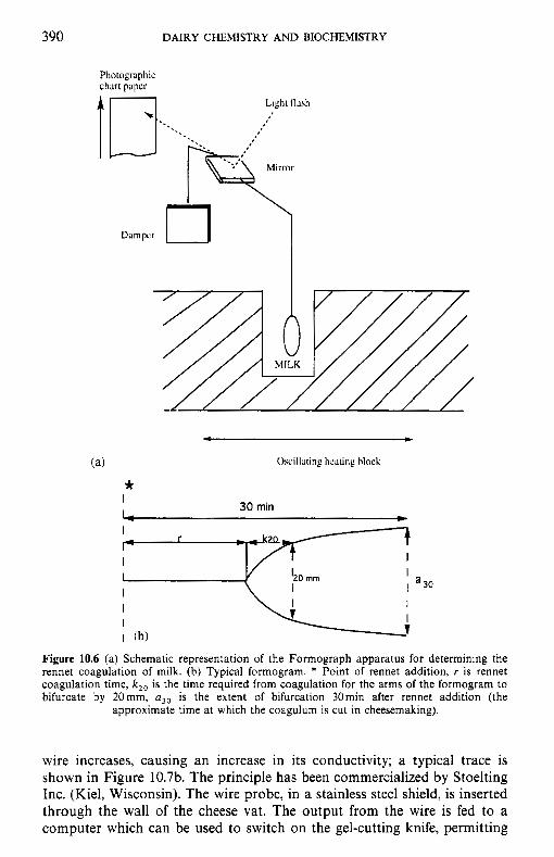
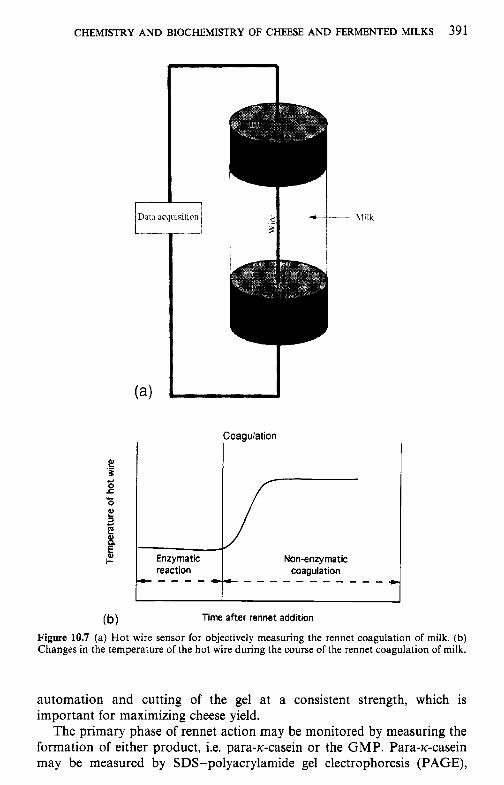
CHEMISTRY AND BIOCHEMISTRY OF CHEESE AND FERMENTED MILKS 391
I 1)ata acquisition
Non-enzymatic coagulation .. - - - - - - - - - - - c Enzymatic
reaction * - - - -
Time after rennet addition
Figure 10.7 (a) Hot wire sensor for objectively measuring the rennet coagulation of milk. (b) Changes in the temperature of the hot wire during the course of the rennet coagulation of milk.
automation and cutting of the gel at a consistent strength, which is important for maximizing cheese yield.
The primary phase of rennet action may be monitored by measuring the formation of either product, i.e. para-lc-casein or the GMP. Para-lc-casein may be measured by SDS-polyacrylamide gel electrophoresis (PAGE),
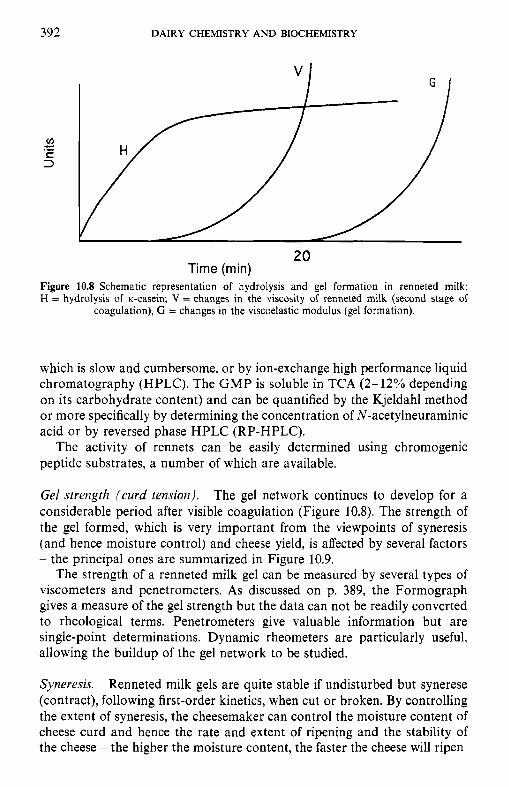
392 DAIRY CHEMISTRY AND BIOCHEMISTRY
20 Time (min)
Figure 10.8 Schematic representation of hydrolysis and gel formation in renneted milk; H = hydrolysis of K-casein; V = changes in the viscosity of renneted milk (second stage of
coagulation), G = changes in the viscoelastic modulus (gel formation).
which is slow and cumbersome, or by ion-exchange high performance liquid chromatography (HPLC). The GMP is soluble in TCA (2-12% depending on its carbohydrate content) and can be quantified by the Kjeldahl method or more specifically by determining the concentration of N-acetylneuraminic acid or by reversed phase HPLC (RP-HPLC).
The activity of rennets can be easily determined using chromogenic peptide substrates, a number of which are available.
Gel strength (curd tension). The gel network continues to develop for a considerable period after visible coagulation (Figure 10.8). The strength of the gel formed, which is very important from the viewpoints of syneresis (and hence moisture control) and cheese yield, is affected by several factors - the principal ones are summarized in Figure 10.9.
The strength of a renneted milk gel can be measured by several types of viscometers and penetrometers. As discussed on p. 389, the Formograph gives a measure of the gel strength but the data can not be readily converted to rheological terms. Penetrometers give valuable information but are single-point determinations. Dynamic rheometers are particularly useful, allowing the buildup of the gel network to be studied.
Syneresis. Renneted milk gels are quite stable if undisturbed but synerese (contract), following first-order kinetics, when cut or broken. By controlling the extent of syneresis, the cheesemaker can control the moisture content of cheese curd and hence the rate and extent of ripening and the stability of the cheese - the higher the moisture content, the faster the cheese will ripen
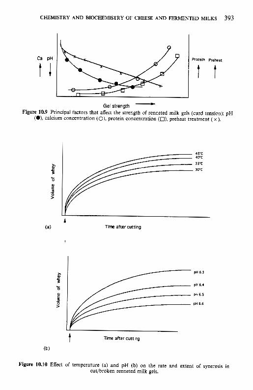
CHEMISTRY AND BIOCHEMISTRY OF CHEESE AND FERMENTED MILKS 393
Gel strength - Figure 10.9 Principal factors that affect the strength of renneted milk gels (curd tension); pH
(O), calcium concentration (O), protein concentration (O), preheat treatment ( x ).
45’C 40’C
3sc 30’c
t Time after cutting
pH 6.3
pH 6.4
pH 6.5
pH 6.6
t Time after cutting
Figure 10.10 Effect of temperature (a) and pH (b) on the rate and extent of syneresis in cut/broken renneted milk gels.
394 DAIRY CHEMISTRY AND BIOCHEMISTRY
but the lower its stability. Syneresis is promoted by:
0 cutting the curd finely, e.g. Emmental (fine cut) versus Camembert (large
0 low pH (Figure 10.1Ob); 0 calcium ions; 0 increasing the cooking temperature (Camembert, c. 30°C; Gouda, c. 36°C;
Cheddar, c. 38°C; Emmental or Parmesan, 52-55OC) (Figure 10.1Oa); 0 stirring the curd during cooking; 0 fat retards syneresis, while increasing the protein content (up to a point)
improves it; at high protein concentrations, the gel is too firm and does not synerese (e.g. UF retentate).
Gels prepared from heated milk synerese poorly (assuming that the milk does coagulate). Such reduced syneresis properties are desirable for fer- mented milk products, e.g. yoghurt (milk for which is severly heated, e.g. 90°C x 10min) but are undesirable for cheese.
Good analytical methods for monitoring syneresis are lacking. Principles that have been exploited include: dilution of an added marker, e.g. a dye, which must not adsorb on to or diffuse into the curd particles, measurement of the electrical conductivity or moisture content of the curd or by measuring the volume of whey released (probably the most commonly used method although only one-point values are obtained).
cut);
10.2.3 Acidification
Acid production is a key feature in the manufacture of all cheese varieties - the pH decreases to about 5 (k0.3, depending on variety) within 5-20h, at a rate depending on the variety (Figure 10.11). Acidification is normally achieved via the bacterial fermentation of lactose to lactic acid, although an acidogen, usually gluconic acid-6-lactone, alone or in combination with acid, may be used in some cases, e.g. Mozzarella.
Traditionally, cheesemakers relied on the indigenous microflora of milk for lactose fermentation, as is still the case for several minor artisanal varieties. However, since the indigenous microflora varies, so does the rate of acidification and hence the quality of the cheese; the indigenous micro- flora is largely destroyed by pasteurization. ‘Slop-back’ or whey cultures (starters; the use of whey from today’s cheesemaking as an inoculum for tomorrow’s milk) have probably been used for a very long time and are still used commercially, e.g. for such famous cheese as Parmigiano-Reggiano and Comte. However, selected ‘pure’ cultures have been used for Cheddar and Dutch-type cheeses for at least 80 years and have become progressively more refined over the years. Single-strain cultures were introduced in New Zealand in the 1930s as part of a bacteriophage control programme. Selected phage-unrelated strains are now widely used for Cheddar cheese;
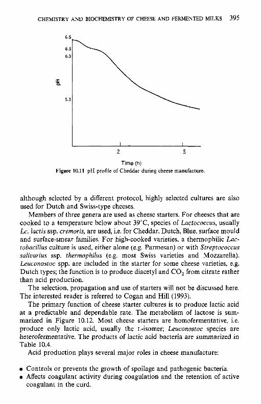
CHEMISTRY AND BIOCHEMISTRY OF CHEESE AND FERMENTED MILKS 395
2 5
Time (h) Figure 10.11 pH profile of Cheddar during cheese manufacture.
although selected by a different protocol, highly selected cultures are also used for Dutch and Swiss-type cheeses.
Members of three genera are used as cheese starters. For cheeses that are cooked to a temperature below about 39"C, species of Lactococcus, usually Lc. lactis ssp. cremoris, are used, i.e. for Cheddar, Dutch, Blue, surface mould and surface-smear families. For high-cooked varieties, a thermophilic Lac- tobacillus culture is used, either alone (e.g. Parmesan) or with Streptococcus saliuarius ssp. therrnophilus (e.g. most Swiss varieties and Mozzarella). Leuconostoc spp. are included in the starter for some cheese varieties, e.g. Dutch types; the function is to produce diacetyl and CO, from citrate rather than acid production.
The selection, propagation and use of starters will not be discussed here. The interested reader is referred to Cogan and Hill (1993).
The primary function of cheese starter cultures is to produce lactic acid at a predictable and dependable rate. The metabolism of lactose is sum- marized in Figure 10.12. Most cheese starters are homofermentative, i.e. produce only lactic acid, usually the L-isomer; Leuconostoc species are heteroferrnentative. The products of lactic acid bacteria are summarized in Table 10.4.
Acid production plays several major roles in cheese manufacture:
0 Controls or prevents the growth of spoilage and pathogenic bacteria. 0 Affects coagulant activity during coagulation and the retention of active
coagulant in the curd.
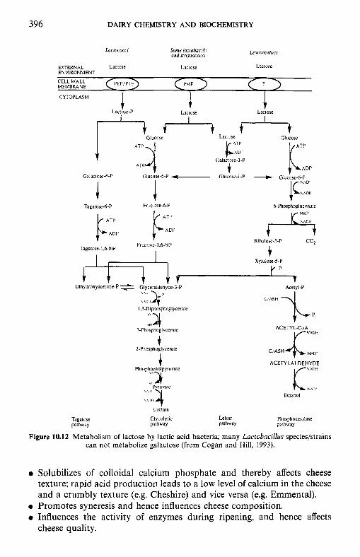
396 DAIRY CHEMISTRY AND BIOCHEMISTRY
LQcrococcr Some lacrobocrlii ood srreprococci kuronosrocs
EXTERNAL Lactose Lactose Lactose ENVIRONMENT
CELL WALL MEMBRANE I’MF
CYTOI’LASM
Laciore-P i
Lactose J.
Laclose I
t Glucose Lactose Glucose
Galactose-6-P p 4 - t ADI’ Glucose-6-P - Galactose-I-P GlUCoSe-I-P KT1’ t AUI’ + - Glucore.6.P LlDP {C::::
6-Phosphogluconate 1
Fructose-6-P J
Tagatose.6-P k”, K ::,
$
t co2
ADI’ t $.
Ribulose-5-P Tagatose.1.6.blP Fructose-1’6-hiP
Xylulose.5-P
p,
v Acetyl-P
1 CuASH
Dthydroxyacetone-P -1 Glyceraldehyde-3-P t :::,yp
1.3-Diphosphoglycerate
3-Phosphoglycerate 24 ACETY L-CoA
CnASH t
?-Phosphoglycerate I
Tagatose pathway
Phosphoenolpyruvate
A,.. ‘\I I’ 2 Fyruvare
Lactate
Glycolytic pathway
ACETYLALDEHYDE NADH K l \ A I l ’
Ethanol
Leloir Phosphoketolase pathway pathway
Figure 10.12 Metabolism of lactose by lactic acid bacteria; many Lactobacillus species/strains can not metabolize galactose (from Cogan and Hill, 1993).
Solubilizes of colloidal calcium phosphate and thereby affects cheese texture; rapid acid production leads to a low level of calcium in the cheese and a crumbly texture (e.g. Cheshire) and vice versa (e.g. Emmental). Promotes syneresis and hence influences cheese composition. Influences the activity of enzymes during ripening, and hence affects cheese quality.
CHEMISTRY AND BIOCHEMISTRY OF CHEESE A N D FERMENTED MILKS 397
Table 10.4 Salient features of lactose metabolism in starter culture organisms (from Cogan and Hill, 1993)
~
Organism Cleavageb Products
Transport” enzyme Pathway‘ (mol mol- lactose)
Lactococcus spp. PTS ppgal G L Y 4 L-Lactate Leuconostoc spp. ? Bgal PK 2 D-Lactate + 2 ethanol + 2 C 0 , Str. salicarius PMF G L Y 2 L-Lactated
Lb. delbrueckii PMF? /?gal G L Y 2 D-Lactated
Lb. delbrueckii PMF? jgal G L Y 2 D-Lactated
Lb. helveticus PMF? Pgal G L Y 4 L- (mainly) + D-lactate
subsp. thermophilus
subsp. lactis
subsp. bulgarrcus
OPTS, phosphotransferase system; PMF, proton motive force. *ppgal, phospho-8-galactosidase; pgal, 8-galactosidase. ‘GLY, glycolysis; PK. phosphoketolase. dThese species metabolize only the glucose moiety of lactose.
The primary starter performs several functions in addition to acid production, especially reduction of the redox potential (Eh, from about +250mV in milk to - 150mV in cheese), and, most importantly, plays a major, probably essential, role in the biochemistry of cheese ripening. Many strains produce bacteriocins which control the growth of contaminating micro-organisms.
The ripening of many varieties is characterized by the action, not of the primary starter, but of other micro-organisms, which we will refer to as a secondary culture. Examples are Propionibacterium in Swiss-type cheeses, Penicillium rogueforti in Blue cheeses, Penicillium camemberti in surface mould-ripened cheeses, e.g. Camembert and Brie, Breuibacterium linens and yeasts in surface smear-ripened cheese, Lactococcus lactis ssp. lactis biovar diacetylactis and Leuconostoc spp. in Dutch-type cheeses. The specific function of these micro-organsims will be discussed in section 10.2.7 on ripening. Traditionally, a secondary culture was not used in Cheddar-type cheeses but there is much current interest in the use of cultures of selected bacteria, usually mesophilic Lactobacillus spp. or lactose-negative Lactococ- cus spp., for Cheddar cheese with the objective of intensifying or modifying flavour or accelerating ripening; such cultures are frequently referred to as ‘adjunct cultures’.
10.2.4 Moulding and shaping
When the desired pH and moisture content have been achieved, the curds are separated from the whey and placed in moulds of traditional shape and size to drain and form a continuous mass; high-moisture curds form a
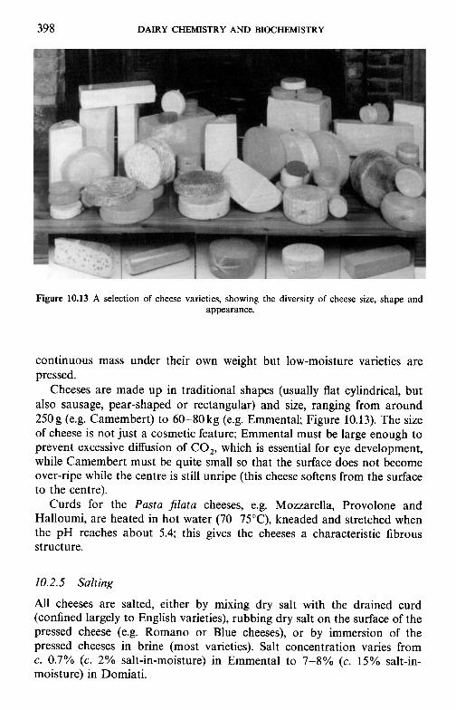
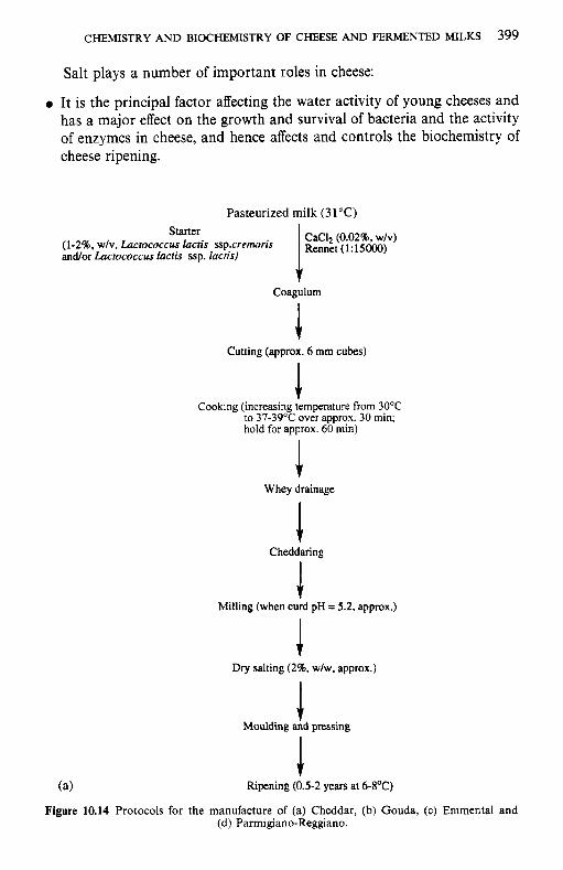
CHEMISTRY AND BIOCHEMISTRY OF CHEESE AND FERMENTED MILKS 399
Starter (1-296, wlv, Lactococcus lacris ssp.cremnris and/or Lactococcus lactis ssp. lactis)
Salt plays a number of important roles in cheese:
0 It is the principal factor affecting the water activity of young cheeses and has a major effect on the growth and survival of bacteria and the activity of enzymes in cheese, and hence affects and controls the biochemistry of cheese ripening.
CaCI, (0.02%. w/v) R~~~~~ (1:15000)
Pasteurized milk (31°C)
f Coagulum
Cutting (approx. 6 mm cubes)
1 Cooking (increasing temperature from 3OoC
to 37-39°C over approx. 30 min; hold for approx. 60 min)
Whey drainage
Cheddaring
Milling (when curd pH = 5.2, approx.)
Dry salting (2%. w/w. approx.)
Moulding a h pressing
(a) Ripening (0.5-2 years at 6-8'C)
Figure 10.14 Protocols for the manufacture of (a) Cheddar, (b) Gouda, (c) Emmental and (d) Parmigiano-Reggiano.
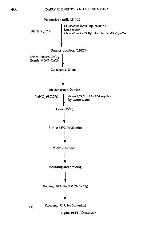
400 DAIRY CHEMISTRY AND BIOCHEMISTRY
Pasteurized milk (31°C)
Lactococcus lactis ssp. cremoris Leuconostoc Lactococcus lactis ssp. lactis biovar diacetylactis Starters (0.7%)
v Rennet addition (0.022%0)
1 Edam, 0.015% CaCI, Gouda, 0.06% CaCI,
Cut (approx. 25 mi,)
Stir (for approx. 20 min)
NaNO, (0.015%) drain 1/3 of whey and replace by warm water i
Cook (35°C)
Stir (at 35°C for 20 min)
1 Whey drainage
Moulding and pressing
Brining (20% NaCI, 0.5% CaC1,)
Ripening (12OC for 3 months) (b)
Figure 10.14 (Continued).
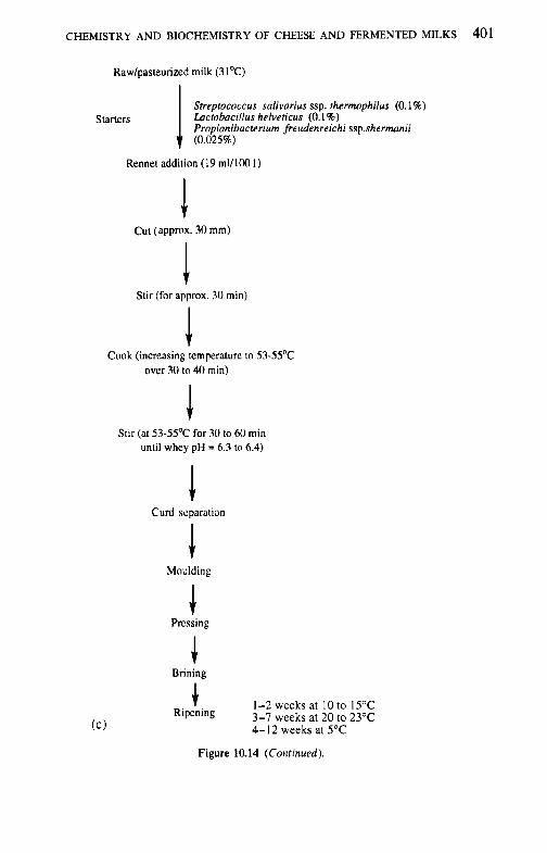
CHEMISTRY AND BIOCHEMISTRY OF CHEESE AND FERMENTED MILKS 401
Rawlpasteunxd milk (3 l0C)
Starters Streptococcus salivarius ssp. thermophilus (0.1%) Lactobacillus helveticus (0.1%) Propionibacterium freudenreichi ssp.shermunii (0.025~
Rennet addition (19 m1/1W 1 )
Cut (approx. 30 mm)
Stir (for approx. 30 min)
Cook (increasing temperature to 53-55OC over 30 to 40 min)
Stir (at 53-55OC for 30 to 60 min until whey pH = 6.3 to 6.4)
Curd scparation
Moulding
Brining
t 1-2 weeks at 10 to 15°C 3-7 weeks at 20 to 23°C 4-12 weeks at 5°C
Figure 10.14 (Continued).
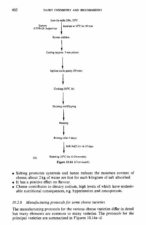
402 DAIRY CHEMISTRY AND BIOCHEMISTRY
Low-fat milk (2%). 32OC
lncuhate at 32OC for 30 min 1 Starters 0.75% Lh. bulgoricus
Rennet addition
Cutting (appmx. 3 mm pieces)
Agitiate curds gently (30 min)
Cooking (55OC lh)
Draining and Dipping
Pressing
Brining (after 3 days)
246 NACI for 14-15 days 1 Ripening (15°C for 10-24 months) (d)
Figure 10.14 (Continued).
0 Salting promotes syneresis and hence reduces the moisture content of cheese; about 2 kg of water are lost for each kilogram of salt absorbed.
0 It has a positive effect on flavour. 0 Cheese contributes to dietary sodium, high levels of which have undesir-
able nutritional consequences, e.g. hypertension and osteoporosis.
10.2.6 Manufacturing protocols for some cheese varieties
The manufacturing protocols for the various cheese varieties differ in detail but many elements are common to many varieties. The protocols for the principal varieties are summarized in Figures 10.14a-d.
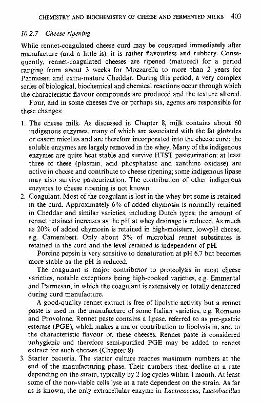
CHEMISTRY AND BIOCHEMISTRY OF CHEESE AND FERMENTED MILKS 403
10.2.7 Cheese ripening
While rennet-coagulated cheese curd may be consumed immediately after manufacture (and a little is), it is rather flavourless and rubbery. Conse- quently, rennet-coagulated cheeses are ripened (matured) for a period ranging from about 3 weeks for Mozzarella to more than 2 years for Parmesan and extra-mature Cheddar. During this period, a very complex series of biological, biochemical and chemical reactions occur through which the characteristic flavour compounds are produced and the texture altered.
Four, and in some cheeses five or perhaps six, agents are responsible for these changes:
1. The cheese milk. As discussed in Chapter 8, milk contains about 60 indigenous enzymes, many of which are associated with the fat globules or casein micelles and are therefore incorporated into the cheese curd; the soluble enzymes are largely removed in the whey. Many of the indigenous enzymes are quite heat stable and survive HTST pasteurization; at least three of these (plasmin, acid phosphatase and xanthine oxidase) are active in cheese and contribute to cheese ripening; some indigenous lipase may also survive pasteurization. The contribution of other indigenous enzymes to cheese ripening is not known.
2. Coagulant. Most of the coagulant is lost in the whey but some is retained in the curd. Approximately 6 % of added chymosin is normally retained in Cheddar and similar varieties, including Dutch types; the amount of rennet retained increases as the pH at whey drainage is reduced. As much as 20% of added chymosin is retained in high-moisture, low-pH cheese, e.g. Camembert. Only about 3% of microbial rennet substitutes is retained in the curd and the level retained is independent of pH.
Porcine pepsin is very sensitive to denaturation at pH 6.7 but becomes more stable as the pH is reduced.
The coagulant is major contributor to proteolysis in most cheese varieties, notable exceptions being high-cooked varieties, e.g. Emmental and Parmesan, in which the coagulant is extensively or totally denatured during curd manufacture.
A good-quality rennet extract is free of lipolytic activity but a rennet paste is used in the manufacture of some Italian varieties, e.g. Romano and Provolone. Rennet paste contains a lipase, referred to as pre-gastric esterase (PGE), which makes a major contribution to lipolysis in, and to the characteristic flavour of, these cheeses. Rennet paste is considered unhygienic and therefore semi-purified PGE may be added to rennet extract for such cheeses (Chapter 8).
3. Starter bacteria. The starter culture reaches maximum numbers at the end of the manufacturing phase. Their numbers then decline at a rate depending on the strain, typically by 2 log cycles within 1 month. At least some of the non-viable cells lyse at a rate dependent on the strain. As far as is known, the only extracellular enzyme in Lactococcus, Lactobacillus
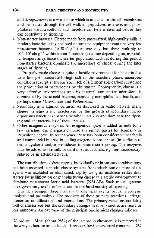
404 DAIRY CHEMISTRY AND BIOCHEMISTRY
and Streptococcus is a proteinase which is attached to the cell membrane and protrudes through the cell wall; all peptidases, esterases and phos- phatases are intracellular and therefore cell lysis is essential before they can contribute to ripening.
4. Non-starter bacteria. Cheese made from pasteurized, high-quality milk in modern factories using enclosed automated equipment contains very few non-starter bacteria ( < 50 cfu g- ') at one day but these multiply to 107-108 cfug-' within about 2 months (at a rate depending on, especial- ly, temperature). Since the starter population declines during this period, non-starter bacteria dominate the microflora of cheese during the later stages of ripening.
Properly made cheese is quite a hostile environment for bacteria due to a low pH, moderate-to-high salt in the moisture phase, anaerobic conditions (except at the surface), lack of a fermentable carbohydrate and the production of bacteriocins by the starter. Consequently, cheese is a very selective environment and its internal non-starter microflora is dominated by lactic acid bacteria, especially mesophilic lactobacilli, and perhaps some Micrococcus and Pediococcus.
5. Secondary and adjunct cultures. As discussed in section 10.2.3, many cheese varieties are characterized by the growth of secondary micro- organisms which have strong metabolic activity and dominate the ripen- ing and characteristics of these cheeses.
6. Other exogenous enzymes. An exogenous lipase is added to milk for a few varieties, e.g. pre-gastric lipase (in rennet paste) for Romano or Provolone cheese. In recent years, there has been considerable academic and commercial interest in adding exogenous proteinases (in addition to the coagulant) and/or peptidases to accelerate ripening. The enzymes may be added to the milk or curd in various forms, e.g. free, microencap- sulated or in attenuated cells.
The contribution of these agents, individually or in various combinations, has been assessed in model cheese systems from which one or more of the agents was excluded or eliminated, e.g. by using an acidogen rather than starter for acidification or manufacturing cheese in a sterile environment to eliminate non-starter lactic acid bacteria (NSLAB). Such model systems have given very useful information on the biochemistry of ripening.
During ripening, three primary biochemical events occur, glycolysis, lipolysis and proteolysis. The products of these primary reactions undergo numerous modifications and interactions. The primary reactions are fairly well characterized but the secondary changes in most varieties are more or less unknown. An overview of the principal biochemical changes follows.
Glycolysis. Most (about 98%) of the lactose in cheese-milk is removed in the whey as lactose or lactic acid. However, fresh cheese curd contains 1-2%
CHEMISTRY AND BIOCHEMISTRY OF CHEESE AND FERMENTED MILKS 405
lactose which is normally metabolized to L-lactic acid by the Lactococcus starter within a day for most varieties or a few weeks for Cheddar. In most varieties, the L-lactate is racemized to DL-lactate by NSLAB within about 3 months and a small amount is oxidized to acetic acid at a rate dependent on the oxygen content of the cheese and hence on the permeability of the packaging material.
In cheese varieties made using Streptococcus salvarius ssp. thermophilus and Lactobacillus spp. as starter, e.g. Swiss types and Mozzarella, the metabolism of lactose is more complex than in cheese in which a Lactococcus starter is used. In these cheeses, the curd is cooked to 52-55"C, which is above the growth temperature for both components of the starter; as the curd cools, the Streptococcus, which is the more heat-tolerant of the two starters, begins to grow, utilizing the glucose moiety of lactose, with the production of L-lactic acid, but not galactose, which accumulates in the curd. When the curd has cooled sufficiently, the Lactobacillus spp. grow, and, if a galactose-positive species/strain is used, it metabolizes galactose, producing DL-lactate (Figure 10.15). If a galactose-negative strain of Lactobaciilus is used, galactose accumulates in the curd and can participate in Maillard browning, especially during heating, which is undesirable, especially in Pizza cheese.
Swiss-type cheeses are ripened at about 22°C for a period to encourage the growth of Propionibacterium spp. which use lactic acid as an energy
2
h 0 Y
c M 0
M 0 . v .- C
3
- I 2 c a,
S
0 0 10 20 I0 20 30 411
Time (h) Time (days)
Figure 10.15 Metabolism of lactose, glucose, galactose, D- and L-lactic acid in Emmental cheese. Cheese transferred to hot room (22-24°C) at 14 days. 0, D-lactate; 0, acetate;
H, galactose; 0, L-lactate; +, glucose; 0, lactose; A, propionate.
406 DAIRY CHEMISTRY AND BIOCHEMISTRY
CH,.CH,-CH,-COOH P. Bulyrate
0 . 4 Acslyl-CoA
Aceiyl-P
A C C I Y ~ ~
ATP CH,-CH~-CH~-CO-CoA t)utyryl.coA
NAI)Hi NAD 1 CHI-CH =CH-CO-CoA
Cmtonyl-CoA
2p, 7 NAD*
~CHI-CO-COOH 2CHyHCOH Pymvale Lactate
t ZCHKO-CoA AcetyI-CoA
+
-COOH
NAD' NADH?
Figure 10.16 Metabolism of glucose or lactic acid by Clostridium tyrobutyricurn with the production of butyric acid, CO, and hydrogen gas.
source, producing propionic acid, acetic acid and CO, (Figure 10.15):
3CH3CHOHCOOH + 2CH,CH,COOH + CH3COOH + CO, + H,O Lactic acid Propionic acid Acetic acid
Propionic and acetic acids probably contribute to the flavour of Swiss- type cheeses, while the CO, is responsible for their large characteristic eyes. Lactic acid may be metabolized by Clostridium tyrobutyricum to butyric acid, CO, and hydrogen (Figure 10.16); butyric acid is responsible for off-flavours and the CO, and H, for late gas blowing. Clostridia are controlled by good hygienic practices, addition of nitrate or lysozyme, bactofugation or microfiltration. The principal sources of clostridia are soil and silage.
In surface mould-ripened cheeses, e.g. Camembert and Brie, Penicillium camemberti, growing on the surface, metabolizes lactic acid as an energy source, causing the pH to increase. Lactic acid diffuses from the centre to the surface, where it is catabolized. Ammonia produced by deamination of amino acids contributes to the increase in pH which reaches about 7.5 at the surface and 6.5 at the centre of the cheese. Ripening of Camembert and Brie is characterized by softening (liquefaction) of the texture from the surface towards the centre. Softening is due to the increase in pH, proteolysis and diffusion of calcium phosphate to the surface, where it precipitates due to the high pH. These events are summarized in Figure 10.17.
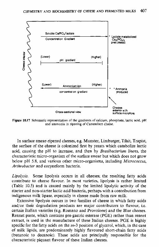
CHEMISTRY AND BIOCHEMISTRY OF CHEESE AND FERMENTED MILKS 407
Soluble CaPOJlactate
Concentration Gradient
5 .- (Lower) (Higher)
pH gradient I
Ammonium ion (Higher)
concentration gradient
i Cross-sectional view
* Lactate metabolized ca3(po4)'2
precipitated
* 'Ammonia produced
Cheese exterior with surface microflora
Figure 10.17 Schematic representation of the gradients of calcium, phosphate, lactic acid, pH and ammonia in ripening of Camembert cheese.
In surface smear-ripened cheeses, e.g. Munster, Limburger, Tilsit, Trapist, the surface of the cheese is colonized first by yeasts which catabolize lactic acid, causing the pH to increase, and then by Breuibucterium linens, the characteristic micro-organism of the surface smear but which does not grow below pH 5.8, and various other micro-organisms, including Micrococcus, Arthrobacter and coryneform bacteria.
Lipolysis. Some lipolysis occurs in all cheeses; the resulting fatty acids contribute to cheese flavour. In most varieties, lipolysis is rather limited (Table 10.5) and is caused mainly by the limited lipolytic activity of the starter and non-starter lactic acid bacteria, perhaps with a contribution from indigenous milk lipase, especially in cheese made from raw milk.
Extensive lipolysis occurs in two families of cheese in which fatty acids and/or their degradation products are major contributors to flavour, i.e. certain Italian varieties (e.g. Romano and Provolone) and the Blue cheeses. Rennet paste, which contains pre-gastric esterase (PGE) rather than rennet extract, is used in the manufacture of these Italian cheeses. PGE is highly specific for the fatty acids on the sn-3 position of glycerol, which, in the case of milk lipids, are predominantly highly flavoured short-chain fatty acids (butanoic to decanoic). These acids are principally responsible for the characteristic piquant flavour of these Italian cheeses.
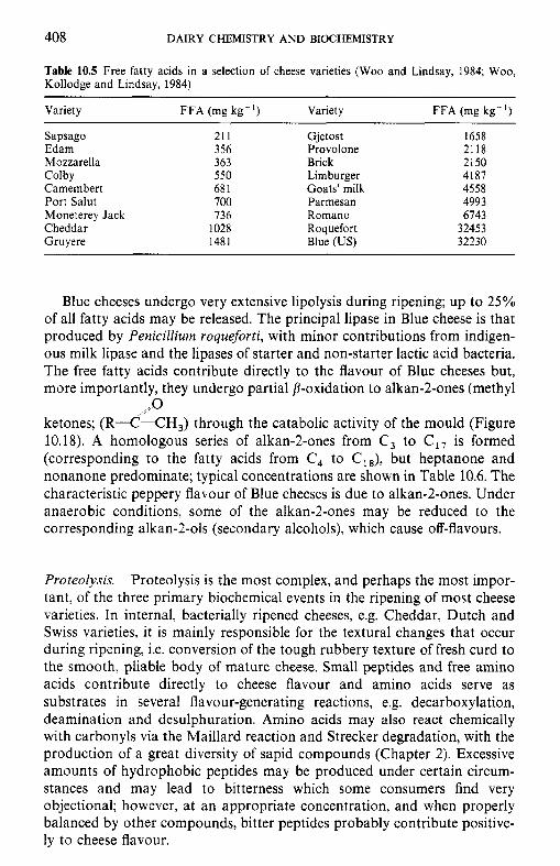
408 DAIRY CHEMISTRY AND BIOCHEMISTRY
Table 10.5 Free fatty acids in a selection of cheese varieties (Woo and Lindsay, 1984; Woo, Kollodge and Lindsay, 1984)
Variety FFA (mg kg-') Variety FFA (mg kg-')
Sapsago Edam Mozzarella Colby Camembert Port Salut Moneterey Jack Cheddar Gruyere
21 1 356 363 550 68 1 700 736
1028 1481
Gjetost Provolone Brick Limburger Goats' milk Parmesan Romano Roquefort Blue (US)
1658 2118 2150 4187 4558 4993 6743
32453 32230
Blue cheeses undergo very extensive lipolysis during ripening; up to 25% of all fatty acids may be released. The principal lipase in Blue cheese is that produced by Penicillium roqueforti, with minor contributions from indigen- ous milk lipase and the lipases of starter and non-starter lactic acid bacteria. The free fatty acids contribute directly to the flavour of Blue cheeses but, more importantly, they undergo partial fl-oxidation to alkan-2-ones (methyl
ketones; (RX-CH,) through the catabolic activity of the mould (Figure 10.18). A homologous series of alkan-2-ones from C, to C,, is formed (corresponding to the fatty acids from C, to CI8), but heptanone and nonanone predominate; typical concentrations are shown in Table 10.6. The characteristic peppery flavour of Blue cheeses is due to alkan-2-ones. Under anaerobic conditions, some of the alkan-2-ones may be reduced to the corresponding alkan-2-01s (secondary alcohols), which cause off-flavours.
, ,o
Proteolysis. Proteolysis is the most complex, and perhaps the most impor- tant, of the three primary biochemical events in the ripening of most cheese varieties. In internal, bacterially ripened cheeses, e.g. Cheddar, Dutch and Swiss varieties, it is mainly responsible for the textural changes that occur during ripening, i.e. conversion of the tough rubbery texture of fresh curd to the smooth, pliable body of mature cheese. Small peptides and free amino acids contribute directly to cheese flavour and amino acids serve as substrates in several flavour-generating reactions, e.g. decarboxylation, deamination and desulphuration. Amino acids may also react chemically with carbonyls via the Maillard reaction and Strecker degradation, with the production of a great diversity of sapid compounds (Chapter 2). Excessive amounts of hydrophobic peptides may be produced under certain circum- stances and may lead to bitterness which some consumers find very objectional; however, at an appropriate concentration, and when properly balanced by other compounds, bitter peptides probably contribute positive- ly to cheese flavour.
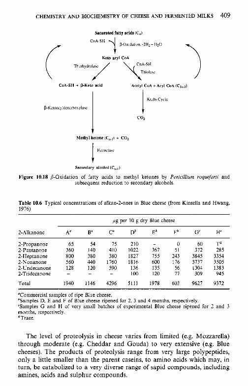
CHEMISTRY AND BIOCHEMISTRY OF CHEESE AND FERMENTED MILKS 409
Saturated fatty acids (CJ
i P-Oxitliltitin. -2H2+ H20
CoA.SH
CoA-SH
Keio acyl CoA
Thiohydrdilsu
CoA-SH + P-Keto acid Acalyl CoA + Acyl CoA (C2,J
Methyl ketone (C,,.]) + CO,
Rcduciasc ! Secondary alcolinl (C,,.])
Figure 10.18 P-Oxidation of fatty acids to methyl ketones by Penicilliwm roqueforti and subsequent reduction to secondary alcohols.
Table 10.6 Typical concentrations of alkan-2-ones in Blue cheese (from Kinsella and Hwang, 1976)
pg per 10 g dry Blue cheese
2-A1 kanone A" Ba C" Db Eb Fb G' H'
2-Propanone 65 54 75 210 - 0 60 Td 2-Pentanone 360 140 410 1022 367 51 372 285 2-Heptanone 800 380 380 1827 755 243 3845 3354 2-Nonanone 560 440 1760 1816 600 176 3737 3505 2-Undecanone 128 120 590 136 135 56 1304 1383 2-Tridecanone - - - 100 120 77 309 945
Total 1940 1146 4296 5111 1978 603 9627 9372
"Commercial samples of ripe Blue cheese. bSamples D, E and F of Blue cheese ripened for 2, 3 and 4 months, respectively. 'Samples G and H of very small batches of experimental Blue cheese ripened for 2 and 3 months, respectively. *Trace.
The level of proteolysis in cheese varies from limited (e.g. Mozzarella) through moderate (e.g. Cheddar and Gouda) to very extensive (e.g. Blue cheeses). The products of proteolysis range from very large polypeptides, only a little smaller than the parent caseins, to amino acids which may, in turn, be catabolized to a very diverse range of sapid compounds, including amines, acids and sulphur compounds.
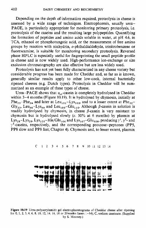
410 DAIRY CHEMISTRY AND BIOCHEMISTRY
Depending on the depth of information required, proteolysis in cheese is assessed by a wide range of techniques. Electrophoresis, usually urea- PAGE, is particularly appropriate for monitoring primary proteolysis, i.e. proteolysis of the caseins and the resulting large polypeptides. Quantifying the formation of peptides and amino acids soluble in water, at pH 4.6, in TCA, ethanol or phosphotungstic acid, or the measurement of free amino groups by reaction with ninhydrin, o-phthaldialdehyde, trinitrobenzene or fluorescarnine, is suitable for monitoring secondary proteolysis. Reversed phase HPLC is especially useful for fingerprinting the small peptide profile in cheese and is now widely used. High-performance ion-exchange or size exclusion chromatography are also effective but are less widely used.
Proteolysis has not yet been fully characterized in any cheese variety but considerable progress has been made for Cheddar and, as far as is known, generally similar results apply to other low-cook, internal bacterially ripened cheeses (e.g. Dutch types). Proteolysis in Cheddar will be sum- marized as an example of these types of cheese.
Urea-PAGE shows that a,,-casein is completely hydrolysed in Cheddar within 3-4 months (Figure 10.19). It is hydrolysed by chymosin, initially at Phe,,-Phe,, and later at Leu,,,-Lys,,,, and to a lesser extent at Phe,,- Gly,,, Leu,,-Lys,, and Leu,,,-Glu, Although p-casein in solution is readily hydrolysed by chymosin, in cheese /3-casein is very resistant to chymosin but is hydrolysed slowly (c. 50% at 6 months) by plasmin at Lys,,-Lys,,, Lys,,,-His/Gln,,, and Lys,,,-Glu,,,, producing yl, y z - and y3-caseins, respectively, and the corresponding proteose-peptones (PP5, PP8 slow and PP8 fast; Chapter 4). Chymosin and, to lesser extent, plasmin
C I 2 3 4 5 6 7 8 9 1 0 1 1 1 2 1 3 1 4
Figure 10.19 Urea-polyacrylamide gel electrophoretograms of Cheddar cheese after ripening for 0, 1, 2, 3, 4, 6, 8, 10, 12, 14, 16, 18 or 20weeks (lanes 1-14); C, sodium caseinate. (Supplied
by S. Mooney.)
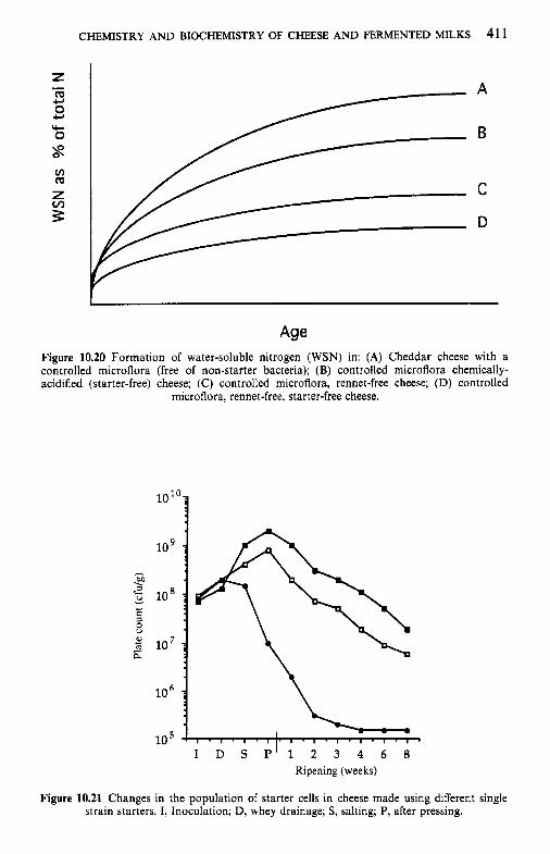
CHEMISTRY AND BIOCHEMISTRY OF CHEESE AND FERMENTED MILKS 41 1
Figure 10.20 Formation of water-soluble nitrogen (WSN) in: (A) Cheddar cheese with a controlled microflora (free of non-starter bacteria); (B) controlled microflora chemically- acidified (starter-free) cheese; (C) controlled microflora, rennet-free cheese; (D) controlled
microflora, rennet-free, starter-free cheese.
h . 00
2 v
10
10 *
10
10
10 I D S P I 1 2 3 4 6 8
Ripening (weeks)
Figure 10.21 Changes in the population of starter cells in cheese made using different single strain starters. I, Inoculation; D, whey drainage; S, salting; P, after pressing.
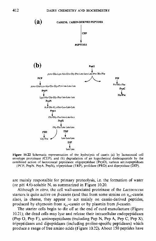
412 DAIRY CHEMISTRY AND BIOCHEMISTRY
CASEINS, CASEIN-DERIVED PEPTIDES
I
PEPTIDES
Pepo
J. pyro-GI u-Lys-Ala-Glx-Gly-Pro-Leu-Leu-Leu-Pro-His-Phe
PIP JL 4 . PCP
i Pro-His-Phe pyro-Glu.Lyr-Ala-Glx-Gly-Pro-Leu~Leu~Leu
Lys- pTN Ala-Glx-Gly-Pro-Leu-Leu-Leu
Glx-GI 7" y-Pro-Leu-Leu-Leu
PepN
*lfGix-Oly-PR1-Leu-Leu.Leu
PepX
i Gly-Pro-Leu-Leu-Leu
Gly-Pro pr J :L DIP
4 Leu-Leu
Figure 10.22 Schematic representation of the hydrolysis of casein (a) by lactococcal cell envelope proteinase (CEP), and (b) degradation of an hypothetical dodecapeptide by the combined action of lactococcal peptidases: oligopeptidase (PepO), various aminopeptidases
(PCP, PepN, PepA, PepX), tripeptidase (TRP), prolidase (PRD) and dipeptidase (DIP).
are mainly responsible for primary proteolysis, i.e. the formation of water (or pH 4.6)-soluble N, as summarized in Figure 10.20.
Although in v i m , the cell wall-associated proteinase of the Lactococcus starters is quite active on /?-casein (and that from some strains on a,,-casein also), in cheese, they appear to act mainly on casein-derived peptides, produced by chymosin from a,,-casein or by plasmin from /?-casein.
The starter cells begin to die off at the end of curd manufacture (Figure 10.21); the dead cells may lyse and release their intracellular endopeptidases (Pep 0, Pep F), arninopeptidases (including Pep N, Pep A, Pep C, Pep X), tripeptidases and dipeptidases (including proline-specific peptidases) which produce a range of free amino acids (Figure 10.22). About 150 peptides have
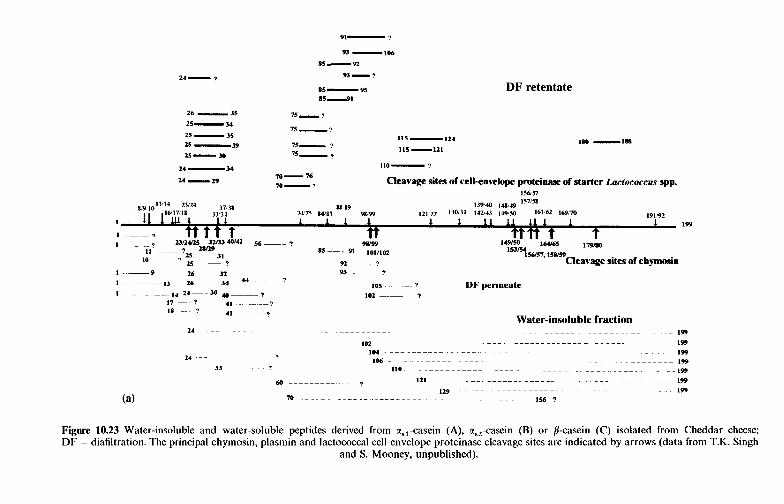
93 -106
85-92
93-?
85 - 95 115'1
26 - 35 75- '!
75-? 25- 34
15 - 35
25 -39 75- ? 15-30 75- ?
115-124
115-121
DF retentate
110- ? 24 -34
24-29 Cleavage sites of cellenvelope pmteinase of starter Lactococcus spp. 70- 76 70-?
1W57
Figure 10.23 Water-insoluble and water-soluble peptides derived from cc,,-casein (A), a,,-casein (B) or /I-casein (C) isolated from Cheddar cheese; DF = diafiltration. The principal chymosin, plasmin and lactococcal cellenvelope proteinase cleavage sites are indicated by arrows (data from T.K. Singh
and S. Mooney, unpublished).
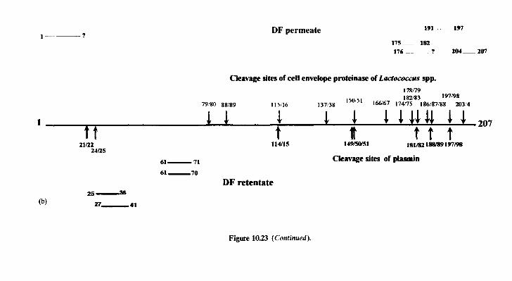
DF permeate 191 ~~ 197
175- 182 176--? 204-207
Cleavage sites of cell envelope proteinase of Ladococas spp. 178/79 182183 197/98
79’80 88!89 115!16 137’38 150’51 166167 17W5 186/87:88 203:4
1 1 1 .1 5. 1 1 l J U 5- 207
t t t t t lslm 188189 197m
t 149/50/51
t 21/22 114/15
2-b25
61- 71
61 -70
Cleavage sites of plasmin
DF retentate
Figure 10.23 (Continued).
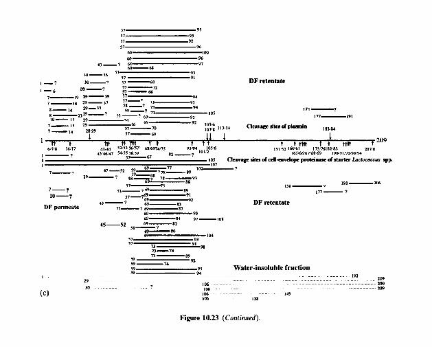
aL-u
16 LS C6 LZ
a6
+Ol
m- - 8S a-69 2s-st
801-L6 V8-6V C6 69
m- ‘-1s
i--01 4-L
- WI 09 % LS
Z6 LS
416 DAIRY CHEMISTRY AND BIOCHEMISTRY
1 3000
AM2 GI IIC25
2000 C J .- a .3
-z 2
1000
. . . A q Thr Ser Glu Pro Gly Ala Cys Val Met Ile Leu T y r Phe His Lys Arg
Amino acid
Figure 10.24 Concentration of individual amino acids in 60-day-old Cheddar cheese, made with a single-strain starter Lactococcus lactis ssp. cremoris AM,, Gll/C25 or HP (from
Wilkinson, 1992).
been isolated from the water-soluble fraction of Cheddar, and characterized (Figure 10.23). These show that both lactococcal proteinase and exopep- tidase contribute to proteolysis in cheese. The proteinases and peptidases of the NSLAB (mainly mesophilic lactobacilli) appear to contribute little to proteolysis in Cheddar, except in the production of amino acids.
The principal amino acids in Cheddar are shown in Figure 10.24.
10.2.8 Cheese Jla vow
Although interest in cheese flavour dates from the beginning of this century, very little progress was made until the development of gas liquid chromato- graphy (GC) in the late 1950s, and especially the coupling of GC and mass spectrometry (MS). More than 200 volatile compounds have been identified in cheese by GC-MS (principal compounds are listed in Table 10.7). The volatile fraction of cheese may be obtained by taking a sample of headspace but the concentration of many compounds is too low, even for modern GC-MS techniques. The volatiles may be concentrated by solvent extrac- tion or distillation. In the former, a large solvent peak may mask important constituents while the latter may generate artefacts, even at moderately low temperatures. Trapping of volatiles, e.g. on adsorbants or in cold traps, is probably the most satisfactory method for concentration.
The taste of cheese is concentrated in the water-soluble fraction (peptides, amino acids, organic acids, amines, NaCl) while the aroma is mainly in the volatile fraction. Initially, it was believed that cheese flavour was due to one
CHEMISTRY AND BIOCHEMISTRY OF CHEESE AND FERMENTED MILKS 4 17
Table 10.7 Volatile compounds which have been identified in Cheedar cheese (modified from Urbach, 1993)
Acetaldehyde Acetoin Acetone Acetophenone 1.2-Butanediol n-Butanol 2-Butanol Butanone n-Butyl acetate 2-Butyl acetate n-Butyl butyrate n-Butyric acid Carbon dioxide p-Cresol y-Decalactone 6-Decalactone n-Decanoic acid Diacetyl Diethyl ether Dimethyl sulfide
Dimethyl disulfide Dimethyl trisulfide &Dodecalactone Ethanol Ethyl butanol 2-Ethyl butanol Ethyl butyrate Ethyl hexanoate 2-Heptanone n-Hexanal n-Hexanoic acid n-Hexanol 2-Hexanone Hexanethiol 2-Hexenal Isobutanol Isohexanal Methanethiol Methional Methyl acetate
2-Methylbutanol 3-Methylbutanol 3-Methyl-2-butanone 3-Methylbutyric acid 2-Nonanone 6-Octalactone n-Octanoic acid 2-Octanol 2,4-Pentanediol n-Pentanoic acid 2-Pentanol Pentan-2-one n-Propanol Propanal Propenal n-Propyl butyrate Tetrahydrofuran Thiophen-2-aldehyde 2-Tridecanone 2-Undecanone
418 DAIRY CHEMISTRY AND BIOCHEMISTRY
or a small number of compounds, but it was soon realized that all cheeses contained essentially the same sapid compounds. Recognition of this led to the component balance theory, i.e. cheese flavour is due to the concentration and balance of a range of compounds. Although considerable information on the flavour compounds in several cheese varieties has been accumulated, it is not possible to fully describe the flavour of any variety, with the possible exception of Blue cheeses, the flavour of which is dominated by alkan-2- ones.
Many cheeses contain the same or similar compounds but at different concentrations and proportions; chromatograms of some cheese varieties are shown in Figure 10.25. The principal classes of components present are aldehydes, ketones, acids, amines, lactones, esters, hydrocarbons and sul- phur compounds; the latter, e.g. H,S, methanethiol (CH,SH), dimethyl sulphide (H,C-S-CH,) and dimethyl disulphide (H,C-S-S-CH,), are con- sidered to be particularly important in Cheddar cheese. The biogenesis of flavour compounds has been reviewed by Fox et al. (1993, 1996a) and FOX, Singh and McSweeney (1995).
10.2.9 Accelerated ripening of cheese
Since the ripening of cheese, especially low moisture varieties, is a slow process, it is expensive in terms of controlled atmosphere storage and stocks. Ripening is also unpredictable. Hence, there are economic and technological incentives to accelerate ripening, while retaining or improving characteristic flavour and texture.
The principal approaches used to accelerate cheese ripening are:
I. Elevated ripening temperatures, especially for Cheddar which is now usually ripened at 6-8°C; most other varieties are ripened at a higher temperature, e.g. around 14°C for Dutch types or 20-22°C for Swiss types and Parmesan, and hence there is little or no scope for increasing the ripening temperature.
2. Exogenous enzymes, usually proteinases and/or peptidases. For several reasons, this approach has had limited success, except for enzyme- modified cheeses (EMC). These are usually high-moisture products which are used as ingredients for processed cheese, cheese spreads, cheese dips or cheese flavourings.
3. Attenuated lactic acid bacteria, e.g. freeze-shocked, heat-shocked or lactose-negative mutants.
4. Adjunct starters, especially mesophilic lactobacilli. 5. Use of fast-lysing starters which die and release their intracellular
6. Genetically modified starters which super-produce certain enzymes; un- enzymes rapidly.
fortunately, the key enzymes are not yet known.
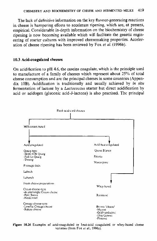
CHEMISTRY AND BIOCHEMISTRY OF CHEESE AND FERMENTED MILKS 419
The lack of definitive information on the key flavour-generating reactions in cheese is hampering efforts to accelerate ripening, which are, at present, empirical. Considerable in-depth information on the biochemistry of cheese ripening is now becoming available which will facilitate the genetic engin- eering of starter cultures with improved cheesemaking properties. Acceler- ation of cheese ripening has been reviewed by Fox et al. (1996b).
10.3 Acid-coagulated cheeses
On acidification to pH 4.6, the caseins coagulate, which is the principle used to manufacture of a family of cheeses which represent about 25% of total cheese consumption and are the principal cheeses in some countries (Appen- dix 10B). Acidification is traditionally and usually achieved by in situ fermentation of lactose by a Lactococcus starter but direct acidification by acid or acidogen (gluconic acid-b-lactone) is also practised. The principal
Quarg-type -Skim milk Quarg -Full l i t Quarg
Fromage t'tais
Ldhneh
Lahanch
Fresh cliccse preparaiiiins
Crcam cheese-type -douhle/singlc Crcnm cliccsc -Petit Suisse -Neufchatcl
Cottage cheese-type
-Bakers cliccsc
- T V W O ~
-Low/lht Cotiagc chccse
Qucso Blanco
Ricotta
Mascarponc
Ricottonc
Brown 'cheese' -Mysost -Gudhrandsalosi -Ek\r: Gcisoat -Floieo~t
Figure 10.26 Examples of acid-coagulated or heat-acid coagulated or whey-based cheese varieties (from Fox e t d., 1996a).
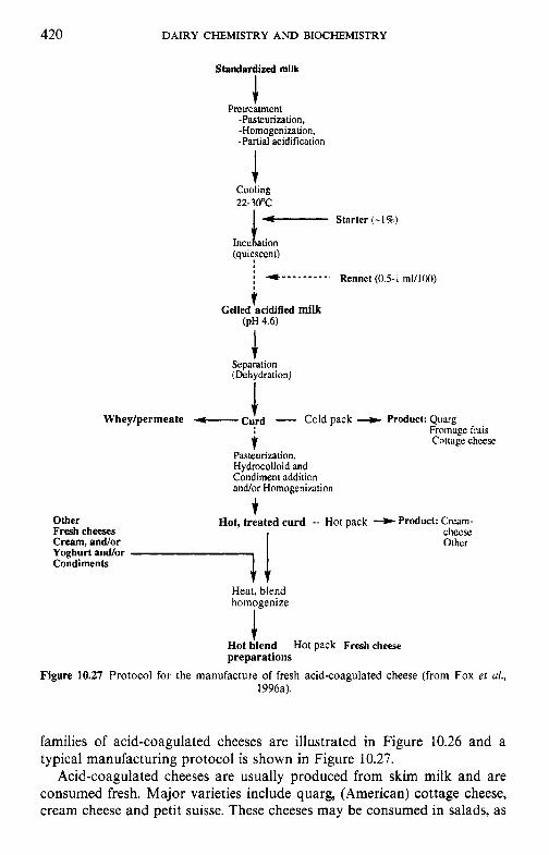
420 DAIRY CHEMISTRY AND BIOCHEMISTRY
Standardized milk
I
Pretreatment -Pasteurization, -Homogenization. -Partial acidification
J Cooling 22-3OoC - Starter (- 15%)
Incu 1 ation (quiescent)
t Gelled acidified milk
(PH 4.6)
Separation (Dehydration)
1 Whey/permeate - C(rd - Cold pack ---t Product: Quarg
Fromuge frnis i Cottage chcese. Pasteurization. Hydrocolloid and Condiment addition and/or Homogenization
t Other Hot, treated curd -- Hot pack + Prodoct: Crcam- Fresh cheeses chccsc: Cream, andor Other Yoghurt andfor Condiments 7 1
Heat, blend homogenize
Hot dlend preparations
Figure 10.27 Protocol for the manufacture of fresh acid-coagulated cheese (from Fox et a/., 1996a).
Hot pack Fresh cheese
families of acid-coagulated cheeses are illustrated in Figure 10.26 and a typical manufacturing protocol is shown in Figure 10.27.
Acid-coagulated cheeses are usually produced from skim milk and are consumed fresh. Major varieties include quarg, (American) cottage cheese, cream cheese and petit suisse. These cheeses may be consumed in salads, as
CHEMISTRY AND BIOCHEMISTRY OF CHEESE AND FERMENTED MILKS 421
food ingredients and serve as the base for a rapidly expanding group of dairy products, i.e. fromage frais-type products.
The casein may also be coagulated at a pH above 4.6, e.g. about 5.2, by using a higher temperature, e.g. 80-90°C. This principle is used to manufac- ture another family of cheeses, which include Ricotta (and variants thereof), Anari, and some types of Queso Blanco. These cheeses may be made exclusively from whey but usually from a blend of milk and whey and are usually used as a food ingredient, e.g. in lasagne or ravioli.
10.4 Processed cheese products
Processed cheese is produced by blending shredded natural cheese of the same or different varieties and at different degrees of maturity with emulsify- ing agents and heating the blend under vacuum with constant agitation until a homogeneous mass is obtained. Other dairy and non-dairy ingredients may be included in the blend. The possibility of producing processed cheese was first assessed in 1895; emulsifying salts were not used and the product was not successful. The first sucessful product, in which emulsifying salts were used, was introduced in Europe in 1912 and in the USA in 1917 by Kraft. Since then, the market for processed cheese has increased and the range of products expanded.
Although established consumers may regard processed cheeses as inferior products compared to natural cheeses, they have numerous advantages compared to the latter:
1. A certain amount of cheese which would otherwise be difficult or impossible to commercialize may be used, e.g. cheese with deformations, cheese trimmings or cheese after removal of localized mould.
2. A blend of cheese varieties and non-cheese components may be used, making it possible to produce processed cheeses differing in consistency, flavour, shape and size.
3. They have good storage stability at moderate temperatures, thus reduc- ing the cost of storage and transport.
4. They are more stable than natural cheeses during storage, which results in less wastage, a feature that may be especially important in remote areas and in households with a low level of cheese consumption.
5. They are amenable to imaginative packing in various conveniently sized units.
6. They are suitable for sandwiches and fast food outlets. 7. They are attractive to children who generally do not like or appreciate
the stronger flavour of natural cheeses.
Today, a wide range of processed cheese products is available, varying in composition and flavour (Table 10.8).
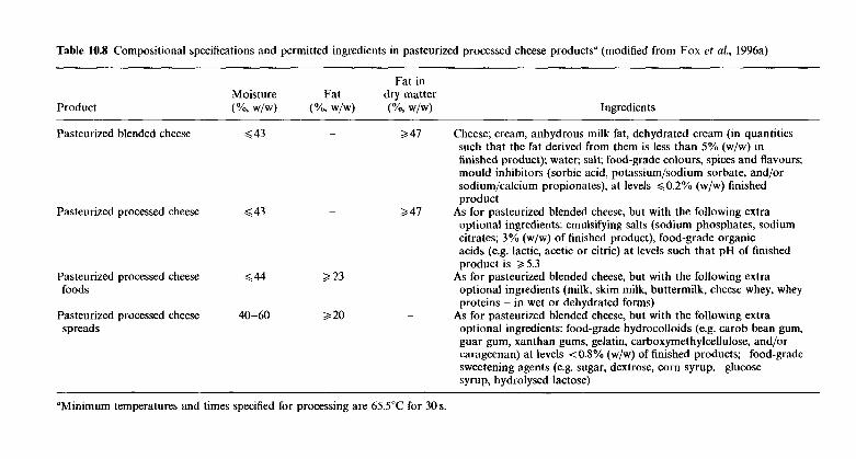
Table 10.8 Compositional specifications and permitted ingredients in pasteurized processed cheese products" (modified from Fox ef al., 1996a)
Fat in Moisture Fat dry matter
Product (Yo, w/w) (%, w/w) (Yo, w/w) Ingredients
Pasteurized processed cheese Q 43
Pasteurized processed cheese foods
Pasteurized processed cheese 40-60 spreads
3 23
3 20
Pasteurized blended cheese Q 43 - 2 4 7 Cheese; cream, anhydrous milk fat, dehydrated cream (in quantities such that the fat derived from them is less than 5% (w/w) in finished product); water; salt; food-grade colours, spices and flavours; mould inhibitors (sorbic acid, potassium/sodium sorbate, and/or sodium/calcium propionates), at levels g0.2% (w/w) finished product
As for pasteurized blended cheese, but with the following extra optional ingredients: emulsifying salts (sodium phosphates, sodium citrates; 3% (w/w) of finished product), food-grade organic acids (e.g. lactic, acetic or citric) at levels such that pH of finished product is 3 5.3
optional ingredients (milk, skim milk, buttermilk, cheese whey, whey proteins - in wet or dehydrated forms)
optional ingredients: food-grade hydrocolloids (e.g. carob bean gum, guar gum, xanthan gums, gelatin, carboxymethylcellulose, and/or carageenan) at levels 4 0.8% (w/w) of finished products; food-grade sweetening agents (e.g. sugar, dextrose, corn syrup, glucose syrup, hydrolysed lactose)
2 4 7
~ As for pasteurized blended cheese, but with the following extra
As for pasteurized blended cheese, but with the following extra ~
"Minimum temperatures and times specified for processing are 65.5"C for 30 s.
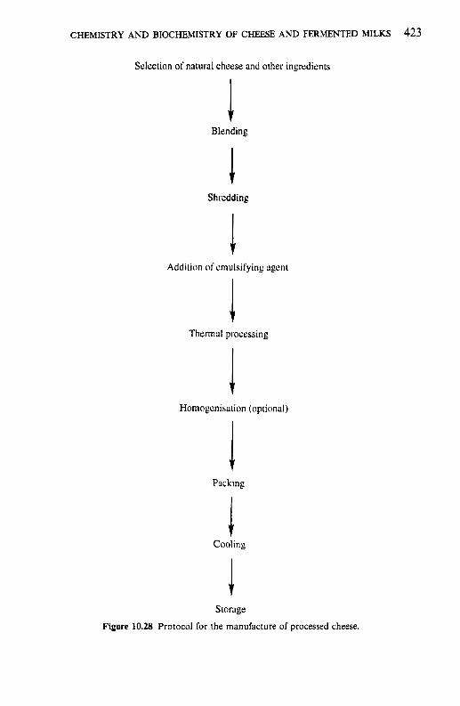
CHEMISTRY AND BIOCHEMISTRY OF CHEESE AND FERMENTED MILKS 423
Selection of natural cheese and other ingicdients
Blending
Shredding
Addition of cmulsifying agcnt
Thermal processing
Homogcnisadon (optional)
PJcking
Cooling
storage
Figure 10.28 Protocol for the manufacture of processed cheese.
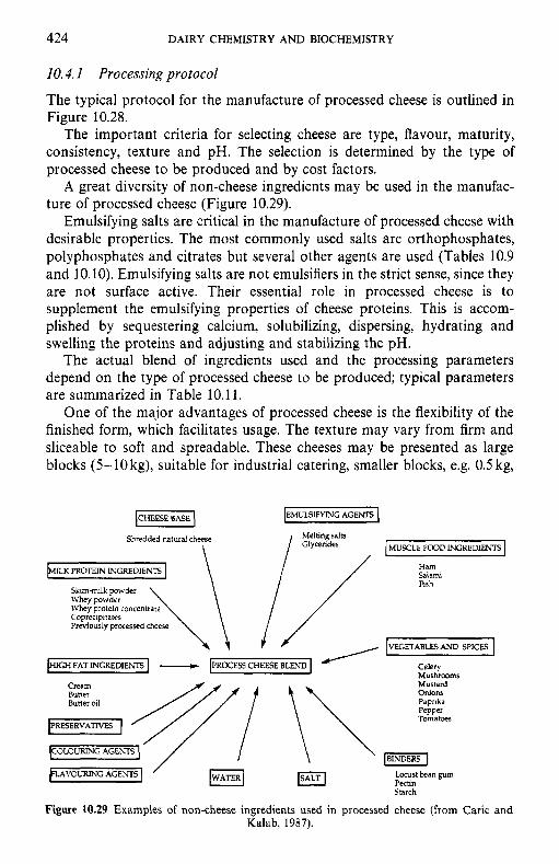
424 DAIRY CHEMISTRY A N D BIOCHEMISTRY
10.4. 1 Processing protocol
The typical protocol for the manufacture of processed cheese is outlined in Figure 10.28.
The important criteria for selecting cheese are type, flavour, maturity, consistency, texture and pH. The selection is determined by the type of processed cheese to be produced and by cost factors.
A great diversity of non-cheese ingredients may be used in the manufac- ture of processed cheese (Figure 10.29).
Emulsifying salts are critical in the manufacture of processed cheese with desirable properties. The most commonly used salts are orthophosphates, polyphosphates and citrates but several other agents are used (Tables 10.9 and 10.10). Emulsifying salts are not emulsifiers in the strict sense, since they are not surface active. Their essential role in processed cheese is to supplement the emulsifying properties of cheese proteins. This is accom- plished by sequestering calcium, solubilizing, dispersing, hydrating and swelling the proteins and adjusting and stabilizing the pH.
The actual blend of ingredients used and the processing parameters depend on the type of processed cheese to be produced; typical parameters are summarized in Table 10.11.
One of the major advantages of processed cheese is the flexibility of the finished form, which facilitates usage. The texture may vary from firm and sliceable to soft and spreadable. These cheeses may be presented as large blocks (5-10kg), suitable for industrial catering, smaller blocks, e.g. 0.5 kg,
Melting salts / Glycerides Shredded natural cheese \
Skim-milk powder Whey powder Whey protein concentrate Coprecipttates Previously processed cheese
HIGH FAT INGREDIENTS - PROCESS CHEESE BLEND J /
MUSCLE FOOD INGREDIENTS
Ham Salami Fish
VEGETABLES AND SPICES I Celery Mushrooms Mustard
Tomatoes
COLOURING AGENTS
Locust bean gum Pectin Starch
VOURING AGENTS
Figure 10.29 Examples of non-cheese ingredients used in processed cheese (from Caric and Kalab, 1987).
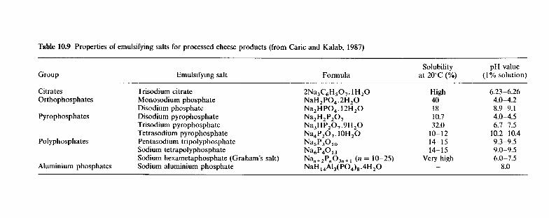
Table 10.9 Properties of emulsifying salts for processed cheese products (from Caric and Kalab, 1987)
Solubility pH value ( I % solution) Group Emulsifying salt Formula at 20°C (%)
Citrates Trisodium citrate 2Na3C,H,0,. 1H,O High 6.23-6.26 Orthophosphates Monosodium phosphate NaH2P0,.2H,0 40 4.0-4.2
Pyrophosphates Disodium pyrophosphate Na2H2P20, 10.7 4.0-4.5 Disodium phosphate Na,HPO,. 12H,O 18 8.9-9.1
Trisodium pyrophosphate Na,HP,O, .YH,O 32.0 6.7-7.5 Tetrasodium pyrophosphate Na,P,O,. 10H20 10-12 10.2-10.4
Sodium tetrapolyphosphate Na,P'tO,, 14- 15 9.0-9.5 Polyphosphates Pentasodium tripolyphosphate Na5P3010 14-15 9.3-9.5
Sodium hexametaphosphate (Graham's salt) Nan+,PnOJn+, (n = 10-25) Very high 6.0-7.5 - Aluminium phosphates Sodium aluminium phosphate NaH ,,AI,(P04),.4H,0 8.0
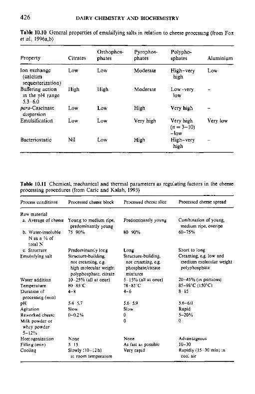
426 DAIRY CHEMISTRY AND BIOCHEMISTRY
Table 10.10 General properties of emulsifying salts in relation to cheese processing (from Fox et al., 1996a,b)
Orthophos- Pyrophos- Polypho- Property Citrates phates phates sphates Aluminium
Ion exchange (calcium sequesterization)
in the pH range Buffering action
5.3-6.0 para-Caseinate
Emulsification dispersion
Bacteriostatic
Low Low Moderate High-very Low high
High High Moderate Low-very - low
Low Low High Very high -
Low Low Very high Very high Very low (n = 3-10) -low
high Nil Low High High-very -
Table 10.11 Chemical, mechanical and thermal parameters as regulating factors in the cheese processing procedures (from Caric and Kalab, 1993)
Process conditions Processed cheese block Processed cheese slice Processed cheese spread
Raw material a. Average of cheese
b. Water-insoluble N as a % of total N
c. Structure Emulsifying salt
Water addition Temperature Duration of processing (min)
PH Agitation Reworked cheese Milk powder or whey powder
Homogenization Filling (min) Cooling
5-12%
Young to medium ripe, predominantly young
75-90%
Predominantly long Structure-building,
not creaming, e.g. high molecular weight polyphosphate, citrate
10-25% (all at once) 80-85’C 4-8
5.4-5.7 Slow 0-0.2%
None 5-15 Slowly (10-12 h) at room temperature
Predominantly young Combination of young, medium ripe, overipe
80-90% 60-75%
Long Short to long Structure-building, Creaming, e.g. low and not creaming, e.g. phosphate/citrate polyphosphate mixtures
medium molecular weight
5- 15% (all at once)
4-6 8-15
5.6-5.9 5.6-6.0 Slow Rapid 0 5-20% 0 0
20-45% (in portions) 78-85°C 85-98°C (150’C)
None Advantageous As fast as possible 10-30 Very rapid Rapidly (15-30 min) in
cool air
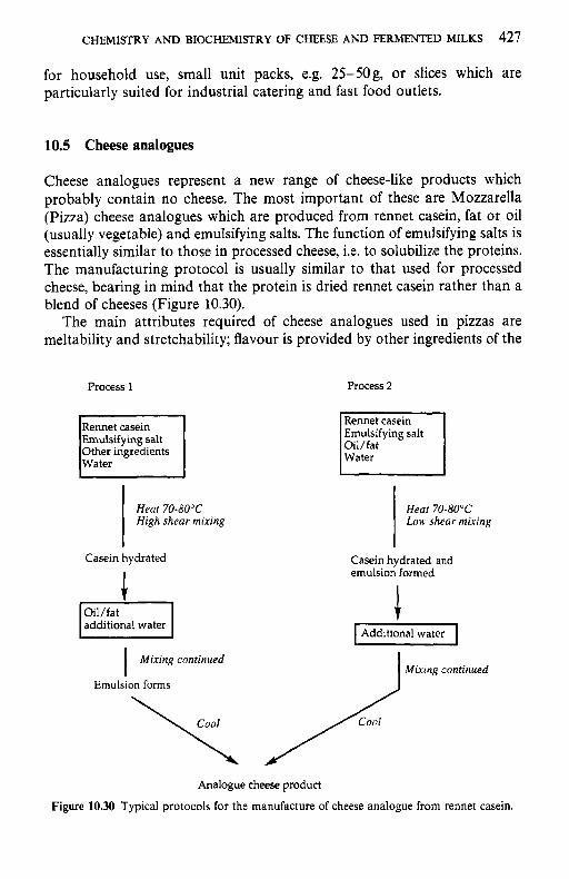
CHEMISTRY AND BIOCHEMISTRY OF CHEESE AND FERMENTED MILKS 427
for household use, small unit packs, e.g. 25-50g, or slices which are particularly suited for industrial catering and fast food outlets.
10.5 Cheese analogues
Cheese analogues represent a new range of cheese-like products which probably contain no cheese. The most important of these are Mozzarella (Pizza) cheese analogues which are produced from rennet casein, fat or oil (usually vegetable) and emulsifying salts. The function of emulsifying salts is essentially similar to those in processed cheese, i.e. to solubilize the proteins. The manufacturing protocol is usually similar to that used for processed cheese, bearing in mind that the protein is dried rennet casein rather than a blend of cheeses (Figure 10.30).
The main attributes required of cheese analogues used in pizzas are meltability and stretchability; flavour is provided by other ingredients of the
Process 1
Emulsifying salt Other ingredients Water
Process 2
Rennet casein 1 I Emulsifying salt
Oil/fat
I Water
Heat 70-80°C High shear mixing
Heat 70-80°C Lnw shear mixing
Casein hydrated
additional water
Mixing continued I Emulsion forms
Casein hydrated and emulsion formed
c
Analogue cheese product
Figure 10.30 Typical protocols for the manufacture of cheese analogue from rennet casein.
428 DAIRY CHEMISTRY AND BIOCHEMISTRY
pizza, e.g. tomato paste, sausage, peppers, spices, anchovies, etc. It may be possible to produce analogues of other cheeses by adding biochemically or chemically generated cheese flavours. Apart from the use of some casein (rennet or acid) in processed cheese blends, cheese analogues, other than Mozzarella, are not widely used at present. As discussed in section 10.2.8, the flavour and texture of natural cheeses are very complex and cannot be simulated readily. The usual approach is to accelerate the ripening of natural cheese (section 10.2.9), although this approach has enjoyed limited success to date.
10.6 Cultured milks
Acidified (cultured) milk products may very well be the oldest dairy products. If removed aseptically from a healthy udder, milk is essentially sterile but, in practice, milk becomes contaminated by various bacteria, including lactic acid bacteria (LAB) during milking. During storage, these contaminants grow at rates dependent on the temperature. LAB probably dominate the microflora of uncooled milk expressed by hand. Since LAB are well suited for growth in milk, they grow rapidly at ambient tempera- ture, metabolizing lactose to lactic acid and reducing the pH of the milk to the isoelectric point of caseins (about pH 4.6), at which they form a gel under quiescent conditions, thus producing cultured milks. Such products have existed since the domestication of dairy animals and some form of cultured milk is produced throughout the world; the principal products are
Table 10.12 Some typical examples of starter cultures employed in the manufacture of fermented milks (from Robinson and Tamime, 1993)
Type of culture Product Micro-organisms involved
Mesophilic Taetrnojolk Lactococcus lactis subsp. lactis Folkjolk
Ymer Lc. h i s subsp. cremoris
Kefir
Lactococcus lactis subsp. lactis biovar. diacetylactis Leuconostoc mesenteroides subsp. cremoris
Lc. lactis subsp. lactis biovar. diacetylactis Kefir grains - thermophilic lactobacilli and Kluyoeromyces marxianus
Typical fermentation temperature 20-22'C
Thermophilic Yoghurt Streptococcus saloarius subsp. thermophilus Lactobacillus delbrueckii subsp. bulgaricus
Yakult Lactobacillus casei subsp. casei Acidophilus milk Lactobacillus acidophilus A/B milk Lb. acidophilus
A/B yoghurt Bifdobacterium bifidum As above plus yoghurt culture
Typical fermentation temperatures 37-42°C
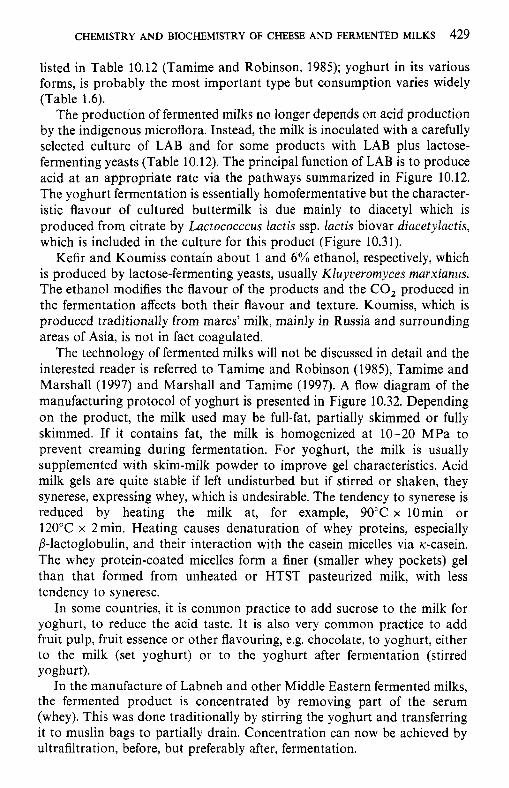
CHEMISTRY AND BIOCHEMISTRY OF CHEESE AND FERMENTED MILKS 429
listed in Table 10.12 (Tamime and Robinson, 1985); yoghurt in its various forms, is probably the most important type but consumption varies widely (Table 1.6).
The production of fermented milks no longer depends on acid production by the indigenous microflora. Instead, the milk is inoculated with a carefully selected culture of LAB and for some products with LAB plus lactose- fermenting yeasts (Table 10.12). The principal function of LAB is to produce acid at an appropriate rate via the pathways summarized in Figure 10.12. The yoghurt fermentation is essentially homofermentative but the character- istic flavour of cultured buttermilk is due mainly to diacetyl which is produced from citrate by Lactococccus lactis ssp. lactis biovar diacetylactis, which is included in the culture for this product (Figure 10.31).
Kefir and Koumiss contain about 1 and 6% ethanol, respectively, which is produced by lactose-fermenting yeasts, usually Kluyveromyces marxianus. The ethanol modifies the flavour of the products and the CO, produced in the fermentation affects both their flavour and texture. Koumiss, which is produced traditionally from mares’ milk, mainly in Russia and surrounding areas of Asia, is not in fact coagulated.
The technology of fermented milks will not be discussed in detail and the interested reader is referred to Tamime and Robinson (1985), Tamime and Marshall (1997) and Marshall and Tamime (1997). A flow diagram of the manufacturing protocol of yoghurt is presented in Figure 10.32. Depending on the product, the milk used may be full-fat, partially skimmed or fully skimmed. If it contains fat, the milk is homogenized at 10-20 MPa to prevent creaming during fermentation. For yoghurt, the milk is usually supplemented with skim-milk powder to improve gel characteristics. Acid milk gels are quite stable if left undisturbed but if stirred or shaken, they synerese, expressing whey, which is undesirable. The tendency to synerese is reduced by heating the milk at, for example, 90°C x 10min or 120°C x 2min. Heating causes denaturation of whey proteins, especially P-lactoglobulin, and their interaction with the casein micelles via K--casein. The whey protein-coated micelles form a finer (smaller whey pockets) gel than that formed from unheated or HTST pasteurized milk, with less tendency to synerese.
In some countries, it is common practice to add sucrose to the milk for yoghurt, to reduce the acid taste. It is also very common practice to add fruit pulp, fruit essence or other flavouring, e.g. chocolate, to yoghurt, either to the milk (set yoghurt) or to the yoghurt after fermentation (stirred yoghurt).
In the manufacture of Labneh and other Middle Eastern fermented milks, the fermented product is concentrated by removing part of the serum (whey). This was done traditionally by stirring the yoghurt and transferring it to muslin bags to partially drain. Concentration can now be achieved by ultrafiltration, before, but preferably after, fermentation.
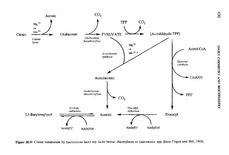
430 D
AIR
Y C
HEM
ISTRY
AN
D BIO
CH
EMISTR
Y
3: G
z 5 +-
i: I i
m
m
m x
9
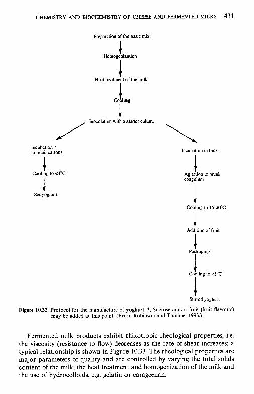
CHEMISTRY AND BIOCHEMISTRY OF CHEESE AND FERMENTED MILKS 431
Preparation of the basic mix
Homogenization I
Cooling .1
Incubation * in retail cartons
Cooling to <4'C
I Set yoghurt
lncuhation in hulk
Agitition to hlrak coagulum
Adjition ol'huit
1 Packaging
I J Cooling to < S T
Stirivd yoghuri
Figure 10.32 Protocol for the manufacture of yoghurt. *, Sucrose and/or fruit (fruit flavours) may be added at this point. (From Robinson and Tamime, 1993.)
Fermented milk products exhibit thixotropic rheological properties, i.e. the viscosity (resistance to flow) decreases as the rate of shear increases; a typical relationship is shown in Figure 10.33. The rheological properties are major parameters of quality and are controlled by varying the total solids content of the milk, the heat treatment and homogenization of the milk and the use of hydrocolloids, e.g. gelatin or carageenan.
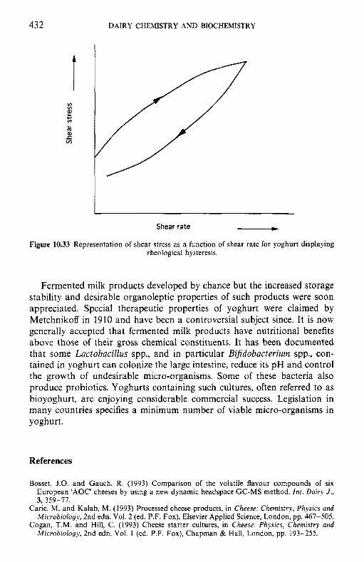
432 DAIRY CHEMISTRY AND BIOCHEMISTRY
Shear rate - Figure 10.33 Representation of shear stress as a function of shear rate for yoghurt displaying
rheological hysteresis.
Fermented milk products developed by chance but the increased storage stability and desirable organoleptic properties of such products were soon appreciated. Special therapeutic properties of yoghurt were claimed by Metchnikoff in 1910 and have been a controversial subject since. It is now generally accepted that fermented milk products have nutritional benefits above those of their gross chemical constituents. It has been documented that some Lactobacillus spp., and in particular Bifidobacterium spp., con- tained in yoghurt can colonize the large intestine, reduce its pH and control the growth of undesirable micro-organisms. Some of these bacteria also produce probiotics. Yoghurts containing such cultures, often referred to as bioyoghurt, are enjoying considerable commercial success. Legislation in many countries specifies a minimum number of viable micro-organisms in yoghurt.
References
Bosset, J.O. and Gauch, R. (1993) Comparison of the volatile flavour compounds of six European 'AOC' cheeses by using a new dynamic headspace GC-MS method. Znt. Dairy J., 3, 359-77.
Caric, M. and Kalab, M. (1993) Processed cheese products, in Cheese: Chemistry, Physics and Microbiology, 2nd edn. Vol. 2 (ed. P.F. Fox), Elsevier Applied Science, London, pp. 467-505.
Cogan, T.M. and Hill, C. (1993) Cheese starter cultures, in Cheese: Physics, Chemistry and Microbiology, 2nd edn, Vol. 1 (ed. P.F. Fox), Chapman & Hall, London, pp. 193-255.
F A 0 (1994) Yearbook-Production, Vol. 48, Food and Agriculture Organization, Rome. Foltmann, B. (1987) General and molecular aspects of rennets, in Cheese: Chemistry, Physics
and Microbiology, Vol. 1 (ed. P.F. Fox), Elsevier Applied Science, London, pp. 33-61. Fox, P.F., Singh, T.K. and McSweeney, P.L.H. (1995) Biogenesis of flavour compounds in
cheese, in Chetnistry of Structure-Function Relationships in Cheese, (eds E.L. Malin and M.H. Tunick), Plenum Press, New York, pp. 59-98.
Fox, P.F., Law, J., McSweeney, P.L.H. and Wallace, J. (1993) Biochemistry of cheese ripening, in Cheese: Chemistry, Physics and Microbiology, Vol. 2: General Aspects, (ed. P.F. Fox), Chapman & Hall, London, pp. 389-483.
Fox, P.F., OConnor, T.P., McSweeney, P.L.H. et a/. (1996a) Cheese: physical, biochemical and nutritional aspects. Adv. Food Nutr. Res., 39, 163-328.
Fox, P.F., Wallace, J.M., Morgan, S. et a/ . (1996b) Acceleration of cheese ripening. Antonie van Leeuu'enhoek, 70, 271-7.
IDF (1992) Bovine Rennets. Determination of Total Milk-clotting Activity, Provisional Standard 157, International Dairy Federation, Brussels.
I D F (1995) Consumption Statisticsfor Milk and Milk Product, Bulletin 301, International Dairy Federation, Brussels.
Kinsella, J.E. and Hwang, D.H. (1976) Enzymes of Penicillium roqueforti involved in the biosynthesis of cheese flavour. C R C Crit. Rev. Food Sci. Nutr., 8, 191-228.
Marshall, V.M.E. and Tamime. A.Y. (1997) Physiology and biochemistry of fermented milks, in Microbiology and Biochemistry of Cheese and Fermented Milk, 2nd edn (ed. B.A. Law), Blackie Academic & Professional, London, pp. 153-92.
Robinson, R.K. and Tamime, A.Y. (1993) Manufacture of yoghurt and other fermented milks, in Modern Dairy Technology, 2nd edn, Vol. 2 (ed. R.K. Robinson), Elsevier Applied Science, London, pp. 1-48.
Tamime, A.Y. and Marshall, V.M.E. (1997) Microbiology and technology of fermented milks, in Microbiology and Biochemistry of Cheese and Fermented Milk, 2nd edn (ed. B.A. Law), Blackie Academic & Professional, London, pp. 57- 152.
Tamime, A.Y. and Robinson, R.K. (1985) Yoghurt Science and Technology, Pergamon Press, Oxford.
Urbach, G. (1993) Relations between cheese flavour and chemical composition. Int. Dairy J . ,
Visser, S., Slangen, C.J. and van Rooijen, P.J. (1987) Peptide substrates for chymosin (rennin). Biochem. J., 244, 553-558.
Visser, S., van Rooijen, P.J., Schattenkerk, C., and Kerling, K.E.T. (1976) Peptide substrates for chymosin (rennin). Kinetic studies with peptides of different chain length including parts of the sequence 101-112 of bovine K-casein. Biochim. Biophys. Acta, 438, 265-72.
Wilkinson, M.G. (1992) Studies on the Acceleration of Cheddar Cheese Ripening, Ph.D. Thesis, National University of Ireland, Cork.
Woo, A.H. and Lindsay, R.C. (1984) Concentrations of major free fatty acids and flavour development in Italian cheese varieties. J . Dairy Sci., 67, 960-8.
Woo, A.H., Kollodge, S. and Lindsay, R.C. (1984) Quantification of major free fatty acids in several cheese varieties. J . Dairy Sci., 67, 874-8.
3, 3899-422.
Suggested reading
Berger, W., Klostermeyer, H., Merkenich, K. and Uhlmann, G. (1989) Die Schmelzkiiseherstel- lung, BenckiserKnapsack GmbH, Ladenburg.
Brown, R.J. and Ernstrom, C.A. (1985) Milk-clotting enzymes and cheese chemistry, Part 1, Milk clotting enzymes, in Fundamentals of Dairy Chemistry, 3rd edn (ed. N.P. Wong), van Nostrand Reinhold, New York, pp. 609-33.
Davies, F.L. and Law, B.A. (eds) (1984) Advances in the Microbiology and Biochemistry of Cheese and Fermented Milk, Elsevier Applied Science Publishers, London.
Eck, A. (ed.) (1984) Le Fromage, Diffusion Lavoisier, Paris. Fox, P.F. (ed.) (1993) Cheese: Chemistry, Physics and Microbiology, 2nd edn, Vols 1 and 2,
Chapman & Hall, London.
434 DAIRY CHEMISTRY AND BIOCHEMISTRY
Frank, J.F. and Marth, E.H. (1988) Fermentations, in Fundamentals of Dairy Chemistry, 3rd edn (ed. N.P. Wong), van Nostrand Reinhold, New York, pp. 655-738.
Johnson, M.E. (1988) Milk-clotting enzymes and cheese chemistry, Part 2, Cheese chemistry, in Fundamentals ofDairy Chemistry, 3rd edn (ed. N.P. Wong), van Nostrand Reinhold, New York, pp. 634-54.
Kosikowski, F.V. (1982) Cheese and Fermented Milk Foods, 2nd edn, F.V. Kosikowski and Associates, Brooktondale, NY.
Law, B.A. (ed.) (1997). Advances in the Microbiology and Biochemistry of Cheese and Fermented Milk, Blackie Academic and Professional, London.
Malin, E.L. and Tunick, M.H. (eds) (1995) Chemistry of Structure-Function Relationships in Cheese, Plenum Press, New York.
Robinson, R.K. (ed.) (1995) Cheese and Fermented Milks, Chapman & Hall, London. Scott, R. (ed.) (1986) Cheesemaking Practice, 2nd edn, Elsevier Applied Science Publishers,
Tamime, A.Y. and Robinson, R.K. (1985) Yoghurt Science and Technology, Pergamon Press,
Waldburg, M. (ed.) (1986) Handbuch der Kiise: Kase der Welt von AaZ; Eine Enzyklopadie,
Zehren, V.L. and Nusbaum, D.D. (eds) (1992) Process Cheese, Cheese Reporter Publishing
London.
Oxford.
Volkswirtschaftlicher Verlag GmbH. Kempten, Germany.
Company, Inc., WI.
Appendices
Appendix IOA
(See facing page)
World cheese production, 1994 (FAO, 1994)
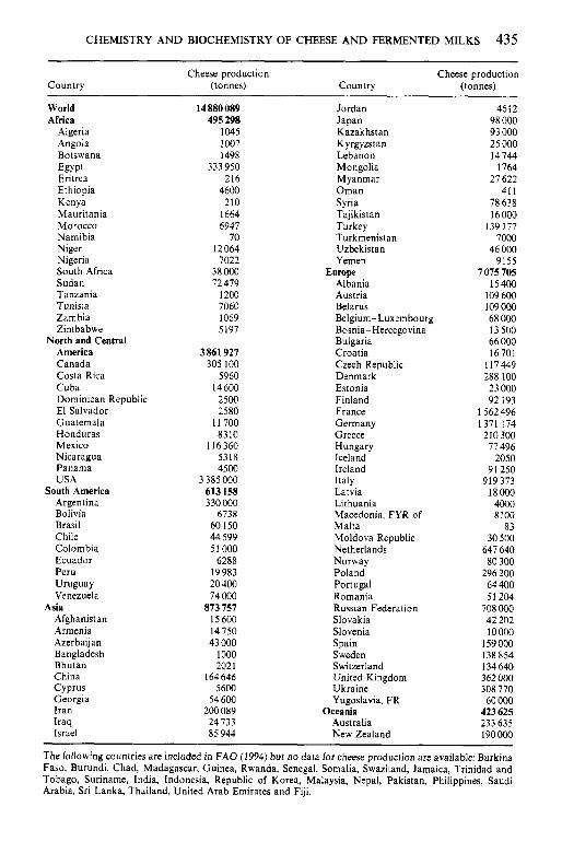
CHEMISTRY AND BIOCHEMISTRY OF CHEESE AND FERMENTED MILKS 435
Cheese production Cheese production Country (tonnes) Country (tonnes)
World Africa
Algeria Angola Botswana
Eritrea Ethiopia Kenya Mauritania Morocco Namibia Niger Nigeria South Africa Sudan Tanzania Tunisia Zambia Zimbabwe
America Canada Costa Rica Cuba Dominican Republic El Salvador Guatemala Honduras Mexico Nicaragua Panama USA
South America Argentina B o I i v i a Brasil Chile Colombia Ecuador Peru Uruguay Venezuela
Afghanistan Armenia Azerbaijan Bangladesh Bhutan China Cyprus Georgia Iran Iraq Israel
Egypt
North and Central
Asia
14 880 089 495 298
1045 1007 1498
333 950 216
4600 210
1664 6947
70 12064
7022 38 000 72 479
1200 7060 1069 5197
3861 921 305 100
5960 14 600
2500 2580
11700 8310
116360 5318 4500
3 385 000 613 158 330000
6738 60 150 44 599 51 000
6288 19983 20 400 74 000
873 151 15600 14750 43 000
1 000 202 1
164 646 5600
54 600 200089 24733 85 944
Jordan Japan Kazakhstan Kyrgyzstan Lebanon Mongolia Myanmar Oman Syria Tajikistan Turkey Turkmenistan Uzbekistan Yemen
Albania Austria Belarus Belgium- Luxembourg Bosnia- Hercegovina Bulgaria Croatia Czech Republic Denmark Estonia Finland France Germany Greece Hungary Iceland Ireland Italy Latvia Lithuania Macedonia, FYR of Malta Moldova Republic Netherlands Norway Poland Portugal Romania Russian Federation Slovakia Slovenia Spain Sweden Switzerland United Kingdom Ukraine Yugoslavia. FR
Australia New Zealand
Europe
Oceania
4612 98 000 93 000 25 000 I4 744
1764 27 622
41 1 78638 16000
139 177 7000
46 000 9155
7075 705 I5 400
109 600 109 000 68 000 I3 500 66 000 16701
117449 288 100 23 000 92 193
1 562 496 1371 174
210300 77 496
2050 91 250
919373 I8000
4000 8100
83 30 500
647 640 80 300
296 200 64 400 51 204
708 000 42 202 10000
159 000 I38 854 134 640 362 OOO 308 770 60 000
423 625 233 635 190000
The following countries are included in F A 0 (1994) but no data for cheese production are available: Burkina Faso, Burundi, Chad, Madagascar, Guinea, Rwanda, Senegal, Somalia, Swaziland, Jamaica, Trinidad and Tobago, Suriname, India, Indonesia, Republic of Korea, Malaysia, Nepal, Pakistan, Philippines, Saudi Arabia, Sri Lanka, Thailand, United Arab Emirates and Fiji.
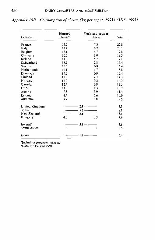
436 DAIRY CHEMISTRY AND BIOCHEMISTRY
Appendix IOB Consumption of cheese (kgper caput, 1993) (IDF, 1995)
Country Ripened Fresh and cottage cheese" cheese Total
France Italy Belgium Germany Iceland Switzerland Sweden Netherlands Denmark Finland Norway Canada USA Austria Estonia Australia
United Kingdom Spain New Zealand Hungary
Irelandb South Africa
Japan
15.5 13.4 15.1 10.5 11.9 13.6 15.5 14.1 14.5 12.0 14.0 12.4 11.9 7.5 4.4 8.7
7.3 6.7 4.7 8.0 5.2 2.8 0.9 1.7 0.9 2.3 0.2 0.9 1.3 3.9 5.6 0.8
8.3 ___ 8.1 ___ 8.1 ~
4.6 3.3
5.6 ~
1.5 0.1
1.4 ~
22.8 20.1 19.8 18.5 17.1 16.4 16.4 15.8 15.4 14.3 14.2 13.3 13.2 11.4 10.0 9.5
8.3 8.1 8.1 7.9
5.6 1.6
1.4
"Including processed cheese. bData for Ireland 1991.
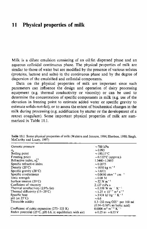
11 Physical properties of milk
Milk is a dilute emulsion consisting of an oil/fat dispersed phase and an aqueous colloidal continuous phase. The physical properties of milk are similar to those of water but are modified by the presence of various solutes (proteins, lactose and salts) in the continuous phase and by the degree of dispersion of the emulsified and colloidal components.
Data on the physical properties of milk are important since such parameters can influence the design and operation of dairy processing equipment (e.g. thermal conductivity or viscosity) or can be used to determine the concentration of specific components in milk (e.g. use of the elevation in freezing point to estimate added water or specific gravity to estimate solids-not-fat), or to assess the extent of biochemical changes in the milk during processing (e.g. acidification by starter or the development of a rennet coagulum). Some important physical properties of milk are sum- marized in Table 11.1.
Table 11.1 Some physical properties of milk (Walstra and Jenness, 1984; Sherbon, 1988; Singh, McCarthy and Lucey, 1997)
Osmotic pressure - 700 kPa a, -0.993 Boiling point - 100.15"C Freezing point -0.522"C (approx.) Refractive index, np 1.3440-1.3485 Specific refractive index -0.2075 Density (20°C) Specific gravity (20°C) .. 1.0321 Specific conductance -0.OO50 ohm-' cm-' Ionic strength -0.08 M Surface tension (20°C) Coefficient of viscosity Thermal conductivity (2.9% fat) Thermal diffusivity (15-20°C) Specific heat pH (at 25°C) - 6.6 Titratable acidity
Coefficient of cubic expansion (273-333 K) Redox potential (25"C, pH 6.6, in equilibrium with air)
- 1030 kg m-3
-52 N m- ' 2.127 mPa s -0.559 W m- ' K- ' - 1.25 x lo-' mz s- ' -3.931 kJ kg-' K-'
1.3-2.0 meq OH- per 100 mi (0.14-0.16% as lactic acid) 0.0008 m3 m-3 K- ' +0.25 to +0.35V
438 DAIRY CHEMISTRY AND BIOCHEMISTRY
11.1 Ionic strength
The ionic strength, I, of a solution is defined as:
(11.1) 1 2 I = zccizi
where ci is the molar concentration of the ion of type i and zi is its charge. The ionic strength of milk is c. 0.08 M.
11.2 Density
The density (p) of a substance is its mass per unit volume, while its specific gravity (SG) or relative density is the ratio of the density of the substance to that of water ( p , ) at a specified temperature:
p = m/V (11.2)
SG = P / P w (11.3)
P = SGPW (11.4)
The thermal expansion coefficient governs the mfluence of temperature on density and therefore it is necessary to specify temperature when discussing density or specific gravity. The density of milk is of consequence since fluid milk is normally retailed by volume rather than by mass. Measurement of the density of milk using a hydrometer (lactometer) has also been used to estimate its total solids content.
The density of bulk milk (4% fat and 8.95% solids-not-fat) at 20°C is approximately 1030kgm-3 and its specific gravity is 1.0321. Milk fat has a density of about 902kgm-3 at 40°C. The density of a given milk sample is influenced by its storage history since it is somewhat dependent on the liquid to solid fat ratio and the degree of hydration of proteins. To minimize effects of thermal history on its density, milk is usually prewarmed to 40-45°C to liquify the milk fat and then cooled to the assay temperature (often 20°C).
The density and specific gravity of milk vary somewhat with breed. Milk from Ayrshire cows has a mean specific gravity of 1.0317 while that of Jersey and Holstein milks is 1.0330. Density varies with the composition of the milk and its measurement has been used to estimate the total solids content of milk. The density of a multicomponent mixture (like milk) is related to the density of its components by:
1lP = C(mx/px> (11.5)
where m, is the mass fraction of component x, and p , its apparent density in the mixture. This apparent density is not normally the same as the true density of the substance since a contraction usually occurs when two components are mixed.
PHYSICAL PROPERTIES OF MILK 439
Equations have been developed to estimate the total solids content of milk based on % fat and specific gravity (usually estimated using a lactometer). Such equations are empirical and suffer from a number of drawbacks; for further discussion see Jenness and Patton (1959). The principal problem is the fact that the coefficient of expansion of milk fat is high and it contracts slowly on cooling and therefore the density of milk fat (Chapter 3) is not constant. Variations in the composition of milk fat and in the proportions of other milk constitiuents have less influence on these equations than the physical state of the fat.
In addition to lactometry (determination of the extent to which a hydrometer sinks), the density of milk can be measured by pycnometry (determination of the mass of a given volume of milk), by hydrostatic weighing of an immersed bulb (e.g. Westphal balance), by dialatometry (measurement of the volume of a known mass of milk) or by measuring the distance that a drop of milk falls through a density gradient column.
11.3 Redox properties of milk
Oxidation-reduction (redox) reactions involve the transfer of an electron from an electron donor (reducing agent) to an electron acceptor (oxidizing agent). The species that loses electrons is said to be oxidized while that which accepts electrons is reduced. Since there can be no net transfer of electrons to or from a system, redox reactions must be coupled and the oxidation reaction occurs simultaneously with a reduction reaction.
The tendency of a system to accept or donate electrons is measured using an inert electrode (typically platinum). Electrons can pass from the system into this electrode, which is thus a half-cell. The Pt electrode is connected via a potentiomenter to another half-cell of known potential (usually, a saturated calomel electrode). All potentials are referred to the hydrogen half-cell:
+H, P H + + e- (11.6)
which by convention is assigned a potential of zero when an inert electrode is placed in a solution of unit activity with respect to H + (i.e. pH = 0) in equilibrium with H, gas at a pressure of 1.013 x lo5 Pa (1 atm). The redox potential of a solution, Eh, is the potential of the half-cell at the inert electrode and is expressed as volts. E , depends not only on the substances present in the half-cell but also on the concentrations of their oxidized and reduced forms. The relationship between E , and the concentrations of the oxidized and reduced forms of the compound is described by the Nernst equation:
E , = E , - RT/nF In ared/aox (11.7)
where E , is the standard redox potential (i.e. potential when reactant and
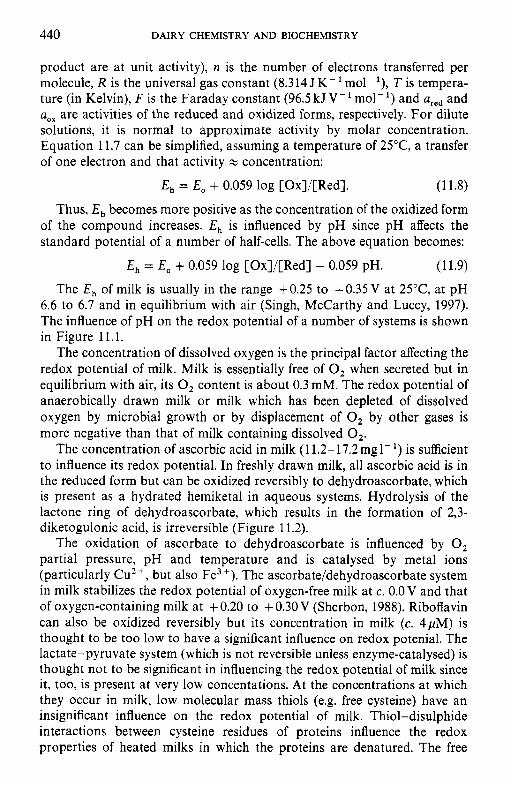
440 DAIRY CHEMISTRY AND BIOCHEMISTRY
product are at unit activity), n is the number of electrons transferred per molecule, R is the universal gas constant (8.314JK-'mol-'), T is tempera- ture (in Kelvin), F is the Faraday constant (96.5 kJ V - ' mol-') and ured and uox are activities of the reduced and oxidized forms, respectively. For dilute solutions, it is normal to approximate activity by molar concentration. Equation 11.7 can be simplified, assuming a temperature of 25"C, a transfer of one electron and that activity
E , = E , + 0.059 log [Ox]/[Red]. (11.8)
Thus, E , becomes more positive as the concentration of the oxidized form of the compound increases. E , is influenced by pH since pH affects the standard potential of a number of half-cells. The above equation becomes:
E , = E, + 0.059 log [Ox]/[Red] - 0.059 pH. (11.9)
The E, of milk is usually in the range + 0.25 to + 0.35 V at 25"C, at pH 6.6 to 6.7 and in equilibrium with air (Singh, McCarthy and Lucey, 1997). The influence of pH on the redox potential of a number of systems is shown in Figure 11.1.
The concentration of dissolved oxygen is the principal factor affecting the redox potential of milk. Milk is essentially free of 0, when secreted but in equilibrium with air, its 0, content is about 0.3 mM. The redox potential of anaerobically drawn milk or milk which has been depleted of dissolved oxygen by microbial growth or by displacement of 0, by other gases is more negative than that of milk containing dissolved 0,.
The concentration of ascorbic acid in milk (1 1.2- 17.2 mgl- ') is sufficient to influence its redox potential. In freshly drawn milk, all ascorbic acid is in the reduced form but can be oxidized reversibly to dehydroascorbate, which is present as a hydrated hemiketal in aqueous systems. Hydrolysis of the lactone ring of dehydroascorbate, which results in the formation of 2,3- diketogulonic acid, is irreversible (Figure 11.2).
The oxidation of ascorbate to dehydroascorbate is influenced by 0, partial pressure, pH and temperature and is catalysed by metal ions (particularly Cu2 +, but also Fe3 +). The ascorbate/dehydroascorbate system in milk stabilizes the redox potential of oxygen-free milk at c. 0.0 V and that of oxygen-containing milk at + 0.20 to + 0.30 V (Sherbon, 1988). Riboflavin can also be oxidized reversibly but its concentration in milk (c. 4pM) is thought to be too low to have a significant influence on redox potenial. The lactate-pyruvate system (which is not reversible unless enzyme-catalysed) is thought not to be significant in influencing the redox potential of milk since it, too, is present at very low concentations. At the concentrations at which they occur in milk, low molecular mass thiols (e.g. free cysteine) have an insignificant influence on the redox potential of milk. Thiol-disulphide interactions between cysteine residues of proteins influence the redox properties of heated milks in which the proteins are denatured. The free
concentration:
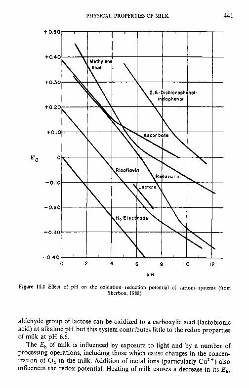
PHYSICAL PROPERTIES OF MILK 44 1
P H
Figure 11.1 Effect of pH on the oxidation-reduction potential of various systems (from Sherbon, 1988).
aldehyde group of lactose can be oxidized to a carboxylic acid (lactobionic acid) at alkaline pH but this system contributes little to the redox properties of milk at pH 6.6.
The E, of milk is influenced by exposure to light and by a number of processing operations, including those which cause changes in the concen- tration of 0, in the milk. Addition of metal ions (particularly C u Z f ) also influences the redox potential. Heating of milk causes a decrease in its E,,
442 DAIRY CHEMISTRY A N D BIOCHEMISTRY
CHPOH
I H-$-OH
Ascorbic acid
Rcduction Oxidation 11 CH2OH
I H-C-OH
0 H k2 Dehydroascorbic acid
11 Hzo
CH20H
I H-C-OH
OH
CHzOH
%O I * H-c-OH I
H-C-OH ,COOH
0 0
‘c-c II II
2, SDiketogulonic acid
Hydrated hemiketal form
Figure 11.2 Chemical structures of ascorbic acid and its derivatives.
due mainly to the denaturation of b-lactoglobulin (and the consequent exposure of -SH groups) and loss of 0,. Compounds formed by the Maillard reaction between lactose and proteins can also influence the E, of heated milk, particularly dried milk products.
Fermentation of lactose during the growth of micro-organisms in milk has a major effect on its redox potential. The decrease in the E , of milk caused by the growth of lactic acid bacteria is shown in Figure 11.3. A rapid decrease in E h occurs after the available 0, has been consumed by the bacteria. Therefore, the redox potential of cheese and fermented milk products is negative. Reduction of redox indicators (e.g. resazurin or
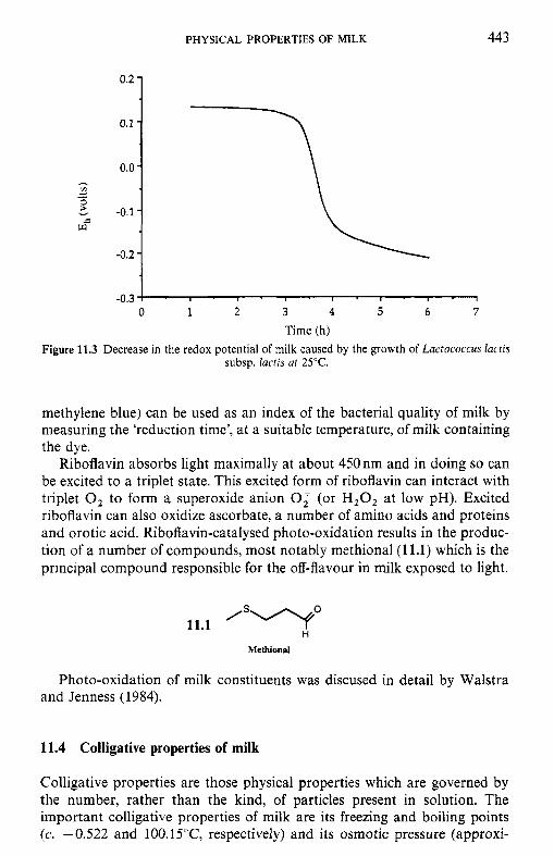
PHYSICAL PROPERTIES OF MILK
0.2 -
0.1 -
0.0 -
-0.1 -
-0.2 -
443
1 -0.31 . I ' I ' I ' I . I . I . I
0 1 2 3 4 5 6 7 Time (h)
Figure 11.3 Decrease in the redox potential of milk caused by the growth of Lactococcus lactis subsp. lactis at 25°C.
methylene blue) can be used as an index of the bacterial quality of milk by measuring the 'reduction time', at a suitable temperature, of milk containing the dye.
Riboflavin absorbs light maximally at about 450nm and in doing so can be excited to a triplet state. This excited form of riboflavin can interact with triplet 0, to form a superoxide anion 0; (or H,O, at low pH). Excited riboflavin can also oxidize ascorbate, a number of amino acids and proteins and orotic acid. Riboflavin-catalysed photo-oxidation results in the produc- tion of a number of compounds, most notably methional(11.1) which is the principal compound responsible for the off-flavour in milk exposed to light.
Methional
Photo-oxidation of milk constituents was discused in detail by Walstra and Jenness (1984).
11.4 Colligative properties of milk
Colligative properties are those physical properties which are governed by the number, rather than the kind, of particles present in solution. The important colligative properties of milk are its freezing and boiling points (c. -0.522 and 100.15"C, respectively) and its osmotic pressure (approxi-
444 DAIRY CHEMISTRY AND BIOCHEMISTRY
mately 700 kPa at 20"C), all of which are interrelated. Since the osmotic pressure of milk remains essentially constant (because it is regulated by that of the cow's blood), the freezing point is also relatively constant.
The freezing point of an aqueous solution is governed by the concentra- tion of solutes in the solution. The relationship between the freezing point of a simple aqueous solution and concentration of solute is described by a relation based on Raoult's law:
Tf = K,m (11.10)
where is the difference between the freezing point of the solution and that of the solvent, K, is the molal depression constant (136°C for water) and m is the molal concentration of solute. However, this equation is valid only for dilute solutions containing undissociated solutes. Raoult's law is thus limited to approximating the freezing point of milk.
The freezing point of bovine milk is usually in the range -0.512 to -O.55O0C, with a mean value close to -02~22°C (Sherbon, 1988) or - 0.540"C (Jenness and Patton, 1959). Despite variations in the concentra- tions of individual solutes, the freezing point depression of milk is quite constant since it is proportional to the osmotic pressure of milk (approxi- mately 700 kPa at 20"C), which is regulated by that of the cow's blood. The freezing point of milk is more closely related to the osmotic pressure of mammary venous blood than to that of blood from the jugular vein.
Owing to their large particle or molecular mass, fat globules, casein micelles and whey proteins do not have a significant effect on the freezing point of milk, to which lactose makes the greatest contribution. The freezing point depression in milk due to lactose alone has been calculated to be 0.296"C. Assuming a mean freezing point depression of 0.522"C, all other constituents in milk depress the freezing point by only 0.226"C. Chloride is also an important contributor to the colligative properties of milk. Assum- ing a C1- concentration of 0.032M and that C1- is accompanied by a monovalent cation (i.e. Na' or K'), the freezing point depression caused by C1- and its associated cation is 0.119"C. Therefore, lactose, chloride and its accompanying cations together account for about 80% of the freezing point depression in milk. Since the total osmotic pressure of milk is regulated by that of the cow's blood, there is a strong inverse correlation between lactose and chloride concentrations (Chapter 5).
Natural variation in the osmotic pressure of milk (and hence freezing point) is limited by the physiology of the mammary gland. Variations in the freezing point of milk have been attributed to seasonality, feed, stage of lactation, water intake, breed of cow, heat stress and time of day. These factors are often interrelated but have relatively little influence on the freezing point of milk. Likewise, unit operations in dairy processing which do not influence the net number of osmotically active molecules/ions in solution do not influence the freezing point. Cooling or heating milk causes
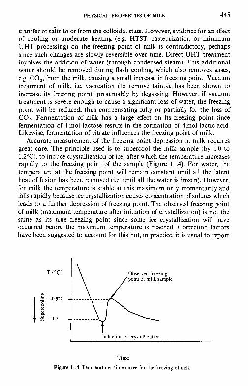
PHYSICAL PROPERTIES OF MILK 445
transfer of sdts to or from the colloidal state. However, evidence for an effect of cooling or moderate heating (e.g. HTST pasteurization or minimum UHT processing) on the freezing point of milk is contradictory, perhaps since such changes are slowly reversible over time. Direct UHT treatment involves the addition of water (through condensed steam). This additional water should be removed during flash cooling, which also removes gases, e.g. CO,, from the milk, causing a small increase in freezing point. Vacuum treatment of milk, i.e. vacreation (to remove taints), has been shown to increase its freezing point, presumably by degassing. However, if vacuum treatment is severe enough to cause a significant loss of water, the freezing point will be reduced, thus compensating fully or partially for the loss of CO,. Fermentation of milk has a large effect on its freezing point since fermentation of 1 mol lactose results in the formation of 4 mol lactic acid. Likewise, fermentation of citrate influences the freezing point of milk.
Accurate measurement of the freezing point depression in milk requires great care. The principle used is to supercool the milk sample (by 1.0 to 1.2"C), to induce crystallization of ice, after which the temperature increases rapidly to the freezing point of the sample (Figure 11.4). For water, the temperature at the freezing point will remain constant until all the latent heat of fusion has been removed (i.e. until all the water is frozen). However, for milk the temperature is stable at this maximum only momentarily and falls rapidly because ice crystallization causes concentration of solutes which leads to a further depression of freezing point. The observed freezing point of milk (maximum temperature after initiation of crystallization) is not the same as its true freezing point since some ice crystallization will have occurred before the maximum temperature is reached. Correction factors have been suggested to account for this but, in practice, it is usual to report
M
v) -1.5 -.
Induction of crystallization
Time Figure 11.4 Temperature-time curve for the freezing of milk.
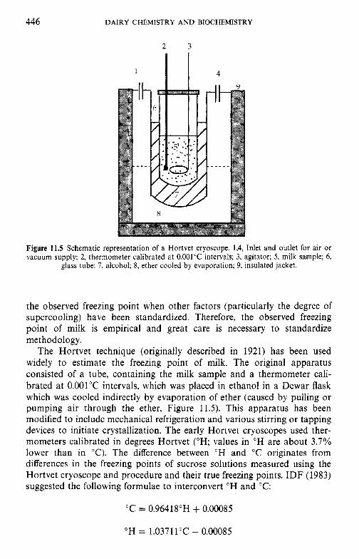
446 DAIRY CHEMISTRY A N D BIOCHEMISTRY
1 I I
Figure 11.5 Schematic representation of a Hortvet cryoscope. 1,4, Inlet and outlet for air o r vacuum supply; 2, thermometer calibrated at 0.001"C intervals; 3, agitator; 5 , milk sample; 6,
glass tube; 7, alcohol; 8, ether cooled by evaporation; 9, insulated jacket.
the observed freezing point when other factors (particularly the degree of supercooling) have been standardized. Therefore, the observed freezing point of milk is empirical and great care is necessary to standardize methodology.
The Hortvet technique (originally described in 1921) has been used widely to estimate the freezing point of milk. The original apparatus consisted of a tube, containing the milk sample and a thermometer cali- brated at 0.001"C intervals, which was placed in ethanol in a Dewar flask which was cooled indirectly by evaporation of ether (caused by pulling or pumping air through the ether, Figure 11.5). This apparatus has been modified to include mechanical refrigeration and various stirring or tapping devices to initiate crystallization. The early Hortvet cryoscopes used ther- mometers calibrated in degrees Hortvet (OH; values in O H are about 3.7% lower than in "C). The difference between OH and "C originates from differences in the freezing points of sucrose solutions measured using the Hortvet cryoscope and procedure and their true freezing points. IDF (1983) suggested the following formulae to interconvert "H and "C:
"C = 0.96418"H + 0.00085
OH = 1.03711"C - 0.00085
PHYSICAL PROPERTIES OF MILK 447
However, it is now recommended that thermometers be calibrated in "C. More recently, thermistors have been used instead of mercury thermom- eters. Cryoscopes based on dew point depression have also been approved for use. These latter instruments also use thermistors and are based on changes in osmotic pressure. Thermistor cryoscopes are now used more widely than Hortvet instruments.
Measurement of the freezing point depression of milk is used to estimate the degree of adulteration of milk with added water. Assuming an average freezing point of O.55O0C, the amount of added water can be calculated from:
0.550 - AT x (100 - TS)
0.550 % added water = (1 1.1 1)
where AT is the observed freezing point depression of the test sample and TS is the YO total solids in the milk. Interpretation of freezing point values when assaying milk suspected of being adulterated with water requires care. Milk with a freezing point of -0.525"C or below is usually presumed to be unadulterated. Due to greater variation in the freezing point of milks drawn from individual animals than of bulk milk, specifications for the freezing point of bulk milk are more stringent than those for milks from individual animals. Finally, it should be noted that estimation of the adulteration of milk with water depends on the constancy of the freezing point (as discussed above). Adulteration of milk with isotonic solutions, e.g. ultrafiltration permeate (which is being considered for standardization of the protein content of milk, see Rattray and Jelen, 1996), will not be detected by this technique.
11.5 Interfacial tension
A phase can be defined as a domain bounded by a closed surface in which parameters such as composition, temperature, pressure and refractive index are constant but change abruptly at the interface. The principal phases in milk are its serum and fat and the most important interfaces are air/serum and fat/serum. If present, air bubbles, and ice, fat or lactose crystals will also constitute phases. Forces acting on molecules or particles in the bulk of a phase differ from those at an interface since the former are attracted equally in all directions while those at an interface experience a net attraction towards the bulk phase (Figure 11.6).
This inward attraction acts to minimize the interfacial area and the force which causes this decrease in area is known as the interfacial tension (7). If one phase is air, the interfacial tension is referred to as surface tension. Interfacial tension can be expressed as force per unit length (N m-') or the energy needed to increase the interfacial area by a unit amount (J mW2 or N m- ').
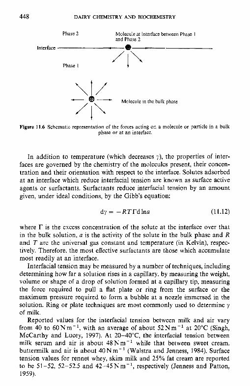
448 DAIRY CHEMISTRY AND BIOCHEMISTRY
Phase 2 Molecule at interface between Phase 1 and Phase 2
Interface
Phase I
\tr Molecule in the bulk phase
t Figure 11.6 Schematic representation of the forces acting on a molecule or particle in a bulk
phase or at an interface.
In addition to temperature (which decreases y), the properties of inter- faces are governed by the chemistry of the molecules present, their concen- tration and their orientation with respect to the interface. Solutes adsorbed at an interface which reduce interfacial tension are known as surface active agents or surfactants. Surfactants reduce interfacial tension by an amount given, under ideal conditions, by the Gibb's equation:
dy = -RTTdlna (1 1.12)
where r is the excess concentration of the solute at the interface over that in the bulk solution, a is the activity of the solute in the bulk phase and R and T are the universal gas constant and temperature (in Kelvin), respec- tively. Therefore, the most effective surfactants are those which accumulate most readily at an interface.
Interfacial tension may be measured by a number of techniques, including determining how far a solution rises in a capillary, by measuring the weight, volume or shape of a drop of solution formed at a capillary tip, measuring the force required to pull a flat plate or ring from the surface or the maximum pressure required to form a bubble at a nozzle immersed in the solution. Ring or plate techniques are most commonly used to determine y of milk.
Reported values for the interfacial tension between milk and air vary from 40 to 60Nm-' , with an average of about 52Nm- ' at 20°C (Singh, McCarthy and Lucey, 1997). At 20-40°C, the interfacial tension between milk serum and air is about 48Nm- ' while that between sweet cream, buttermilk and air is about 40Nm- ' (Walstra and Jenness, 1984). Surface tension values for rennet whey, skim milk and 25% fat cream are reported to be 51-52, 52-52.5 and 42-45 N m-', respectively (Jenness and Patton, 1959).
PHYSICAL PROPERTIES OF MILK 449
The principal surfactants in milk are its proteins, phospholipids, mono- and diglycerides and salts of free fatty acids. The immunoglobulins are less effective surfactants than other milk proteins. Salts and lactose do not contribute significantly to the interfacial tension of milk. The difference in interfacial tension between milk serum/air and buttermilk/air can be at- tributed to the higher concentration of very surface active proteins and protein-phospholipid complexes of the fat globule membrane in buttermilk. The interfacial tension between milk fat globules and the milk serum is about 2 N m- while the interfacial tension between non-globular, liquid milk fat and milk serum is about 15Nm-' , indicating the effectiveness of milk fat globule membrane material in reducing interfacial tension. The surface tension of whole milk is a little lower than that of skim milk, possibly due to the presence of higher levels of material from the fat globule membrane and traces of free fat in the former. Surface tension decreases with increasing fat content up to about 4%. Lipolysis reduces the surface tension of milk due to the liberation of free fatty acids and attempts have been made to estimate hydrolytic rancidity by exploiting this fact, although such approaches have not been very successful (see Sherbon (1988) for refer- ences).
In addition to its composition, various processing parameters can influ- ence the surface tension of milk. The surface tension of whole and skim milk decreases with increasing temperature. Surface tension also varies with the temperature history and age of the milk and with the time required for measurement. Homogenization of raw milk reduces surface tension because lipolysis by the indigenous milk lipase is stimulated and surface-active fatty acids released. Homogenization of pasteurized milk causes a slight increase in surface tension. Pasteurization of milk has little effect on its surface tension although heating milk to sterilization temperatures causes a slight increase in surface tension, resulting from denaturation and coagulation of proteins which are then less effective as surfacants.
11.6 Acid-base equilibria
The acidity of a solution is normally expressed as its pH, which may be defined as:
(11.13)
(1 1.14) where a,,- is the activity of the hydrogen ion, [H'] its concentration and fH
its activity coefficient. For many dilute solutions, fH x 1 and pH can thus be closely approximated by the negative logarithm of the hydrogen ion concentration.
450 DAIRY CHEMISTRY A N D BIOCHEMISTRY
The pH of milk at 25°C is usually in the range 6.5-6.7, with a mean value of 6.6. The pH of milk is influenced much more by temperature than is the pH of dilute buffers, principally due to the temperature dependence of the solubility of calcium phosphate (Chapter 5). pH varies with stage of lactation; colostrum can have a pH as low as 6.0. Mastitis tends to increase the pH since increased permeability of the mammary gland membranes means that more blood constituents gain access to the milk; the pH of cow’s blood is 7.4. The difference in pH between blood and milk results from the active transport of various ions into the milk, precipitation of colloidal calcium phosphate (CCP; which results in the release of H’) during the synthesis of casein micelles, higher concentrations of acidic groups in milk and the relatively low buffering capacity of milk between pH 6 and 8 (Singh, McCarthy and Lucey, 1997).
An important characteristic of milk is its buffering capacity, i.e. resistance to changes in pH on addition of acid or base. A pH buffer resists changes in the [H’] (ApH) in the solution and normally consists of a weak acid (HA) and its corresponding anion (A-, usually present as a fully dissociat- able salt). An equilibrium thus exists:
H A = H + + A - (11.15)
The addition of H + to this solution favours the back reaction while the addition of base favours the forward reaction. The weak acid/salt pair thus acts to minimize ApH. An analogous situation exists for buffers consisting of a weak base and its salt. The pH of a buffer can be calculated from the concentration of its components by the Henderson-Hasselbalch equation
(11.16)
where pK, is the negative logarithm of the dissociation constant of the weak acid, HA. A weak acid/salt pair is most effective in buffering against changes in pH when the concentrations of acid and salt are equal, i.e. at pH = pK, of HA. The effectiveness of a buffer is expressed as its buffering index
(11.17)
Milk contains a range of groups which are effective in buffering over a wide pH range. The principal buffering compounds in milk are its salts (particularly soluble calcium phosphate, citrate and bicarbonate) and acidic and basic amino acid side-chains on proteins (particularly the caseins). The contribution of these components to the buffering of milk was discussed in detail by Singh, McCarthy and Lucey (1997).
In theory, it should be possible to calculate the overall buffering proper- ties of milk by combining the titration curves for all components but in
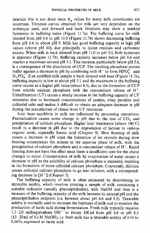
PHYSICAL PROPERTIES OF MILK 45 1
practice this is not done since K , values for many milk constituents are uncertain. Titration curves obtained for milk are very dependent on the technique used, and forward and back titrations may show a marked hysteresis in buffering index (Figure 11.7a). The buffering curve for milk titrated from pH 6.6 to pH 11.0 (Figure 11.7b) shows decreasing buffering from pH 6.6 to about pH 9. Milk has good buffering capacity at high pH values (above pH lo), due principally to lysine residues and carbonate anions. When milk is back titrated from pH 11.0 to pH 3.0, little hysteresis is apparent (Figure 11.7b). Buffering capacity increases below pH 6.6 and reaches a maximum around pH 5.1. This increase, particularly below pH 5.6, is a consequence of the dissolution of CCP. The resulting phosphate anions buffer against a decrease in pH by combining with H' to form HPOi- and H,PO,. If an acidified milk sample is back titrated with base (Figure 11.7a), buffering capacity is low at about pH 5.1 and the maximum in the buffering curve occurs at a higher pH value (about 6.3), due to the formation of CCP from soluble calcium phosphate with the concomitant release of H +. Ultrafiltration (UF) causes a steady increase in the buffering capacity of U F retentates due to increased concentrations of caseins, whey proteins and colloidal salts and makes it difficult to obtain an adequate decrease in pH during the manufacture of cheese from U F retentates.
Acid-base equilibria in milk are influenced by processing operations. Pasteurization causes some change in pH due to the loss of CO, and precipitation of calcium phosphate. Higher heat treatments (above 100°C) result in a decrease in pH due to the degradation of lactose to various organic acids, especially formic acid (Chapter 9). Slow freezing of milk causes a decrease in pH since the formation of ice crystals during slow freezing concentrates the solutes in the aqueous phase of milk, with the precipitation of calcium phosphate and a concomitant release of H'. Rapid freezing does not have this effect since there is insufficient time for the above changes to occur. Concentration of milk by evaporation of water causes a decrease in pH as the solubility of calcium phosphate is exceeded, resulting in the formation of more colloidal calcium phosphate. Conversely, dilution causes colloidal calcium phosphate to go into solution, with a correspond- ing decrease in [H'] (Chapter 5) .
The buffering capacity of milk is often estimated by determining its titratable acidity, which involves titrating a sample of milk, containing a suitable indicator (usually phenolphthalein), with NaOH and thus is a measure of the buffering capacity of the milk between its natural pH and the phenolphthalein endpoint (i.e. between about pH 6.6 and 8.3). Titratable acidity is normally used to estimate the freshness of milk and to monitor the production of lactic acid during fermentation. Fresh milk typically requires 1.3-2.0 milliequivalents OH- to titrate 100ml from pH 6.6 to pH 8.3 (13-20ml of 0.1 M NaOH), i.e. fresh milk has a titratable acidity of 0.14 to O.16%, expressed as lactic acid.
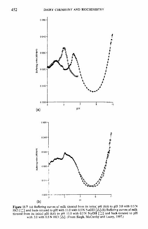
452 DAIRY CHEMISTRY AND BIOCHEMISTRY
0.050 -
0 040-
0
^^^ 0 . y t k \ / "& 1
I B
010 , '-u 0.000 / 11
(4 PH
0 050 -
0040- 0 0
I J
0 000
PH (b) Figure 11.7 (a) Buffering curves of milk titrated from its initial pH (6.6) to pH 3.0 with 0.5N HCI [a] and back-titrated to pH with 11.0 with 0.5 N NaOH [A].(b) Buffering curves of milk titrated from its initial pH (6.6) to pH 11.0 with 0.5 N NaOH [ I 3 3 and back-titrated to pH
with 3.0 with 0.5 N HCI [A]. (From Singh, McCarthy and Lucey, 1997.)
PHYSICAL PROPERTIES OF MILK 453
A high titratable acidity for fresh milk suggests high concentrations of proteins and/or other buffering constituents. Titratable acidity varies only slightly with the breed of cow, although the values for individual cows can vary more widely (0.08-0.25% as lactic acid). The liberation of fatty acids on lipolysis can interfere with the estimation of titratable acidity in high-fat products. Precipitation of calcium phosphate (with a concomitant decrease in pH) and 'fading of the phenolphthalein end-point' can occur during titration and thus the titratable acidity value obtained is influenced by the speed of titration.
11.7 Rheological properties
11.7.1 Newtonian behaviour
Under certain conditions (e.g. moderate shear rates, at fat contents below 40% and at temperatures above 40°C, at which the fat is liquid and no cold agglutination occurs) milk, skim milk and cream are, in effect, fluids with Newtonian rheological properties. Newtonian behaviour can be described by the equation:
z = qp (11.18)
where 5 is the shear stress (force per unit area, Pa), $J the shear rate (rate of change of velocity across the stream, s-') and q is the Coefficient of viscosity (Pas). The coefficient of viscosity for a Newtonian fluid is independent of shear rate but is influenced by temperature and pressure.
The coefficient of viscosity for whole milk at 20°C, but not affected by cold agglutination of fat globules, is about 2.127mPa s. Values for water and milk plasma at 20°C are 1.002 and 1.68mPas, respectively. Casein, and to a lesser extent fat, are the principal contributors to the viscosity of milk; whey proteins and low molecular mass species have less influence.
The viscosity of milk and Newtonian milk products is influenced by composition, concentyation, pH, temperature, thermal history and process- ing operations.
The Newtonian coefficient of viscosity at a given temperature for milk, creams and some concentrated milk products is related to the concentration of individual components by Eiler's equation:
(11.19)
where qo is the coefficient of viscosity of the portion of the fluid consisting of water and low molecular mass species other than lactose and 4 is the volume fraction of all dispersed particles that are at least an order of magnititude larger than water. The volume fraction of any component is
454 DAIRY CHEMISTRY AND BIOCHEMISTRY
given by
4i = Ycv,i (11.20)
where is the voluminosity of component i (in m3kg- ' dry com- ponent) and cv.i is the volume concentration of the component in the product (m3kg- ' product). The voluminosity of fat in fat globules is c. 1.11 x 1OW3m3kg-', that of casein micelles is c. 3.9 x 10⁻³m3kg-' , whey proteins c. 1.5 x 10⁻³m3kg-1 and lactose c. 1 x 10⁻³m3kg-1. For milk
4 = 4f + 4 c + 4 w + 4, (11.21)
where df, 4c , &, 4, are the volume fractions of fat, casein, whey proteins and lactose, respectively. +,,, is the assumed value of L(I$~) for maximum packing of all dispersed particles (0.9 for fluid milk products).
Increasing pH increases viscosity slightly (perhaps as a consequence of micellar swelling) while a small decrease in pH reduces viscosity, although a large decrease in pH causes aggregation of casein micelles. Viscosity is inversely related to temperature. The viscosity of milk shows thermal hysteresis; it usually shows greater viscosity during heating than during subsequent cooling, probably due to the melting and crystallization behav- iour of milk triglycerides.
The viscosity of milk and creams tends to increase slightly with age, due in part to changes in ionic equilibria. Heating skim milk to an extent that denatures most of the whey proteins increases its viscosity by about 10%. Homogenization of whole milk has little effect on its viscosity. The increase in the volume fraction of fat on homogenization is compensated by a decrease in the volume fractions of casein and whey proteins because some skim milk proteins are adsorbed at the fat-oil interface. Pasteurization has no significant effect on the rheology of whole milk.
11.7.2 Non-Newtonian behaviour
Raw milks and creams exhibit non-Newtonian rheological properties when they are held under conditions which favour cold agglutination of fat globules (below 40°C and low shear rates). Under such conditions, they show thixotropic (shear thinning) behaviour, i.e. their apparent viscosity (qapp) is inversely related to shear rate. Aggregates of fat globules and the milk serum trapped in their interstitial spaces have a large effective volume due to their irregular shapes. Increasing the shear rate causes increased shear stress to be applied to the aggregates which can disperse, yielding smaller or more rounded ones. Disaggregation reduces the interstitial space between fat globules, thereby reducing the total volume fraction of the fat phase and consequently reducing the qaPp of the product. When the shearing force applied to the fluid increases in excess of the forces which hold the aggregates together, increases in shear rate cause increasingly smaller
PHYSICAL PROPERTIES OF MILK 455
changes in apparent viscosity. Thus, at high shear rates the fluid will exhibit Newtonian behaviour.
Increasing the fat content and/or reducing the temperature favours non-Newtonian behaviour. Low temperatures promote cold agglutination of fat globules and thus increase both qapp and deviation from Newtonian behaviour. The temperature at cream separation also influences the rheological properties of the resulting cream. Cream prepared by separation above 40°C shows less deviation from Newtonian behaviour since cryo- globulins are lost in the skim milk, resulting in less agglutination. Apparent viscosity is also influenced by the shear history of the product. The reformation of bonds between fat globules in aggregates requires time and thus the qapp versus shear rate ( 9 ) curves exhibit hysteresis. ylaPp increases after cessation of shearing (as aggregates are reformed) but usually does not return to its original value. Hysteresis is apparent in products containing aggregates caused by cold agglutination or homogenization.
Coalescence of fat globules does not change qapp since the volume fraction of the fat is not changed. However, partial coalescence can result in an increase in qaPp due to entrapment of milk serum in aggregates. Indeed, high-fat creams can exhibit rheopectic (shear thickening) behaviour since shearing can cause partial coalescence of fat globules.
In addition to the general decrease in viscosity with increasing tempera- ture, heating milk can also influence its rheology by heat-induced denatura- tion of cryoglobulins and/or other whey proteins. Concentration of milk, e.g. by ultrafiltration, prior to heating results in a greater increase in qapp than in milk heated before concentration.
The addition of hydrocolloids (e.g. carrageenans, pectins or car- boxymethyl cellulose) as thickening agents will greatly increase the apparent viscosity of the product. The production of extracellular polysaccharides by certain bacteria will also increase the viscosity of milk products.
11.7.3 Rheology of milk gels
Gels are viscoelastic bodies, the rheological properties of which can be described by two parameters, the storage modulus (G’, which is a measure of its elasticity) and the loss modulus (G”, which is a measure of its viscous nature). The combined viscoelastic modulus (G*) is a measure of the overall resistance of a gel to deformation. These moduli are often highly dependent on the time-scale of deformation. Another important parameter of a food gel is its yield stress.
Although the gelation properties of whey proteins are of great impor- tance in many foods (Mulvihill, 1992) and it is possible to form a weak gel in creams by the formation of a continuous network of fat globules, most important milk gels are those involving casein micelles which can be made to form a gel matrix either by isoelectric precipitation (acid-induced gel) or by the action of a proteolytic enzyme (rennet-induced gel). Both gel types
456 DAIRY CHEMISTRY AND BIOCHEMISTRY
are relatively similar but, over long deformation times, rennet-induced gels have more liquid character than acid gels, which means that the former can flow under their own weight while acid gels are more likely to retain their shape. Rennet-induced gels also have a greater tendency to synerese and have a higher yield stress than acid-induced gels.
The firmness of acid- and rennet-induced milk gels is increased by such factors as time elapsed after aggregation of the micelles, gelation at elevated temperature, increasing casein and calcium phosphate concentrations and reduced pH (Walstra and Jenness, 1984). Heat-induced denaturation of whey proteins decreases the firmness of rennet-induced gels but increases the firmness of acid-induced gels. Fat globules weaken casein gels by interrupt- ing the gel matrix. Casein molecules on the surface of fat globules in homogenized milk can participate in gel network formation. However, in practice this is influenced by a number of other factors, including preheating, homogenization pressure and temperature, and type of gel (Walstra and Jenness, 1984). Indeed, the yield stress of a rennet-induced milk gel may be reduced by homogenization.
11.7.4
The rheological properties of milk fat are greatly influenced by the ratio of solid to liquid fat and by the crystal form of the solid fat. At room temperature (20°C), milk fat is partially solid and has a plastic consistency, i.e. it exhibits viscoelastic properties; at small deformations (below 1 %), it is almost completely elastic due to interactions between the fat crystals which form a weak network but it will begin to flow when subjected to greater deformations. As discussed by Walstra and Jenness (1984), the important parameters in determining the firmness of milk fat include the fraction of solid fat, the shape and size of fat crystals, heterogeneity throughout the fat and the extent to which fat crystals form a network throughout the mass of fat.
The structure of butter and other dairy spreads are further complicated by the presence of aqueous phase droplets and intact fat globules. Water droplets tend to weaken the structure and fat crystals inside intact fat globules cannot participate in the formation of a network thoughout the product (Chapter 3).
Rheological properties of milk fat
11.8 Electrical conductivity
The specific resistance ( p , ohm cm) of a substance is related to its dimensions by:
p = ctR/l (11.22)
PHYSICAL PROPERTIES OF MILK 457
where c( is the cross-sectional area (cm'), 1 is length (cm) and R the measured resistance (ohms). The specific conductance, K (ohm-' cm-'), is the recip- rocal of specific resistance. The specific conductance of milk is usually in the range 0.0040-0.0055 ohm-' cm-'. Ions (particularly Na', K f and C1-) are responsible for most of the electrical conductivity of milk which is increased by the bacterial fermentation of lactose to lactic acid. Measurement of the specific conductance of milk has been used as a rapid method for detecting subclinical mastitis. The conductivity of solutions is altered by concentra- tion and dilution. However, the usefulness of this in the context of milk (e.g. to detect adulteration with water) is reduced considerably by the influence of concentration or dilution on the precipitation or solubilization of colloidal calcium phosphate. Direct conductivity measurements are thus unsuitable for assessing the amount of water added to milk.
11.9 Thermal properties of milk
The specific heat of a substance is the amount of heat energy, in kJ, required to increase the temperature of 1 kg of the substance by 1 K. The specific heat of skim milk increases from 3.906 to 3.993 kJ kg- ' K-' from 1 to 50°C. Values of 4.052 and 3.931 kJkg- 'K- ' have been reported for skim and whole milks, respectively, at 80°C (Sherbon, 1988). The specific heat of milk is inversely related to its total solids content, although discontinuities have been observed around 70-80°C. Skim-milk powder usually has a specific heat in the range 1.172-1.340kJkg-'K-' at 18-30°C.
The specific heat of milk fat (solid or liquid) is about 2.177kJkg-' K-I . The specific heat of milk and cream is therefore strongly influenced by their fat content. Over most commonly encountered temperature ranges, the specific heat of high-fat dairy products is complicated by the latent heat absorbed by melting fat (about 84 J g- '). The observed specific heat of these products, at temperatures over which milk fat melts is thus the sum of the true specific heat and the energy absorbed to provide the latent heat of fusion of milk fat. Specific heat is thus influenced by factors such as the proportion of fat in the solid phase at the beginning of heating, and thus the composition of the fat and its thermal history. The apparent specific heat of high-fat dairy products (sum of 'true' specific heat and the energy absorbed by melting of fat) is usually maximal at 15-20°C and often has a second maximum or inflexion around 35°C.
The rate of heat transfer through a substance by conduction is given by the Fourier equation for heat conduction:
d T dt dx dQ = - k A - (11.23)
458 DAIRY CHEMISTRY AND BIOCHEMISTRY
where dQ/dt is the quantity of heat energy (Q) transferred per unit time (t) , A is the cross-sectional area of the path of heat flow, dT/dx is the temperature gradient and k is the thermal conductivity of the medium. The thermal conductivity of whole milk (2.9% fat), cream and skim milk is roughly 0.559, 0.384 and 0.568 Wm- ' K-', respectively. The thermal con- ductivity of skim milk, whole milk and cream increases with increasing temperature but decreases with increasing levels of total solids or fat, particularly at higher temperatures. In addition to their composition, the thermal conductivity of dried-milk products depends on bulk density (weight per unit volume) due to differences in the amount of air entrapped in the powder.
Thermal diffusivity is a measure of the ability of a material to dissipate temperature gradients within it. Thermal diffusivity (a , m2 s-') is defined as the ratio of thermal conductivity ( k ) to volumetric specific heat (density times specific heat, pc):
a = k/pc (11.24)
The thermal diffusivity of milk (at 15-20°C) is about 1.25 x lo-' m2 s-'.
11.10 Interaction of light with milk and dairy products
The refractive index (a) of a transparent substance is expressed by the relation:
sin i n=-
sin r (11.25)
where i and r are the angles between the incident ray and the refracted ray of light, respectively, and a perpendicular to the surface of the substance. The refractive index of milk is difficult to estimate due to light scattering by casein micelles and fat globules. However, it is possible to make accurate measurements of the refractive index of milk using refractometers in which a thin layer of sample is used, e.g. the Abbe refractometer. The refractive index of milk at 20°C using the D-line of the sodium spectrum (- 589 nm), n p , is normally in the range 1.3440-1.3485. The refractive index of milk fat is usually in the range 1.4537-1.4552 at 40°C. Although there is a linear relationship between the solids content (weight per unit volume) and refractive index, determination of percentage solids in milk by refractometry is difficult, since the contributions of various milk components differ and are additive. The relationship between the refractive index of milk and its total solids content varies with changes in the concentration and composition of the solutes in milk. However, attempts have been made to measure the total contribution of solids and casein in milk and milk products by estimating
PHYSICAL PROPERTIES OF MILK 459
the refractive index (Sherbon, 1988). The specific refrative index (refractive constant), K , is calculated from:
(11.26)
where n is the refractive index and p is density. Milk has a specific refractive index of about 0.2075.
Milk contains not only numerous dissolved chemical components but it is also an emulsion with a colloidal continuous phase. Therefore, milk absorbs light of a wide range of wavelengths and also scatters ultraviolet (UV) and visible light due to the presence of particles. Milk absorbs light of wavelengths between 200 and about 380nm due to the proteins present and between 400 and 520 nm due to fat-soluble pigments (carotenoids). A number of functional groups in milk constituents absorb in the infrared (IR) region of the spectrum; the OH groups of lactose absorb at c. 9.61 pm, the amide groups of proteins at 6.465 pm and the ester carbonyl groups of lipids at 5.723pm (Singh, McCarthy and Lucey, 1997). Since light scattering is reduced at longer wavelenghts in the IR region, the absorbance of IR light of specific wavelengths can be used to measure the concentrations of fat, protein and lactose in milk. Instruments using this principle are now widely used in the dairy industry. However, since milk contains about 87.5% water (which absorbs IR light strongly), it is opaque to light throughout much of the IR region of the spectrum.
Milk contains about 1.62 mg kg- riboflavin which fluoresces strongly on excitation by light of wavelenghts from 400 to 500 nm, emitting light with a Lmax = 530 nm. Milk proteins also fluoresce due to the presence of aromatic amino acid residues; part of the light absorbed at wavelengths around 280 nm is emitted at longer wavelengths.
Scattering of light by the colloidal fat particles present in milk has been used to estimate its fat content. A commercial apparatus (Milko-TesterTM) has been developed which exploits this principle. Milk is diluted (to avoid multiple scatterings) using an EDTA solution which disperses the casein micelles. The milk sample is homogenized to ensure a uniform fat globule size and the extent of scattering of white light is determined.
11.11 Colour of milk and milk products
The white colour of milk results from scattering of visible light by casein micelles and fat globules. Homogenization of milk results in a whiter product due to increased scattering of light by smaller, homogenized, fat globules. The serum phase of milk is greenish due to the presence of riboflavin which is responsible for the characteristic colour of whey.
460 DAIRY CHEMISTRY AND BIOCHEMISTRY
HOOC
\\ Bixin
LOOCH,
HOOC
Norhixin
coon
Figure 11.8 Structures of cis-bixin and norbixin, apocarotenoid pigments in annatto.
The colour of dairy products such as butter and cheese is due to fat-soluble pigments, especially carotenoids, which are not synthesized by the animal but are obtained from plant sources in the diet. Therefore, feed has a major effect on the colour of milk-fat. Cows fed on grass produce a more yellow-coloured fat than animals fed on hay or concentrates. The ability of cattle to metabolize carotenes to vitamin A varies between breeds and between individuals (Chapter 6).
The most widely used added colorant in dairy products is annatto (E160b) which is a yellow-orange preparation containing apocarotenoid pigments obtained form the pericarp of the seeds of the tropical shrub, Bixa orellana. The principal pigment in annatto is cis-bixin (methyl 9’-cis-6,6‘- diapocarotene-6,6’-diooate) with smaller amounts of norbixin (cis-6,6-dia- pocarotene-6,6’-dioic acid) (Figure 113). The heat treatment used in extrac- tion normally converts cis-bixin to trans-bixin which is red and soluble in oil. Annatto is used to give a yellow colour to margarine and to colour ‘red’ Cheddar and other cheeses.
References
IDF (1983) Measurement of extraneous water by the freezing point test, Bulletin 154, Interna- tional Dairy Federation, Brussels.
Jenness, R. and Patton, S. (1959) Principles of Dairy Chemistry, John Wiley and Sons, New York.
Mulvihill, D.M. (1992) Production, functional properties and utilization of milk protein products, in Aduanced Dairy Chemistry, Vol, 1: Proteins, (ed. P.F. Fox), Elsevier Applied Science, London, pp. 369-404.
Rattray, W. and Jelen, P. (1996) Thermal stability of skim milk with protein content standardized by the addition of ultrafiltration permeates. ln t . Dairy J. , 6, 157-70.
PHYSICAL PROPERTIES OF MILK 46 1
Sherbon, J.W. (1988) Physical properties of milk, in Fundamentals of Dairy Chemistry, 3rd edn (eds.) N.P. Wong, R. Jenness, M. Keeney and E.H. Marth), Van Nostrand Reinhold, New York, pp. 409-60.
Singh, H., McCarthy, O.J. and Lucey, J.A. (1997) Physico-chemical properties of milk, in Advanced Dairy Chemistry, Vol. 3: Lactose, Water, Salts and Vitamins, (ed. P.F. Fox), Chapman & Hall, London, pp. 469-518.
Walstra, P. and Jenness, R. (1984) Dairy Chemistry and Physics, John Wiley & Sons, New York.
Suggested reading
Atkins, P.W. (1994) Physical Chemistry, 5th edn, Oxford University Press, Oxford. Sherbon, J.W. (1988) Physical properties of milk, in Fundamentals ojDairy Chemistry, 3rd edn
(eds. N.P. Wong, R. Jenness, M. Keeney and E.H. Marth), Van Nostrand Reinhold, New York, pp. 409-60.
Singh, H., McCarthy, O.J. and Lucey, J.A. (1997) Physicochemical properties of milk, in Adaanced Dairy Chemistry, Vol. 3: Lactose, Water, Salts and Vitamins, (ed. P.F. Fox), Chapman & Hall, London, pp. 469-518.
Walstra, P. and Jenness, R. (1984) Dairy Chemistry and Physics, John Wiley & Sons, New York.
Index
A/B milk and yoghurt, 428 Abbe refractometer, 458 Accelerated ripening of cheese, 418
adjunct starters, 418 attenuated lactic acid bacteria, 418 elevated ripening temperature, 418 exogenous enzymes, 418 genetically modified starters, 418
Acetyl CoA, , 81, 82, 87, 276 Acetyl CoA carboxylase, 98 Acetylcholine, 276 N-Acetyl-glucosamidase, 247 N-Acetylglucosarnine, 327 N-Acetyl-CI-D-glucoaminidase, 318, 328 Acid-base equilibria in milk, 449, 451 Acid casein, 211, 212, 213
Acid-coagulated cheeses, 419 see also Casein
fermentation of lactose, 419 fresh acid-curd cheeses, 419, 420
Acid coagulation of milk, see Isoelectric
Acid degree value (ADV), 108 Acid milk gels, 455-6 Acidophilus milk, 428 Acid phosphatase, see Phosphatases, Activity coefficient, 449 Adenosine triphosphatase, 98, 333 Age gelation of UHT milks, 317, 321, 374 Age thickening of cream, 117 Agglutination
Aldolase, 98, 333 Alkaline phosphatase, see Phosphatases Alkan-2-ones (methyl ketones), 408 Alnarp process, 137 Amino acids
coagulation of milk
see also Milk fat globules, 455
essential, 201 non-essential, 201
8-Aminoalanine, 367 Aminopeptidase, 320 Amorphous calcium phosphate, 185 a-Amylase, 333 8-Amylase, 333 Analogue cheese, see Cheese analogues Analytical ultracentrifugation, 155 Antioxidant effect of sulphydryl groups, 377 Antioxidants, 265
Annatto, 460 Apo-lactoferrin, 364 Apparent viscosity, 454, 455 Arachidonic acid, 142 Arteriovenous difference studies, 201 Arthrobacter spp., 407 Attrition dryers, 21 1 Ascorbic acid (vitamin C), 132,265,289-90,
315 concentration in milk and dairy products,
290, 292, 293 deficiency syndromes, 290 dehydroascorbate, 289 2,3-diketogluonic acid, 289 function, 290 oxidation of, 440-2 reducing agent, 290 role as antioxidant, 290 RDA values, 290 RNI values, 290 stability, 290
Ash, 239,240 Aspartyl (acid) proteinase, 384
tertiary structure, 385 see also Chymosin; Cathepsin D
Aspergillus niger, 340 Auto-oxidation, 134 Avidin, 28 1
Babcock butyrometer, 118 Bactofugation, 113, 382, 406 Bakery products, 220 Beriberi, 276 Bicarbonate, 239 Bijidobacterium spp., 43, 45, 49, 231 Biologically active proteins, peptides, 146,
228 angiotensin converting enzyme (ACE)
inhibitor, 234 bifidus factors, 231 calrnodulin-binding peptide, 234 glucose oxidase, 229 growth factors, 231
bornbasin, 231 fibroblast growth factors, 231 IFG2, 231 IGF1, 231
464 INDEX
Biologically active proteins, (conr.) mammary-derived growth factors,
platelet-derived factor, 231 transforming growth factors, 231
23 1
lactoperoxidase, 229 nutraceutical, 23 1 vitamin-binding proteins, 230 xanthine oxidase, 229 see also Immunoglobulins; Vitamin-
binding proteins; Metal binding proteins; Protein hormones; Caseinomorphins; Immunomodulating peptides; Platlet modifying peptides
Biotin, 265, 275, 281 binding proteins, 281 concentration in milk and dairy products,
28 1 deficiency, 281 food sources, 281 intake, 281 RDA value, 281 role in gluconeogenesis, 281
‘Bitty cream’defect, 117, 118, 317 Bixin, 131, 460 Blood serum albumin (BSA), 187, 195
binding of fatty acids, 195 binding of metals, 195 stimulation of lipase activity. 195
Blue cheese, 76 Boiling point of milk. 437, 443 Brassica. 332 Braunauer-Emmett-Teller model, 307, 308 Brevibacterium linens. 407, 432 Brie, 406 Brushite, 259 Buffering capacity, 450, 451
bicarbonate, 450 citrate, 450 soluble calcium phosphate, 450
Buffering curves, 452 Buffering index, 450 Bulk density of milk powders, 458 Butanoic scid, 87, 142 Butter, 78
blending, 138 blending with vegetable oils, 138 polyunsaturated fatty acids, 139
buttermaking, 118, 119, 125 butter churns, 123 churning, 118 phase inversion, 121
frozen cream, use of, 126 hardness of, 90, 134 lactic butter, 120
diacetyl in, 120 seasonal variations in firmness, 78
structure of butter, 120, 121, 456 sweet-cream butter, 120 water droplets in butter, 121
Buttermilk, 71 Buttermilk, cultured, 429 Butter oil, 120, 126 Butylated hydroxytoluene, 132 Butyrophilin, 94, 95, 102
Caking of whey and milk powders, 33 Calciferols (vitamin D), 269
cholecalciferol (vitamin DJ, 269, 270 deficiency of, 271, 272
osteomalacia, 271 rickets, 271
7-dehydrocholesterol, 269, 270 1,25-dihydroxycholecalciferol, 271 effect on plasma calcium, 271 ergocalciferol, 271 25-hydroxycholecalciferol, 270 hypervitaminosis D, 272 metabolites of vitamin D,, 271 pre-vitamin D,, 269 principal sources, 272 RDA values, 271 RNI values, 271 stability, 272
Calcium, 239, 241, 253, 261-2 measurement of, 254
Calcium-ion electrode, 256 Calcium phosphate, 38, 453 Camembert, 215, 406 Carbon tetrachloride, 118 Carbonate, 239, 241, 251 Cardiolipin, 143 Carotenoids, 73, 74, 97, 131, 132, 266, 268,
P-carotene, 73, 74, 131, 266, 267, 268
amino acid composition, 163, 164 applications of caseins, 219 association, 180 chemical composition, 163 degree of phosphorylation, 160 disulphide bonding, 160 electrophoresis of, 159 functional (physicochemical) properties
of caseins, 218 gelation of, 218 genetic polymorphism (variants), 162 heterogeneity and fractionation, 155, 163 heterogeneity of bovine casein, 163 homology, 171 hydration of, 218 hydrolysis of primary caseins by plasmin,
hydrophobicity, 178 industrial fractionation of casein, 216
269,459, 460
Casein, definition, 149
160
INDEX 465
industrial production of caseins, 21 1 enzymatic (rennet) coagulation, 21 1 isoelectric precipitation, 21 1
influence of Ca2 * on caseins, 179 microheterogeneity of the caseins, 160 models of tertiary structures, 176 molecular size, 178 nomenclature of the caseins, 162 phosphorus in, 171 preparation
acid (isoelectric) precipitation, 152 cryoprecipitation, 154 gel filtration (gel permeation)
novel methods for casein production,
precipitation with ethanol, 154 rennet coagulation, 154 salting-out method, 153 specific absorbance, 157 ultracentrifugation, 153
primary structures, 165-8 rennet action on casein, 179 rheological properties, 218 solubility, 218 surface activity, 219 variations in the degree of glycosylation,
chromatography, 154
215
161 a-Casein, 155 a,,-Casein, 161, 165, 217
see also Casein a,,-Casein, 161, 217
see also Casein /?-Casein, 155, 160, 217
see also Casein y-Casein, 155, 160, 187, 217
see also Casein K-Casein, 150, 161, 183, 217
see also Casein Caseinate
ammonium, 21 1 calcium, 21 1 potassium, 21 1 sodium, 211, 214, 310
Casein gels, 456 Casein kinase, 206 Casein micelles, 109, 153, 310
characteristics of casein micelles, 181 composition and general features, 180 cryodestabilization, 182, 215 destabilization, 39 electrostatic intractions, 184 electron microscopy, 184 models of micelle structure, 184
core-coat, 184 internal structure, 184 subunit (submicelles), 184
principal micelle characteristics, 183
role of colloidal calcium phosphate
stability, 182 structure, 180 submicelle model of the casein micelle,
184 hairy layer, 185 hydrophobic bonds, 185 protruding K-casein hairs, 186 zeta potential. 185
surface (zeta) potential, 184
(CCP), 182, 183
Caseinomorphins, 233 Catalase, 128, 131, 247, 98, 318, 319, 330,
331, 336,339 destruction of hydrogen peroxide, 339 role as pro-oxidant, 331
Cathepsin D (acid milk proteinase), 320, 322
Cell-wall associated proteinase, see Lactococcus
Ceramides, 71, 143 Cerebrosides, 71, 95, 96, 97, 143 Cheddar cheese, 399
flavour, 330, 339 Cheese, 379
Appellation d'origine Protegee status,
composition, 380 consumption, 436 conversion of milk to cheese, 382 principal families, 379 rennet-coagulated cheeses, 380 world production, 435 see also Processed cheese products, 379
381
Cheese analogues, 427 Cheese flavour, 416
amines, 4 16 amino acids, 416 biogenesis of flavour compounds, 418 dimethyl sulphide, 418 dimethyl disulphide, 418 gas liquid chromatography (GC), 416 H,S, 418 mass spectrometry (MS), 416 methanethiol, 418 organic acids, 416 peptides. 416 role of water-soluble fraction, 416 taste of cheese, 416 volatile compounds in Cheddar cheese,
see also Cheese ripening Cheese manufacture, 382-401
cheddaring, 399 milling, 399 moulding, 397 salting, 398 shaping, 397
41 7
466 INDEX
Cheese manufacture (cont.) treatment of cheesemilk, 380 use of NaNO,, 382, 400 whey drainage, 399 see also Cheese starters and acidification:
Rennet coagulation of milk Cheese ripening, 403
assessment of ripening fluorescamine, 410 high performance ion-exchange
chromatography, 410 ninhydrin, 410 phosphotungstic acid, 410 o-phthaldialdehyde, 410 reversed-phase HPLC, 410 trinitrobenzene sulphonic acid, 410 urea-polyacrylamide gel
electrophoresis, 410 water-soluble nitrogen, 41 1
deamination, 408 decarboxylation, 408 degradation of a,,-casein, 410-12 degradation of B-casein, 410, 412 desulphuration, 408 glycolysis, 404 lipolysis during, 407 production of amines, 409 production of bitter peptides, 408 production of butyric acid, 406 production of large polypeptides, 409 production of sulphur compounds, 409 production of yl-, 7’- and y3-caseins, 410 proteolysis during, 408 role of coagulant, 403 role of Maillard reaction and Strecker
degradation, 408 role of phosphatase, xanthine oxidase,
403 role of plasmin, 403, 412 textural changes, 408 see also Cheese flavour; Accelerated
ripening of cheese Cheese starters and acidification, 395
see also Lactococcus; Luctobacillus acidification, 394 galactose-positive/negative species/
homofermentative, 395 Leuconostoc spp., 395, 397 metabolism of lactose by, 396 pH profile during Cheddar cheese
Slreptococcus sakarius ssp. thermophilus,
strains, 405
manufacture, 395
395 Chloride, 239, 241 Cholesterol, 71, 73, 95
Cholinesterase, 98 cholesteryl esters, 71, 73, 80, 83
Churning, see Butter Chylomicrons, 85, 87, 100 Chymosin, 149, 183, 322, 383, 384,410
see also Rennets; Rennet coagulation of
Citrate, 239, 241, 251, 254,. 261, 373, 445 Clausius-Clapeyron equation, 302,303,304 Clostridiurn tyrobutyricum, 339, 382, 406 Cobalamin, see Vitamin B,, Co-enzyme A (CoA), 281 Colligative properties of milk, 443, 444 Colloidal calcium phosphate (CCP), 150,
milk
152, 171,257,259, 260-2,374, 451, 457
association with casein, 257 CCP-free milk, 257 composition and structure, 257
human, 229
74,459 benzoyl peroxide, 75 chlorophyll, 75 titanium oxide, 75 see also Carotenoids
Colostrum, 75, 209, 328, 450
Colour of milk, milk fat, milk products, 73,
Comte cheese, 394 Conalbumin, 229 Conjugated linoleic acid, isomers, 351-2 Co-precipitates, milk protein, 220, 221, 227 Co-translational modifications, 206 Cottage cheese, 420 Coulter counter, 90 Cream cheese, 420 Creaming, 104-8, 117
creaming properties of milk, 113 cryoglobulins, role in, 107, 108, 113, t 17 electrokinetic potential, 107 immunoglobulins, role in, 107
Cream plug, 117, 350 Cryoglobulins, 349
see also Creaming Cryoprecipitation of casein, 215 Cryoscopy, 447
Crystallization behaviour of milk
Cultured buttermilk
Cultured milks, 428
adulteration of milk, 447
triglycerides, 454
role of diacetyl, citrate, 429
see also Fermented dairy products, yoghurt
Cysteine, 440 Cytochromes, 128, 131 Cytoplasmic crescents, 102 Cytoplasmic lipid droplets, 99 Cytoskeletal elements, 99
Deamination of amino acids, 408
INDEX 467
Decanting centrifuge, 21 1 Decarboxylation of amino acids, 408 Dehydroalanine, 367, 368 Dehydroascorbate, 289, 440 Density of milk, 437, 438 Desulphuration of amino acids, 408 Diacylglycerols (diglycerides), 71, 97, 449 Dialatometry, 439 2,6-Dichlorophenol indophenol, 441 2,3-Diketogulonic acid, 440 Diisopropylflurophosphate, 321 Dispersibility/wettability of milk powder,
Dissolved oxygen in milk, 440 Dictyosomes, 101 Direct acidification of dairy products, 341 Domiati cheese, 398 Dutch-type cheese, 113
33,351
Ectopic mineralization, 186 Eiler’s equation, 453 Electrical conductivity of milk, 456, 457 Electrodialysis, 187, 224 Electron microscopy, 102 Electrophoresis, see Polyacrylamide gel
8-Elimination, 368 Emmental cheese, 394, 399 Emulsifying salts, see Processed cheese
Emulsion, milk fat, 437, 459
electrophoresis
products
aqueous colloidal continuous phase, 437.
oil/fat dispersed phase, 437 459
Enzyme-linked immunosorbent assays (ELISA), 343
authentication of cheese, 344 competitive ELISA, 343 detection of enzymes from psychrotrophs
monitoring proteolysis, 345 noncompetitive ELISA, 343 quantification of denaturation of 8-
lactoglobulin, 344 use of ELISA in dairy analyses, 344
Enzyme-modified cheeses, 418 Enzymes in milk and milk products, 317-46
344
enzymes of psychrotrophs, 317, 321 exogenous enzymes, 317, 333 exogenous enzymes in food analysis, 342
enzyme electrodes, 342 examples of compounds analysed,
lactic acid, 342 luciferase, 342
indigenous enzymes, 317, 318 minor enzymes, 334, 335 use in accelerated cheese ripening, 336
342
use of exogenous proteinases and
use of in modification of protein peptidases, 337
functionality, 337 Epilactose, 353 Erythrocyte haemolysis, 273, 274 Essential fatty acids, 67, 78 Esterase, 322
see also Lipase A-type carboxylic ester hydrolases
B-type esterases (glycerol tricarboxyl (arylesterases), 322
esterases, aliphatic esterases, lipases), 322
C-type esterases (cholinesterase), 322 Ethanol, 50 Ethylene diamine tetraacetic acid (EDTA),
240,256,459
Faraday constant, 440 Fat, see Lipids in milk Fat content of milks, 67
mastitic infection, effect of, 68 milk yield, effect of, 69 seasonality, effect of, 68, 70
Fat globules, see Milk fat globules Fat globule membrane, see Milk fat globule
membrane Fat-soluble vitamins (A, D, E, K), 67,73, 74,
75, 97, 265, 266, 268, 269, 270, 211,272, 273, 274, 275
see also Vitamins
8-keto acid, 87 cis isomers, 77, 137 6-hydroxy acid, 87 elongation/desaturation of fatty acids, 86 fatty acid distribution, 90 hydroxy acids, 77, 351 keto acids, 77 lactones, 351 malonyl CoA pathway, 77, 81, 82, 87 medium-chain, 77 melting point, 77 methyl ketones, 77 sources of fatty acids, 84 synthesis, 81
Fatty acids, 76
acetate, role in, 81, 85 acetyl CoA-ACP transacetylase
acetyl CoA carboxylase, 82 acyl carrier protein (ACP), 82, 83 ATP citrate lyase, 81 8-hydroxyacyl-ACP-dehydrase
8-hydroxyburyryl-S-ACP, 82 8-keto-ACP synthase (KS), 83 j?-ketoacyl-ACP reductase (KR), 83
(AT), 83
(HD), 83
468 INDEX
Fatty acids (cont.) enoyl-ACP reductase (ER), 83 malonyl CoA-ACP transferase
pentose pathway, 82 role of blood glucose in, 81
(MT), 83
thioacylases, 77 trans isomers, 77, 137 type of fatty acid in triglycerides and
esterification position, 89 Fatty acid lactones, 81 Feathering, 1 17 Fermented dairy products, 22
Flavin adenine dinucleotide (FAD), 277,
Flavin mononucleotide (FMN), 277, 278 Flocculation, see Milk fat globules Fluidized bed dryer, 33, 21 1 Fluorescence, 459 Folate, 265, 275, 285
concentration in milk, 286 deficiency, 286 dietary sources, 286 folate-binding proteins, 287 interaction with vitamin B,,, 285 5-methyltetrahydrofolate, 285 oxidation to p-aminobenzoglutamic acid,
RDA values, 286 stability, 286, 287
structure, 285 tetrahydrofolate, 285
see also Cheese; Cultured milks; Yoghurt
278
286
protection by antioxidants, 286
Folkjolk, 428 Forewarming (for sterilization), 347
Formograph, 389, 390, 392 Formol titration, 259 N-Formyl methionine, 204 Fourier equation for heat conduction, 457 Fractional crystallization of milk fat, 138 Fractionation, 137 Free boundary electrophoresis, 155 Free (non-globular) fat, 1 17, 1 18, 126 Free fatty acids in cheese, 408 Freezing point depression, 444
effect of chloride, 444 effect of HTST pasteurization on, 444 effect on UHT processing, 445 measurement, 447 seasonality, 444 vacuum treatment, 445
effect of added water, 437 Freezing point of milk, 437, 443, 444, 445
Fromage frais, 419 Fructose, 21 Functional milk proteins, 210
Furfural, 352
Galactosaemia, 56, 61 Galactosamine, 21, 56 Galactose, 62, 65, 353
Galactose- 1-phosphate (Gal-1 -P):uridyl
Galactokinase, 60 Galacto-oligosaccharides, 43
metabolism, 61
transferase, 61, 62
b-Galactosidase, 38,42-5, 56, 58, 60, 65, 98, 317, 332. 336, 338. 341
Galactosyl transferase, 98 Gangliosides, 71, 96 Gel filtration, 224 Geotrichum candidum, 118 Gerber butyrometer, 118 Ghee, 120 Gibb’s equation, 448 Glass transition, 3 1 1, 3 12
changes that occur on glass transition,
viscosity of lactose in the glassy state, 313 311
Gluconic acid-d-lactone, 381, 419 Glucosamine, 21 Glucose, 21, 59, 65 Glucose-6-phosphatase, 98 Glucose-fructose syrups, 39 Glucose-galactose syrups, 43 Glucose oxidase, 332, 336, 340, 341 P-Glucosidase, 98 j-Glucuronidase, 3 3 3 .~-Glutamyl transpeptidase (transferase), 98,
318, 328 Glutathione peroxidase, 333 Glycomacropeptides, 232, 382 Glycosylation, 206 N-Glycosylation, 205 0-Glycosylation, 205. 206 Golgi apparatus, 100. 101 Gouda cheese, 399 Growth factors, see Biologically active
Gruggenheim- Andersson-De Boer model, protein, peptides
307, 308
H, electrode, effect of pH on redox potential, 441
Halloumi cheese, 398 Heat-acid coagulated cheese, 419 Heat-induced changes in milk, 347
association and shattering of casein
changes in hydration, 371 changes in surface (zeta) potential, 371 chemical changes, 351
micelles, 371
dephosphorylation, 355
INDEX 469
interesterification, 351 loss of c o , , 355 precipitation of Ca,(POJZ, 355 production of acid, 355 production of organic acids, 353, 354
proteins, 363
371
denaturation of other biologically active
denaturation of whey proteins, 363-5,
dephosphorylation of casein, 371 effect on caseins, 368 effect on milk salts, 359 effect on rennet coagulation of milk, 373 enzymes, 360 exposure of sulphydryl groups, 367 heat-induced changes in flavour of milk,
hydrolysis of caseins, 371 microbial enzymes, 361, 362 physicochemical changes, 349
376
changes to the fat globule membrane, 349, 350
effect on creaming, 349 effect on lipids, 349
sulphydryl-disulphide interchange, 365 temperature dependence of reactions, 348 vitamins, 360
effect of pH, 370 Heat stability of milk, 369
additives, 373 concentration, 372 forewarming (preheating), 372 homogenization, 372
precipitation of soluble calcium phosphate. 371
role of pyrolysis of lactose, 370 type A and type B milks, 370
Henderson-Hasselbalch equation, 251, 450 Hexokinase, 65 High performance liquid chromatography
Homogenization of milk, 108, 113-17, 126,
denaturation of immunoglobulins, 114 FGM of homogenized milk, 114, 116 foaming, 116 light scattering, 11 5
Hormones in mammary development, 7 Hortvet cryoscope, 446 Hydrocolloids, 455 Hydrolytic rancidity, 68, 108, 133, 317 Hydrometer (lactometer), 438, 439 Hydrophilic interactions, 298 Hydrophobic interaction chromatography,
Hydrophobic interactions, 298 j-Hydroxyanisole, 132 Hydroxymethyfurfural, 352
(HPLC), 155, 354, 392
131,449,454,455
155
j-Hydroxybutyrate, 75, 81, 82, 85 Hypoxanthine, 332
Ice, 294, 297 basal planes, 297, 299 crystals, 298 non-equilibrium ice formation, 312 physical constants, 296 structure, 297, 300 unit cell, 297
metastable lactose, 37 spontaneous lactose crystallization in, 37
Immobilized enzymes, 43 Immunoglobulins, 146, 187, 195, 196, 198,
Ice-cream, 37
230, 449 concentration in colostrum, 195 in utero transfer, 197 role in immunity, 196 secretion of immunoglobulins, 209 structure, 196
Immunomodulating peptides, 233 Indigenous milk lipase, 449 ‘Instantized’ milk powder, 33 Interfacial tension of milk, 447, 448 International Dairy Federation (IDF), 13 Iodine number of milk fat, 77, 79 Ion-exchange chromatography, 155, 187,
Ionic strength of milk, 437, 438 Isoelectric (acid) coagulation of milk, 379
224
Kefir, 428,429 Kjeldahl, 151 Kluyveromyces marxianus, 429 Koestler number, 21, 22, 247 Koumiss, 429 Kuhn model, 307, 308
Labaneh, 419 Labnah, 419,429 Lactalbumin, 187
I-Lactalbumin, 150, 187, 192 manufacture of, 223
amino acid composition, 192 calcium content, 194 genetic variants, 192 heat stability of, 194, 364 influence on heat stability of milk, 369 metal binding, 194 methods for isolation, 226 primary structure, 192 quaternary structure, 193 role in biosynthesis of lactose, 194 role in control of osmotic pressure, 194 secondary structure, 193 specifier protein, 194
470 INDEX
Lactic acid, (cont.) tertiary structure, 193
Lactase, 42 see also P-Galactosidase
Lactenins, 332 Lactic acid, 50, 120, 445, 451
Lactic acid bacteria, 428 effect of pH on redox potential, 441
see also Lactococcus; Non-starter lactic acid bacteria: Cheese starters and acidification
Lactitol, 48, 51 Lactobacillus spp., 397, 432 Lactobionic acid, 48 Lactococcus, 211, 412, 419
peptides, 412 action of enzymes on casein-derived
aminopeptidase, 412 cell wall-associated proteinase, 412 dipeptidase, 412 intracellular endopeptidase, 412 proline-specific peptidases, 412 tripeptidase, 412
Lactococcus lactis ssp. lactis biovar diacetylactis, 397
citrate metabolism by, 430
amino acid composition, 188 denaturation, 192 genetic variants, 188 effect on heat stability of milk, 369 heat stability of, 364 hydrophobic interaction, 190 methods for isolation, 226 occurrence and microheterogeneity, 188 physiological function, 191 primary structure, 189 quaternary structure, 190
/?-Lactoglobulin, 132, 150, 183, 187, 188,442
effect of pH on quaternary structure, 191
191 relationship to retinol (vitamin A), 190,
secondary structure. 189 tertiary structure, 189
Lactometer, see Hydrometer Lactonase, 340 Lactoperoxidase, 131, 199, 318, 319, 331-3,
336, 341, 363 index of flash and super-HTST
pasteurisation, 331 index of mastitic infection, 332 non-enzymic oxidation, 332 significance of lactoperoxidase, 332
1-lactose, 23, 24 Lactose, 11, 14, 17, 21-66
anhydrous, 30 monohydrate, 29 sandiness, 30
fi-lactose, 23, 24
biosynthesis, 23 anhydride, 30
blood, 23 galactosyl transferase, 23 Leloir pathway, 23 osmotic pressure, 23 role of a-lactalbumin, 23 UDP-galactose, 23
chemical modifications, 43 concentration in milk, 21 crystallization, 28
nucleation, 28 crystal shape, 27 derivatives of lactose, 42
anthrone, 64, 65 chloramine-T, 63 chromatographic methods, 65 chromatography, 62 colorimetry, 62 enzymatic methods, 62, 65 Fehling’s solution, 63, 64 oxidation-reduction titration, 62 phenol, 64 polarimetry, 62
effect of temperature on solubility, 27 fermentation products, 50 food applications, 41 heat-induced changes in, 352 hygroscopicity, 27, 31, 32, 33 intolerance, 23, 56, 58, 59 lactose glass, 31 mutarotation, 25 nutritional aspects, 56-62 physical properties, 31 production, 39, relative humectancy, 42 relative sweetness, 41, 42 solubility, 27
determination of concentration, 62
supersaturation, 29, 38 unsaturated, metastable, labile zones,
28 specific rotation, 25 structural formulae, 24 structure, 23 thermoplasticity, 35
Lactosyl urea, 50 Lactotransferrin, 187, 229, 363
bacteriocidal peptides from lactotransferrin, 234
Lactulose, 43, 46-8, 353 as bifidus factor, 45 cariogenicity, 45 effect on intestinal microflora, 48 formation, 354 production during sterilization, 45
Lactyl palmitate, 51
INDEX 471
Lanthionine, 367 consequences for intra- or intermolecular
cross-linking, 368 Laplace principle, 104 Late gas blowing in cheese, 406 Lecithin, 143 Leucocytes, 116
Light-activated flavour, 108 Limburger cheese, 407 Linoleic acid, 67, 87, 142 Linolenic acid, 87, 142 Lipase, 110, 199, 317-19,322,323,336, 338
activation by blood serum albumin and Ca2', 322
gastric, 339 microbial, 339 pancreatic, 323 technological significance, 323 see also Lipoprotein lipase
see also Microlipid droplets
addition of fish oils, 69 classes of lipids, 7 1 crystalline form of fat, 137 effect of dehydration, 126
de-emulsification of milk fat, 127 dispersibility, 127 wettability, 127
see also Somatic cells
Lipid droplets, 99-100
Lipids in milk, 67
effect of freezing, 126 lipids of marine mammal milks, 77 monogastric milk fat, 77 principal fatty acids in milk, 76 ruminant milk fat, 69 structure of milk lipids, 87, 90 see also Fatty acids, 67
Lipid oxidation, 127, 133 antioxidants, 129, 132 autolcatalysis, 129, 132 autooxidation of fatty acids, 128 compounds contributing to oxidized
flavour, 130 effect on polyunsaturated fatty acids
(PUFA), 127 factors affecting rate of, 131
free-radical chain, 127 hydroperoxides, 129 initiation, 127 measurement, 134
chemiluminescence, 134 Kreis test, 134 peroxide value, 134 thiobarbituric acid value (TBA), 134
propagation, 127 rate of oxidation, 130 role of peroxy radicals, 129 termination, 127
Lipoamide dehydrogenase, 98 Lipolysis in milk, 323, 449 Lipoprotein co-factor, 110 Lipoprotein lipase, 85, 109, 361 Lipoprotein particles, 85
high density lipoprotein (HDL), 85 low density lipoprotein (LDL), 85, 87 very low density lipoprotein (VLDL), 85,
87, 100 Lipoxygenase, 129 Listeria spp., 3 15, 339 Lobry de Bruyn-Alberda van Ekenstein
Loss modulus (G"), 455 Low-fat spreads, 139 Low-lactose milk, 60 Low-temperature inactivation of enzymes,
rearrangement, 352
362 Lumiflavin, 279 Lysinoalanine, 367 Lysosomes, 101 Lysozyme, 318, 319, 327, 336, 339, 363
action on mucopolysaccharides, 327 egg-white lysozyme, 327 human milk lysozyme, 327 significance, 327
Machine milking, 109 Macropeptides, see Glycomacropeptides Magnesium, 239, 241, 253
measurement of, 254 Maillard browning, 32, 57, 131, 134, 350,
356, 357, 358, 360, 371, 377, 313, 314, 442
effect on available lysine, 357 glycosylamine, 56 in Mozarella (Pizza) cheese, 356 production of
carboxymethyl lysine, 357, 358 2,4-dinitrofluorobenzene, 357 erythronic acid, 358 fructosylysine, 357, 358 furfural, 357 furosine, 357, 358 hydroxymethyfurfural, 357 melanoidins, 356 pyridosine, 357, 358
strecker degradation, 58, 357, 360 Malvern Mastersizer, 90, 116 Mammals
classification, 3 eutherians, 3 marsupials, 3 prototheria, 3
alveoli, 4, 11 blood, 4, 8 effect of hormones, 4, 6
Mammary gland, 3, 5, 6, 8-10, 450
472 INDEX
Mammary gland, ( c o w ) effect of mastitis on permeability of, 450 endoplasmic reticulum, 5, 7 Golgi apparatus, 5 , 7 lumen, 4 lysosomes, 8 mammocytes, 4-5, 100 mitochondria, 5, 7 secretory cell, 6, 7 uptake of blood constituents, 84
r-Mannosidase, 333 Mastitis, I , 21, 247, 328, 331, 450 Membrane lipase, 323 Membrane processing of milk proteins, 216 Menadione (vitamin K3), 275 Menaquinone (vitamin K2), 274, 275 Mercaptans, 377 Melting point of fat, 136, 139 Mesophilic lactobacilli, see Non-starter
Metal-binding proteins, 146 Metal-catalysed oxidation, 108, 115 Methional, 133 Methionine, 132 Methylene blue, 441, 443 Micrococcus spp., 340. 404, 407 Microfilaments, 100 Microfiltration, 382, 406 Microfixation, 137, 138 Microlipid droplets, 99 Microsomes, 103 Microtubules, 100 Milk fat, see Lipids in milk Milk fat emulsion
lactic acid bacteria
demulsification, 105 emulsion interfacial area, 113 interfacial tension, 104 stability, 104, 105
agglutination, 107. 108 average diameter, 91 clustering of, 106 coalescence, 105 emulsifiers, 92, 104 flocculation, 105 interfacial area, 92 interfacial tension, 92 milk fat as an emulsion, 90, 92
Coulter counter, 90 light microscopy, 90 light scattering, 90 Malvern Mastersizer, 90, 116
Milk fat globules, 90, 98, 99, 107, 116. 117
number, 91 secretion, 100
Milk fat globule membrane (MFGM), 71, 92, 93, 94, 318, 323, 329
composition, 94 cytochrome, 96
enzymatic activities, 98 fluid-mosaic model, 103 free fatty acids, 96 glycoproteins of, 94, 102 hexosamines, 96 hexoses, 96 influence of processing operations, 108 isolation, 93 lipid fraction of, 95 membrane structure, 97 metal ions of, 96, 11 5 origin as Golgi membranes, 318 plasmin in, 94 pro-oxidants, 115, 128-30, 132 protein fraction of MFGM, 94, 102, 104,
RNA, 96
trilaminar structure, 102, 103 uronic acids, 96
Milk and milk products composition and variability, 1-3 consumption, 12-15
butter, 13 cheese, 12 cream, 14 fermented milks, 15 liquid milk, 12
109, 113
SDS-PAGE Of, 94,99
diversity, 17 production and utilization, 1, 11 trade, 18-19
Milko-TesterTM, 459 Milk protein hydrolysate, 232 Milk serum, 260 Milk synthesis, 8
ATP, 11 UTP, 11 see also Lactose; Lipids in milk; Proteins
of milk
see also Enzymes Minor milk proteins, 199
Monoacylglycerols (monoglycerides), 71,
Mozzarella cheese, 215, 394, 395, 398, 428 Munster cheese, 407 Murexide, 255 Mycobactcrium tuberculosis, 324
NADH oxidase, 98 NADPH oxidase, 98 Native casein, 216 Nernst equation, 439 Neuraminic acid, 21 Newtonian behaviour, 453 Niacin, 265, 275, 279, 280
85,90,449
concentration in milk and dairy products, 280
deficiency, 280
INDEX 473
RDA values, 280 RNI values, 280 stability, 280 synthesis from tryptophan, 279
nicotinamide adenine dinucleotide (NAD), 279
nicotinamide adenine dinucleotide phosphate (NADP), 279
see also Niacin Nitrate, 339
formation of nitrosamines, 339 Non-casein nitrogen, 149, 186, 199
concentration in bovine milk, 200 Non-globular fat, see Free fat Non-Newtonian rheological behaviour, 454 Non-protein nitrogen, 150 Non-starter lactic acid bacteria (NSLAB),
404, 41 1, 416
Nicotinic acid, nicotinamide
mesophilic lactobacilli, 416, 418 Norbixin, 460 Nucleotidases, 98, 333 Nutraceuticals, 14
Oiling-off, 108, 117, 350 Oleic acid, 87, 97, 142 Omega-3 (w-3) fatty acids, 139 Organophosphates, 322 Ornithinoalanine, 367 Osmotic pressure, 21, 437, 443, 444 Ovotransferrin, 229 Oxalate titration, 257 P-Oxidation, 87, 408, 409 &Oxidation, 87 Oxidation-reduction potential, 441 Oxidative rancidity, 68
Palmitic acid, 142 Palmitoleic acid, 87, 142 Panthothenate, 265, 275, 282
concentration in milk and dairy products, 282
deficiency, 282 food sources, 282 RDA values, 282 RNI values, 282 stability, 282
Para-K-casein see also rennet coagulation of milk, 391
Parmigiano-Reggiano (Parmesan), 76, 394,
Pasta filata cheese varieties, 398 Pasteurization, 449, 454
399
flash, 331 high temperature short time (HTST), 45,
low-temperature, long time (LTLT), 347 super-HTST, 331
116, 117, 126, 134, 347
Pediococcus spp., 404 Pellagra, 280 Penicillium camemberti, 397 Penicillium candidum, 338 Penicillium glaucum, 340 Penicillium roqueforti, 77, 338, 397, 408 Peroxidase, 128, 330 Petit suisse, 420 pH of milk, 2. 437, 449, 450, 451, 454 Phenolphthalein, 451 Phenylmethyl sulphonyl fluoride, 321 Phosphatases
acid phosphomonoesterase (acid phosphatase), 318,319,324,326-8
dephosphorylation of casein, 326 dephosphorylated peptides in cheese,
significance, 326 similarity to phosphoprotein
326
phosphatase, 326 alkaline phosphomonoesterase (alkaline
phosphatase), 318, 319, 324, 326 characteristics, 325 dephosphorylation of casein, 325 index of pasteurization, 324 isolation and characterization, 324 reactivation, 325
phenolphthalein phosphate, 324 p-Nitrophenyl phosphate, 324
assay methods, 324
in fat globule membrane, 98 Phosphates, 239, 254, 261, 262
organic, 150 orthophosphates, 373 soluble, 171
Phosphatidic acid, 143 Phosphatidic acid phosphatase, 98 Phosphatidylcholine, 71, 80, 95, 96 Phosphatidylethanolamine, 71, 80, 95, 96,
Phosphatidylglycerol, 143 Phosphatidylinositol, 96 Phosphatidylserine, 96, 143 Phosphodiesterase I, 98 Phospholipids, 71, 72, 73, 85,93, 95, 96,449 Phosphopeptides, 232 Phosphoric acid, 251 Phosphorus, 241 0-Phosphorylation, 205, 206 Phosphoserine, 259 Photo-oxidation, 443 Phylloquinone (vitamin Kl) , 274, 275
143
concentration in milk, 275 jaundice, 274 menaquinone (vitamin K2), 274, 275 menadione (vitamin KJ, 275 osteocalcin synthesis, 274 prothrombin synthesis, 274
474 INDEX
Phylloquinone (vitamin K,) (cont.) RDA values, 274 stability, 275 toxicity, 274
Physical properties of milk, 438 Piquant flavour, 407 Plasmin, 94, 98, 319, 320-1, 374
activity on milk proteins, 321 effect on cheesemaking properties of milk,
321 inhibitors, 321 importance in Swiss, Italian cheeses, 321 role in age gelation in UHT milk, 321 significance of activity in milk, 321 structure, 321
Plasminogen, 320 plasminogen inhibitors, 320
Plasminogen activators, 320 inhibitors of plasminogen activators, 320
Platelet-modifying peptide, 233 Polenski number, 75, 77 Polyacrylamide gel electrophoresis
(PAGE), 157, 159,410 Polyenoic acids, 87 Poly-proline helix, 169 Polyunsaturated fatty acids, 69, 77 Post-transitional modification, 206 Potassium, 239, 241 Pregastric esterase, 338, 339, 407 Processed cheese products, 421
aluminium phosphate, 425 bacteriostatic effect of salts, 426 cheese base, 424 emulsifying agents, 424 non-cheese ingredients, 424 process cheese blends, 424 processed cheese, 424 pyrophosphate, 425 polyphosphate, 425 use of citrate, 425
orthophosphate, 425 Pro-oxidants, 329 Propionibacterium spp., 50, 397 Propionic acid, 50 Prostaglandins, 75 Proteinase, 199, 318, 322, 336
proteinases from psychrotrophs, 374 see also Plasmin; Cathepsin D; Thiol
proteinase Protein hormones, 146 Protein hydrolysates, 337
bitterness in, 337 debittering with carboxylpeptidase,
aminopeptidase, 337 Proteins of milk, 11, 14, 146
amino acid composition of major milk
applications of milk proteins in food proteins, 164
products, 220 beverages, 220 convenience foods, 221 dairy products, 220 dessert products, 221 meat products, 221 pharmaceutical and medical
products, 222 textural products, 221
changes in protein concentration, 146 comparison of human and bovine milk
heterogeneity of milk proteins, 148 intracellular transport of proteins, 209 properties of some milk proteins, 158
proteins, 200,201
A,,,, 158 amino acids, 158 carbohydrate, 158 distribution, 158 hydrophobicity (kJ/residue), 158 intermolecular disulphide bonds, 158 mol % residues, 158 molecular weight, 158 net chargeiresidue, 158 number of residues/molecule, 158 phosphate, 158
species, 148 protein content in the milk of some
secretion of milk-specific proteins, 207 structure and expression of milk protein
genes, 206 synthesis and secretion of milk proteins,
201,203 sources of amino acids, 201 amino acid transport into mammary
protein release factor, 205 signal sequence, 205 modification of the polypeptide
chain, 205, 206
cell, 203
see also Whey protein; Caseins Proteolysis in cheese ripening, see Cheese
ripening Proteose-peptone, 152, 187, 160, 186, 187,
32 1 Provolone cheese, 398, 407 Pycnometry, 439 Pyridoxine (vitamin B6), 265, 275, 282-3
concentration in milk and dairy products, 284
deficiency, 282 dietary sources, 284 pyridoxal, 282, 283 pyridoxal phosphate, 283 pyridoxamine, 282-3 pyridoxamine phosphate, 283 RDA values, 283 RNI values, 284
INDEX 475
role in niacin synthesis, 282 stability, 284 thiazolidine derivative of pyridoxal, 284 transaminase co-factor, 282
Pyrophosphatase inorganic, 98 nucleotide, 98
Quarg, 420
Rancidity, spontaneous, 323 role of membrane lipase, 323
Raoult’s law, 444 Redox potential of cheese, 397 Redox potential of milk, 2, 437, 439, 440,
439, 442 see also Oxidation-reduction
Reducing sugar, 23 Refractive index of milk, 437, 458, 459 Reichert Meissl number, 75, 77 Relative density, see Specific gravity Relative humidity, equilibrium, 302 Rennet casein, 212 Rennet coagulation of milk, 379, 382
chymosin action, 383 coagulation of rennet-altered micelles,
factors that affect rennet coagulation, 387,
formation of para-rc-casein, 382 kinetic parameters for hydrolysis of
measurement of rennet coagulation time,
386
388
K-casein, 384
387 Formograph, 389, 390, 392 gel strength, 389, 392 hot wire sensor, 391 pentrometers, 392
primary (first) phase, 383 rennet-coagulated cheeses, 380 secondary phase, 383 syneresis, 382, 392, 393
Rennet gels, 456 Rennet paste, 338, 403, 407 Rennets, 149, 317, 333, 336, 384
rennet substitutes, 385 Resazurin, 441, 443 Retinol (vitamin A), 73, 265, 266, 269, 315
fi-ionone ring, 266, 268 carotenoids, 268 concentration in human milk and
deficiency, 268 colostrum, 268
keratinization, 268 night blindness, 268 xerophthalmia, 268
dietary sources, 268 hypervitaminosis A, 268
retinal, 266, 268 1 1-cis-retinal, 268 retinoic acid, 266, 268 retinyl esters, 266 retinyl plamitate, 267
Rhizomucor spp., 338 Riboflavin (vitamin B2), 265, 275, 277-9,
315,440,441,443,459 concentration in milk and dairy products,
279 deficiency, 277 flavoprotein enzymes, 277 RDA values, 277 RNI values, 277 role as co-enzyme precursor, 277 stability of riboflavin, 279
Riboflavin-binding protein (RfBP), 279 Ribonuclease, 98, 318, 324, 333 Ricotta cheese, 421 Ring dryer, 21 1 Rheological properties of milk, 453 Rheology of milk fat, 134, 456
effect of fatty acid profile and distribution, 134
modification by encapsulating fat in protein, 134
position of the double bond, 136 Rheology of milk gels, 455 Rheopectic (shear thickening) behaviour, 455 Romano cheese, 76,407 Rose Gottlieb method, 118
Salts of milk, 11, 239 changes in equilibria, 260
addition of acid or alkali, 260 effect of temperature, 261
changes in pH induced by temperature, 262
composition, 240, 241 factors affecting variation, 243
dissociation constants, 251 effect of dilution and concentration, 262 effect of freezing, 263 effect of temperature on pH, 262 interrelations of constituents, 247, 248 methods of analysis, 239 methods used to separate colloidal and
partition of milk salts, 249 secretion, 242
Golgi apparatus, 242 solubility product, 260 soluble salts, 250 see also Phosphate; Citrate; Chloride;
Sulphate; Carbonate; Bicarbonate; Sodium; Potassium; Calcium; Magnesium; Ash; Colloidal calcium phosphate
soluble phases, 249
476 INDEX
Seeding for lactose crystallization, 34 Separation of milk, mechanical, 11 1, 112
Serotransferrin, 187 Serum protein, see Whey protein Short-chain fatty acids, 75 Signal peptidase, 205 Signal sequence, 205 Signet, 102 Singlet oxygen, 128 Skim milk, 71 Skim milk powder, 457 Slop-back/whey cultures, 394 Sodium, 239, 241 Sodium caseinate, see Caseinate, sodium Soret band, 332 Spray dryers, 34 Specific conductance, 457 Specific gravity of milk, 437, 438 Specific heat of milk, 437, 457 Specific heat of milk fat, 457 Specific refractive index (refractive
constant), 437. 459 Specific resistance, 456 Sphingomyelin, 71, 95, 96, 143 Sphingosine, 143 Spontaneous oxidation, 133 Spontaneous rancidity, 109 Squalene, 97 Staphylococcus aureus, 31 5 Starters, see Cheese starters and
acidification Stearic acid, 87, 90, 142 Sterilization (UHT, in-container), 347, 354,
Steroid hormones, 73 Sterols, 96, 97
steryl esters, 96, 97 see also Cholesterol
Sticking temperature, 35 Stokes’ equation, 104, 106, 11 1 Storage modulus (G’), 455 Sweetened condensed milk, 36 Swiss-type cheese, 1 13 Sulphate, 239, 241 Sulphydryl oxidase, 318, 319, 330, 377 Sunlight oxidized flavour, 115, 133 Supercooling, 445 Superoxide dismutase, 318, 319, 330-1, 336,
34 1
centrifugal force, 113
356
effect of EDTA, 331 Superoxide radicals, 132, 330 Surface active agents (surfactants), 448 Surface tension of milk, 437, 449 Syneresis, 382, 392, 393
Taetmojolk, 428 Tagatose, 353
Talose, 353 Thermal conductivity of milk and/or
Thermal diffusivity of milk, 437, 458 Thermal expansion coefficient of milk, 438 Thermal properties of milk, 457 Thermization, 347, 377, 381 Thiamin (vitamin B,), 265, 275-7, 315
products, 437, 458
concentration in milk, 276, 277 deficiency, 276 RDA values, 276 RNI values, 276 role in Krebs cycle, 276 sources, 276 thiamin pyrophosphate (TPP), 275-6
Thiobarbituric acid, 357 Thiol oxidase, 98, 330 Thiol proteinase, 320 Thixotropic (shear thinning) behaviour, 454 Thrombin, 320 Titratable acidity, 437, 451 Titration curves, 451 Tocopherols (vitamin E), 272-4
6-chromanol, 272 concentration in milk, 274 deficiency, 273 food sources, 273 hypervitaminosis E, 273 neuromuscular dysfunction, 273 RDA values, 273 RNI values, 273 role as antioxidant, 273, 279
free radicals, 273 protection of polyunsaturated fatty
acids, 273 stability, 274
role of lipoxygenase, 274 role of pro-oxidants, 274
tocopherol equivalents (TE), 272 tocotrienol, 272
Transesterification, 90, 137 Transferrin, 229 Triacylglycerols, 71, 85, 87
Tricalcium phosphate, 257, 258 Trichloroacetic acid (TCA), 240 Triglycerides, see triacyglycerols Triplet oxygen (302), 129, 329, 443
UDP-galactosyl transferase, 194 UDP-glycosyl hydrolase, 98 UHT sterilized products
biosynthesis, 88
see also sterilization, 134, 445 flavour of UHT milk, 375 UHT milk, 38, 45
Ultracentrifugation, 2 16 Ultrafiltration, 153, 187, 380, 381, 451 Urea, 200
INDEX 477
Utrecht phenomenon, 368
Vacreation, 377, 445 Viscoelastic modulus (G*), 455 Viscoelastic properties of milk fat, 456 Viscosity of milk, 437, 453, 454
coefficient of viscosity, 453 thermal hysteresis of, 454
Vitamins, 11, 265 B group, 275
role as co-enzymes, 275 vitamin B,, see Thiamin vitamin B,, see Riboflavin vitamin B,, see Pyridoxine
vitamin A, see Retinol vitamin D, see Calciferols vitamin E, see Tocopherols vitamin K, see Phylloquinone
vitamin and vitamin precursor concentrations, 292-3
water-soluble, 265, 275 vitamin C , see Ascorbic acid
fat-soluble, 265
Vitamin A, see retinol Vitamin B,, (cobalamin and its
B,,-binding proteins, 288
concentration in milk and dairy products,
cyanocobalamin, 287 5’-deoxyadenosylcobalamin, 287 hydroxocobalamin, 287 methylcobalamin, 287 RDA values, 287 RNI values, 287 vitamin B,, deficiency, 287 stability, 288, 289
Vitamin-binding proteins, 146 Voluminosity, 454 Votator, 123
derivatives), 265, 275, 287-8
transcobalamin 11, 288
287
Water, 294-3 16 bulk-phase water, 300 bulk water, 298 constitutional water, 298, 301 free and bound, 298 interaction with functional groups, 301 rnultilayer water, 299 physical constants, 296 physical properties, 294, 296 polarity, 294 properties of water and other
role in crystallization of lactose, 313 role in plasticization, 31 1, 312 role in stickiness and caking of powders,
compounds, 296
313
structure of water, 296, 299, 300, 301 vapour pressure, 301 vicinal (monolayer) water, 299, 301 water-water hydrogen bonding, 301
Water activity (aw), 133, 294, 301, 302, 304, 313,437
control of the growth of microorganisms, 3 13
denaturation of proteins, 313, 315 effect on Maillard browning, 313-15 effect on rate of growth of
microorganisms, 3 15 lipid oxidation, 3 13, 3 15 loss of certain vitamins, 313, 315 physical changes in lactose, 313 pigment stability, 313
measurement, 304, 305 cryoscopy, 304 hygroscopicity of various salts, 304 isopiestic equilibration, 304 manometry, 304 measurement of ERH, 304 nomograph for estimation, 305
relationship with temperature, 303 Water in milk and dairy products, 11,
emulsification state in butter, 315 role in stability of dairy products, 313 water content of dairy products, 295 see also Water; Water activity; Ice
absorption, 305 desorption, 305 models for isotherms. 307, 308 sorption behaviour of dairy products, 307 sorption by casein micelles and sodium
caseinate, 309-11 sorption hysteresis, 306, 307 sorption isotherms, 306, 307, 308 sorption isotherm for P-lactoglobulin, 309 sorption by WPC, 309 water sorption by lactose, 31 1 water sorption by whey fractions, 308
influence on stability, 313
294
Water sorption, 305, 306, 31 1
Westphal balance, 439 Wettability of milk powders, see
Dispersibility and wettability of milk powders
Whey-based cheese, 419 Whey powder, 33 Whey proteins, 149, 186, 219
heat stability, 364 heterogeneity of whey proteins, 187 fractionation of whey proteins, 224 whole whey protein products, 222 see also u-Lactalbumin; 8-Lactoglobulin;
Blood serum albumin; Immunoglobulins
478 INDEX
Whey protein concentrate, 187, 337 Whey protein isolate, 187, 225
Xanthine oxidase, 94, 97, 98, 102, 115, 118, 128, 131, 133, 318, 319, 330, 332, 341
activity in milk, 329 effect of homogenization on activity, 329 isolation, 329 possible role in atherosclerosis, 330 role in lipid oxidation, 329
Xerophilic yeasts, 315
Yakult, 428 Yield stress, 456 Ymer, 428 Yoghurt, 59,428,429, 431
8-lactoglobulin denaturation in, 429 flavouring, 429 fruit essence, 429 fruit pulp, 429