controlled nanometric phase transitions of phospholipid ...users.df.uba.ar/bragas/optica en la...
TRANSCRIPT
Controlled Nanometric PhaseTransitions of Phospholipid Membranesby Plasmonic Heating of Single GoldNanoparticlesA. S. Urban,† M. Fedoruk,† M. R. Horton,‡ J. O. Radler,‡ F. D. Stefani,*,†
and J. Feldmann*,†
Photonics and Optoelectronics Group, Fakultat fur Physik and CeNS,Ludwig-Maximilians-UniVersitat Munchen, Amalienstrasse 54,80799 Munich, Germany, and Soft Condensed Matter Group, Fakultat fur Physik andCeNS, Ludwig-Maximilians-UniVersitat Munchen, Geschwister-Scholl Platz-1,80799 Munich, Germany
Received April 15, 2009; Revised Manuscript Received May 20, 2009
ABSTRACT
The development of remotely controlled nanoscopic sources of heat is essential for investigating and manipulating temperature sensitiveprocesses at the nanoscale. Here, we use single gold nanoparticles to rapidly deposit controlled amounts of heat in nanoscopic regions ofdefined size. This allows us to induce and control nanoscale reversible gel-fluid phase transitions in phospholipid membranes. We exploit theoptical control over the phase transition to determine the velocity of the fluid phase front into the gel phase membrane and to guide thenanoparticles to specific locations. These results illustrate how single gold nanoparticles enable local thermodynamic investigation andmanipulation on nanoscale (bio-) systems.
Impressive advances have recently been made in imagingstructural properties of cellular and subcellular systems1,2 andin visualizing their pathways3,4 by optical techniques withnanometric resolution. Full understanding of these systemsrequires additional information on the dynamics and energyof the molecular interactions involved. This information hasbeen traditionally obtained by bulk calorimetry techniques.5,6
Metallic nanoparticles can be efficiently heated by illuminat-ing them at their plasmon resonances.7,8 In recent years, thisoptical heating has found a number of applications includingremote release,9 microscopy,10 biomolecular analysis,11 andeven as a prospect for cancer therapy by destroying biologicalcells.12 However, the full potential of metallic nanoparticlesto heat nanoscopic portions of matter, thereby providingnanoscale local information and control capabilities, remainslargely unexplored. In this work, we combine optical(plasmonic) heating, single particle tracking, and finiteelement calculations to demonstrate the use of single goldnanoparticles for nanoscale thermodynamic investigation and
manipulation on phospholipid membranes. The nanoparticlessimultaneously induce and characterize the gel-fluid phasetransition of the membrane in defined nanometric regions.
Many vital processes such as photosynthesis, cellularrespiration, signal transduction as well as endo- and exocy-tosis take place in cellular or subcellular membranes.13 Eventhough biological membranes vary considerably in theircomposition, their basic structural unit is the phospholipidbilayer.13 Pure phospholipid bilayer membranes exhibit asharp thermal transition between a gel and a fluid phase.Below the melting temperature (Tm), the membrane is in thegel phase where the phospholipids are bound tightly togetherby van der Waals forces between their acyl chains. AboveTm, the intermolecular interaction between phospholipids isreduced. In this fluid phase, the mobility of phospholipidswithin the membrane is typically 2 orders of magnitudehigher than in the gel phase.14 The value of Tm depends onthe lipid composition of the membrane; double bonds in theacyl chains as well as shorter acyl chains cause lowertransition temperatures. The presence of cholesterol inbiological membranes makes a different kind of phasepossible: the liquid-ordered phase. In this phase, the acylchains are extended and tightly packed, as in the gel phase,but have a high degree of lateral mobility.15 The membrane
* To whom correspondence should be addressed. E-mail: (F.D.S.)[email protected]; (J.F.) [email protected].
† Photonics and Optoelectronics Group, Fakultat fur Physik and CeNS,Ludwig-Maximilians-Universitat Munchen.
‡ Soft Condensed Matter Group, Fakultat fur Physik and CeNS, Ludwig-Maximilians-Universitat Munchen.
NANOLETTERS
2009Vol. 9, No. 82903-2908
10.1021/nl901201h CCC: $40.75 2009 American Chemical SocietyPublished on Web 07/07/2009
phase of a biological cell is thought to influence or regulatea number of processes including protein partitioning andendocytotic trafficking.15,16
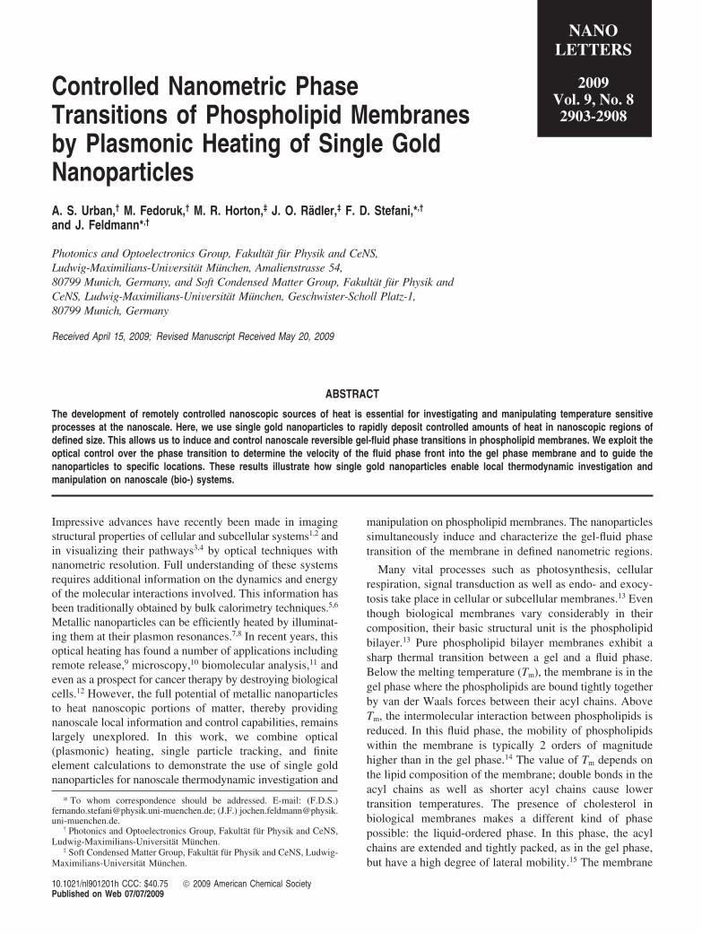
Artificial model membranes have played an important rolein revealing physical and chemical characteristics of mem-brane function.17,18 We used phospholipid giant unilamellarvesicles (GUVs; 15 to 50 µm in diameter) prepared byelectroformation19 as a model system for the biological cellmembrane. Vesicles in the gel phase generally present facetedshapes due to the rigidity of the membrane, whereas vesiclesin the fluid phase take a spherical shape (Figure 1A). Goldnanoparticles with a diameter of 80 nm were coated withcetyl trimethyl ammonium bromide (CTAB) and attachedto the surface of the vesicles (Figure 1B). We used opticaldark-field microscopy (Figure 1C) to characterize the nano-particle-modified vesicles, to perform spectroscopy on thegold nanoparticles, and to directly visualize the effects ofnanoparticle heating. A continuous-wave (heating) laser at532 nm was used to illuminate the nanoparticles near theirplasmon resonance (Figure 1C,D). We observed that incomparison to gold nanoparticles with other surface ligands(e.g., citrate molecules) the CTAB-coated nanoparticles havea considerably higher affinity to the phospholipid membranes.The precise origin of this interaction is not fully understood.
However, a strong interaction between the CTAB andphospholipid molecules is indicated by the observation thatthe gold nanoparticles are immobile when attached to vesiclesin the gel phase.
In a first experiment, gold nanoparticles were attached to1,2-dipalmitoyl-sn-glycero-3-phosphocholine (DPPC) vesicles,which have a Tm of 41 °C14 and are therefore in the gel phaseat room temperature (Figure 1B). We optically heated thegold nanoparticles with power densities P < 350 kW/cm2.Higher values of P led to the destruction of the vesicles.Figure 1B shows the effect of the simultaneous opticalheating of several gold nanoparticles attached to a DPPCvesicle. Within a short period of time the illuminated vesiclerelaxed into the energetically favorable spherical shape. Inaddition, during the optical heating, the beforehand immobilenanoparticles started to move over the membrane.20 Laserillumination at 633 nm, where the gold nanoparticles do notabsorb substantially, produced no effect.
In order to understand the light-induced heat depositionand the behavior of the nanoparticle-modified vesicles, wesimulated the heat transfer in our system by finite-elementcalculations. These calculations require the absorption cross-section of the gold nanoparticle, the laser power density, thegeometry of the nanoparticle-membrane system, and the
Figure 1. Optically induced phase transition of a nanoparticle-modified giant unilamellar vesicle. (A) Schematic of a vesicle in the gel(left) and in the fluid (right) phase modified with gold nanoparticles (Au-NPs). For clarity, the schematics are not drawn to scale (thephospholipid membrane has a thickness of 5 nm and the gold nanoparticles a diameter of 80 nm). (B) Dark-field micrograph of twoadjacent gel-phase vesicles modified with gold nanoparticles (left). Membrane and nanoparticles appear with a size of about 1 µm due tothe diffraction limited detection of the scattering signal. The brightness scale is saturated in order to visualize both the strongly scatteringgold nanoparticles and the weakly scattering phospholipid membrane. The lower vesicle is illuminated with the heating laser (green circle)for about 1 min with a power density P = 100 kW/cm2. This vesicle relaxes to a spherical shape (right). (C) Dark-field microscope adaptedfor optical heating. (D) Calculated (Mie theory) absorption (AMie) and scattering (SMie) spectra together with the measured scattering spectrum(Sexp) of an 80 nm gold nanoparticle. A vertical line marks the 532 nm wavelength of the heating laser.
2904 Nano Lett., Vol. 9, No. 8, 2009
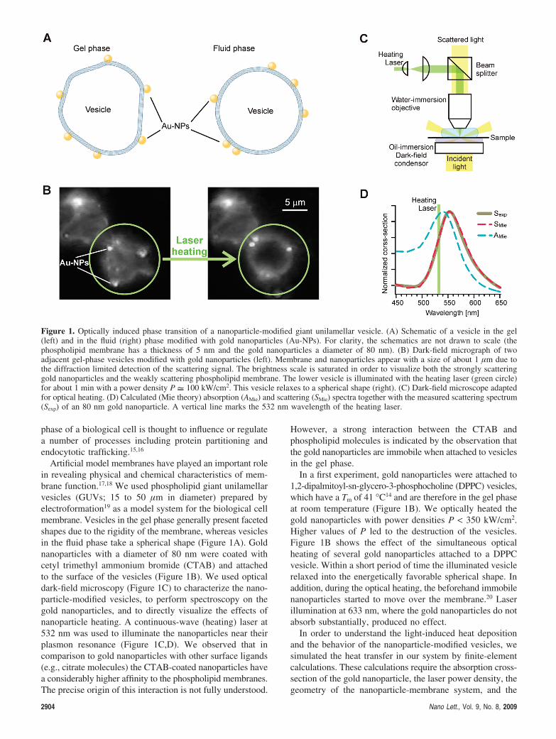
thermal capacity and conductivity of all components. Wedetermined the absorption cross-section from experimentallymeasured scattering spectra (Figure 1D). We used standardvalues for the thermal properties of gold and of thesurrounding solution. The extent to which the membranebends around the gold nanoparticle is not known.21 Wetherefore simulated the two extreme situations of a nano-particle lying over a flat membrane and of a nanoparticlefully wrapped by the membrane (Figure 2A). Since thethermal conductivity of phospholipid membranes is alsounknown, we performed calculations using a range of valuesfrom those of hydrocarbons of similar acyl chains to thoseof common polymers. Likewise, the heat capacity was variedby an order of magnitude around reported values.22,23 It turnsout that the thermal properties of the membrane as well asthe degree of wrapping only have a small influence on thethermal behavior of the full system. The variation of theseparameters produces temperature deviations of 2-8% onlyin the close proximity of the gold nanoparticle (R < 20 nm,Figure 2A). Illumination with the heating laser results in ahighly localized temperature increase around the nanoparticle(Figure 2B). In such small nanometric regions, water canreach temperatures above 100 °C without boiling.24 A definedcircular region of the membrane around the nanoparticle ishotter than Tm (Figure 2C). The radius of this fluid region(Rf) increases with P and lies in the 0-350 nm range. Theheating of the phospholipid membrane is extremely rapid;95% of the steady-state temperature is typically reachedwithin 100 ns. These calculations explain the experimentalobservations. Gold nanoparticles free in solution undergothree-dimensional Brownian motion. On the surface of a gelmembrane, the nanoparticle motion is stopped as a result ofthe low mobility of the phospholipids in the membrane andthe strong interaction of CTAB and phospholipid molecules.When optically heated, the gold nanoparticles melt a regionof radius Rf in the vesicle membrane. The much higher lipidmobility in the surrounding fluid region re-enables thenanoparticle motion, this time in two dimensions over themembrane surface. The nanoparticles melt other regions as
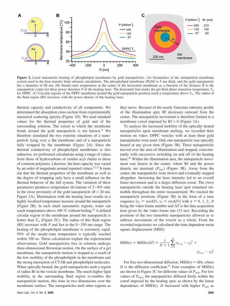
they move. Because of the nearly Gaussian intensity profileof the illumination spot, Rf decreases outward from thecenter. The nanoparticle movement is therefore limited to amembrane corral imposed by Rf > 0 (Figure 3A).
To analyze the increased mobility of the optically heatednanoparticles upon membrane melting, we recorded theirmotion on video. DPPC vesicles with at least three goldnanoparticles were used. Only one nanoparticle was opticallyheated at any given time (Figure 3B). These nanoparticlesmoved over the area of illumination and stopped, concomi-tantly with successive switching on and off of the heatinglaser.20 Within the illumination area, the nanoparticle move-ment was fastest in the center, where Rf and the powerdensity are maximal (Pmax) (Figure 3B). Away from thecenter, the nanoparticles were slower and eventually stoppedaltogether. Increasing the laser intensity led to an overallfaster movement and to a larger diffusion corral. The (two)nanoparticles outside the heating laser spot remained im-mobile throughout the entire measurement. We tracked thenanoparticle positions (Figure 3B) in the form of a timesequence [xn ) x(n∆T), yn ) y(n∆T)] with n ) 0, 1, 2...Nbeing the video frame number and ∆T is the data acquisitiontime given by the video frame rate (33 ms). Recording thepositions of the two immobile nanoparticles allowed us tosubtract movements of the vesicle as a whole. From therecorded trajectories we calculated the time dependent meansquare displacement (MSD)
For free two-dimensional diffusion, MSD(t) ) 4Dt, whereD is the diffusion coefficient.25 Four examples of MSD(t)are shown in Figure 3C for different values of Pmax. For lowvalues of Pmax, the nanoparticles diffused freely within thecorral imposed by the heating spot, as shown by the lineardependence of MSD(t). D increased with higher Pmax as
Figure 2. Local nanometric heating of phospholipid membranes by gold nanoparticles. (A) Geometries of the nanoparticle-membranesystem used in the heat transfer finite element calculations. The phospholipid membrane (PLM) is 5 nm thick, and the gold nanoparticlehas a diameter of 80 nm. (B) Steady-state temperature at the center of the horizontal membrane as a function of the distance R to thenanoparticle center for three power densities P of the heating laser. The horizontal line marks the gel-fluid phase transition temperature Tm
for DPPC. (C) Circular regions of the DPPC membrane around the gold nanoparticle position reach a temperature above Tm. The radius ofthe fluid region (Rf) increases with the power density of the heating laser.
MSD(t) ) MSD(n∆T) ) 1N + 1 ∑
i)0
N
(xi+n - xi)2 +
(yi+n - yi)2
Nano Lett., Vol. 9, No. 8, 2009 2905
reflected in the larger slopes of MSD(t). For sufficiently highPmax (e.g., Pmax ) 265 kW/cm2) the gold nanoparticlesexhibited a subdiffusive behavior. This is a result of theirmobility being so high that within the observation time thegold nanoparticles reached the limits of the diffusion corral.
We obtained further insight into the nanoscale phasetransition by measuring the diffusion coefficient of nano-particles on DPPC and 1,2-dioleoyl-sn-glycero-3-phospho-choline (DOPC) vesicles as a function of Pmax (Figure 3D).The membranes of DOPC have a Tm of approximately -20°C14 and are thus in the fluid phase at room temperature.Measurements on DOPC vesicles serve as a control for theinfluence of optical heating on the mobility of gold nano-particles on a fluid phospholipid membrane. Gold nanopar-ticles on DOPC vesicles display an exponential increase ofD with Pmax (Figure 3D), reflecting the reduction of the fluidmembrane viscosity with temperature. The values of Dcorrespond well to reported diffusion coefficients of lipid-tagged gold nanoparticles on fluid artificial bilayers26 andbiological membranes.27 In contrast, measurements on DPPCvesicles reveal three different regimes of D as a function of
Pmax (Figure 3D). In the high Pmax regime, an exponentialdependence analogous to the one of gold nanoparticles onDOPC is observed. For a given Pmax, the viscosity of thefluid DPPC membrane is higher than that of a DOPCmembrane due to the lower relative temperature (T - Tm).Thus, the values of D for nanoparticles on DPPC are lowerthan for DOPC. In the intermediate regime, D exhibits asubstantially faster exponential increase than in the high Pmax
regime. And in the low Pmax regime, nanoparticle movementis undetectable. The scatter of the data for Pmax < 80 kW/cm2 results from the experimental error of 150 nm in thelocalization of the nanoparticles. In order to understand theseresults, we need to account for the dynamics of the phasetransition. The nanoparticle heats up a region of radius Rfto a temperature above Tm on a submicrosecond time scale.However, the complete melting of that region may takeconsiderably longer. In the high Pmax regime, the nanopar-ticles are extremely hot. The fluid region around them isgenerated so rapidly that they diffuse freely as the nanopar-ticles on the full fluid membranes of DOPC do. In theintermediate regime, the movement of the nanoparticle is
Figure 3. Nanometric gel-fluid phase transitions of phospholipid membranes. (A) An optically heated gold nanoparticle melts a region ofthe membrane with radius Rf and may move over the membrane. For a Gaussian heating spot, Rf decreases outward from the center. Theheated nanoparticle movement is restricted to the area where Rf > 0. (B) Dark-field micrograph of a gel phase DPPC vesicle with three goldnanoparticles on its surface. Only the nanoparticle on the left is optically heated. A 10 s trajectory of this nanoparticle (red; Pmax ) 200kW/cm2) and of one of the nonheated nanoparticles (light blue) are shown. The green circles denote the calculated radius Rf of the fluidregion around the heated nanoparticle for different positions within the illumination area (Gaussian, 9 µm fwhm). (C) MSD vs time of goldnanoparticles on a DPPC vesicle for four values of Pmax (Rfmax). (D) Diffusion coefficient D and mean square velocity ⟨V2⟩ of gold nanoparticleson the surface of DPPC and DOPC vesicles as a function of Pmax (Rfmax). Each value of D is the average obtained from at least 10 nanoparticletrajectories. The statistical error bars are smaller than the plot markers.
2906 Nano Lett., Vol. 9, No. 8, 2009
limited by the rate of the phase transition. A gold nanoparticlemoving along the membrane cannot progress faster than thefluid front advances from the hot nanoparticle surface intothe gel phase. In this regime the velocity of the fluid phasefront is directly obtained from the relation ⟨ν2⟩ ) D/∆T25
(Figure 3D, right axis). We determine fluid phase frontvelocities ranging from 0.9 to 4.5 µm/s. In the low Pmax
regime, either the heat produced by the gold nanoparticle isnot sufficient to produce a fluid region or the time neededby the fluid front to advance is too long to enable a noticeablenanoparticle motion.
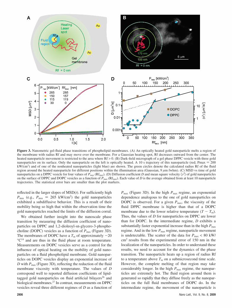
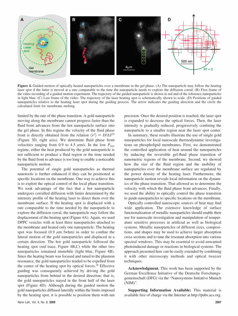
The potential of single gold nanoparticles as thermalnanotools is further enhanced if they can be positioned atspecific locations on the membrane. One way to achieve thisis to exploit the optical control of the local phase transition.We took advantage of the fact that a hot nanoparticleundergoes corralled diffusion with limits determined by theintensity profile of the heating laser to direct them over themembrane surface. If the heating spot is displaced with arate comparable to the time needed by the nanoparticle toexplore the diffusion corral, the nanoparticle may follow thedisplacement of the heating spot (Figure 4A). Again, we usedDPPC vesicles with at least three nanoparticles attached tothe membrane and heated only one nanoparticle. The heatingspot was focused (0.9 µm fwhm) in order to confine thelateral motion of the gold nanoparticles and displaced in acertain direction. The hot gold nanoparticle followed theheating spot (red trace, Figure 4B,C) while the other twonanoparticles remained immobile (light blue, Figure 4B).Since the heating beam was focused and tuned to the plasmonresonance, the gold nanoparticles tended to be expelled fromthe center of the heating spot by optical forces.28 Effectiveguiding was consequently achieved by driving the goldnanoparticles from behind in the desired direction; that is,the gold nanoparticles stayed in the front half of the laserspot (Figure 4D). Although during the guided motion thegold nanoparticles diffused laterally within the limits imposedby the heating spot, it is possible to position them with nm
precision. Once the desired position is reached, the laser spotis expanded to decrease the optical forces. Then, the laserintensity is gradually reduced, progressively confining thenanoparticle to a smaller region near the laser spot center.
In summary, these results illustrate the use of single goldnanoparticles for local nanoscale thermodynamic investiga-tions on phospholipid membranes. First, we demonstratedthe controlled application of heat around the nanoparticlesby inducing the reversible gel-fluid phase transition ofnanometric regions of the membrane. Second, we showedhow the size of the fluid region and the mobility ofnanoparticles over the membrane surface are regulated bythe power density of the heating laser. Furthermore, thenanoparticle motion reveals local information on the dynam-ics of the phase transition. That allowed us to determine thevelocity with which the fluid phase front advances. Finally,we used the ability to optically control the phase transitionto guide nanoparticles to specific locations on the membrane.
Optically controlled nanoscopic sources of heat may findwide application. The extensive knowledge of surfacefunctionalization of metallic nanoparticles should enable theiruse for nanoscale investigation and manipulation of temper-ature sensitive processes in artificial as well as biologicalsystems. Metallic nanoparticles of different sizes, composi-tions, and shapes may be used to achieve larger absorptioncross sections and to tune the resonant absorption into variousspectral windows. This may be essential to avoid unwantedphotoinduced damage or reactions in biological systems. Theapproach presented here can be easily extended by combiningit with other microscopy methods and optical tweezertechniques.
Acknowledgment. This work has been supported by theGerman Excellence Initiative of the Deutsche Forschungs-gemeinschaft (DFG) via the “Nanosystems Initiative Munich(NIM)”.
Supporting Information Available: This material isavailable free of charge via the Internet at http://pubs.acs.org.
Figure 4. Guided motion of optically heated nanoparticles over a membrane in the gel phase. (A) The nanoparticle may follow the heatinglaser spot if the latter is moved at a rate comparable to the time the nanoparticle needs to explore the diffusion corral. (B) First frame ofthe video recording of a guided motion experiment. The trajectory of the guided nanoparticle is shown in red and of the reference nanoparticlesin light blue. (C) Last frame of the video. The trajectory of the laser heating spot is schematically shown to scale. (D) Positions of guidednanoparticles relative to the heating laser spot during the guiding process. The arrow indicates the guiding direction and the circle thecalculated limit for membrane melting.
Nano Lett., Vol. 9, No. 8, 2009 2907
References(1) Nagerl, U. V.; Willig, K. I.; Hein, B.; Hell, S. W.; Bonhoeffer, T.
Proc. Natl. Acad. Sci. U.S.A. 2008, 105 (48), 18982–18987.(2) Huang, B.; Wang, W. Q.; Bates, M.; Zhuang, X. W. Science 2008,
319 (5864), 810–813.(3) Yildiz, A.; Forkey, J. N.; McKinney, S. A.; Ha, T.; Goldman, Y. E.;
Selvin, P. R. Science 2003, 300 (5628), 2061–2065.(4) Lidke, D. S.; Nagy, P.; Heintzmann, R.; Arndt-Jovin, D. J.; Post, J. N.;
Grecco, H. E.; Jares-Erijman, E. A.; Jovin, T. M. Nat. Biotechnol.2004, 22 (2), 198–203.
(5) Weber, P. C.; Salemme, F. R. Current Opinion in Structural Biology2003, 13 (1), 115–121.
(6) Leavitt, S.; Freire, E. Curr. Opin. Struct. Biol. 2001, 11 (5), 560–566.
(7) Richardson, H. H.; Carlson, M. T.; Tandler, P. J.; Hernandez, P.;Govorov, A. O. Nano Lett. 2009, 9 (3), 1139–1146.
(8) Perner, M.; Gresillon, S.; Marz, J.; von Plessen, G.; Feldmann, J.;Porstendorfer, J.; Berg, K. J.; Berg, G. Phys. ReV. Lett. 2000, 85 (4),792–795.
(9) Skirtach, A. G.; Dejugnat, C.; Braun, D.; Susha, A. S.; Rogach, A. L.;Parak, W. J.; Mohwald, H.; Sukhorukov, G. B. Nano Lett. 2005, 5(7), 1371–1377.
(10) Cognet, L.; Tardin, C.; Boyer, D.; Choquet, D.; Tamarat, P.; Lounis,B. Proc. Natl. Acad. Sci. U.S.A. 2003, 100 (20), 11350–11355.
(11) Stehr, J.; Hrelescu, C.; Sperling, R. A.; Raschke, G.; Wunderlich, M.;Nichtl, A.; Heindl, D.; Kurzinger, K.; Parak, W. J.; Klar, T. A.;Feldmann, J. Nano Lett. 2008, 8 (2), 619–623.
(12) Hirsch, L. R.; Stafford, R. J.; Bankson, J. A.; Sershen, S. R.; Rivera,B.; Price, R. E.; Hazle, J. D.; Halas, N. J.; West, J. L. Proc. Natl.Acad. Sci. U.S.A. 2003, 100 (23), 13549–13554.
(13) Lodish, H. F.; Darnell, J. E. Molecular cell biology, 3rd ed.; ScientificAmerican Books: New York, 1995.
(14) Heimburg, T. Thermal biophysics of membranes; Wiley-VCH Verlag:Weinheim, 2007.
(15) Brown, D. A.; London, E. J. Biol. Chem. 2000, 275 (23), 17221–17224.
(16) Rajendran, L.; Simons, K. J. Cell Sci. 2005, 118 (6), 1099–1102.(17) Chan, Y. H. M.; Boxer, S. G. Curr. Opin. Chem. Biol. 2007, 11 (6),
581–587.(18) Lipowsky, R.; Sackmann, E. Structure and dynamics of membranes;
Elsevier Science: Amsterdam, 1995.(19) Bagatolli, L. A.; Gratton, E. Biophys. J. 1999, 77 (4), 2090–2101.(20) See Supporting Information.(21) Deserno, M.; Gelbart, W. M. J. Phys. Chem. B 2002, 106 (21), 5543–
5552.(22) Blume, A. Biochemistry 1983, 22 (23), 5436–5442.(23) Gershfeld, N. L.; Mudd, C. P.; Tajima, K.; Berger, R. L. Biophys. J.
1993, 65 (3), 1174–1179.(24) Skripov, V. P.; Torstveit, S. Thermophysical properties of liquids in
the metastable (superheated) state; Gordon and Breach SciencePublishers: New York, 1988.
(25) Hong, Q. A.; Sheetz, M. P.; Elson, E. L. Biophys. J. 1991, 60 (4),910–921.
(26) Lee, G. M.; Ishihara, A.; Jacobson, K. A. Proc. Natl. Acad. Sci. U.S.A.1991, 88 (14), 6274–6278.
(27) Lee, G. M.; Zhang, F.; Ishihara, A.; Mcneil, C. L.; Jacobson, K. A.J. Cell Biol. 1993, 120 (1), 25–35.
(28) Agayan, R. R.; Gittes, F.; Kopelman, R.; Schmidt, C. F. Appl. Opt.2002, 41 (12), 2318–2327.
NL901201H
2908 Nano Lett., Vol. 9, No. 8, 2009