colorectal cancer screening - journal of the national ... · colon cancer and 40,870 new cases of...
TRANSCRIPT

© Journal of the National Comprehensive Cancer Network | Volume 8 Number 1 | January 2010
8
Gideon Steinbach, MD, PhD; Jonathan P. Terdiman, MD; and David Weinberg, MD, MSc
OverviewColorectal cancer (CRC) is the third most frequently diagnosed cancer in men and women in the United States. In 2009, an estimated 106,100 new cases of colon cancer and 40,870 new cases of rectal cancer will occur in the United States, and 49,920 people will die of colon and rectal cancers.1 Patients with lo-calized colon cancer have a 90% 5-year survival rate.
CRC mortality can be reduced through early di-agnosis and cancer prevention with polypectomy.2 Therefore, the goal of CRC screening is to detect cancer at an early, curable stage and to detect and remove clinically significant adenomas. Screening
The NCCN
Colorectal Cancer ScreeningClinical Practice Guidelines in OncologyTM
Randall W. Burt, MD; James S. Barthel, MD; Kelli Bullard Dunn, MD; Donald S. David, MD; Ernesto Drelichman, MD; James M. Ford, MD; Francis M. Giardiello, MD, MBA; Stephen B. Gruber, MD, PhD, MPH; Amy L. Halverson, MD; Stanley R. Hamilton, MD; Mohammad K. Ismail, MD; Kory Jasperson, MS, CGC; Audrey J. Lazenby, MD; Patrick M. Lynch, MD, JD; Edward W. Martin, Jr., MD; Robert J. Mayer, MD; Reid M. Ness, MD, MPH; Dawn Provenzale, MD, MS; M. Sambasiva Rao, MD; Moshe Shike, MD;
Colorectal Cancer Screening Clinical Practice Guidelines in Oncology
Key WordsNCCN Clinical Practice Guidelines, colorectal cancer, fecal oc-cult blood test, adenoma, HNPCC, APC, FAP, colon polyp, stool DNA, Lynch syndrome, FIT test, colonoscopy, colon cancer screening (JNCCN 2010;8:8–61)
NCCN Categories of Evidence and ConsensusCategory 1: The recommendation is based on high-level evidence (e.g., randomized controlled trials) and there is uniform NCCN consensus.Category 2A: The recommendation is based on lower-level evidence and there is uniform NCCN consensus.Category 2B: The recommendation is based on lower-level evidence and there is nonuniform NCCN consensus (but no major disagreement).Category 3: The recommendation is based on any level of evidence but reflects major disagreement.
All recommendations are category 2A unless otherwise noted.
Clinical trials: The NCCN believes that the best management for any cancer patient is in a clinical trial. Participation in clinical trials is especially encouraged.
Please NoteThese guidelines are a statement of consensus of the
authors regarding their views of currently accepted ap-proaches to treatment. Any clinician seeking to apply or consult these guidelines is expected to use independent medical judgment in the context of individual clinical cir-cumstances to determine any patient’s care or treatment. The National Comprehensive Cancer Network makes no representation or warranties of any kind regarding their content, use, or application and disclaims any responsibil-ity for their applications or use in any way.
These guidelines are copyrighted by the National Comprehensive Cancer Network. All rights reserved. These guidelines and the illustrations herein may not be reproduced in any form without the express written per-mission of the NCCN © 2010.Disclosures for the NCCN Colorectal Cancer Screening Guidelines Panel
At the beginning of each NCCN guidelines panel meeting, panel members disclosed any financial support they have received from industry. Through 2008, this information was published in an aggregate statement in JNCCN and online. Furthering NCCN’s commitment to public transparency, this disclosure process has now been expanded by listing all potential conflicts of interest respective to each individual expert panel member.
Individual disclosures for the NCCN Colorectal Cancer Screen-ing Guidelines Panel members can be found on page 61. (To view the most recent version of these guidelines and accom-panying disclosures, visit the NCCN Web site at NCCN.org.)
These guidelines are also available on the Internet. For the latest update, please visit NCCN.org.

Colorectal Cancer Screening
NCCNClinical Practice Guidelines
© Journal of the National Comprehensive Cancer Network | Volume 8 Number 1 | January 2010
9
Journal of the National Comprehensive Cancer Network
Text continues on p. 43
tests that can detect both early cancer and adeno-matous polyps are encouraged, although the panel recognizes that patient preference and resource ac-cessibility play a large role in test selection.
Current technology falls into 2 broad categories: structural and stool/fecal-based tests. Although some techniques are better established than others, panel-ists agreed that any screening is better than none.
Structural Screening TestsStructural tests are able to detect both early can-cer and adenomatous polyps using endoscopic or radiologic imaging. These have several limitations, including their relative invasiveness, the need for dietary preparation and bowel cleansing, and the
time dedicated to the examination (typically a day). Endoscopic examinations require informed consent and sedation, and have related risks, including per-foration and bleeding. Recently, a large cohort study of 53,220 Medicare patients between ages 66 and 95 years showed that risk for adverse events after colo-noscopy increases with age.3
ColonoscopyColonoscopy is the most complete screening proce-dure, allowing the entire large bowel to be examined and polyps to be removed in one session. It is cur-rently the preferred screening method and also the required procedure for confirming positive findings from other tests. Colonoscopy is also considered the current gold standard for assessing the efficacy of other screening methods. Although no randomized
NCCN Colorectal Cancer Screening Panel Members*Randall W. Burt, MD/Chair¤∆
Huntsman Cancer Institute at the University of UtahJames S. Barthel, MD¤Þ
H. Lee Moffitt Cancer Center & Research InstituteKelli Bullard Dunn, MD¶
Roswell Park Cancer InstituteDonald S. David, MD¤
City of Hope Comprehensive Cancer CenterErnesto Drelichman, MD¶
University of Alabama at BirminghamComprehensive Cancer Center
James M. Ford, MD†Þ∆Stanford Comprehensive Cancer Center
Francis M. Giardiello, MD, MBA¤The Sidney Kimmel Comprehensive Cancer Center atJohns Hopkins
Stephen B. Gruber, MD, PhD, MPH†∆University of Michigan Comprehensive Cancer Center
Amy L. Halverson, MD¶Robert H. Lurie Comprehensive Cancer Center ofNorthwestern University
Stanley R. Hamilton, MD≠The University of Texas M. D. Anderson Cancer Center
Mohammad K. Ismail, MD¤St. Jude Children’s Research Hospital/University of Tennessee Cancer Institute
Kory Jasperson, MS, CGC∆Huntsman Cancer Institute at the University of Utah
Audrey J. Lazenby, MD≠UNMC Eppley Cancer Center atThe Nebraska Medical Center
Patrick M. Lynch, MD, JD¤The University of Texas M. D. Anderson Cancer Center
Edward W. Martin, Jr, MD¶The Ohio State University Comprehensive Cancer Center –James Cancer Hospital and Solove Research Institute
Robert J. Mayer, MD†Dana-Farber/Brigham and Women’s Cancer Center
Reid M. Ness, MD, MPH¤Vanderbilt-Ingram Cancer Center
Dawn Provenzale, MD, MS¤ÞDuke Comprehensive Cancer Center
M. Sambasiva Rao, MD≠¤Robert H. Lurie Comprehensive Cancer Center ofNorthwestern University
Moshe Shike, MD¤ÞMemorial Sloan-Kettering Cancer Center
Gideon Steinbach, MD, PhD¤ÞFred Hutchinson Cancer Research Center/Seattle Cancer Care Alliance
Jonathan P. Terdiman, MD¤UCSF Helen Diller Family Comprehensive Cancer Center
David Weinberg, MD, MSc¤Fox Chase Cancer Center
KEY:
*Writing Committee Member
Specialties: ¤Gastroenterology; ∆Cancer Genetics; ÞInternal Medicine; ¶Surgery/Surgical Oncology; †Medical Oncology; ≠Pathology
© Journal of the National Comprehensive Cancer Network | Volume 8 Number 1 | January 2010
10
Colorectal Cancer Screening Version 1:2010
Clinical trials: The NCCN believes that the best management for any cancer patient is in a clinical trial. Participation in clinical trials is especially encouraged. All recommendations are category 2A unless otherwise noted.
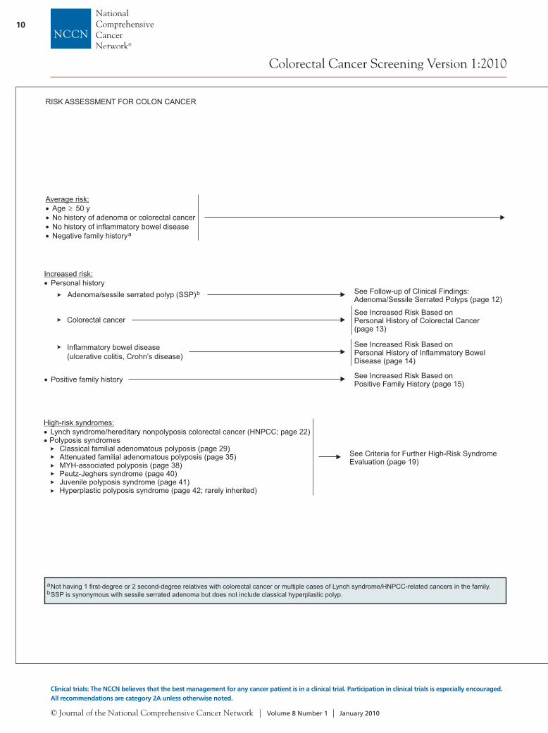
See Criteria for Further High-Risk SyndromeEvaluation (page 19)
Average risk:Age 50 yNo history of adenoma or colorectal cancerNo history of inflammatory bowel diseaseNegative family historya
ab
Not having 1 first-degree or 2 second-degree relatives with colorectal cancer or multiple cases of Lynch syndrome/HNPCC-related cancers in the family.SSP is synonymous with sessile serrated adenoma but does not include classical hyperplastic polyp.
Increased risk:Personal history
RISK ASSESSMENT FOR COLON CANCER
Adenoma/sessile serrated polyp (SSP)b
Colorectal cancer
Inflammatory bowel disease(ulcerative colitis, Crohn’s disease)
See Follow-up of Clinical Findings:Adenoma/Sessile Serrated Polyps (page 12)
See Increased Risk Based onPersonal History of Colorectal Cancer(page 13)
See Increased Risk Based onPersonal History of Inflammatory BowelDisease (page 14)
Positive family history See Increased Risk Based onPositive Family History (page 15)
High-risk syndromes:
Polyposis syndromesClassical (page 29)
MYH-associated polyposis (page 38)Peutz-Jeghers syndrome (page 40)Juvenile polyposis syndrome (page 41)Hyperplastic polyposis syndrome (page 42; rarely inherited)
Lynch syndrome/hereditary nonpolyposis colorectal cancer (HNPCC; page 22)
familial adenomatous polyposisAttenuated familial adenomatous polyposis (page 35)
SCREENING MODALITYAND SCHEDULEc,d
Colonoscopyf
See Follow-up ofClinical Findings:Adenoma/SSP(page 12)
EVALUATION OF POSITIVESCREENING FINDINGS
Biopsy
Hyperplastic
Adenoma/SSPi
See Follow-up ofClinical Findings:Adenoma/SSP(page 12)
Negative/no polyps
Repeatcolonoscopyin 10 yc
Positive/polyps Polypectomy
Adenoma/SSPi
HyperplasticRepeatcolonoscopyin 10 yc
Positive
Colonoscopy(preferred if available)
e,f
Stool based:Guaiac-based (category 1) orimmunohistochemical-basedtesting annually± flexible sigmoidoscopy every 5 y
g
h
or
or
Flexible sigmoidoscopy
Repeat flexiblesigmoidoscopy in 5 yc
Negative/no polyps
Positive/polyps
Colonoscopyf
Repeat flexiblesigmoidoscopy in 5 yc
cd
e
fg
hi
See Screening Modality and Schedule (pages 16 and 17).Currently there is no consensus on the use of CT colonography as a primary screening modality and it is evolving with regards to recommended/programmatic frequency, polyp size leading to referral for colonoscopy, and protocol for evaluating extracolonic lesions. However, the data availablesuggests that if CT colonography is negative/no polyps, then CT colonography should be repeated in 5 y, and if positive/polyps lesions, colonoscopyshould be performed.
Other screening modalities, such as double-contrast barium enema, should be reserved for those who are not able to undergo colonoscopy, orcolonoscopy is technically incomplete.
If colonoscopy incomplete, consider other screening modalities or repeat colonoscopy at discretion of physician.Emerging technologies, such as stool DNA, have shown increasing evidence as reasonably accurate screening tests but data are limited for determining aninterval between screening. Currently, stool DNA is not considered a first-line screening test except in specific circumstances.
Studies at the present time have shown that fecal immunohistochemical testing (FIT) is as good as, if not superior to, guaiac-based testing.SSPs are managed the same as adenomas.

NCCN Clinical Practice Guidelines in Oncology
© Journal of the National Comprehensive Cancer Network | Volume 8 Number 1 | January 2010
11
Colorectal Cancer Screening Version 1:2010
Version 1.2010, 10-23-09 ©2010 National Comprehensive Cancer Network, Inc. All rights reserved. These guidelines and this illustration may not be reproduced in any form without the express written permission of NCCN.
See Criteria for Further High-Risk SyndromeEvaluation (page 19)
Average risk:Age 50 yNo history of adenoma or colorectal cancerNo history of inflammatory bowel diseaseNegative family historya
ab
Not having 1 first-degree or 2 second-degree relatives with colorectal cancer or multiple cases of Lynch syndrome/HNPCC-related cancers in the family.SSP is synonymous with sessile serrated adenoma but does not include classical hyperplastic polyp.
Increased risk:Personal history
RISK ASSESSMENT FOR COLON CANCER
Adenoma/sessile serrated polyp (SSP)b
Colorectal cancer
Inflammatory bowel disease(ulcerative colitis, Crohn’s disease)
See Follow-up of Clinical Findings:Adenoma/Sessile Serrated Polyps (page 12)
See Increased Risk Based onPersonal History of Colorectal Cancer(page 13)
See Increased Risk Based onPersonal History of Inflammatory BowelDisease (page 14)
Positive family history See Increased Risk Based onPositive Family History (page 15)
High-risk syndromes:
Polyposis syndromesClassical (page 29)
MYH-associated polyposis (page 38)Peutz-Jeghers syndrome (page 40)Juvenile polyposis syndrome (page 41)Hyperplastic polyposis syndrome (page 42; rarely inherited)
Lynch syndrome/hereditary nonpolyposis colorectal cancer (HNPCC; page 22)
familial adenomatous polyposisAttenuated familial adenomatous polyposis (page 35)
SCREENING MODALITYAND SCHEDULEc,d
Colonoscopyf
See Follow-up ofClinical Findings:Adenoma/SSP(page 12)
EVALUATION OF POSITIVESCREENING FINDINGS
Biopsy
Hyperplastic
Adenoma/SSPi
See Follow-up ofClinical Findings:Adenoma/SSP(page 12)
Negative/no polyps
Repeatcolonoscopyin 10 yc
Positive/polyps Polypectomy
Adenoma/SSPi
HyperplasticRepeatcolonoscopyin 10 yc
Positive
Colonoscopy(preferred if available)
e,f
Stool based:Guaiac-based (category 1) orimmunohistochemical-basedtesting annually± flexible sigmoidoscopy every 5 y
g
h
or
or
Flexible sigmoidoscopy
Repeat flexiblesigmoidoscopy in 5 yc
Negative/no polyps
Positive/polyps
Colonoscopyf
Repeat flexiblesigmoidoscopy in 5 yc
cd
e
fg
hi
See Screening Modality and Schedule (pages 16 and 17).Currently there is no consensus on the use of CT colonography as a primary screening modality and it is evolving with regards to recommended/programmatic frequency, polyp size leading to referral for colonoscopy, and protocol for evaluating extracolonic lesions. However, the data availablesuggests that if CT colonography is negative/no polyps, then CT colonography should be repeated in 5 y, and if positive/polyps lesions, colonoscopyshould be performed.
Other screening modalities, such as double-contrast barium enema, should be reserved for those who are not able to undergo colonoscopy, orcolonoscopy is technically incomplete.
If colonoscopy incomplete, consider other screening modalities or repeat colonoscopy at discretion of physician.Emerging technologies, such as stool DNA, have shown increasing evidence as reasonably accurate screening tests but data are limited for determining aninterval between screening. Currently, stool DNA is not considered a first-line screening test except in specific circumstances.
Studies at the present time have shown that fecal immunohistochemical testing (FIT) is as good as, if not superior to, guaiac-based testing.SSPs are managed the same as adenomas.

© Journal of the National Comprehensive Cancer Network | Volume 8 Number 1 | January 2010
12
Colorectal Cancer Screening Version 1:2010
Clinical trials: The NCCN believes that the best management for any cancer patient is in a clinical trial. Participation in clinical trials is especially encouraged. All recommendations are category 2A unless otherwise noted.
Malignant polyp See NCCN Clinical Practice Guidelines in Oncology:Colon Cancer*
FOLLOW-UP OF CLINICAL FINDINGSc
Increased-riskpatients: personalhistory ofadenomas/SSPsfound atcolonoscopy
Advanced or multiple adenomas:High-grade dysplasia
1 cmVillous (> 25% villous)Between 3-10 polypsj
> 10 cumulative adenomas j
Incomplete or piecemealpolypectomy or polypectomy oflarge sessile polyps
k
Repeatcolonoscopywithin 3 y
Repeatcolonoscopywithin 5 y
Individual managementConsider a polyposis syndrome
Repeat colonoscopy within 2-6 mo(timing dependent on endoscopic and pathologic findings)
See HereditaryColorectal CancerPathway (page 19)
Low-risk adenoma:2 polyps
< 1 cmTubular
Repeatcolonoscopywithin 5 y
Repeatcolonoscopyevery 5-10 y
Positive/polyps
Negative/no polyps
Negative/no polyps
CLINICAL FINDINGS
INCREASED RISK BASED ON PERSONAL HISTORY OF ADENOMA/SESSILE SERRATED POLYPS (SSP)i
RISKSTATUS
Personal history of curativeintent resected colorectalcancerl,m
Colonoscopy in 1 y,(within 3-6 mo if there was noor incomplete preoperativecolonoscopy)
Adenoma/SSP Repeat colonoscopy in 1-3 yn
Repeat colonoscopy in 2-3 y, thenevery 3-5 y based on findings
SURVEILLANCE
INCREASED RISK BASED ON PERSONAL HISTORY OF COLORECTAL CANCER
RISKSTATUS FOLLOW-UP OF CLINICAL FINDINGS
Negative/no polyps
l
m
n
Identify colorectal patients who meet Bethesda criteria; these patients may require genetic counseling or individualized management. (See High-RiskSyndromes [page 19] and Lynch Syndrome [page 22].)In addition to the colonoscopy, patients with rectal cancer should also undergo periodic limited endoscopic evaluation of the rectal anastamosis to identifylocal recurrence. Optimal timing for surveillance is not known. Expert opinion supports repeat evaluation every 3-6 mo for the first 2-3 y of surveillance.No specific data clearly support rigid versus flexible sigmoidoscopy. The usefulness of routine endoscopic ultrasound for early surveillance is not defined.See surveillance section of NCCN Clinical Practice Guidelines in Oncology: (to view the most recent version of these guidelines, visit theNCCN Website at www.NCCN.org).
The recommendation for intensive surveillance immediately after resection is based on studies that found a high rate of metachronous colorectal cancerand/or resectable recurrences in the 4-5 y after colorectal cancer resections, although the studies did not fully exclude patients with HNPCC.
Rectal Cancercijk
See Screening Modality and Schedule (pages 16 and 17).SSPs are managed the same as adenomas.Fewer than 10 polyps in the setting of a strong family history or younger age (< 40 y) may be associated with an inherited polyposis syndrome.Ink lesion for later identification.
*To view the most recent version of these guidelines, visit the NCCN Web site at www.NCCN.org.

NCCN Clinical Practice Guidelines in Oncology
© Journal of the National Comprehensive Cancer Network | Volume 8 Number 1 | January 2010
13
Colorectal Cancer Screening Version 1:2010
Version 1.2010, 10-23-09 ©2010 National Comprehensive Cancer Network, Inc. All rights reserved. These guidelines and this illustration may not be reproduced in any form without the express written permission of NCCN.
Malignant polyp See NCCN Clinical Practice Guidelines in Oncology:Colon Cancer*
FOLLOW-UP OF CLINICAL FINDINGSc
Increased-riskpatients: personalhistory ofadenomas/SSPsfound atcolonoscopy
Advanced or multiple adenomas:High-grade dysplasia
1 cmVillous (> 25% villous)Between 3-10 polypsj
> 10 cumulative adenomas j
Incomplete or piecemealpolypectomy or polypectomy oflarge sessile polyps
k
Repeatcolonoscopywithin 3 y
Repeatcolonoscopywithin 5 y
Individual managementConsider a polyposis syndrome
Repeat colonoscopy within 2-6 mo(timing dependent on endoscopic and pathologic findings)
See HereditaryColorectal CancerPathway (page 19)
Low-risk adenoma:2 polyps
< 1 cmTubular
Repeatcolonoscopywithin 5 y
Repeatcolonoscopyevery 5-10 y
Positive/polyps
Negative/no polyps
Negative/no polyps
CLINICAL FINDINGS
INCREASED RISK BASED ON PERSONAL HISTORY OF ADENOMA/SESSILE SERRATED POLYPS (SSP)i
RISKSTATUS
Personal history of curativeintent resected colorectalcancerl,m
Colonoscopy in 1 y,(within 3-6 mo if there was noor incomplete preoperativecolonoscopy)
Adenoma/SSP Repeat colonoscopy in 1-3 yn
Repeat colonoscopy in 2-3 y, thenevery 3-5 y based on findings
SURVEILLANCE
INCREASED RISK BASED ON PERSONAL HISTORY OF COLORECTAL CANCER
RISKSTATUS FOLLOW-UP OF CLINICAL FINDINGS
Negative/no polyps
l
m
n
Identify colorectal patients who meet Bethesda criteria; these patients may require genetic counseling or individualized management. (See High-RiskSyndromes [page 19] and Lynch Syndrome [page 22].)In addition to the colonoscopy, patients with rectal cancer should also undergo periodic limited endoscopic evaluation of the rectal anastamosis to identifylocal recurrence. Optimal timing for surveillance is not known. Expert opinion supports repeat evaluation every 3-6 mo for the first 2-3 y of surveillance.No specific data clearly support rigid versus flexible sigmoidoscopy. The usefulness of routine endoscopic ultrasound for early surveillance is not defined.See surveillance section of NCCN Clinical Practice Guidelines in Oncology: (to view the most recent version of these guidelines, visit theNCCN Website at www.NCCN.org).
The recommendation for intensive surveillance immediately after resection is based on studies that found a high rate of metachronous colorectal cancerand/or resectable recurrences in the 4-5 y after colorectal cancer resections, although the studies did not fully exclude patients with HNPCC.
Rectal Cancercijk
See Screening Modality and Schedule (pages 16 and 17).SSPs are managed the same as adenomas.Fewer than 10 polyps in the setting of a strong family history or younger age (< 40 y) may be associated with an inherited polyposis syndrome.Ink lesion for later identification.
*To view the most recent version of these guidelines, visit the NCCN Web site at www.NCCN.org.

© Journal of the National Comprehensive Cancer Network | Volume 8 Number 1 | January 2010
14
Colorectal Cancer Screening Version 1:2010
Clinical trials: The NCCN believes that the best management for any cancer patient is in a clinical trial. Participation in clinical trials is especially encouraged. All recommendations are category 2A unless otherwise noted.
INCREASED RISK BASED ON POSITIVE FAMILY HISTORY
SCREENINGFAMILY HISTORY CRITERIAt
tu
v
w
If a patient meets the criteria for an inherited colorectal syndrome, see Criteria for Further Risk Evaluation for High-Risk Syndromes (page 19).First-degree relatives with advanced adenoma may confer the same risk as first-degree relatives with colorectal cancer, and any adenoma < age 40 y mayconfer a similar risk to colorectal cancer < age 50 y.
In this circumstance or if any one of the revised Bethesda criteria (page 26) are met, IHC/MSI testing should be performed on the colon tumor of youngestfamily member with available colorectal cancer tissue. Also see Lynch syndrome guidelines (page 22) .Shorter intervals may be needed depending upon the family history.
Personal history ofinflammatorybowel disease
Ulcerative colitisCrohn’s disease
o8-10 y afteronset ofsymptomsp
Dysplasia/intraepithelialneoplasia
Confirmation by expertGI pathologist desirable
Colonoscopy every 1–2 yWhen clinically quiescent,4 quadrant biopsies every10 cm with > 30 totalsamples (preferred)Additional extensivesampling of strictures andmassesEndoscopic polypectomywhen appropriate withbiopsies of surroundingmucosa for the assessmentof dysplasia
INITIATION OFSCREENING
SCREENING MODALITYAND SCHEDULE
EVALUATION OF POSITIVESCREENING FINDINGS
Surgicalconsultation forresections
FOLLOW-UP OFCLINICALFINDINGSq,r
INCREASED RISK BASED ON PERSONAL HISTORY OF INFLAMMATORY BOWEL DISEASE
RISKSTATUS
op
q
r
s
Information regarding the value of endoscopic surveillance of long-standing Crohn’s disease is limited. Surveillance is at the discretion of the physician.Winawer S, Fletcher R, Rex D, et al. Colorectal cancer screening and surveillance: clinical guidelines and rationale--update based on new evidence.Gastroenterology 2003;124:544-560.
Optimal management of Crohn’s-related dysplasia remains undefined. Patient and physician preference should be considered. Extent of resection forCrohn’s-related dysplasia needs to be based on the individual findings.
Appropriate management of adenomatous polyps in the setting of ulcerative colitis is dependent on various factors and should be at the discretion of thetreating physician.
See Definitions of Common Colorectal Resections (page 18).
First-degree relative with colorectal cancerat age 50-60 yu Colonoscopy beginning at age 40 y
First-degree relative with colorectal cancerat age 60 yu Colonoscopy beginning at age 50 y Repeat every 5 y
Repeat every 5 yw
Treat as average-risk patientsColonoscopy is preferred screening
First-degree relative with colorectal cancerat < age 50 yu,v
Repeat every 3-5 ydepending on otherfamily history
Colonoscopy beginning at age 40 y, or 10 ybefore earliest diagnosis of colorectal cancer
Two related first-degree relatives with colorectalcancer at any agev
Colonoscopy beginning at age 40 y, or 10 ybefore earliest diagnosis of colorectal cancer
Repeat every 3-5 ydepending on otherfamily history
Two related second-degree relatives with colorectalat any age Colonoscopy beginning at age 50 y Repeat every 5 y
One second-degree relative or any third-degreerelative with colorectal cancerorFirst-degree relative with non-advanced adenomas

NCCN Clinical Practice Guidelines in Oncology
© Journal of the National Comprehensive Cancer Network | Volume 8 Number 1 | January 2010
15
Colorectal Cancer Screening Version 1:2010
Version 1.2010, 10-23-09 ©2010 National Comprehensive Cancer Network, Inc. All rights reserved. These guidelines and this illustration may not be reproduced in any form without the express written permission of NCCN.
INCREASED RISK BASED ON POSITIVE FAMILY HISTORY
SCREENINGFAMILY HISTORY CRITERIAt
tu
v
w
If a patient meets the criteria for an inherited colorectal syndrome, see Criteria for Further Risk Evaluation for High-Risk Syndromes (page 19).First-degree relatives with advanced adenoma may confer the same risk as first-degree relatives with colorectal cancer, and any adenoma < age 40 y mayconfer a similar risk to colorectal cancer < age 50 y.
In this circumstance or if any one of the revised Bethesda criteria (page 26) are met, IHC/MSI testing should be performed on the colon tumor of youngestfamily member with available colorectal cancer tissue. Also see Lynch syndrome guidelines (page 22) .Shorter intervals may be needed depending upon the family history.
Personal history ofinflammatorybowel disease
Ulcerative colitisCrohn’s disease
o8-10 y afteronset ofsymptomsp
Dysplasia/intraepithelialneoplasia
Confirmation by expertGI pathologist desirable
Colonoscopy every 1–2 yWhen clinically quiescent,4 quadrant biopsies every10 cm with > 30 totalsamples (preferred)Additional extensivesampling of strictures andmassesEndoscopic polypectomywhen appropriate withbiopsies of surroundingmucosa for the assessmentof dysplasia
INITIATION OFSCREENING
SCREENING MODALITYAND SCHEDULE
EVALUATION OF POSITIVESCREENING FINDINGS
Surgicalconsultation forresections
FOLLOW-UP OFCLINICALFINDINGSq,r
INCREASED RISK BASED ON PERSONAL HISTORY OF INFLAMMATORY BOWEL DISEASE
RISKSTATUS
op
q
r
s
Information regarding the value of endoscopic surveillance of long-standing Crohn’s disease is limited. Surveillance is at the discretion of the physician.Winawer S, Fletcher R, Rex D, et al. Colorectal cancer screening and surveillance: clinical guidelines and rationale--update based on new evidence.Gastroenterology 2003;124:544-560.
Optimal management of Crohn’s-related dysplasia remains undefined. Patient and physician preference should be considered. Extent of resection forCrohn’s-related dysplasia needs to be based on the individual findings.
Appropriate management of adenomatous polyps in the setting of ulcerative colitis is dependent on various factors and should be at the discretion of thetreating physician.
See Definitions of Common Colorectal Resections (page 18).
First-degree relative with colorectal cancerat age 50-60 yu Colonoscopy beginning at age 40 y
First-degree relative with colorectal cancerat age 60 yu Colonoscopy beginning at age 50 y Repeat every 5 y
Repeat every 5 yw
Treat as average-risk patientsColonoscopy is preferred screening
First-degree relative with colorectal cancerat < age 50 yu,v
Repeat every 3-5 ydepending on otherfamily history
Colonoscopy beginning at age 40 y, or 10 ybefore earliest diagnosis of colorectal cancer
Two related first-degree relatives with colorectalcancer at any agev
Colonoscopy beginning at age 40 y, or 10 ybefore earliest diagnosis of colorectal cancer
Repeat every 3-5 ydepending on otherfamily history
Two related second-degree relatives with colorectalat any age Colonoscopy beginning at age 50 y Repeat every 5 y
One second-degree relative or any third-degreerelative with colorectal cancerorFirst-degree relative with non-advanced adenomas

© Journal of the National Comprehensive Cancer Network | Volume 8 Number 1 | January 2010
16
Colorectal Cancer Screening Version 1:2010
Clinical trials: The NCCN believes that the best management for any cancer patient is in a clinical trial. Participation in clinical trials is especially encouraged. All recommendations are category 2A unless otherwise noted.
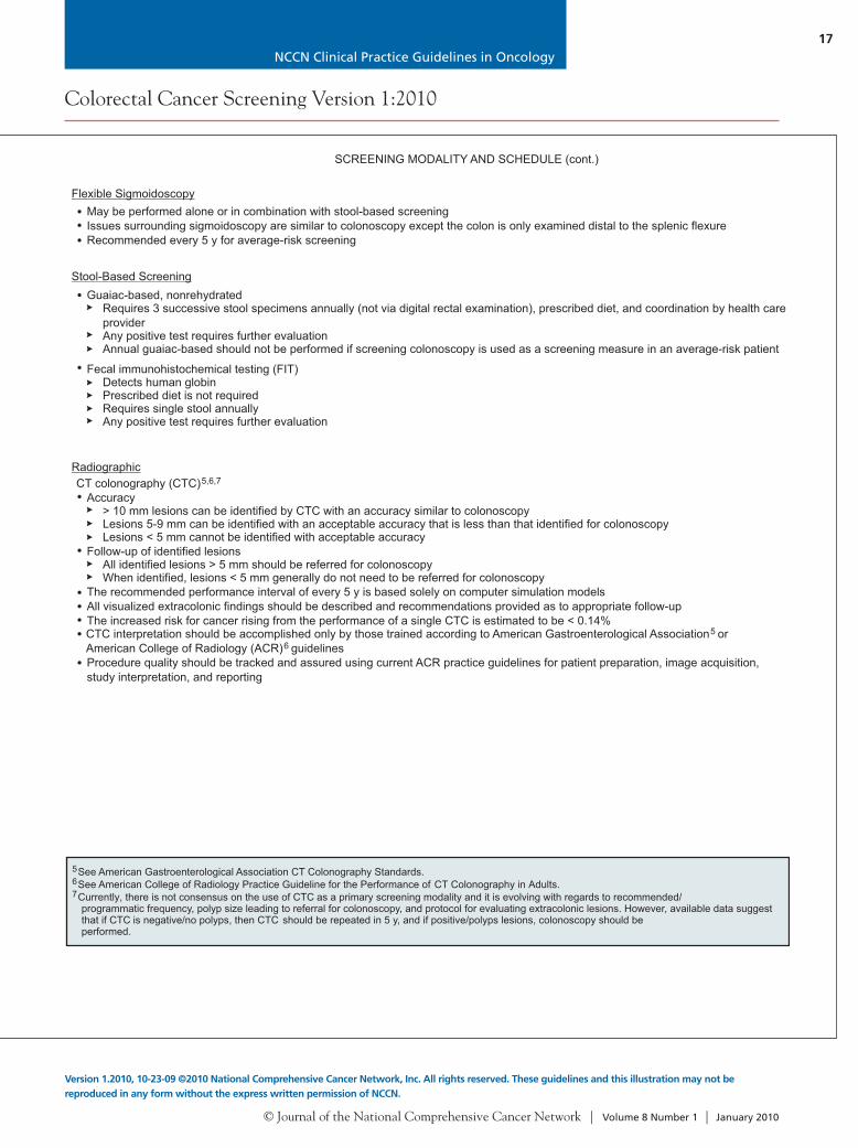
Guaiac-based, nonrehydratedRequires 3 successive stool specimens annually (not via digital rectal examination), prescribed diet, and coordination by health careprovider
Annual guaiac-based should not be performed if screening colonoscopy is used as a screening measure in an average-risk patientAny positive test requires further evaluation
SCREENING MODALITY AND SCHEDULE (cont.)
CT colonography (CTC)Accuracy
> 10 mm lesions can be identified by CTC with an accuracy similar to colonoscopyLesions 5-9 mm can be identified with an acceptable accuracy that is less than that identified for colonoscopy
Follow-up of identified lesionsAll identified lesions > 5 mm should be referred for colonoscopy
The recommended performance interval of every 5 y is based solely on computer simulation modelsAll visualized extracolonic findings should be described and recommendations provided as to appropriate follow-upThe increased risk for cancer rising from the performance of a single CTC is estimated to be < 0.14%CTC interpretation should be accomplished only by those trained according to
guidelinesProcedure quality should be tracked and assured using current ACR practice guidelines for patient preparation, image acquisition,study interpretation, and reporting
5,6,7
Lesions < 5 mm cannot be identified with acceptable accuracy
When identified, lesions < 5 mm generally do not need to be referred for colonoscopy
American Gastroenterological Association orAmerican College of Radiology (ACR)
56
Fecal immunohistochemical testing (FIT)Detects human globinPrescribed diet is not requiredRequires single stool annuallyAny positive test requires further evaluation
Stool-Based Screening
Radiographic
56
See American Gastroenterological Association CT Colonography Standards.See American College of Radiology Practice Guideline for the Performance of CT Colonography in Adults.Currently, there is not consensus on the use of CTC as a primary screening modality and it is evolving with regards to recommended/programmatic frequency, polyp size leading to referral for colonoscopy, and protocol for evaluating extracolonic lesions. However, available data suggestthat if CTC is negative/no polyps, then CTC should be repeated in 5 y, and if positive/polyps lesions, colonoscopy should beperformed.
7
Flexible Sigmoidoscopy
SCREENING MODALITY AND SCHEDULE
Colon cancer prevention should be the primary goal of colorectal cancer screening.Screening of average-risk individuals can reduce colorectal cancer mortality by detecting cancer at an early, curable stage anddetecting and removing clinically significant adenomas. It has been shown to be cost-effective compared with other screeningprograms.Although patient preferences and availability of resources play an important role in the selection of screening options, tests that aredesigned to detect both early cancer and adenomatous polyps should be encouraged.
Colonoscopy every 10 y, orFlexible sigmoidoscopy every 5 y, orCT colonography every 5 y
nnually, or
Screening Modalities that Detect Adenomatous Polyps and Cancer1
2
Screening Modalities that Primarily Detect Cancer3
Stool-based screeningGuaiac-based testing, aImmunochemical-based testing, annually, orStool DNA test with high sensitivity for cancer (interval for screening is uncertain)4
ColonoscopyColonoscopy is the primary method employed for colorectal cancer screening in average- and high-risk populations. However,screening with any of the available modalities is preferable to no screening.
Accumulating data suggest that substantial variability exists in the quality and, by extension, the clinical effectiveness ofcolonoscopy. Improving the overall impact of screening colonoscopy requires a programmatic approach that addresses qualityissues at several levels.These colonoscopy quality indicators include:
Cecal intubation ratesWithdrawal timeAdenoma detection ratesAppropriate intervals between endoscopic studies based on family and personal history, number, and histologic type of polypson last colonoscopyMinor and major complications ratesPre-procedure medical evaluationAppropriate preparation instructions
Standardized colonoscopy reports that contain, at a minimum:Patient demographic and clinical factorsProcedure indicationsEndoscopic findingsPhotographic documentation of endoscopic landmarksEstimate of quality of bowel preparationDocumentation of follow-up planning, including pathology resultsSedation administered
Caveats for every-10-years interval:A 1-year interval is appropriate for average-risk patients who had an optimal procedure.Shorter intervals may be indicated based on the quality and completeness of the colonoscopy.Individual risk factors and physician judgment should be included in the interval determination.The number and characteristics of polyps, family history, and medical assessment should influence judgment regarding theinterval between colonoscopies.Colonoscopy has limitations and may not detect all cancers and polyps.
12
34
If other modalities are not available, double-contrast barium enema every 5 y may be useful.Currently, there is no consensus on the use of CT colonography as a primary screening modality and it is evolving with regards to recommended/programmatic frequency, polyp size leading to referral for colonoscopy, and protocol for evaluating extracolonic lesions. However, available data suggestthat if CT colonography is negative/no polyps, then CT colonography should be repeated in 5 y, and if positive/polyps lesions, colonoscopy should beperformed.
Annual stool-based testing with every 5-y flexible sigmoidoscopy can be used in combination for screening.Emerging technologies, such as stool DNA, have shown increasing evidence as a reasonably accurate screening test, but data are limited for determiningan interval between screening. Currently, stool DNA is not considered a first-line screening test except in specific circumstances.
May be performed alone or in combination with stool-based screeningIssues surrounding sigmoidoscopy are similar to colonoscopy except the colon is only examined distal to the splenic flexureRecommended every 5 y for average-risk screening
NCCN Clinical Practice Guidelines in Oncology
© Journal of the National Comprehensive Cancer Network | Volume 8 Number 1 | January 2010
17
Colorectal Cancer Screening Version 1:2010
Version 1.2010, 10-23-09 ©2010 National Comprehensive Cancer Network, Inc. All rights reserved. These guidelines and this illustration may not be reproduced in any form without the express written permission of NCCN.
Guaiac-based, nonrehydratedRequires 3 successive stool specimens annually (not via digital rectal examination), prescribed diet, and coordination by health careprovider
Annual guaiac-based should not be performed if screening colonoscopy is used as a screening measure in an average-risk patientAny positive test requires further evaluation
SCREENING MODALITY AND SCHEDULE (cont.)
CT colonography (CTC)Accuracy
> 10 mm lesions can be identified by CTC with an accuracy similar to colonoscopyLesions 5-9 mm can be identified with an acceptable accuracy that is less than that identified for colonoscopy
Follow-up of identified lesionsAll identified lesions > 5 mm should be referred for colonoscopy
The recommended performance interval of every 5 y is based solely on computer simulation modelsAll visualized extracolonic findings should be described and recommendations provided as to appropriate follow-upThe increased risk for cancer rising from the performance of a single CTC is estimated to be < 0.14%CTC interpretation should be accomplished only by those trained according to
guidelinesProcedure quality should be tracked and assured using current ACR practice guidelines for patient preparation, image acquisition,study interpretation, and reporting
5,6,7
Lesions < 5 mm cannot be identified with acceptable accuracy
When identified, lesions < 5 mm generally do not need to be referred for colonoscopy
American Gastroenterological Association orAmerican College of Radiology (ACR)
56
Fecal immunohistochemical testing (FIT)Detects human globinPrescribed diet is not requiredRequires single stool annuallyAny positive test requires further evaluation
Stool-Based Screening
Radiographic
56
See American Gastroenterological Association CT Colonography Standards.See American College of Radiology Practice Guideline for the Performance of CT Colonography in Adults.Currently, there is not consensus on the use of CTC as a primary screening modality and it is evolving with regards to recommended/programmatic frequency, polyp size leading to referral for colonoscopy, and protocol for evaluating extracolonic lesions. However, available data suggestthat if CTC is negative/no polyps, then CTC should be repeated in 5 y, and if positive/polyps lesions, colonoscopy should beperformed.
7
Flexible Sigmoidoscopy
SCREENING MODALITY AND SCHEDULE
Colon cancer prevention should be the primary goal of colorectal cancer screening.Screening of average-risk individuals can reduce colorectal cancer mortality by detecting cancer at an early, curable stage anddetecting and removing clinically significant adenomas. It has been shown to be cost-effective compared with other screeningprograms.Although patient preferences and availability of resources play an important role in the selection of screening options, tests that aredesigned to detect both early cancer and adenomatous polyps should be encouraged.
Colonoscopy every 10 y, orFlexible sigmoidoscopy every 5 y, orCT colonography every 5 y
nnually, or
Screening Modalities that Detect Adenomatous Polyps and Cancer1
2
Screening Modalities that Primarily Detect Cancer3
Stool-based screeningGuaiac-based testing, aImmunochemical-based testing, annually, orStool DNA test with high sensitivity for cancer (interval for screening is uncertain)4
ColonoscopyColonoscopy is the primary method employed for colorectal cancer screening in average- and high-risk populations. However,screening with any of the available modalities is preferable to no screening.
Accumulating data suggest that substantial variability exists in the quality and, by extension, the clinical effectiveness ofcolonoscopy. Improving the overall impact of screening colonoscopy requires a programmatic approach that addresses qualityissues at several levels.These colonoscopy quality indicators include:
Cecal intubation ratesWithdrawal timeAdenoma detection ratesAppropriate intervals between endoscopic studies based on family and personal history, number, and histologic type of polypson last colonoscopyMinor and major complications ratesPre-procedure medical evaluationAppropriate preparation instructions
Standardized colonoscopy reports that contain, at a minimum:Patient demographic and clinical factorsProcedure indicationsEndoscopic findingsPhotographic documentation of endoscopic landmarksEstimate of quality of bowel preparationDocumentation of follow-up planning, including pathology resultsSedation administered
Caveats for every-10-years interval:A 1-year interval is appropriate for average-risk patients who had an optimal procedure.Shorter intervals may be indicated based on the quality and completeness of the colonoscopy.Individual risk factors and physician judgment should be included in the interval determination.The number and characteristics of polyps, family history, and medical assessment should influence judgment regarding theinterval between colonoscopies.Colonoscopy has limitations and may not detect all cancers and polyps.
12
34
If other modalities are not available, double-contrast barium enema every 5 y may be useful.Currently, there is no consensus on the use of CT colonography as a primary screening modality and it is evolving with regards to recommended/programmatic frequency, polyp size leading to referral for colonoscopy, and protocol for evaluating extracolonic lesions. However, available data suggestthat if CT colonography is negative/no polyps, then CT colonography should be repeated in 5 y, and if positive/polyps lesions, colonoscopy should beperformed.
Annual stool-based testing with every 5-y flexible sigmoidoscopy can be used in combination for screening.Emerging technologies, such as stool DNA, have shown increasing evidence as a reasonably accurate screening test, but data are limited for determiningan interval between screening. Currently, stool DNA is not considered a first-line screening test except in specific circumstances.
May be performed alone or in combination with stool-based screeningIssues surrounding sigmoidoscopy are similar to colonoscopy except the colon is only examined distal to the splenic flexureRecommended every 5 y for average-risk screening

© Journal of the National Comprehensive Cancer Network | Volume 8 Number 1 | January 2010
18
Colorectal Cancer Screening Version 1:2010
Clinical trials: The NCCN believes that the best management for any cancer patient is in a clinical trial. Participation in clinical trials is especially encouraged. All recommendations are category 2A unless otherwise noted.
Lynch syndrome (see page 22)Individual meeting the revised Bethesdaguidelines (see page 26)
Individual from a family meetingAmsterdam criteria (see page 27)
> 10 adenomas in same individual (seepages 29 and 38)
Individual with multiple gastrointestinalhamartomatous polyps (see pages 40 and41) or hyperplastic polyps (see page 42)
Individual from a family with a knownhereditary syndrome associated withcolorectal cancer, with or without knownmutation (see appropriate hereditarysyndrome)
a
Obtain detailed family historyObtain detailed medical and surgicalhistoryDirected examination for relatedmanifestationsPsychosocial assessment and supportRisk counselingEducation supportDiscussion of genetic testingObtain informed consent
b
CRITERIA FOR FURTHER RISKEVALUATION FOR HIGH-RISKSYNDROMES
RISK ASSESSMENT/ GENETIC COUNSELINGb,c
Classical FAP (see page 29)
MYH-associated polyposis(see page 38)
a
bc
d
Endometrial cancer < 50 y is not included in the revised Bethesda guidelines; however, recent evidence suggests that these individuals should be evaluatedfor Lynch syndrome.
See Obtaining a Comprehensive Risk Assessment for Hereditary Colorectal Cancer (page 20).A genetic counselor and/or medical geneticist should be involved early in counseling patients who (potentially) meet criteria for an inherited syndrome.Genetic counseling is advised when genetic testing is offered.Referral to a specialized team is recommended.
Attenuated FAP (see page 35)
HEREDITARYSYNDROME
Peutz-Jeghers syndrome(page 40)
d
No syndromes,but familial riskpresent
See PositiveFamily History(page 15)
Juvenile polyposis syndrome(page 41)
d
Hyperplastic polyposis syndrome(page 42)
or
or
or
or
The extent of colorectal resection depends upon the location of the tumor, any underlying condition (e.g., inflammatorybowel disease, hereditary syndrome), and the vascular supply to the colorectum.
K L Abdominoperineal resection
1Adapted and reprinted with permission from Bullard KM, Rothenberger DA. Colon, rectum, and anus. In: Brunicardi D, ed. Schwartz's Principles of Surgery, eighth edition. New York: McGraw Hill; 2004.
DEFINITIONS OF COMMON COLORECTAL RESECTIONS
ABC
DD
EF G
H
I
J
K
L
A C Ileocecetomy
A D Ascending colectomy
A F Right hemicolectomy
A G Extended right hemicolecotmy
E H Transverse colectomy
G I Left hemicolectomy
F I Extended left hemicolectomy
J K Sigmoid colectomy
A J Subtotal colectomy
A K Total colectomy
K L Low anterior resectionwith sphincter preservation
without sphincter preservation
Definitions of common colorectal resections are as follows:1

NCCN Clinical Practice Guidelines in Oncology
© Journal of the National Comprehensive Cancer Network | Volume 8 Number 1 | January 2010
19
Colorectal Cancer Screening Version 1:2010
Version 1.2010, 10-23-09 ©2010 National Comprehensive Cancer Network, Inc. All rights reserved. These guidelines and this illustration may not be reproduced in any form without the express written permission of NCCN.
HIGH RISK SYNDROMES
Lynch syndrome (see page 22)Individual meeting the revised Bethesdaguidelines (see page 26)
Individual from a family meetingAmsterdam criteria (see page 27)
> 10 adenomas in same individual (seepages 29 and 38)
Individual with multiple gastrointestinalhamartomatous polyps (see pages 40 and41) or hyperplastic polyps (see page 42)
Individual from a family with a knownhereditary syndrome associated withcolorectal cancer, with or without knownmutation (see appropriate hereditarysyndrome)
a
Obtain detailed family historyObtain detailed medical and surgicalhistoryDirected examination for relatedmanifestationsPsychosocial assessment and supportRisk counselingEducation supportDiscussion of genetic testingObtain informed consent
b
CRITERIA FOR FURTHER RISKEVALUATION FOR HIGH-RISKSYNDROMES
RISK ASSESSMENT/ GENETIC COUNSELINGb,c
Classical FAP (see page 29)
MYH-associated polyposis(see page 38)
a
bc
d
Endometrial cancer < 50 y is not included in the revised Bethesda guidelines; however, recent evidence suggests that these individuals should be evaluatedfor Lynch syndrome.
See Obtaining a Comprehensive Risk Assessment for Hereditary Colorectal Cancer (page 20).A genetic counselor and/or medical geneticist should be involved early in counseling patients who (potentially) meet criteria for an inherited syndrome.Genetic counseling is advised when genetic testing is offered.Referral to a specialized team is recommended.
Attenuated FAP (see page 35)
HEREDITARYSYNDROME
Peutz-Jeghers syndrome(page 40)
d
No syndromes,but familial riskpresent
See PositiveFamily History(page 15)
Juvenile polyposis syndrome(page 41)
d
Hyperplastic polyposis syndrome(page 42)
or
or
or
or
The extent of colorectal resection depends upon the location of the tumor, any underlying condition (e.g., inflammatorybowel disease, hereditary syndrome), and the vascular supply to the colorectum.
K L Abdominoperineal resection
1Adapted and reprinted with permission from Bullard KM, Rothenberger DA. Colon, rectum, and anus. In: Brunicardi D, ed. Schwartz's Principles of Surgery, eighth edition. New York: McGraw Hill; 2004.
DEFINITIONS OF COMMON COLORECTAL RESECTIONS
ABC
DD
EF G
H
I
J
K
L
A C Ileocecetomy
A D Ascending colectomy
A F Right hemicolectomy
A G Extended right hemicolecotmy
E H Transverse colectomy
G I Left hemicolectomy
F I Extended left hemicolectomy
J K Sigmoid colectomy
A J Subtotal colectomy
A K Total colectomy
K L Low anterior resectionwith sphincter preservation
without sphincter preservation
Definitions of common colorectal resections are as follows:1

© Journal of the National Comprehensive Cancer Network | Volume 8 Number 1 | January 2010
20
Colorectal Cancer Screening Version 1:2010
Clinical trials: The NCCN believes that the best management for any cancer patient is in a clinical trial. Participation in clinical trials is especially encouraged. All recommendations are category 2A unless otherwise noted.
HIGH RISK SYNDROMES
COMMON PEDIGREE SYMBOLS2
Male, Female
Proband (patientinitiating geneticworkup)
Mating
Deceased
Sibship
Affectedwith trait
Adopted intoa family
Dizygotictwins
Monozygotictwins
2 Niece ornephew
1
1
2
3
2
2
2 2 2
1
1
1
Paternalgrandfather
Maternalgrandfather
Paternalgrandmother
Maternalgrandmother
Aunt Father Mother Uncle
Sister Proband
Son
Brother First cousin(male)
PEDIGREE: FIRST-, SECOND-, AND THIRD-DEGREE RELATIVES OF PROBAND3
2Bennett RL, Steinhaus KA, Uhrich SB, et al. Recommendations for standardized human pedigree nomenclature. Am J Hum Genet 1995;56:745-752.3First-degree relatives: parents, siblings, and children; second-degree relatives: grandparents, aunts, uncles, nieces, nephews, and half-siblings;
third-degree relatives: great-grandparents and cousins.
It is essential to obtain a detailed family history, including:ParentsChildrenSiblings/half-siblingsAunts and uncles
Minimal data set on each relative:Current age and age at diagnosis of cancer (medical record documentation of cancer strongly encouraged)Age/availability of tumor sample and cause of deathType of cancer (note multiple primaries)Ethnicity/country of originConsanguinitySuspected colon cancer syndromes and additional syndrome-specific features(e.g., Muir-Torre, Turcot, Peutz-Jeghers, juvenile polyposis)All other inherited conditions and birth defects
1
GrandparentsGreat-grandparentsCousinsNieces and nephews
See Common Pedigree Symbols (facing page) andPedigree: First-, Second-, and Third-degree Relativesof Proband (facing page)
1Burt R, Neklason DW. Genetic testing for inherited colon cancer. Gastroenterology 2005;128:1696-1716.
Detailed Medical and Surgical HistoryPolypsInflammatory bowel diseaseInherited syndromes:
Lynch syndrome
FAP and associated syndromes
MYH-associated polyposis (MAP)Peutz-Jeghers syndromeJuvenile polyposis syndromePTEN-associated syndromes
Pathology verification strongly encouraged
Muir-Torre syndrome
Attenuated FAPGardner syndromeTurcot syndrome
Cowden syndromeBannayan-Riley-Ruvalcaba syndrome
Directed Examination for Related ManifestationsColonoscopyEsophagogastroduodenoscopyEye examinationSkin, soft-tissue, and bone examinationOral examination
Family History of Colorectal Cancer and Expanded Pedigree
OBTAINING A COMPREHENSIVE ASSESSMENT FOR HEREDITARY COLORECTAL CANCER OBTAINING A COMPREHENSIVE ASSESSEMNT FOR HEREDITARY COLORECTAL CANCER (cont.)

NCCN Clinical Practice Guidelines in Oncology
© Journal of the National Comprehensive Cancer Network | Volume 8 Number 1 | January 2010
21
Colorectal Cancer Screening Version 1:2010
Version 1.2010, 10-23-09 ©2010 National Comprehensive Cancer Network, Inc. All rights reserved. These guidelines and this illustration may not be reproduced in any form without the express written permission of NCCN.
COMMON PEDIGREE SYMBOLS2
Male, Female
Proband (patientinitiating geneticworkup)
Mating
Deceased
Sibship
Affectedwith trait
Adopted intoa family
Dizygotictwins
Monozygotictwins
2 Niece ornephew
1
1
2
3
2
2
2 2 2
1
1
1
Paternalgrandfather
Maternalgrandfather
Paternalgrandmother
Maternalgrandmother
Aunt Father Mother Uncle
Sister Proband
Son
Brother First cousin(male)
PEDIGREE: FIRST-, SECOND-, AND THIRD-DEGREE RELATIVES OF PROBAND3
2Bennett RL, Steinhaus KA, Uhrich SB, et al. Recommendations for standardized human pedigree nomenclature. Am J Hum Genet 1995;56:745-752.3First-degree relatives: parents, siblings, and children; second-degree relatives: grandparents, aunts, uncles, nieces, nephews, and half-siblings;
third-degree relatives: great-grandparents and cousins.
It is essential to obtain a detailed family history, including:ParentsChildrenSiblings/half-siblingsAunts and uncles
Minimal data set on each relative:Current age and age at diagnosis of cancer (medical record documentation of cancer strongly encouraged)Age/availability of tumor sample and cause of deathType of cancer (note multiple primaries)Ethnicity/country of originConsanguinitySuspected colon cancer syndromes and additional syndrome-specific features(e.g., Muir-Torre, Turcot, Peutz-Jeghers, juvenile polyposis)All other inherited conditions and birth defects
1
GrandparentsGreat-grandparentsCousinsNieces and nephews
See Common Pedigree Symbols (facing page) andPedigree: First-, Second-, and Third-degree Relativesof Proband (facing page)
1Burt R, Neklason DW. Genetic testing for inherited colon cancer. Gastroenterology 2005;128:1696-1716.
Detailed Medical and Surgical HistoryPolypsInflammatory bowel diseaseInherited syndromes:
Lynch syndrome
FAP and associated syndromes
MYH-associated polyposis (MAP)Peutz-Jeghers syndromeJuvenile polyposis syndromePTEN-associated syndromes
Pathology verification strongly encouraged
Muir-Torre syndrome
Attenuated FAPGardner syndromeTurcot syndrome
Cowden syndromeBannayan-Riley-Ruvalcaba syndrome
Directed Examination for Related ManifestationsColonoscopyEsophagogastroduodenoscopyEye examinationSkin, soft-tissue, and bone examinationOral examination
Family History of Colorectal Cancer and Expanded Pedigree
OBTAINING A COMPREHENSIVE ASSESSMENT FOR HEREDITARY COLORECTAL CANCER OBTAINING A COMPREHENSIVE ASSESSEMNT FOR HEREDITARY COLORECTAL CANCER (cont.)
HIGH RISK SYNDROMES
© Journal of the National Comprehensive Cancer Network | Volume 8 Number 1 | January 2010
22
Colorectal Cancer Screening Version 1:2010
Clinical trials: The NCCN believes that the best management for any cancer patient is in a clinical trial. Participation in clinical trials is especially encouraged. All recommendations are category 2A unless otherwise noted.
LYNCH SYNDROME
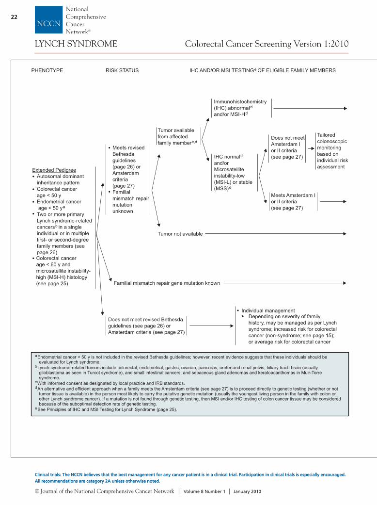
PHENOTYPE RISK STATUS IHC AND/OR MSI TESTING OF ELIGIBLE FAMILY MEMBERSe
Extended PedigreeAutosomal dominantinheritance patternColorectal cancerage < 50 yEndometrial cancerage < 50 y
Colorectal cancerage < 60 y andmicrosatellite instability-high (MSI-H) histology(see page 25)
a
Two or more primaryLynch syndrome-relatedcancers in a singleindividual or in multiplefirst- or second-degreefamily members (seepage 26)
b
Familial mismatch repair gene mutation known
Meets revisedBethesdaguidelines(page 26) orAmsterdamcriteria(page 27)Familialmismatch repairmutationunknown
Tumor availablefrom affectedfamily memberc,d
Does not meet revised Bethesdaguidelines (see page 26) orAmsterdam criteria (see page 27)
Individual managementDepending on severity of familyhistory, may be managed as per Lynchsyndrome; increased risk for colorectalcancer (non-syndrome; see page 15);or average risk for colorectal cancer
Tumor not available
Immunohistochemistry(IHC) abnormaland/or MSI-H
dd
IHC normaland/orMicrosatelliteinstability-low(MSI-L) or stable(MSS)
d
d
Tailoredcolonoscopicmonitoringbased onindividual riskassessment
Does not meetAmsterdam Ior II criteria(see page 27)
Meets Amsterdam Ior II criteria(see page 27)
a
b
cd
e
Endometrial cancer < 50 y is not included in the revised Bethesda guidelines; however, recent evidence suggests that these individuals should beevaluated for Lynch syndrome.
Lynch syndrome-related tumors include colorectal, endometrial, gastric, ovarian, pancreas, ureter and renal pelvis, biliary tract, brain (usuallyglioblastoma as seen in Turcot syndrome), and small intestinal cancers, and sebaceous gland adenomas and keratoacanthomas in Muir-Torresyndrome.
With informed consent as designated by local practice and IRB standards.
See Principles of IHC and MSI Testing for Lynch Syndrome (page 25).
An alternative and efficient approach when a family meets the Amsterdam criteria is to proceed directly to genetic testing (whether or nottumor tissue is available) in the person most likely to carry the putative genetic mutation (usually the youngest living person in the family with colon orother Lynch syndrome cancer). If a mutation is not found through genetic testing, then MSI and/or IHC testing of colon cancer tissue may be consideredbecause of the suboptimal detection rate of genetic testing.
(see page 27)
GENETIC COUNSELING/TESTING OF ELIGIBLEFAMILY MEMBERS
Mismatch repair gene mutation unknown
Positive familial mutation MLH1,MSH2, MSH6, or PMS2 found
Familial mismatch repair gene mutation known
Positive gene test(mutation present)
Negative gene test(mutation not present)
Not tested
No familial mutation found
Average-risk screening(see page 11)
See Lynch SyndromeSurveillance and Follow-up(page 24) andSee pathway below to considergenetic testing for at-risk familymembers
Mutation of unknownsignificance found
Tailored surveillancebased on individual-and family-riskassessment
See Lynch SyndromeSurveillance andFollow-up (page 24)
MUTATION STATUS
Not tested
Consider genetic testing of affected family memberif possible to find a disease causing mutation
Genetic testing should begin with the gene mostlikely to harbor the mutation based on IHCresults. If IHC testing cannot be performed or isuninformative, first test MSH2 or MLH1 and thenMSH6 or PMS2 if a mutation is not found in thefirst 2 genes
Consider genetic testing of at-risk familymemberf to find a disease causing mutation
fAt-risk family member can be defined as a first-degree relative of an affected individual and/or proband. If a first-degree relative is unavailable or unwillingto be tested, more distant relatives should be offered testing for the known mutation in the family.

NCCN Clinical Practice Guidelines in Oncology
© Journal of the National Comprehensive Cancer Network | Volume 8 Number 1 | January 2010
23
Colorectal Cancer Screening Version 1:2010
Version 1.2010, 10-23-09 ©2010 National Comprehensive Cancer Network, Inc. All rights reserved. These guidelines and this illustration may not be reproduced in any form without the express written permission of NCCN.
LYNCH SYNDROME
PHENOTYPE RISK STATUS IHC AND/OR MSI TESTING OF ELIGIBLE FAMILY MEMBERSe
Extended PedigreeAutosomal dominantinheritance patternColorectal cancerage < 50 yEndometrial cancerage < 50 y
Colorectal cancerage < 60 y andmicrosatellite instability-high (MSI-H) histology(see page 25)
a
Two or more primaryLynch syndrome-relatedcancers in a singleindividual or in multiplefirst- or second-degreefamily members (seepage 26)
b
Familial mismatch repair gene mutation known
Meets revisedBethesdaguidelines(page 26) orAmsterdamcriteria(page 27)Familialmismatch repairmutationunknown
Tumor availablefrom affectedfamily memberc,d
Does not meet revised Bethesdaguidelines (see page 26) orAmsterdam criteria (see page 27)
Individual managementDepending on severity of familyhistory, may be managed as per Lynchsyndrome; increased risk for colorectalcancer (non-syndrome; see page 15);or average risk for colorectal cancer
Tumor not available
Immunohistochemistry(IHC) abnormaland/or MSI-H
dd
IHC normaland/orMicrosatelliteinstability-low(MSI-L) or stable(MSS)
d
d
Tailoredcolonoscopicmonitoringbased onindividual riskassessment
Does not meetAmsterdam Ior II criteria(see page 27)
Meets Amsterdam Ior II criteria(see page 27)
a
b
cd
e
Endometrial cancer < 50 y is not included in the revised Bethesda guidelines; however, recent evidence suggests that these individuals should beevaluated for Lynch syndrome.
Lynch syndrome-related tumors include colorectal, endometrial, gastric, ovarian, pancreas, ureter and renal pelvis, biliary tract, brain (usuallyglioblastoma as seen in Turcot syndrome), and small intestinal cancers, and sebaceous gland adenomas and keratoacanthomas in Muir-Torresyndrome.
With informed consent as designated by local practice and IRB standards.
See Principles of IHC and MSI Testing for Lynch Syndrome (page 25).
An alternative and efficient approach when a family meets the Amsterdam criteria is to proceed directly to genetic testing (whether or nottumor tissue is available) in the person most likely to carry the putative genetic mutation (usually the youngest living person in the family with colon orother Lynch syndrome cancer). If a mutation is not found through genetic testing, then MSI and/or IHC testing of colon cancer tissue may be consideredbecause of the suboptimal detection rate of genetic testing.
(see page 27)
GENETIC COUNSELING/TESTING OF ELIGIBLEFAMILY MEMBERS
Mismatch repair gene mutation unknown
Positive familial mutation MLH1,MSH2, MSH6, or PMS2 found
Familial mismatch repair gene mutation known
Positive gene test(mutation present)
Negative gene test(mutation not present)
Not tested
No familial mutation found
Average-risk screening(see page 11)
See Lynch SyndromeSurveillance and Follow-up(page 24) andSee pathway below to considergenetic testing for at-risk familymembers
Mutation of unknownsignificance found
Tailored surveillancebased on individual-and family-riskassessment
See Lynch SyndromeSurveillance andFollow-up (page 24)
MUTATION STATUS
Not tested
Consider genetic testing of affected family memberif possible to find a disease causing mutation
Genetic testing should begin with the gene mostlikely to harbor the mutation based on IHCresults. If IHC testing cannot be performed or isuninformative, first test MSH2 or MLH1 and thenMSH6 or PMS2 if a mutation is not found in thefirst 2 genes
Consider genetic testing of at-risk familymemberf to find a disease causing mutation
fAt-risk family member can be defined as a first-degree relative of an affected individual and/or proband. If a first-degree relative is unavailable or unwillingto be tested, more distant relatives should be offered testing for the known mutation in the family.
© Journal of the National Comprehensive Cancer Network | Volume 8 Number 1 | January 2010
24
Colorectal Cancer Screening Version 1:2010
Clinical trials: The NCCN believes that the best management for any cancer patient is in a clinical trial. Participation in clinical trials is especially encouraged. All recommendations are category 2A unless otherwise noted.
LYNCH SYNDROME
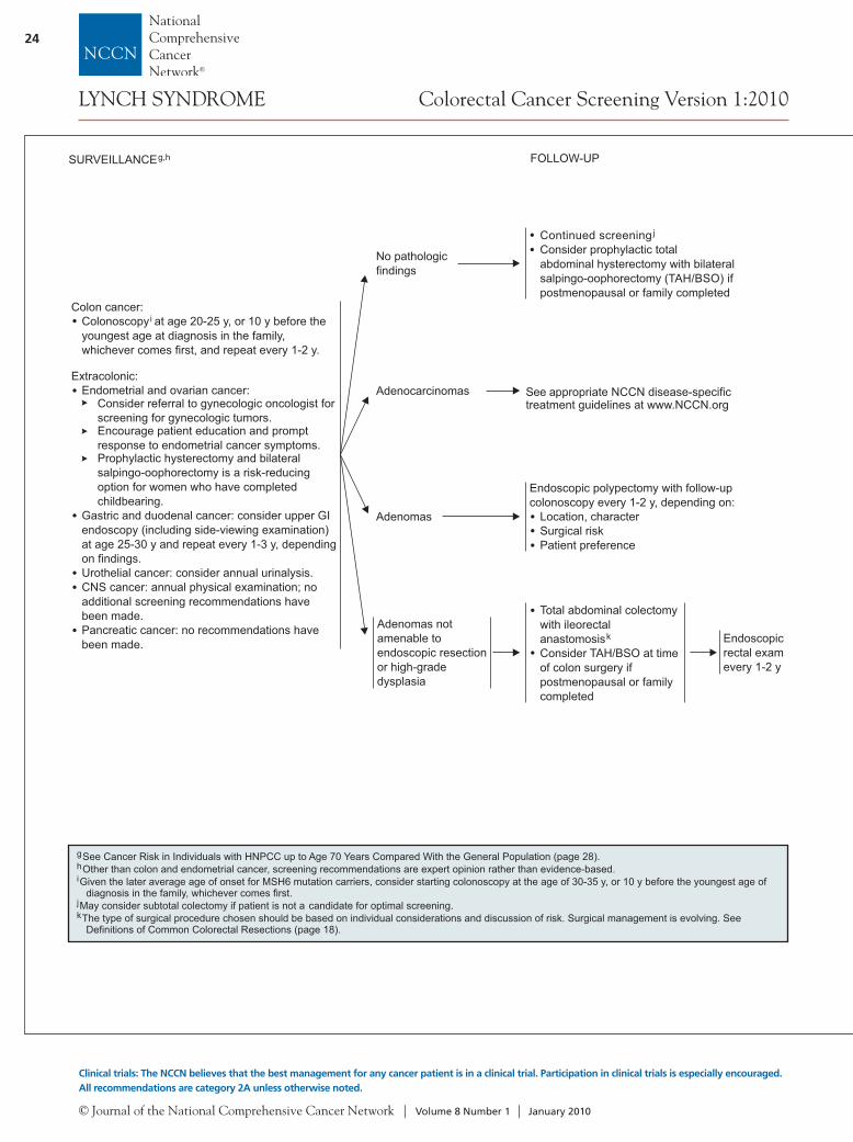
Colon cancer:Colonoscopy at age 20-25 y, or 10 y before theyoungest age at diagnosis in the family,whichever comes first, and repeat every 1-2 y.
Extracolonic:Endometrial and ovarian cancer:
Consider referral to gynecologic oncologist forscreening for gynecologic tumors.Encourage patient education and promptresponse to endometrial cancer symptoms.Prophylactic hysterectomy and bilateralsalpingo-oophorectomy is a risk-reducingoption for women who have completedchildbearing.
Gastric and duodenal cancer: consider upper GIendoscopy (including side-viewing examination)at age 25-30 y and repeat every 1-3 y, dependingon findings.Urothelial cancer: consider annual urinalysis.CNS cancer: annual physical examination; noadditional screening recommendations havebeen made.Pancreatic cancer: no recommendations havebeen made.
i
No pathologicfindings
Adenocarcinomas
Continued screeningConsider prophylactic totalabdominal hysterectomy with bilateralsalpingo-oophorectomy (TAH/BSO) ifpostmenopausal or family completed
j
See appropriate NCCN disease-specifictreatment guidelines at www.NCCN.org
Adenomas
Endoscopic polypectomy with follow-upcolonoscopy every 1-2 y, depending on:
Location, characterSurgical riskPatient preference
Adenomas notamenable toendoscopic resectionor high-gradedysplasia
Total abdominal colectomywith ileorectalanastomosisConsider TAH/BSO at timeof colon surgery ifpostmenopausal or familycompleted
k Endoscopicrectal examevery 1-2 y
ghi
See Cancer Risk in Individuals with HNPCC up to Age 70 Years Compared With the General Population (page 28).Other than colon and endometrial cancer, screening recommendations are expert opinion rather than evidence-based.
Given the later average age of onset for MSH6 mutation carriers, consider starting colonoscopy at the age of 30-35 y, or 10 y before the youngest age ofdiagnosis in the family, whichever comes first.
May consider subtotal colectomy if patient is not a candidate for optimal screening.The type of surgical procedure chosen should be based on individual considerations and discussion of risk. Surgical management is evolving. SeeDefinitions of Common Colorectal Resections (page 18).
jk
SURVEILLANCEg,h FOLLOW-UP PRINCIPLES OF IHC AND MSI TESTING FOR LYNCH SYNDROME
IHC and MSI analyses are screening tests (either by themselves or in conjunction), typically performed on colon cancer tissueto identify individuals at risk for Lynch syndrome.
IHC refers to staining tumor tissue for protein expression of the 4 mismatch genes known to be mutated in Lynch syndrome:MLH1, MSH2, MSH6, and PMS2. A normal IHC test implies all 4 mismatch repair proteins are normally expressed and thus nounderlying mismatch repair gene mutation is present. An abnormal test means that 1 of the proteins is not expressed and aninherited mutation may be present in the related gene. Loss of protein expression by IHC in any one of the mismatch repair genesguides genetic testing (mutation detection) to the gene where protein expression is not observed.10% to 15% of sporadic colon cancers exhibit abnormal IHC, often from abnormal methylation of the MLH1 gene promoter, butoccasionally from an inherited mutation from 1 of the mismatch repair genes.Thus the presence of an abnormal IHC testincreases the possibility of Lynch syndrome but does not make a definitive diagnosis. Genetic testing of peripheral blood DNA tofind a disease-causing mutation of 1 of the mismatch repair genes should then be performed. Most patients will be found tohave sporadic colon cancer and not a germline mutation. Those with a germline mutation are then identified as having Lynchsyndrome.There is a 5%–10% false-negative rate with IHC testing.
MSI-H in tumors refers to changes in 2 or more of the 5 National Cancer Institute-recommended panels of microsatellitemarkers in tumor tissue. Its significance, use, and implications are similar to those of IHC, although the tests are slightlycomplementary.There is a 5%–10% false-negative rate with MSI testing.
The Bethesda Criteria were developed in response to the emerging understanding of the pathologic spectrum and molecularcharacteristics of Lynch syndrome-related tumors. These criteria were intended to help identify colon cancer whosetumors should be tested for MSI, thereby identifying patients with a greater chance of having Lynch syndrome. The revisedBethesda Guidelines (see page 26) are now widely used to identify tumors that should be tested for mismatch repair defects,either by MSI and/or IHC analysis. Although more sensitive than the Amsterdam criteria (see page 27), up to 30% of patients withLynch syndrome fail to meet even the revised Bethesda Guidelines.
Recently, MSI screening of all endometrial and colorectal cancers, regardless of age at diagnosis or family history, hasbeen implemented at some centers to identify individuals at risk for Lynch syndrome.
Immunohistochemistry
Microsatellite Instability
patients with
IHC and/or

NCCN Clinical Practice Guidelines in Oncology
© Journal of the National Comprehensive Cancer Network | Volume 8 Number 1 | January 2010
25
Colorectal Cancer Screening Version 1:2010
Version 1.2010, 10-23-09 ©2010 National Comprehensive Cancer Network, Inc. All rights reserved. These guidelines and this illustration may not be reproduced in any form without the express written permission of NCCN.
LYNCH SYNDROME
Colon cancer:Colonoscopy at age 20-25 y, or 10 y before theyoungest age at diagnosis in the family,whichever comes first, and repeat every 1-2 y.
Extracolonic:Endometrial and ovarian cancer:
Consider referral to gynecologic oncologist forscreening for gynecologic tumors.Encourage patient education and promptresponse to endometrial cancer symptoms.Prophylactic hysterectomy and bilateralsalpingo-oophorectomy is a risk-reducingoption for women who have completedchildbearing.
Gastric and duodenal cancer: consider upper GIendoscopy (including side-viewing examination)at age 25-30 y and repeat every 1-3 y, dependingon findings.Urothelial cancer: consider annual urinalysis.CNS cancer: annual physical examination; noadditional screening recommendations havebeen made.Pancreatic cancer: no recommendations havebeen made.
i
No pathologicfindings
Adenocarcinomas
Continued screeningConsider prophylactic totalabdominal hysterectomy with bilateralsalpingo-oophorectomy (TAH/BSO) ifpostmenopausal or family completed
j
See appropriate NCCN disease-specifictreatment guidelines at www.NCCN.org
Adenomas
Endoscopic polypectomy with follow-upcolonoscopy every 1-2 y, depending on:
Location, characterSurgical riskPatient preference
Adenomas notamenable toendoscopic resectionor high-gradedysplasia
Total abdominal colectomywith ileorectalanastomosisConsider TAH/BSO at timeof colon surgery ifpostmenopausal or familycompleted
k Endoscopicrectal examevery 1-2 y
ghi
See Cancer Risk in Individuals with HNPCC up to Age 70 Years Compared With the General Population (page 28).Other than colon and endometrial cancer, screening recommendations are expert opinion rather than evidence-based.
Given the later average age of onset for MSH6 mutation carriers, consider starting colonoscopy at the age of 30-35 y, or 10 y before the youngest age ofdiagnosis in the family, whichever comes first.
May consider subtotal colectomy if patient is not a candidate for optimal screening.The type of surgical procedure chosen should be based on individual considerations and discussion of risk. Surgical management is evolving. SeeDefinitions of Common Colorectal Resections (page 18).
jk
SURVEILLANCEg,h FOLLOW-UP PRINCIPLES OF IHC AND MSI TESTING FOR LYNCH SYNDROME
IHC and MSI analyses are screening tests (either by themselves or in conjunction), typically performed on colon cancer tissueto identify individuals at risk for Lynch syndrome.
IHC refers to staining tumor tissue for protein expression of the 4 mismatch genes known to be mutated in Lynch syndrome:MLH1, MSH2, MSH6, and PMS2. A normal IHC test implies all 4 mismatch repair proteins are normally expressed and thus nounderlying mismatch repair gene mutation is present. An abnormal test means that 1 of the proteins is not expressed and aninherited mutation may be present in the related gene. Loss of protein expression by IHC in any one of the mismatch repair genesguides genetic testing (mutation detection) to the gene where protein expression is not observed.10% to 15% of sporadic colon cancers exhibit abnormal IHC, often from abnormal methylation of the MLH1 gene promoter, butoccasionally from an inherited mutation from 1 of the mismatch repair genes.Thus the presence of an abnormal IHC testincreases the possibility of Lynch syndrome but does not make a definitive diagnosis. Genetic testing of peripheral blood DNA tofind a disease-causing mutation of 1 of the mismatch repair genes should then be performed. Most patients will be found tohave sporadic colon cancer and not a germline mutation. Those with a germline mutation are then identified as having Lynchsyndrome.There is a 5%–10% false-negative rate with IHC testing.
MSI-H in tumors refers to changes in 2 or more of the 5 National Cancer Institute-recommended panels of microsatellitemarkers in tumor tissue. Its significance, use, and implications are similar to those of IHC, although the tests are slightlycomplementary.There is a 5%–10% false-negative rate with MSI testing.
The Bethesda Criteria were developed in response to the emerging understanding of the pathologic spectrum and molecularcharacteristics of Lynch syndrome-related tumors. These criteria were intended to help identify colon cancer whosetumors should be tested for MSI, thereby identifying patients with a greater chance of having Lynch syndrome. The revisedBethesda Guidelines (see page 26) are now widely used to identify tumors that should be tested for mismatch repair defects,either by MSI and/or IHC analysis. Although more sensitive than the Amsterdam criteria (see page 27), up to 30% of patients withLynch syndrome fail to meet even the revised Bethesda Guidelines.
Recently, MSI screening of all endometrial and colorectal cancers, regardless of age at diagnosis or family history, hasbeen implemented at some centers to identify individuals at risk for Lynch syndrome.
Immunohistochemistry
Microsatellite Instability
patients with
IHC and/or

© Journal of the National Comprehensive Cancer Network | Volume 8 Number 1 | January 2010
26
Colorectal Cancer Screening Version 1:2010
Clinical trials: The NCCN believes that the best management for any cancer patient is in a clinical trial. Participation in clinical trials is especially encouraged. All recommendations are category 2A unless otherwise noted.
LYNCH SYNDROME
THE REVISED BETHESDA GUIDELINESFOR TESTING COLORECTAL TUMORS FOR MICROSATELLITE INSTABILITY (MSI)1
Tumors from individuals should be tested for MSI in the following situations:
Colorectal cancer diagnosed in a patient < 50 years.
Presence of synchronous or metachronous Lynch syndrome-associated tumors regardless of age.
Colorectal cancer with the MSI-H histology diagnosed in a patient < 60 years.
Colorectal cancer diagnosed in a patient with 1 or more first-degree relatives with a Lynch syndrome-related cancer, with 1of the cancers being diagnosed < 50 years of age.
Colorectal cancer diagnosed in a patient with 2 or more first- or second-degree relatives with Lynch syndrome-relatedcancers regardless of age.
2
4
3
3
,3
1
2
3
4
Adapted with permission from Umar A, Boland CR, Terdiman JP, et al. Revised Bethesda Guidelines for hereditary nonpolyposis colorectal cancer (Lynchsyndrome) and microsatellite instability. J Natl Cancer Inst 2004;96:261-268.
Endometrial cancer at age < 50 y is not included in the revised Bethesda Guidelines; however, recent evidence suggests that these individuals should beevaluated for Lynch syndrome.
Lynch syndrome-related cancers include colorectal, endometrial, gastric, ovarian, pancreas, ureter and renal pelvis, biliary tract, brain (usually glioblastomaas seen in Turcot syndrome), and small intestinal cancers, and sebaceous gland adenomas and keratoacanthomas, as seen in Muir-Torre syndrome.
Presence of tumor infiltrating lymphocytes, Crohn's-like lymphocytic reaction, mucinous/signet-ring differentiation, or medullary growth pattern.
MINIMUM CRITERIA FOR CLINICAL DEFINITION OF HNPCC(AMSTERDAM CRITERIA I)1,2
At least 3 relatives with colorectal cancer; all of the following criteria should be present:
One should be a first-degree relative of the other 2
At least 2 successive generations must be affected
At least 1 of the relatives with colorectal cancer must have received the diagnosis < 50 years of age
Familial adenomatous polyposis (FAP) should be excluded
Tumors should be verified by pathologic examination
1From Vasen HFA. Clinical diagnosis and management of hereditary colorectal cancer syndromes. J Clin Oncol 2000;18(Suppl 1):81S-92S.
At least 3 relatives must have a cancer associated with HNPCC (colorectal, cancer of endometrium, small bowel, ureter orrenal pelvis); all of the following criteria should be present:
One must be a first-degree relative of the other 2
At least 2 successive generations must be affected
At least 1 of the relatives with cancer associated with HNPCC should be diagnosed < 50 years of age
FAP should be excluded in the colorectal cancer cases (if any)
Tumors should be verified whenever possible
REVISED MINIMUM CRITERIA FOR CLINICAL DEFINITION OF HNPCC(AMSTERDAM CRITERIA II)1,2
2Approximately 50% of patients with HNPCC will be missed by these criteria and approximately 50% of patients will meet the criteria and not have HNPCC, but have a high familial risk of uncertain etiology .

NCCN Clinical Practice Guidelines in Oncology
© Journal of the National Comprehensive Cancer Network | Volume 8 Number 1 | January 2010
27
Colorectal Cancer Screening Version 1:2010
Version 1.2010, 10-23-09 ©2010 National Comprehensive Cancer Network, Inc. All rights reserved. These guidelines and this illustration may not be reproduced in any form without the express written permission of NCCN.
LYNCH SYNDROME
THE REVISED BETHESDA GUIDELINESFOR TESTING COLORECTAL TUMORS FOR MICROSATELLITE INSTABILITY (MSI)1
Tumors from individuals should be tested for MSI in the following situations:
Colorectal cancer diagnosed in a patient < 50 years.
Presence of synchronous or metachronous Lynch syndrome-associated tumors regardless of age.
Colorectal cancer with the MSI-H histology diagnosed in a patient < 60 years.
Colorectal cancer diagnosed in a patient with 1 or more first-degree relatives with a Lynch syndrome-related cancer, with 1of the cancers being diagnosed < 50 years of age.
Colorectal cancer diagnosed in a patient with 2 or more first- or second-degree relatives with Lynch syndrome-relatedcancers regardless of age.
2
4
3
3
,3
1
2
3
4
Adapted with permission from Umar A, Boland CR, Terdiman JP, et al. Revised Bethesda Guidelines for hereditary nonpolyposis colorectal cancer (Lynchsyndrome) and microsatellite instability. J Natl Cancer Inst 2004;96:261-268.
Endometrial cancer at age < 50 y is not included in the revised Bethesda Guidelines; however, recent evidence suggests that these individuals should beevaluated for Lynch syndrome.
Lynch syndrome-related cancers include colorectal, endometrial, gastric, ovarian, pancreas, ureter and renal pelvis, biliary tract, brain (usually glioblastomaas seen in Turcot syndrome), and small intestinal cancers, and sebaceous gland adenomas and keratoacanthomas, as seen in Muir-Torre syndrome.
Presence of tumor infiltrating lymphocytes, Crohn's-like lymphocytic reaction, mucinous/signet-ring differentiation, or medullary growth pattern.
MINIMUM CRITERIA FOR CLINICAL DEFINITION OF HNPCC(AMSTERDAM CRITERIA I)1,2
At least 3 relatives with colorectal cancer; all of the following criteria should be present:
One should be a first-degree relative of the other 2
At least 2 successive generations must be affected
At least 1 of the relatives with colorectal cancer must have received the diagnosis < 50 years of age
Familial adenomatous polyposis (FAP) should be excluded
Tumors should be verified by pathologic examination
1From Vasen HFA. Clinical diagnosis and management of hereditary colorectal cancer syndromes. J Clin Oncol 2000;18(Suppl 1):81S-92S.
At least 3 relatives must have a cancer associated with HNPCC (colorectal, cancer of endometrium, small bowel, ureter orrenal pelvis); all of the following criteria should be present:
One must be a first-degree relative of the other 2
At least 2 successive generations must be affected
At least 1 of the relatives with cancer associated with HNPCC should be diagnosed < 50 years of age
FAP should be excluded in the colorectal cancer cases (if any)
Tumors should be verified whenever possible
REVISED MINIMUM CRITERIA FOR CLINICAL DEFINITION OF HNPCC(AMSTERDAM CRITERIA II)1,2
2Approximately 50% of patients with HNPCC will be missed by these criteria and approximately 50% of patients will meet the criteria and not have HNPCC, but have a high familial risk of uncertain etiology .

© Journal of the National Comprehensive Cancer Network | Volume 8 Number 1 | January 2010
28
Colorectal Cancer Screening Version 1:2010
Clinical trials: The NCCN believes that the best management for any cancer patient is in a clinical trial. Participation in clinical trials is especially encouraged. All recommendations are category 2A unless otherwise noted.
LYNCH SYNDROME
1Kohlmann W, Gruber SB (Updated November 29, 2006). Hereditary non-polyposis colon cancer. In: GeneReviews at GeneTests: Medical GeneticsInformation Resource (database online). Copyright, University of Washington, Seattle. 1997-2009. Available at: http://www.genetests.org. AccessedOctober 21, 2009.
Cancer General Population Risk
HNPCC
Risks Mean Age of Onset
Colon
Endometrium
Stomach
Ovary
Hepatobiliary tract
Urinary tract
Small bowel
Brain/central nervoussystem
5.5%
2.7%
< 1%
1.6%
< 1%
< 1%
< 1%
< 1%
80%
20%-60%
11%-19%
9%-12%
2%-7%
4%-5%
1%-4%
1%-3%
44 y
46 y
56 y
42.5 y
Not reported
~ 55 y
49 y
~ 50 y
1 RISK ASSESSMENT
Classical Familial Adenomatous Polyposis (FAP):Presence of 100 polyps (sufficient for clinical diagnosis)or fewer polyps at younger ages, especially in a familyknown to have FAPAutosomal dominant inheritance (except with de novomutation)Possible associated additional findings
Congenital hypertrophy of retinal pigment epithelium(CHRPE)Osteomas, supernumerary teeth, odontomasDesmoids, epidermoid cystsDuodenal and other small bowel adenomasGastric fundic gland polyps
Increased risk for medulloblastoma, papillary carcinoma ofthe thyroid (< 2%), hepatoblastoma (usually age 5 y)Pancreatic cancers (< 2%)Gastric cancers (< 1%)
a
b
Personal historyclassical FAP
No symptoms orfindings (no adenomas),positive family historyclassical FAP
See Treatment andSurveillance(page 30)
See Genetic Testingand Surveillance(page 32)
See Genetic Testingand Surveillance(page 33)
Familymutationknown
Familymutationunknown
ab
Individuals with 100 or more polyps occurring at older ages ( 35-40 years) may be found to have attenuated FAP.30% spontaneous new mutation rate, thus family history may be negative. Especially noteworthy if onset < 50 years of age.
Attenuated FAPPresence of < 100 adenomas (average of 30 polyps)Frequent right-sided distribution of polypsAdenomas and cancers at age older than classic FAP (mean age > 50)Upper GI findings and thyroid cancer risk are similar toclassic FAPOther extraintestinal manifestations, including CHRPEand desmoids, are rare
aPersonal historyattenuated FAP
No symptoms or findings(no adenomas),positive family historyattenuated FAP
See Treatment andSurveillance (page35)
See Genetic Testingand Surveillance(page 36)
See Genetic Testingand Surveillance(page 37)
Familymutationknown
Familymutationunknown
PHENOTYPE
CANCER RISK IN INDIVIDUALS WITH HNPCC UP TO AGE 70 YEARS COMPARED WITH THE GENERAL POPULATION
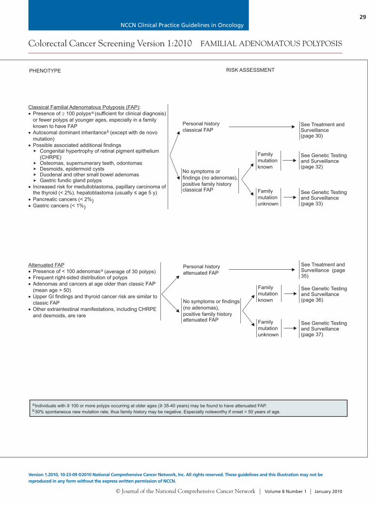
NCCN Clinical Practice Guidelines in Oncology
© Journal of the National Comprehensive Cancer Network | Volume 8 Number 1 | January 2010
29
Colorectal Cancer Screening Version 1:2010
Version 1.2010, 10-23-09 ©2010 National Comprehensive Cancer Network, Inc. All rights reserved. These guidelines and this illustration may not be reproduced in any form without the express written permission of NCCN.
FAMILIAL ADENOMATOUS POLYPOSIS
1Kohlmann W, Gruber SB (Updated November 29, 2006). Hereditary non-polyposis colon cancer. In: GeneReviews at GeneTests: Medical GeneticsInformation Resource (database online). Copyright, University of Washington, Seattle. 1997-2009. Available at: http://www.genetests.org. AccessedOctober 21, 2009.
Cancer General Population Risk
HNPCC
Risks Mean Age of Onset
Colon
Endometrium
Stomach
Ovary
Hepatobiliary tract
Urinary tract
Small bowel
Brain/central nervoussystem
5.5%
2.7%
< 1%
1.6%
< 1%
< 1%
< 1%
< 1%
80%
20%-60%
11%-19%
9%-12%
2%-7%
4%-5%
1%-4%
1%-3%
44 y
46 y
56 y
42.5 y
Not reported
~ 55 y
49 y
~ 50 y
1 RISK ASSESSMENT
Classical Familial Adenomatous Polyposis (FAP):Presence of 100 polyps (sufficient for clinical diagnosis)or fewer polyps at younger ages, especially in a familyknown to have FAPAutosomal dominant inheritance (except with de novomutation)Possible associated additional findings
Congenital hypertrophy of retinal pigment epithelium(CHRPE)Osteomas, supernumerary teeth, odontomasDesmoids, epidermoid cystsDuodenal and other small bowel adenomasGastric fundic gland polyps
Increased risk for medulloblastoma, papillary carcinoma ofthe thyroid (< 2%), hepatoblastoma (usually age 5 y)Pancreatic cancers (< 2%)Gastric cancers (< 1%)
a
b
Personal historyclassical FAP
No symptoms orfindings (no adenomas),positive family historyclassical FAP
See Treatment andSurveillance(page 30)
See Genetic Testingand Surveillance(page 32)
See Genetic Testingand Surveillance(page 33)
Familymutationknown
Familymutationunknown
ab
Individuals with 100 or more polyps occurring at older ages ( 35-40 years) may be found to have attenuated FAP.30% spontaneous new mutation rate, thus family history may be negative. Especially noteworthy if onset < 50 years of age.
Attenuated FAPPresence of < 100 adenomas (average of 30 polyps)Frequent right-sided distribution of polypsAdenomas and cancers at age older than classic FAP (mean age > 50)Upper GI findings and thyroid cancer risk are similar toclassic FAPOther extraintestinal manifestations, including CHRPEand desmoids, are rare
aPersonal historyattenuated FAP
No symptoms or findings(no adenomas),positive family historyattenuated FAP
See Treatment andSurveillance (page35)
See Genetic Testingand Surveillance(page 36)
See Genetic Testingand Surveillance(page 37)
Familymutationknown
Familymutationunknown
PHENOTYPE
CANCER RISK IN INDIVIDUALS WITH HNPCC UP TO AGE 70 YEARS COMPARED WITH THE GENERAL POPULATION

© Journal of the National Comprehensive Cancer Network | Volume 8 Number 1 | January 2010
30
Colorectal Cancer Screening Version 1:2010
Clinical trials: The NCCN believes that the best management for any cancer patient is in a clinical trial. Participation in clinical trials is especially encouraged. All recommendations are category 2A unless otherwise noted.
FAMILIAL ADENOMATOUS POLYPOSIS
Proctocolectomy orcolectomyc,d,e
TREATMENT
Personalhistory ofclassical FAP
c
e
APC genetic testing is recommended in a proband to confirm a diagnosis of FAP and allow for mutation-specific testing in other family members.Additionally, knowing the location of the mutation in the APC gene can be helpful for predicting severity of polyposis, rectal involvement, and desmoidtumors.
See Surgical Options for Treating the Colon And Rectum in Patients with FAP (page 34).Timing of colectomy in patients < 18 y is not established. In patients < 18 y with mild polyposis and without family history of early cancer or severe genotype,the timing of colectomy can be individualized. Annual colonoscopy if surgery is delayed.
d
f
g
It is recommended that patients be managed by physicians or centers with expertise in FAP and that management be individualized to account for genotype,phenotype, and personal considerations.
Other than colon cancer, screening recommendations are expert opinion rather than-evidence based.
CLASSICAL FAP TREATMENT AND SURVEILLANCE: PERSONAL HISTORY
Colon cancer:Annual sigmoidoscopy and polypectomy or polyp ablation ifadenoma burden is low (in patients with retained rectum).Consider NSAID chemoprevention to reduce polyp burdenas pharmacologic adjunct to endoscopic surveillance.Clinical trial is encouraged.
Extracolonic:Duodenal or periampullary cancer: baseline upperendoscopy (including side-viewing examination) at the timeof colectomy or at age 25-30 y, whichever comes first, andrepeat every 1-3 y, depending on severity of polyposis.Gastric cancer: same as duodenal cancer.Thyroid cancer: annual thyroid examination, starting in lateteenage years.CNS cancer: annual physical examination; no additionalscreening recommendations have been made.Intra-abdominal desmoids: annual abdominal palpation;after colectomy, consider abdominal and pelvic CT or MRIevery 3 y, especially if family history of desmoids.Small bowel polyps and cancer: consider adding smallbowel visualization to CT or MRI for desmoids as outlinedabove, especially if duodenal polyposis is advanced.Hepatoblastoma: c
AFP5 . Screening in a clinical trial is preferred.
Pancreatic cancer: no recommendations have been made.
onsider liver palpation, abdominalultrasound and measurement of , every 3-6 mo, duringfirst y of life
Proctectomy ifdense polyposisor severedysplasia
SURVEILLANCE(POSTCOLECTOMY)
f,g
Stage 0,No polyposis
Stage I,Minimal polyposis (1-4 tubular adenomas, size 1-4 mm)
Stage II,Mild polyposis (5-19 tubular adenomas, size 5-9 mm)
Stage III,Moderate polyposis ( 20 lesions, or size 1 cm)
Stage IV,Dense polyposis or high-grade dysplasia
Repeat endoscopy every 4 y
Repeat endoscopy every 2-3 y
Repeat endoscopy every 1-3 y
Repeat endoscopy every 6-12 mo
SURVEILLANCEhDUODENOSCOPIC FINDINGS
hDuodenal Surveillance:It is recommended that patients be managed by physicians or centers with expertise in FAP and that management be individualized to account forgenotype, phenotype, and personal considerations, including potential risks and benefits. Management that includes endoscopic treatment may requireshorter intervals.Recommend examination with side-viewing endoscope, use of Spigelman's or other standardized staging, and extensive biopsy of dense lesions toevaluate for advanced histology. More intensive surveillance and/or treatment is required in patients with large or villous adenomas, and with advancingage > 50 y. Surgical counseling is advisable for patients with stage IV polyposis. (Spigelman AD, Williams CB, Talbot IC, et al. Upper gastrointestinalcancer in patients with familial adenomatous polyposis. Lancet 1989;2:783-785.)Endoscopic treatment options include endoscopic papillectomy, in addition to excision or ablation of resectable large (> 1 cm) or villous adenomas, andmucosectomy of resectable advanced lesions, including contained high-grade dysplasia, to potentially avert surgery while observing pathology guidelinesfor adequate resection.Surgery is recommended for invasive carcinoma and dense polyposis or high-grade dysplasia that cannot be managed endoscopically.
Surgical evaluationExpert surveillance every 6-12 moComplete mucosectomy or duodenectomy, orWhipple procedure if duodenal papilla is involved

NCCN Clinical Practice Guidelines in Oncology
© Journal of the National Comprehensive Cancer Network | Volume 8 Number 1 | January 2010
31
Colorectal Cancer Screening Version 1:2010
Version 1.2010, 10-23-09 ©2010 National Comprehensive Cancer Network, Inc. All rights reserved. These guidelines and this illustration may not be reproduced in any form without the express written permission of NCCN.
FAMILIAL ADENOMATOUS POLYPOSIS
Proctocolectomy orcolectomyc,d,e
TREATMENT
Personalhistory ofclassical FAP
c
e
APC genetic testing is recommended in a proband to confirm a diagnosis of FAP and allow for mutation-specific testing in other family members.Additionally, knowing the location of the mutation in the APC gene can be helpful for predicting severity of polyposis, rectal involvement, and desmoidtumors.
See Surgical Options for Treating the Colon And Rectum in Patients with FAP (page 34).Timing of colectomy in patients < 18 y is not established. In patients < 18 y with mild polyposis and without family history of early cancer or severe genotype,the timing of colectomy can be individualized. Annual colonoscopy if surgery is delayed.
d
f
g
It is recommended that patients be managed by physicians or centers with expertise in FAP and that management be individualized to account for genotype,phenotype, and personal considerations.
Other than colon cancer, screening recommendations are expert opinion rather than-evidence based.
CLASSICAL FAP TREATMENT AND SURVEILLANCE: PERSONAL HISTORY
Colon cancer:Annual sigmoidoscopy and polypectomy or polyp ablation ifadenoma burden is low (in patients with retained rectum).Consider NSAID chemoprevention to reduce polyp burdenas pharmacologic adjunct to endoscopic surveillance.Clinical trial is encouraged.
Extracolonic:Duodenal or periampullary cancer: baseline upperendoscopy (including side-viewing examination) at the timeof colectomy or at age 25-30 y, whichever comes first, andrepeat every 1-3 y, depending on severity of polyposis.Gastric cancer: same as duodenal cancer.Thyroid cancer: annual thyroid examination, starting in lateteenage years.CNS cancer: annual physical examination; no additionalscreening recommendations have been made.Intra-abdominal desmoids: annual abdominal palpation;after colectomy, consider abdominal and pelvic CT or MRIevery 3 y, especially if family history of desmoids.Small bowel polyps and cancer: consider adding smallbowel visualization to CT or MRI for desmoids as outlinedabove, especially if duodenal polyposis is advanced.Hepatoblastoma: c
AFP5 . Screening in a clinical trial is preferred.
Pancreatic cancer: no recommendations have been made.
onsider liver palpation, abdominalultrasound and measurement of , every 3-6 mo, duringfirst y of life
Proctectomy ifdense polyposisor severedysplasia
SURVEILLANCE(POSTCOLECTOMY)
f,g
Stage 0,No polyposis
Stage I,Minimal polyposis (1-4 tubular adenomas, size 1-4 mm)
Stage II,Mild polyposis (5-19 tubular adenomas, size 5-9 mm)
Stage III,Moderate polyposis ( 20 lesions, or size 1 cm)
Stage IV,Dense polyposis or high-grade dysplasia
Repeat endoscopy every 4 y
Repeat endoscopy every 2-3 y
Repeat endoscopy every 1-3 y
Repeat endoscopy every 6-12 mo
SURVEILLANCEhDUODENOSCOPIC FINDINGS
hDuodenal Surveillance:It is recommended that patients be managed by physicians or centers with expertise in FAP and that management be individualized to account forgenotype, phenotype, and personal considerations, including potential risks and benefits. Management that includes endoscopic treatment may requireshorter intervals.Recommend examination with side-viewing endoscope, use of Spigelman's or other standardized staging, and extensive biopsy of dense lesions toevaluate for advanced histology. More intensive surveillance and/or treatment is required in patients with large or villous adenomas, and with advancingage > 50 y. Surgical counseling is advisable for patients with stage IV polyposis. (Spigelman AD, Williams CB, Talbot IC, et al. Upper gastrointestinalcancer in patients with familial adenomatous polyposis. Lancet 1989;2:783-785.)Endoscopic treatment options include endoscopic papillectomy, in addition to excision or ablation of resectable large (> 1 cm) or villous adenomas, andmucosectomy of resectable advanced lesions, including contained high-grade dysplasia, to potentially avert surgery while observing pathology guidelinesfor adequate resection.Surgery is recommended for invasive carcinoma and dense polyposis or high-grade dysplasia that cannot be managed endoscopically.
Surgical evaluationExpert surveillance every 6-12 moComplete mucosectomy or duodenectomy, orWhipple procedure if duodenal papilla is involved

© Journal of the National Comprehensive Cancer Network | Volume 8 Number 1 | January 2010
32
Colorectal Cancer Screening Version 1:2010
Clinical trials: The NCCN believes that the best management for any cancer patient is in a clinical trial. Participation in clinical trials is especially encouraged. All recommendations are category 2A unless otherwise noted.
FAMILIAL ADENOMATOUS POLYPOSIS
Family history ofclassical FAP,mutation known
APC gene testingfor at-risk familymemberi
Flexible sigmoidoscopy orcolonoscopy every 12 mobeginning at age 10-15 y
Flexible sigmoidoscopy orcolonoscopy beginning atage 10-15 y:
Every 12 mo until age 24 yEvery 2 y until age 34 yEvery 3 y until age 44 yThen every 3-5 y thereafter
Consider substituting colonoscopyevery 5 y beginning at age 20 yfor the chance that patient mayhave attenuated FAP
If adenomas, followpathway for PersonalHistory of ClassicalFAP (page 30)
If adenomas, followpathway for PersonalHistory of ClassicalFAP (page 30)If no polyps, continuescreening
GENETIC TESTING SURVEILLANCE
Average-risk screening
APC genepositive
APC genenegative
Not tested
CLASSICAL FAP GENETIC TESTING AND SURVEILLANCE: FAMILY HISTORY OF CLASSICAL FAP MUTATION KNOWN
iAt-risk family member can be defined as a first-degree relative of an affected individual and/or proband. If a first-degree relative is unavailable or unwilling tobe tested, more distant relatives should be offered testing for the known mutation in the family.
GENETIC TESTING
See APC Gene Positive(opposite page)
Nomutationfound
Not tested
Family historyof classicalFAP, mutationunknown
If mutation found, follow pathway for Family Historyof Classical FAP, Mutation Known (opposite page)
Mutation in family not found
Affected familymember notavailable
ConsiderAPC genetesting forat-risk familymemberi
APC gene testingof affected familymemberConsider MYHtestingj,k
APC genepositive
SURVEILLANCE
CLASSICAL FAP GENETIC TESTING AND SURVEILLANCE: FAMILY HISTORY OF CLASSICAL FAP MUTATION UNKNOWN
Flexible sigmoidoscopy orcolonoscopy beginning at age10-15 y:
Every 12 mo until age 24 yEvery 2 y until age 34 yEvery 3 y until age 44 yThen every 3-5 y thereafter
Consider substitutingcolonoscopy every 5 ybeginning at age 20 y inaddition to the sigmoidoscopyexaminationsIf polyposis detected, followpathway on (page 30)
i
jk
At-risk family member can be defined as a first-degree relative of an affected individual and/or proband. If a first-degree relative is unavailable or unwilling tobe tested, more distant relatives should be offered testing for the known mutation in the family.
See MYH-Associated Polyposis (page 38).When polyposis is present in a single person with negative family history, testing for a de novo APC mutation should be considered; if negative, testing forMYH should follow. When family history is positive only for a sibling, recessive inheritance should be considered and testing for MYH should occur first. In apolyposis family with clear autosomal dominant inheritance and absence of APC mutation, MYH testing is unlikely to be informative. These families aretreated according to the polyposis phenotype, including classical or attenuated FAP.

NCCN Clinical Practice Guidelines in Oncology
© Journal of the National Comprehensive Cancer Network | Volume 8 Number 1 | January 2010
33
Colorectal Cancer Screening Version 1:2010
Version 1.2010, 10-23-09 ©2010 National Comprehensive Cancer Network, Inc. All rights reserved. These guidelines and this illustration may not be reproduced in any form without the express written permission of NCCN.
FAMILIAL ADENOMATOUS POLYPOSIS
Family history ofclassical FAP,mutation known
APC gene testingfor at-risk familymemberi
Flexible sigmoidoscopy orcolonoscopy every 12 mobeginning at age 10-15 y
Flexible sigmoidoscopy orcolonoscopy beginning atage 10-15 y:
Every 12 mo until age 24 yEvery 2 y until age 34 yEvery 3 y until age 44 yThen every 3-5 y thereafter
Consider substituting colonoscopyevery 5 y beginning at age 20 yfor the chance that patient mayhave attenuated FAP
If adenomas, followpathway for PersonalHistory of ClassicalFAP (page 30)
If adenomas, followpathway for PersonalHistory of ClassicalFAP (page 30)If no polyps, continuescreening
GENETIC TESTING SURVEILLANCE
Average-risk screening
APC genepositive
APC genenegative
Not tested
CLASSICAL FAP GENETIC TESTING AND SURVEILLANCE: FAMILY HISTORY OF CLASSICAL FAP MUTATION KNOWN
iAt-risk family member can be defined as a first-degree relative of an affected individual and/or proband. If a first-degree relative is unavailable or unwilling tobe tested, more distant relatives should be offered testing for the known mutation in the family.
GENETIC TESTING
See APC Gene Positive(opposite page)
Nomutationfound
Not tested
Family historyof classicalFAP, mutationunknown
If mutation found, follow pathway for Family Historyof Classical FAP, Mutation Known (opposite page)
Mutation in family not found
Affected familymember notavailable
ConsiderAPC genetesting forat-risk familymemberi
APC gene testingof affected familymemberConsider MYHtestingj,k
APC genepositive
SURVEILLANCE
CLASSICAL FAP GENETIC TESTING AND SURVEILLANCE: FAMILY HISTORY OF CLASSICAL FAP MUTATION UNKNOWN
Flexible sigmoidoscopy orcolonoscopy beginning at age10-15 y:
Every 12 mo until age 24 yEvery 2 y until age 34 yEvery 3 y until age 44 yThen every 3-5 y thereafter
Consider substitutingcolonoscopy every 5 ybeginning at age 20 y inaddition to the sigmoidoscopyexaminationsIf polyposis detected, followpathway on (page 30)
Flexible sigmoidoscopy orcolonoscopy beginning at age10-15 y:
Every 12 mo until age 24 yEvery 2 y until age 34 yEvery 3 y until age 44 yThen every 3-5 y thereafter
Consider substitutingcolonoscopy every 5 ybeginning at age 20 y inaddition to the sigmoidoscopyexaminationsIf polyposis detected, followpathway on (page 30)
i
jk
At-risk family member can be defined as a first-degree relative of an affected individual and/or proband. If a first-degree relative is unavailable or unwilling tobe tested, more distant relatives should be offered testing for the known mutation in the family.
See MYH-Associated Polyposis (page 38).When polyposis is present in a single person with negative family history, testing for a de novo APC mutation should be considered; if negative, testing forMYH should follow. When family history is positive only for a sibling, recessive inheritance should be considered and testing for MYH should occur first. In apolyposis family with clear autosomal dominant inheritance and absence of APC mutation, MYH testing is unlikely to be informative. These families aretreated according to the polyposis phenotype, including classical or attenuated FAP.

© Journal of the National Comprehensive Cancer Network | Volume 8 Number 1 | January 2010
34
Colorectal Cancer Screening Version 1:2010
Clinical trials: The NCCN believes that the best management for any cancer patient is in a clinical trial. Participation in clinical trials is especially encouraged. All recommendations are category 2A unless otherwise noted.
FAMILIAL ADENOMATOUS POLYPOSIS
SURGICAL OPTIONS FOR TREATING THE COLON AND RECTUM IN PATIENTS WITH FAP
Total Abdominal Colectomy With Ileorectal Anastomosis (TAC/IRA)Indications:
Asymptomatic patient with few (< 20) rectal polyps and mildcolonic disease (< 100) polypsAttenuated FAP with rectal sparing
Curable cancer in colon or rectumSevere rectal or colon disease (size or number of polyps)Patient not reliable for follow-up surveillance of retained rectum
Advantages:Technically straightforwardRelatively low complication rateGood function outcomeNo permanent or temporary stomaAvoids risk for proctectomy (sexual or bladder dysfunction)
Contraindications:
Total Proctocolectomy With End Ileostomy (TPC/EI)Indications:
Very low, advanced rectal cancerInability to perform IPAAPatient with IPAA with unacceptable functionPatient with contraindication to IPAA
Advantages:Removes risk for colorectal cancerOne operation
Disadvantages:Risks for proctectomyPermanent stomaMay discourage family members from seeking evaluation for fearof permanent stoma
Total Proctocolectomy With Ileal Pouch Anal Anastomosis(TPC/IPAA)
Indications:After TAC/IRA with unstable rectumPatient unreliable for follow-up after TAC/IRASevere disease in colon and/or rectumCurable colon or rectal cancer
Contraindications:Incurable cancerIntra-abdominal desmoidAdvanced low rectal cancerPatient not a candidate for IPAA (e.g., hasconcomitant Crohn’s disease or anal sphincterdysfunction)
Advantages:Negligible risk for rectal cancerNo permanent stomaReasonable bowel function
Disadvantages:Complex operationUsually involves temporary stomaRisks of proctectomy (sexual or bladder dysfunction)Functional results can be unpredictable
Personal historyconsistent withattenuated FAP
APC genetestinga,b
< 21 y withsmall adenomaburdenc
21 y withsmall adenomaburdenc
Significantpolyposis notmanageable withpolypectomy
Colonoscopy andpolypectomy every1-2 ySurgical evaluationand counseling
Colectomy andileorectal anastomosis(IRA)orcolonoscopy andpolypectomy every 1-2 ySurgical evaluation andcounseling
d
e
Colectomy with IRA(preferred in mostcases) orproctocolectomy withileal J-pouch analanastomosis based onburden of disease inrectum
d
e
If colectomy, annualendoscopic examination ofthe rectumAnnual physical examinationConsider NSAID andchemoprevention to reducepolyp burden aspharmacologic adjunct toendoscopic surveillance.Clinical trial is encouraged.
a
bc
def
g
APC gene testing is recommended in a proband to confirm a diagnosis of attenuated FAP and allow for mutation-specific testing in other family members.Additionally, knowing the location of the APC mutation can be helpful in determining extracolonic cancer risks in affected individuals.
Consider MYH testing if APC mutation not found (see page 38).Small adenoma burden is defined (somewhat arbitrarily) as < 20 adenomas, all < 1 cm in diameter, and none with advanced histology, so that colonoscopywith polypectomy can be used to effectively eliminate the polyps. Colectomy may be indicated before this level of polyp profusion, especially ifcolonoscopy is difficult. Surgery is strongly advised when polyp burden is > 20, some polyps have reached a size > 1 cm, or advanced histology isencountered in any polyp.
See Surgical Options for Treating the Colon and Rectum in Patients with FAP (opposite page).Earlier surgical intervention should be considered in patients with family history of cancer or those who are noncompliant.It is recommended that patients be managed by physicians or centers with expertise in FAP and that management be individualized to account for
genotype, phenotype, and personal considerations.Surveillance for upper gastointestinal findings for attenuated FAP is similar to that for classical FAP.
< 40 years
TREATMENT
ATTENUATED FAP TREATMENT AND SURVEILLANCE: PERSONAL HISTORY
40 y withsmall adenomaburdenc
Strongly considercolectomy andIRASurgical evaluationand counseling
de
SURVEILLANCEf,gADENOMA/POLYPBURDEN
Baseline upperendoscopy beginningat age 25-30 y
SeeDuodenoscopicFindings(page 31)

NCCN Clinical Practice Guidelines in Oncology
© Journal of the National Comprehensive Cancer Network | Volume 8 Number 1 | January 2010
35
Colorectal Cancer Screening Version 1:2010
Version 1.2010, 10-23-09 ©2010 National Comprehensive Cancer Network, Inc. All rights reserved. These guidelines and this illustration may not be reproduced in any form without the express written permission of NCCN.
SURGICAL OPTIONS FOR TREATING THE COLON AND RECTUM IN PATIENTS WITH FAP
Total Abdominal Colectomy With Ileorectal Anastomosis (TAC/IRA)Indications:
Asymptomatic patient with few (< 20) rectal polyps and mildcolonic disease (< 100) polypsAttenuated FAP with rectal sparing
Curable cancer in colon or rectumSevere rectal or colon disease (size or number of polyps)Patient not reliable for follow-up surveillance of retained rectum
Advantages:Technically straightforwardRelatively low complication rateGood function outcomeNo permanent or temporary stomaAvoids risk for proctectomy (sexual or bladder dysfunction)
Contraindications:
Total Proctocolectomy With End Ileostomy (TPC/EI)Indications:
Very low, advanced rectal cancerInability to perform IPAAPatient with IPAA with unacceptable functionPatient with contraindication to IPAA
Advantages:Removes risk for colorectal cancerOne operation
Disadvantages:Risks for proctectomyPermanent stomaMay discourage family members from seeking evaluation for fearof permanent stoma
Total Proctocolectomy With Ileal Pouch Anal Anastomosis(TPC/IPAA)
Indications:After TAC/IRA with unstable rectumPatient unreliable for follow-up after TAC/IRASevere disease in colon and/or rectumCurable colon or rectal cancer
Contraindications:Incurable cancerIntra-abdominal desmoidAdvanced low rectal cancerPatient not a candidate for IPAA (e.g., hasconcomitant Crohn’s disease or anal sphincterdysfunction)
Advantages:Negligible risk for rectal cancerNo permanent stomaReasonable bowel function
Disadvantages:Complex operationUsually involves temporary stomaRisks of proctectomy (sexual or bladder dysfunction)Functional results can be unpredictable
Personal historyconsistent withattenuated FAP
APC genetestinga,b
< 21 y withsmall adenomaburdenc
21 y withsmall adenomaburdenc
Significantpolyposis notmanageable withpolypectomy
Colonoscopy andpolypectomy every1-2 ySurgical evaluationand counseling
Colectomy andileorectal anastomosis(IRA)orcolonoscopy andpolypectomy every 1-2 ySurgical evaluation andcounseling
d
e
Colectomy with IRA(preferred in mostcases) orproctocolectomy withileal J-pouch analanastomosis based onburden of disease inrectum
d
e
If colectomy, annualendoscopic examination ofthe rectumAnnual physical examinationConsider NSAID andchemoprevention to reducepolyp burden aspharmacologic adjunct toendoscopic surveillance.Clinical trial is encouraged.
a
bc
def
g
APC gene testing is recommended in a proband to confirm a diagnosis of attenuated FAP and allow for mutation-specific testing in other family members.Additionally, knowing the location of the APC mutation can be helpful in determining extracolonic cancer risks in affected individuals.
Consider MYH testing if APC mutation not found (see page 38).Small adenoma burden is defined (somewhat arbitrarily) as < 20 adenomas, all < 1 cm in diameter, and none with advanced histology, so that colonoscopywith polypectomy can be used to effectively eliminate the polyps. Colectomy may be indicated before this level of polyp profusion, especially ifcolonoscopy is difficult. Surgery is strongly advised when polyp burden is > 20, some polyps have reached a size > 1 cm, or advanced histology isencountered in any polyp.
See Surgical Options for Treating the Colon and Rectum in Patients with FAP (opposite page).Earlier surgical intervention should be considered in patients with family history of cancer or those who are noncompliant.It is recommended that patients be managed by physicians or centers with expertise in FAP and that management be individualized to account for
genotype, phenotype, and personal considerations.Surveillance for upper gastointestinal findings for attenuated FAP is similar to that for classical FAP.
< 40 years
TREATMENT
ATTENUATED FAP TREATMENT AND SURVEILLANCE: PERSONAL HISTORY
40 y withsmall adenomaburdenc
Strongly considercolectomy andIRASurgical evaluationand counseling
de
SURVEILLANCEf,gADENOMA/POLYPBURDEN
Baseline upperendoscopy beginningat age 25-30 y
SeeDuodenoscopicFindings(page 31)
ATTENUATED FAMILIAL ADENOMATOUS POLYPOSIS

© Journal of the National Comprehensive Cancer Network | Volume 8 Number 1 | January 2010
36
Colorectal Cancer Screening Version 1:2010
Clinical trials: The NCCN believes that the best management for any cancer patient is in a clinical trial. Participation in clinical trials is especially encouraged. All recommendations are category 2A unless otherwise noted.
Colonoscopy beginning inlate teens, then every 2-3 y
If adenomas, followpathway for Personalhistory of attenuatedFAP, adenoma/polypburden (page 35)
If adenomas, followpathway for Personalhistory of attenuatedFAP, adenoma/polypburden (page 35)If no polyps, continuescreening
GENETIC TESTING SURVEILLANCE
APC positive
APC negative
Not tested
Family history ofattenuated FAP,mutation known
APC testing forat-risk familymemberh
Colonoscopy beginning in lateteens, then every 2-3 y
Average-risk screening
hAt-risk family member can be defined as a first-degree relative of an affected individual and/or proband. If a first-degree relative is unavailable or unwillingto be tested, more distant relatives should be offered testing for the known mutation in the family.
ATTENUATED FAP GENETIC TESTING AND SURVEILLANCE: FAMILY HISTORY OF ATTENUATED FAP MUTATION KNOWN
No mutationfound
Not tested
Family history ofattenuated FAP,mutationunknown
If APC mutation found, follow pathway forfamily mutation known (opposite page)
APC mutation in family not found
Affected familymember notavailable
Consider APC testingfor at-risk familymemberConsider MYH testingif APC mutationnegative
hi
Consider APC testing ofaffected family memberConsider MYH testingi,j
APCmutationfound
GENETIC TESTING
h
ij
At-risk family member can be defined as a first-degree relative of an affected individual and/or proband.
See MYH-Associated Polyposis (page 38).When polyposis is present in a single person with negative family history, consider testing for a de novo APC mutation; if negative, follow with testing for
MYH. When family history is positive only for a sibling, recessive inheritance should be considered and testing for MYH should occur first. In a polyposisfamily with clear autosomal dominant inheritance and absence of APC mutation, MYH testing is unlikely to be informative. These families are treatedaccording to the polyposis phenotype, including classical or attenuated FAP.
If a first-degree relative is unavailable or unwilling tobe tested, more distant relatives should be offered testing for the known mutation in the family.
ATTENUATED FAP GENETIC TESTING AND SURVEILLANCE: FAMILY HISTORY OF ATTENUATED FAP MUTATION UNKNOWN
Colonoscopy beginning inlate teens, then every 2-3 yIf adenoma, see page 35
Colonoscopybeginning inlate teens, thenevery 2-3 yIf adenoma,see page 35
SURVEILLANCE
ATTENUATED FAMILIAL ADENOMATOUS POLYPOSIS

NCCN Clinical Practice Guidelines in Oncology
© Journal of the National Comprehensive Cancer Network | Volume 8 Number 1 | January 2010
37
Colorectal Cancer Screening Version 1:2010
Version 1.2010, 10-23-09 ©2010 National Comprehensive Cancer Network, Inc. All rights reserved. These guidelines and this illustration may not be reproduced in any form without the express written permission of NCCN.
Colonoscopy beginning inlate teens, then every 2-3 y
If adenomas, followpathway for Personalhistory of attenuatedFAP, adenoma/polypburden (page 35)
If adenomas, followpathway for Personalhistory of attenuatedFAP, adenoma/polypburden (page 35)If no polyps, continuescreening
GENETIC TESTING SURVEILLANCE
APC positive
APC negative
Not tested
Family history ofattenuated FAP,mutation known
APC testing forat-risk familymemberh
Colonoscopy beginning in lateteens, then every 2-3 y
Average-risk screening
hAt-risk family member can be defined as a first-degree relative of an affected individual and/or proband. If a first-degree relative is unavailable or unwillingto be tested, more distant relatives should be offered testing for the known mutation in the family.
ATTENUATED FAP GENETIC TESTING AND SURVEILLANCE: FAMILY HISTORY OF ATTENUATED FAP MUTATION KNOWN
No mutationfound
Not tested
Family history ofattenuated FAP,mutationunknown
If APC mutation found, follow pathway forfamily mutation known (opposite page)
APC mutation in family not found
Affected familymember notavailable
Consider APC testingfor at-risk familymemberConsider MYH testingif APC mutationnegative
hi
Consider APC testing ofaffected family memberConsider MYH testingi,j
APCmutationfound
GENETIC TESTING
h
ij
At-risk family member can be defined as a first-degree relative of an affected individual and/or proband.
See MYH-Associated Polyposis (page 38).When polyposis is present in a single person with negative family history, consider testing for a de novo APC mutation; if negative, follow with testing for
MYH. When family history is positive only for a sibling, recessive inheritance should be considered and testing for MYH should occur first. In a polyposisfamily with clear autosomal dominant inheritance and absence of APC mutation, MYH testing is unlikely to be informative. These families are treatedaccording to the polyposis phenotype, including classical or attenuated FAP.
If a first-degree relative is unavailable or unwilling tobe tested, more distant relatives should be offered testing for the known mutation in the family.
ATTENUATED FAP GENETIC TESTING AND SURVEILLANCE: FAMILY HISTORY OF ATTENUATED FAP MUTATION UNKNOWN
Colonoscopy beginning inlate teens, then every 2-3 yIf adenoma, see page 35
Colonoscopybeginning inlate teens, thenevery 2-3 yIf adenoma,see page 35
SURVEILLANCE
ATTENUATED FAMILIAL ADENOMATOUS POLYPOSIS

© Journal of the National Comprehensive Cancer Network | Volume 8 Number 1 | January 2010
38
Colorectal Cancer Screening Version 1:2010
Clinical trials: The NCCN believes that the best management for any cancer patient is in a clinical trial. Participation in clinical trials is especially encouraged. All recommendations are category 2A unless otherwise noted.
MYH-ASSOCIATED POLYPOSIS
MYH-Associated Polyposis (MAP):autosomal recessive
inheritance )< 100 adenomas (range 0-100’s and uncommonly > 1000)Adenomas and colorectal cancer at age older than classical FAP(median CRC age > 50 y)Duodenal adenomas are uncommon
Polyposis or colon cancers consistent with(i.e., parents unaffected, siblings affected
Attenuated polyposis with negative APC gene mutation
Personal historyorFamily history(i.e., known mutation in patient or sibling)
RISK ASSESSMENTPHENOTYPE
Family history of sibling with MYHpolyposis, asymptomatic;Counseling and testing for thefamilial mutations is recommended
Mutation status unknownor biallelic MYH mutationsknown
Begin colonoscopy at age 25-30 yand every 3-5 y if negative(consider shorter intervals withadvancing age)Consider upper endoscopy andside-viewing duodenoscopy at age30-35 y and every 3-5 yPatients with duodenal adenomasare treated as FAP (SeeDuodenoscopic Findings; page 31)
Personal history ofmultiple adenomatouspolyps (> 10 adenomaswith negative APCmutation testing)a
a
b
When polyposis is present in a single person with negative family history, consider testing for a de novo APC mutation; if negative, follow with testing forMYH. When family history is positive only for a sibling, consider recessive inheritance and test for MYH first. In a polyposis family with clear autosomaldominant inheritance, and absence of APC mutation, MYH testing is unlikely to be informative. Such families are treated according to the polyposisphenotype, including classical or attenuated FAP.
In patients with MYH, the absolute risk of colorectal cancer and the role of surgery and endoscopically manageable adenomas is not known. The lifetimecolon cancer risk may be very high.
Counseling andtesting for MYHmutations
Biallelic MYHmutationnegative
Manage individuallyas multipleadenomatous polyps(page 29)
Biallelic MYHmutationpositive
Small adenomaburden manageableby colonoscopy andpolypectomy
TREATMENT/SURVEILLANCE
Colonoscopy and polypectomy every1-2 yConsider upper endoscopy and side-viewing duodenoscopy beginning atage 30-35 y and every 3-5 yPatients with duodenal adenomas aretreated as FAP (See DuodenoscopicFindings; page 31)
b
Dense polyposis orlarge polyps notmanageable bypolypectomy
Counseling regarding surgical optionsSubtotal colectomy orproctocolectomy depending onadenoma density and distributiona
Upper endoscopy and side-viewingduodenoscopy at age 30-35 y andevery 3-5 yPatients with duodenal adenomas aretreated as FAP (See DuodenoscopicFindings; page 31)
GENETIC COUNSELING/TESTINGOF ELIGIBLE FAMILY MEMBERS

NCCN Clinical Practice Guidelines in Oncology
© Journal of the National Comprehensive Cancer Network | Volume 8 Number 1 | January 2010
39
Colorectal Cancer Screening Version 1:2010
Version 1.2010, 10-23-09 ©2010 National Comprehensive Cancer Network, Inc. All rights reserved. These guidelines and this illustration may not be reproduced in any form without the express written permission of NCCN.
MYH-ASSOCIATED POLYPOSIS
MYH-Associated Polyposis (MAP):autosomal recessive
inheritance )< 100 adenomas (range 0-100’s and uncommonly > 1000)Adenomas and colorectal cancer at age older than classical FAP(median CRC age > 50 y)Duodenal adenomas are uncommon
Polyposis or colon cancers consistent with(i.e., parents unaffected, siblings affected
Attenuated polyposis with negative APC gene mutation
Personal historyorFamily history(i.e., known mutation in patient or sibling)
RISK ASSESSMENTPHENOTYPE
Family history of sibling with MYHpolyposis, asymptomatic;Counseling and testing for thefamilial mutations is recommended
Mutation status unknownor biallelic MYH mutationsknown
Begin colonoscopy at age 25-30 yand every 3-5 y if negative(consider shorter intervals withadvancing age)Consider upper endoscopy andside-viewing duodenoscopy at age30-35 y and every 3-5 yPatients with duodenal adenomasare treated as FAP (SeeDuodenoscopic Findings; page 31)
Personal history ofmultiple adenomatouspolyps (> 10 adenomaswith negative APCmutation testing)a
a
b
When polyposis is present in a single person with negative family history, consider testing for a de novo APC mutation; if negative, follow with testing forMYH. When family history is positive only for a sibling, consider recessive inheritance and test for MYH first. In a polyposis family with clear autosomaldominant inheritance, and absence of APC mutation, MYH testing is unlikely to be informative. Such families are treated according to the polyposisphenotype, including classical or attenuated FAP.
In patients with MYH, the absolute risk of colorectal cancer and the role of surgery and endoscopically manageable adenomas is not known. The lifetimecolon cancer risk may be very high.
Counseling andtesting for MYHmutations
Biallelic MYHmutationnegative
Manage individuallyas multipleadenomatous polyps(page 29)
Biallelic MYHmutationpositive
Small adenomaburden manageableby colonoscopy andpolypectomy
TREATMENT/SURVEILLANCE
Colonoscopy and polypectomy every1-2 yConsider upper endoscopy and side-viewing duodenoscopy beginning atage 30-35 y and every 3-5 yPatients with duodenal adenomas aretreated as FAP (See DuodenoscopicFindings; page 31)
b
Dense polyposis orlarge polyps notmanageable bypolypectomy
Counseling regarding surgical optionsSubtotal colectomy orproctocolectomy depending onadenoma density and distributiona
Upper endoscopy and side-viewingduodenoscopy at age 30-35 y andevery 3-5 yPatients with duodenal adenomas aretreated as FAP (See DuodenoscopicFindings; page 31)
GENETIC COUNSELING/TESTINGOF ELIGIBLE FAMILY MEMBERS

© Journal of the National Comprehensive Cancer Network | Volume 8 Number 1 | January 2010
40
Colorectal Cancer Screening Version 1:2010
Clinical trials: The NCCN believes that the best management for any cancer patient is in a clinical trial. Participation in clinical trials is especially encouraged. All recommendations are category 2A unless otherwise noted.
PEUTZ-JEGHERS SYNDROME
12
Tomlinson IP, Houlston RS: Peutz-Jeghers syndrome. J Med Genet 1997;34:1007-1011.Because of the rarity of the syndrome and complexities of diagnosing and managing individuals with PJS, referral to a specialized team is recommended.
Peutz-Jeghers Syndrome (PJS) Definition:A clinical diagnosis of PJS can be made when an individual has 2 or more of the following features:
Two or more PJS-type hamartomatous polyps of the small intestineMucocutaneous hyperpigmentation of the mouth, lips, nose, eyes, genitalia, or fingersFamily history of PJS
1,2
Surveillance Considerations:More cases occur because of mutations in the gene and clinical genetic testing is available.Referral to a specialized team is recommended and participation in clinical trials is especially encouraged.Surveillance should begin at the approximate ages (see below) if symptoms have not already occurred; any earlysymptoms should be evaluated thoroughly.
ata are limited regarding theefficacy of various screening modalities in PJS.
STK11(LKB1)
The surveillance guidelines (see below) for the multiple organs at risk for cancer are provisional, but may beconsidered in view of the cancer risks in PJS and the known efficacy of the tests. D
Peutz-Jeghers Syndrome: Cancer Risk and Surveillance Guidelines
Site (% Risk) Screening Procedure and Interval Initiation Age (y)
Breast (45%-50%)Mammogram and breast MRI annuallyClinical breast exam every 6 mo ~ 25 y
Colon (39%) Colonoscopy every 2-3 y ~ Late teens
Pancreas (11%-36%)Magnetic resonance cholangiopancreatographyand/or endoscopic ultrasound every 1-2 yearsCA 19-9 at similar intervals
~ 30 y
Stomach (29%) andsmall intestine (13%)
Upper endoscopy every 2-3 y and small bowel visualization (CTenterography, small bowel enteroclysis) every 2-3 y, or with symptoms ~ 10 y
Ovary (18%-21%), cervix(10%), uterus (9%)
Pelvic examination and PAP smear annuallyConsider transvaginal ultrasound ~ 18-20 y
Testes Annual testicular exam and observation for feminizing changes ~ 10 y
Lung (15%-17%)No specific recommendations have been made. Provideeducation about symptoms and smoking cessation.
Juvenile Polyposis Syndrome (JPS) Definition:1
A clinical diagnosis of JPS is considered in an individual who meets at least 1 of the following criteria:At least 3-5 juvenile polyps of the colonMultiple juvenile polyps found throughout the gastrointestinal tractAny number of juvenile polyps in an individual with a family history of JPS
Surveillance Considerations:
The following surveillance for the multiple organs at risk for cancer may be considered.
Some JPS cases occur due to mutations in the and genes and clinical genetic testing is available.Referral to a specialized team is recommended and participation in clinical trials is especially encouraged.Surveillance should begin at these approximate ages listed below if symptoms have not already occurred; any earlysymptoms should be evaluated thoroughly.
guidelines Limited data existregarding the efficacy of various screening modalities in juvenile polyposis syndrome.
BMPR1A SMAD4
Juvenile Polyposis Syndrome: Cancer Risk and Surveillance Guidelines
Site (% Risk) Screening Procedure and Interval Initiation Age (y)
Colon (40%-50%) Colonoscopy: repeat annually if polyps are found; if no polyps,repeat every 2-3 y ~ 15 y
Pancreas(rare, undefined)
No recommendations have been made
Stomach (21% ifmultiple polyps)
Upper endoscopy: repeat annually if polyps are found; if no polyps,repeat every 2-3 y ~ 15 y
Small intestine(rare, undefined)
No recommendations have been made
1Because of the rarity of the syndrome and complexities of diagnosing and managing individuals with JPS, referral to a specialized team is recommended.

NCCN Clinical Practice Guidelines in Oncology
© Journal of the National Comprehensive Cancer Network | Volume 8 Number 1 | January 2010
41
Colorectal Cancer Screening Version 1:2010
Version 1.2010, 10-23-09 ©2010 National Comprehensive Cancer Network, Inc. All rights reserved. These guidelines and this illustration may not be reproduced in any form without the express written permission of NCCN.
JUVENILE POLYPOSIS SYNDROME
12
Tomlinson IP, Houlston RS: Peutz-Jeghers syndrome. J Med Genet 1997;34:1007-1011.Because of the rarity of the syndrome and complexities of diagnosing and managing individuals with PJS, referral to a specialized team is recommended.
Peutz-Jeghers Syndrome (PJS) Definition:A clinical diagnosis of PJS can be made when an individual has 2 or more of the following features:
Two or more PJS-type hamartomatous polyps of the small intestineMucocutaneous hyperpigmentation of the mouth, lips, nose, eyes, genitalia, or fingersFamily history of PJS
1,2
Surveillance Considerations:More cases occur because of mutations in the gene and clinical genetic testing is available.Referral to a specialized team is recommended and participation in clinical trials is especially encouraged.Surveillance should begin at the approximate ages (see below) if symptoms have not already occurred; any earlysymptoms should be evaluated thoroughly.
ata are limited regarding theefficacy of various screening modalities in PJS.
STK11(LKB1)
The surveillance guidelines (see below) for the multiple organs at risk for cancer are provisional, but may beconsidered in view of the cancer risks in PJS and the known efficacy of the tests. D
Peutz-Jeghers Syndrome: Cancer Risk and Surveillance Guidelines
Site (% Risk) Screening Procedure and Interval Initiation Age (y)
Breast (45%-50%)Mammogram and breast MRI annuallyClinical breast exam every 6 mo ~ 25 y
Colon (39%) Colonoscopy every 2-3 y ~ Late teens
Pancreas (11%-36%)Magnetic resonance cholangiopancreatographyand/or endoscopic ultrasound every 1-2 yearsCA 19-9 at similar intervals
~ 30 y
Stomach (29%) andsmall intestine (13%)
Upper endoscopy every 2-3 y and small bowel visualization (CTenterography, small bowel enteroclysis) every 2-3 y, or with symptoms ~ 10 y
Ovary (18%-21%), cervix(10%), uterus (9%)
Pelvic examination and PAP smear annuallyConsider transvaginal ultrasound ~ 18-20 y
Testes Annual testicular exam and observation for feminizing changes ~ 10 y
Lung (15%-17%)No specific recommendations have been made. Provideeducation about symptoms and smoking cessation.
Juvenile Polyposis Syndrome (JPS) Definition:1
A clinical diagnosis of JPS is considered in an individual who meets at least 1 of the following criteria:At least 3-5 juvenile polyps of the colonMultiple juvenile polyps found throughout the gastrointestinal tractAny number of juvenile polyps in an individual with a family history of JPS
Surveillance Considerations:
The following surveillance for the multiple organs at risk for cancer may be considered.
Some JPS cases occur due to mutations in the and genes and clinical genetic testing is available.Referral to a specialized team is recommended and participation in clinical trials is especially encouraged.Surveillance should begin at these approximate ages listed below if symptoms have not already occurred; any earlysymptoms should be evaluated thoroughly.
guidelines Limited data existregarding the efficacy of various screening modalities in juvenile polyposis syndrome.
BMPR1A SMAD4
Juvenile Polyposis Syndrome: Cancer Risk and Surveillance Guidelines
Site (% Risk) Screening Procedure and Interval Initiation Age (y)
Colon (40%-50%) Colonoscopy: repeat annually if polyps are found; if no polyps,repeat every 2-3 y ~ 15 y
Pancreas(rare, undefined)
No recommendations have been made
Stomach (21% ifmultiple polyps)
Upper endoscopy: repeat annually if polyps are found; if no polyps,repeat every 2-3 y ~ 15 y
Small intestine(rare, undefined)
No recommendations have been made
1Because of the rarity of the syndrome and complexities of diagnosing and managing individuals with JPS, referral to a specialized team is recommended.

© Journal of the National Comprehensive Cancer Network | Volume 8 Number 1 | January 2010
42
Colorectal Cancer Screening Version 1:2010
Clinical trials: The NCCN believes that the best management for any cancer patient is in a clinical trial. Participation in clinical trials is especially encouraged. All recommendations are category 2A unless otherwise noted.
HYPERPLASTIC POLYPOSIS SYNDROME
Hyperplastic Polyposis Syndrome Definition:1
2 3A clinical diagnosis of hyperplastic polyposis is considered in anyone who meets at least 1 of the following criteria:
> 20-30 cumulative hyperplastic polyps distributed throughout the colon> 5 hyperplastic polyps proximal to the sigmoid colon with 2 > 1 cm> 1 hyperplastic polyp proximal to the sigmoid colon and a first-degree relative with hyperplastic polyposis
Hyperplastic polyposis syndrome is rarely inherited
Surveillance Recommendations for Individuals With Hyperplastic Polyposis
Surveillance Recommendations for Individuals With a Family History of Hyperplastic Polyposis
:Colonoscopy with polypectomy until all polyps 5 mm are removed, then colonoscopy every 1-3 y depending onnumber and size of polyps. Clearing of all polyps is preferable but not always possible.Consider surgical referral if colonoscopic treatment and/or surveillance inadequate or if high-grade dysplasia occurs.
:The risk for colorectal cancer in relatives of individuals with hyperplastic polyposis is still unclear.First-degree relatives should be encouraged to undergo average-risk screening colonoscopy. Increased screeningmay be warranted for individuals with a family history of multiple adenomas and/or colorectal cancer.
12
3
Sessile serrated polyps, traditional serrated polyps, and/or mixed adenomas can be substituted for hyperplastic polyps in all parts of the definition.The total number of polyps necessary to make a diagnosis of hyperplastic polyposis is unclear. A lower threshold of polyps (> 20) has also been used todiagnose hyperplastic polyposis.
Multiple hyperplastic polyps localized to the rectal sigmoid is unlikely to be hyperplastic polyposis syndrome.

NCCN Clinical Practice Guidelines in Oncology
Colorectal Cancer Screening
© Journal of the National Comprehensive Cancer Network | Volume 8 Number 1 | January 2010
43
Text continued from p. 9
controlled trials directly show mortality reduction with colonoscopy, findings from case-control and co-hort studies show significant impact of colonoscopy and polypectomy on CRC incidence, estimated at a more than 50% reduction.4–6
In a recent Canadian case-control study that matched each of 10,292 individuals who died of CRC to 5 controls, colonoscopy was associated with lower mortality from left-sided CRC (adjusted con-ditional odds ratio [OR], 0.33; 95% CI, 0.28–0.39) but not right-sided (OR, 0.99; CI, 0.86–1.14).7 Part of this finding may be related to significant variation in the quality of this widely used procedure within the community, which can lead to variable effective-ness.8,9 The quality indicators of colonoscopy (e.g., withdrawal times) and minimum requirements of a colonoscopy report are listed on pages 16 and 17. Recommendations made by the panel are based on the premise of complete, high-quality colonoscopies, as reflected by colonoscopy to cecum, rectal retro-flexion, excellent preparation or endoscopic clearing of residual stool, sufficient distention and full 360° view of front and back side of all folds, withdrawal time longer than 10 minutes, and complete excision of polyps (may require extra snare/biopsy or cautery after initial polypectomy).
An optimal screening protocol should have an interval during which there is a low likelihood of de-veloping cancer and be cost-effective based on the duration of risk reduction after an initial negative colonoscopy. The general consensus is that a 10-year interval is appropriate for most individuals (average-risk), although shorter intervals may be indicated depending on the completeness and quality of the colonoscopy. The panel emphasizes the importance of family history in the screening scheme. Individual risk factors, the number or characteristics of polyps found, and physician judgment should also be in-cluded in the interval determination. A 1996 study reported that 27% of individuals had adenomatous polyps identified on repeat colonoscopy a mean of 66 months after an initial negative colonoscopy.10 Only 1 of 154 individuals had a polyp 1 cm or larger. These results suggest that an interval of repeat colonoscopy after an initial negative colonoscopy beyond 5 years is safe.
Imperiale et al.11 reported on 2436 individuals with no adenomas at baseline colonoscopy. No can-cers were found at rescreening at a mean of 5.3 years
later. Adenomas were identified in 16%, and only 1.3% had advanced adenomas. The authors recom-mended a rescreening interval of 5 years or longer. Lieberman et al.12 reported that advanced adenomas were found in only 2.4% of individuals on repeat colonoscopy within 5.5 years after a baseline normal colonoscopy. In this study, individuals with adeno-matous polyps smaller than 1 cm at baseline also had a low rate of developing advanced neoplasia.
Further studies have evaluated the durability of risk reduction 10 years after an endoscopic evalua-tion. In an early study evaluating the value of rigid sigmoidoscopy for CRC screening, individuals who underwent rigid sigmoidoscopy within 10 years be-fore being diagnosed with colon cancer were com-pared with a cohort of matched controls.13 The negative association was 0.41 risk reduction (95% CI, 0.25–0.69) in the proctosigmoidoscopy group. In a population-based retrospective analysis using a physician billing claims database of individuals who underwent a screening colonoscopy, Singh et al.14 also assessed the time that risk reduction persists af-ter colonoscopy. They compared incidence of CRC among patients in the surveillance cohort with that in the general population. A negative colonoscopy was associated with a standardized incidence ratio of 0.28 (95% CI, 0.09–0.65) at 10 years.
A similar study calculated the adjusted relative risk for CRC among subjects who had a previous negative colonoscopy;15 the adjusted OR was 0.26 (95% CI, 0.16–0.40). The low risk was seen even if the colonoscopy had been performed up to 20 or more years previously. In a study of 564 participants, Robertson et al.16 recently reported that combining results of 2 prior colonoscopies can help predict the likelihood of high-risk findings (advanced adenomas or cancers) on the third screen.
Flexible SigmoidoscopyFlexible sigmoidoscopy followed by colonoscopic polypectomy significantly reduced mortality risk in earlier case-control studies.6,17 Currently, 4 random-ized studies are ongoing in the United States and Europe.18–21 Compared with colonoscopy, this tech-nique requires no sedation and less bowel prepara-tion, but is limited to examination of the lower half of the colon tract. Flexible sigmoidoscopy should be performed using a scope 60 cm or longer. Polyps identified should be biopsied by trained personnel to determine if they are hyperplastic, adenomatous, or

NCCN Clinical Practice Guidelines in Oncology
Colorectal Cancer Screening
© Journal of the National Comprehensive Cancer Network | Volume 8 Number 1 | January 2010
44
sessile serrated polyps (SSP; flat adenomas are unusu-al and may be missed during colonoscopy). Patients with lesions larger than 1 cm should be referred di-rectly for a colonoscopy, because these lesions are almost always adenomas associated with a risk for proximal colonic neoplasms.
Double-Contrast Barium EnemaThe availability of and physicians’ experience with double-contrast barium enema are decreasing. This technique is typically only used as an alternative for patients who cannot undergo colonoscopy.
CT ColonographyCT colonography (CTC; see page 17), also known as virtual colonoscopy, is evolving as a promising tech-nique for CRC screening. CTC has the advantages of being noninvasive and not requiring sedation, and the risk for test-related complications is very low. However, a positive finding requires a colonos-copy, and extra-colonic findings—which are pres-ent in up to 16% of patients—pose a dilemma.22,23 These findings require further investigation and have a potential for benefit and harm. Data are cur-rently insufficient to determine the clinical impact of these findings.
The recently completed National CT Colonog-raphy Trial (ACRIN 6664) organized by the Ameri-can College of Radiology assessed the accuracy of CTC in detecting polyps or cancers measuring 10 mm or more.24 Among the 2531 patients participat-ing in the study, 128 large adenomas or carcinomas were found in 109. CTC detected polyps or cancers in 90% of patients who had lesions measuring 10 mm or larger found with colonoscopy. There were 18 le-sions found on CTC, but not colonoscopy, for which a subsequent colonoscopy was performed; 5 of these lesions were confirmed to include 4 adenomas and 1 inflammatory polyp. CTC performance in this study was better than that reported in earlier studies25,26 and similar to what was reported by Pickhardt et al.27
In 2005, 2 meta-analysis reviewed the perfor-mance of CTC for detecting colorectal polyps.28,29 In one meta-analysis, CTC showed high average sen-sitivity (93%) and specificity (97%), both of which decreased to 86% when medium polyps were includ-ed in the analysis. The sensitivity of CTC was het-erogenous but improved as the polyp size increased in another meta-analysis (48% for polyps < 6 mm, 70% for those 6–9 mm, and 85% for those > 9 mm).
The specificity was uniform (92%) for detection of all the polyps. Kim et al.30 compared CTC with colo-noscopy for detecting advanced neoplasia. Although this study was not randomized, detection rates were comparable between the groups (3.2% for CTC and 3.4% for colonoscopy).
The technical aspects of CTC, including imag-ing, preprocedure preparation, use of stool tagging, and expertise of the interpreter, differ among studies and have not been standardized. Long-term follow-up studies of patients screened with CTC are not yet available.
The issue of radiation exposure also requires con-sideration. The reported dose of radiation exposure during CTC is approximately 10 mSv. The precise risk from life-long screening cannot be estimated, but experts have suggested that 1 additional person in 1000 would develop cancer in a lifetime after be-ing exposed to radiation at the dose used for CTC.31
Available data indicate that CTC may be useful for detecting larger polyps. However, this technique is still evolving in terms of screening intervals, polyp size leading to referral for colonoscopy, and protocol for evaluating extracolonic lesions. The best evi-dence currently available seems to support repeating the procedure every 5 years and referring patients with identified polyps larger than 5 mm to colonos-copy. The panel views colonoscopy as the preferred screening modality, and there is no consensus on the use of CTC as a primary screening tool.
Fecal-Based Screening TestsFecal tests are designed to detect signs of cancer in stool samples, specifically occult blood or, more re-cently, alterations in exfoliated DNA. In contrast with structural tests, they are noninvasive and no bowel clearance is necessary. However, stool tests are less likely to detect adenomatous polyps for cancer prevention. Also, sensitivity can be limited by inad-equate specimen collection or suboptimal processing and interpretation, and is significantly lower than for structural tests. Any positive stool test must be fol-lowed by colonoscopy.
Fecal Occult Blood TestTwo fecal occult blood tests (FOBT) are currently available: guaiac-based and immunochemical. These may be used alone annually or in combination with flexible sigmoidoscopy. Based on the pseudoperoxi-
NCCN Clinical Practice Guidelines in Oncology
Colorectal Cancer Screening
© Journal of the National Comprehensive Cancer Network | Volume 8 Number 1 | January 2010
45
dase activity of heme in human blood, guaiac FOBT is the most common stool test for CRC screening. A meta-analysis of 4 randomized controlled trials involving more than 320,000 participants showed a 16% reduction in relative risk for CRC death with guaiac FOBT screening (95% CI, 0.78–0.90).32 In a study of approximately 8000 candidates, Allison et al.33 showed that the sensitivity of different guaiac FOBT for detecting cancer ranged from 37% to 79%. The technique currently recommended is Hemoc-cult II SENSA, which is the most sensitive and read-er-friendly. One major disadvantage of the guaiac FOBT is that it may miss tumors that bleed in smaller amounts, intermittently, or not at all. To compensate for intermittent bleeding, guaiac FOBTs should be performed on 3 successive stool specimens obtained while the patient adheres to a prescribed diet. An-other limitation is the high false-positive rate result-ing from reaction with non-human heme in food and blood from the upper gastrointestinal tract.
The fecal immunochemical test (FIT), approved by the FDA in 2001, directly detects human globin within hemoglobin. Unlike guaiac FOBT, FIT does not require dietary restrictions and a single testing sample is sufficient. However, sensitivity (25%–72%) and specificity (59%–97%) vary widely, as il-lustrated by a recent German study that assessed 6 different FIT methods on 1319 participants.34 Com-parative studies generally show that detection rates for FIT are comparable, if not superior, to guaiac FOBT depending on the test used. For example, in the study by Allison et al.,33 FIT had a sensitivity of 69% for cancer, which was between that for He-moccult II and Hemoccult II Sensa. An update study by the same group showed that a newer FIT had a higher sensitivity for detecting cancer than Hemoc-cult Sensa (82% vs. 64%). A recent Dutch random-ized study also showed that FIT had higher detec-tion rates for advanced neoplasia (2.4%) than guaiac FOBT (1.1%), although both were less reliable than flexible sigmoidoscopy (8.0%).35
To ensure adequate follow-up, health care profes-sionals should coordinate FOBT testing so that pa-tients with positive results enter the health care sys-tem in a responsible way. FOBT of a single specimen obtained at digital rectal examination is not recom-mended because of exceptionally low sensitivity.36,37
Stool DNA TestStool DNA test is an emerging screening tool for
CRC. It detects the presence of known DNA altera-tions during colorectal carcinogenesis in tumor cells sloughed into stool. Early proof-of-principle tests in-volving single-target markers such as K-ras produced less than 40% sensitivity.38 To improve sensitivity, newer tests with multipanel markers were developed. In a large multicenter study of 4404 patients, eligible subjects submitted 1 stool specimen for DNA analysis, underwent standard Hemoccult II testing, and then had a colonoscopy.39 In a subgroup analysis, the mul-titarget DNA assay SDT-1 (21 mutations) detected 52% of CRC compared with 13% by Hemoccult at specificities of 94% and 95%, respectively. The SDT-1 assay did not perform as well in another large mul-ticenter, prospective, triple-blinded trial that also as-sessed a second-generation combination test: SDT-2 (APC, K-ras, and vimentin methylation).40 A total of 3764 average-risk healthy adults underwent screening colonoscopy, Hemoccult, Hemoccult Sensa, SDT-1, and SDT-2. The sensitivities of SDT-1 and Hemoc-cult Sensa were very similar for screen-relevant neo-plasms (20% and 21%, respectively), whereas the sen-sitivity of SDT-2 was 40%.
For individuals unwilling or unable to have screening colonoscopy, increasing evidence shows that a stool DNA test may provide a valuable non-invasive option. However, stool DNA has not yet been approved by the FDA and is currently not con-sidered a first-line screening tool. More research is also necessary to determine the optimal testing interval.
Risk AssessmentThese guidelines stratify patients into 3 groups de-pending on their risk for CRC (page 10). Colorec-tal screening is particularly important for African Americans because of a higher risk for incidence and mortality. Communicating to the patient and refer-ring physician any updated CRC risk assessment and screening plan based on family history, colonoscopy, and pathology findings is highly encouraged.
Average RiskIndividuals at average risk for developing CRC are those 50 years or older with no history of adenoma, CRC, or inflammatory bowel disease, and a negative family history.
Increased RiskIndividuals with personal history of adenomas/
NCCN Clinical Practice Guidelines in Oncology
Colorectal Cancer Screening
© Journal of the National Comprehensive Cancer Network | Volume 8 Number 1 | January 2010
46
SSP, CRC, or inflammatory bowel disease, and those with a positive family history of CRC or ad-vanced adenomas are considered at increased risk for developing CRC.
High-Risk SyndromesIndividuals with family history of Lynch syndrome (also known as hereditary nonpolyposis colorectal cancer [HNPCC]), or personal or family history of polyposis syndromes are considered high-risk.
Individuals at Average RiskCRC risk assessment in persons without known fam-ily history is advisable by 40 years of age to deter-mine the appropriate age for initiating screening. It is recommended that screening for persons at aver-age risk begin at age 50 years after discussion of avail-able options (page 11).
Currently recommended options include colo-noscopy every 10 years, annual fecal-based tests, flexible sigmoidoscopy every 5 years using a 60 cm or longer scope, combination of annual fecal tests and sigmoidoscopy every 5 years, or CTC every 5 years. However, any screening is better than none.
If available, colonoscopy is the preferred screen-ing modality for individuals at average risk. If the colonoscopy is incomplete, other screening meth-ods (including double-contrast barium enema) or repeat colonoscopy should be considered based on physician judgment.
Interpretation of FindingsColonoscopy is indicated as follow-up of abnormal findings from other screening modalities, such as stool tests, flexible sigmoidoscopy (biopsy-proven adenoma), CTC, and double-contrast barium ene-ma. During colonoscopy, any polyps found should be removed, and follow-up strategies should be based on the endoscopic and pathologic findings. Special at-tention should be given to polyps on the right side of the colon tract, because these tend to be associated with microsatellite instability (MSI) and hence great-er cancer risk that warrants additional surveillance.Adenoma/Adenomatous Polyps: Adenomas or ad-enomatous polyps (most often found to be tubular), the most common form of polyps, are associated with an increased risk for CRC (see Individuals at Increased Risk, opposite column). Villous adenomas have a greater chance of harboring cancer and hav-
ing adenomas or cancer found on follow-up.SSP and Flat Adenoma: SSP, also known as sessile serrated adenomas, are rare forms of polyps associated with adenocarcinoma. The pathologic classification is currently controversial and guidelines for screen-ing these polyps are not clearly established. Patients with SSP may need more frequent screening than those with hyperplastic polyps. Pending additional data, individuals with SSP should be treated follow-ing the same guidelines for those with adenomas.
Flat adenomas are unusual and can be eas-ily missed during colonoscopy because they are not protruding from the colon wall.41 More prospective studies are required to clarify their contribution to CRC risk. Meanwhile, all identified flat adenomas should be removed and patients should undergo rou-tine post-adenoma follow-up.Hyperplastic Polyps: A large body of literature indi-cates that hyperplastic polyps are not associated with significantly increased risk for CRC, and supports the recommendation that persons with hyperplastic polyps be screened as average risk. Recent literature, however, suggests that a small subset of persons with multiple or large hyperplastic polyps—the hyper-plastic polyposis syndrome (HPP; see page 42 for definition)—have a 26% to 70% risk for developing CRC.42–44 Most of these had concomitant adenomas or SSP.45 Additionally, evidence suggests that some cancers with extensive DNA methylation and MSI might derive from hyperplastic polyps.46
Based on these observations, all hyperplastic polyps larger than 5 mm should be removed during the initial colonoscopy and polypectomy session, and patients should undergo colonoscopy every 1 to 3 years thereafter. Ideally, all detected polyps should be removed, but this is not always possible. Removed polyps should also be examined for degree of dysplasia and histologic features of SSP. HPP is rarely reported to be inherited, and the risk for developing CRC in individuals with affected relatives remains unclear.
Individuals at Increased Risk
Personal History of Adenoma/SSPIndividuals with adenomas are at increased risk for developing recurrent adenomas and CRC. To mini-mize risk for developing CRC, a surveillance pro-gram is recommended after screening colonoscopy and complete polypectomy.47 For patients with a

NCCN Clinical Practice Guidelines in Oncology
Colorectal Cancer Screening
© Journal of the National Comprehensive Cancer Network | Volume 8 Number 1 | January 2010
47
completely resected adenomatous polyp, the surveil-lance schedule depends on risk for recurrence, which is in turn related to the number of adenomatous pol-yps and their size and histology.
Low-risk adenomas are tubular, 2 or fewer in number, and smaller than 1 cm. In this group, colo-noscopy should be repeated within 5 years. Emerging data suggest that longer intervals are usually appro-priate. If this examination is normal, colonoscopy should be repeated every 5 to 10 years.
Advanced or multiple adenomas (3–10 polyps, ≥ 10 mm, with > 25% villous histology or high-grade dysplasia) have been associated with increased risk. High-grade dysplasia is defined as an adenoma that shows features of severe dysplasia (marked reduction of interglandular stromas with complex irregularity of glands, papillary infolding, and cytogenetic abnor-malities) or carcinoma in situ (severe architectural disturbance of glands along with cytologic features of dysplasia).48 Carcinoma in situ is a term previously used by pathologists to describe colon polyps and cancer, and is currently being replaced by the term high-grade dysplasia. A study by Golembeski et al.49 showed that identification of villous architecture and high-grade dysplasia is associated with poor re-producibility among pathologists.
Because studies have used 1 cm as the standard measure, data are lacking on the relative significance of intermediate-sized adenomas (5–10 mm). Individu-als with high-risk adenomas are recommended to re-peat colonoscopy within 3 years. Subsequent surveil-lance colonoscopies are recommended within 5 years, depending on colonoscopic findings. Longer intervals are recommended for persons with normal follow-up colonoscopies. Reassessing risk, including contribut-ing medical and personal factors, number and charac-teristics of adenomas, and family history, is appropri-ate at each interval before and after procedures.
Individuals with more than 10 cumulative ad-enomas are recommended to undergo evaluation for a polyposis syndrome, although only a small fraction of those with no family history and low adenoma bur-den will have a defined hereditary syndrome. Fewer than 10 polyps may infrequently be associated with an inherited polyposis syndrome, especially in patients younger than 40 years or with a strong family history. Hence, a detailed family history is crucial in patients with multiple adenomas. Individual management is emphasized.
Polypectomy of large sessile polyps is associated with a high rate of recurrence, attributed to the pres-ence of residual adenoma tissue at polypectomy.50 Hence, follow-up colonoscopy within 2 to 6 months is appropriate, or when polypectomy is incomplete or involves removal of large sessile polyps.
The NCCN Clinical Practice Guidelines in On-cology: Colon Cancer (to view the most recent ver-sion of these guidelines, visit the NCCN Web site at www.NCCN.org) provide suggestions for manage-ment if a malignant polyp is found at colonoscopy.
Personal History of CRCIndividuals with a personal history of CRC who un-derwent colonic resection with curative intent are at increased risk for recurrent adenomas and can-cer. The recommendation for intensive surveillance immediately after resection is based on studies that found a high recurrence rate in the 4 to 5 years after CRC resections.51–54
An analysis of 3278 patients with resected stage II and III CRC found that recurrence rate is espe-cially high in the immediate 5 years after surgery, suggesting that intense surveillance be considered during that period (Intergroup 0089 study).51 How-ever, the studies did not exclude patients with Lynch syndrome who are at greater than 30% risk for syn-chronous and metachronous cancers. The guidelines recommend a complete colonoscopy preoperatively and at 1 year after surgery (within 3–6 months if pre-operative colonoscopy was incomplete). If this ex-amination is normal, colonoscopy should be repeat-ed in 2 to 3 years. Shorter intervals (1–3 years) are recommended if adenomas or SSP are found. Subse-quent colonoscopic intervals are individualized and generally should not exceed 5 years.
Patients with rectal cancer should also undergo periodic rigid or flexible sigmoidoscopy of the rec-tal anastomosis to identify local recurrence. Expert opinion supports repeat evaluation every 3 to 6 months for the first 2 to 3 years. The usefulness of routine endoscopic ultrasound for early surveillance is not defined.
Inflammatory Bowel DiseaseIndividuals with a personal history of inflammatory bowel disease are known to be at increased risk for developing CRC. Screening with colonoscopy every 1 to 2 years should be initiated 8 to 10 years after the onset of symptoms and be performed by an en-

NCCN Clinical Practice Guidelines in Oncology
Colorectal Cancer Screening
© Journal of the National Comprehensive Cancer Network | Volume 8 Number 1 | January 2010
48
doscopist who is familiar with the appearance of ul-cerative colitis or Crohn’s disease. When the disease is clinically quiescent, multiple 4-quadrant biopsies (every 10 cm with ≥ 30 samples) should be taken for histologic examination using large-cup forceps. Strictures, particularly those in ulcerative colitis, that are suggestive should be evaluated thoroughly using biopsy and brush cytology. Any masses, includ-ing so-called “dysplasia-associated” lesions, are of extreme concern. Endoscopic polypectomy should be performed when appropriate with biopsies of sur-rounding mucosa to assess dysplasia.
Interpretation of dysplasia or intraepithelial neoplasia can be difficult. Pathologists experienced in interpreting inflammatory bowel disease lesions should evaluate biopsies. Most findings of high-grade, multifocal, or repeat low-grade dysplasia place patients with ulcerative colitis at high-risk for de-veloping CRC. Prophylactic proctocolectomy with ileoanal anastomosis is preferred. These individuals should be referred to a surgeon experienced in in-flammatory bowel disease to discuss surgical options. See page 18 for common colorectal resections.
Family HistoryFamily history is the most important risk factor for CRC. A detailed family history must be obtained, including first-degree relatives (parents, siblings, and offspring), second-degree relatives (aunts, uncles, grandparents, and half-siblings), and other relatives with cancer (cousins, great-grandparents, nieces, and nephews). Sometimes a great deal of informa-tion can be obtained by looking at first cousins. Grandchildren are often not old enough to manifest any of the clinical phenotypes of cancer syndromes.
For each relative, current age and age at diagno-sis of any cancer; availability of a tumor sample; or date, age, and cause of death are very important in discerning whether relatives were at risk for devel-oping cancer, how long they were at risk, and what type of cancer they had. It is particularly important to note the occurrence of multiple primary tumors. Other inherited conditions and birth defects should be included in this family history. Ethnicity and country of origin are also important.Positive Family History: Individuals with 1 or more first-degree relative diagnosed with CRC are at in-creased risk for developing the malignancy (see page 15). Colonoscopy every 5 years beginning at 40 years of age is recommended for those with a first-degree
relative diagnosed with CRC between 50 and 60 years. If the relative was diagnosed at 60 years of age or older, screening should begin at 50 years and re-peated every 5 years. The same screening schedule is recommended for individuals who have 2 second-degree relatives diagnosed with CRC at any age.
For individuals with 1 first-degree relative diag-nosed with CRC before 50 years of age or 2 diag-nosed with CRC at any age, screening colonoscopy is recommended beginning at 40 years, or 10 years before earliest CRC in family, whichever is earlier. Screening interval will depend on other family his-tory but should be between every 3 and 5 years. Ad-ditionally, this family history (or if any of the revised Bethesda criteria are met) suggests the possibility of Lynch syndrome (page 22), and immunohistochemi-cal (IHC)/MSI testing on the colon tumor of the youngest affected family member is warranted.
Having a first-degree relative with advanced ad-enomas may confer similar CRC risk as having one with CRC. Similarly, having one with early-onset adenomas (< 40 years of age) may confer similar CRC risk as having one with early-onset CRC (< 50 years of age).
Persons who have a second-degree or any num-ber of third-degree relatives with CRC, or individu-als with a first-degree relative with nonadvanced adenomas are screened as average-risk individuals, with colonoscopy as the preferred method. However, individualized risk assessment is recommended and should include a careful family history to determine whether a familial clustering of cancers is present in the extended family.
In all cases, further risk evaluation and counseling as outlined on page 19 are required for patients who meet the criteria for an inherited CRC syndrome.
Inherited Colon CancerGenetic susceptibility to CRC includes well-de-fined inherited syndromes, such as Lynch syndrome (HNPCC), familial adenomatous polyposis (FAP), and MYH-associated polyposis (MAP).
Other entities that are important to recognize include suspected colon cancer syndromes, such as Muir-Torre, Turcot, and Peutz-Jeghers syndromes, juvenile polyposis, or HPP.55–57 These syndromes are also critical to understanding what could be the potential genetic basis for cancer in the family. If

NCCN Clinical Practice Guidelines in Oncology
Colorectal Cancer Screening
© Journal of the National Comprehensive Cancer Network | Volume 8 Number 1 | January 2010
49
a concern exists about the presence of a hereditary syndrome, the guidelines recommend referring the patient to a genetic service or counselor. Inclusion criteria and details for risk evaluation are listed on pages 20 through 21.
The newly developed test for I1307K, a muta-tion found among Ashkenazi Jews that predisposes them to CRC, has been excluded from the guidelines because very little evidence is available on what kind of screening should be offered to individuals with this mutation.
After evaluation, individuals with Lynch syn-drome, FAP, or MAP are managed as described in following sections. Referral to a specialized team is recommended for those with Peutz-Jeghers syn-drome (page 40) or juvenile polyposis (page 41). Individuals with a familial risk and no syndrome should be managed as described for those with posi-tive family history.
Lynch Syndrome (HNPCC)Lynch syndrome is the most common form of a ge-netically determined colon cancer predisposition, accounting for 2% to 3% of all CRC cases.58,59 This hereditary syndrome results from germline mutations in DNA mismatch repair (MMR) genes (MLH1, MSH2, MSH6, and PMS2), although associations with 3 other genes (MLH3, PMS1, EXO1) have also been found. MMR mutations are detected in more than 50% of individuals meeting the clinical criteria of Lynch syndrome, and the lifetime risk for CRC approaches 80% in affected individuals carrying a mutation in 1 of these genes.60 MSI occurs in 85% to 90% of resulting CRCs.61 Surveillance has been shown to reduce the risk for developing CRC and may be beneficial for early diagnosis of endometrial cancer.62,63 Site-specific evaluation and heightened attention to symptoms are also advised for other cancers that occur with increased frequency in af-fected persons, including gastric, ovarian, pancreas, urethral, brain (glioblastoma), and small intestinal cancers, and sebaceous gland adenomas and kera-toacanthomas, although efficacy of surveillance for these sites has not been clearly shown (reviewed by Lindor et al.63).
Risk factors for the presence of Lynch syndrome related to the extended family history for individu-als are listed on page 22. Because of the high risk for
CRC in individuals with Lynch syndrome, intensive screening is essential, though the exact interval has not been fully established in clinical trials. Recom-mendations in this area are based on the best avail-able evidence, but more data are still needed.
Molecular WorkupGenetic testing for Lynch syndrome is somewhat complicated because several different genes con-tribute to its development. MSI and IHC analysis of CRC specimens are frequently first used to identify individuals who might have Lynch syndrome (page 25).64 IHC analysis is used to detect the protein expression of the 4 MMR genes in tumor tissue. If one of the genes is not expressed, then underlying dysfunction of that gene is indicated. Both MSI and IHC have false-negative rates of 5% to 10%. Some studies have shown that both IHC and MSI are cost-effective and useful for selecting high-risk patients who may have MLH1, MSH2, and MSH6 germline mutations.65,66 However, conclusive data are not yet available on which strategy is optimal.67–71
MSI analysis of colon cancer tissue is very sensi-tive but less specific than IHC testing. The classi-cal Bethesda guidelines provide several criteria for testing colorectal tumors for MSI.72 The National Cancer Institute introduced the revised Bethesda guidelines in 2002 (see page 26) to clarify selection criteria for the MSI testing.73 Bethesda guidelines are useful for determining which individuals should have MSI testing, leading to subsequent identifica-tion of MLH1 and MSH2 germline mutations. One study reported that MLH1 and MSH2 mutations were detected in 65% of patients with MSI of the colon cancer tissue who met the Bethesda criteria.74
MSI test is particularly helpful when the fam-ily history is not strongly suggestive of Lynch syn-drome. Families that meet the minimal criteria for consideration, such as diagnosis before the age of 50 years (but meet no other criteria), may not repre-sent the disorder. Microsatellite stable tumors arising within a patient with disease onset at a young age is unlikely to represent the disorder. Proceeding with genetic testing in this setting will probably not yield informative results.
IHC analysis is usually performed for family members who meet the Amsterdam criteria I or II (page 27), because there is a 50% to 92% chance of identifying a mutation in 1 of the 4 MMR genes in these individuals.64 The first version of the mini-

NCCN Clinical Practice Guidelines in Oncology
Colorectal Cancer Screening
© Journal of the National Comprehensive Cancer Network | Volume 8 Number 1 | January 2010
50
mum criteria for clinical definition of HNPCC was introduced in 1991 (Amsterdam criteria), and these criteria were modified in 1999 (Amsterdam II cri-teria).75 MMR gene mutations are found in 88% of patients with MSI-positive tumors and who meet the Amsterdam criteria.76 Among patients with MSI-negative tumors, only 29% were found to have germline MMR gene mutations.
Genetic TestingSome centers now perform IHC (and sometimes MSI) testing on all CRCs to determine which pa-tients should undergo genetic testing for Lynch syn-drome. The cost-effectiveness of this approach is un-certain. If Lynch syndrome is suspected based on the extended pedigree, an analysis of the tumor block for MSI provides diagnostic information and guidance on the likelihood of informative predictive testing. Screening for MSI is cost-effective for patients with newly diagnosed CRC and for the siblings and chil-dren of mutation carriers.77
Mutations found within a family provide an op-portunity to perform predictive testing for at-risk family members, which can prevent many unneces-sary procedures. Genetic testing should be consid-ered for at-risk family members when the family mu-tation is known (page 23). At-risk family members can be defined as first-degree relatives of an affected individual and/or proband. If a first-degree relative is unavailable or unwilling to be tested, more distant relatives should be offered testing for the known family mutation.
If the familial mutation is not known in an in-dividual who meets the revised Bethesda guidelines, the management plan depends on the results of IHC staining and/or MSI analysis as outlined in page 22. Almost all tumors arising within the context of Lynch syndrome are microsatellite unstable. If tumor is avail-able from the affected relative and shows abnormal IHC and/or high MSI, genetic testing for mutations should be considered (page 23). Testing should pro-ceed in the following order: the gene mostly likely mu-tated according to IHC results (if available), MLH1 or MSH2, then MSH6 or PMS2. Those with normal IHC and/or low/stable MSI but do not meet the more stringent Amsterdam criteria should be monitored with colonoscopy based on individual risk assessment. If they meet the Amsterdam criteria or if tumor from an affected family member is not available, then ge-netic testing for MMR mutations is recommended.
Because of the challenges that exist with us-ing MSI testing and IHC staining of tumor tis-sue, several computer models have been devel-oped to predict detection of a priori risk for Lynch syndrome mutation.78–80
For individuals who do not meet the revised Bethesda guidelines or Amsterdam criteria, individ-ualized management should be considered based on the severity of family history.
Many other issues are involved in genetic coun-seling of individuals undergoing presymptomatic test-ing for cancer susceptibility. Many individuals elect not to undergo testing, and these individuals must be counselled to continue with increased surveillance.
Surveillance and Treatment OptionsIndividuals with Lynch syndrome are at an increased lifetime risk for developing CRC compared with the general population (80% vs. 6%, respectively), en-dometrial cancer (20%–60% vs. 3%, respectively), and other cancers (page 28).81,82 Screening study data in the literature mainly concern colon and endome-trial cancers. More data are needed to evaluate the risk and benefits of extracolonic cancer screening. If Lynch syndrome can be confirmed, colonoscopy is advised to start between ages 20 and 25 years, or 10 years younger than the youngest age at diagnosis in the family, whichever comes first, and be repeated every 1 to 2 years. This recommendation is based on a systematic review of data between 1996 and 2006 on the reduction in cancer incidence and mortality using colonoscopy.63 For women, referral to a gyne-cologic oncologist should be considered to screen for endometrial and ovarian cancers. Total abdominal hysterectomy and bilateral salpingo-oophorectomy (TAH/BSO) is an option for women who have com-pleted childbearing. Because patients are also at in-creased risk for gastric and duodenal cancer, upper gastrointestinal endoscopy every 1 to 3 years based on findings starting at age 25 to 30 years should be considered.83 The endoscopy session should include side-viewing examination for ampullary cancers. Annual urinalysis, which provides relative ease and low cost compared with other tests, should also be considered to screen for urothelial cancers. Because of the current lack of data, annual history and physi-cal examination is appropriate for screening of other cancers (e.g., brain, pancreatic).
If there are no pathologic findings, continued screening is recommended. If the patient is not a
NCCN Clinical Practice Guidelines in Oncology
Colorectal Cancer Screening
© Journal of the National Comprehensive Cancer Network | Volume 8 Number 1 | January 2010
51
candidate for routine screening, subtotal colectomy may be considered. This important feature often aris-es clinically because some people cannot undergo a colonoscopy or decline to have one on a regular basis.
Patients with confirmed adenocarcinoma should be treated following the appropriate NCCN dis-ease-specific guidelines (see the NCCN Clinical Practice Guidelines in Oncology Table of Contents at www.NCCN.org).
For patients with adenomas, recommendations include endoscopic polypectomy with a follow-up colonoscopy every 1 to 2 years. This option depends on the tumor location and characteristics, surgical risk, and patient preference. If the adenomas identi-fied cannot be endoscopically resected, or high-grade dysplasia is identified, total abdominal colectomy with an ileorectal anastomosis (IRA) is recommend-ed (page 18). Because surgical management is evolv-ing, the option of segmental or extended segmental colectomy is based on individual considerations and discussion of risks. These patients should be fol-lowed up with endoscopic rectal examination every 1 to 2 years.
When an individual tests negative for a known familial Lynch syndrome mutation and has no symp-toms, they are no longer at a heightened cancer risk based on the mutation identified in their family. This does not mean they are at zero risk; rather they are at average risk, and routine screening is recommended.
FAPFAP is an autosomal dominant condition character-ized by a germline mutation in the APC gene, locat-ed on chromosome 5q21.84 Although FAP accounts for less than 1% of all CRC, its importance has been recognized as a paradigm for treating individuals at increased risk for cancer.
Diagnosis: Classical Versus Attenuated FAPDiagnosis of classical FAP is based on the presence of more than 100 polyps, or fewer polyps at younger ages, especially in patients with a family history of FAP.84 When fully developed, patients exhibit hun-dreds to thousands of colonic adenomas. The life-time risk for cancer in individuals with classic FAP approaches 100% by 50 years of age. Most of the re-sulting cancers occur in the left colon. Family mem-bers are increasingly being diagnosed at adolescence through genetic testing for their specific familial
mutation or through sigmoidoscopic screening in the second decade of life. Because cancer incidence rises dramatically early in the third decade, prophy-lactic proctocolectomy is usually indicated in the second decade.
Attenuated FAP is a recognized variant of FAP characterized by a later onset of disease and fewer adenomas, typically fewer than 100.84 These adeno-mas are more prone to occur in the right colon and may take the form of diminutive sessile adenomas.85 Phenotypic expression is often variable within fami-lies. The onset of CRC is typically delayed, but the incidence of cancer rises sharply after the age of 40 years and approaches 70% by 80 years of age.
Management of FAP includes early screening and colectomy or proctocolectomy after the onset of polyposis. Genetic testing can guide management of cancer risk in patients and their family members. It is important to note the distinction between individu-als with a personal history of FAP and individuals who are considered at high risk based on a family history of FAP (but no symptoms or findings). This distinction makes a significant difference in clinical management. Again, at-risk family members can be defined as first-degree relatives of an affected indi-vidual and/or proband. If a first-degree relative is unavailable or unwilling to be tested, more distant relatives should be offered testing for the known family mutation.
Fundic gland polyps (FGP) occur in most patients with FAP, classical and attenuated, and often are too numerous to count. In FAP, FGPs usually have bial-lelic inactivation of the APC gene, and often display foci of dysplasia or microadenomas of the foveolar epithelium.86 However, malignant progression in FGPs is uncommon and the lifetime risk for gastric cancer in patients with FAP in Western countries is reported to range from 0.5% to 1%. Endoscopic bi-opsies of FGP are not routinely recommended. How-ever, the recommendation is to observe carefully for polyps that stand out because they have an irregular shape or texture or are large, suggesting adenomas. Removal of polyps in the antrum or immediate pre-antrum is also recommended, if possible. These are less common and often adenomas.
Surgical OptionsThree different surgical options are available for in-dividuals with classical and attenuated FAP (page 34).87 The prime factors when choosing an opera-

NCCN Clinical Practice Guidelines in Oncology
Colorectal Cancer Screening
© Journal of the National Comprehensive Cancer Network | Volume 8 Number 1 | January 2010
52
tion for FAP are the personal and familial pheno-type, including the rectal polyp burden and whether colon or rectal cancer is present at diagnosis (page 18). In patients presenting with the classical FAP phenotype, proctocolectomy, if possible, is the pre-ferred procedure because it prevents both colon and rectal cancer.Total Proctocolectomy With Ileal Pouch Anal Anastomosis: Total proctocolectomy with ileal pouch anal anastomosis (IPAA), usually with a tem-porary loop ileostomy, is offered to patients with classical FAP, those with attenuated FAP with se-vere phenotypes resulting in carpeting of the rectum, those with curable colon or rectal cancer complicat-ing the polyposis, and those who underwent IRA and now have an unstable rectum in terms of polyp number, size, or histology. The operation is generally not offered to patients with incurable cancer, those with an intra-abdominal desmoid or low rectal can-cer, or patients who have an anatomic, physiologic, or pathologic contraindication to an IPAA.
The advantages of this operation are that the risks for developing rectal cancer are negligible and a permanent stoma is not needed. The disadvantages are that it is a complex operation, a temporary stoma is usually needed, and it is associated with a small risk for bladder and sexual dysfunction after proctec-tomy. Bowel function, although usually reasonable, is also somewhat unpredictable. The ileal pouch re-quires surveillance, and the area of the IPAA should still be examined because of the imperfect nature of mucosectomy. The remaining controversy over per-forming total abdominal colectomy with IRA versus total proctocolectomy with IPAA centers on issues of relative quality of life.88–93 A modest reduction in life expectancy is expected in patients with classical FAP with rectal preservation.94,95 Proctoscopic ex-amination of a retained rectum is indicated annually.Total Abdominal Colectomy With IRA: A total ab-dominal colectomy with IRA is a fairly quick and straightforward operation with an overall low mor-bidity rate, and generally results in good bowel func-tion. Most patients have 3 to 4 bowel movements per day after this procedure, and the risk for urgency, seepage, or fecal incontinence is low. Without proc-tectomy, no risk exists for bladder or sexual function problems, and even a temporary stoma is obviated. The major disadvantages of total abdominal colec-tomy with IRA are high risk for rectal cancer de-
velopment and associated morbidity and mortality, frequent need to undergo subsequent proctectomy because of severe rectal polyposis, and real need for regular endoscopic surveillance of the retained rec-tum (every 6–12 months).
In a review of 659 patients in the Dutch-Scan-dinavian collaborative national polyposis registries who underwent colectomy with IRA, Vasen et al.96 found a high rate of advanced and fatal rectal can-cers even though 88% of the patients underwent a diagnostic proctoscopy within 18 months of pre-sentation. The investigators estimated that 12.5% of patients undergoing this procedure would die of rectal cancer by 65 years of age even if compliant with endoscopic screening. They concluded that proctocolectomy is the preferred procedure for most patients with the classical FAP phenotype, although some controversy remains. They and others also ob-served that patients could not be reliably selected for colectomy based on genotype alone. However, studies have reported that the risk for rectal cancer development associated with total abdominal colec-tomy and IRA has declined since the 1980s when IPAA first became available for high-risk patients with severe polyposis.97,98
However, IRA is the preferred surgery for most patients with attenuated FAP who have either rectal-sparing or endoscopically manageable rectal polyposis. It is not recommended for patients with curable colon or rectal cancer or those with exten-sive rectal or colonic polyposis. Patients and fami-lies must be absolutely reliable in attending follow-up endoscopic examinations. The risk to the rectal stump rises considerably after the age of 50 years, and if the rectum becomes unstable a proctectomy with either an IPAA or end ileostomy is recommended.99
Total Proctocolectomy With Ileostomy: A total proctocolectomy with end ileostomy is rarely indi-cated as a prophylactic procedure because good op-tions are available that do not involve a permanent stoma, which has implications for the patient and family. Fear of a permanent stoma may make family members reluctant to undergo screening. The op-eration removes all risk of colon and rectal cancer, but is associated with the risk for bladder or sexual function disorders. This operation may be offered to patients with a low, locally advanced rectal cancer, those who cannot have an ileal pouch because of a desmoid tumor, those with a poorly functioning ileal

NCCN Clinical Practice Guidelines in Oncology
Colorectal Cancer Screening
© Journal of the National Comprehensive Cancer Network | Volume 8 Number 1 | January 2010
53
pouch, and those who have a contraindication for an IPAA (e.g., concomitant Crohn’s disease, poor sphincter function).
Total proctocolectomy with continent ileostomy is offered to patients who are motivated to avoid end ileostomy because they either are not suitable for total proctocolectomy with IPAA or have a poorly functioning IPAA. This is a complex operation with a significant risk for re-operation.
Surveillance After SurgeryFor all patients with FAP, proctocolectomy surveil-lance involves annual physical examination, sig-moidoscopy, and polypectomy. In practice, some pa-tients have few or no recurrent polyps in the rectum and the follow-up interval can probably be cautious-ly lengthened. Major surveillance in patients with a personal history of FAP or attenuated FAP after colectomy involves the upper gastrointestinal tract. It is recommended that physicians or centers with expertise in FAP manage patients, and that manage-ment should be individualized based on genotype, phenotype, and other personal considerations. In patients with retained rectum, polyp ablation can be performed if adenoma burden is low. In the case of at-tenuated FAP, surveillance involves annual physical and endoscopic examination if colectomy has been performed. Again, if rectal adenomas do not recur, longer follow-up intervals may be safely followed.
APC testing is recommended in the proband for several reasons: it confirms the diagnosis and allows other family members to clarify their risks through mutation-specific testing. Additionally, identifying the location of the APC mutation can be useful in predicting the severity of colonic polyposis generally and the severity of rectal involvement (for FAP) and risks for extracolonic cancers in affected patients.
Most classical and attenuated FAP patients will develop duodenal adenomas that can become cancer-ous (see Duodenoscopic Findings, opposite column).
Patients with classical FAP also are at elevat-ed risk for developing other extracolonic cancers, which warrants attention during surveillance (page 30).100 Patients are at heightened risk for thyroid cancer, with a lifetime risk of approximately 2% and female predominance (95%).100 Peak incidence is in the third decade of life, with a mean age of around 30 years. Yearly thyroid physical examination is recom-mended and considered adequate for timely diagno-sis and treatment. Annual thyroid ultrasound can be
used to supplement physical examination. These pa-tients also have an increased risk for intra-abdominal desmoid tumors, most of which present within 5 years of colectomy. Because significant morbidity and mortality are associated with advanced tumors, early diagnosis is likely of benefit.101 If family history of desmoids is present, pelvic CT or MRI every 3 years postcolectomy should be considered, and per-formed immediately if abdominal symptoms are pres-ent. Data on screening for small bowel polyps and cancer are lacking, but adding small bowel visualiza-tion to CT or MRI for desmoids can be considered.
The risk for hepatoblastoma is much higher in young children predisposed to FAP.102 Although the absolute risk is approximately 1.5%, given the le-thality of the disease (25% mortality), active screen-ing by liver palpation, ultrasound, and AFP measure-ments every 3 to 6 months during the first 5 years of life may be considered. The optimal approach would be to perform this screening in a clinical trial.
Medulloblastoma accounts for most brain tu-mors found in patients with FAP, predominantly in women younger than 20 years.103 The incidence of pancreatic cancer in FAP is not well defined and likely very low. Giardiello et al.104 reported 4 retro-spective cases (histology not documented) of 1391 FAP-related subjects. More studies are needed to elucidate the risk and benefit of screening for brain and pancreatic cancers; no specific recommenda-tions are provided other than to undergo annual physical examation.Duodenoscopic Findings: Duodenal adenomas de-velop in more than 90% of patients with FAP, and are classified into stages 0 to IV, as defined by Spi-gelman et al.105 based on macroscopic and histologic criteria (page 31). Duodenal cancer risk is uncom-mon in individuals younger than 40 years, and are rare in those younger than 30 years. The cumulative risk for developing severe duodenal polyposis (stage IV) has been estimated to be approximately 40% by 60 years of age.106 The risk for duodenal cancer in-creases dramatically with stage IV disease.
Surveillance using end- and side-view duodenos-copy, Spigelman’s or other standardized staging sys-tem, and extensive biopsy of dense lesions to evalu-ate advanced histology are recommended starting at age 25 to 30 years, although efficacy of surveillance of these sites has not been shown. More intensive surveillance and/or treatment is required in patients

NCCN Clinical Practice Guidelines in Oncology
Colorectal Cancer Screening
© Journal of the National Comprehensive Cancer Network | Volume 8 Number 1 | January 2010
54
older than 50 years with large or villous adenomas.Endoscopic treatment options include endo-
scopic papillectomy, in addition to excision or ab-lation of resectable large or villous adenomas, and mucosectomy of resectable advanced lesions to po-tentially avert surgery. The appropriate period for follow-up endoscopy relates to the burden of polyps, varying from every 4 years if no polyps are found to every 6 to 12 months for Spigelman’s stage IV pol-yposis. Surgical evaluation and expert surveillance every 6 to 12 months is recommended for stage IV polyps, invasive carcinoma, and high-grade dyspla-sia or dense polyposis that cannot be managed en-doscopically. Surgical counseling is also advised for those with stage IV polyposis.Chemoprevention: Nonsteroidal anti-inflamma-tory drugs (NSAIDs) have been shown to reduce the incidence and recurrence of colorectal adeno-mas,107–109 although long-term follow-up is needed to more precisely determine the role of this type of therapy. Long-term use of sulindac seems to be effec-tive in polyp regression and preventing recurrence of higher-grade adenomas in the retained rectal seg-ment of patients with FAP.110 However, in a random-ized, double-blind, placebo-controlled study, sulin-dac did not prevent the development of adenomas in those with FAP.111 NSAIDs are not effective as primary treatment of FAP. Prophylactic colectomy remains the preferred treatment to prevent CRC in patients with FAP. Chemoprevention with NSAIDs can be considered after initial prophylactic surgery for both classical and attenuated FAP as an adjunct to endoscopic surveillance to reduce the rectal polyp burden.
Cyclooxygenase-2 (COX-2) inhibitors are a form of NSAID that have been shown to be over-expressed in colorectal adenomas and cancers. Inter-national multicenter studies have been conducted to study the effect of these agents as chemopreven-tion of colorectal adenomas.112–115 Results from the Prevention of Colorectal Sporadic Adenomatous Polyps (PreSAP) trial showed that the use of cele-coxib, a COX-2 inhibitor, significantly reduced the occurrence of colorectal adenomas within 3 years af-ter polypectomy.112 The Adenoma Prevention with Celecoxib (APC) trial showed that celecoxib signifi-cantly lowered the formation of adenomas during a 3-year period in patients at high risk for CRC who had their polyps removed.114 Five-year safety and effi-
cacy results showed that compared with placebo, the reduction in incidence of advanced adenomas over 5 years was 41% for those who received a lower dose of celecoxib compared with 26% in patients who re-ceived a higher dose (both P < .0001).116 However, because of the increased risk for cardiovascular events associated with their use, COX-2 inhibitors are not recommended routinely for sporadic adenomas.117,118
Personal History of Classical FAPProphylactic surgery (proctocolectomy or colecto-my) remains the primary preferred treatment for this group of individuals (page 30). Surgery is performed either at the onset of polyposis or later, depending on the severity of the familial phenotype and genotype, the extent of polyposis at diagnosis, individual con-siderations, and local practices and expertise. Proper postsurgical surveillance should be followed. In pa-tients who are younger than 18 years with mild pol-yposis and without family history of early cancers or genetic disposition, the timing of colectomy can be individualized, but annual colonoscopy is essential.
Personal History of Attenuated FAPTreating patients with a personal history consistent with attenuated FAP varies depending on the pa-tient’s age and adenoma burden (page 35). For young patients younger than 21 years with a small adenoma burden, colonoscopy and polypectomy are recom-mended every 1 to 2 years with appropriate surgical evaluation and counseling. In patients aged 21 years and older with small adenomas, colectomy and IRA are alternative treatment options to colonoscopy and polypectomy. Patients older than 40 years should strongly consider colectomy and IRA. Even patients older than 40 years with what seems to be an endo-scopically manageable adenoma burden, particularly if responsive to a chemopreventative agent such as sulindac or celecoxib, may choose to defer consid-eration of colectomy. For those who have significant polyposis that is not manageable with polypectomy, colectomy with IRA is preferred, but IPAA is an op-tion. Annual physical examination is recommended, and NSAIDs may be considered as chemopreven-tion. Surveillance of the rectum and upper gastroin-testinal tract is similar to that for classical FAP.
Family History of Classical FAPManagement of individuals with a family history of FAP depends on whether the familial mutation is known or unknown. Individuals who have a fam-

NCCN Clinical Practice Guidelines in Oncology
Colorectal Cancer Screening
© Journal of the National Comprehensive Cancer Network | Volume 8 Number 1 | January 2010
55
ily history of FAP have 2 possible situations: known specific mutation that has caused familial polypo-sis or family history of polyposis with no known familial mutation.Familial Mutation Known:Genetic Counseling: When the mutation responsible for FAP within a family is known, screening can be appropriately directed to those at highest risk and APC testing can be considered for at-risk fam-ily members. At-risk individuals should be provided counseling so that they are able to make informed decisions about the implications of genetic testing and on their own management.Genetic Testing: Genetic testing for the APC gene in individuals with familial polyposis should be consid-ered before or at the age of screening. The age at which to begin screening should be based on symp-toms, family phenotype, and other individual con-siderations. Fatal CRC is rare before 18 years of age.
If an APC gene mutation is found, individuals have virtually a 100% probability that they will even-tually develop familial polyposis. Truncating muta-tion of the APC gene is detectable in approximately 80% of patients with FAP using protein-truncating tests.119,120 These patients need to undergo flexible sigmoidoscopy or colonoscopy every 12 months, be-ginning at 10 to 15 years of age.
If an individual at risk is found not to carry the APC gene mutation responsible for familial polypo-sis in the family, screening as an average-risk indi-vidual is recommended.No Genetic Testing: Some people who undergo ge-netic counseling decide, for one reason or another, not to undergo genetic testing, which influences how their screening is managed. These individuals are considered to be potentially at risk and should be offered annual flexible sigmoidoscopy or colonos-copy beginning at age 10 to 15 years until they are 24 years of age. Then, if results continue to be negative, screening is scaled down to every 2 years until 34 years of age, every 3 years until 44 years of age, and every 3 to 5 years thereafter. Substituting colonos-copy every 5 years should be considered beginning at age 20 years because of the possibility that the pa-tient may have attenuated FAP.
Screening is recommended so often for these individuals for several reasons. First, adenomas may begin to develop in adolescence. Most people with classic FAP present with polyps before 25 years of
age, so annual screening with sigmoidoscopy will de-tect FAP in most of these patients. Less often, people with FAP will not develop polyps until a later age. The probability of FAP in a person without any pol-yps on annual screening begins to decrease with age around this time, so that screening does not need to be as frequent between 24 and 34 years of age and can be even less frequent between 34 and 44 years of age. However, even this recommended screening schedule is more rigorous than screening guidelines for the general population, because serial negative examinations up to 35 years of age do not exclude the diagnosis of FAP. Individuals with attenuated polyposis may not present until a later age and may have fewer polyps than those with classic FAP, but enhanced screening is still warranted.Familial Mutation Unknown: In some families, mu-tations cannot be found with available testing tech-nology, recognizing that the sensitivity to identify APC gene mutations is currently only approximately 80%. In other families, affected individuals have died or are not immediately available. Under these circumstances, APC testing should be considered for at-risk family members. If the mutation responsible for FAP within a family is not found, it is impor-tant to remember the limitations of interpreting a gene test in a presymptomatic individual. Evaluating presymptomatic individuals at risk in these families presents a difficult problem, because the mutation responsible for FAP within the family is not known. Certainly, a positive test in a presymptomatic person is informative even when the familial mutation has not been previously identified. But interpreting a test in which “no mutation is found” in a presymptom-atic person is not the same as a “negative test.”
The best approach in this situation is to attempt to identify the APC mutation in an affected family member, even if the available person is not a first-degree relative. Without this information, genetic testing in an unaffected individual offers less pre-cision in estimating a person’s risk. If a mutation is found, they should be managed similar to those with known familial mutations. FAP is only excluded in a person at risk whose genetic testing results indi-cate no mutation is found when a mutation has been previously identified in an affected family member (a “true-negative” test result). Physicians have rec-ognized this particular issue as a source of confu-sion and misinterpretation. Thus, it is critical that

NCCN Clinical Practice Guidelines in Oncology
Colorectal Cancer Screening
© Journal of the National Comprehensive Cancer Network | Volume 8 Number 1 | January 2010
56
patients receive appropriate genetic counseling to avoid false-negative interpretations of test results.121 MYH testing should be considered if APC gene mu-tation is negative and family history is consistent with a recessive inheritance.
Surveillance for these at-risk individuals who are not tested or have no mutation found after testing is identical to that in untested individuals with known familial mutation (see No Genetic Testing, page 55). Again, if polyposis is detected, then individuals should be managed the same as those with personal history of classical FAP.
Family History of Attenuated FAPThe same genetic counseling, testing, and surveil-lance considerations discussed previously for patients with a classical FAP family history apply to patients with a family history of attenuated FAP, except for the endoscopy approach (pages 36 and 37). Individ-uals with attenuated polyposis may not present un-til a later age and may have fewer polyps than those with classical FAP. However, enhanced screening is still warranted for these patients. The recommended endoscopic schedule involves colonoscopy begin-ning in late teens, with repeat examinations every 2 to 3 years. Thus, the late onset and right colon involvement is accommodated, in contrast to clas-sical FAP. These recommendations apply to patients who have known gene familial mutations, those not tested, and those for whom a familial mutation is not known. Individuals should continue with screen-ing until adenomas are found, at which point they should be managed as patients with personal history of attenuated FAP.
MAPMAP is an autosomal recessive hereditary syndrome that predisposes some individuals to attenuated ad-enomatous polyposis and CRC.122,123 It is caused by biallelic germline mutations in the MutY human homolog (MYH) gene. MYH is an excision repair protein responsible for excising adenine nucleotides mismatched with 8-oxo-guanine, a product of oxida-tive damage to DNA. Dysfunctional MYH protein can thus result in G:C to T:A transversions during DNA replication. Adenomatous polyposis is be-lieved to result from these transversions occurring within the APC gene.
Most individuals with MAP generally have few-
er than 100 polyps, although a minority can present with more than 1000. The median age of presenta-tion is in the mid-40s to late 50s. Duodenal polyposis is reported less frequently than in FAP, and the mag-nitude of risk for duodenal cancer is not yet defined. Individuals with MYH mutations also require colec-tomy at a later age than those with FAP.
Guidelines for screening and surveillance are based on limited retrospective data.124,125 Balaguer et al.126 recently reported that patients with CRC and more than 15 synchronous colorectal adenomas or those younger than 50 years might benefit from MYH genetic testing.
NCCN guidelines recommend genetic coun-seling and testing for germline MYH mutations for siblings of affected patients, and for patients who test negative for APC mutation with more than 10 cumulative adenomas (page 39). Genetic testing for MYH mutations may precede APC gene testing for families in which only siblings are affected (suggest-ing recessive inheritance).
Colonoscopy screening of asymptomatic indi-viduals with affected siblings is recommended begin-ning at age 25 to 30 years at 3- to 5-year intervals (shorter intervals with advancing age). Patients with multiple adenomatous polyps but who tested negative for MYH mutation should be managed individually.
The lifetime risk for CRC in patients with MAP may be very high. If the individual tests MYH-positive, those with small adenoma burden are fol-lowed up with colonoscopy and complete polypecto-my every 1 to 2 years. Surgery in the form of subtotal colectomy or proctocolectomy, depending on ad-enoma distribution and density, is recommended for patients with dense or large polyposis not man-ageable with polypectomy. Upper endoscopy and side-viewing duodenoscopy at 3- to 5-year intervals beginning at age 30 to 35 years are recommended for patients with dense polyposis and should also be considered for patients with small adenoma burden or those with unknown mutation status. If duodenal adenomas are identified, management is similar to that for FAP.
References 1. Jemal A, Siegel R, Ward E, et al. Cancer statistics, 2009. CA
Cancer J Clin 2009;59:225–249. 2. Levin B, Lieberman DA, McFarland B, et al. Screening and
surveillance for the early detection of colorectal cancer and

NCCN Clinical Practice Guidelines in Oncology
Colorectal Cancer Screening
© Journal of the National Comprehensive Cancer Network | Volume 8 Number 1 | January 2010
57
adenomatous polyps, 2008: a joint guideline from the American Cancer Society, the US Multi-Society Task Force on Colorectal Cancer, and the American College of Radiology. CA Cancer J Clin 2008;58:130–160.
3. Warren JL, Klabunde CN, Mariotto AB, et al. Adverse events after outpatient colonoscopy in the Medicare population. Ann Intern Med 2009;150:849–857, W152.
4. Citarda F, Tomaselli G, Capocaccia R, et al. Efficacy in standard clinical practice of colonoscopic polypectomy in reducing colorectal cancer incidence. Gut 2001;48:812–815.
5. Winawer SJ, Zauber AG, Ho MN, et al. Prevention of colorectal cancer by colonoscopic polypectomy. The National Polyp Study Workgroup. N Engl J Med 1993;329:1977–1981.
6. Muller AD, Sonnenberg A. Prevention of colorectal cancer by flexible endoscopy and polypectomy. A case-control study of 32,702 veterans. Ann Intern Med 1995;123:904–910.
7. Baxter NN, Goldwasser MA, Paszat LF, et al. Association of colonoscopy and death from colorectal cancer. Ann Intern Med 2009;150:1–8.
8. Barclay RL, Vicari JJ, Doughty AS, et al. Colonoscopic withdrawal times and adenoma detection during screening colonoscopy. N Engl J Med 2006;355:2533–2541.
9. Radaelli F, Meucci G, Sgroi G, Minoli G. Technical performance of colonoscopy: the key role of sedation/analgesia and other quality indicators. Am J Gastroenterol 2008;103:1122–1130.
10. Rex DK, Cummings OW, Helper DJ, et al. 5-year incidence of adenomas after negative colonoscopy in asymptomatic average-risk persons [see comment]. Gastroenterology 1996;111:1178–1181.
11. Imperiale TF, Glowinski EA, Lin-Cooper C, et al. Five-year risk of colorectal neoplasia after negative screening colonoscopy. N Engl J Med 2008;359:1218–1224.
12. Lieberman DA, Weiss DG, Harford WV, et al. Five-year colon surveillance after screening colonoscopy. Gastroenterology 2007;133:1077–1085.
13. Selby JV, Friedman GD, Quesenberry CP Jr, Weiss NS. A case-control study of screening sigmoidoscopy and mortality from colorectal cancer. N Engl J Med 1992;326:653–657.
14. Singh H, Turner D, Xue L, et al. Risk of developing colorectal cancer following a negative colonoscopy examination: evidence for a 10-year interval between colonoscopies. JAMA 2006;295:2366–2373.
15. Brenner H, Chang-Claude J, Seiler CM, et al. Does a negative screening colonoscopy ever need to be repeated? Gut 2006;55:1145–1150.
16. Robertson DJ, Burke CA, Welch HG, et al. Using the results of a baseline and a surveillance colonoscopy to predict recurrent adenomas with high-risk characteristics. Ann Intern Med 2009;151:103–109.
17. Newcomb PA, Norfleet RG, Storer BE, et al. Screening sigmoidoscopy and colorectal cancer mortality. J Natl Cancer Inst 1992;84:1572–1575.
18. Weissfeld JL, Schoen RE, Pinsky PF, et al. Flexible sigmoidoscopy in the PLCO cancer screening trial: results from the baseline screening examination of a randomized trial. J Natl Cancer Inst 2005;97:989–997.
19. Investigators UFSST. Single flexible sigmoidoscopy screening to prevent colorectal cancer: baseline findings of a UK multicentre randomised trial. Lancet 2002;359:1291–1300.
20. Segnan N, Senore C, Andreoni B, et al. Baseline findings of the Italian multicenter randomized controlled trial of “once-only sigmoidoscopy”--SCORE. J Natl Cancer Inst 2002;94:1763–1772.
21. Gondal G, Grotmol T, Hofstad B, et al. The Norwegian Colorectal Cancer Prevention (NORCCAP) screening study: baseline findings and implementations for clinical work-up in age groups 50-64 years. Scand J Gastroenterol 2003;38:635–642.
22. Kim DH, Pickhardt PJ, Taylor AJ, Menias CO. Imaging evaluation of complications at optical colonoscopy. Curr Probl Diagn Radiol 2008;37:165–177.
23. Whitlock EP, Lin JS, Liles E, et al. Screening for colorectal cancer: a targeted, updated systematic review for the U.S. Preventive Services Task Force. Ann Intern Med 2008;149:638–658.
24. Johnson CD, Chen MH, Toledano AY, et al. Accuracy of CT colonography for detection of large adenomas and cancers. N Engl J Med 2008;359:1207–1217.
25. Johnson CD, Harmsen WS, Wilson LA, et al. Prospective blinded evaluation of computed tomographic colonography for screen detection of colorectal polyps. Gastroenterology 2003;125:311–319.
26. Rockey DC, Paulson E, Niedzwiecki D, et al. Analysis of air contrast barium enema, computed tomographic colonography, and colonoscopy: prospective comparison. Lancet 2005;365:305–311.
27. Pickhardt PJ, Choi JR, Hwang I, et al. Computed tomographic virtual colonoscopy to screen for colorectal neoplasia in asymptomatic adults. N Engl J Med 2003;349:2191–2200.
28. Halligan S, Altman DG, Taylor SA, et al. CT colonography in the detection of colorectal polyps and cancer: systematic review, meta-analysis, and proposed minimum data set for study level reporting. Radiology 2005;237:893–904.
29. Mulhall BP, Veerappan GR, Jackson JL. Meta-analysis: computed tomographic colonography. Ann Intern Med 2005;142:635–650.
30. Kim DH, Pickhardt PJ, Taylor AJ, et al. CT colonography versus colonoscopy for the detection of advanced neoplasia. N Engl J Med 2007;357:1403–1412.
31. Committee to Assess Health Risks from Exposure to Low Levels of Ionizing Radiation BoRER, Division on Earth and Life Sciences, and National Research Council of the National Academies. Health risks from exposure to low levels of ionizing radiation: BEIR VII, phase 2. Available at: http://www.nap.edu/catalog.php?record_id=11340. Washington, DC: The National Academies Press; 2006.
32. Hewitson P, Glasziou P, Watson E, et al. Cochrane systematic review of colorectal cancer screening using the fecal occult blood test (hemoccult): an update. Am J Gastroenterol 2008;103:1541–1549.
33. Allison JE, Tekawa IS, Ransom LJ, Adrain AL. A comparison of fecal occult-blood tests for colorectal-cancer screening. N Engl J Med 1996;334:155–159.
34. Hundt S, Haug U, Brenner H. Comparative evaluation of immunochemical fecal occult blood tests for colorectal adenoma detection. Ann Intern Med 2009;150:162–169.
35. Hol L, Van Leerdam ME, Van Ballegooijen M, et al. Screening For colorectal cancer; randomised trial comparing guaiac-based and immunochemical fecal occult blood testing and flexible sigmoidoscopy. Gut 2009; in press.
36. Sox HC. Office-based testing for fecal occult blood: do only in case of emergency. Ann Intern Med 2005;142:146–148.

NCCN Clinical Practice Guidelines in Oncology
Colorectal Cancer Screening
© Journal of the National Comprehensive Cancer Network | Volume 8 Number 1 | January 2010
58
37. Collins JF, Lieberman DA, Durbin TE, Weiss DG. Accuracy of screening for fecal occult blood on a single stool sample obtained by digital rectal examination: a comparison with recommended sampling practice. Ann Intern Med 2005;142:81–85.
38. Osborn NK, Ahlquist DA. Stool screening for colorectal cancer: molecular approaches. Gastroenterology 2005;128:192–206.
39. Imperiale TF, Ransohoff DF, Itzkowitz SH, et al. Fecal DNA versus fecal occult blood for colorectal-cancer screening in an average-risk population. N Engl J Med 2004;351:2704–2714.
40. Ahlquist DA, Sargent DJ, Loprinzi CL, et al. Stool DNA and occult blood testing for screen detection of colorectal neoplasia. Ann Intern Med 2008;149:441–450, W481.
41. Heresbach D, Barrioz T, Lapalus MG, et al. Miss rate for colorectal neoplastic polyps: a prospective multicenter study of back-to-back video colonoscopies. Endoscopy 2008;40:284–290.
42. Chow E, Lipton L, Lynch E, et al. Hyperplastic polyposis syndrome: phenotypic presentations and the role of MBD4 and MYH. Gastroenterology 2006;131:30–39.
43. Rubio CA, Stemme S, Jaramillo E, Lindblom A. Hyperplastic polyposis coli syndrome and colorectal carcinoma. Endoscopy 2006;38:266–270.
44. Yeoman A, Young J, Arnold J, et al. Hyperplastic polyposis in the New Zealand population: a condition associated with increased colorectal cancer risk and European ancestry. N Z Med J 2007;120:U2827.
45. Ferrandez A, Samowitz W, DiSario JA, Burt RW. Phenotypic characteristics and risk of cancer development in hyperplastic polyposis: case series and literature review. Am J Gastroenterol 2004;99:2012–2018.
46. Leggett BA, Devereaux B, Biden K, et al. Hyperplastic polyposis: association with colorectal cancer. Am J Surg Pathol 2001;25:177–184.
47. Winawer SJ, Zauber AG, Fletcher RH, et al. Guidelines for colonoscopy surveillance after polypectomy: a consensus update by the US Multi-Society Task Force on Colorectal Cancer and the American Cancer Society. CA Cancer J Clin 2006;56:143–159.
48. O’Brien MJ, Winawer SJ, Zauber AG, et al. The National Polyp Study. Patient and polyp characteristics associated with high-grade dysplasia in colorectal adenomas. Gastroenterology 1990;98:371–379.
49. Golembeski C, McKenna B, Appelman HD. Advanced adenomas: pathologists don’t agree. Modern Pathology 2007;20:115A.
50. Brooker JC, Saunders BP, Shah SG, et al. Total colonic dye-spray increases the detection of diminutive adenomas during routine colonoscopy: a randomized controlled trial. Gastrointest Endosc 2002;56:333–338.
51. Green RJ, Metlay JP, Propert K, et al. Surveillance for second primary colorectal cancer after adjuvant chemotherapy: an analysis of Intergroup 0089. Ann Intern Med 2002;136:261–269.
52. Rex DK, Kahi CJ, Levin B, et al. Guidelines for colonoscopy surveillance after cancer resection: a consensus update by the American Cancer Society and US Multi-Society Task Force on Colorectal Cancer. CA Cancer J Clin 2006;56:160–167.
53. Rodriguez-Moranta F, Salo J, Arcusa A, et al. Postoperative surveillance in patients with colorectal cancer who have undergone curative resection: a prospective, multicenter, randomized, controlled trial. J Clin Oncol 2006;24:386–393.
54. Shureiqi I, Cooksley CD, Morris J, et al. Effect of age on risk of second primary colorectal cancer. J Natl Cancer Inst 2001;93:1264–1266.
55. Burt R, Neklason DW. Genetic testing for inherited colon cancer. Gastroenterology 2005;128:1696–1716.
56. Giardiello FM, Offerhaus JG. Phenotype and cancer risk of various polyposis syndromes. Eur J Cancer 1995;31A:1085–1087.
57. Hamilton SR, Liu B, Parsons RE, et al. The molecular basis of Turcot’s syndrome. N Engl J Med 1995;332:839–847.
58. Hampel H, Frankel WL, Martin E, et al. Feasibility of screening for Lynch syndrome among patients with colorectal cancer. J Clin Oncol 2008;26:5783–5788.
59. Lynch HT, de la Chapelle A. Hereditary colorectal cancer. N Engl J Med 2003;348:919–932.
60. Chung DC, Rustgi AK. The hereditary nonpolyposis colorectal cancer syndrome: genetics and clinical implications. Ann Intern Med 2003;138:560–570.
61. Loukola A, Eklin K, Laiho P, et al. Microsatellite marker analysis in screening for hereditary nonpolyposis colorectal cancer (HNPCC). Cancer Res 2001;61:4545–4549.
62. Jarvinen HJ, Mecklin JP, Sistonen P. Screening reduces colorectal cancer rate in families with hereditary nonpolyposis colorectal cancer. Gastroenterology 1995;108:1405–1411.
63. Lindor NM, Petersen GM, Hadley DW, et al. Recommendations for the care of individuals with an inherited predisposition to Lynch syndrome: a systematic review. JAMA 2006;296:1507–1517.
64. Hendriks YM, de Jong AE, Morreau H, et al. Diagnostic approach and management of Lynch syndrome (hereditary nonpolyposis colorectal carcinoma): a guide for clinicians. CA Cancer J Clin 2006;56:213–225.
65. Caldes T, Godino J, Sanchez A, et al. Immunohistochemistry and microsatellite instability testing for selecting MLH1, MSH2 and MSH6 mutation carriers in hereditary non-polyposis colorectal cancer. Oncol Rep 2004;12:621–629.
66. Vasen HF, Hendriks Y, de Jong AE, et al. Identification of HNPCC by molecular analysis of colorectal and endometrial tumors. Dis Markers 2004;20:207–213.
67. Lindor NM, Burgart LJ, Leontovich O, et al. Immunohistochemistry versus microsatellite instability testing in phenotyping colorectal tumors. J Clin Oncol 2002;20:1043–1048.
68. Pinol V, Castells A, Andreu M, et al. Accuracy of revised Bethesda guidelines, microsatellite instability, and immunohistochemistry for the identification of patients with hereditary nonpolyposis colorectal cancer. JAMA 2005;293:1986–1994.
69. Shia J, Klimstra DS, Nafa K, et al. Value of immunohistochemical detection of DNA mismatch repair proteins in predicting germline mutation in hereditary colorectal neoplasms. Am J Surg Pathol 2005;29:96–104.
70. Hampel H, Frankel W, Panescu J, et al. Screening for Lynch syndrome (hereditary nonpolyposis colorectal cancer) among endometrial cancer patients. Cancer Res 2006;66:7810–7817.
71. Reyes CM, Allen BA, Terdiman JP, Wilson LS. Comparison of selection strategies for genetic testing of patients with hereditary nonpolyposis colorectal carcinoma: effectiveness and cost-effectiveness. Cancer 2002;95:1848–1856.
72. Rodriguez-Bigas MA, Boland CR, Hamilton SR, et al. A National Cancer Institute Workshop on Hereditary Nonpolyposis Colorectal Cancer Syndrome: meeting highlights and Bethesda guidelines. J Natl Cancer Inst 1997;89:1758–1762.
73. Umar A, Boland CR, Terdiman JP, et al. Revised Bethesda guidelines for hereditary nonpolyposis colorectal cancer (Lynch

NCCN Clinical Practice Guidelines in Oncology
Colorectal Cancer Screening
© Journal of the National Comprehensive Cancer Network | Volume 8 Number 1 | January 2010
59
syndrome) and microsatellite instability. J Natl Cancer Inst 2004;96:261–268.
74. Raedle J, Trojan J, Brieger A, et al. Bethesda guidelines: relation to microsatellite instability and MLH1 promoter methylation in patients with colorectal cancer. Ann Intern Med 2001;135:566–576.
75. Vasen HF. Clinical diagnosis and management of hereditary colorectal cancer syndromes. J Clin Oncol 2000;18:81S–92S.
76. Lagerstedt Robinson K, Liu T, Vandrovcova J, et al. Lynch syndrome (hereditary nonpolyposis colorectal cancer) diagnostics. J Natl Cancer Inst 2007;99:291–299.
77. Ramsey SD, Clarke L, Etzioni R, et al. Cost-effectiveness of microsatellite instability screening as a method for detecting hereditary nonpolyposis colorectal cancer. Ann Intern Med 2001;135:577–588.
78. Balmana J, Stockwell DH, Steyerberg EW, et al. Prediction of MLH1 and MSH2 mutations in Lynch syndrome. JAMA 2006;296:1469–1478.
79. Barnetson RA, Tenesa A, Farrington SM, et al. Identification and survival of carriers of mutations in DNA mismatch-repair genes in colon cancer. N Engl J Med 2006;354:2751–2763.
80. Chen S, Wang W, Lee S, et al. Prediction of germline mutations and cancer risk in the Lynch syndrome. JAMA 2006;296:1479–1487.
81. Kohlmann W, Gruber S. Hereditary Non-Polyposis Colon Cancer. Available at http://www.genetests.org. Accessed 09/18/09. GeneReviews at GeneTests: Medical Genetics Information Reseource (datebase online). Seattle: University of Washington; 1997–2009.
82. Watson P, Vasen HF, Mecklin JP, et al. The risk of extra-colonic, extra-endometrial cancer in the Lynch syndrome. Int J Cancer 2008;123:444–449.
83. Schulmann K, Engel C, Propping P, Schmiegel W. Small bowel cancer risk in Lynch syndrome. Gut 2008;57:1629–1630.
84. Galiatsatos P, Foulkes WD. Familial adenomatous polyposis. Am J Gastroenterol 2006;101:385–398.
85. Burt RW, Leppert MF, Slattery ML, et al. Genetic testing and phenotype in a large kindred with attenuated familial adenomatous polyposis. Gastroenterology 2004;127:444–451.
86. Abraham SC, Nobukawa B, Giardiello FM, et al. Fundic gland polyps in familial adenomatous polyposis: neoplasms with frequent somatic adenomatous polyposis coli gene alterations. Am J Pathol 2000;157:747–754.
87. Guillem JG, Wood WC, Moley JF, et al. ASCO/SSO review of current role of risk-reducing surgery in common hereditary cancer syndromes. J Clin Oncol 2006;24:4642–4660.
88. Ambroze WL Jr, Dozois RR, Pemberton JH, et al. Familial adenomatous polyposis: results following ileal pouch-anal anastomosis and ileorectostomy. Dis Colon Rectum 1992;35:12–15.
89. Madden MV, Neale KF, Nicholls RJ, et al. Comparison of morbidity and function after colectomy with ileorectal anastomosis or restorative proctocolectomy for familial adenomatous polyposis. Br J Surg 1991;78:789–792.
90. Soravia C, Klein L, Berk T, et al. Comparison of ileal pouch-anal anastomosis and ileorectal anastomosis in patients with familial adenomatous polyposis. Dis Colon Rectum 1999;42:1028–1033.
91. Van Duijvendijk P, Slors JF, Taat CW, et al. Quality of life after total colectomy with ileorectal anastomosis or proctocolectomy and
ileal pouch-anal anastomosis for familial adenomatous polyposis. Br J Surg 2000;87:590–596.
92. van Duijvendijk P, Slors JF, Taat CW, et al. Functional outcome after colectomy and ileorectal anastomosis compared with proctocolectomy and ileal pouch-anal anastomosis in familial adenomatous polyposis. Ann Surg 1999;230:648–654.
93. Ziv Y, Church JM, Oakley JR, et al. Surgery for the teenager with familial adenomatous polyposis: ileo-rectal anastomosis or restorative proctocolectomy? Int J Colorectal Dis 1995;10:6–9.
94. Bjork JA, Akerbrant HI, Iselius LE, Hultcrantz RW. Risk factors for rectal cancer morbidity and mortality in patients with familial adenomatous polyposis after colectomy and ileorectal anastomosis. Dis Colon Rectum 2000;43:1719–1725.
95. De Cosse JJ, Bulow S, Neale K, et al. Rectal cancer risk in patients treated for familial adenomatous polyposis. The Leeds Castle Polyposis Group. Br J Surg 1992;79:1372–1375.
96. Vasen HF, van Duijvendijk P, Buskens E, et al. Decision analysis in the surgical treatment of patients with familial adenomatous polyposis: a Dutch-Scandinavian collaborative study including 659 patients. Gut 2001;49:231–235.
97. Bulow S, Bulow C, Vasen H, et al. Colectomy and ileorectal anastomosis is still an option for selected patients with familial adenomatous polyposis. Dis Colon Rectum 2008;51:1318–1323.
98. Church J, Burke C, McGannon E, et al. Risk of rectal cancer in patients after colectomy and ileorectal anastomosis for familial adenomatous polyposis: a function of available surgical options. Dis Colon Rectum 2003;46:1175–1181.
99. Nugent KP, Phillips RK. Rectal cancer risk in older patients with familial adenomatous polyposis and an ileorectal anastomosis: a cause for concern. Br J Surg 1992;79:1204–1206.
100. Groen EJ, Roos A, Muntinghe FL, et al. Extra-intestinal manifestations of familial adenomatous polyposis. Ann Surg Oncol 2008;15:2439–2450.
101. Church J, Lynch C, Neary P, et al. A desmoid tumor-staging system separates patients with intra-abdominal, familial adenomatous polyposis-associated desmoid disease by behavior and prognosis. Dis Colon Rectum 2008;51:897–901.
102. Aretz S, Koch A, Uhlhaas S, et al. Should children at risk for familial adenomatous polyposis be screened for hepatoblastoma and children with apparently sporadic hepatoblastoma be screened for APC germline mutations? Pediatr Blood Cancer 2006;47:811–818.
103. Attard TM, Giglio P, Koppula S, et al. Brain tumors in individuals with familial adenomatous polyposis: a cancer registry experience and pooled case report analysis. Cancer 2007;109:761–766.
104. Giardiello FM, Offerhaus GJ, Lee DH, et al. Increased risk of thyroid and pancreatic carcinoma in familial adenomatous polyposis. Gut 1993;34:1394–1396.
105. Spigelman AD, Williams CB, Talbot IC, et al. Upper gastrointestinal cancer in patients with familial adenomatous polyposis. Lancet 1989;2:783–785.
106. Saurin JC, Gutknecht C, Napoleon B, et al. Surveillance of duodenal adenomas in familial adenomatous polyposis reveals high cumulative risk of advanced disease. J Clin Oncol 2004;22:493–498.
107. Baron JA, Cole BF, Sandler RS, et al. A randomized trial of aspirin to prevent colorectal adenomas. N Engl J Med 2003;348:891–899.
108. Chan AT, Giovannucci EL, Meyerhardt JA, et al. Long-term use of aspirin and nonsteroidal anti-inflammatory drugs and risk of colorectal cancer. JAMA 2005;294:914–923.
NCCN Clinical Practice Guidelines in Oncology
Colorectal Cancer Screening
© Journal of the National Comprehensive Cancer Network | Volume 8 Number 1 | January 2010
60
109. Sandler RS, Halabi S, Baron JA, et al. A randomized trial of aspirin to prevent colorectal adenomas in patients with previous colorectal cancer. N Engl J Med 2003;348:883–890.
110. Cruz-Correa M, Hylind LM, Romans KE, et al. Long-term treatment with sulindac in familial adenomatous polyposis: a prospective cohort study. Gastroenterology 2002;122:641–645.
111. Giardiello FM, Yang VW, Hylind LM, et al. Primary chemoprevention of familial adenomatous polyposis with sulindac. N Engl J Med 2002;346:1054–1059.
112. Arber N, Eagle CJ, Spicak J, et al. Celecoxib for the prevention of colorectal adenomatous polyps. N Engl J Med 2006;355:885–895.
113. Baron JA, Sandler RS, Bresalier RS, et al. A randomized trial of rofecoxib for the chemoprevention of colorectal adenomas. Gastroenterology 2006;131:1674–1682.
114. Bertagnolli MM, Eagle CJ, Zauber AG, et al. Celecoxib for the prevention of sporadic colorectal adenomas. N Engl J Med 2006;355:873–884.
115. Steinbach G, Lynch PM, Phillips RK, et al. The effect of celecoxib, a cyclooxygenase-2 inhibitor, in familial adenomatous polyposis. N Engl J Med 2000;342:1946–1952.
116. Bertagnolli MM, Eagle CJ, Zauber AG, et al. Five-year efficacy and safety analysis of the Adenoma Prevention with Celecoxib Trial. Cancer Prev Res (Phila Pa) 2009;2:310–321.
117. Bresalier RS, Sandler RS, Quan H, et al. Cardiovascular events associated with rofecoxib in a colorectal adenoma chemoprevention trial. N Engl J Med 2005;352:1092–1102.
118. Solomon SD, McMurray JJ, Pfeffer MA, et al. Cardiovascular risk associated with celecoxib in a clinical trial for colorectal adenoma prevention. N Engl J Med 2005;352:1071–1080.
119. Ballhausen WG. Genetic testing for familial adenomatous polyposis. Ann N Y Acad Sci 2000;910:36–47.
120. Mihalatos M, Apessos A, Papadopoulou E, et al. Genetic alterations of the APC gene in familial adenomatous polyposis patients of the hellenic group for the study of colorectal cancer. Anticancer Res 2003;23:2191–2193.
121. Giardiello FM, Brensinger JD, Petersen GM, et al. The use and interpretation of commercial APC gene testing for familial adenomatous polyposis. N Engl J Med 1997;336:823–827.
122. Al-Tassan N, Chmiel NH, Maynard J, et al. Inherited variants of MYH associated with somatic G:C-->T:A mutations in colorectal tumors. Nat Genet 2002;30:227–232.
123. Jones S, Emmerson P, Maynard J, et al. Biallelic germline mutations in MYH predispose to multiple colorectal adenoma and somatic G:C-->T:A mutations. Hum Mol Genet 2002;11:2961–2967.
124. Jo WS, Bandipalliam P, Shannon KM, et al. Correlation of polyp number and family history of colon cancer with germline MYH mutations. Clin Gastroenterol Hepatol 2005;3:1022–1028.
125. Wang L, Baudhuin LM, Boardman LA, et al. MYH mutations in patients with attenuated and classic polyposis and with young-onset colorectal cancer without polyps. Gastroenterology 2004;127:9–16.
126. Balaguer F, Castellvi-Bel S, Castells A, et al. Identification of MYH mutation carriers in colorectal cancer: a multicenter, case-control, population-based study. Clin Gastroenterol Hepatol 2007;5:379–387.

NCCN Clinical Practice Guidelines in Oncology
Colorectal Cancer Screening
© Journal of the National Comprehensive Cancer Network | Volume 8 Number 1 | January 2010
61
The NCCN guidelines staff have no conflicts to disclose.
Individual Disclosures for the NCCN Colorectal Cancer Screening Panel Members
Panel MemberClinical Research Support
Advisory Boards, Speakers Bureau, Expert Witness, or Consultant
Patent, Equity, or Royalty Other
Date Completed
James S. Barthel, MD CSA, Inc. None None None 11/20/09
Kelli Bullard Dunn, MD None None None None 12/9/09
Randall W. Burt, MD None Myriad Genetic Laboratories, Inc.; and Archimedes Inc.
None None 8/15/09
Donald S. David, MD None None None None 11/18/09
Ernesto Drelichman, MD None None None None 1/12/09
James M. Ford, MD None None None None 11/18/09
Francis M. Giardiello, MD, MBA
None None None None 7/1/09
Stephen B. Gruber, MD, PhD, MPH
National Cancer Institute
Myriad Genetic Laboratories, Inc.
None None 11/30/09
Amy Halverson, MD None None None None 9/28/09
Stanley R. Hamilton, MD None None None None 11/18/09
Mohammad K. Ismail, MD None None None None 11/18/09
Kory Jasperson, MS, CGC None None None None 7/22/09
Audrey J. Lazenby, MD None None None None 11/18/09
Patrick M. Lynch, MD, JD None Myriad Genetic Laboratories, Inc.
None None 12/7/09
Edward W. Martin, MD None None None None 11/18/09
Robert J. Mayer, MD None None None None 7/14/09
Reid M. Ness, MD, MPH None None None None 7/1/09
Dawn Provenzale, MD, MS None None None None 12/4/09
M. Sambasiva Rao, MD None None None None 11/18/09
Moshe Shike, MD None None None None 11/30/09
Gideon Steinbach, MD, PhD None None None None 11/30/09
Jonathan P. Terdiman, MD None None None None 11/18/09
David Weinberg, MD, MSc National Cancer Institute
Mode Diagnostics
None None 11/23/09