colon, rectal, and anal cancers - ons.org · above the peritoneal reflection are classified as...
TRANSCRIPT
1
CHAPTER 1
Colon, Rectal, and Anal Cancers
Steve Malangone, MSN, FNP-C, AOCNP®
IntroductionColorectal adenocarcinoma is an epithelial-derived cancer that arises
from the colonic mucosa. More than 90% of colorectal cancers arise from adenomatous polyps (Levin et al., 2008). On the cellular level, the transformation of normal colonic mucosa to invasive cancer occurs over a decade or longer, with an identified multistep progression from nor-mal colonic mucosa to adenomatous polyp to invasive cancer (Sagiv et al., 2006). This process is associated with defined, albeit heterogeneous, genetic events (Pino & Chung, 2010). Once invasive biology occurs, colorectal adenocarcinoma can invade both locally through direct inva-sion and distantly through lymphatic and hematogenous spread.
The location of the tumor in the colorectum (see Figure 1-1) has therapeutic and prognostic relevance. Tumors located in the rectum, defined as below the peritoneal reflection (typically, the 12–15 cm above the anal verge), are classified as rectal cancer, whereas tumors located above the peritoneal reflection are classified as colon cancer (Kenig & Richter, 2013). In comparison to colon cancer, rectal cancer is associated with a relatively high rate of local recurrence, resulting in considerable morbidity and mortality (Sauer et al., 2004). This critical distinction in diagnosis is needed to identify a proper interprofessional management strategy (see Preoperative Management).
Anal cancer represents a less common form of gastrointestinal malig-nancy that arises from the epithelium of the anal canal. Anal cancers are distinct from rectal cancers and perianal skin cancers in that they origi-nate from the epithelium between the anorectal ring and the anal verge (Czito, Ahmed, Kalady, & Eng, 2015). Anal cancer epidemiology, risk factors, prognosis, and histology represent a distinct entity and are dis-cussed in the following sections.
Copyright 2019 by Oncology Nursing Society. All rights reserved.

2 Gastrointestinal Cancer Care for Oncology Nurses
Incidence and EpidemiologyThe lifetime risk of developing colorectal cancer in the United States
is estimated at 5% (Siegel, Miller, & Jemal, 2018). As the fourth most frequently diagnosed cancer for both sexes combined, colorectal cancer is a common malignancy, with 140,250 new cases estimated for 2018 in the United States. It is also the second leading cause of cancer death for both sexes combined (Siegel et al., 2018).
Colorectal cancer represents significant morbidity and mortality globally. The estimated global incidence is 1.4 million new cases per year, with approximately 694,000 deaths estimated annually (Torre et al., 2015).
Overall, colorectal cancer incidence in the United States is declining. Between 2005 and 2014, incidence declined 3% per year (Siegel et al., 2018). This decrease is attributable to relative reduction in risk factors and increased use of screening colonoscopy with removal of precancer-ous polyps (Siegel, Ward, & Jemal, 2012).
In contrast, colorectal cancer incidence is rising among people younger than age 50 in the United States. From the mid-1980s through
Figure 1-1. Colorectal Cancer
Note. Image courtesy of Blausen Medical Communications, Inc., via Wikimedia Commons. Retrieved from https://commons.wikimedia.org/wiki/File:Blausen_0246_ColorectalCancer .png. Used under the Creative Commons Attribution 3.0 Unported (CC BY 3.0) license (https:// creativecommons.org/licenses/by/3.0/legalcode).
Copyright 2019 by Oncology Nursing Society. All rights reserved.

Chapter 1. Colon, Rectal, and Anal Cancers 3
2013, colon and rectal cancer rates in adults aged 20–29 years and 40–54 years increased at an annual rate of 1%–2.4% and 0.5%–1.3%, respectively. Rectal cancer in people younger than age 55 now repre-sents one-third of all new diagnoses, with a disproportionately higher increase of 3.2% annually from 1974 to 2013 in the 20–29-year age group. The proportion of rectal cancer cases in people younger than age 55 in the United States has doubled in the past three decades, from 14.6% to 29.2% (Siegel et al., 2017). Familial syndromes account for approximately 20% of these cases while 80% are sporadic (Ahnen et al., 2014). Young-onset colorectal cancers occur more frequently in the distal colon and rectum and are more likely to be poorly differentiated, have signet ring features, and present at advanced stages compared with those diagnosed at a later onset (Ahnen et al., 2014).
Mortality from colorectal cancer is decreasing in the United States. Mortality decreased by 35% from 1990 to 2007 (Henley et al., 2015) and by 50% from 1970 to 2015 (Siegel et al., 2018). This trend is attributable to earlier detection and improved therapies (Jemal et al., 2011).
Anal cancer is a relatively uncommon malignancy, with 8,580 new cases estimated for 2018 in the United States (Siegel et al., 2018). Despite the rarity of anal cancer, the incidence has almost doubled in recent years, from 1.2 cases per 100,000 per year during 1973–1996 to 2.2 cases per 100,000 per year during 1997–2009 (Nelson, Levine, Bernstein, Smith, & Lai, 2013). The increased rates result from increased risk fac-tors, including infection with oncogenic human papillomavirus (HPV), immunocompromised conditions, and smoking.
Etiology and Risk FactorsModifiable and nonmodifiable risk factors contribute to the devel-
opment of colorectal adenocarcinoma. Several clearly defined familial syndromes have been identified, the most common of which are famil-ial adenomatous polyposis, hereditary nonpolyposis colorectal cancer (also known as Lynch syndrome), Peutz-Jeghers syndrome, and juvenile polyposis syndrome (Burt & Neklason, 2005; see Table 1-1).
The National Comprehensive Cancer Network® (NCCN®, 2018d) rec-ommends that whenever possible, patients with a significant family his-tory of colorectal or associated cancer, a known family history of familial cancer syndrome, or a history of multiple cancer primaries be referred to a genetic counselor. The purpose of this referral is for review of his-tory, consideration of germline genetic testing, and provision of screen-ing recommendations for patients and potentially affected family mem-bers.
Copyright 2019 by Oncology Nursing Society. All rights reserved.

4 Gastrointestinal Cancer Care for Oncology Nurses
Tabl
e 1-
1. F
amili
al S
yndr
omes
Ass
ocia
ted
With
Incr
ease
d R
isk
for C
olor
ecta
l Can
cer
Synd
rom
ePr
eval
ence
Clin
ical
Fea
ture
s
Life
time
Ris
k of
Dev
elop
ing
Col
orec
tal
Can
cer
Gen
es In
volv
ed
Scre
enin
g an
d Tr
eatm
ent
Rec
omm
enda
tions
Fam
ilial
aden
omat
ous
po
lypo
sis
(FAP
)
< 1%
of a
ll co
lore
ctal
cas
es>
10–2
0 po
lyps
, ofte
n as
so
ciat
ed w
ith 1
00s
to 1
,000
s of
col
onic
pol
yps;
als
o as
so
ciat
ed w
ith d
esm
oid
tum
or,
hepa
tobl
asto
ma,
and
thyr
oid
canc
er
100%
in c
lass
ic
FAP
APC
(ade
nom
atou
s po
lypo
sis
coli)
MU
TYH
Refe
rral t
o ge
netic
s;
refe
rral t
o su
rger
y fo
r co
nsid
erat
ion
of to
tal
proc
toco
lect
omy
Here
dita
ry n
onpo
lypo
sis c
olor
ecta
l ca
ncer
(Lyn
ch s
yndr
ome)
3%–5
% o
f all
colo
rect
al c
an
cer c
ases
Not
ass
ocia
ted
with
pol
ypo
sis;
stro
ng fa
mily
his
tory
of
colo
rect
al, e
ndom
etria
l, an
d ur
othe
lial m
alig
nanc
y
50%
–80%
(MLH
1,
MSH
2)15
%–2
0% (M
SH6)
10%
–22%
(PM
S2)
Mis
mat
ch re
pair
gene
s: M
LH1,
M
SH2,
MSH
6,
PMS2
Refe
rral t
o ge
netic
s
Juve
nile
pol
ypos
is
synd
rom
eRa
re; <
1%
of a
ll co
lore
ctal
can
ce
r cas
es
Auto
som
al d
omin
ant,
stro
ng
fam
ily h
isto
ry, r
ight
sid
ed
ham
arto
mat
ous
poly
ps, r
ec
tal b
leed
ing
is c
omm
on
50%
BMPR
1A
MAD
H4
Refe
rral t
o ge
netic
s
Peut
zJe
gher
s
synd
rom
eRa
re; <
1%
of a
ll co
lore
ctal
can
ce
r cas
es
Pers
onal
or f
amily
his
tory
of
bre
ast,
colo
rect
al, s
mal
l in
test
ine,
pan
crea
tic, o
varia
n,
and
lung
can
cers
39%
STK1
1 (L
KB1)
Refe
rral t
o ge
netic
s
Not
e. B
ased
on
info
rmat
ion
from
Bur
t & N
ekla
son,
200
5; N
atio
nal C
ompr
ehen
sive
Can
cer N
etwo
rk, 2
018c
.
Copyright 2019 by Oncology Nursing Society. All rights reserved.

Chapter 1. Colon, Rectal, and Anal Cancers 5
Inflammatory bowel disease, including Crohn disease and ulcerative colitis, is associated with a risk approximately six times that of the gen-eral population’s risk of developing colorectal cancer (Ekbom, Helmick, Zack, & Adami, 1990). Because of the high risk for developing colorec-tal cancer, earlier and more frequent endoscopic surveillance is recom-mended in patients with inflammatory bowel disease (Rutter, 2011).
Environmental modifiable factors play a significant role in the eti-ologic development of colorectal cancer. Inactivity, a diet high in red and processed meats, and a diet high in refined starches and sugars are related to increased risk of colorectal cancer (Chan & Giovannucci, 2010). This is demonstrated in the highly variable global patterns of prevalence. Regions following a Western lifestyle, such as Australia and New Zealand, Europe, and North America, show the highest rates of colorectal cancer (35–45 cases per 100,000 per year), compared with regions not following a Western lifestyle, such as Africa, Eastern Asia, and Eastern Europe (3–14 cases per 100,000 per year), which show the lowest rates (Jemal et al., 2011).
Diets high in red and processed meats have been associated with significantly higher rates of colorectal cancer, especially after high-temperature cooking (Martinez et al., 2007). The exact mech-anism for this increased risk is unknown, but a hypothesis includes increased exposure of the colonic mucosa to mutagenic substances (Chan & Giovannucci, 2010). Obesity is a correlative risk factor, with higher levels of obesity showing higher correlative risk of developing colorectal cancer (Karahalios, English, & Simpson, 2015). Vitamin D deficiency also is associated with increased risk of developing colorectal cancer (Chung, Lee, Terasawa, Lau, & Trikalinos, 2011).
Colonic polyps are the premalignant precursor to colorectal cancer in at least 90% of cases (Libutti, Saltz, Willett, & Levine, 2015). A per-sonal history of large (larger than 1 cm) tubulovillous, villous, or adeno-matous polyps is associated with a higher risk for developing subsequent colorectal adenocarcinoma, whereas polyps smaller than 1 cm are not associated with higher risk (Libutti et al., 2015).
Family history of colorectal adenocarcinoma is associated with increased risk for developing colorectal cancer. Having a single first-degree relative with a history of colorectal adenocarcinoma is asso-ciated with approximately double the risk, with further increases in risk as the number of first- and second-degree relatives with colorectal can-cer increases (Taylor, Burt, Williams, Haug, & Cannon-Albright, 2010).
The risk factors for developing anal cancer are etiologically distinct from those for developing colorectal cancer. The most significant risk factor for the development of anal cancer is chronic infection with onco-genic strains of HPV, which has been identified in up to 88% of anal squamous cell tumors (Daling et al., 2004). Etiologically, chronic infec-
Copyright 2019 by Oncology Nursing Society. All rights reserved.
6 Gastrointestinal Cancer Care for Oncology Nurses
tion with oncogenic HPV type 16 is associated with the development of premalignant dysplastic changes analogous to those seen in cervical cancers. These lesions, termed high-grade intraepithelial neoplasia, prog-ress further to undergo malignant transformation (Czito et al., 2015). Additional associated risk factors include chronic immunosuppression, HIV infection, and cigarette smoking (Daling et al., 2004; Frisch, 2000).
Signs and SymptomsIn the early stage, colorectal adenocarcinoma rarely causes signifi-
cant symptoms. Colonoscopic surveillance, when performed as rec-ommended, can identify and cure the majority of early colonic neo-plasms (Brenner, Chang-Claude, Seiler, Stürmer, & Hoffmeister, 2007). Unfortunately, despite improvements in screening colonoscopy use, the majority of colorectal cancers are diagnosed at a more advanced stage: when symptoms develop (Moreno et al., 2016). In more advanced dis-ease, these symptoms include abdominal pain, change in bowel hab-its, iron-deficiency anemia, hematochezia, melena, and colonic obstruc-tion (Hamilton, Round, Sharp, & Peters, 2005). Additionally, 20% of colorectal adenocarcinoma is diagnosed after development of met-astatic disease, and in these cases, symptoms secondary to metastatic sites, such as jaundice secondary to biliary obstruction from liver metas-tases, can be the presenting symptom (Kanas et al., 2012).
The most common presenting symptom of anal cancer is bleeding; other common symptoms include pain, pruritus, and change in bowel pattern (Hamilton et al., 2005). Patients often self-treat for presumed hemorrhoid-related symptoms, thus delaying seeking medical attention until self-treatment fails. Frequently, patients and healthcare providers mistakenly attribute these symptoms to benign anal conditions such as hemorrhoids or anal fissures, which also may be coexistent. Unfortu-nately, the delay from symptom onset to diagnosis is common, rang-ing from two weeks to more than four years and averaging six months (Czito et al., 2015).
Diagnostic EvaluationEvaluation of colorectal adenocarcinoma includes the patient’s
medical history, family history, physical assessment, and laboratory analysis of complete blood count, renal and hepatic function, and mea-surement of carcinoembryonic antigen (CEA). To assess the extent of
Copyright 2019 by Oncology Nursing Society. All rights reserved.

Chapter 1. Colon, Rectal, and Anal Cancers 7
local disease and evaluate for the presence of distant disease, imag-ing of the chest, abdomen, and pelvis with either computed tomogra-phy (CT) or magnetic resonance imaging is also indicated (Libutti et al., 2015). A tissue sample is used to establish a diagnosis and evaluate for molecular and genetic markers. It is typically obtained during colo-noscopy but may be obtained surgically in the event of an urgent indi-cation for resection, such as colonic perforation. Colonoscopy is also needed to differentiate colon versus rectal cancer, which has therapeu-tic implications, and to evaluate for synchronous (more than one) pri-maries, which are present in approximately 4% of cases (Mulder et al., 2011; Sauer et al., 2004).
The diagnosis of anal cancer can be initially made on rectal exam-ination and typically is identified as an intraluminal mass. For pain-ful lesions, examination under anesthesia is performed, with inci-sional biopsy required to establish pathologic diagnosis. Palpation of inguinal lymph nodes is also performed, with fine needle aspiration of any enlarged inguinal lymph nodes to assess for local lymph node involvement (Czito et al., 2015). Once diagnosis is confirmed, advanced imaging with CT of the chest and abdomen, as well as magnetic reso-nance imaging of the pelvis, is recommended to assess for the extent of local disease and evaluate for the presence of metastatic disease. Positron-emission tomography may offer additional sensitivity in identi-fication of lymph node metastases that may not be found with CT alone (Jones, Hruby, Solomon, Rutherford, & Martin, 2015).
HistologyColorectal adenocarcinoma arises from gland-forming epithelium.
The degree of differentiation is related to the degree of gland forma-tion on histologic examination at the cellular level, with poorly differ-entiated tumors being associated with worse prognosis (Compton et al., 2000). Mucinous adenocarcinomas are a histologic subtype associated with increase in intracellular and extracellular mucin, which is associ-ated with a higher likelihood of peritoneal and lymphatic spread and a poorer prognosis overall (Kanemitsu et al., 2003). Carcinomas that have a predominant accumulation of mucin within the cell are classified as signet ring cell carcinomas. Signet ring cell carcinomas represent an uncommon, very aggressive variant associated with significantly lower rates of survival (Libutti et al., 2015). Invasion into the lymphatic, peri-neural, and vascular space also is assessed upon standard pathologic review. Tumors that invade the lymphovascular space are associated with more invasive disease in general and poorer five-year survival (Lim et al., 2010). Perineural invasion also confers poorer prognosis, with approximately double the rates of metastatic recurrence when present (Knijn, Mogk, Teerenstra, Simmer, & Nagtegaal, 2016).
Copyright 2019 by Oncology Nursing Society. All rights reserved.

8 Gastrointestinal Cancer Care for Oncology Nurses
Anal cancers arise from the epithelium of the anal canal and are pre-dominantly of squamous cell histology. Less commonly, glandular cells of the anal canal may lead to adenocarcinomas, which are associated with a biology and treatment paradigm most similar to rectal adenocar-cinoma. Pathologic specimens associated with chronic HPV infection may be stained for p16, which is a marker for HPV infection identified in the majority of cases of anal squamous cell carcinoma (Serup-Hansen et al., 2014).
Clinical StagingBoth colon and rectal adenocarcinoma are staged using the Amer-
ican Joint Committee on Cancer (AJCC) tumor-node-metastasis, or TNM, staging system (Jessup et al., 2017). The T stage defines the extent of the primary tumor, N designates the number of regional lymph nodes, and M denotes the presence of metastatic disease (Jessup et al., 2017). T1 invasion is defined as involving the submucosa; T2 as penetra-tion through the submucosa into the muscularis propria; T3 as penetra-tion through the muscularis propria into the pericolorectal tissues; T4a as penetration into the visceral peritoneum; and T4b as directly invad-ing adjacent organs. Regional lymph nodes are classified by N, with N0 indicating no lymph node involvement. N1 is classified as metastasis in one to three regional lymph nodes and is further divided into N1a, N1b, and N1c. N1a describes metastasis in one lymph node. N1b designates two to three lymph nodes, whereas N1c denotes tumoral deposits in the pericolonic tissue. N2 is defined as metastasis in four or more lymph nodes, with N2a and N2b describing metastasis in four to six regional lymph nodes and seven or more lymph nodes, respectively. M1 desig-nation indicates the presence of distant metastatic disease, with M1a describing metastasis in one site or organ without peritoneal metastasis, M1b as metastasis to two more sites without peritoneal metastasis, and M1c as metastasis to the peritoneal surface. Stage I disease includes T1 or T2, lymph node–negative, nonmetastatic disease. Stage IIA, IIB, and IIIC disease includes T3–T4, lymph node–negative, nonmetastatic dis-ease. Stage IIIA, IIIB, and IIIC disease includes lymph node–positive, nonmetastatic disease, and stage IVA, IVB, and IVC disease includes all cancers with known distant metastases. AJCC staging also contains pre-fix designations, in which p indicates pathologic stage, c indicates clini-cal stage, and yp indicates pathologic stage after receiving neoadjuvant treatment (Jessup et al., 2017).
Anal squamous cell staging is clinical in nature and based on clinical examination and diagnostic imaging studies. Tis, or in situ, is reserved for high-grade squamous intraepithelial lesions. T1, T2, and T3 indicate the size of the primary tumor and are 2 cm or less, 2–5 cm, and greater than 5 cm, respectively. T4 is reserved for direct tumor invasion to adja-
Copyright 2019 by Oncology Nursing Society. All rights reserved.

Chapter 1. Colon, Rectal, and Anal Cancers 9
cent organs. Anal cancer lymph node staging is based on location: N1 indicates metastasis in inguinal, mesorectal, internal iliac, or external iliac nodes. N1a signifies metastasis in inguinal, mesorectal, or inter-nal iliac lymph nodes; N1b specifies metastasis in external iliac lymph nodes; and N1c denotes metastasis in external iliac with any N1a nodes (Welton et al., 2017). Stage I is reserved to T1 N0 M0. Stage IIA is T2 N0 M0, whereas stage IIB is T3 N0 M0. Stage IIIA includes T1–2 N1 M0; stage IIIB indicates T4 N0 M0 disease; and stage IIIC denotes T3 N1 M0. Stage IV signifies the presence of any distant metastases, or M1 (Welton et al., 2017).
Treatment
SurgeryIn early-stage cT1 or cT2 N0 rectal cancer, transanal excision can be
considered in highly selected patients. For localized T3 N0 or greater adenocarcinoma of the colorectum, total mesorectal excision is univer-sally recommended, which includes complete surgical excision with en bloc removal of local lymph nodes, vasculature, and lymphatics. Total mesorectal excision is the only established potentially curative treat-ment modality for T3 N0 and more advanced colorectal cancer (West et al., 2010). At minimum, 12 regional lymph nodes are needed from a sur-gical specimen to adequately assess N stage (NCCN, 2018b).
For potentially resectable colon cancer, this may be achieved through either a laparoscopic or open approach. When possible, a laparoscopic approach is associated with reduced perioperative morbidity and mor-tality and an earlier initiation of adjuvant chemotherapy when indicated (Zheng, Jemal, Lin, Hu, & Chang, 2015).
For potentially resectable rectal cancer, surgical excision should occur after delivery of neoadjuvant therapy with chemoradiation to reduce the risk of local recurrence (Sauer et al., 2004). Transabdomi-nal resection technique varies depending on location. When adequate surgical margins can be achieved with sphincter preservation, low ante-rior resection is completed. When the tumor location is too low for ade-quate margins and sphincter preservation, an abdominoperineal resec-tion (APR) is performed, which results in the placement of a permanent colostomy (NCCN, 2018d).
Definitive combination chemotherapy with radiation therapy is the current standard of care in the treatment of anal carcinoma (Czito et al., 2015). Prior to the development of effective chemoradiation, sur-gical resection with APR was the primary treatment of rectal and anal
Copyright 2019 by Oncology Nursing Society. All rights reserved.

10 Gastrointestinal Cancer Care for Oncology Nurses
cancer and was associated with five-year survival of 50%–70% and over-all recurrence rate of 40%–60% (Czito et al., 2015). Definitive chemora-diation is associated with improvement in five-year overall survival rates as well as reduced morbidity. However, in patients with persistent or locally recurrent and nonmetastatic disease after completion of chemo-radiation, salvage resection with APR remains the standard of care and is associated with five-year survival rates of approximately 50% (Ryan, Compton, & Mayer, 2000).
Preoperative ManagementUpfront surgical resection of stage II or III disease is standard prac-
tice for potentially resectable cancer of the colon. In contrast, preopera-tive (neoadjuvant) therapy is the recommended approach in rectal can-cer. This recommendation is based on the high local recurrence rates typically seen in rectal cancer not treated with preoperative (neoadju-vant) therapy (Sauer et al., 2004). Current recommendations in man-agement of rectal cancer are for neoadjuvant therapy with a combina-tion of fluoropyrimidine chemotherapy and radiation therapy (NCCN, 2018d). An acceptable alternative is preoperative chemotherapy fol-lowed by chemoradiation. The delivery of radiation to the pelvis in con-junction with fluoropyrimidine chemotherapy in the preoperative set-ting has demonstrated improved rates of local recurrence (6% vs. 13%) and higher rates of sphincter-preserving surgery as compared to chemo-radiation given postoperatively (39% vs. 19% sphincter preservation) (Sauer et al., 2004).
Preoperative nursing care involves assessment of patient history and comorbidities, particularly those that may affect patients’ ability to suc-cessfully recover from surgery. These include personal or family history of myocardial infarction, stroke, chronic kidney disease, diabetes, or other cardiovascular risk factors. Additionally, nurses may screen for history of pulmonary disease and encourage smoking cessation. Assess-ment of hematologic status is also indicated, and patients with anemia secondary to blood loss from a direct effect of colorectal cancer may require iron replacement or transfusion. Patients who received neoadju-vant treatment should also be screened for recovery of absolute neutro-phil count to 1,500/mm3 or greater, which is needed for infection pre-vention, and platelet count of at least 50,000/mm3, which is needed to achieve postoperative hemostasis (Lester, 2018).
Postoperative ManagementPostoperative care of patients with colorectal cancer requires an
interprofessional approach. Nursing care includes assessment of air-way, surgical site, and sensorium; pain and nausea management; nutri-tion support; and monitoring for postoperative complications, includ-
Copyright 2019 by Oncology Nursing Society. All rights reserved.

Chapter 1. Colon, Rectal, and Anal Cancers 11
ing thromboembolism, infection, ileus, and urinary retention. Patient assessment and monitoring of renal function, intake and output, and vital signs are priorities in the immediate postoperative setting (Les-ter, 2018).
The average length of stay for patients undergoing surgical resec-tion of colorectal cancer is less than five days (Wilkes, 2018). During this window, nurses provide significant patient education. In patients who require an ostomy, a wound and ostomy nurse can provide assess-ment and individual education. Nurses have a vital role in not only direct patient care, but also patient education. Nurses educate patients regarding reportable symptoms such as fever, shortness of breath, dehiscence, or obstruction. By providing education regarding nor-mal and abnormal symptoms, nurses empower patients to participate in recovery and identify potential complications after discharge. An important component of this education is care coordination, as well as discharge planning and review of the hospital follow-up plan (Wil-kes, 2018).
Postoperative (adjuvant) chemotherapy recommendations for local-ized, completely resected colon cancer vary depending on pathologic stage. Adjuvant chemotherapy is not recommended in patients with stage I or microsatellite instability (MSI)-high (MSI-H) stage II colon cancer (NCCN, 2018b). MSI-H, or deficient mismatch repair (dMMR), colorectal cancer is associated with an overall more favorable prognosis and, in stage II disease, is also associated with lack of benefit from adju-vant chemotherapy (Popat, Hubner, & Houlston, 2005). See High-Risk Assessment: Screening and Genetic Testing for further discussion on MSI.
In patients with low-risk stage II colon cancer, adjuvant chemotherapy with a fluoropyrimidine (either capecitabine or 5-fluorouracil [5-FU]) alone may be considered, but observation is also an appropriate option (NCCN, 2018b). In high-risk stage II colon cancer (T4, poorly differen-tiated histology, lymphovascular or perineural invasion) and stage III colon cancer, adjuvant chemotherapy with a fluoropyrimidine with or without oxaliplatin can be offered. These recommendations are largely based on the large phase 3 MOSAIC (Multicenter International Study of Oxaliplatin/5-Fluorouracil/Leucovorin in the Adjuvant Treatment of Colon Cancer) study, in which patients with stage II and III colon can-cer were randomized to receive six months of adjuvant chemotherapy with 5-FU alone or 5-FU with oxaliplatin (FOLFOX). The study showed an advantage in five-year disease-free survival (DFS) of 73.3% in the FOLFOX group versus 67.4% in the 5-FU group (André et al., 2009). However, this advantage is isolated primarily in patients with lymph node–positive (stage III) resected colon cancer. Patients with stage III colon cancer experienced a higher six-year overall survival of 72.9% in
Copyright 2019 by Oncology Nursing Society. All rights reserved.

12 Gastrointestinal Cancer Care for Oncology Nurses
the FOLFOX group, compared with 68.7% in those receiving a fluoro-pyrimidine alone. This pattern was not seen in a stage II subgroup anal-ysis (André et al., 2009). Adjuvant chemotherapy with a fluoropyrim-idine with or without oxaliplatin is also recommended in all patients with clinical stage II or III rectal cancer following neoadjuvant chemo-radiation and surgery (NCCN, 2018d). Adjuvant chemotherapy should be initiated as soon as possible because delays in initiation of adjuvant chemotherapy are associated with reductions in survival (Biagi et al., 2011).
Treatment of Metastatic DiseaseColorectal cancer is metastatic at the time of diagnosis in 21% of
cases (Siegel et al., 2018). Up to half of patients will develop metastatic disease at some time during the course of the disease (Kanas et al., 2012). Although most cases of metastatic disease are ultimately incur-able, resection of metastases has been associated with improvement in overall survival and DFS in patients with resectable metastatic disease to the liver.
In patients with potentially resectable colorectal metastases to the liver, consideration for surgical resection is recommended in patients who are candidates when complete resection is deemed technically fea-sible. With chemotherapy alone, five-year survival is approximately 10% (Pietrantonio, Garassino, Torri, & Braud, 2012). In oligometastatic liver-only metastatic disease, surgical resection of hepatic metastases is associated with 5- and 10-year survival rates of 25%–40% and 10%–21%, respectively (Abbas, Lam, & Hollands, 2011). Although less com-mon, resection of oligometastatic disease to the lungs is also possible and is associated with improvements in survival (Shah et al., 2006).
A variety of locoregional therapies are available for the treatment of patients with oligometastatic disease that is not resectable because of tumor location. These include external beam radiation, hepatic arterial infusion, radioembolization (radiation-emitting microspheres), trans-catheter arterial chemoembolization with drug-eluding beads, tumor ablation with radiofrequency ablation, cryoablation, microwave abla-tion, and percutaneous ethanol ablation. Although radiofrequency ablation is the most studied, similar response rates have been found across the aforementioned modalities (Zacharias et al., 2015).
In combination with chemotherapy, liver-directed treatment with radiofrequency ablation to liver metastases has been shown to offer improvements in progression-free and overall survival as compared with chemotherapy alone in patients with liver-only unresectable dis-ease that is amenable to liver-directed therapies (Ruers et al., 2017).
Surgical resection and liver-directed therapies are reserved for cases of stage IV disease that are completely addressed by resection or ablative
Copyright 2019 by Oncology Nursing Society. All rights reserved.
Chapter 1. Colon, Rectal, and Anal Cancers 13
approaches, and resection is not performed in cases in which all known sites of disease cannot be treated with appropriate margins (NCCN, 2018b). Chemotherapy and locoregional approaches can be used with a goal of conversion to resectability (NCCN, 2018b). In cases of unresect-able metastatic disease, supportive care with or without chemotherapy is integral, with the goal of improving quality of life and life expectancy.
Metastatic anal squamous cell cancer is relatively rare, occurring in approximately 5% of cases at the time of diagnosis and another 20% of cases after primary treatment (Eng et al., 2014). Because of the rar-ity of the diagnosis, large randomized studies evaluating systemic ther-apies are lacking. The most studied combination is 5-FU with cisplatin, which remains the standard first-line regimen in the treatment of met-astatic anal cancer.
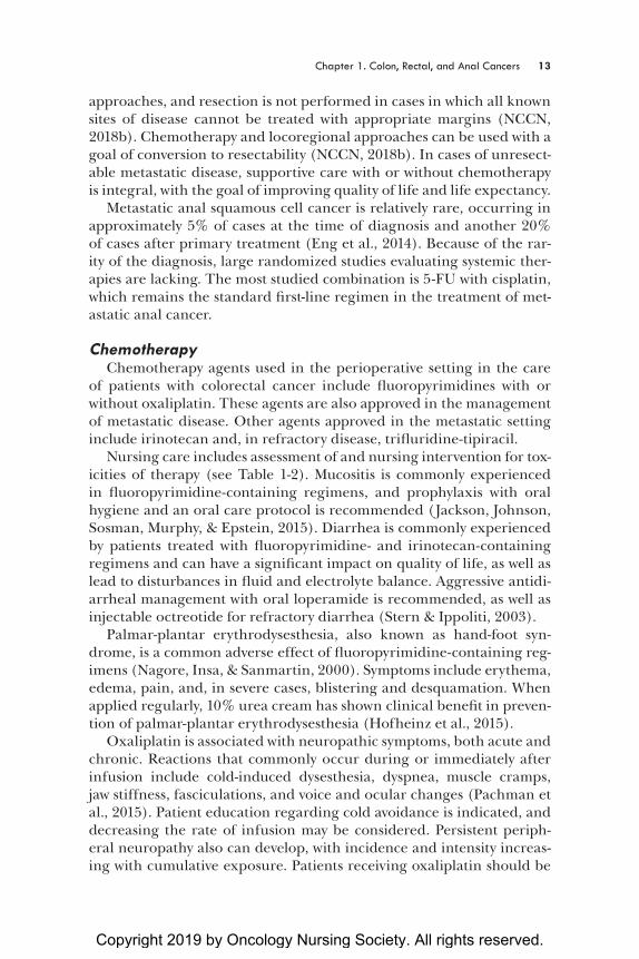
ChemotherapyChemotherapy agents used in the perioperative setting in the care
of patients with colorectal cancer include fluoropyrimidines with or without oxaliplatin. These agents are also approved in the management of metastatic disease. Other agents approved in the metastatic setting include irinotecan and, in refractory disease, trifluridine-tipiracil.
Nursing care includes assessment of and nursing intervention for tox-icities of therapy (see Table 1-2). Mucositis is commonly experienced in fluoropyrimidine-containing regimens, and prophylaxis with oral hygiene and an oral care protocol is recommended (Jackson, Johnson, Sosman, Murphy, & Epstein, 2015). Diarrhea is commonly experienced by patients treated with fluoropyrimidine- and irinotecan-containing regimens and can have a significant impact on quality of life, as well as lead to disturbances in fluid and electrolyte balance. Aggressive antidi-arrheal management with oral loperamide is recommended, as well as injectable octreotide for refractory diarrhea (Stern & Ippoliti, 2003).
Palmar-plantar erythrodysesthesia, also known as hand-foot syn-drome, is a common adverse effect of fluoropyrimidine-containing reg-imens (Nagore, Insa, & Sanmartin, 2000). Symptoms include erythema, edema, pain, and, in severe cases, blistering and desquamation. When applied regularly, 10% urea cream has shown clinical benefit in preven-tion of palmar-plantar erythrodysesthesia (Hofheinz et al., 2015).
Oxaliplatin is associated with neuropathic symptoms, both acute and chronic. Reactions that commonly occur during or immediately after infusion include cold-induced dysesthesia, dyspnea, muscle cramps, jaw stiffness, fasciculations, and voice and ocular changes (Pachman et al., 2015). Patient education regarding cold avoidance is indicated, and decreasing the rate of infusion may be considered. Persistent periph-eral neuropathy also can develop, with incidence and intensity increas-ing with cumulative exposure. Patients receiving oxaliplatin should be
Copyright 2019 by Oncology Nursing Society. All rights reserved.

14 Gastrointestinal Cancer Care for Oncology Nurses
routinely assessed for the presence or worsening of persistent numbness or paresthesias. Patients on oxaliplatin often require dose reduction or permanent discontinuation, especially when pain, motor weakness, or interference with instrumental activities of daily living is present (Her-shman, Lacchetti, & Loprinzi, 2014).
Definitive combination chemoradiation is the standard of care in the treatment of nonmetastatic squamous cell carcinoma of the anus. The chemotherapeutic component of therapy includes 5-FU 1,000 mg/m2/day on days 1–4 and 29–32 and mitomycin C 10 mg/m2 on days 1 and 29 (Flam et al., 1996). Colostomy-free survival and DFS with this approach were 71% and 73%, respectively, in a large randomized phase 3 study (Flam et al., 1996). Alternatively, capecitabine may be administered at a
Table 1-2. Chemotherapy for Colorectal Cancer
Agent Indications Common Toxicities
Fluoropyrimidines (including 5FU and capecitabine)
Adjuvant, monotherapy, in combination with radiation; metastatic, in combination with oxaliplatin or irinotecan
Fatigue, palmarplantar erythrodysesthesia, diarrhea, stomatitis/mucositis, anemia, neutropenia, thrombocytopenia
Irinotecan Metastatic, as monotherapy or in combination with 5FU
Cholinergic syndrome (diaphoresis, flushing, increased peristalsis, lacrimation, miosis, rhinitis, sialorrhea), alopecia, diarrhea, nausea, abdominal pain, abdominal cramping, vomiting, anemia, neutropenia, thrombocytopenia, increased bilirubin
Oxaliplatin Adjuvant, in combination with fluoropyrimidine; metastatic, in combination with fluoropyrimidine
Fatigue, acute neuropathy, chronic neuropathy, paresthesia, neutropenia, thrombocytopenia, anemia, nausea, allergic reaction
Trifluridinetipiracil Metastatic, after progression on fluoropyrimidine, oxaliplatin, irinotecan, and agents targeted against VEGF and EGFR (in RAS wild type)
Neutropenia, anemia, thrombocytopenia, fatigue, asthenia
EGFR—epidermal growth factor receptor; 5FU—5fluorouracil; VEGF—vascular endothelial growth factorNote. Based on information from André et al., 2004; Cassidy et al., 2008; Mayer et al., 2015; Pfizer Inc., 2016.
Copyright 2019 by Oncology Nursing Society. All rights reserved.

Chapter 1. Colon, Rectal, and Anal Cancers 15
dose of 825 mg/m2 twice a day on days of radiation along with mitomy-cin C (Glynne-Jones et al., 2008). Toxicities frequently related to these regimens include fluoropyrimidine-associated toxicity as discussed pre-viously and in Table 1-2. Additional toxicities include high-grade cyto-penias and neutropenia, which are related to the addition of mitomy-cin C. Nursing care includes monitoring and intervention for signs and symptoms of anemia, thrombocytopenia, and neutropenia, as well as supportive measures for perianal discomfort related to both the tumor effect and radiation effect (see Radiation Therapy).
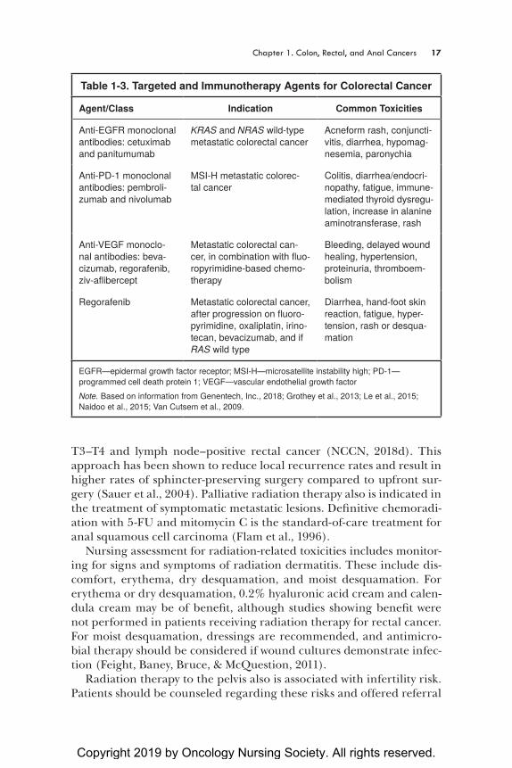
Targeted TherapyActive targeted therapies in the management of metastatic disease
include agents targeting vascular endothelial growth factor (VEGF) and epidermal growth factor receptor (EGFR). Agents targeting VEGF include bevacizumab, ziv-aflibercept, ramucirumab, and, in refractory disease, regorafenib.
The management of metastatic colorectal cancer is biomarker driven. Molecular testing for mutations in the biomarkers NRAS and KRAS is standard, and mutations in these genes confer resistance to agents tar-geting EGFR (Allegra, Rumble, & Schilsky, 2016). The anti-EGFR mono-clonal antibodies cetuximab and panitumumab are approved in the set-ting of metastatic colorectal cancer without mutations in NRAS or KRAS (with RAS wild-type disease).
An important nursing consideration in the care of patients receiv-ing targeted therapies is monitoring for the specific toxicities associated with these agents and implementing management strategies. In patients treated with VEGF-targeted agents, blood pressure and urine protein monitoring is indicated. Anti-EGFR monoclonal antibodies are associ-ated with treatment-related acneform eruption. Reducing sun exposure through the use of protective clothing or sunblock, application of mois-turizing lotion, and prophylactic therapy with oral doxycycline and top-ical hydrocortisone cream has proved effective in reducing rash severity (Lacouture et al., 2010).
To date, no targeted therapies have been approved in the treatment of anal squamous cell cancer. The anti-EGFR monoclonal antibody cetuximab, which has demonstrated activity in other HPV-related can-cers, was investigated in combination with standard chemoradiation. Unfortunately, the investigation was discontinued because of unaccept-able toxicity observed in the patients receiving standard therapy plus cetuximab (Deutsch et al., 2013).
ImmunotherapyThe development of cancer involves a multistep process. One compo-
nent of oncogenesis is the evasion of cancer cells from normal, appro-
Copyright 2019 by Oncology Nursing Society. All rights reserved.

16 Gastrointestinal Cancer Care for Oncology Nurses
priate immune response, which is destruction of malignant and prema-lignant cells (Vinay et al., 2015). Complex signaling between human T cells and antigen-presenting cells occurs during normal immune sur-veillance. Programmed cell death protein 1 (PD-1) is an inhibitory mol-ecule located on T lymphocytes. Anti-PD-1 monoclonal antibodies act by blocking immune inhibition. These agents have shown activity in a molecular subset of MSI-H metastatic colorectal cancer (Le et al., 2015).
The anti-PD-1 monoclonal antibodies pembrolizumab and nivolumab are indicated in the setting of metastatic MSI-H colorectal cancer that has progressed on chemotherapy (Le et al., 2016; Overman et al., 2017). Clin-ical benefit was shown for pembrolizumab in heavily pretreated MSI-H metastatic colon cancer in the form of a 50% objective response rate and an 89% disease control rate, defined as objective response or stable dis-ease at six-month follow-up (Le et al., 2016). Nivolumab showed simi-lar activity in heavily pretreated MSI-H metastatic colon cancer with an objective response rate of 31% and a disease control rate of 61% (Over-man et al., 2017).
The activity of anti-PD-1 therapy has also been demonstrated in met-astatic anal squamous cell carcinoma. One factor in the oncogenesis of chronic HPV infection is upregulation of immune checkpoint pro-teins, including PD-1. Nivolumab demonstrated a response rate of 24%, including two patients with complete response, in a small, single-arm study in patients with chemorefractory, metastatic anal squamous cell carcinoma (Morris et al., 2017).
The PD-1 monoclonal antibodies are associated with a unique, immune-associated toxicity profile. Nurses should assess for the pres-ence of immune-related toxicities associated with anti-PD-1 antibod-ies. These include generalized symptoms such as fatigue and rash. Immune-mediated colitis has been identified, ranging from diarrhea to hematochezia. Autoimmune thyroiditis also can occur, manifested as alterations in thyroid-stimulating hormone, which is routinely monitored. Less commonly, autoimmune pneumonitis, hypophysitis, pancreatitis, arthritis, hepatitis, and nephritis have been identified. Interventions for immune-mediated toxicity vary depending on severity but include hold-ing therapy and administering corticosteroids (Naidoo et al., 2015).
See Table 1-3 for a listing of targeted and immunotherapy agents used in the treatment of colorectal cancer, as well as their common tox-icities.
Radiation TherapyRadiation therapy is indicated in several applications in the treat-
ment of colon, rectal, and anal carcinomas. Neoadjuvant chemoradia-tion is the standard approach in the treatment of potentially resectable
Copyright 2019 by Oncology Nursing Society. All rights reserved.
Chapter 1. Colon, Rectal, and Anal Cancers 17
T3–T4 and lymph node–positive rectal cancer (NCCN, 2018d). This approach has been shown to reduce local recurrence rates and result in higher rates of sphincter-preserving surgery compared to upfront sur-gery (Sauer et al., 2004). Palliative radiation therapy also is indicated in the treatment of symptomatic metastatic lesions. Definitive chemoradi-ation with 5-FU and mitomycin C is the standard-of-care treatment for anal squamous cell carcinoma (Flam et al., 1996).
Nursing assessment for radiation-related toxicities includes monitor-ing for signs and symptoms of radiation dermatitis. These include dis-comfort, erythema, dry desquamation, and moist desquamation. For erythema or dry desquamation, 0.2% hyaluronic acid cream and calen-dula cream may be of benefit, although studies showing benefit were not performed in patients receiving radiation therapy for rectal cancer. For moist desquamation, dressings are recommended, and antimicro-bial therapy should be considered if wound cultures demonstrate infec-tion (Feight, Baney, Bruce, & McQuestion, 2011).
Radiation therapy to the pelvis also is associated with infertility risk. Patients should be counseled regarding these risks and offered referral
Table 1-3. Targeted and Immunotherapy Agents for Colorectal Cancer
Agent/Class Indication Common Toxicities
AntiEGFR monoclonal antibodies: cetuximab and panitumumab
KRAS and NRAS wildtype metastatic colorectal cancer
Acneform rash, conjunctivitis, diarrhea, hypomagnesemia, paronychia
AntiPD1 monoclonal antibodies: pembrolizumab and nivolumab
MSIH metastatic colorectal cancer
Colitis, diarrhea/endocrinopathy, fatigue, immunemediated thyroid dysregulation, increase in alanine aminotransferase, rash
AntiVEGF monoclonal antibodies: bevacizumab, regorafenib, zivaflibercept
Metastatic colorectal cancer, in combination with fluoropyrimidinebased chemotherapy
Bleeding, delayed wound healing, hypertension, proteinuria, thromboembolism
Regorafenib Metastatic colorectal cancer, after progression on fluoropyrimidine, oxaliplatin, irinotecan, bevacizumab, and if RAS wild type
Diarrhea, handfoot skin reaction, fatigue, hypertension, rash or desquamation
EGFR—epidermal growth factor receptor; MSIH—microsatellite instability high; PD1— programmed cell death protein 1; VEGF—vascular endothelial growth factorNote. Based on information from Genentech, Inc., 2018; Grothey et al., 2013; Le et al., 2015; Naidoo et al., 2015; Van Cutsem et al., 2009.
Copyright 2019 by Oncology Nursing Society. All rights reserved.

18 Gastrointestinal Cancer Care for Oncology Nurses
to a fertility preservation specialist for consideration of sperm, oocyte, or ovarian tissue banking. Additionally, radiation therapy to the pelvis may cause vaginal stenosis. The use of a vaginal dilator is recommended following completion of radiation therapy, but not during (Morris, Do, Chard, & Brand, 2017).
Radiation-induced colitis results from radiation-induced stem cell damage to colonic epithelium. Symptoms include diarrhea, nausea, vomiting, anorexia, and pain. Supportive treatment directed at control-ling symptoms is indicated, including antidiarrheal management with loperamide or octreotide, and analgesia (Zimmerer, Böcker, Wenz, & Singer, 2008).
Guidelines for TreatmentNCCN guidelines summarize expert recommendations for the man-
agement of various cancers. Surgical resection followed by consideration of adjuvant chemotherapy, as summarized in the preceding sections, is the NCCN recommendation for stage II and III colorectal adenocarci-noma (NCCN, 2018b). Similarly, NCCN outlines recommendations for neoadjuvant chemoradiation followed by surgical resection in stage T3 N0 or greater rectal cancer, as detailed previously (NCCN, 2018d).
NCCN guidelines for the management of stage IV colon and rec-tal cancer include the recommendation for surgical consideration for metastasectomy for liver and lung metastases and consideration of liver-directed therapies only in patients with disease that can be com-pletely addressed by directed approaches (NCCN, 2018b, 2018d). Che-motherapy, targeted therapy, and immunotherapy options are listed in detail, as have been summarized earlier in this chapter.
Finally, NCCN guidelines for the management of anal cancer advo-cate for definitive chemoradiation with a fluoropyrimidine and mito-mycin C, as described previously (NCCN, 2018a). Additionally, NCCN guidelines for colon, rectal, and anal cancer describe consensus recom-mendations for survivorship and surveillance (see Surveillance and Sur-vivorship).
Clinical Trials Influencing Current TreatmentThe current standard duration of adjuvant chemotherapy after resec-
tion of stage III colon cancer is six months. The persistent neurotoxicity associated with oxaliplatin represents a significant quality-of-life issue in colorectal cancer survivors treated with oxaliplatin. This is related to cumulative dose and was persistent at four years for more than 13% of patients treated with six months of oxaliplatin in the adjuvant setting enrolled in the MOSAIC study (André et al., 2009). Given these find-ings, six collaborating clinical trials are currently investigating the com-parison of three months’ duration of adjuvant chemotherapy with 5-FU
Copyright 2019 by Oncology Nursing Society. All rights reserved.

Chapter 1. Colon, Rectal, and Anal Cancers 19
and oxaliplatin versus six months’ duration (Shi et al., 2017). If found noninferior, three months of adjuvant therapy may become a new stan-dard, which could potentially result in a considerable reduction in neu-ropathic morbidity, but more mature data are needed before these find-ings can be applied to standard practice.
The current standard in the management of locally advanced rec-tal cancer involves neoadjuvant chemoradiation followed by surgi-cal resection (see Preoperative Management). The rates of pathologic complete response of approximately 25% raise the clinical question as to whether surgical resection is required in patients with a complete response to chemoradiation. An ongoing area of investigation involves the watch-and-wait approach to follow-up, which offers the potential for patients who have a complete pathologic response to avoid the mor-bidity associated with surgery (Plummer, Leake, & Albert, 2017). Chal-lenges to this approach include the lack of criteria for determining com-plete pathologic response, and NCCN (2018d) does not advocate for this practice outside of clinical trials.
Recent approvals in immunotherapy with the anti-PD-1 monoclonal antibodies pembrolizumab and nivolumab in the treatment of MSI-H/dMMR metastatic refractory colorectal cancer are based on interim analysis at 6 and 12 months, respectively, of ongoing clinical trials show-ing significant benefit, leading to accelerated approvals. Long-term follow-up is ongoing, as these trials remain open at the time of this pub-lication (Le et al., 2016; Overman et al., 2017).
Although these breakthroughs in immunotherapy are promis-ing, current benefit from anti-PD-1 therapies in colorectal cancer is restricted to patients with MSI-H/dMMR tumors, which represent 10%–20% of all cases of colorectal cancer but only 3.5%–5% of the total number of patients with metastatic colorectal cancer (Koopman et al., 2009). An important focus for future investigation is expand-ing the utility of immunotherapy approaches in microsatellite stable (MSS) metastatic colon cancer. One ongoing trial is investigating the combination of atezolizumab, an anti–programmed cell death-ligand 1 antibody, with cobimetinib, an MEK inhibitor, to increase immune destruction of cancer cells. Preliminary data confirmed responses in a small number of patients, including patients with MSS disease (Bendell et al., 2016).
Nursing CareThe care of patients with colorectal and anal cancer involves assess-
ment and intervention for symptoms related to both the cancer and
Copyright 2019 by Oncology Nursing Society. All rights reserved.

20 Gastrointestinal Cancer Care for Oncology Nurses
side effects from therapy. Nurses have an invaluable role in the safe administration of chemotherapy that encompasses verification of dos-ing accuracy, assessment of contraindications and toxicities, patient education regarding reportable side effects, and symptom manage-ment (Wilkes, 2018). Prior to administration of chemotherapy, the oncology nurse assessment includes review of laboratory results to ensure that appropriate hematologic, renal, and hepatic parameters are met. Nurses also perform the key role of ensuring safe and appro-priate administration of chemotherapy and immunotherapy. Oncol-ogy nurses monitor for acute and delayed toxicities throughout and after administration. Knowledge of toxicities specific to given thera-pies is critical to appropriately assess patient tolerance and outcomes (see Chemotherapy, Targeted Therapy, and Immunotherapy). Chemo-therapy side effects can be severe and even life threatening, and oncol-ogy nurses are critical in promoting patient safety and positive out-comes through assessment, education, and implementation of nursing interventions (Wilkes, 2018).
Oncology nurses also perform a critical role in interprofessional man-agement of symptoms secondary to colorectal cancer. Bowel obstruc-tion is a complication of colorectal cancer that can result from internal or external compression by tumor on the bowel, leading to blockage. This can present as pain, nausea, vomiting, reduction or absence of bowel movements, and reduced bowel sounds on physical assessment. If obstruction is suspected, abdominal x-ray or CT can be used for iden-tification and is aided by ingestion of oral contrast (Wilkes, 2018). The management of obstruction includes gut rest with placement of a naso-gastric tube for decompression and administration of IV fluids. Surgical consultation is indicated to evaluate the possibility of surgical interven-tion to relieve obstruction. Additional management includes pharmaco-therapy to palliate pain, nausea, and vomiting (Wilkes, 2018).
Symptoms related to colorectal cancer can result from direct mass effect of the primary or metastatic tumors. The liver is the most com-mon site of metastases, and the presence of metastatic lesions within the liver can progress to liver failure. This presents as jaundice, nausea, anorexia, edema, hypoalbuminemia, ascites, and altered mentation. Peritoneal involvement can also lead to the development of ascites. From either cause, ascites can cause shortness of breath, pain, and early satiety. Interventions to address ascites include paracentesis, diuretic adminis-tration, and nutrition interventions to improve serum albumin (Wilkes, 2018). An additional symptom experienced is anorexia-cachexia syn-drome. Small, frequent meals and antiemetic administration are recom-mended. Total parenteral nutrition is not recommended unless aggres-sive treatment can result in reversing the disease state (Wilkes, 2018). Patients with advanced disease causing visceral pain often require
Copyright 2019 by Oncology Nursing Society. All rights reserved.

Chapter 1. Colon, Rectal, and Anal Cancers 21
opioid analgesia for palliation. Assessing for and preventing constipa-tion through an appropriate bowel regimen is a critical nursing inter-vention, as constipation can result from opioid analgesia and exacerbate abdominal pain symptoms (Wilkes, 2018).
Throughout the continuum of care, oncology nurses are tasked with critical roles in the assessment and management of patients with colorectal and anal cancer.
PrognosisThe five-year overall survival rate for colorectal cancer for all stages
combined is 65% (Siegel et al., 2018). The five-year survival rates for localized, regional, and distant stages at diagnosis are 90%, 71%, and 14%, respectively (Siegel et al., 2018).
In rectal cancer treated with neoadjuvant chemoradiation, degree of treatment response is highly prognostic. This was demonstrated by long-term data from the CAO/ARO/AIO-94 trial, showing that patients with the greatest degree of tumor regression had a 10-year DFS of 90%, whereas those with no tumor regression had a 10-year DFS of 63% (Fokas et al., 2014).
MSI-H status refers to alterations in mismatch repair genes associated with repeated sequences called microsatellites. Microsatellite stable, or MSS, refers to tumors with intact mismatch repair genes. MSI is a useful marker in screening for hereditary nonpolyposis colorectal cancer (see High-Risk Assessment: Screening and Genetic Testing) and is predictive of potential benefit from anti-PD-1 antibodies. In addition, in stage II and III colorectal cancer, MSI-H is a useful prognostic indicator. The six-year survival rates for MSI-H versus MSS resected stage II colorectal cancer are 97% and 82%, respectively. Similarly, the six-year survival rates for stage III resected colorectal cancer are 78% in MSI-H and 56% in MSS (Lanza et al., 2006).
Additional pathologic prognostic indicators can be gathered from pathologic features, with positive surgical margins, lymphovascular or perineural invasion, and poorly differentiated histology, as discussed previously (see Histology).
The prognosis for anal squamous cell carcinoma is relatively good with definitive chemoradiation, with reported DFS of 73%–80% after completion of treatment with combination radiation therapy and flu-oropyrimidine plus mitomycin C chemotherapy (Bartelink et al., 1997; Flam et al., 1996). Despite relatively good outcomes overall, retrospec-tive review of the large RTOG 98-11 trial demonstrated that higher-risk features are associated with less favorable prognosis. T stage is a signif-
Copyright 2019 by Oncology Nursing Society. All rights reserved.
22 Gastrointestinal Cancer Care for Oncology Nurses
icant prognostic indicator; T2 stage is associated with 82% overall sur-vival, whereas patients with T4 lymph node–negative disease had 57% overall survival. Lymph node–positive disease is also associated with reduced overall survival of 42%–57% (Gunderson et al., 2013). In addi-tion, skin ulceration and male sex are also associated with poorer prog-nosis overall (Bartelink et al., 1997).
PreventionScreening colonoscopy allows for the direct visualization and removal
of precancerous polyps and early-stage cancers. An estimated 73%–91% of colorectal cancer is preventable by screening colonoscopy (Brenner et al., 2007). Although colorectal adenocarcinoma rates are trending downward overall in the United States, colonoscopic screening remains underutilized. Unfortunately, up to 90% of colorectal adenocarcinoma continues to be diagnosed in the more advanced, symptomatic stage (Moreno et al., 2016).
A number of lifestyle factors have been associated with reduced risk for developing colorectal cancer. A systematic review and meta-analysis demonstrated that the most physically active people have an approxi-mately 25% reduced risk for developing colorectal cancer as compared with the least physically active (Boyle, Keegel, Bull, Heyworth, & Frit-schi, 2012). Epidemiologic evidence supports the association between diet and colorectal cancer. Vegetarian, pescatarian, and semi-vegetarian diets were associated with lower risk of colorectal cancer. In the large, prospective Adventist Health Study-2, the respective hazard ratios for development in these groups were 0.82, 0.57, and 0.92, compared with matched nonvegetarians (Orlich et al., 2015). Diets high in fiber are also associated with a reduced risk of developing colorectal cancer (Aune et al., 2011).
Regular aspirin use has also been associated with reduced risk of colorectal cancer. A systematic review and meta-analysis demonstrated significantly reduced rates of colon and rectal cancer in individu-als who took aspirin 75 mg or more daily (Rothwell et al., 2010). The U.S. Preventive Services Task Force recommends low-dose daily aspi-rin in people aged 50–59 on the basis of established chemoprotec-tive effect, citing both cardiovascular and colorectal cancer outcomes (Bibbins-Domingo, 2016). Aspirin use may also have value in secondary prevention in patients diagnosed with colorectal cancer and has been demonstrated to be associated with improved disease-specific and over-all survival in this population (Bains et al., 2016).
Copyright 2019 by Oncology Nursing Society. All rights reserved.

Chapter 1. Colon, Rectal, and Anal Cancers 23
The majority of anal squamous cell cancers result from chronic infec-tion with oncogenic HPV type 16 (Daling et al., 2004). Anal intercourse is associated with the development of HPV-related dysplastic cytology and anal HPV infection (Moscicki et al., 1999). A sequence of progres-sion from low-grade to high-grade neoplasia has been identified, and high-grade, p16-positive anal intraepithelial neoplasia is considered a premalignant condition analogous to neoplasia identified in cervi-cal specimens. Given the biologic similarity with cervical cancer (HPV related, squamous histology), screening for anal intraepithelial neopla-sia in a similar fashion in at-risk populations has been proposed, with sensitivity and specificity comparable to that seen in cervical cancer screening (Czito et al., 2015). At this time, data from large, randomized trials supporting cytologic screening are lacking, and further study is needed before large-scale cytological screening programs can be insti-tuted (Chiao, Giordano, Palefsky, Tyring, & Serag, 2006).
Vaccination against HPV types 6, 11, 16, and 18 has demonstrated effectiveness in the prevention of infection with these strains of HPV and the development of genital warts (Giuliano et al., 2011). Further-more, a study in a high-risk population demonstrated immunization with vaccination against HPV types 6, 11, 16, and 18 to be associated with a 78% reduction in the development of high-grade anal intraepi-thelial neoplasia and an 84% reduction in the detection of HPV DNA (Palefsky et al., 2011). This supports the hypothesis that HPV vaccina-tion has the potential to prevent anal carcinoma through prevention of chronic HPV infection, which is the most common etiology for the development of anal carcinoma.
High-Risk Assessment: Screening and Genetic TestingAlthough the majority of colorectal cancer cases are sporadic in
nature, a number of inheritable familial syndromes are known (see Table 1-1). Of these, Lynch syndrome is the most common, account-ing for approximately 2%–4% of all cases of colorectal cancer (Lynch & Chapelle, 2003). Lynch syndrome is an autosomal dominant disorder that results from inherited mutation in one of four DNA MMR genes: MLH1, MSH2, MSH6, and PMS2 (Palomaki, McClain, Melillo, Hampel, & Thibodeau, 2009). These genes code for DNA MMR proteins, which normally correct errors in DNA replication occurring during cellular division.
Alteration in MMR genes results in increased accumulation of redun-dant genetic nucleotide mismatch mutations known as microsatellite insta-bility, or MSI (Hendriks et al., 2006). Accumulation of increased muta-
Copyright 2019 by Oncology Nursing Society. All rights reserved.

24 Gastrointestinal Cancer Care for Oncology Nurses
tions in cancer-related genes leads to an increased rate of oncogenesis (Lynch & Chapelle, 2003). Tumor specimens can be tested for expres-sion of MMR proteins or MSI. Although both methodologies share high sensitivity and specificity, MSI testing has a disadvantage in that it does not provide insight into which gene or protein is implicated (Hendriks et al., 2006).
It is important to identify Lynch syndrome in patients and families because of the high rates of second Lynch syndrome–associated pri-mary cancers in the same individual and its dominant inheritance pat-tern, which is commonly passed on to first-degree relatives. Lynch syn-drome–associated colorectal cancers typically develop at a younger age and are more likely to be right sided (Lynch & Chapelle, 2003). In indi-viduals with Lynch syndrome, the lifetime risk for developing colorec-tal cancer varies from 12% to 48% depending on the genes involved (Bonadona et al., 2011). Lynch syndrome is also associated with an increased risk for the development of multiple extracolonic cancers, including endometrial, gastric, small bowel, urothelial, brain (glioma), and ovarian cancer (Lynch & Chapelle, 2003).
NCCN (2018c) recommends universal screening of all colorec-tal cancer specimens for Lynch syndrome–associated molecular pro-file. This screening can be completed with one of two methodologies: (a) immunohistochemistry analysis for MMR protein (MLH1, MSH2, MSH6, and PMS2) expression, or (b) analysis for MSI, a result of MMR protein deficiency. More than 90% of Lynch syndrome specimens are MSI-H and lack normal expression of one of the four MMR proteins. Abnormal initial screening results must be interpreted in clinical con-text, as 10%–15% of sporadic, nonfamilial colorectal cancers also express this molecular profile (French et al., 2008). Sporadic MSI-H colorectal cancer is associated with hypermethylation of MLH1, a fea-ture that can be identified by the presence of BRAF mutation, which rules out Lynch syndrome in MSI-H colorectal cancers (Deng et al., 2004). In addition to molecular markers, clinical criteria exist to iden-tify individuals at risk for Lynch syndrome. The Amsterdam II crite-ria identify at-risk individuals as having the following: (a) three rel-atives with a Lynch syndrome–associated cancer, one of which is a first-degree relative of the other two, (b) two successive generations affected, and (c) at least one case being diagnosed before age 50. In addition, familial adenomatous polyposis should be excluded (Vasen, Watson, Mecklin, & Lynch, 1999). When clinical concern for Lynch syndrome is present on the basis of clinical context and MSI-H or abnormality in MMR proteins, germline testing may be conducted to differentiate sporadic MSI-H cancers from Lynch syndrome. This should be performed in consultation with a genetics expert (NCCN, 2018c).
Copyright 2019 by Oncology Nursing Society. All rights reserved.

Chapter 1. Colon, Rectal, and Anal Cancers 25
If a deleterious germline mutation is identified, institution of Lynch syndrome surveillance is indicated. This includes surveillance for colon cancer, endometrial and ovarian cancer, and less frequently associated cancers, including gastric, small bowel, and urothelial cancers. Screen-ing colonoscopy every one to two years starting at age 20–25 is recom-mended because of the relatively rapid progression from polyp to can-cer seen in Lynch syndrome (Lynch & Chapelle, 2003). Although no active surveillance for endometrial or ovarian cancer is established in this population, patient education regarding reportable signs and symp-toms, such as dysfunctional uterine bleeding, is advised. Prophylactic total abdominal hysterectomy with bilateral salpingo-oophorectomy is an option in women who have completed childbearing (Schmeler et al., 2006). Urinalysis is also recommended annually for screening of urothelial cancers (Mork et al., 2015). In addition to screening of the affected individual, screening of at-risk family members is indicated. This includes first-degree relatives, as well as more distant family mem-bers if first-degree relatives are unwilling or unable to be tested (NCCN, 2018c). Finally, chemoprevention with aspirin is associated with reduc-tion in cancer incidence in carriers of Lynch hereditary colon cancer genes (Burn et al., 2011).
SurveillanceAfter curative intent therapy is complete, consensus recommenda-
tions are for disease surveillance (Meyerhardt et al., 2013). The goals of surveillance are to identify recurrences that may be amenable to cure, identify new primary colorectal neoplasms before they become invasive, and monitor for delayed complications of therapies received. In all stages treated with curative intent, endoscopic surveillance one year from resection is indicated, with subsequent endoscopy at three- and five-year intervals afterward. When premalignant polyps are identi-fied, annual endoscopic surveillance is recommended until the patient is polyp free. Serial CEA monitoring is also recommended and has been shown, when applying a threshold of 5 mcg/L, to provide sensitiv-ity of 71% and specificity of 88% in identification of recurrent disease (Nicholson et al., 2015). Although CEA testing is helpful, the sensitiv-ity of CEA monitoring with the goal of early identification of potentially curable recurrences is below the acceptable threshold to function as a stand-alone approach to surveillance (Nicholson et al., 2015). Thus, in patients with a history of stage II and III colorectal cancer, strategies incorporating both CEA testing and advanced imaging of the chest, abdomen, and pelvis with CT are recommended, in addition to history
Copyright 2019 by Oncology Nursing Society. All rights reserved.
26 Gastrointestinal Cancer Care for Oncology Nurses
and physical examination, serum chemistries including liver function panel, and complete blood count (NCCN, 2018b).
The approach of more intensive surveillance has been demonstrated to identify a significant number of patients with potentially resectable metastatic recurrences. When identified, these patients then receive definitive therapy for the recurrences with the goal of achieving remis-sion, with 30% of patients identified as having liver-only metastases during surveillance imaging achieving remission at five-year follow-up (Primrose et al., 2014).
The frequency of recommended monitoring is based on recurrence patterns. In a pooled analysis of 20,898 survivors of stage II and III colorectal cancer, 80% of recurrences were identified to have occurred within three years of surgery, and 8% occurred between years 3 and 5 (Sargent et al., 2005). Thus, intensive surveillance with CEA monitoring and CT imaging is advised every three to six months for two years, then every 6–12 months for five years (NCCN, 2018b).
Surveillance after primary treatment of anal cancer with definitive chemoradiation involves monitoring for local response or treatment fail-ure and periodic monitoring for metastatic disease. Digital rectal exam-ination and inguinal lymph node palpation should occur 8–12 weeks after completion of chemoradiation to assess for response. If a com-plete clinical response has occurred at 8–12 weeks, this examination is repeated every 3–6 months for 5 years (NCCN, 2018a). If biopsy-proven residual disease is present at the time of the first follow-up, the deter-mination regarding salvage surgery with APR versus close observation is based on whether evidence of progressive disease is present (NCCN, 2018a). This recommendation is based on findings that 72% of patients who had residual disease on examination at 11 weeks after chemoradi-ation went on to show a complete response by 26 months (James et al., 2013). If persistent residual disease, progressive local disease, or local recurrence is identified, surgical resection with APR is recommended, which has resulted in a 50% five-year DFS (Czito et al., 2015). Additional surveillance recommendations include anoscopy every 6–12 months for five years, and chest, abdomen, and pelvis CT imaging with contrast yearly for three years (NCCN, 2018a).
SurvivorshipUpon completion of primary treatment of cancer, the focus of can-
cer care shifts to survivorship care. Colorectal cancer survivors are the third largest survivorship population, representing 11% of the total sur-vivorship population (Hewitt, Greenfield, & Stovall, 2006). Focused
Copyright 2019 by Oncology Nursing Society. All rights reserved.

Chapter 1. Colon, Rectal, and Anal Cancers 27
assessment for quality-of-life components is essential in providing qual-ity survivorship care. Colorectal cancer survivors have been identified as having a higher risk for developing depression, indicating a need for distress screening and appropriate intervention (Ramsey, Berry, Moin-pour, Giedzinska, & Andersen, 2002). Patients with colorectal cancer may experience chronic bowel pattern changes. Patients may report loose stools or an increase in stool frequency after colonic resection, which tends to improve over time (Hewitt et al., 2006). Adhesions sec-ondary to surgery or radiation therapy can lead to abdominal pain or bowel obstruction. Education regarding measures to improve bowel pat-tern, such as including fiber and the use of over-the-counter stool soft-eners, is of benefit (Hewitt et al., 2006).
Rectal cancer survivors may experience sexual dysfunction second-ary to injury to pelvic nerves and vasculature from cancer and cancer therapy. Male patients can experience erectile dysfunction, which may require medications or referral to urology. Female patients can experi-ence painful coitus. Interventions include use of a vaginal dilator and over-the-counter lubricants (Vogel, 2017). A significant percentage of patients who received oxaliplatin also experience chronic peripheral neuropathy (Hershman et al., 2014).
Survivorship care includes educating patients and promoting lifestyle modifications known to improve colorectal cancer outcomes and overall health. Lifestyle factors including smoking cessation, maintenance of a healthy body mass index, and regular physical exercise have been iden-tified as beneficial for improving colorectal cancer outcomes. Increased physical activity has been shown to reduce mortality in colorectal can-cer survivor populations (Campbell, Patel, Newton, Jacobs, & Gapstur, 2013). Increases in activity by any amount in colorectal cancer survivors have been associated with decreased mortality, with greater improve-ments shown in patients who engaged in moderate exercise for at least 150 minutes per week (Schmid & Leitzmann, 2014). A diet higher in vegetables and fruit, whole grains, poultry, and fish and lower in refined grains, red and processed meat, and refined sugars is associated with lower recurrence and improved overall survival (Meyerhardt et al., 2007).
Survivorship care in patients treated for anal cancers involves mon-itoring for late toxicities of pelvic radiation. Anal cancer survivors commonly report global reduction in quality of life, fatigue, dyspnea, insomnia, diarrhea, fecal incontinence, increased stool frequency, but-tock pain, flatulence, erectile dysfunction (males), dyspareunia, and reduced sexual interest following therapy with combined chemother-apy and pelvic radiation (Bentzen et al., 2013). Unique screening and prevention elements of survivorship care planning in anal cancer sur-vivors includes considering screening for HIV status. Additionally, cer-
Copyright 2019 by Oncology Nursing Society. All rights reserved.

28 Gastrointestinal Cancer Care for Oncology Nurses
vical screening for female patients is also recommended because of the shared etiology between anal and cervical cancers.
Goals of survivorship care include prevention of new and recurrent cancers, prevention of late effects of cancer and cancer therapy, inter-vention for consequences of cancer and cancer therapy, and coordina-tion of care between the oncology team and primary care provider to ensure that all healthcare needs are comprehensively met (Hewitt et al., 2006). The Health and Medicine Division of the National Acade-mies of Sciences, Engineering, and Medicine (formerly the Institute of Medicine) and other bodies have promoted written survivorship care plans as a standard of care for facilitating improved outcomes in cancer survivors. The survivorship care plan provides a written sum-mary of the treatment received, possible late side effects of cancer and cancer therapy, information on reportable signs of recurrence, rec-ommendations for follow-up, lifestyle recommendations, and defined roles between the oncology and primary care provider. The plan should be given to the patient and primary care provider (Hewitt et al., 2006).
Survivorship care also includes disease prevention, such as immuni-zations, screening for second cancers, and routine health care, in collab-oration with the primary care team.
SummaryCollectively, cancers of the colon, rectum, and anus represent a sig-
nificant health burden. Although colon and rectal cancer rates have declined overall, they remain the third most common form of cancer, as well as the third leading cause of cancer death in men and women (Sie-gel et al., 2018). Additionally, colon and rectal cancer rates are increas-ing rapidly in people younger than age 50 (Siegel et al., 2017). Despite being a relatively uncommon malignancy, anal cancer rates have more than doubled in recent decades (Nelson et al., 2013).
Evidence-based interprofessional care is needed for the promotion of improved outcomes for patients affected by cancers of the colon, rec-tum, and anus. Oncology nurses can promote prevention of these can-cers by encouraging screening colonoscopy in asymptomatic patients. Nurses may also educate patients on high-risk familial syndromes and provide referral to genetic counseling as indicated. Furthermore, nurses have a role in educating patients regarding the signs and symptoms that indicate evaluation.
Nurses are better suited to provide appropriate patient care and edu-cation in the context of understanding disease biology, therapeutic
Copyright 2019 by Oncology Nursing Society. All rights reserved.
Chapter 1. Colon, Rectal, and Anal Cancers 29
options, and goals of therapy. Nurses can both educate and advocate for patients in the setting of the overall clinical picture. By remaining apprised of the most current therapies, nurses have the opportunity to promote best practice.
Patients with cancers of the colon, rectum, and anus commonly expe-rience significant disease-related symptoms, including pain, fatigue, anorexia, nausea, vomiting, diarrhea, obstruction, and depression. In addition, patients also may experience significant toxicities related to radiation, chemotherapy, and surgery. Oncology nurses have a vital role in assessment for these related symptoms. When symptoms are identi-fied, nurses collaborate with the patient, family, and medical team to develop a plan that includes nursing interventions to address these tox-icities. In doing so, nurses have the opportunity to promote improved quality of life and, potentially, disease outcomes.
ReferencesAbbas, S., Lam, V., & Hollands, M. (2011). Ten-year survival after liver resection for
colorectal metastases: Systematic review and meta-analysis. ISRN Oncology, 2011, 1–11. https://doi.org/10.5402/2011/763245
Ahnen, D.J., Wade, S.W., Jones, W.F., Sifri, R., Silveiras, J.M., Greenamyer, J., … You, Y.N. (2014). The increasing incidence of young-onset colorectal cancer: A call to action. Mayo Clinic Proceedings, 89, 216–224. https://doi.org/10.1016/j.mayocp.2013.09.006
Allegra, C.J., Rumble, R.B., & Schilsky, R.L. (2016). Extended RAS gene mutation testing in metastatic colorectal carcinoma to predict response to anti–epidermal growth factor receptor monoclonal antibody therapy: American Society of Clinical Oncology provisional clinical opinion update 2015 summary. Journal of Oncology Practice, 12, 180–181. https://doi.org/10.1200/JOP.2015.007898
André, T., Boni, C., Mounedji-Boudiaf, L., Navarro, M., Tabernero, J., Hickish, T., … de Gramont, A. (2004). Oxaliplatin, fluorouracil, and leucovorin as adjuvant treatment for colon cancer. New England Journal of Medicine, 350, 2343–2351. https://doi.org /10.1056/NEJMoa032709
André, T., Boni, C., Navarro, M., Tabernero, J., Hickish, T., Topham, C., … de Gramont, A. (2009). Improved overall survival with oxaliplatin, fluorouracil, and leucovorin as adjuvant treatment in stage II or III colon cancer in the MOSAIC trial. Journal of Clinical Oncology, 27, 3109–3116. https://doi.org/10.1200/JCO.2008.20.6771
Aune, D., Chan, D.S.M., Lau, R., Vieira, R., Greenwood, D.C., Kampman, E., & Norat, T. (2011). Dietary fibre, whole grains, and risk of colorectal cancer: Systematic review and dose-response meta-analysis of prospective studies. BMJ, 343, d6617. https:// doi.org/10.1136/bmj.d6617
Bains, S.J., Mahic, M., Myklebust, T.Å., Småstuen, M.C., Yaqub, S., Dørum, L.M., … Taskén, K. (2016). Aspirin as secondary prevention in patients with colorectal cancer: An unselected population-based study. Journal of Clinical Oncology, 34, 2501–2508. https://doi.org/10.1200/JCO.2015.65.3519
Bartelink, H., Roelofsen, F., Eschwege, F., Rougier, P., Bosset, J.F., Gonzalez, D.G., … Pierart, M. (1997). Concomitant radiotherapy and chemotherapy is superior to radiotherapy alone in the treatment of locally advanced anal cancer: Results of a
Copyright 2019 by Oncology Nursing Society. All rights reserved.
30 Gastrointestinal Cancer Care for Oncology Nurses
phase III randomized trial of the European Organization for Research and Treatment of Cancer Radiotherapy and Gastrointestinal Cooperative Groups. Journal of Clinical Oncology, 15, 2040–2049. https://doi.org/10.1200/JCO.1997.15.5.2040
Bendell, J.C., Kim, T.W., Goh, B.C., Wallin, J., Oh, D.-Y., Han, S.-W., … Bang, Y.-J. (2016). Clinical activity and safety of cobimetinib (cobi) and atezolizumab in colorectal cancer (CRC). Journal of Clinical Oncology, 34(Suppl.), Abstract 3502. Retrieved from http://meetinglibrary.asco.org/record/123622/abstract
Bentzen, A.G., Balteskard, L., Wanderås, E.H., Frykholm, G., Wilsgaard, T., Dahl, O., & Guren, M.G. (2013). Impaired health-related quality of life after chemoradiotherapy for anal cancer: Late effects in a national cohort of 128 survivors. Acta Oncologica, 52, 736–744. https://doi.org/10.3109/0284186X.2013.770599
Biagi, J.J., Raphael, M.J., Mackillop, W.J., Kong, W., King, W.D., & Booth, C.M. (2011). Association between time to initiation of adjuvant chemotherapy and survival in colorectal cancer: A systematic review and meta-analysis. JAMA, 305, 2335–2342. https://doi.org/10.1001/jama.2011.749
Bibbins-Domingo, K. (2016). Aspirin use for the primary prevention of cardiovascular disease and colorectal cancer: U.S. Preventive Services Task Force recommendation statement. Annals of Internal Medicine, 164, 836–845. https://doi.org/10.7326/M16 -0577
Bonadona, V., Bonaïti, B., Olschwang, S., Grandjouan, S., Huiart, L., Longy, M., … Bonaïti-Pellié, C. (2011). Cancer risks associated with germline mutations in MLH1, MSH2, and MSH6 genes in Lynch syndrome. JAMA, 305, 2304–2310. https://doi .org/10.1001/jama.2011.743
Boyle, T., Keegel, T., Bull, F., Heyworth, J., & Fritschi, L. (2012). Physical activity and risks of proximal and distal colon cancers: A systematic review and meta-analysis. Journal of the National Cancer Institute, 104, 1548–1561. https://doi.org/10.1093 /jnci/djs354
Brenner, H., Chang-Claude, J., Seiler, C.M., Stürmer, T., & Hoffmeister, M. (2007). Potential for colorectal cancer prevention of sigmoidoscopy versus colonoscopy: Population-based case control study. Cancer Epidemiology, Biomarkers and Prevention, 16, 494–499. https://doi.org/10.1158/1055-9965.EPI-06-0460
Burn, J., Gerdes, A.-M, Macrae, F., Mecklin, J.-P., Moeslein, G., Olschwang, S., … Bishop, D.T. (2011). Long-term effect of aspirin on cancer risk in carriers of hereditary colorectal cancer: An analysis from the CAPP2 randomised controlled trial. Lancet, 378, 2081–2087. https://doi.org/10.1016/S0140-6736(11)61049-0
Burt, R., & Neklason, D.W. (2005). Genetic testing for inherited colon cancer. Gastro-enterology, 128, 1696–1716. https://doi.org/10.1053/j.gastro.2005.03.036
Campbell, P.T., Patel, A.V., Newton, C.C., Jacobs, E.J., & Gapstur, S.M. (2013). Associa-tions of recreational physical activity and leisure time spent sitting with colorectal cancer survival. Journal of Clinical Oncology, 31, 876–885. https://doi.org/10.1200 /JCO.2012.45.9735
Cassidy, J., Clarke, S., Díaz-Rubio, E., Scheithauer, W., Figer, A., Wong, R., … Saltz, L. (2008). Randomized phase III study of capecitabine plus oxaliplatin compared with fluorouracil/folinic acid plus oxaliplatin as first-line therapy for metastatic colorectal cancer. Journal of Clinical Oncology, 26, 2006–2012. https://doi.org/10 .1200/JCO.2007.14.9898
Chan, A.T., & Giovannucci, E.L. (2010). Primary prevention of colorectal cancer. Gastroenterology, 138, 2029–2043.e10. https://doi.org/10.1053/j.gastro.2010.01.057
Chiao, E.Y., Giordano, T.P., Palefsky, J.M., Tyring, S., & Serag, H.E. (2006). HIV/AIDS: Screening HIV-infected individuals for anal cancer precursor lesions: A systematic review. Clinical Infectious Diseases, 43, 223–233. https://doi.org/10.1086/505219
Chung, M., Lee, J., Terasawa, T., Lau, J., & Trikalinos, T.A. (2011). Vitamin D with or without calcium supplementation for prevention of cancer and fractures: An updated meta-analysis for the U.S. Preventive Services Task Force. Annals of Internal Medicine, 155, 827–838. https://doi.org/10.7326/0003-4819-155-12-201112200-00005
Copyright 2019 by Oncology Nursing Society. All rights reserved.

Chapter 1. Colon, Rectal, and Anal Cancers 31
Compton, C.C., Fielding, L.P., Burgart, L.J., Conley, B., Cooper, H.S., Hamilton, S.R., … Willett, C. (2000). Prognostic factors in colorectal cancer. Archives of Pathology and Laboratory Medicine, 124, 979–994.
Czito, B.G., Ahmed, S., Kalady, M., & Eng, C. (2015). Cancer of the anal region. In V.T. DeVita Jr., T.S. Lawrence, & S.A. Rosenberg (Eds.), DeVita, Hellman, and Rosenberg’s cancer: Principles and practice of oncology (10th ed., pp. 842–856). Philadelphia, PA: Wolters Kluwer Health.
Daling, J.R., Madeleine, M.M., Johnson, L.G., Schwartz, S.M., Shera, K.A., Wurscher, M.A., … McDougall, J.K. (2004). Human papillomavirus, smoking, and sexual practices in the etiology of anal cancer. Cancer, 101, 270–280. https://doi.org/10 .1002/cncr.20365
Deng, G., Bell, I., Crawley, S., Gum, J., Terdiman, J.P., Allen, B.A., … Kim, Y.S. (2004). BRAF mutation is frequently present in sporadic colorectal cancer with methylated hMLH1, but not in hereditary nonpolyposis colorectal cancer. Clinical Cancer Research, 10, 191–195. https://doi.org/10.1158/1078-0432.CCR-1118-3
Deutsch, E., Lemanski, C., Pignon, J.P., Levy, A., Delarochefordiere, A., Martel-Lafay, I., … Azria, D. (2013). Unexpected toxicity of cetuximab combined with conventional chemoradiotherapy in patients with locally advanced anal cancer: Results of the Unicancer Accord 16 phase II trial. Annals of Oncology, 24, 2834–2838. https://doi .org/10.1093/annonc/mdt368
Ekbom, A., Helmick, C., Zack, M., & Adami, H.-O. (1990). Ulcerative colitis and colorectal cancer—A population-based study. New England Journal of Medicine, 323, 1228–1233. https://doi.org/10.1056/NEJM199011013231802
Eng, C., Chang, G.J., You, Y.N., Das, P., Rodriguez-Bigas, M., Xing, Y., … Wolff, R.A. (2014). The role of systemic chemotherapy and multidisciplinary management in improving the overall survival of patients with metastatic squamous cell car-cinoma of the anal canal. Oncotarget, 5, 11133–11142. https://doi.org/10.18632 /oncotarget.2563
Feight, D., Baney, T., Bruce, S., & McQuestion, M. (2011). Putting evidence into practice: Evidence-based interventions for radiation dermatitis. Clinical Journal of Oncology Nursing, 15, 481–492. https://doi.org/10.1188/11.CJON.481-492
Flam, M., John, M., Pajak, T.F., Petrelli, N., Myerson, R., Doggett, S., … Murray, K. (1996). Role of mitomycin in combination with fluorouracil and radiotherapy, and of salvage chemoradiation in the definitive nonsurgical treatment of epidermoid carcinoma of the anal canal: Results of a phase III randomized intergroup study. Journal of Clinical Oncology, 14, 2527–2539. https://doi.org/10.1200/jco.1996.14 .9.2527
Fokas, E., Liersch, T., Fietkau, R., Hohenberger, W., Beissbarth, T., Hess, C., … Rödel, C. (2014). Tumor regression grading after preoperative chemoradio-therapy for locally advanced rectal carcinoma revisited: Updated results of the CAO/ARO/AIO-94 trial. Journal of Clinical Oncology, 32, 1554–1562. https://doi.org/10.1200/JCO.2013.54.3769
French, A.J., Sargent, D.J., Burgart, L.J., Foster, N.R., Kabat, B.F., Goldberg, R., … Thibodeau, S.N. (2008). Prognostic significance of defective mismatch repair and BRAF V600E in patients with colon cancer. Clinical Cancer Research, 14, 3408–3415. https://doi.org/10.1158/1078-0432.CCR-07-1489
Frisch, M. (2000). Human papillomavirus–associated cancers in patients with human im-munodeficiency virus infection and acquired immunodeficiency syndrome. Journal of the National Cancer Institute, 92, 1500–1510. https://doi.org/10.1093/jnci/92.18.1500
Genentech, Inc. (2018). Avastin® (bevacizumab) [Package insert]. Retrieved from https://www.gene.com/download/pdf/avastin_prescribing.pdf
Giuliano, A.R., Palefsky, J.M., Goldstone, S., Moreira, E.D., Penny, M.E., Aranda, C., … Guris, D. (2011). Efficacy of quadrivalent HPV vaccine against HPV infection and
Copyright 2019 by Oncology Nursing Society. All rights reserved.

32 Gastrointestinal Cancer Care for Oncology Nurses
disease in males. New England Journal of Medicine, 364, 401–411. https://doi.org/10 .1056/NEJMoa0909537
Glynne-Jones, R., Meadows, H., Wan, S., Gollins, S., Leslie, M., Levine, E., … Sebag-Montefiore, D. (2008). EXTRA—A multicenter phase II study of chemoradiation using a 5 day per week oral regimen of capecitabine and intravenous mitomycin C in anal cancer. International Journal of Radiation Oncology, Biology, Physics, 72, 119–126. https://doi.org/10.1016/j.ijrobp.2007.12.012
Grothey, A., Van Cutsem, E., Sobrero, A., Siena, S., Falcone, A., Ychou, M., … Laurent, D. (2013). Regorafenib monotherapy for previously treated metastatic colorectal cancer (CORRECT): An international, multicentre, randomised, placebo-controlled, phase 3 trial. Lancet, 381, 303–312. https://doi.org/10.1016/S0140-6736(12)61900-X
Gunderson, L.L., Moughan, J., Ajani, J.A., Pedersen, J.E., Winter, K.A., Benson, A.B., III, … Willett, C.G. (2013). Anal carcinoma: Impact of TN category of disease on survival, disease relapse, and colostomy failure in US Gastrointestinal Intergroup RTOG 98-11 phase 3 trial. International Journal of Radiation Oncology, Biology, Physics, 87, 638–645. https://doi.org/10.1016/j.ijrobp.2013.07.035
Hamilton, W., Round, A., Sharp, D., & Peters, T.J. (2005). Clinical features of colorectal cancer before diagnosis: A population-based case–control study. British Journal of Cancer, 93, 399–405. https://doi.org/10.1038/sj.bjc.6602714
Hendriks, Y.M.C., de Jong, A.E., Morreau, H., Tops, C.M.J., Vasen, H.F., Wijnen, J.T., … Bröcker-Vriends, A.H.J.T. (2006). Diagnostic approach and management of Lynch syndrome (hereditary nonpolyposis colorectal carcinoma): A guide for clinicians. CA: A Cancer Journal for Clinicians, 56, 213–225. https://doi.org/10.3322/canjclin .56.4.213
Henley, S.J., Singh, S.D., King, J., Wilson, R.J., O’Neil, M.E., & Ryerson, A.B. (2015). Invasive cancer incidence and survival—United States, 2012. Morbidity and Mortality Weekly Report, 64, 1353–1358. https://doi.org/10.15585/mmwr.mm6449a1
Hershman, D.L., Lacchetti, C., & Loprinzi, C.L. (2014). Prevention and management of chemotherapy-induced peripheral neuropathy in survivors of adult cancers: American Society of Clinical Oncology clinical practice guideline summary. Journal of Oncology Practice, 10, e421–e424. https://doi.org/10.1200/JOP.2014.001776
Hewitt, M., Greenfield, S., & Stovall, E. (Eds.). (2006). From cancer patient to cancer survivor: Lost in transition. Retrieved from https://www.nap.edu/catalog/11468 /from-cancer-patient-to-cancer-survivor-lost-in-transition
Hofheinz, R., Gencer, D., Schulz, H., Stahl, M., Hegewisch-Becker, S., Loeffler, L.M., … Schneeweiss, A. (2015). Mapisal versus urea cream as prophylaxis for capecitabine-associated hand-foot syndrome: A randomized phase III trial of the AIO Quality of Life Working Group. Journal of Clinical Oncology, 33, 2444–2449. https://doi.org/10 .1200/JCO.2014.60.4587
Jackson, L.K., Johnson, D.B., Sosman, J.A., Murphy, B.A., & Epstein, J.B. (2015). Oral health in oncology: Impact of immunotherapy. Supportive Care in Cancer, 23, 1–3. https://doi.org/10.1007/s00520-014-2434-6
James, R.D., Glynne-Jones, R., Meadows, H.M., Cunningham, D., Myint, A.S., Saunders, M.P., … Sebag-Montefiore, D. (2013). Mitomycin or cisplatin chemoradiation with or without maintenance chemotherapy for treatment of squamous-cell carcinoma of the anus (ACT II): A randomised, phase 3, open-label, 2×2 factorial trial. Lancet Oncology, 14, 516–524. https://doi.org/10.1016/S1470-2045(13)70086-X
Jemal, A., Bray, F., Center, M.M., Ferlay, J., Ward, E., & Forman, D. (2011). Global cancer statistics. CA: A Cancer Journal for Clinicians, 61, 69–90. https://doi.org/10 .3322/caac.20107
Jessup, J.M., Goldberg, R.M., Asare, E.A., Benson, A.B., III, Brierley, J.D., Chang, G.J., … Washington, M.K. (2017). Colon and rectum. In M.B. Amin (Ed.), AJCC cancer staging manual (8th ed., pp. 251–274). Chicago, IL: Springer.
Copyright 2019 by Oncology Nursing Society. All rights reserved.

Chapter 1. Colon, Rectal, and Anal Cancers 33
Jones, M., Hruby, G., Solomon, M., Rutherford, N., & Martin, J. (2015). The role of FDG-PET in the initial staging and response assessment of anal cancer: A systematic review and meta-analysis. Annals of Surgical Oncology, 22, 3574–3581. https://doi.org /10.1245/s10434-015-4391-9
Kanas, G.P., Taylor, A., Primrose, J.N., Langeberg, W.J., Kelsh, M.A., Mowat, F.S., … Poston, G. (2012). Survival after liver resection in metastatic colorectal cancer: Review and meta-analysis of prognostic factors. Clinical Epidemiology, 283, 283–301. https://doi.org/10.2147/CLEP.S34285
Kanemitsu, Y., Kato, T., Hirai, T., Yasui, K., Morimoto, T., Shimizu, Y., … Yamamura, Y. (2003). Survival after curative resection for mucinous adenocarcinoma of the colorec-tum. Diseases of the Colon and Rectum, 46, 160–167. https://doi.org/10.1007/s10350 -004-6518-0
Karahalios, A., English, D.R., & Simpson, J.A. (2015). Weight change and risk of colorectal cancer: A systematic review and meta-analysis. American Journal of Epidemiol-ogy, 181, 832–845. https://doi.org/10.1093/aje/kwu357
Kenig, J., & Richter, P. (2013). Definition of the rectum and level of the peritoneal reflection—Still a matter of debate? Videosurgery and Other Miniinvasive Techniques, 3, 183–186. https://doi.org/10.5114/wiitm.2011.34205
Knijn, N., Mogk, S.C., Teerenstra, S., Simmer, F., & Nagtegaal, I.D. (2016). Perineural invasion is a strong prognostic factor in colorectal cancer: A systematic review. American Journal of Surgical Pathology, 40, 103–112. https://doi.org/10.1097/PAS .0000000000000518
Koopman, M., Kortman, G.A.M., Mekenkamp, L., Ligtenberg, M.J.L., Hoogerbrugge, N., Antonini, N.F., … van Krieken, J.H.J.M. (2009). Deficient mismatch repair system in patients with sporadic advanced colorectal cancer. British Journal of Cancer, 100, 266–273. https://doi.org/10.1038/sj.bjc.6604867
Lacouture, M.E., Mitchell, E.P., Piperdi, B., Pillai, M.V., Shearer, H., Iannotti, N., … Yassine, M. (2010). Skin toxicity evaluation protocol with panitumumab (STEPP), a phase II, open-label, randomized trial evaluating the impact of a pre-emptive skin treatment regimen on skin toxicities and quality of life in patients with metastatic colorectal cancer. Journal of Clinical Oncology, 28, 1351–1357. https://doi.org/10 .1200/JCO.2008.21.7828
Lanza, G., Gafà, R., Santini, A., Maestri, I., Guerzoni, L., & Cavazzini, L. (2006). Immunohistochemical test for MLH1 and MSH2 expression predicts clinical outcome in stage II and III colorectal cancer patients. Journal of Clinical Oncology, 24, 2359–2367. https://doi.org/10.1200/JCO.2005.03.2433
Le, D.T., Uram, J.N., Wang, H., Bartlett, B.R., Kemberling, H., Eyring, A.D., … Diaz, L.A., Jr. (2015). PD-1 blockade in tumors with mismatch-repair deficiency. New England Journal of Medicine, 372, 2509–2520. https://doi.org/10.1056/NEJMoa1500596
Le, D.T., Uram, J.N., Wang, H., Bartlett, B.R., Kemberling, A., Eyring, A.D., … Diaz, L.A. (2016). Programmed death-1 blockade in mismatch repair deficient colorectal cancer. Journal of Clinical Oncology, 34(Suppl. 15), 103. Retrieved from http:// ascopubs.org/doi/abs/10.1200/JCO.2016.34.15_suppl.103
Lester, J. (2018). Surgical oncology. In C.H. Yarbro, D. Wujcik, & B.H. Gobel (Eds.), Cancer nursing: Principles and practice (8th ed., pp. 243–262). Burlington, MA: Jones & Bartlett Learning.
Levin, B., Lieberman, D.A., McFarland, B., Smith, R.A., Brooks, D., Andrews, K.S., … Winawer, S.J. (2008). Screening and surveillance for the early detection of colorectal cancer and adenomatous polyps, 2008: A joint guideline from the American Cancer Society, the US Multi-Society Task Force on Colorectal Cancer, and the American College of Radiology. CA: A Cancer Journal for Clinicians, 58, 130–160. https://doi .org/10.3322/CA.2007.0018
Libutti, S.K., Saltz, L.B., Willett, C.G., & Levine, R.A. (2015). Cancer of the colon. In V.T. DeVita Jr., T.S. Lawrence, & S.A. Rosenberg (Eds.), DeVita, Hellman, and Rosenberg’s
Copyright 2019 by Oncology Nursing Society. All rights reserved.

34 Gastrointestinal Cancer Care for Oncology Nurses
cancer: Principles and practice of oncology (10th ed., pp. 768–812). Philadelphia, PA: Wolters Kluwer Health.
Lim, S.-B., Yu, C.S., Jang, S.J., Kim, T.W., Kim, J.H., & Kim, J.C. (2010). Prognostic significance of lymphovascular invasion in sporadic colorectal cancer. Diseases of the Colon and Rectum, 53, 377–384. https://doi.org/10.1007/DCR.0b013e3181cf8ae5
Lynch, H.T., & Chapelle, A.D. (2003). Hereditary colorectal cancer. New England Journal of Medicine, 348, 919–932. https://doi.org/10.1056/NEJMra012242
Martinez, M.E., Jacobs, E.T., Ashbeck, E.L., Sinha, R., Lance, P., Alberts, D.S., & Thompson, P.A. (2007). Meat intake, preparation methods, mutagens and colorectal adenoma recurrence. Carcinogenesis, 28, 2019–2027. https://doi.org/10.1093/carcin /bgm179
Mayer, R.J., Van Cutsem, E., Falcone, A., Yoshino, T., Garcia-Carbonero, R., Mizunuma, N., … Ohtsu, A. (2015). Randomized trial of TAS-102 for refractory metastatic colorectal cancer. New England Journal of Medicine, 372, 1909–1919. https://doi.org /10.1056/NEJMoa1414325
Meyerhardt, J.A., Mangu, P.B., Flynn, P.J., Korde, L., Loprinzi, C.L., Minsky, B.D., … Benson, A.B., III. (2013). Follow-up care, surveillance protocol, and secondary prevention measures for survivors of colorectal cancer: American Society of Clinical Oncology clinical practice guideline endorsement. Journal of Clinical Oncology, 31, 4465–4470. https://doi.org/10.1200/JCO.2013.50.7442
Meyerhardt, J.A., Niedzwiecki, D., Hollis, D., Saltz, L.B., Hu, F.B., Mayer, R.J., … Fuchs, C.S. (2007). Association of dietary patterns with cancer recurrence and survival in patients with stage III colon cancer. JAMA, 298, 754–764. https://doi.org/10.1001 /jama.298.7.754
Moreno, C.C., Mittal, P.K., Sullivan, P.S., Rutherford, R., Staley, C.A., Cardona, K., … Votaw, J.R. (2016). Colorectal cancer initial diagnosis: Screening colonoscopy, diagnostic colonoscopy, or emergent surgery, and tumor stage and size at initial presentation. Clinical Colorectal Cancer, 15, 67–73. https://doi.org/10.1016/j.clcc .2015.07.004
Mork, M., Hubosky, S.G., Rouprêt, M., Margulis, V., Raman, J., Lotan, Y., … Matin, S.F. (2015). Lynch syndrome: A primer for urologists and panel recommendations. Journal of Urology, 194, 21–29. https://doi.org/10.1016/j.juro.2015.02.081
Morris, L., Do, V., Chard, J., & Brand, A.H. (2017). Radiation-induced vaginal stenosis: Current perspectives. International Journal of Women’s Health, 9, 273–279. https://doi .org/10.2147/IJWH.S106796
Morris, V.K., Salem, M.E., Nimeiri, H., Iqbal, S., Singh, P., Ciombor, K., … Eng, C. (2017). Nivolumab for previously treated unresectable metastatic anal cancer (NCI9673): A multicentre, single-arm, phase 2 study. Lancet Oncology, 18, 446–453. https://doi.org/10.1016/S1470-2045(17)30104-3
Moscicki, A., Hills, N.K., Shiboski, S., Darragh, T.M., Jay, N., & Palefsky, J. (1999). Risk factors for abnormal anal cytology in young heterosexual women. Cancer Epidemiology, Biomarkers and Prevention, 8, 173–178.
Mulder, S.A., Kranse, R., Damhuis, R.A., Wilt, J.H., Ouwendijk, R.J., Kuipers, E.J., & Leerdam, M.E. (2011). Prevalence and prognosis of synchronous colorectal cancer: A Dutch population-based study. Cancer Epidemiology, 35, 442–447. https://doi.org /10.1016/j.canep.2010.12.007
Nagore, E., Insa, A., & Sanmartin, O. (2000). Antineoplastic therapy–induced palmar plantar erythrodysesthesia (“hand-foot”) syndrome: Incidence, recognition and management. American Journal of Clinical Dermatology, 1, 225–234. https://doi.org /10.2165/00128071-200001040-00004
Naidoo, J., Page, D.B., Li, B.T., Connell, L.C., Schindler, K., Lacouture, M.E., … Wolchok, J.D. (2015). Toxicities of the anti-PD-1 and anti-PD-L1 immune checkpoint antibod-ies. Annals of Oncology, 383, 2375–2391. https://doi.org/10.1093/annonc/mdv383
Copyright 2019 by Oncology Nursing Society. All rights reserved.

Chapter 1. Colon, Rectal, and Anal Cancers 35
National Comprehensive Cancer Network. (2018a). NCCN Clinical Practice Guidelines in Oncology (NCCN Guidelines®): Anal carcinoma [v.2.2018]. Retrieved from https:// www.nccn.org/professionals/physician_gls/pdf/anal.pdf
National Comprehensive Cancer Network. (2018b). NCCN Clinical Practice Guidelines in Oncology (NCCN Guidelines®): Colon cancer [v.2.2018]. Retrieved from https://www .nccn.org/professionals/physician_gls/pdf/colon.pdf
National Comprehensive Cancer Network. (2018c). NCCN Clinical Practice Guidelines in Oncology (NCCN Guidelines®): Genetic/familial high-risk assessment: Colorectal [v.1.2018]. Retrieved from https://www.nccn.org/professionals/physician_gls/pdf/genetics _colon.pdf
National Comprehensive Cancer Network. (2018d). NCCN Clinical Practice Guidelines in Oncology (NCCN Guidelines®): Rectal cancer [v.1.2018]. Retrieved from https://www .nccn.org/professionals/physician_gls/pdf/rectal.pdf
Nelson, R.A., Levine, A.M., Bernstein, L., Smith, D.D., & Lai, L.L. (2013). Changing patterns of anal canal carcinoma in the United States. Journal of Clinical Oncology, 31, 1569–1575. https://doi.org/10.1200/JCO.2012.45.2524
Nicholson, B.D., Shinkins, B., Pathiraja, I., Roberts, N.W., James, T.J., Mallett, S., … Mant, D. (2015). Blood CEA levels for detecting recurrent colorectal cancer. Cochrane Database of Systematic Reviews, 2015(12). https://doi.org/10.1002/14651858 .CD011134.pub2
Orlich, M.J., Singh, P.N., Sabaté, J., Fan, J., Sveen, L., Bennett, H., … Fraser, G.E. (2015). Vegetarian dietary patterns and the risk of colorectal cancers. JAMA Internal Medicine, 175, 767–776. http://doi.org/10.1001/jamainternmed.2015.59
Overman, M.J., McDermott, R., Leach, J.L., Lonardi, S., Lenz, H.-J., Morse, M.A., … André, T. (2017). Nivolumab in patients with metastatic DNA mismatch repair-deficient or microsatellite instability-high colorectal cancer (CheckMate 142): An open-label, multicentre, phase 2 study. Lancet Oncology, 18, 1182–1191. https://doi .org/10.1016/S1470-2045(17)30422-9
Pachman, D.R., Qin, R., Seisler, D.K., Smith, E.M.L., Beutler, A.S., Ta, L.E., … Loprinzi, C.L. (2015). Clinical course of oxaliplatin-induced neuropathy: Results from the randomized phase III trial N08CB (Alliance). Journal of Clinical Oncology, 33, 3416–3422. http://doi.org/10.1200/JCO.2014.58.8533
Palefsky, J.M., Giuliano, A.R., Goldstone, S., Moreira, E.D., Aranda, C., Jessen, H., … Garner, E.I. (2011). HPV vaccine against anal HPV infection and anal intraepithelial neoplasia. New England Journal of Medicine, 365, 1576–1585. https://doi.org/10.1056 /NEJMoa1010971
Palomaki, G.E., McClain, M.R., Melillo, S., Hampel, H.L., & Thibodeau, S.N. (2009). EGAPP supplementary evidence review: DNA testing strategies aimed at reducing morbidity and mortality from Lynch syndrome. Genetics in Medicine, 11, 42–65. https://doi.org/10.1097/GIM.0b013e31818fa2db
Pfizer Inc. (2016). Camptosar® (irinotecan) [Package insert]. Retrieved from http:// labeling.pfizer.com/ShowLabeling.aspx?id=533
Pietrantonio, F., Garassino, M.C., Torri, V., & Braud, F.D. (2012). Reply to FOLFIRI plus cetuximab versus FOLFIRI plus bevacizumab as first-line treatment for patients with metastatic colorectal cancer-subgroup analysis of patients with KRAS-mutated tumours in the randomised German AIO study KRK-0306. Annals of Oncology, 23, 2771–2772. https://doi.org/10.1093/annonc/mds332
Pino, M.S., & Chung, D.C. (2010). The chromosomal instability pathway in colon cancer. Gastroenterology, 138, 2059–2072. https://doi.org/10.1053/j.gastro.2009.12.065
Plummer, J.M., Leake, P.-A., & Albert, M.R. (2017). Recent advances in the management of rectal cancer: No surgery, minimal surgery or minimally invasive surgery. World Journal of Gastrointestinal Surgery, 9, 139–148. https://doi.org/10.4240/wjgs.v9.i6 .139
Copyright 2019 by Oncology Nursing Society. All rights reserved.

36 Gastrointestinal Cancer Care for Oncology Nurses
Popat, S., Hubner, R., & Houlston, R.S. (2005). Systematic review of microsatellite instability and colorectal cancer prognosis. Journal of Clinical Oncology, 23, 609–618. https://doi.org/10.1001/jama.2013.285718
Primrose, J.N., Perera, R., Gray, A., Rose, P., Fuller, A., Corkhill, A., … Mant, D. (2014). Effect of 3 to 5 years of scheduled CEA and CT follow-up to detect recurrence of colorectal cancer. JAMA, 311, 263–270. https://doi.org/10.1001/jama.2013 .285718
Ramsey, S.D., Berry, K., Moinpour, C., Giedzinska, A., & Andersen, M.R. (2002). Quality of life in long term survivors of colorectal cancer. American Journal of Gastroenterology, 97, 1228–1234. https://doi.org/10.1111/j.1572-0241.2002.05694.x
Rothwell, P.M., Wilson, M., Elwin, C., Norrving, B., Algra, A., Warlow, C.P., & Meade, T.W. (2010). Long-term effect of aspirin on colorectal cancer incidence and mortality: 20-year follow-up of five randomised trials. Lancet, 376, 1741–1750. https://doi.org /10.1016/S0140-6736(10)61543-7
Ruers, T., Van Coevorden, F., Punt, C.J.A., Pierie, J.-P.E.N., Borel-Rinkes, I., Ledermann, J.A., … Nordlinger, B. (2017). Local treatment of unresectable colorectal liver metastases: Results of a randomized phase II trial. Journal of the National Cancer Institute, 109, djx015. https://doi.org/10.1093/jnci/djx015
Rutter, M.D. (2011). Surveillance programmes for neoplasia in colitis. Journal of Gastroenterology, 46(Suppl. 1), 1–5. https://doi.org/10.1007/s00535-010-0309-2
Ryan, D.P., Compton, C.C., & Mayer, R.J. (2000). Carcinoma of the anal canal. New England Journal of Medicine, 342, 792–800. https://doi.org/10.1056 /NEJM200003163421107
Sagiv, E., Memeo, L., Karin, A., Kazanov, D., Jacob-Hirsch, J., Mansukhani, M., … Arber, N. (2006). CD24 is a new oncogene, early at the multistep process of colorectal cancer carcinogenesis. Gastroenterology, 131, 630–639. https://doi.org/10.1053/ j.gastro.2006.04.028
Sargent, D.J., Wieand, H.S., Haller, D.G., Gray, R., Benedetti, J.K., Buyse, M., … De Gramont, A. (2005). Disease-free survival versus overall survival as a primary end point for adjuvant colon cancer studies: Individual patient data from 20,898 patients on 18 randomized trials. Journal of Clinical Oncology, 23, 8664–8670. https://doi.org /10.1200/JCO.2005.01.6071
Sauer, R., Becker, H., Hohenberger, W., Rödel, C., Wittekind, C., Fietkau, R., … Raab, R. (2004). Preoperative versus postoperative chemoradiotherapy for rectal cancer. New England Journal of Medicine, 351, 1731–1740. https://doi.org/10.1056 /NEJMoa040694
Schmeler, K.M., Lynch, H.T., Chen, L., Munsell, M.F., Soliman, P.T., Clark, M.B., … Lu, K.H. (2006). Prophylactic surgery to reduce the risk of gynecologic cancers in the lynch syndrome. New England Journal of Medicine, 354, 261–269. https://doi.org /10.1056/NEJMoa052627
Schmid, D., & Leitzmann, M.F. (2014). Association between physical activity and mortality among breast cancer and colorectal cancer survivors: A systematic review and meta-analysis. Annals of Oncology, 25, 1293–1311. https://doi.org/10.1093 /annonc/mdu012
Serup-Hansen, E., Linnemann, D., Skovrider-Ruminski, W., Høgdall, E., Geertsen, P.F., & Havsteen, H. (2014). Human papillomavirus genotyping and p16 expression as prognostic factors for patients with American Joint Committee on Cancer stages I to III carcinoma of the anal canal. Journal of Clinical Oncology, 32, 1812–1817. https:// doi.org/10.1200/JCO.2013.52.3464
Shah, S.A., Haddad, R., Al-Sukhni, W., Kim, R.D., Grant, P.D., Taylor, B.R., & Wei, A.C. (2006). Surgical resection of hepatic and pulmonary metastases from colorectal carcinoma. Journal of the American College of Surgeons, 202, 468–475. https://doi.org /10.1016/j.jamcollsurg.2005.11.008
Copyright 2019 by Oncology Nursing Society. All rights reserved.

Chapter 1. Colon, Rectal, and Anal Cancers 37
Shi, Q., Sobrero, A.F., Shields, A.F., Yoshino, T., Paul, J., Taieb, J., … Iveson, T. (2017). Prospective pooled analysis of six phase III trials investigating duration of adjuvant (adjuv) oxaliplatin-based therapy (3 vs 6 months) for patients (pts) with stage III colon cancer (CC): The IDEA (International Duration Evaluation of Adjuvant Chemotherapy) collaboration. Journal of Clinical Oncology, 35(Suppl.), Abstract LBA1. Retrieved from http://meetinglibrary.asco.org/record/147028/abstract
Siegel, R.L., Fedewa, S.A., Anderson, W.F., Miller, K.D., Ma, J., Rosenberg, P.S., & Jemal, A. (2017). Colorectal cancer incidence patterns in the United States, 1974–2013. Jour-nal of the National Cancer Institute, 109, djw322. https://doi.org/10.1093/jnci/djw322
Siegel, R.L., Miller, K.D., & Jemal, A. (2018). Cancer statistics, 2018. CA: A Cancer Journal for Clinicians, 68, 7–30. https://doi.org/10.3322/caac.21442
Siegel, R.L., Ward, E.M., & Jemal, A. (2012). Trends in colorectal cancer incidence rates in the United States by tumor location and stage, 1992–2008. Cancer Epidemiology, Biomarkers and Prevention, 21, 411–416. https://doi.org/10.1158/1055-9965.EPI-11 -1020
Stern, J., & Ippoliti, C. (2003). Management of acute cancer treatment-induced diarrhea. Seminars in Oncology Nursing, 19, 11–16. https://doi.org/10.1053/ j.soncn.2003.09.009
Taylor, D.P., Burt, R.W., Williams, M.S., Haug, P.J., & Cannon-Albright, L.A. (2010). Population-based family history–specific risks for colorectal cancer: A constellation approach. Gastroenterology, 138, 877–885. https://doi.org/10.1053/j.gastro.2009 .11.044
Torre, L.A., Bray, F., Siegel, R.L., Ferlay, J., Lortet-Tieulent, J., & Jemal, A. (2015). Global cancer statistics, 2012. CA: A Cancer Journal for Clinicians, 65, 87–108. https:// doi.org/10.3322/caac.21262
Van Cutsem, E., Köhne, C., Hitre, E., Zaluski, J., Chien, C.C., Makhson, A., … Rougier, P. (2009). Cetuximab and chemotherapy as initial treatment for metastatic colorectalcancer. New England Journal of Medicine, 360, 1408–1417. https://doi.org/10.1056/NEJMoa0805019
Vasen, H.F., Watson, P., Mecklin, J.P., & Lynch, H.T. (1999). New clinical criteria for hereditary nonpolyposis colorectal cancer (HNPCC, Lynch syndrome) proposed by the International Collaborative Group on HNPCC. Gastroenterology, 116, 1453–1456.
Vinay, D.S., Ryan, E.P., Pawelec, G., Talib, W.H., Stagg, J., Elkord, E., … Kwon, B.S. (2015). Immune evasion in cancer: Mechanistic basis and therapeutic strategies. Seminars in Cancer Biology, 35(Suppl.), S185–S198. https://doi.org/10.1016/ j.semcancer.2015.03.004
Vogel, W.H. (2017). Sexuality alterations. In S. Newton, M. Hickey, & J.M. Brant (Eds.), Mosby’s oncology nursing advisor: A comprehensive guide to clinical practice (2nd ed., pp. 347–349). St. Louis, MO: Elsevier.
Welton, M.L., Steele, S.R., Goodman, K.A., Gunderson, L.L., Asare, E.A., Brierley, J.D., … Jessup, J.M. (2017). Anus. In M.B. Amin (Ed.), AJCC cancer staging manual (8th ed., pp. 275–284). Chicago, IL: Springer.
West, N.P., Hohenberger, W., Weber, K., Perrakis, A., Finan, P.J., & Quirke, P. (2010). Complete mesocolic excision with central vascular ligation produces an oncologically superior specimen compared with standard surgery for carcinoma of the colon. Journal of Clinical Oncology, 28, 272–278. https://doi.org/10.1200/JCO.2009.24.1448
Wilkes, G.M. (2018). Colon, rectal, and anal cancers. In C.H. Yarbro, D. Wujcik, & B.H. Gobel (Eds.), Cancer nursing: Principles and practice (8th ed., pp. 243–262). Burlington, MA: Jones & Bartlett Learning.
Zacharias, A.J., Jayakrishnan, T.T., Rajeev, R., Rilling, W.S., Thomas, J.P., George, B., … Turaga, K.K. (2015). Comparative effectiveness of hepatic artery based therapies for unresectable colorectal liver metastases: A meta-analysis. PLOS ONE, 10. https:// doi.org/10.1371/journal.pone.0139940
Copyright 2019 by Oncology Nursing Society. All rights reserved.

38 Gastrointestinal Cancer Care for Oncology Nurses
Zheng, Z., Jemal, A., Lin, C.C., Hu, C.-Y., & Chang, G.J. (2015). Comparative effective-ness of laparoscopy vs open colectomy among nonmetastatic colon cancer patients: An analysis using the National Cancer Data Base. Journal of the National Cancer Institute, 107, dju491. https://doi.org/10.1093/jnci/dju491
Zimmerer, T., Böcker, U., Wenz, F., & Singer, M.V. (2008). Medical prevention and treatment of acute and chronic radiation induced enteritis—Is there any proven therapy? A short review. Zeitschrift für Gastroenterologie, 46, 441–448. https://doi.org /10.1055/s-2008-1027150
Copyright 2019 by Oncology Nursing Society. All rights reserved.