clinical study small-gauge pars plana vitrectomy for...
TRANSCRIPT
Clinical StudySmall-Gauge Pars Plana Vitrectomy for the Management ofSymptomatic Posterior Vitreous Detachment afterPhacoemulsification and Multifocal Intraocular LensImplantation: A Pilot Study from the Pan-AmericanCollaborative Retina Study Group
Rodrigo M. Navarro,1 Leonardo M. Machado,2 Ossires Maia Jr.,1 Lihteh Wu,3
Michel E. Farah,2 Octaviano Magalhaes Jr.,2 J. Fernando Arevalo,4 and Mauricio Maia1,2
1Brazilian Institute of Fight Against Blindness, 901 Otto Ribeiro, 19815-040 Assis, SP, Brazil2Department of Ophthalmology, Vitreoretinal Surgery Unit, Federal University of Sao Paulo, 822 Botucatu Street,04023-062 Sao Paulo, SP, Brazil3Retina and Vitreous Service, Instituto de Cirugia Ocular, Costa Rica Diagonal a la Sala Garbo, Paseo Colon,P.O. BOX 3971-1000, San Jose, Costa Rica4Retina Division, Wilmer Eye Institute, Johns Hopkins University School of Medicine, 600 NWolfe Street No. 327 Baltimore,MD 21287, USA
Correspondence should be addressed to Mauricio Maia; [email protected]
Received 17 April 2015; Revised 21 July 2015; Accepted 6 August 2015
Academic Editor: Hyeong Gon Yu
Copyright © 2015 Rodrigo M. Navarro et al. This is an open access article distributed under the Creative Commons AttributionLicense, which permits unrestricted use, distribution, and reproduction in any medium, provided the original work is properlycited.
Purpose. To determine the efficacy of 23-gauge pars plana vitrectomy (PPV) for symptomatic posterior vitreous detachment (PVD)on visual acuity (VA) and quality after multifocal intraocular lenses (IOLs).Methods. In this prospective case series, patients whodeveloped symptomatic PVD and were not satisfied with visual quality due to floaters and halos after multifocal IOL implantationunderwent PPV. Examinations included LogMAR uncorrected visual acuity (UCVA), intraocular pressure, biomicroscopy, andindirect ophthalmoscopy at baseline and 1, 7, 30, and 180 days postoperatively. Ultrasonography and aberrometry were performed.The Visual Functioning Questionnaire 25 (VFQ-25) was administered preoperatively and at 30 days postoperatively. Both thepostoperative UCVA and questionnaire results were compared to preoperative findings using the Wilcoxon test. Results. Sixteeneyes of 8 patients were included. VA significantly improved from 0.17 to 0.09 postoperatively (𝑃 = 0.017). All patients reportedimprovement of halos, glare, and floaters. VFQ-25 scores significantly improved in general vision (𝑃 = 0.023), near activities(𝑃 = 0.043), distance activities (𝑃 = 0.041), mental health (𝑃 = 0.011), role difficulties (𝑃 = 0.042), and driving (𝑃 = 0.016).Conclusion. PPVmay increase UCVA and quality of vision in patients with bilateral multifocal IOLs and symptomatic PVD. Largerstudies are advised.
1. IntroductionPosterior vitreous detachment (PVD) is defined as the sep-aration of the posterior hyaloid from the internal limitingmembrane [1]. It is age-related and becomes noticeable afterthe sixth decade of life (up to 63% prevalence) and is relatedto synchysis senilis [2]. Vitreous separation can cause visual
symptoms such as photopsia from vitreoretinal traction andfloaters resulting from the presence of condensed vitreouscollagen [3]. About 30% of patients have floaters afterdevelopment of PVD; however, most patients tolerate theirsymptoms [1, 2, 4]. A minority of patients report that thesefloaters are troublesome, in particular young myopic patients
Hindawi Publishing CorporationJournal of OphthalmologyVolume 2015, Article ID 156910, 7 pageshttp://dx.doi.org/10.1155/2015/156910
2 Journal of Ophthalmology
and those whose work requires detailed visual tasks. Pseu-dophakic patients frequently report floaters, which may beexplained by the improved postoperative contrast sensitivityrelated to the intraocularmonofocal lens, which increases theperception of floaters in the visual field [2, 4]. PVD incidenceis also increased after cataract surgery by phacoemulsificationwith implantation of a posterior chamber intraocular lens(IOL) [4].
Multifocal IOLs were designed to reduce the need forspectacles by providing two or more points of focus. Adverseeffects include reduced contrast sensitivity and the subjectiveexperience of halos around lights [5].
The apodized diffractivemultifocal AcrySof ReSTOR IOL(Alcon, Fort Worth, TX) was designed specifically to reduceglare and halos and provide increased dominant distancevision for patients with large pupils [5].This lens has a central3.6mm apodized optic area with 12 concentric diffractivezones on the anterior surface for gradual reduction of thediffractive increments from the center to the periphery [6].Rayner M-flex multifocal IOLs (Rayner, London, UK) arebased on multizoned refractive aspheric optic technology,with either four or five annular zones (depending on the IOLbase power [7, 8]).
Themultifocal IOLdesignwith concentric rings of opticalzones creates positive dysphotopsias, also called photic phe-nomena. Visual phenomena interfering with vision stronglyaffect patient satisfaction [9]. Tolerance to visual phenom-ena caused by multifocal IOLs usually improves over time.Researchers believe the brain adjusts to the altered visualinput over time through neural adaptation [9]. To experiencethe full visual benefits of multifocal IOLs, most patientsrequire a neuroadaptation period of about 6 months [10, 11].
In the present study, the authors investigated the roleof sutureless pars plana vitrectomy treatment in patientswith symptomatic PVD in terms of visual acuity (VA) andvisual satisfaction (measured through a standardized ques-tionnaire) in patients who previously underwent bilateralimplantation of the ReSTOR +3 (Alcon, Fort Worth, TX) orthe M-Flex IOLs (Rayner, London, UK).
2. Methods
This prospective case series included patients who were notsatisfied with their VA and complained of halos, glare, andfloaters for at least 6 months after having undergone bilateralphacoemulsification using a 2.2mmmicroincision techniqueand implantation of ReSTOR +3 (Alcon, Fort Worth, TX) orM-Flex IOLs (Rayner, London, UK).
Patients underwent a complete preoperative ophthal-mologic examination including evaluation of refractive sta-tus, measurement of far uncorrected VA (UCVA) andbest-corrected VA (BCVA), slit-lamp examination, Gold-mann applanation tonometry, and indirect ophthalmoscopy.Ultrasonography, automated visual field measurement usingthe Humphrey 750i Visual Field Analyzer (Zeiss, Ger-many), and optical coherence tomography (OCT, Spec-tralis OCT, Heidelberg, Germany) were also performed, aswell as a corneal aberrometry measurement (Galilei DualScheimpflugAnalyzer, ZiemerOphthalmology, Switzerland).
The National Eye Institute Visual Function Questionnaire 25(NEI VFQ-25) was administered before and 30 days afterpars plana vitrectomy (PPV). All eyes were submitted toYAG-laser capsulotomy (Alcon,USA) at distinct periods aftercataract surgery. Patients were informed about the possibilityof being submitted to a sutureless PPV surgery to clear mediaopacities to obtain better focus of the multifocal IOL on theretina. All patients provided informed consent for sutureless23-gauge PPV assisted by triamcinolone acetonide.The studyprotocol was approved by the Ethics Committees of theFederal University of Sao Paulo. The study was conductedaccording to the Declaration of Helsinki.
Patients were evaluated on postoperative days 1 and 7and at 1, 3, and 6 months postoperatively. Ultrasonographywas repeated at 7 days and 1 month after surgery. A newaberrometry was performed 6 months postoperatively.
Patients were included in the study if they were pre-viously submitted to cataract surgery with multifocal IOLimplantation and had been diagnosed with symptomaticPVD of at least 6 months of duration. Symptomatic PVDwas defined as PVD detected by indirect ophthalmoscopyand fundus biomicroscopy revealing vitreous debris (whichcould include a Weiss Ring but that was not mandatory) ina patient with PVD-related visual symptoms (floaters and/orphotopsias). Additionally, ultrasonography had to disclosePVD in the axial, temporal, nasal, inferior, and superiorplanes. The refractive error based on objective and subjectivedynamic refraction could not exceed ±0.25 of spherical andno cylindric refractive errors. Patients also had to haveaberrometry showing a root mean square (RMS) of lessthan 1.2 in both eyes. In addition, macular/retinal diseaseshad to be ruled out by spectral-domain OCT, fluoresceinangiography, and automated visual fields (patients excludedif mean deviation values were not between +1.00 and −7.00decibels).
Exclusion criteria were diabetes mellitus, age less than45 years, a previous stroke or neurosurgical procedure, glau-coma or uveitis, previous ocular surgery other than cataractand multifocal IOL implantation, intraoperative complica-tions during a previous phacoemulsification such as capsulartears, previous corneal diseases or scars, irregular cornealastigmatism, iris abnormalities, macular degeneration, neu-roophthalmic disease, and postoperative complications suchas macular edema and retinal detachment.
Vitreoretinal surgeries were performed by the sameexperienced surgeon (MM). Four-port PPV using three23-gauge valved trocars (DORC, Amsterdam, Netherlands)was performed. A Tornambe (Synergetics, Missouri, USA)chandelier light pipe connected to a Photon II light source(Synergetics, MO, USA) was inserted into an additional 25-gauge sclerotomy. The surgical procedure was performedusing the Stellaris PC vitrectomy system (Bausch & Lomb,USA) and a standard 23-gauge vitrectomy probe and thebinocular indirect ophthalmomicroscope (BIOM) (Oculus,Germany) for visualization of the vitreous cavity. A corevitrectomy was performed using 5,000 cuts/minute and anaspiration rate of 200mmHg for approximately 5minutes fol-lowed by central posterior capsulectomy. In all cases, a flushof 0,3mL of triamcinolone acetonide 4mg/mL (Ophthalmos,

Journal of Ophthalmology 3
Table 1: Baseline patients’ characteristics and uncorrected visual acuity testing in both eyes prior to and after 23-gauge sutureless pars planavitrectomy.
Patient Gender Age(years)
Time intervalbetween
symptoms onsetand PPV(months)
EyePreoperative
UCVA(logMAR)
PostoperativeUCVA
(logMAR)
Preoperativecorneal
aberrometry(RMS)
Postoperativecorneal
aberrometry(RMS)
PreoperativeOCT central
fovealthickness(𝜇m)
Preoperativeautomatedvisual fieldMD (dB)
1 F 67 12 OD 0.17 0.17 1.1 1.1 226 −1.68OS 0.17 0.17 1.0 1.0 246 −1.54
2 M 58 12 OD 0.30 0.09 1.1 1.1 250 −1.0OS 0.30 0.09 1.1 1.0 227 −0.5
3 F 54 6 OD 0.09 0.00 0.8 0.7 260 −0.2OS 0.09 0.00 0.8 0.8 264 −0.4
4 M 44 8 OD 0.09 0.00 0.4 0.4 246 −1.0OS 0.09 0.00 0.6 0.5 249 −0.5
5 F 50 7 OD 0.30 0.17 0.4 0.3 232 +0.5OS 0.30 0.17 0.5 0.4 230 0.0
6 M 62 10 OD 0.09 0.09 1.0 1.0 228 −2.92OS 0.09 0.09 1.1 1.1 244 −2.32
7 M 64 12 OD 0.30 0.17 0.6 0.4 262 −0.54OS 0.30 0.17 0.4 0.4 248 −0.64
8 M 62 11 OD 0.17 0.09 1.0 0.9 196 −6.73OS 0.30 0.17 0.9 0.9 198 −5.72
RMS: root mean square.MD: mean deviation.
Brazil) was used to confirm the posterior hyaloid removal;if still present, it was detached by direct aspiration withthe vitrectomy probe with a vacuum rate set at 500mmHg.Scleral indentation was performed and aspiration was resetat 200mmHg for complete removal of the vitreous base andidentification of possible retinal breaks. In bilateral cases, thesame surgical technique was used 7 days later to treat thefellow eye.
The questionnaire of vision quality (NEI VFQ-25) wasapplied before and after PPV, in a 1-month interval. NEIVFQ-25 includes 25 questions that measure different componentsof visual function, with six additional optional items thatenhance the reliability of the near and distance activity sub-scales. These were included in the Minimally Classic/OccultTrial of the Anti-VEGF Antibody Ranibizumab in the Treat-ment of Neovascular AMD (age-related macular degener-ation) study and Anti-VEGF Antibody for the Treatmentof Predominantly Classic Choroidal Neovascularization inAMD clinical trial [12]. The scores ranged from 0 (worstvision) to 100 (best, perfect vision-related function). Thereare 12 subscales: one general health subscale and 11 visionsubscales, including general vision, difficulties related tonear and distance vision activities, difficulties related todriving, vision-specific dependency, social functioning, roledifficulties, limitations in peripheral and color vision, ocularpain, and mental health issues related to vision. The overallcomposite score was calculated using the mean of the sub-scales, excluding the general health subscale.
A Portuguese version of the questionnaire was appliedand reliability was assessed using Cronbach’s alpha coeffi-cient, intraclass correlation coefficient, and interrater reli-ability coefficient. To estimate the power of the test, weconsidered simple random sampling and normal distributionfor the VFQ-25 subscale scores. Given these assumptions,the estimated power of the test was between 50 and 90%,depending on the subscale [13].
The preoperative and postoperative NEI VFQ-25 scoresand UCVA using logarithm of the minimum angle of reso-lution (LogMAR) charts were compared using the nonpara-metric Wilcoxon test. 𝑃 < 0.05 was considered significant.The analysis was performed using SPSS software v. 21.0 (IBM,New York, USA).
3. Results
Sixteen eyes of eight patients were included in the analysisand all patients had improvement in UCVA after vitrectomy.All studied eyes were submitted to previous YAG-laser capsu-lotomy during the 6-month evaluation before the vitrectomy.Baseline data andUCVAvalues are shown inTable 1. All of therefraction values ranged from −0.25 to +0.25 diopters withno astigmatism preoperatively and did not change after PPV;hence, there were no differences between UCVA and BCVAvalues. Table 2 shows the changes in the postoperative VFQ-25 scores on its subscales and the comparison between the
4 Journal of Ophthalmology
Table 2: Results of the VFQ-25 subscales and uncorrected visual acuity (UCVA) used to compare the preoperative period to the postoperativeone.
Parameter PreoperativeMedian (range)
PostoperativeMedian (range) 𝑃
1𝑛2
General health 75 (50–100) 75 (50–100) 0.157 2 (25%)General vision 60 (40–80) 80 (60–100) 0.023∗ 6 (75%)Ocular pain 81 (38–100) 81 (63–100) 0.180 2 (25%)Near activities 75 (42–100) 92 (58–100) 0.043∗ 5 (63%)Distance activities 67 (58–100) 100 (75–100) 0.041∗ 5 (63%)Social functioning 94 (75–100) 100 (88–100) 0.083 3 (38%)Mental health 75 (38–81) 94 (50–100) 0.011∗ 8 (100%)Role difficulties 88 (25–100) 100 (75–100) 0.042∗ 5 (63%)Dependency 100 (58–100) 100 (83–100) 0.102 3 (38%)Driving 67 (50–67) 92 (75–100) 0.016∗ 7 (100%)Color vision 100 (50–100) 100 (100–100) 0.317 1 (13%)Peripheral vision 75 (50–100) 100 (75–100) 0.059 4 (50%)UCVA (logMAR) 0.17 (0.09–0.30) 0.09 (0.00–0.17) 0.0171Wilcoxon test.2Number of patients that got better scores after surgery.UCVA: uncorrected visual acuity.∗𝑃 < 0.05.
preoperative and postoperative scores of the VFQ-25. TheLogMAR UCVA levels are also presented (Tables 1 and 2).
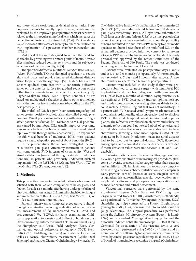
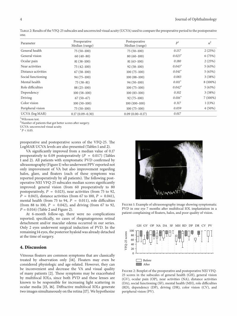
VA significantly improved from a median value of 0.17preoperatively to 0.09 postoperatively (𝑃 = 0.017) (Tables1 and 2). All patients with symptomatic PVD confirmed byultrasonography (Figure 1) who underwent PPV reported notonly improvement of VA but also improvement regardinghalos, glare, and floaters (each of these symptoms wasreported preoperatively by all patients). The following post-operative NEI VFQ-25 subscales median scores significantlyimproved: general vision (from 60 preoperatively to 80postoperatively, 𝑃 = 0.023), near activities (from 75 to 92,𝑃 = 0.043), distance activities (from 67 to 100, 𝑃 = 0.041),mental health (from 75 to 94, 𝑃 = 0.011), role difficulties(from 88 to 100, 𝑃 = 0.042), and driving (from 67 to 92,𝑃 = 0.016) (Table 2 and Figure 2).
At 6-month follow-up, there were no complicationsreported; specifically, no cases of rhegmatogenous retinaldetachment and/or macular edema occurred in our series.Only 2 eyes underwent surgical induction of PVD. In theremaining 14 eyes, the posterior hyaloid was already detachedat the time of surgery.
4. Discussion
Vitreous floaters are common symptoms that are classicallytreated by observation only [14]. Floaters may even beconsidered physiologic and age-related. However, they canbe inconvenient and decrease the VA and visual qualityof many patients [2]. These symptoms may be exacerbatedby multifocal IOLs, since both PVD and these lenses areknown to be responsible for increasing light scattering inocular media [15, 16]. Diffractive multifocal IOLs generatetwo images simultaneously on the retina [17].We hypothesize
Figure 1: Example of ultrasonography image showing symptomaticPVD in one eye 7 months after multifocal IOL implantation in apatient complaining of floaters, halos, and poor quality of vision.
GH GV
BeforeAfter
OP NA DA SF MH RD DP DR CV PV100
80604020
0
VFQ
-25
scor
es
Figure 2: Boxplot of the preoperative and postoperative NEI VFQ-25 scores in the subscales of general health (GH), general vision(GV), ocular pain (OP), near activities (NA), distance activities(DA), social functioning (SF), mental health (MH), role difficulties(RD), dependency (DP), driving (DR), color vision (CV), andperipheral vision (PV).
Journal of Ophthalmology 5
1
2
34
(a) (b)
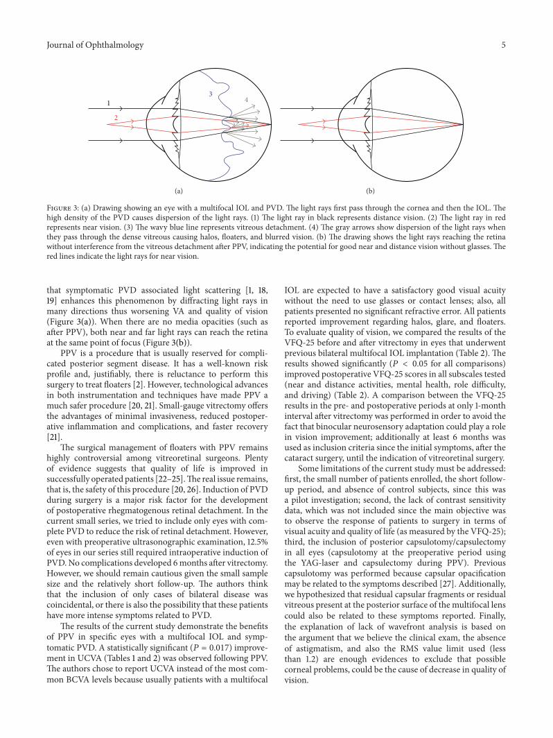
Figure 3: (a) Drawing showing an eye with a multifocal IOL and PVD. The light rays first pass through the cornea and then the IOL. Thehigh density of the PVD causes dispersion of the light rays. (1) The light ray in black represents distance vision. (2) The light ray in redrepresents near vision. (3) The wavy blue line represents vitreous detachment. (4) The gray arrows show dispersion of the light rays whenthey pass through the dense vitreous causing halos, floaters, and blurred vision. (b) The drawing shows the light rays reaching the retinawithout interference from the vitreous detachment after PPV, indicating the potential for good near and distance vision without glasses. Thered lines indicate the light rays for near vision.
that symptomatic PVD associated light scattering [1, 18,19] enhances this phenomenon by diffracting light rays inmany directions thus worsening VA and quality of vision(Figure 3(a)). When there are no media opacities (such asafter PPV), both near and far light rays can reach the retinaat the same point of focus (Figure 3(b)).
PPV is a procedure that is usually reserved for compli-cated posterior segment disease. It has a well-known riskprofile and, justifiably, there is reluctance to perform thissurgery to treat floaters [2]. However, technological advancesin both instrumentation and techniques have made PPV amuch safer procedure [20, 21]. Small-gauge vitrectomy offersthe advantages of minimal invasiveness, reduced postoper-ative inflammation and complications, and faster recovery[21].
The surgical management of floaters with PPV remainshighly controversial among vitreoretinal surgeons. Plentyof evidence suggests that quality of life is improved insuccessfully operated patients [22–25].The real issue remains,that is, the safety of this procedure [20, 26]. Induction of PVDduring surgery is a major risk factor for the developmentof postoperative rhegmatogenous retinal detachment. In thecurrent small series, we tried to include only eyes with com-plete PVD to reduce the risk of retinal detachment. However,even with preoperative ultrasonographic examination, 12.5%of eyes in our series still required intraoperative induction ofPVD.No complications developed 6months after vitrectomy.However, we should remain cautious given the small samplesize and the relatively short follow-up. The authors thinkthat the inclusion of only cases of bilateral disease wascoincidental, or there is also the possibility that these patientshave more intense symptoms related to PVD.
The results of the current study demonstrate the benefitsof PPV in specific eyes with a multifocal IOL and symp-tomatic PVD. A statistically significant (𝑃 = 0.017) improve-ment in UCVA (Tables 1 and 2) was observed following PPV.The authors chose to report UCVA instead of the most com-mon BCVA levels because usually patients with a multifocal
IOL are expected to have a satisfactory good visual acuitywithout the need to use glasses or contact lenses; also, allpatients presented no significant refractive error. All patientsreported improvement regarding halos, glare, and floaters.To evaluate quality of vision, we compared the results of theVFQ-25 before and after vitrectomy in eyes that underwentprevious bilateral multifocal IOL implantation (Table 2). Theresults showed significantly (𝑃 < 0.05 for all comparisons)improved postoperative VFQ-25 scores in all subscales tested(near and distance activities, mental health, role difficulty,and driving) (Table 2). A comparison between the VFQ-25results in the pre- and postoperative periods at only 1-monthinterval after vitrectomy was performed in order to avoid thefact that binocular neurosensory adaptation could play a rolein vision improvement; additionally at least 6 months wasused as inclusion criteria since the initial symptoms, after thecataract surgery, until the indication of vitreoretinal surgery.
Some limitations of the current study must be addressed:first, the small number of patients enrolled, the short follow-up period, and absence of control subjects, since this wasa pilot investigation; second, the lack of contrast sensitivitydata, which was not included since the main objective wasto observe the response of patients to surgery in terms ofvisual acuity and quality of life (as measured by the VFQ-25);third, the inclusion of posterior capsulotomy/capsulectomyin all eyes (capsulotomy at the preoperative period usingthe YAG-laser and capsulectomy during PPV). Previouscapsulotomy was performed because capsular opacificationmay be related to the symptoms described [27]. Additionally,we hypothesized that residual capsular fragments or residualvitreous present at the posterior surface of the multifocal lenscould also be related to these symptoms reported. Finally,the explanation of lack of wavefront analysis is based onthe argument that we believe the clinical exam, the absenceof astigmatism, and also the RMS value limit used (lessthan 1.2) are enough evidences to exclude that possiblecorneal problems, could be the cause of decrease in quality ofvision.

6 Journal of Ophthalmology
Despite these, our preliminary results are exciting. Tothe best of our knowledge, this is the first study to addressthe importance of the posterior vitreous surgery in VA andquality of vision in eyes implanted with bilateral multifocalIOLs. On the basis of these preliminary data, it is reasonableto conclude that the status of the vitreous is important inpatients whomay undergo multifocal IOL implantation. Pre-operative evaluation of candidates for these intraocular lensesimplantations should include an assessment of the vitreous.Patients with symptomatic PVD who are not satisfied withtheir VA and quality of vision may benefit from small-gaugePPV in specific situations reported. However, additionalclinical trials with large series of patients performed bydifferent surgeons are necessary to confirm these preliminaryobservations.
Summary Statement
Pars plana vitrectomy improves visual acuity and quality ofvision in patients implantedwith amultifocal intraocular lenswho have clinically relevant posterior vitreous detachmentand are not satisfied with their vision.
Conflict of Interests
The authors have no financial or proprietary interest in any ofthe products or techniques mentioned in this paper.
Acknowledgments
The current research was conducted with funding resourcesprovided by the following institutions in Brazil: Con-selho Nacional de Pesquisa (CNPq) and Coordenacao deAperfeicoamento de Pessoal de Nıvel Superior (CAPES). Acomplete listing of participating members of PACORES isas follows. The following investigators belong to the Pan-American Collaborative Retina Study Group (PACORES): L.Wu (principal investigator, PI), Instituto de Cirugia Ocular,San Jose, Costa Rica; J. F. Arevalo (PI), King Khalid EyeSpecialistHospital, Riyadh, SaudiArabia, and theWilmer EyeInstitute, Johns Hopkins University, Baltimore, MD, USA;M. A. Serrano (PI) and A. F. Lasave, Clinica OftalmologicaCentro Caracas and the Arevalo-Coutinho Foundation forResearch in Ophthalmology, Caracas, Venezuela; M. Farah(PI), M. Maia, F. M. Penha, and E. B. Rodrigues, Depar-tamento de Oftalmologia, Instituto da Visao, UniversidadeFederal de Sao Paulo, Sao Paulo, Brazil; V. Morales-Canton(PI), J. Fromow-Guerra, J. L. Guerrero-Naranjo, and J.Dalma-Weiszhausz, Asociacion para Evitar la Ceguera enMexico,MexicoCity,Mexico;H.Quiroz-Mercado (PI) andR.Velez-Montoya, University of Colorado, School of Medicine,Denver, Colorado; F. J. Rodriguez (PI), F. E. Gomez, andA.C. Brieke, FundacionOftalmologicaNacional, Universidaddel Rosario, Bogota, Colombia; M. H. Berrocal (PI) and V.Cruz-Villegas, University of Puerto Rico, San Juan, PuertoRico; F. Graue-Wiechers (PI), D. Lozano-Rechy, and E. Ariza-Camacho, Fundacion Conde Valenciana, Mexico City, Mex-ico; J. A. Roca (PI) and R. G. Chico, Clınica Ricardo Palma,
Lima, Peru; M. J. Saravia (PI), A. Schlaen, A. Lupinacci,and M. N. Gabin, Hospital Universitario Austral, BuenosAires, Argentina; M. Avila (PI) and L. Carla, Departamentode Oftalmologia, Universidade Federal de Goias, Goiania,Brazil; J. Cardillo (PI), Hospital de Olhos Araraquara andUniversidade de Sao Paulo, Sao Paulo, Brazil; J. Verdaguer(PI), C. Carpentier, J. I. Verdaguer, and G. Sepulveda, Funda-cion Oftalmologica Los Andes, Santiago de Chile, Chile; A.Alezzandrini (PI), B. Garcia, and G. Bregliano, OFTALMOS,Catedra de Oftalmologıa, Universidad de Buenos Aires,Buenos Aires, Argentina; G. Alvira (PI), P. Flor, and F.Jaramillo, Hospital Metropolitano, Quito, Ecuador; M. Diaz-Llopis (PI), R. Gallego-Pinazo, P. Udaondo, and D. Salom,Hospital La Fe, Universidad de Valencia, Spain; M. Figueroa(PI), I. Contreras, and D. Ruiz-Casas, Departamento deRetina, Hospital Universitario Ramon y Cajal and VISSUMMadrid Mirasierra de Oftalmologıa Integral, Madrid, Spain.
References
[1] R. Y. Foos, “Posterior vitreous detachment,” Transactions—American Academy of Ophthalmology and Otolaryngology, vol.76, no. 2, pp. 480–497, 1972.
[2] R. J. Foos and N. C. Wheeler, “Vitreoretinal juncture. Synchysissenilis and posterior vitreous detachment,”Ophthalmology, vol.89, no. 12, pp. 1502–1512, 1982.
[3] R. E. Coffee, A. C. Westfall, G. H. Davis, W. F. Mieler, andE. R. Holz, “Symptomatic posterior vitreous detachment andthe incidence of delayed retinal breaks: case series and meta-analysis,” American Journal of Ophthalmology, vol. 144, no. 3,pp. 409–413, 2007.
[4] A. Mirshahi, F. Hoehn, K. Lorenz, and L.-O. Hattenbach, “Inci-dence of posterior vitreous detachment after cataract surgery,”Journal of Cataract andRefractive Surgery, vol. 35, no. 6, pp. 987–991, 2009.
[5] F. Vega, F. Alba-Bueno, and M. S. Millan, “Energy distributionbetween distance and near images in apodized diffractivemultifocal intraocular lenses,” Investigative Ophthalmology &Visual Science, vol. 52, no. 8, pp. 5695–5701, 2011.
[6] M. R. Santhiago, M. V. Netto, J. Barreto Jr., B. A. F. Gomes,A. Schaefer, and N. Kara-Junior, “Wavefront analysis andmodulation transfer function of three multifocal intraocularlenses,” Indian Journal of Ophthalmology, vol. 58, no. 2, pp. 109–113, 2010.
[7] J. C. Prieto and M. J. Bautista, “Visual outcomes after implan-tation of a refractive multifocal intraocular lens with a +3.00 Daddition,” Journal of Cataract and Refractive Surgery, vol. 36, no.9, pp. 1508–1516, 2010.
[8] J. S. M. Chang, J. C. M. Ng, and S. Y. F. Lau, “Visual outcomesand patient satisfaction after presbyopic lens exchange witha diffractive multifocal intraocular lens,” Journal of RefractiveSurgery, vol. 28, no. 7, pp. 468–474, 2012.
[9] P. Sood andM.A.Woodward, “Patient acceptability of the tecnismultifocal intraocular lens,” Clinical Ophthalmology, vol. 5, no.1, pp. 403–410, 2011.
[10] C. P. Bautista, D. C. Gonzalez, A. C. Gomez, and J. A. C. Bescos,“Evolution of visual performance in 250 eyes implanted withTecnis ZM900 multifocal IOL,” European Journal of Ophthal-mology, vol. 19, no. 5, pp. 762–768, 2009.
Journal of Ophthalmology 7
[11] M. Yoshino, M. Inoue, N. Kitamura, and H. Bissen-Miyajima,“Diffractive multifocal intraocular lens interferes with intraop-erative view,” Clinical Ophthalmology, vol. 4, no. 1, pp. 467–469,2010.
[12] I. J. Suner, G. T. Kokame, E. Yu, J. Ward, C. Dolan, and N. M.Bressler, “Responsiveness of NEI VFQ-25 to changes in visualacuity in neovascular AMD: validation studies from two phase3 clinical trials,” Investigative Ophthalmology & Visual Science,vol. 50, no. 8, pp. 3629–3635, 2009.
[13] L. M. Simao, M. A. Lana-Peixoto, C. R. Araujo, M. A. Mor-eira, and A. L. Teixeira, “The Brazilian version of the 25-Item National Eye Institute Visual Function Questionnaire:translation, reliability and validity,” Arquivos Brasileiros deOftalmologia, vol. 71, no. 4, pp. 540–546, 2008.
[14] F. Martınez-Sanz, J. I. Velarde, P. Casuso, and J. N. F. Dez-Cotero, “Surgical solution to vitreous floaters visual problem,”Archivos de la Sociedad Espanola de Oftalmologıa, vol. 84, no. 5,pp. 259–262, 2009.
[15] D. P. Pinero, D. Ortiz, and J. L. Alio, “Ocular scattering,”Optometry & Vision Science, vol. 87, no. 9, pp. E682–E696, 2010.
[16] M. J. Langeslag,M. van derMooren, G.H. Beiko, and P. A. Piers,“Impact of intraocular lens material and design on light scatter:in vitro study,” Journal of Cataract & Refractive Surgery, vol. 40,no. 12, pp. 2120–2127, 2014.
[17] J. Ji, X. Huang, X. Fan, and M. Luo, “Visual performance ofacrysof ReSTOR compared with a monofocal intraocular lensfollowing implantation in cataract surgery,” Experimental andTherapeutic Medicine, vol. 5, no. 1, pp. 277–281, 2013.
[18] J. J. Kanski, “Complications of acute posterior vitreous detach-ment,” The American Journal of Ophthalmology, vol. 80, no. 1,pp. 44–46, 1975.
[19] M. Castilla-Marti, T. J. T. P. van den Berg, and M. D. deSmet, “Effect of vitreous opacities on straylight measurements,”Retina, vol. 35, no. 6, pp. 1240–1246, 2015.
[20] C. P. Wilkinson, “Safety of vitrectomy for floaters—how safe issafe?” American Journal of Ophthalmology, vol. 151, no. 6, pp.919.e1–920.e1, 2011.
[21] S. Khanduja, A. Kakkar, S. Majumdar, R. Vohra, and S. Garg,“Small gauge vitrectomy: recent update,” Oman Journal ofOphthalmology, vol. 6, no. 1, pp. 3–11, 2013.
[22] Y. M. Delaney, A. Oyinloye, and L. Benjamin, “Nd:YAG vitreol-ysis and pars plana vitrectomy: surgical treatment for vitreousfloaters,” Eye, vol. 16, no. 1, pp. 21–26, 2002.
[23] S. Schulz-Key, J.-O. Carlsson, and S. Crafoord, “Longtermfollow-up of pars plana vitrectomy for vitreous floaters: com-plications, outcomes and patient satisfaction,”Acta Ophthalmo-logica, vol. 89, no. 2, pp. 159–165, 2011.
[24] J. Sebag, “Floaters and the quality of life,” American Journal ofOphthalmology, vol. 152, no. 1, pp. 3.e1–4.e1, 2011.
[25] K. F. de Nie, N. Crama, M. A. D. Crama, B. J. Klevering, andC. J. F. Boon, “Pars plana vitrectomy for disturbing primaryvitreous floaters: clinical outcome and patient satisfaction,”Graefe’s Archive for Clinical and Experimental Ophthalmology,vol. 251, no. 5, pp. 1373–1382, 2013.
[26] H. S. Tan, M. Mura, S. Y. L. Oberstein, and H. M. Bijl, “Safetyof vitrectomy for floaters,” American Journal of Ophthalmology,vol. 151, no. 6, pp. 995–998, 2011.
[27] V. C. Shah, C. Russo, R. Cannon, R. Davidson, and M. J. Tar-avella, “Incidence of Nd:YAG capsulotomy after implantationof AcrySof multifocal and monofocal intraocular lenses: a casecontrolled study,” Journal of Refractive Surgery, vol. 26, no. 8, pp.565–568, 2010.

Submit your manuscripts athttp://www.hindawi.com
Stem CellsInternational
Hindawi Publishing Corporationhttp://www.hindawi.com Volume 2014
Hindawi Publishing Corporationhttp://www.hindawi.com Volume 2014
MEDIATORSINFLAMMATION
of
Hindawi Publishing Corporationhttp://www.hindawi.com Volume 2014
Behavioural Neurology
EndocrinologyInternational Journal of
Hindawi Publishing Corporationhttp://www.hindawi.com Volume 2014
Hindawi Publishing Corporationhttp://www.hindawi.com Volume 2014
Disease Markers
Hindawi Publishing Corporationhttp://www.hindawi.com Volume 2014
BioMed Research International
OncologyJournal of
Hindawi Publishing Corporationhttp://www.hindawi.com Volume 2014
Hindawi Publishing Corporationhttp://www.hindawi.com Volume 2014
Oxidative Medicine and Cellular Longevity
Hindawi Publishing Corporationhttp://www.hindawi.com Volume 2014
PPAR Research
The Scientific World JournalHindawi Publishing Corporation http://www.hindawi.com Volume 2014
Immunology ResearchHindawi Publishing Corporationhttp://www.hindawi.com Volume 2014
Journal of
ObesityJournal of
Hindawi Publishing Corporationhttp://www.hindawi.com Volume 2014
Hindawi Publishing Corporationhttp://www.hindawi.com Volume 2014
Computational and Mathematical Methods in Medicine
OphthalmologyJournal of
Hindawi Publishing Corporationhttp://www.hindawi.com Volume 2014
Diabetes ResearchJournal of
Hindawi Publishing Corporationhttp://www.hindawi.com Volume 2014
Hindawi Publishing Corporationhttp://www.hindawi.com Volume 2014
Research and TreatmentAIDS
Hindawi Publishing Corporationhttp://www.hindawi.com Volume 2014
Gastroenterology Research and Practice
Hindawi Publishing Corporationhttp://www.hindawi.com Volume 2014
Parkinson’s Disease
Evidence-Based Complementary and Alternative Medicine
Volume 2014Hindawi Publishing Corporationhttp://www.hindawi.com