clinical photography manual - dentsply sirona implants · for dental photography, the best lens is...
TRANSCRIPT

Clinical Photography Manualby Kris Chmielewski

2
Dental photography requires basic knowledge about general photographic rules, but also proper equipment and a digital workflow are important. In this manual you will find practical information about recommended equipment, settings, and accessories.
For success with clinical photo documentation, consistency is the key. The shots and views presented here are intended as recommendations. While documenting cases, it is very important to compose the images in a consistent manner, so that the results or stages of the treatment can easily be compared. Don’t stop documenting if a failure occurs. It’s even more important to document such cases because of their high educational value.
Dr. Kris Chmielewski, DDS, MSc Educational Director of Dental Photo Master
About the author
Kris Chmielewski is a dentist and professional photographer. Highly experienced in implantology and esthetic dentistry, he has more than 20 years experience with dental photography. He is also a freelance photographer and filmmaker, involved with projects for the Discovery Channel.
Introduction

3
Equipment 4
Camera 5
Initial camera settings for dental photography 7
Lens 8
Flash 10
Brackets 14
Accessories Retractors 15
Mirrors 16
Contrasters 17
Camera & instrument positioning 18
Intraoral photography Recommended settings 22
Frontal views 23
Occlusal views 23
Lateral views 24
Portraits Recommended settings 26
Views 27
Post-production 29
How to prepare pictures for lectures and for print 30
CONTENT

4
For dental photography, you need a camera with a dedicated macro lens and flash. The equipment presented in these pages is intended to serve as a guide that can help with selection of similar products from other manufacturers.
Equipment
Equipment

5
Camera
The best results can be achieved with a digital single lens reflex (DSLR) camera. This type of camera has interchangeable lenses, providing great flexibility. To choose the proper body for dental photography, important elements to consider include size, weight, custom shooting modes, sensor type and size, built-in wireless controller, and manufacturer.
Size and weight
A mid-size camera is easy and comfortable to hold, even with one hand. The total weight will include the camera, lens, and flash. Each of these components can influence the total weight.
Custom shooting mode
This option allows you to record and save the settings that are used most often. For dental photography, settings for capturing intraoral images can be saved so that the camera is always ready, no matter who is using it or when. Some cameras have more than one custom shooting mode. This makes it possible to record and save one mode for intraoral photography and a second one for portraits. Some Canon cameras have three modes available. In Nikon models there are two modes: U1 and U2. Please refer to your specific camera’s manual to go through the process of recording your favorite settings to the user modes.
DSLR camera body with interchangeable lens Two custom shooting modes: U1 and U2
Equipment | Camera
This manual reflects the author’s experience with certain camera brands and is not necessarily the opinion of Dentsply Sirona. The inclusion of these experiences does not constitute an endorsement, guarantee, warranty or recommendation of, and Dentsply Sirona make no representations and/or warranties about, any of the camera brands mentioned herein.

6
Camera
DSLR Sensor – type and size
In general, DSLRs have one of two types of sensors: full-frame sensors or APS-C sensors. The first is bigger. A full-frame DSLR has a sensor that is the same size as a frame of traditional 35mm film, measuring 36x24mm. The more popular APS-C sensor cameras have much smaller 22x15mm sensors. This means a full-frame sensor has over 2.5 times the surface area of an APS-C sensor.
Both types can have the same number of pixels and resolution. The size of the pixels is crucial to image quality. In dental photography, the difference in quality of the final photograph is not recognizable.
The advantage of a full-frame camera is mostly noticeable in portrait photography with shallow depth of field. The advantage of an APS-C sensor is bigger depth of field and lower price.
Wireless controller
Some cameras have built-in wireless controllers. This feature enables communication between the camera and a wireless external flash without any additional wires. The user only must set the communication between the wireless flash and the camera.
Cameras that have built-in wireless controllers include:Nikon: D70, D70s, D80, D90, D200, D300, D300s, D7000, D7100, D7200, D600, D610, D700, D750, D800/D800E and D810.Canon: - Canon EOS 600D (Rebel T3i / Kiss X5)- Canon EOS 650D (Rebel T4i / Kiss X6i)- Canon EOS 700D (Rebel T5i / Kiss X7i)- Canon EOS 750D (Rebel T6i / Kiss X8i)- Canon EOS 760D (Rebel T6s / EOS 8000D)- Canon EOS 60D- Canon EOS 70D- Canon EOS 7D- Canon EOS 7D Mark II
Manufacturer
The most common brands used in dental photography are Canon and Nikon. The main reason for this is their long presence on the photo market with a full portfolio of cameras, lenses, flashes, and accessories used by both professionals and amateurs. New models are available every year, but used equipment is also available at reasonable prices. Other companies such as Sony, Pentax, Sigma, Olympus, and others also offer some options but are not as common in dental photography.
Recommended camera bodies for dental photography
These models have features that will help to make your dental photography an easy and pleasant experience.APS-C Sensor:- Canon EOS 7D Mark II , - Canon EOS 70D, - Nikon D7100- Nikon D7200Full Frame Sensor:- Canon EOS 6D- Nikon D750
Dimensions of the full-frame and APS-C sensors.
Equipment | Camera
Full frame sensor size36 mm x 24 mm
APS-C sensor size22,5 mm x 15 mm

7
Initial camera settings for dental photography
When you buy the camera, you should get familiar with your new equipment. The best approach is to read the manual carefully, but the reality is often very different. Initial settings will influence management of your files and the quality of the pictures. In the majority of camera models, access to these settings is achieved through the Menu.
Here is the short list of the recommended settings that you should apply:
• Set correct date and time
• Change color space to Adobe RGB
• Image quality: RAW + jpg (Canon), NEF + jpg (Nikon)
• White balance: flash or 5500K
• ISO 200
• Metering mode: evaluative metering (Canon), 3D Matrix (Nikon)
• Select single Auto Focus point
ISO 200Set correct date and time
White balance: flash or 5500K
Image quality: RAW + jpg (Canon), NEF + jpg (Nikon)
Change color space to Adobe RGB
Equipment | Initial camera settings for dental photography

8
On the market you can buy system macro lenses such as Nikon and Canon (meaning that the lens and camera are produced by the same company). Or lenses can be purchased from independent companies such as Sigma, Tamron, or Tokina. Lenses generally are not inexpensive, but it should be possible to find one that will fit your budget.
A few features to consider before buying the lens are the desired focal length, magnification ratio, image stabilization, and lens speed.
Focal length
In photography we can differentiate between prime lenses and zoom lenses. A prime lens has a fixed focal length and is described in mm.When you select the lens, please be aware that lenses can have different mounts. Before you buy your lens, make sure it has the connection that will fit your camera. Although a prime lens has only one focal length, it can come in lengths ranging from wide angle ones through longer telephoto ones.A zoom lens has a range of focal lengths available to the photographer in one lens; it is described as the focal range in mm for instance: 70-200mm.
Magnification ratio
For dental photography, the best lens is a macro lens with a magnification ratio of 1:1. This means that with minimum focusing distance, which is the closest distance from the lens to the object to get a still sharp image, you can capture a real life 1cm object with the same size on your sensor. Be careful. While lenses with the “macro” description can be found on the market, that doesn’t mean they always have a 1:1 magnification ratio.
Image stabilization
Newer lenses have built-in systems that can compensate for image blur caused by small, involuntary movements known as camera shake. In dental photography, this feature doesn’t have a big impact because the shutter speeds vary in the range from 1/60 to 1/250 of a second. If your lens has an image stabilization system, you should read the lens manual carefully to know the criteria for using it. These lenses are more expensive. Every company has a different acronym for image stabilization (e.g. Nikon-VR, Canon-IS, Sigma-OS, Tamron-VC).
Tip: when you shoot from a tripod, please switch off the image stabilization mode on your lens to avoid blurry images. Otherwise your lens by default will try to detect shaking and can give a false reading.
Lens speed
Two features influence the so-called lens speed. First is the maximum lens opening described with the aperture number, such as f/2.8. This means that the specific lens is not built to open more than the size of the aperture. The aperture number influences the amount of light that comes into the lens barrel and the camera and, ultimately, how easy it is for you to look through the camera’s viewfinder and see a bright image that is easy to compose.
Second is the auto focus speed. More expensive lenses have ultrasonic motors built-in. They allow for very fast and accurate adjustment of the focus to get a sharp image when you are shooting in auto focus mode.
Lens
Equipment | Lens

9
Recommended lenses for a Nikon DSLR camera:
• Nikon AF-S VR Micro-NIKKOR 105mm f/2.8G IF-ED Lens
Other macro lenses available for a Nikon DSLR camera:
• Nikon AF-S DX Micro NIKKOR 85mm f/3.5G ED VR Lens
• Sigma 105mm f/2.8 EX DG OS HSM Macro Lens for Nikon
• Tokina 100mm f/2.8 AT-X M100 AF Pro D Macro Autofocus Lens for Nikon
• Tamron 90mm f/2.8 SP AF Di Macro Lens for Nikon
• Tamron 90mm f/2.8 SP Di MACRO 1:1 VC USD Lens for Nikon
• Samyang 100mm f/2.8 ED UMC Macro Lens for Nikon
• Rokinon 100mm f/2.8 Macro Lens for Nikon F
Recommended lenses for a Canon DSLR camera:
• Canon EF 100mm f/2.8L Macro IS USM Lens
• Canon EF 100mm f/2.8 Macro USM Lens
Other macro lenses available for a Canon:
• Sigma 105mm f/2.8 EX DG OS HSM Macro Lens for Canon EOS Cameras
• Tamron 90mm f/2.8 SP Di MACRO 1:1 VC USD Lens for Canon EOS
• Tamron SP 90mm f/2.8 Di Macro Autofocus Lens for Canon EOS
• Samyang 100mm f/2.8 ED UMC Macro Lens for Canon EF
Canon EF 100mm f/2.8L Macro IS USM Lens
Equipment | Lens

10
In dental photography, flash is used to illuminate the oral cavity and the patient’s face. When choosing flash equipment, consideration should be given to the type of flash, availability of a wireless option, and optional accessories.
Flash types For dental applications, macro flashes are recommended, especially for intraoral photography. Their construction allows the source of light to be positioned close to the subject and the lens.
A ring flash is mounted on the front of the lens with dedicated mount rings. It is available with wireless communication.
A dual flash is more advanced, and with the help of additional accessories like brackets it enables control over the distance of the flash head from the lens.
External flash is a very good choice for portrait photography.
Flash
Ring Flash Dual Flash External Flash
Equipment | Flash

11
Recommendations
For surgery, a ring flash is the top choice. Its compact size and the position of the flash tubes next to the lens makes it possible to perfectly illuminate all the areas in the limited space of the oral cavity.
A dual flash can also be used during surgical procedures, but essential in this case is the close position of the flash heads next to the lens axis. Such a position can be set on the dedicated mount ring for the specific flash models (Canon MT-24EX and Nikon R1C1) or with brackets.
A ring flash has more limitations for documenting prosthetic procedures. The straight direction of the light creates a “flat” picture flooded with light. The glossy surface of tooth enamel reflects the light, and the information on such a picture is limited (like color, transparency and texture). A better choice is dual flash on a bracket. By controlling the distance and flash head position, you can easily capture many more details.
Portraits – external flash with diffuser
Equipment | Flash
12
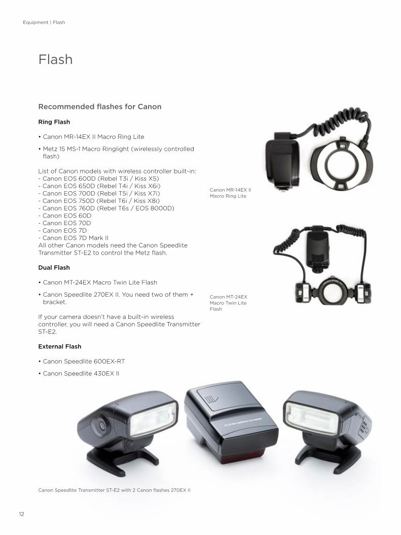
Recommended flashes for Canon
Ring Flash
• Canon MR-14EX II Macro Ring Lite
• Metz 15 MS-1 Macro Ringlight (wirelessly controlled flash)
List of Canon models with wireless controller built-in: - Canon EOS 600D (Rebel T3i / Kiss X5)- Canon EOS 650D (Rebel T4i / Kiss X6i)- Canon EOS 700D (Rebel T5i / Kiss X7i)- Canon EOS 750D (Rebel T6i / Kiss X8i)- Canon EOS 760D (Rebel T6s / EOS 8000D)- Canon EOS 60D- Canon EOS 70D- Canon EOS 7D- Canon EOS 7D Mark IIAll other Canon models need the Canon Speedlite Transmitter ST-E2 to control the Metz flash.
Dual Flash
• Canon MT-24EX Macro Twin Lite Flash
• Canon Speedlite 270EX II. You need two of them + bracket.
If your camera doesn’t have a built-in wireless controller, you will need a Canon Speedlite Transmitter ST-E2.
External Flash
• Canon Speedlite 600EX-RT
• Canon Speedlite 430EX II
Flash
Canon Speedlite Transmitter ST-E2 with 2 Canon flashes 270EX II
Canon MT-24EX Macro Twin Lite Flash
Canon MR-14EX II Macro Ring Lite
Equipment | Flash

13
Recommended flashes for Nikon
Ring Flash:
• Metz 15 MS-1 Macro Ring light Digital Flash (wirelessly controlled flash)
Nikon models with a wireless controller built-in include: D70, D70s, D80, D90, D200, D300, D300s, D7000, D7100, D7200, D600, D610, D700, D750, D800/D800E and D810. If you have one of them, than you do not need any other equipment to work with the Metz 15 MS-1 flash. Other camera models require Nikon’s SU-800 Wireless Speedlight Commander.
Dual Flash on the bracket:
• Nikon R1C1 Wireless Close-Up Speedlight System
Portraits:
• Nikon SB-910 AF Speedlight
• Nikon SB-700 AF Speedlight
Nikon D7100 with mounted Metz 15 MS-1 flash
Nikon R1C1 Wireless Close-Up Speedlight System mounted on the Photomed bracket with LumiQuest Pocket Bouncers
Equipment | Flash

14
Brackets are used to mount the flash heads at a distance from the lens and camera body. The bracket is mounted to the tripod socket of the camera body with the fixing screw. The arms can be positioned in various ways and enable control over the distance and angulation of the light source from the object and the lens.
The most common suppliers on the market are Photomed and Agno’s.
When choosing a bracket, pay attention to the system compatibility. The mounts for Nikon flash heads SB-R200 and for Canon differ.
Brackets
Photomed bracket
Agno’s bracket
Equipment | Brackets

15
Accessories for dental photography help to get better pictures. They include retractors, mirrors, and contrasters. Because they come in contact with the patient’s oral cavity, they all should be autoclavable.
Retractors
Retractors are used to lift the lips away and give better access to the oral cavity. They can be made of plastic or metal. Two main types exist: self-retracting retractors and single sided retractors. They come in different shapes and sizes.Self-retracting retractors are used mostly for frontal views and also for occlusal shots.Single sided retractors are mostly used for lateral views.
RecommendationPlastic retractors are more comfortable for the patients.
Accessories
Single sided retractor
Self-retracting retractor
Accessories | Retractors

16
Mirrors
Mirrors are used to capture the reflection of the teeth. They can be made from different materials and with different techniques. The most common mirrors are made of polished metal or glass coated with a highly reflective surface. Depending on the surface coating type, a different reflection percentage will result. A chromium coating reflects 65% of the light, rhodium 75%, titanium 75%, and dielectric coatings such as Ultrabright reflect 99%. All reflective surfaces will scratch over the time; because of this, mirrors should be replaced from time to time. The coating material does not influence the final picture unless it is scratched or distorted. A less reflective surface needs more energy from the flash to get a proper exposure. Chromium coated mirrors are most common and are cheaper than the others. Mirrors come in different shapes and sizes, however in daily practice, two shapes are used most often: occlusal and lateral.
RecommendationBuy mirrors in different sizes to fit different individuals. For some mirror shapes, separate handles are offered. This is helpful in holding and positioning the mirror.
Accessories
Lateral mirror
Occlusal mirror
Accessories | Mirrors

17
Contrasters
Contrasters allow photos of the teeth to be taken with a black background. They come in different shapes and sizes. They are made from black anodized aluminum or from metal plates covered with soft black silicone. Three types are available: anterior, occlusal, and lateral.
RecommendationSelect contrasters coated with black silicone. It’s more durable, will not scratch, and is more pleasant for the patient. The anterior contraster is used most often.
Lateral contraster
Occlusal contraster
Anterior contraster
Accessories | Contrasters

18
Correct positioning of instruments such as mirrors, retractors, and contrasters is very important for good access and to avoid obstacles interfering with the photo axis. Instruments can be held by the patient but also by the assistant or photographer. To avoid distortion of portraits and images of the teeth, the photo/lens axis should be perpendicular to the teeth.
Camera and instrument positioning
Camera & instruments positioning
Correct position of the photographer and the patient. The axis of the lens is at the eye level of the patient.

19
Positioning of the camera for intraoral photography in the front area. The axis of the lens is perpendicular to the front teeth.
Positioning of the anterior contraster for shooting upper front teeth.
Camera & instruments positioning

20
Positioning of the anterior contraster for shooting lower front teeth.
Positioning of the occlusal mirror for a lower occlusal photo. The camera axis is at a 45° angle to the surface of the mirror.
Camera & instruments positioning

21
Example of positioning the occlusal mirror for an upper occlusal arch photo. The camera axis is at a 45° angle to the surface of the mirror.
Positioning of the buccal mirror and retractor for a lateral/buccal view.
Camera & instruments positioning

22
Camera and Flash Settings
Camera settings in Aperture Priority Mode
Mode: A (Aperture priority mode. In Canon it is described with the letters Av. In Nikon cameras, it’s with A.) Aperture f22 or higher – depends on the construction of the lens.Shutter Speed: Will be set automatically by the camera (in Nikon by default it is 1/60 of a second; in Canon cameras you have to change the Flash Synchronization speed in Av Mode to the fixed value of 1/200th of a second).Flash setting: TTL mode (fully automatic mode – camera will measure and calculate the proper amount of light for the exposure). With some of the flashes, Manual mode is necessary and requires individual tests to determine which settings will be appropriate for the final exposure.
Camera settings in the Manual ModeMode: MAperture: f22 or higherShutter Speed: 1/200sFlash: Stay with TTL
Intraoral photography
Intraoral photography | Settings

23
Frontal views
Equipment:
• DSLR Camera set with 100mm macro lens and ring flash or dual flash
Accessories:
• Self-retracting retractor
• Anterior contraster
Advice:
• Hold the camera with the back of the camera parallel to the plane of the front teeth.
• Focus on the front teeth.
Shots:
• Retracted view in the MIP position: upper and lower teeth are in the full contact. Upper and lower teeth are visible at least from first molar to first molar.
• Retracted view with teeth apart.
• Upper teeth from canine to canine (with and without contraster).
• Lower teeth from canine to canine (with and without contraster).
Occlusal views
Equipment:
• DSLR Camera set with 100mm macro lens and ring flash or dual flash
Accessories:
• Self-retracting retractor
• Occlusal mirror
Advice:
• 45 degree angle between the mirror and the camera for occlusal shot is recommended.
• Focus on the occlusal surface of the first bicuspids (premolars).
• Full arch should be visible from the central incisors to the mesial of the second molars.
• Eliminate the fog on the mirror (air blow or heat the mirror).
Shots:
• Full upper arch
• Full lower arch
Intraoral photography | Views

24
Lateral views
Equipment:
• DSLR Camera set with 100mm macro lens and ring flash or dual flash
Accessories:
• Single sided retractor
• Buccal mirror
Advice:
• Upper and lower teeth should be visible from the central incisors to the mesial of the second molars.
• Focus on the canine (cuspid)
• Eliminate the fog on the mirror (air blow or heat the mirror)
Shots:
• Right lateral retracted view in the MIP position: upper and lower teeth are in full contact.
• Right lateral retracted view with teeth apart: upper and lower teeth are not in contact.
• Left lateral retracted view in the MIP position: upper and lower teeth are in full contact.
• Left lateral retracted view with teeth apart: upper and lower teeth are not in contact.
Intraoral photography | Views

25
Intraoral photography | Views
Upper & lower teeth MIP position / retracted view
f>22, 1/200s
Maxillary anterior teeth retracted view
f>22, 1/200s
Maxillary anterior teeth retracted view with contraster
f>22, 1/200s
Upper & lower teeth mouth open / retracted view
f>22, 1/200s
Mandibular anterior teeth retracted view
f>22, 1/200s
Mandibular anterior teeth retracted view with contraster
f>22, 1/200s
Maxillary arch / occlusal view retracted view with mirror
f>22, 1/200s
Right lateral MIP viewretracted view with mirror
f>22, 1/200s
Left lateral MIP viewretracted view with mirror
f>22, 1/200s
Mandibular arch / occlusal view retracted view with mirror
f>22, 1/200s
Right lateral open viewretracted view with mirror
f>22, 1/200s
Left lateral open viewretracted view with mirror
f>22, 1/200s

26
In portrait photography, the main goal is to capture the full face from the top of the head to the neck in 3 basic positions: lips together, mouth open, and full smile. These types of pictures are used to analyze the esthetics and symmetry of the face.
Camera and flash settings
Mode: Aperture Priority Mode: A (Nikon), Av (Canon) or Manual (M)Aperture f11 Shutter Speed: will be set automatically by the camera in A/Av Mode. In Manual Mode, set the speed to 1/200s Flash setting: TTL modeAuto Focus mode
Portraits
Portraits | Settings

27
Portrait views
Equipment:
• DSLR Camera set with 100mm macro lens and external flash
Accessories:
• Solid/uniform background (black, white, or grey)
• Flash diffuser
Advice:
• Full face from the top of the head to the neck
• Patient should look into the lens or straight ahead (in 45- and 90-degree positions)
• Auto focus on the eye
Shots:
• Front views in 3 positions (lips together, apart, and full smile)
• 45-degree views (left and right) in 3 positions (lips together, apart, and full smile)
• 90-degree views (left and right) in 3 positions (lips together, apart, and full smile)
Portraits | Views

28
Portraits | Views
Frontal view / not smiling
f/11, 1/200s
Frontal view / mouth open
f/11, 1/200s
Frontal view / full smile
f/11, 1/200s
45° right profile / not smiling
f/11, 1/200s
45° right profile / mouth open
f/11, 1/200s
45° right profile / full smile
f/11, 1/200s
90° right profile / not smiling
f/11, 1/200s
90° right profile / mouth open
f/11, 1/200s
90° right profile / full smile
f/11, 1/200s
45° left profile / not smiling
f/11, 1/200s
45° left profile / mouth open
f/11, 1/200s
45° left profile / full smile
f/11, 1/200s
90° left profile / not smiling
f/11, 1/200s
90° left profile / mouth open
f/11, 1/200s
90° left profile / full smile
f/11, 1/200s
29
Post-production is the process that occurs after taking the photographs. It covers multiple actions like importing the pictures to the computer, managing the files, developing the pictures, adjusting the exposure, correcting the color, cropping, cleaning, and also exporting the files for different media.
In dental photography it is very important to establish a digital workflow to help stay organized and maintain control over file locations and security. For all of these, proper hardware and software is essential.
One of the best software packages available on the market for this purpose is Adobe Photoshop Lightroom. The software was created for photo-graphers. Modules include Library, Develop, and sharing modes such as Slideshow, Book, and Print. Each of these modules is designed to meet the photographer’s needs.
In the Library Mode, you can organize your pictures, apply keywords, and organize other metadata. An advanced search engine is incorporated. This is a very important tool for quickly finding specific pictures among the thousands.
The Develop Mode has all the necessary tools for correcting exposures, white balance, and color; cropping; cleaning up dust and scratches; and making other improvements. One of the most important features of the Lightroom software is the way it treats files. It is not destructive, meaning that when you develop your pictures, the software creates a recipe. It records all your actions, which are saved in the history. Permanent changes are applied to the picture when files are exported. But the original files remain untouched, so you can always return to the original file in the file history. The software allows for working with different file types, including RAW, jpg, TIFF, psd, and png.
Post-production
Post-production

30
The size of the recorded picture is represented in the number of pixels (points). The pixel is the smallest single component of a digital image. Each pixel holds information about color. The number of pixels in an image is sometimes called the resolution.
The average sensor of digital cameras used in dental photography is close to 20 megapixels.For different media, images must be optimized to achieve the best results. Pictures should be prepared differently for lectures and for print.
If you are using dedicated software for photography like Adobe Photoshop Lightroom, then the process is very easy. If not, the rules are the same and are applied to the image during export. In that case, the information below can be used as a reference.
Optimal image settings for lectures:
File settings:
• Format: .jpg
• Quality: 100 (best)
• Limit files size to 500kb
• Color space: sRGB
Image sizing:
• Resize to fit: long edge 2000 pixels
• Don’t enlarge
• Resolution: 72 pixels per inch
Output sharpening:
• Sharpen for screen
• Amount: high
Optimal image settings for print:
File settings for TIFF:
• Format: TIFF (ask the editor or print house)
• Compression: none
• Bit depth: 16 bits/component
• Color space: Adobe RGB
File settings for jpg:
• Format: .jpg
• Quality: 100 (best)
• Limit files size: unchecked (no limit)
• Color space: Adobe RGB
Image sizing:
• Original size
• Resolution: 240 pixels per inch
Output sharpening:
• Sharpen for print for Matte Paper
• Amount: high
How to prepare pictures for lectures and for print
How to prepare pictures for lectures and for print


Notes

Notes

Notes

Notes

Den
tsp
ly S
iro
na d
oes
no
t w
aive
any
rig
ht t
o it
s tr
adem
arks
by
not
usin
g t
he s
ymb
ols
® o
r ™
. 32
671
374
-USX
-170
8 ©
20
17 D
ents
ply
Sir
ona
. All
rig
hts
rese
rved
.
About Dentsply Sirona Implants
Dentsply Sirona Implants offers comprehensive solutions for all phases of implant therapy, including Ankylos®, Astra Tech Implant System® and Xive® implant lines, digital technologies, such as Atlantis® patient-specific solutions and Simplant® guided surgery, Symbios® regenerative solutions, and professional and business development programs, such as STEPPS™. Dentsply Sirona Implants creates value for dental professionals and allows for predictable and lasting implant treatment outcomes, resulting in enhanced quality of life for patients.
About Dentsply Sirona
Dentsply Sirona is the world’s largest manufacturer of professional dental products and technologies, with a 130-year history of innovation and service to the dental industry and patients worldwide. Dentsply Sirona develops, manufactures, and markets a comprehensive solutions offering including dental and oral health products as well as other consumable medical devices under a strong portfolio of world class brands. As The Dental Solutions Company™, Dentsply Sirona’s products provide innovative, high-quality and effective solutions to advance patient care and deliver better, safer and faster dentistry. Dentsply Sirona’s global headquarters is located in York, Pennsylvania, and the international headquarters is based in Salzburg, Austria. The company’s shares are listed in the United States on NASDAQ under the symbol XRAY.
Visit www.dentsplysirona.com for more information about Dentsply Sirona and its products.
THE DENTAL SOLUTIONS COMPANY™