clavicle or collarbone -...
TRANSCRIPT

Clavicle or Collarbone
►The clavicle, or collar bone, holds the shoulder joint away from the rest of the upper body and is only as thick as your little finger.

Clavicle The clavicle (collarbone) extends between the sternum and theacromion of the scapula.It is classed as a long bone, and can be palpated along its length. Inthin individuals, it is visible under the skin. The clavicle has threemain functions:•Attaches the upper limb to the trunk.•Protects the underlying neurovascular structures supplying the upper limb.•Transmits force from the upper limb to the axial skeleton.
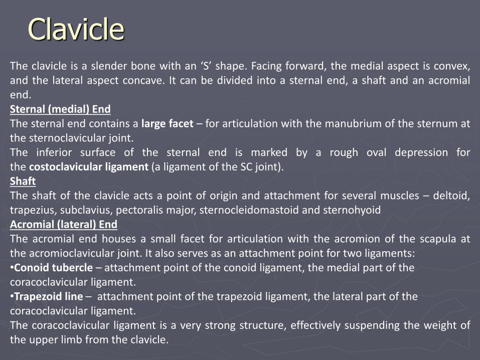
Clavicle The clavicle is a slender bone with an ‘S’ shape. Facing forward, the medial aspect is convex,and the lateral aspect concave. It can be divided into a sternal end, a shaft and an acromialend.Sternal (medial) EndThe sternal end contains a large facet – for articulation with the manubrium of the sternum atthe sternoclavicular joint.The inferior surface of the sternal end is marked by a rough oval depression forthe costoclavicular ligament (a ligament of the SC joint).ShaftThe shaft of the clavicle acts a point of origin and attachment for several muscles – deltoid,trapezius, subclavius, pectoralis major, sternocleidomastoid and sternohyoidAcromial (lateral) EndThe acromial end houses a small facet for articulation with the acromion of the scapula atthe acromioclavicular joint. It also serves as an attachment point for two ligaments:•Conoid tubercle – attachment point of the conoid ligament, the medial part of the coracoclavicular ligament.•Trapezoid line – attachment point of the trapezoid ligament, the lateral part of the coracoclavicular ligament.The coracoclavicular ligament is a very strong structure, effectively suspending the weight ofthe upper limb from the clavicle.

Rib Cage
Surrounds the thoracic and upper abdominal cavitiesThe rib cage is essential for protecting your heart and lung, providing a place for your shoulder bones to attach, and it aids in the breathing process.

5
Bones of the Rib Cage
12 pairs of ribs
❑ Attached posteriorly to
thoracic vertebrae
❑ True ribs 1-7
❑ False ribs 8-10
❑ Floating ribs 11&12
Sternum
❑ Forms the front, middle
portion of the rib cage

Sternum (Breastbone)
The sternum can be divided into three parts; the manubrium, body and xiphoid process. In children, these elements are joined by cartilage. The cartilage ossifies to bone during adulthood.

Sternum
ManubriumThe manubrium is the most superior portion of the sternum. It is trapezoid inshape.The superior aspect of the manubrium is concave, producing a depressionknown as the jugular notch – this is visible underneath the skin.On the lateral edges of the manubrium, there is a facet (cartilage lineddepression in the bone), for articulation with the costal cartilage of the 1st rib,and a demifacet (half-facet) for articulation with part of the costal cartilage ofthe 2nd rib.Inferiorly, the manubrium articulates with the body of the sternum, formingthe sternal angle. This can be felt as a transverse ridge of bone on the anterioraspect of the sternum. The sternal angle is commonly used as an aid to countribs, as it marks the level of the 2nd costal cartilage.

Sternum
► Body
► The body is flat and elongated – the largest part of the sternum. It articulates with the manubrium superiorly (manubriosternal joint) and the xiphoid process inferiorly (xiphisternal joint).
► Xiphoid Process
► The xiphoid process is the most inferior and smallest part of the sternum. It is variable is shape and size, located at the level of the T10 vertebrae. The xiphoid process is largely cartilaginous in structure, and completely ossifies late in life – around the age of 40.
► In some individuals, the xiphoid process articulates with part of the costal cartilage of the seventh rib.

Scapula
►The scapula is located on the back side of the ribcage and helps provide part of the shoulder joint and movement for the arms.

Scapula The scapula is also known as the shoulder blade. Itarticulates with the humerus at the glenohumeral joint,and with the clavicle at the acromioclavicular joint. Indoing so, the scapula connects the upper limb to thetrunk.It is a triangular, flat bone, which serves as a site forattachment for many (17!) muscles.In this article, we shall look at the bony landmarks on thecostal, lateral and posterior surfaces of the scapula.
The anterior surface of the scapula is termed ‘costal’, this is because it is the side facing the ribcageOriginating from the superolateral surface of the costal scapula is the coracoid process. It is a hook-like projection, which lies just underneath the clavicle. The pectoralis minor attaches here, while the coracobrachialis and biceps brachii muscles originates from this projection.

Scapula The lateral surface of the scapula faces thehumerus. It is the site of the glenohumeral joint,and of various muscle attachments.Bony landmarksGlenoid fossa – A shallow cavity, which articulateswith the humerus to form the glenohumeral joint.The superior part of the lateral border is veryimportant clinically, as it articulates with thehumerus to make up the shoulder joint, orglenohumeral joint.Supraglenoid tubercle – A roughening immediatelysuperior to the glenoid fossa, this is the place ofattachment of the long head of the biceps brachii.Infraglenoid tubercle – A roughening immediatelyinferior to the glenoid fossa, this is the place ofattachment of the long head of the triceps brachii.

Scapula
The posterior surface of the scapula faces outwards. It is a site of attachment for themajority of the rotator cuff muscles of the shoulder.Bony landmarksSpine – The most prominent feature of the posterior scapula. It runs transversely across thescapula, dividing the surface into two.Infraspinous fossa – The area below the spine of the scapula, it displays a convex shape.The infraspinatus muscle originates from this area.Supraspinous fossa – The area above the spine of the scapula, it is much smaller that theinfraspinous fossa, and is more convex in shape. The supraspinatus muscle originates fromthis area.Acromion – projection of the spine that arches over the glenohumeral joint and articulateswith the clavicle.

Humerus (Upper Arm Bone)
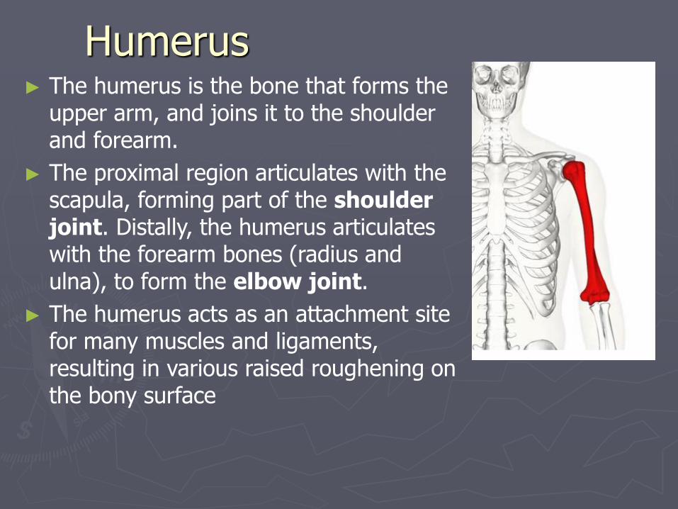
Humerus ► The humerus is the bone that forms the
upper arm, and joins it to the shoulder and forearm.
► The proximal region articulates with the scapula, forming part of the shoulder joint. Distally, the humerus articulates with the forearm bones (radius and ulna), to form the elbow joint.
► The humerus acts as an attachment site for many muscles and ligaments, resulting in various raised roughening on the bony surface

Humerus ► The important anatomical features of the proximal humerus are the head,
anatomical neck, surgical neck, greater and lesser tubercles and intertubercular sulcus. A tubercle is a round nodule, and signifies an attachment site of a muscle or ligament.
► The head of the humerus is connected to the greater and lesser tubercles by the anatomical neck, which is short in width and nondescript.
► The greater tubercle is located laterally on the humerus. It has an anterior and posterior face. The greater tubercle serves as attachment site for three of the rotator cuff muscles – supraspinatus, infraspinatus and teres minor.
► The lesser tubercle is much smaller, and more medially located on the bone. It only has an anterior face. It is a place of attachment for the last rotator cuff muscle – subscapularis.

Humerus ► On the lateral side of the humeral shaft is a roughened surface where
the deltoid muscle attaches. This is known is as the deltoid tuberosity
► Immediately distal to the supraepicondylar ridges are the lateral and medial epicondyles – projections of bone. Both can be palpated at the elbow (the medial more so, as it is much larger). The ulnar nerve passes into the forearm along the posterior side of the medial epicondyle, and can also be palpated there.

Humerus► Distally, the trochlea is located medially, and extends onto
the posterior of the bone. Lateral to the trochlea is the capitulum, which articulates with the radius.
► Also found on the distal portion of the humerus are three depressions, known as the coronoid, radial and olecranon fossae. They accommodate the forearm bones during movement at the elbow.


Ulna ► The ulna is a long bone in the forearm. It lies medially
and parallel to the radius, the second of the forearm bones. The ulna acts as the stabilizing bone, with the radius pivoting to produce movement.
► Proximally, the ulna articulates with the humerus at the elbow joint. Distally, the ulna articulates with the radius, forming the distal radio-ulnar joint.

Ulna ► The proximal end of the ulna articulates with the trochlea of the humerus. To enable
movement at the elbow joint, the ulna has a specialized structure, with bony prominences for muscle attachment.
► Olecranon – A large projection of bone that extends proximally, forming part of trochlear notch. It can be palpated as the ‘tip’ of the elbow. The triceps brachii muscle attaches to its superior surface.
► Coronoid process – This ridge of bone projects outwards anteriorly, forming part of the trochlear notch.
► Trochlear notch – Formed by the olecranon and coronoid process. It is wrench shaped, and articulates with the trochlea of the humerus.
► Radial notch – Located on the lateral surface of the trochlear notch, this area articulates with the head of the radius.
► Tuberosity of ulna – A roughening immediately distal of the coronoid process. It is where the brachialis muscle attaches.

Radius ► The radius is a long bone in the forearm. It lies laterally and parallel to ulna, the
second of the forearm bones. The radius pivots around the ulna to produce movement at the proximal and distal radio-ulnar joints.
► The radius articulates in four places:
► Elbow joint – Partly formed by an articulation between the head of the radius, and the capitulum of the humerus.
► Proximal radioulnar joint – An articulation between the radial head, and the radial notch of the ulna.
► Wrist joint – An articulation between the distal end of the radius and the carpal bones.
► Distal radioulnar joint – An articulation between the ulnar notch and the head of the ulna

Radius ► Important bony landmarks include
the head, neck and radial tuberosity:
► Head of radius – A disk shaped structure, with a concave articulating surface. It is thicker medially, where it takes part in the proximal radioulnar joint.
► Neck – A narrow area of bone, which lies between the radial head and radial tuberosity.
► Radial tuberosity – A bony projection, which serves as the place of attachment of the biceps brachiimuscle.

Bones of the hand ► The bones of the hand provide support and flexibility to the
soft tissues. They can be divided into three categories:
► Carpal bones (Most proximal) – A set of eight irregularly shaped bones. These are located in the wrist area.
► Metacarpals – There are five metacarpals, each one related to a digit
► Phalanges (Most distal) – The bones of the fingers. Each finger has three phalanges, except for the thumb, which has two.


Carpals ► The carpal bones are a group of
eight, irregularly shaped bones. They
are organized into two rows – proximal
and distal.
► In the proximal row, the bones
are (lateral to medial):
► Scaphoid, Lunate, Triquetrum
► Pisiform – A sesamoid bone,
formed within the tendon of the flexor
carpi ulnaris
► In the distal row, the bones
are (lateral to medial):
► Trapezium, Trapezoid, Capitate
► Hamate – has a projection on its palmar surface called the hook of hamate
► Proximally, the scaphoid and lunate articulate with the radius to form the wrist joint. In the distal row, all of the carpal bones articulate with the metacarpals

Metacarpals ► The metacarpal bones articulate proximally with the carpals, and
distally with the proximal phalanges. They are numbered, and each associated with a digit:
► Metacarpal I – Thumb.
► Metacarpal II – Index finger.
► Metacarpal III – Middle finger.
► Metacarpal IV – Ring finger.
► Metacarpal V – Little finger.
► Each metacarpal consists of a base, shaft and a head. The medial and lateral surfaces of the metacarpals are concave, allowing attachment of the interoessei muscles.

Phalanges (Fingers)
The phalanges are the bones of the fingers. The thumb has a proximal and distal phalanx, while the rest of the digits have proximal, middle and distal phalanges.

28
Bones of the Hips, Legs and FeetHipbones - pelvic girdle or coxal bones
Bones of the lower limb or leg - femur,
the patella, the tibia, and the fibula
Bones of the foot - the tarsal,the
metatarsals, and
the phalanges.

Femur ►The femur is the only bone in the thigh. It is
classed as a long bone, and is the longest bone in the body. The main function of the femur is to transmit forces from the tibia to the hip joint.
► It acts as the site of origin and attachment of many muscles and ligaments, and can be divided into three areas; proximal, shaft and distal.

Femur ► The proximal area of the femur forms the hip joint with the pelvis. It
consists of a head and neck, and two bony processes called trochanters. There are also two bony ridges connecting the two trochanters.
► Head – Articulates with the acetabulum of the pelvis to form the hip joint. It has a smooth surface with a depression on the medial aspect; for the attachment of the ligament of head of femur.
► Neck – Connects the head of the femur with the shaft. It is cylindrical, projecting in a superior and medial direction – this angle of projection allows for an increased range of movement at the hip joint.

Femur ► Greater trochanter – A projection of bone that originates from the
anterior aspect, just lateral to the neck. It is angled superiorly and posteriorly, and can be found on both the anterior and posterior sides of the femur.
▪ Site of attachment for many of the muscles in the gluteal region, such as gluteus medius, gluteus minimus and piriformis.
► Lesser trochanter – Smaller than the greater trochanter. It projects from the posteromedial side of the femur, just inferior to the neck-shaft junction.
▪ Site of attachment for the psoas major and iliacus muscles.

Femur ► The distal end is characterized by the presence of the medial and lateral
condyles, which articulate with the tibia and patella, forming the knee joint.
► Medial and lateral condyles – Rounded areas at the end of the femur. The posterior and inferior surfaces articulate with the tibia and menisci of the knee, while the anterior surface articulates with the patella.
► Medial and lateral epicondyles – Bony elevations on the non-articular areas of the condyles. They are the area of attachment of some muscles and the collateral ligaments of the knee joint.
► Intercondylar fossa – A depression found on the posterior surface of the femur, it lies in between the two condyles. It contains two facets for attachment of internal knee ligaments.

Patella (knee cap)► The patella (knee-cap) is located at the front of the knee
joint, within the patellofemoral groove of the femur. It attaches superiorly to the quadriceps tendon and inferiorly to the patellar ligament.
► It is classified as a sesamoid type bone due to its position within the quadriceps tendon, and is the largest sesamoid bone in the body.
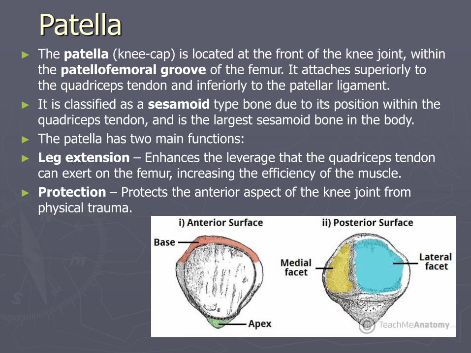
Patella ► The patella (knee-cap) is located at the front of the knee joint, within
the patellofemoral groove of the femur. It attaches superiorly to the quadriceps tendon and inferiorly to the patellar ligament.
► It is classified as a sesamoid type bone due to its position within the quadriceps tendon, and is the largest sesamoid bone in the body.
► The patella has two main functions:
► Leg extension – Enhances the leverage that the quadriceps tendon can exert on the femur, increasing the efficiency of the muscle.
► Protection – Protects the anterior aspect of the knee joint from physical trauma.
The Tibia and Fibula
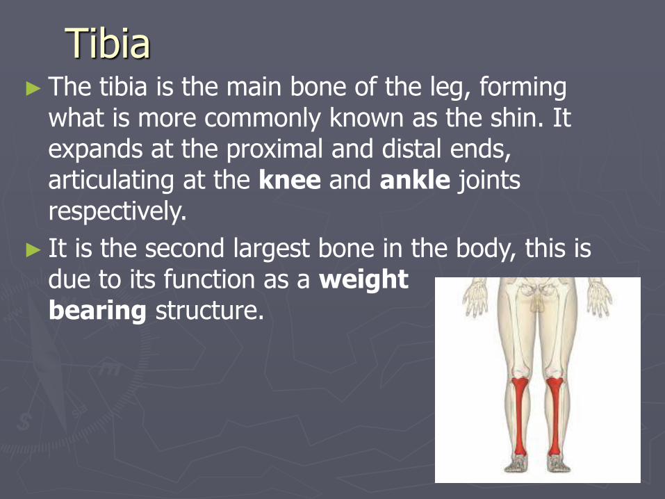
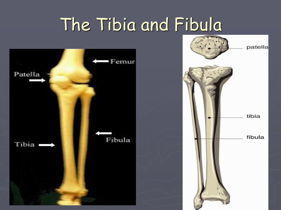
Tibia ►The tibia is the main bone of the leg, forming
what is more commonly known as the shin. It expands at the proximal and distal ends, articulating at the knee and ankle joints respectively.
► It is the second largest bone in the body, this is due to its function as a weight bearing structure.
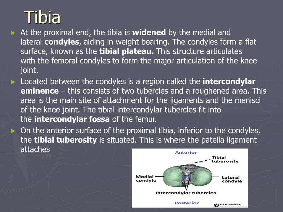
Tibia ► At the proximal end, the tibia is widened by the medial and
lateral condyles, aiding in weight bearing. The condyles form a flat surface, known as the tibial plateau. This structure articulates with the femoral condyles to form the major articulation of the knee joint.
► Located between the condyles is a region called the intercondylar eminence – this consists of two tubercles and a roughened area. This area is the main site of attachment for the ligaments and the menisci of the knee joint. The tibial intercondylar tubercles fit into the intercondylar fossa of the femur.
► On the anterior surface of the proximal tibia, inferior to the condyles, the tibial tuberosity is situated. This is where the patella ligament attaches

Tibia ► The distal end of the tibia, like the proximal, widens to
help with weight bearing.
► There is a bony projection continuing inferiorly on the medial side – this is called the medial malleolus. It articulates with the tarsal bones to form part of the ankle joint. On the posterior surface of the tibia, there is a groove where the tibialis posterior muscle attaches.

Fibula ► The fibula, along with the tibia,
makes up the bones of the leg. The fibula is found laterally to the tibia, and is much thinner. As it does not articulate with the femur at the knee joint, its main function is to act as an attachment for muscles, and not as a weight bearer.
► At the proximal end, the fibula has an enlarged head, which contains a facet for articulation with the lateral condyle of the tibia. On the posterior and lateral surface of the fibular neck, the common fibular nerve can be found.

Fibula ► The fibular shaft has three surfaces –
anterior, lateral and posterior. The leg is split into three compartments, and each surface faces its respective compartment e.g anterior surface faces the anterior compartment of the leg.
► Distally, the lateral surface continues inferiorly, and is called the lateral malleolus. The lateral malleolus is more prominent than the medial malleolus, and can be palpated at the ankle on the lateral side of the leg

Bones of the foot ► The human foot is a very complex and highly developed structure. The
bones of the foot provide mechanical support for the soft tissues, helping the foot withstand the weight of the body.
► The bones of the foot can be divided into three categories:
► Tarsals – A set of seven irregularly shaped bones. They are situated proximally in the foot, in the ankle area.
► Metatarsals – These bones connect the phalanges to the tarsals. There are five in number – one for each digit.
► Phalanges – The bones of the toes. Each toe has three phalanges – a proximal, intermediate and distal (except the big toe, which only has two phalanges).

TarsalsThe tarsal bones of the foot are organised into three rows; proximal, intermediate and distal.Proximal GroupThe proximal tarsal bones are the talus and the calcaneus. They form the bony frameworkaround the proximal ankle and heel area.The talus is the most superior of the tarsal bones. It has three articulations:•Superiorly: Ankle joint – between the talus and the bones of the leg (the tibia and fibula).•Inferiorly: Subtalar joint – between the talus and calcaneus.•Anteriorly: Talonavicular joint – between the talus and the navicular.
The main function of the talus is to transmit forces from the tibia to the heel bone (known asthe calcaneus). Whilst numerous ligaments attach to the talus, it is not a site of muscleattachment or origin.

Tarsals ► The calcaneus lies underneath the talus, and has two
articulations:
► Superiorly: Subtalar joint – between the calcaneus and the talus.
► Anteriorly: Calcaneocuboid joint – between the calcaneus and the cuboid.
► It is thick and sturdy, acting to transmit forces from the talus to the ground. The posterior aspect of the calcaneus is marked by calcaneal tuberosity, to which the Achilles tendon attaches

Metatarsals
The metatarsals are located in the midfoot, between the tarsals andphalanges. They are numbered I-V (medial to lateral).Each metatarsal has a similar structure. They consist of a distal head andproximal base, which are joined by a shaft of bone. They have three orfour articulations:•Proximally: Tarsometatarsal joint – between the metatarsal bases and the cuneiforms or cuboid bones.•Laterally: Intermetatarsal joint(s) – between the metatarsal and the adjacent metatarsals.•Distally: Metatarsophalangeal joint – between the metatarsal head and the proximal phalanx.

PhalangesThe phalanges are the bones of the toes.Most toes have three phalanges –proximal, intermediate and distal. Thegreat toe only has proximal and distalphalanges.Each phalanx consists of a body, a proximalextremity and a distal extremity.