1 the circulatory system the circulatory system...
TRANSCRIPT
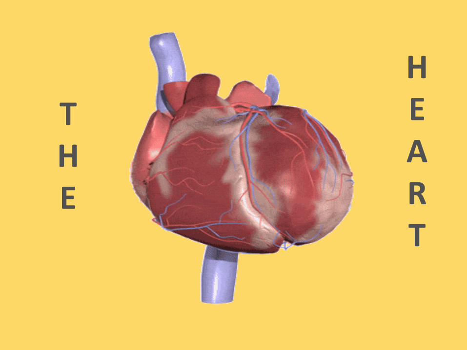
THE
HEART

37–1 The Circulatory System
The circulatory system and respiratory system work together to supply cells with the nutrients and oxygenthey need to stay alive.
a) The respiratory system:
● picks up the oxygen and absorbs it into the blood.
● It changes oxygen-poor blood (deoxygenated) into oxygen-rich blood
(oxygenated)
b) The circulatory system:
● then pumps the blood to the lungs & rest of body

Functions of the Circulatory
System
Functions of the Circulatory System
Organisms with many cells need a way to get oxygen & nutrients to each and every cell of their body. The circulatory system is the transport system of the body that can do this.
Humans and other vertebrates have a closed circulatory system, meaning that the blood is always contained within a system of vessels.

Functions of the Circulatory
System
The human circulatory system consists of:
•the heart
•blood vessels
•blood

Blood Vessels
As blood flows through the
circulatory system, it moves
through three types of blood
vessels:
•arteries
•capillaries
•veins

Blood Vessels
Arteries
Large vessels that carry blood AWAY from
the heart to the tissues of the body are
called arteries.
Except for the pulmonary arteries, all
arteries carry oxygenated blood.
Arteries have thick muscular walls.
They contain the following tissues from
outside to inside: connective tissue, smooth
muscle, and endothelium.

Blood VesselsIn the circulatory system,
there are three types of
blood vessels—arteries,
capillaries, and veins. The
walls of these vessels contain
connective tissue, smooth
muscle, and endothelium.

Blood Vessels
Capillaries
The smallest of the blood vessels are
the capillaries. No cells are far from a
capillary.
Their walls are only one cell thick, and
most are so narrow that only one red
blood cell can pass through at a time.

Blood Vessels
The capillaries are exchange vessels:
They bring nutrients and oxygen to
the tissues of body
They absorb carbon dioxide and other
waste products from body cells and
bring these compounds away from cells
so the body can dispose of them.

Blood Vessels
In the circulatory
system, there are
three types of blood
vessels—arteries,
capillaries, and
veins. The walls of
these vessels contain
connective tissue,
smooth muscle, and
endothelium.

Blood Vessels
VeinsBlood vessels that carry blood back to the heart are called veins.
Except for the pulmonary veins, all veins carry deoxygenated blood.
Veins have thinner walls than arteries, containing less muscle than arteries.
The walls of veins contain connective tissue, smooth muscle and endothelium.

Blood Vessels
In the circulatory
system, there are
three types of
blood vessels—
arteries,
capillaries, and
veins. The walls of
these vessels
contain connective
tissue, smooth
muscle, and
endothelium.

Blood Vessels
Large veins contain one-way valves that keep blood moving toward the heart.
Many veins are located near and between skeletal muscles.
The movement of these skeletal muscles helps to return the blood to our hearts when we are standing.
Contraction of skeletal muscles helps
move blood in veins toward the heart.
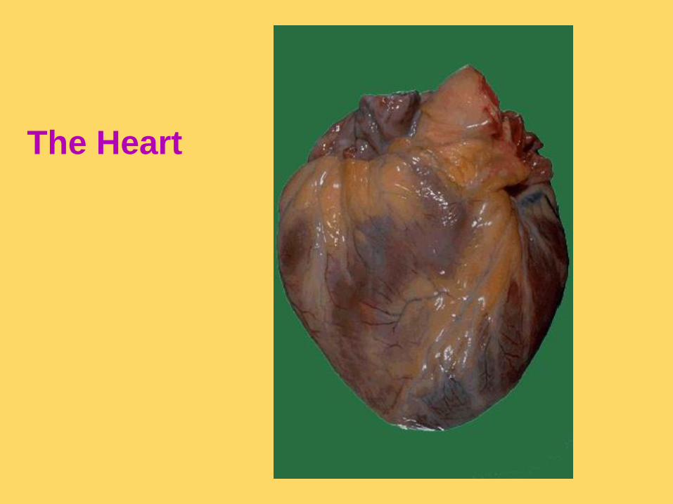
The Heart

Structure of the Heart• The heart is a muscle that contracts to pump blood throughout the
body.
• The heart is enclosed in a
protective sac called
the PERICARDIUM.-Keeps the heart contained in the chest cavity.
-Prevents the heart from over-expanding when blood
volume increases.
-Limits heart motion.
-made up of three layers Fibrous Pericardium,
Parietal Pericardium,Visceral Pericardium
• In most animals, the heart is located between the 3rd and 7th rib in the ventral chest cavity.
• The pointed end of the heart- APEX
• The flattened end of the heart- BASE.
The Heart
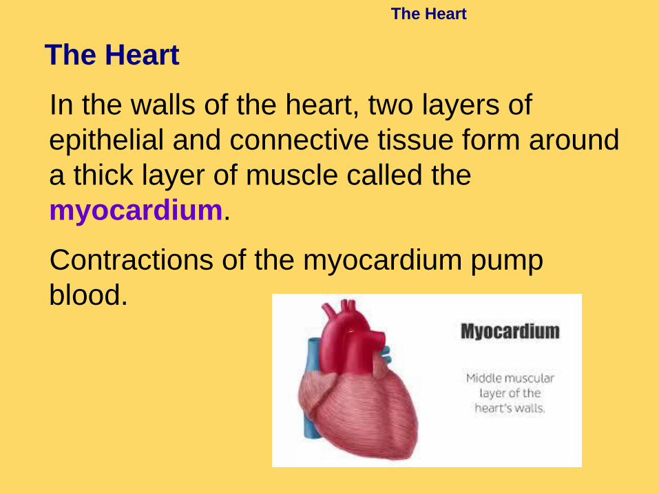
The Heart
In the walls of the heart, two layers of
epithelial and connective tissue form around
a thick layer of muscle called the
myocardium.
Contractions of the myocardium pump
blood.

•When referring to the heart, always refer to it from the animal’s point of view. SO………
Right sideLeft side
•A thick wall of MYOCARDIUM, referredto as the SEPTUM,

The Heart
The septum divides the right side of the
heart from the left.
It prevents the mixing of deoxygenated
and oxygenated blood.
Septum

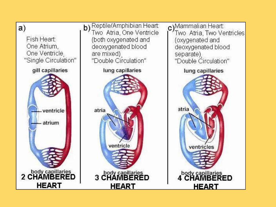
The heart is divided into chambers and valves that help pump the blood and prevent backflow.
Fish have a 2-chambered heart: 1 atrium receives
blood1 ventricle pumps blood

Amphibians and Reptiles:Have a 3-chambered heart:
2 Atria1 Ventricle

Birds and Mammals:Have 4- chambered hearts:
2- Atria2- Ventricles
We will be focusing on the mammalian heart!!

The Heart
The heart has four chambers — two atria
and two ventricles.
There are two chambers on each side of
the septum.
The upper chamber, which receives the
blood, is the atrium.
The lower chamber, which pumps blood
out of the heart, is the ventricle.

Right Atrium
Right Ventricle
Left Atrium
Left Ventricle

Chambers and valves
bicuspid valve:
contains two
cusps


The Heart
Circulation Through the Heart
Large veins, the vena cava, bring blood
back to the heart from the rest of the
body. These enter either the right
atrium.
There are 2 vena cava:
Superior (from head region)
Inferior (from lower body)

The Heart
Large vein that
brings deoxygenated
blood from the upper
part of the body to
the right atrium
Right Atrium
Superior Vena Cava:

The Heart
Vein that brings
deoxygenated blood
from the lower part of
the body to the right
atrium.
Right Atrium
Inferior Vena Cava:

The Heart
As the heart contracts, blood flows from the atria into the ventricles.
Then the ventricles pump the blood out
of the heart into two large arteries
(aorta & pulmonary artery). Blood then
moves to either the body or the lungs.

The Heart
There are flaps of connective tissue called valves between the atria and
the ventricles.
Valve on left side is called the mitralor bicuspid valve.
Valve on the right side is called the tricuspid valve.
When the ventricles contract, the valves close, which prevents blood from flowing back into the atria.

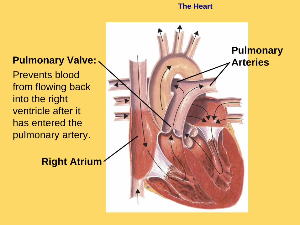
The Heart
Prevents blood
from flowing back
into the right
atrium after blood
has entered the
right ventricle
Right Atrium
Tricuspid Valve:

The Heart
Mitral Valve:
Prevents blood
from flowing back
into the left atrium
after blood has
entered the left
ventricle
Left Atrium
Left Ventricle

The Heart
At the exits from the right and left ventricles, different valves prevent blood that flows out of the heart from flowing back in.
Blood leaves the left ventricle, and enters the aorta. This is the largest artery in your body and begins the bloods journey to the rest of the body.
●Valve at base the aorta is called the aortic valve.

The Heart
Aortic Valve:
Prevents blood
from flowing
back into the left
ventricle after it
has entered the
aorta
Left Atrium
Left Ventricle
Aorta

The Heart
Brings oxygenated
blood from the left
ventricle to the
body
Aorta:

The Heart
Blood leaves the right ventricle, and enters the pulmonary artery. This goes to the lungs to pick up oxygen and drop off carbon dioxide.
●Valve at base of the pulmonary artery is called the pulmonary valve.
The Heart
Prevents blood
from flowing back
into the right
ventricle after it
has entered the
pulmonary artery.
Right Atrium
Pulmonary
ArteriesPulmonary Valve:
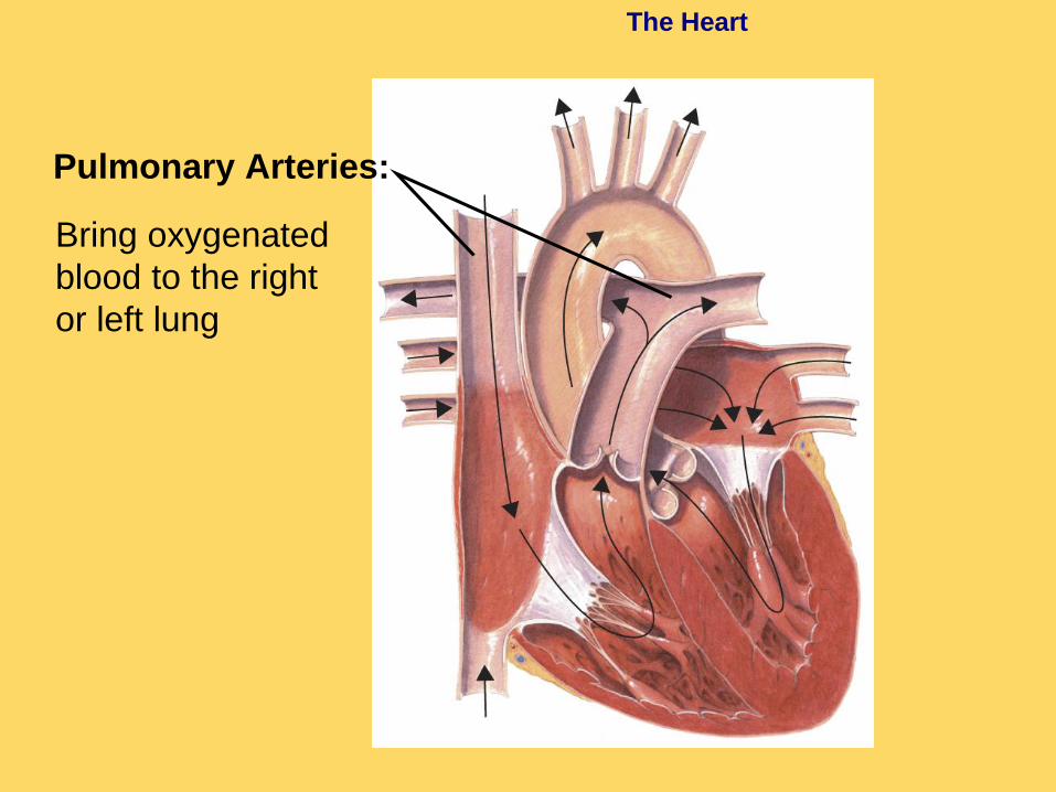
The Heart
Bring oxygenated
blood to the right
or left lung
Pulmonary Arteries:

Aortic Valve Pulmonary Valve
Bicuspid Valve Tricuspid Valve

The Aortic Valve

Mechanical Heart Valves

Mechanical Heart Valves

The Heart
Blood returns to the heart from the lungs in the pulmonary veins. This brings back oxygenated blood to the left atrium for distribution to the rest of the body.

The Heart
Bring deoxygenated
blood from each of
the lungs to the left
atrium
Left Atrium
Pulmonary
Veins:

The Heart
Circuits Through the Body
The heart functions as two separate pumps:
One pumps deoxygenated blood from the
right side of the heart to the lungs and
back to the left side of the heart. This is
called the pulmonary circuit.
The other pumps oxygenated blood from
the left side of the heart to the cells of the
body and then back to the right side of
the heart. This is called the systemic
circuit.

The Heart
Circulation of
Blood through
the Body
Capillaries of
head and arms
Superior
vena cava Aorta
Pulmonary
veinCapillaries of
right lungs
Inferior
vena cava
Capillaries of
abdominal organs
and legs
Capillaries
of left lung
Pulmonary
artery

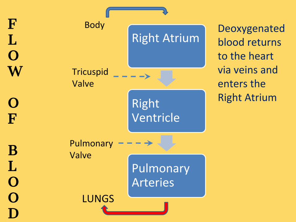
Right Atrium
Right Ventricle
Pulmonary Arteries
Body
Tricuspid Valve
Pulmonary Valve
FLOW
OF
BLOOD
Deoxygenated blood returns to the heart via veins and enters the Right Atrium
LUNGS

Left Atrium
Left Ventricle
Aorta
Pulmonary Veins
Mitral Valve
Aortic Valve
Body
FLOW
OF
BLOOD
Oxygenated blood from the lungs returns to the heart and enters the Left Atrium.


The Heart
Blood Pathway:
Here is a listing of all of the places that the blood flows as it moves through the heart and body in order:
Vena cava, right atrium, tricuspid valve, right ventricle, pulmonary valve, pulmonary artery, lungs, pulmonary veins, left atrium, bicuspid (mitral) valve, left ventricle, aortic valve, aorta, body tissues,vena cava

Heart Conduction
• The cardiac muscle has the ability to expand and contract and is regulated by the heart’s own conduction system.
• The conduction system is similar to an electrical outlet: Once “plugged in” electricity flows through the heart causing it to beat.
• The sounds of the heart “lub dub” are actually the heart valves opening and closing with each electrical impulse.

The Heart
Heartbeat
Each contraction begins in a small cluster of cells in the right atrium called the sinoatrial (SA) node.
● Cells act like a pacemaker
● Spontaneously sets off impulses, about 72 beats/minute
Contraction spreads quickly from atria to ventricles.
● Spread by a system of fibers called the Purkinje fibers.

The Heart
Sinoatrial (SA)
node Conducting fibers
The impulse spreads from the pacemaker
(SA node) to a network of fibers in the atria. The signal to contract spreads from the sinoatrial node to the cardiac
muscle cells of the atria, causing the atria to contract. The impulse is
picked up by the atrioventricular node, which transmits the impulse to
muscle fibers in the ventricles, causing the ventricles to contract.

The Heart
Conducting fibers
Atrioventricular
(AV) node
The impulse is picked up by a bundle of fibers
called the atrioventricular (AV) node and carried
to the network of Purkinje fibers in the ventricles.The signal to contract spreads from the sinoatrial node to the cardiac
muscle cells of the atria, causing the atria to contract. The impulse is
picked up by the atrioventricular node, which transmits the impulse to
muscle fibers in the ventricles, causing the ventricles to contract.

Artificial
Pacemaker
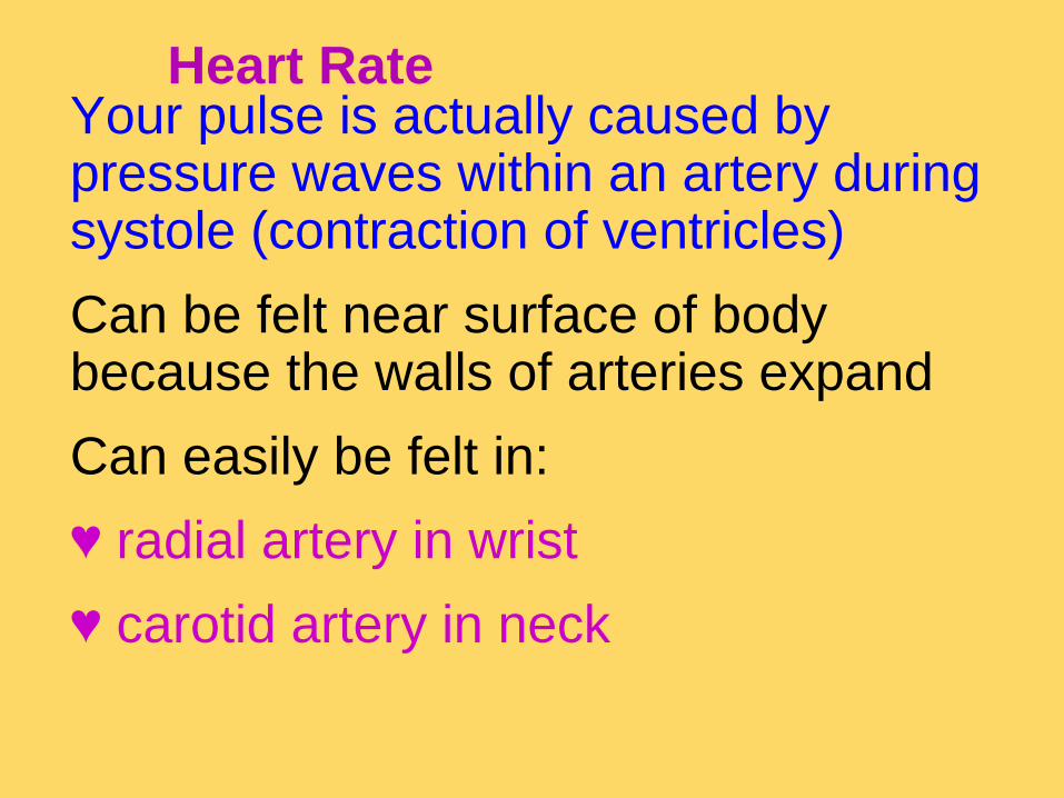
Heart RateYour pulse is actually caused by pressure waves within an artery during systole (contraction of ventricles)
Can be felt near surface of body because the walls of arteries expand
Can easily be felt in:
♥ radial artery in wrist
♥ carotid artery in neck


Contraction Phases of Heartbeat
Systole
● The contraction
phase of the
heart cycle
● When the ventricles
actively pump
the blood

● The relaxation phase of the heart cycle
● When the ventricles fill with blood
Diastole

Heart Contraction & Blood Flow

Blood Pressure
Blood Pressure
When the ventricle of the heart contracts, it
produces a wave of fluid pressure in the
arteries.
The force of the blood on the arteries’ walls is
blood pressure.
Blood pressure keeps blood flowing through
the body.

Blood Pressure
Blood pressure is measured with a machine called a:
sphygmomanometer.
A typical blood pressure for a healthy person is 120/80.
● 1st # = systolic pressure
Pressure during systole
● 2nd # = diastolic pressure
Pressure during diastole


Electrocardiograms (EKGs)
Can measure tiny electrical impulses
that are produced by the heart
Electrocardiograph is an instrument that
can measure these impulses
The written record is called an
electrocardiogram (EKG or ECG)

Electrocardiogram (ECG or EKG)
A test used to measure the electrical activity of the heart. Informs us if the heart is beating too fast, too slow, normally or irregularly.

How to read an EKG
To briefly summarize the features used in reading EKGs, they consist of
waveform components which indicate electrical events during one heartbeat.
These waveforms are labeled P, Q, R, S, and T .
P wave is the first short upward movement of the EKG tracing. It indicates that
the atria are contracting, pumping blood into the ventricles.
The QRS complex, normally beginning with a downward deflection, Q; a larger
upwards deflection, a peak (R); and then a downwards S wave. The QRS
complex represents ventricular depolarization and contraction.
The PR interval indicates the transit time for the electrical signal to travel from
the AV node to the ventricles.
T wave is normally a modest upwards waveform representing ventricular
repolarization

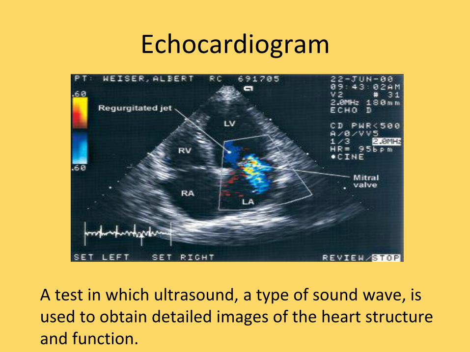
Echocardiogram
A test in which ultrasound, a type of sound wave, is used to obtain detailed images of the heart structure and function.

Diseases of the Circulatory
System
Diseases of the Circulatory System
Cardiovascular diseases are among the
leading causes of death and disability in the
U.S.
Atherosclerosis is a condition in which fatty
deposits called plaque build up on the inner
walls of the arteries.
Atherosclerosis can lead to heart attacks
and strokes in the brain.

Normal
Coronary
Artery
Artery withplaque
Atherosclerosis
Clogging of the arteries

Diseases of the Circulatory
System
Heart Attack and Stroke
If one of the coronary arteries in heart
becomes blocked, part of the heart muscle
may begin to die from a lack of oxygen.
If enough heart muscle is damaged, a heart
attack occurs.
Diseases of the Circulatory
System
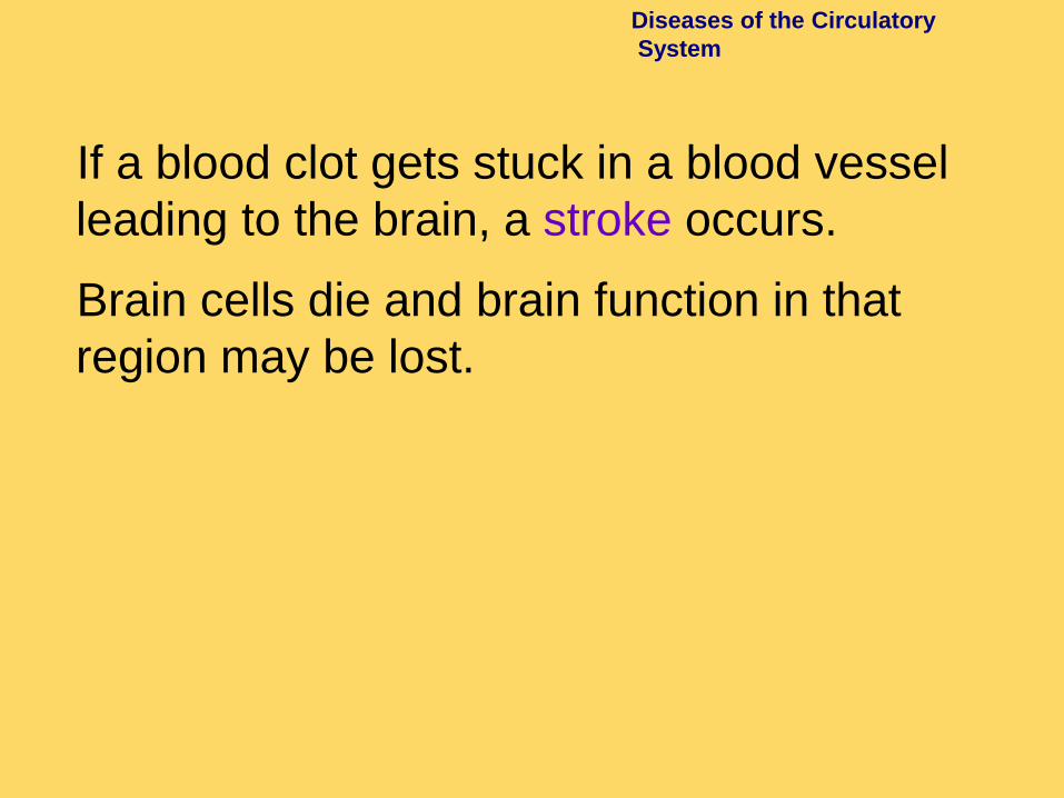
If a blood clot gets stuck in a blood vessel
leading to the brain, a stroke occurs.
Brain cells die and brain function in that
region may be lost.

Diseases of the Circulatory
System
Hypertension, commonly called high
blood pressure, is a serious problem.
● It puts strain on walls of arteries &
increases chances they might burst
● It makes the heart work too hard
● It can lead to heart damage, brain
damage and kidney failure

Diseases of the Circulatory
System
Circulatory System Health
Ways of avoiding cardiovascular disease
include:
• getting regular exercise.
• eating a balanced diet.
• avoiding smoking.

CPR/AEDSometimes when the heart stops, it can be restarted. Professionals are taught to read the rhythms of an EKG and decide if the heart is in a rhythm that can be restarted. An
Automatic External Defibrillator or AED is also programmed to read
these specific rhythms.


While waiting for an AED to arrive, to give the person the best possible chance of surviving,
Cardio Pulmonary Resuscitation or CPR should be performed. CPR keeps the heart pumping
and the oxygenated blood circulating.


