characterization of intracytoplasmic prokaryote infections
TRANSCRIPT

DISEASES OF AQUATIC ORGANISMSDis Aquat Org
Vol. 44: 203–216, 2001 Published April 10
INTRODUCTION
The invasion and spread of zebra mussels Dreissenapolymorpha and quagga mussels Dreissena bugensis
throughout North American freshwaters since the mid-1980s has had significant adverse economic and eco-logical impacts. As major macrofoulers of raw-waterconduits within infrastructures, they have caused dam-age and increased operating expenses totaling hun-dreds of millions of dollars (O’Neill 1996, 1997). Envi-
© Inter-Research 2001
*E-mail: [email protected]
Characterization of intracytoplasmic prokaryoteinfections in Dreissena sp. (Bivalvia: Dreissenidae)
Daniel P. Molloy1,*, Laure Giamberini2, J. Frank Morado3, Sergei I. Fokin4, Franck Laruelle5
1New York State Museum, The State Education Department, Cultural Education Center, Albany, New York 12230, USA2Equipe de Production des Ecosystèmes et Ecotoxicologie, Laboratoire EBSE, Université de Metz, Campus Bridoux,
rue du Gal Délestraint, 57070 Metz Cedex, France3National Oceanic & Atmospheric Administration, National Marine Fisheries Service, Alaska Fisheries Science Center,
Resource Assessment & Conservation Engineering Division, 7600 Sand Point Way NE, Seattle, Washington 98115-0070, USA4Biological Research Institute, St. Petersburg State University, St. Petersburg 198904, Russia
5UMR CNRS 6539, Institut Universitaire Européen de la Mer, UBO, Place Nicolas Copernic, Technopôle Brest-Iroise, 29280 Plouzané, France
ABSTRACT: This study characterizes intracytoplasmic infections with prokaryote microorganisms inDreissena sp. (near Dreissena polymorpha) from northeastern Greece and represents the first reportof such infections in freshwater bivalves. Light microscope observations of stained tissues revealedbasophilic, cytoplasmic inclusion bodies in 87.5% (28/32) of the mussels sectioned. Inclusions inepithelial cells and connective tissues were noted, respectively, in 34.4 and 71.9% of the sample, with5 mussels (15.6%) having both tissue types infected. Epithelial cell infections were observed in histo-logical sections only in digestive gland tubules and ducts; within tubules, inclusions were presentmore often in secretory than digestive cells. Connective tissue infections, however, were systemic;among the 32 mussels sectioned, inclusions were found in the gills (65.6%), foot (12.5%), mantle(9.4%), labial palps (6.3%), digestive gland (6.3%), stomach (6.3%), and gonads (3.1%). Cytoplasmicinclusions (maximum dimension, 138 µm) were prominent enough in the gills to be visible in 17.0%of the 247 mussels dissected. Ultrastructurally, prokaryote cells in gill connective tissues were clearlycharacteristic of Chlamydiales-like organisms, with each intracytoplasmic inclusion containing aloosely packed mixture of elementary, reticulate, intermediate bodies, and blebs. Prokaryote coloniesin digestive gland epithelial cells exclusively contained 1 of 4 morphological cell types and wereconsidered Rickettsiales-like. Hexagonal, virus-like particles were present in the cytoplasm of thelargest of these Rickettsiales-like prokaryotes. Although host stress was evident from localized cellnecrosis and dense hemocyte infiltration, overall infection was fairly benign, with no major, adverseimpact on body condition evident among sectioned or dissected mussels. A possible negative effectwas partial constriction of gill water tubes, but at the infection intensity observed (typical range 1 to7 inclusion bodies per section), significant interference with respiration and other metabolic functionsof the gills was highly unlikely.
KEY WORDS: Zebra mussels · Intracytoplasmic prokaryote · Epithelium · Connective tissue · Virus-like particles · Rickettsiales-like · Chlamydiales-like
Resale or republication not permitted without written consent of the publisher

Dis Aquat Org 44: 203–216, 2001
ronmental impacts in North America (Strayer 1999)are similar to European experiences (Karatayev et al.1997), including decreased phytoplankton productivity(Fahnenstiel et al. 1995), increased native bivalve mor-tality (Schloesser et al. 1996), and restructured benthiccommunities (Stewart et al. 1998).
After introduction into a waterbody, populationgrowth of Dreissena spp. can be explosive because oftheir life history traits of high fecundity, high growthrate, and tolerance of a range of environmental condi-tions. The absence of their Eurasian natural enemycomplex of predators, parasites, and benthic competi-tors has also contributed to their rapid populationgrowth in North America, altough to what degree isopen to debate. Molloy et al. (1997) suggested thatamong these 3 natural enemy groups, research onDreissena’s parasites had received the least attention.This information gap is currently being addressed by anetwork of over a dozen scientists from the formerSoviet Union, Europe, and North America as a projectof the International Research Consortium on Mollus-can Symbionts (IRCOMS; http//www.nysm.nysed.gov/biology/ircoms/bio_ircoms.html). In this IRCOMS pro-ject, initial focus is on developing a database charac-terizing the systematics, biology, ecology, and distribu-tion of Dreissena’s parasites, as well as other symbioticorganisms encountered within these mussels (Molloyet al. 1996, Burlakova et al. 1998, Laruelle et al. 1999,Karatayev et al. 2000a,b, Laruelle et al. 2001). Thesedata on parasites will help define the role that in-fectious diseases play in the population dynamics ofDreissena spp. and to what degree the relativeabsence of parasites in North American Dreissenapopulations (Toews et al. 1993, Camp et al. 1999) aidstheir successful colonization of waterbodies on thiscontinent. This database may also enhance determina-tion of whether any of Dreissena’s parasites could beused as biological control agents (Molloy 1998).
The present study characterizes infection by intra-cytoplasmic prokaryote microorganisms (i.e., with nomembrane-bound nucleus) observed in a Dreissena sp.in northeastern Greece. Prokaryotes have frequentlybeen recorded as intracytoplasmic parasites of marinebivalves (reviewed in Bower et al. 1994, Fryer & Lan-nan 1994), but this is the first report from freshwaterbivalves. We present evidence that this population ofzebra mussels was infected by at least 2 prokaryotespecies, 1 of which was hyperinfected with virus-likeparticles.
MATERIALS AND METHODS
Dreissena sp. were sampled from Lake Volvi, Greece(40° 37’ N, 23° 21’ E) at a depth of 4 m on October 27,
1995, and shipped live overnight under refrigeratedconditions to a temporary field laboratory in Seillans,France. All mussels possessed uniform shell morpho-logy, including a flat ventral surface, and thus resem-bled D. polymorpha. Their species determination,however, is still undergoing analysis since their valvesformed an unusually high dorsal keel that is uncharac-teristic for this species (A. Y. Karatayev pers. comm.).
Mussels were examined for endosymbionts usingvarious techniques, including immediate dissection of247 specimens and fixation of others for subsequenthistological and ultrastructural analyses at IRCOMSmember laboratories. Only data on the intracytoplas-mic prokaryote infections observed are presentedherein. Information on other endosymbionts within thispopulation will be published separately.
Dissections. Live mussels were sorted into 5 mmlength size classes, and their tissues examined by dis-section for gross signs of disease using a stereomicro-scope (≤70×). If parasitism was suspected (e.g., abnor-mal size, shape, and/or color of an organ), their tissueswere further inspected using a phase contrast com-pound microscope (≤1000×). In addition, small piecesof gill and digestive gland tissues were excised fromother live mussels and fixed in 2.5% glutaraldehydebuffered in sodium cacodylate for subsequent ultra-structural analysis (protocol below).
Histological analysis. Thirty-two randomly chosenmussels were fixed in 10% neutral buffered formalin,dehydrated in a graded series of alcohols and toluene,and embedded in paraffin. Approximately 4 serial,oblique-longitudinal sections (5 µm thick) were cut permussel and stained with hematoxylin and eosin (H&E)and examined by light microscopy (≤1000×). To furthercharacterize suspected prokaryote colonies, additionalsections were cut from selected mussels and processedfor Giemsa, Ordway-Macchiavello, and Brown-BrennGram staining (Humason 1979).
Ultrastructural analysis. Some paraffin blocks withsuspected prokaryote colonies were deparaffinizedfollowing Meyers (1981), and small pieces of deparaf-finized tissues immersed in 2% glutaraldehyde (GradeI, Sigma Chemical Co.) in 0.025 M sodium cacodylatebuffer (pH 7.4) for 90 min at 4°C. These deparaffinizedtissues and small pieces of tissue fixed in glutaralde-hyde during dissections, were rinsed in buffer solution(0.05 M) and post-fixed with 1% osmium tetroxide(Sigma) buffered with sodium cacodylate. After dehy-dration through graded alcohols, tissues were embed-ded in Epon-Araldite (Sigma). Ultra-thin sections (60 to80 nm), cut with a diamond knife on a LKB Ultratom Vultramicrotome, were placed on copper grids andstained with uranyl acetate and lead citrate. Sectionswere examined with a Jeol CX100 (80 kV) transmis-sion electron microscope (TEM).
204

Molloy et al.: Prokaryote infections in Dreissena sp.
RESULTS
Dissections
Translucent, round-to-oval inclusions were visiblewithin the gills of 17.0% (42/247) of all dissected mus-sels and in 28.6% (42/147) of those ≥10 mm in length(Fig. 1, Table 1). Although some inclusions measuredup to 138 µm in maximum dimension, most were<60 µm. Such large inclusions were not noticeable inany other organs during dissection. Phase contrast ex-amination of a smear of the contents of some of these in-clusions revealed fine, homogeneous material within;the levels of magnification (1000×) and resolution wereinsufficient to discern the presence of microorganisms.
Histological observations
Prevalence
Light microscope observations of H&E-stained tissuesrevealed basophilic, cytoplasmic inclusion bodies typicalof prokaryote infections (Bower et al. 1994) in 87.5%(28/32) of the mussels. Inclusions in epithelial cells andconnective tissues were noted, respectively, in 34.4 and71.9% of the sample, with 5 mussels (15.6%) havingboth tissue types infected. Epithelial cell infections wereobserved only in digestive gland tubules and ducts;within tubules, inclusions were present more often insecretory than digestive cells. Connective tissue infec-tions, however, were systemic; among the 32 musselssectioned, inclusions were found in the gills (65.6%), foot(12.5%), mantle (9.4%), labial palps (6.3%), digestivegland (6.3%), stomach (6.3%), and gonads (3.1%).
Intensity
Most infected mussels had 1 to 7 inclusions per section,but a few mussels had 12 to 30 inclusions per section inthe digestive gland alone. Highest infection intensities
were recorded in 2 mussels which had, respectively,>90 and >150 inclusions per digestive gland section,including ≥3 inclusions in the same tubule. Amongmussels which had concurrent gill and digestive glandinfections (5 of 28 infected mussels), the highest infectionintensity per section was >50 inclusions in the gill and>20 inclusions in the digestive gland.
Cytoplasmic inclusion bodies
Inclusion bodies contained Gram-negative prokar-yotes (i.e., red colonies with Brown-Brenn stain) andwere sometimes surrounded by a clear zone in bothconnective tissue (Fig. 2) and epithelial cells (Fig. 3).Inclusion bodies were usually spherical (diametersranging from 5 to 25 µm) in digestive gland epithelialcells, and their texture was finely granular (Fig. 3), butoccasionally coarsely granular (Fig. 4). In contrast,inclusion bodies in gill connective tissues (Figs. 2, 5& 6) appeared round (diameter range, 13 to 67 µm) tooval (maximum length × width, 75 × 42 µm), and theirtexture was uniformly finely granular, never coarse.
Histopathology
Host response was observed in Dreissena sp. tissuesonly in intense infections. In such cases, connective tis-sues in the digestive gland, the mantle, and the gillsexhibited cell necrosis. Additionally, connective tis-sues in the digestive gland, gills, and mantle were infil-trated with numerous hemocytes, but only in regionscontaining relatively numerous inclusions (Fig. 5). In-clusions in gill connective tissues were also observedto partially constrict water tubes (Fig. 6).
Ultrastructural observations
Gill connective tissue
TEM observations confirmed that the cytoplasmic in-clusion bodies observed histologically in cells of the gillconnective tissue were vacuoles containing colonies ofprokaryote cells (Fig. 7). Prokaryote cell morphologieswere clearly characteristic of Chlamydiales-like organ-isms (Figs. 7 to 9), with each vacuole containing a looselypacked mixture of elementary bodies (i.e., nonmultiply-ing cells specialized for transmission), reticulate bodies(i.e., initial, noninfectious forms that specialize in intra-cellular multiplication and elementary body production),and their intermediate forms (i.e., bodies undergoingnucleoid condensation). Vacuoles did not appear to behost membrane-limited, but rather surrounded by a
205
Table 1. Percentage of Dreissena sp. with inclusion bodies visible in gills during dissection (≤70×)
Size class No. Percentage withlength (L) (mm) dissected visible inclusions
0 < L < 5 50 0.05 ≤ L < 10 50 0.010 ≤ L < 15 41 34.115 ≤ L < 20 53 24.520 ≤ L < 25 50 30.025 ≤ L < 30 3 0.0
Total (0 < L < 30) 2470 17.0

Dis Aquat Org 44: 203–216, 2001206
50 µm
20 µm
100 µm 20 µm
20 µm
100 µm
DC
DC
SC
SC
1 2
3 4
5 6

Molloy et al.: Prokaryote infections in Dreissena sp.
thick layer of fibrillar material (Fig. 8). Reticulate bodies(Figs. 7 to 9) were irregularly rounded (mean diameter±SD [n = 15] = 256 ± 60 nm) and possessed an electron-lucent cytoplasm, typically with peripheral condensa-tions. Elementary bodies (Figs. 7 to 9) averaged (±SD,n = 56) 88 ± 27 nm × 184 ± 30 nm, were coccoid tofusiform (possibly reflecting cross-sectional to longitu-dinal sectioning, respectively), had rippled cell walls,and possessed a condensed, finely granular cytoplasmwhich sometimes contained electron-lucent regions.Intermediate bodies (Figs. 7 & 9) had a mean diameter(±SD, n = 10) of 157 ± 52 nm. Blebs, which were expelledfrom reticulate bodies (Harshbarger et al. 1977), werescattered throughout the vacuoles, had a mean diameter(±SD, n = 57) of 50 ± 9 nm, and appeared irregularlyrounded, thin walled, and electron-lucent (Figs. 7 to 9).In contrast to the many inclusion bodies in gill connectivetissue observed histologically, none were detected in gillepithelial cells. During ultrastructural analysis, however,a small vacuole (2.3 × 1.5 µm) containing Chlamydiales-like organisms (morphologically similar to those in con-nective tissue cells) was noted in a single gill epithelialcell, suggesting that these tissues might on rare occasionalso harbor minute infections.
Digestive gland epithelial cells
In contrast to gill tissues, inclusions in digestive glandcells were densely packed with prokaryote cells, andeach inclusion exclusively contained only 1 of 4 mor-phological cell types (referred to here as Types I to IV).The inclusion bodies in epithelial cells that had ap-peared finely granular in light microscopy containedeither Type I or II prokaryote cells, and those coarselygranular possessed either Type III or IV. Dimensions(mean maximum × minimum ± SD) and other character-istic features of these 4 types of prokaryote cells were:
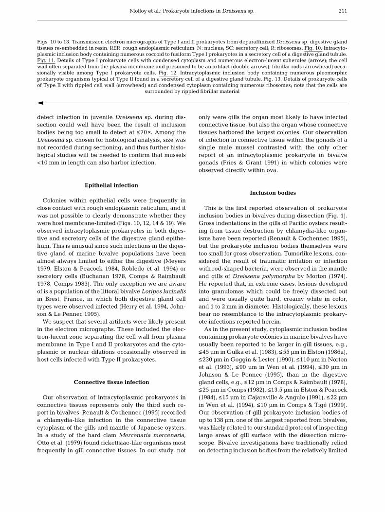
Type I. Typically hundreds of cells per inclusion body(Fig. 10); cells (Fig. 11) relatively small, measuring (n =49) 0.28 ± 0.09 × 0.13 ± 0.04 µm; coccoid to fusiform(possibly reflecting cross-sectional to longitudinal sec-tioning, respectively); cytoplasm condensed and finelygranular, with numerous electron-lucent spherules; cellwall separated from plasma membrane by electron-
lucent zone (presumed to be an artifact); cells occasion-ally surrounded by straight and curved fibrillar rods.
Type II. Typically hundreds of cells per inclusionbody (Fig. 12); cells (Fig. 13) pleomorphic, measuring(n = 49) 0.39 ± 0.06 × 0.19 ± 0.05 µm; highly rippled cellwall separated from plasma membrane by electron-lucent zone (presumed to be an artifact); cytoplasmcondensed, finely granular, and homogeneous (with-out electron-lucent spherules); numerous and denselystaining ribosomes at periphery; cytoplasmic or nuc-lear dilations occasionally observed in host cells (pre-sumed to be an artifact); cells surrounded by largequantities of rippled fibrillar material.
Type III. Typically dozens of cells per inclusion body(Fig. 14); cells (Fig. 15) pleomorphic, but often irregu-larly rounded; relatively large, measuring (n = 45)0.58 ± 0.09 × 0.42 ± 0.07 µm; cytoplasm filled withprominent ribosomes and irregular electron-lucentpatches likely corresponding to fine DNA strands (as inAnderson et al. 1965); plasma membrane in close prox-imity to cell wall, with no electron-lucent zone separat-ing them; cell wall flexible as evidenced by flatteningdue to crowding within vacuole.
Type IV. Typically dozens of cells per inclusion body(Fig. 16); cells (Fig. 17) large and irregularly rounded,measuring (n = 36) 1.05 ± 0.24 × 0.8 ± 0.15 µm; plasmamembrane in close proximity to cell wall, with no elec-tron-lucent zone separating them; cytoplasm homoge-neous and without electron-lucent areas; cell wall flex-ible as evidenced by flattening due to crowding withinvacuole; virus-like particles in cytoplasm, occasionallyvisible in dense paracrystalline array (Fig. 17); virus-like particles hexagonal (Fig. 18) with mean diameter(mean ±SD, n = 52) of 45 (±4) nm.
DISCUSSION
Evidence of more than one prokaryote species
The following observations, listed approximatelyin declining order of importance, provided evidencethat the Dreissena sp. population was infected with atleast 2 prokaryote species, one infecting epithelia andanother connective tissues:
207
Figs. 1 to 6. Light microscope observations of prokaryote inclusion bodies in either gill or digestive gland tissues of Dreissena sp.H&E: hematoxylin & eosin; SC: secretory cell; DC: digestive cell. Fig. 1. Numerous round-to-oval inclusion bodies (arrows) arevisible within water-mount of freshly dissected gill filaments. Fig. 2. Paraffin section (H&E) of gill; oval, basophilic inclusion body(arrow) in the connective tissue is surrounded by a clear zone (double arrows). Fig. 3. Paraffin section (H&E) of digestive tubuleshowing round, basophilic inclusion body (arrow) surrounded by a clear zone (double arrows) in a secretory cell. Fig. 4. Paraffinsection (H&E) of digestive tubule showing coarsely granular inclusion body (arrow). Fig. 5. Paraffin section (H&E) of heavilyinfected gill; the prokaryote inclusion bodies (black arrows) are surrounded by numerous infiltrated hemocytes (white arrows).Fig. 6. Paraffin section (H&E) of heavily infected gill; inclusion bodies (large arrows) in connective tissues partially constrict water
tubes (small arrows)

Dis Aquat Org 44: 203–216, 2001208
1 µm
0.05 µm0.1 µm
C
E
I
R
E
E
B
R
I
R
H
7
8 9

Molloy et al.: Prokaryote infections in Dreissena sp.
(1) Prokaryote cell morphologies observed in the 2types of tissues were substantially different.
(2) Based on previous studies, it would be very un-usual if the same prokaryote species was present inboth epithelial and connective tissues. A bivalve pop-ulation with presumably the same intracytoplasmicprokaryote species in both epithelial and connectivetissues has been reported only once before (Wu & Pan1999). Otto et al. (1979) did report intracytoplasmicinfection in both tissue types in Mercenaria merce-naria, but the epithelial and connective tissues exclu-sively harbored different prokaryote species (i.e.,chlamydial and rickettsial infections, respectively). Inother reports of intracytoplasmic prokaryotes in amarine bivalve population (minimum of 38 publica-tions, Fryer & Lannan 1994), either epithelial or con-nective tissues were reported infected, never both.
(3) There was no significant correlation between theoccurrence of infection in epithelial and connectivetissues (r = 0.28, p = 0.122, Spearman’s coefficient ofrank correlation). Of the 28 mussels observed fromhistological sections to be infected, 78.6% had pro-karyotes in only 1 tissue type (17.9% epithelial and60.7% connective). Moreover, when 1 tissue type wasintensely infected, the other sometimes exhibitedminimal to no infection; this was true for the 2 mostheavily infected mussels where little to no infectionwas observed in their connective tissues, yet >90 and>150 inclusions per section, respectively, were presentin their digestive gland epithelia.
(4) Following staining with hematoxylin and eosin, thefinely granular inclusion bodies were routinely more ba-sophilic in digestive gland epithelium than those in gillconnective tissue. Otto et al. (1979) also noted a subtledifference in basophilic staining of inclusion bodies be-tween 2 prokaryote species (rickettsial and chlamydial)observed in a population of the hard clam Mercenariamercenaria, but in their study the reverse was true, i.e.,inclusion bodies were slightly less basophilic in digestivegland epithelium than in gill connective tissue.
Taxonomic assignment of prokaryotes
Based on ultrastructural observations, the prokary-otes in gill connective tissue were clearly Chlamydi-ales-like (Storz & Spears 1977). Elementary bodies and
reticulate bodies were morphologically similar to theChlamydiales-like infections reported from marinebivalves (Harshbarger et al. 1977). In contrast, theprokaryotes routinely observed in the epithelial cellswere more difficult to assign taxonomically. Accurateclassification of Gram-negative, intracellular prokary-otes based on morphology observed by light and elec-tron microscopy can be difficult (Fryer & Lannan 1994),including these epithelial prokaryotes. Their cell pleo-morphism, trilaminar cell wall and plasmalemma, andinternal bacterium-like organization indicated thatthey were either in the order Rickettsiales or Chlamy-diales, but unfortunately there is no histological stainto unequivocally distinguish between these 2 pro-karyote orders. Thus, the results of the Giemsa (i.e.,colonies blue) and Ordway-Macchiavello (i.e., coloniesred) staining performed herein only confirmed that theprokaryotes might be in either order. Although 4 mor-phological types of cells were observed, a mixture ofcell types resembling electron-lucent reticulate bodiesand elementary bodies was never observed within anyinclusion body. By default, these prokaryotes must beconsidered Rickettsiales-like.
Infection prevalence
As would be expected due to the small size of cyto-plasmic inclusion bodies, overall prokaryote infectionintensity as determined from dissection data (17.0%)was substantially less than indicated by histologicalanalysis (87.5%). Considering that typically only 4sections per mussel were examined, it is highly likelythat even histological analysis underestimated trueinfection prevalence. A wide range of prevalenceswith intracytoplasmic prokaryotes occur in marinebivalves, including similar extensive infections, e.g.,ca 100% by both Gulka & Chang (1984) and Le Gallet al. (1991), >85% by Johnson & Le Pennec (1995),and ≤75% by Buchanan (1978).
During dissections, inclusion bodies were observedin the gills (Fig. 1) of 28.6% (42/147) of the mussels≥10 mm in length versus 0% (0/100) in smaller mussels(Table 1). This does not indicate that mussels <10 mmin length were uninfected. In their histological study,Norton et al. (1993) recorded smaller inclusions injuvenile than in adult giant clams. Thus, the inability to
209
Figs. 7 to 9. Transmission electron micrographs from deparaffinized Dreissena sp. gill tissues re-embedded in resin. C: cytoplasm;H: hemocyte; R: reticulate body; I: intermediate body; E: elementary body; B: bleb. Fig. 7. Spherical vacuole containing Chlamy-diales-like organisms in connective cell cytoplasm in the gill; hemocyte extending pseudopodium (double arrows) toward con-nective cell. Fig. 8. Details of reticulate and elementary bodies; note that the prokaryote inclusion is limited only by a thick layerof electron-dense fibrillar material (arrow). Fig. 9. Details of intermediate, reticulate, and elementary bodies; note the occurrence
of blebs which presumably originate from reticulate bodies

Dis Aquat Org 44: 203–216, 2001210
0.5 µm
0.5 µm
0.1 µm0.2 µm
RER
SC
N
R
bb
bb
RER
N
10
12
11 13
Molloy et al.: Prokaryote infections in Dreissena sp.
detect infection in juvenile Dreissena sp. during dis-section could well have been the result of inclusionbodies being too small to detect at ≤70×. Among theDreissena sp. chosen for histological analysis, size wasnot recorded during sectioning, and thus further histo-logical studies will be needed to confirm that mussels<10 mm in length can also harbor infection.
Epithelial infection
Colonies within epithelial cells were frequently inclose contact with rough endoplasmic reticulum, and itwas not possible to clearly demonstrate whether theywere host membrane-limited (Figs. 10, 12, 14 & 19). Weobserved intracytoplasmic prokaryotes in both diges-tive and secretory cells of the digestive gland epithe-lium. This is unusual since such infections in the diges-tive gland of marine bivalve populations have beenalmost always limited to either the digestive (Meyers1979, Elston & Peacock 1984, Robledo et al. 1994) orsecretory cells (Buchanan 1978, Comps & Raimbault1978, Comps 1983). The only exception we are awareof is a population of the littoral bivalve Loripes lucinalisin Brest, France, in which both digestive gland celltypes were observed infected (Herry et al. 1994, John-son & Le Pennec 1995).
We suspect that several artifacts were likely presentin the electron micrographs. These included the elec-tron-lucent zone separating the cell wall from plasmamembrane in Type I and II prokaryotes and the cyto-plasmic or nuclear dilations occasionally observed inhost cells infected with Type II prokaryotes.
Connective tissue infection
Our observation of intracytoplasmic prokaryotes inconnective tissues represents only the third such re-port in bivalves. Renault & Cochennec (1995) recordeda chlamydia-like infection in the connective tissuecytoplasm of the gills and mantle of Japanese oysters.In a study of the hard clam Mercenaria mercenaria,Otto et al. (1979) found rickettsiae-like organisms mostfrequently in gill connective tissues. In our study, not
only were gills the organ most likely to have infectedconnective tissue, but also the organ whose connectivetissues harbored the largest colonies. Our observationof infection in connective tissue within the gonads of asingle male mussel contrasted with the only otherreport of an intracytoplasmic prokaryote in bivalvegonads (Fries & Grant 1991) in which colonies wereobserved directly within ova.
Inclusion bodies
This is the first reported observation of prokaryoteinclusion bodies in bivalves during dissection (Fig. 1).Gross indentations in the gills of Pacific oysters result-ing from tissue destruction by chlamydia-like organ-isms have been reported (Renault & Cochennec 1995),but the prokaryote inclusion bodies themselves weretoo small for gross observation. Tumorlike lesions, con-sidered the result of traumatic irritation or infectionwith rod-shaped bacteria, were observed in the mantleand gills of Dreissena polymorpha by Morton (1974).He reported that, in extreme cases, lesions developedinto granulomas which could be freely dissected outand were usually quite hard, creamy white in color,and 1 to 2 mm in diameter. Histologically, these lesionsbear no resemblance to the intracytoplasmic prokary-ote infections reported herein.
As in the present study, cytoplasmic inclusion bodiescontaining prokaryote colonies in marine bivalves haveusually been reported to be larger in gill tissues, e.g.,≤45 µm in Gulka et al. (1983), ≤55 µm in Elston (1986a),≤230 µm in Goggin & Lester (1990), ≤110 µm in Nortonet al. (1993), ≤90 µm in Wen et al. (1994), ≤30 µm inJohnson & Le Pennec (1995), than in the digestivegland cells, e.g., ≤12 µm in Comps & Raimbault (1978),≤25 µm in Comps (1982), ≤13.5 µm in Elston & Peacock(1984), ≤15 µm in Cajaraville & Angulo (1991), ≤22 µmin Wen et al. (1994), ≤10 µm in Comps & Tigé (1999).Our observation of gill prokaryote inclusion bodies ofup to 138 µm, one of the largest reported from bivalves,was likely related to our standard protocol of inspectinglarge areas of gill surface with the dissection micro-scope. Bivalve investigations have traditionally reliedon detecting inclusion bodies from the relatively limited
211
Figs. 10 to 13. Transmission electron micrographs of Type I and II prokaryotes from deparaffinized Dreissena sp. digestive glandtissues re-embedded in resin. RER: rough endoplasmic reticulum; N: nucleus; SC: secretory cell; R: ribosomes. Fig. 10. Intracyto-plasmic inclusion body containing numerous coccoid to fusiform Type I prokaryotes in a secretory cell of a digestive gland tubule.Fig. 11. Details of Type I prokaryote cells with condensed cytoplasm and numerous electron-lucent spherules (arrow); the cellwall often separated from the plasma membrane and presumed to be an artifact (double arrows); fibrillar rods (arrowhead) occa-sionally visible among Type I prokaryote cells. Fig. 12. Intracytoplasmic inclusion body containing numerous pleomorphicprokaryote organisms typical of Type II found in a secretory cell of a digestive gland tubule. Fig. 13. Details of prokaryote cellsof Type II with rippled cell wall (arrowhead) and condensed cytoplasm containing numerous ribosomes; note that the cells are
surrounded by rippled fibrillar material

Dis Aquat Org 44: 203–216, 2001212
0.5 µm 0.5 µm
0.2 µm
0.2 µm1 µm
0.05 µm
N
N
DC
SC
R
RER14
15
17
18
1916
bb
bb
bb6
RER

Molloy et al.: Prokaryote infections in Dreissena sp.
material provided by serial sections. If we had relied onhistological material alone, we would have reported amaximum inclusion body size of only 75 µm.
The clear zones sometimes observed surroundinginclusion bodies (Figs. 2 & 3) were not likely related tohost-parasite interactions. We did not observe theseclear zones in our TEM observations, and thus theywere probably an artifact of histological processing.Similar clear zones have also been present in paraffinsections of prokaryote infections in marine bivalves(Comps et al. 1977, Buchanan 1978).
Virus-like particles
The presence of virus-like particles in electron-denseparacrystalline arrays within intracytoplasmic prokary-otes has been reported in marine bivalves, all from in-fections within digestive gland epithelium. Records ofsuch hyperparasitism include both Rickettsiales-like(Buchanan 1978, Wen et al. 1994, Comps & Tigé 1999)and Chlamydiales-like organisms (Harshbarger et al.1977, Meyers 1979, Johnson & Le Pennec 1995). Thesereports also noted that prokaryote cells containing suchvirus-like particles, like our Type IV cells, were largerthan uninfected cells and were likely a terminal phasein the prokaryote’s life cycle, with host cells eventuallylysing. Mean diameters of these virus-like particles inbivalves have ranged from 41 nm (Wen et al. 1994) to67 nm (Buchanan 1978), and thus the virus-like parti-cles we observed (mean diameter, 45 mm) were rela-tively small and similar in size to those observed byComps & Tigé (1999) in Mytilus galloprovincialis. Eachparticle had a hexagonal outline (Fig. 18) similar tovirus-like particles within prokaryotes in the intertidalbivalve Tellina tenuis (Buchanan 1978).
Host pathology
Infection was fairly benign in Dreissena sp. Althoughindividual infected epithelial cells appeared to suffercytopathic effects in the digestive gland, the epithe-
lium in general did not appear damaged, even wheninfection intensity was high. The cycle of shedding andregeneration of digestive gland epithelium in bivalveslikely serves to minimize the impact of such prokaryoteinfections. Although it is the likely course of events innature, there was no direct evidence of the breakdownof host cell membranes and release of prokaryotes asobserved for other bivalve intracytoplasmic prokary-otes in cells within the gill (Fries & Grant 1991, Villalbaet al. 1999) and digestive gland (Meyers 1979, Herry etal. 1994, Wen et al. 1994). Although infection preva-lence was very high (ca ≥87.5%), no major, adverseimpact on body condition was evident among the32 Dreissena sp. sectioned or 247 dissected. Host re-sponse was clear, however, from dense hemocyte infil-tration into infected tissues (Fig. 5). The most apparentadverse effect of infection was partial constriction ofgill water tubes (Fig. 6). At the densities of inclusionbodies observed, however, significant interferencewith respiration and other metabolic functions of thegills was highly unlikely. A similar conclusion wasreached in a study of intracytoplasmic prokaryoteinfection in gills of a marine bivalve (Renault &Cochennec 1994). In the only report of intranuclearinfection by prokaryote-like organisms in bivalves,Elston (1986b) observed occlusion of gill water tubesin the Pacific razor clam Siliqua patula in associationwith massive mortalities. In heavy infections, tubeswere blocked by the release of nuclei infected withthese microorganisms as well as by epithelial cellhypertrophy.
Other than mild cytopathic effects, marine bivalveinvestigations, however, have typically recorded nosignificant, detrimental effects on hosts from intra-cytoplasmic prokaryote infections. Although, intra-cytoplasmic prokaryote infections have occasionallybeen associated with mass mortalities of marine bi-valves in Europe (Le Gall et al. 1991, Villalba et al.1999), North America (Gulka et al. 1983), and Asia (Wu& Pan 1999), studies to date (Gulka & Chang 1984)have yet to definitively demonstrate that the prokary-otes per se were the primary lethal etiological agents.Norton et al. (1993), for example, suggested that al-
213
Figs. 14 to 19. Transmission electron micrographs of Type III and IV prokaryotes from deparaffinized Dreissena sp. digestivetissues re-embedded in resin. RER: rough endoplasmic reticulum; N: nucleus; R: ribosomes; SC: secretory cell; DC: digestive cell.Fig. 14. Intracytoplasmic inclusion body containing irregularly rounded prokaryote organisms typical of Type III found in a secre-tory cell of a digestive gland tubule. Fig. 15. Higher magnification of large prokaryote cells of Type III; the cytoplasm of Type IIIprokaryotes contains ribosomes and electron-lucent patches corresponding to DNA strands (arrows). Fig. 16. Intracytoplasmicinclusion body containing large rounded prokaryote organisms typical of Type IV found in a secretory cell of a digestive glandtubule. Fig. 17. Details of large Type IV prokaryote cells containing virus-like particles occasionally occurring in paracrystallinearray (arrowheads). Fig. 18. Higher magnification of hexagonal virus-like particles in paracrystalline array each with a clearcentral zone occurring in the cytoplasm of Type IV prokaryote cells. Fig. 19. High magnification of the peripheral zone of aninclusion containing Type IV prokaryote cells (arrowhead) and their virus-like particles; note the single tubule of rough endo-plasmic reticulum in the cytoplasm of this digestive cell and that no membrane apparently separates it from the prokaryote cells

Dis Aquat Org 44: 203–216, 2001
though Rickettsiales-like organisms were common ingill epithelium of dying giant clams Hippopus hippo-pus, stress from rearing conditions may have beenlargely responsible for the mortalities. Stress due toconcurrent trematode infection was also considered tobe the most probable cause for expression of a Rick-ettsiales-like infection in a freshwater snail (Adam etal. 1994). Although Elston (1986b) presented evidenceof a link between intranuclear prokaryote-like infec-tion and razor clam mortalities, he indicated that hismorphological study could not be used to conclusivelyestablish cause and effect. One of the most convincingrecords of bivalve mortality directly due to intracyto-plasmic prokaryotes was a Chlamydiales infection inhatchery-reared larval (ca 80 to 100% mortality) andpostmetamorphic (ca 40 to 60% mortality) bay scallopsin which infection was associated with the completedestruction of their digestive tract epithelial lining;the prevalence and importance of this disease for wildlarval and postmetamorphic stocks of scallops, how-ever, remains unknown (Leibovitz 1989).
Although there may be no definitive, experimentalevidence that prokaryote infections in bivalves caninduce lethality, the symbiotic relationship of theseintracytoplasmic prokaryotes to their hosts, includingDreissena, is still generally considered parasitic. Insupport of this, Le Gall et al. (1991) provided evidenceof energetic loss due to rickettsial infection in Pectenmaximus. In their study of the effect of a Chlamydia-like prokaryote in Crassostrea gigas, Soletchnik et al.(1998) demonstrated that clearance rates were posi-tively correlated with gill abnormalities and suggestedthat this compensated for the loss in gill surface func-tion. In contrast, Johnson & Le Pennec (1995) havesuggested that Chlamydiales-like prokaryotes in thelittoral clam Loripes lucinalis could be a beneficialsymbiont. They speculated that host clams might sys-temically benefit by obtaining some essential nutrientssuch as fatty acids or vitamins via prokaryote colonydigestion, and the specialized gill bacteriocyte cells ofthese clams might directly benefit by phagocytosis ofprokaryotes.
Research imbalance
This is the first report of intracytoplasmic prokaryoteorganisms from a freshwater bivalve. In contrast, atleast 39 primary research papers (see Fryer & Lannan1994) have been published over the last 20 years re-porting on intracytoplasmic prokaryote infections in atleast 25 marine bivalve species (e.g., oysters, scallops,clams, and mussels). This freshwater versus marinepublication imbalance is almost certainly the result ofthe relative lack of parasitological studies of fresh-
water bivalves—a group of no major commercial im-portance. Bivalve pathology has traditionally been anapplied science funded in large part to understand andminimize diseases of commercially valuable species.The present paper actually follows in this tradition, butwith a twist. The fundamental research reported here-in emerged out of a need to understand and possiblymaximize diseases of a commercially harmful species.
Future research
Could intracytoplasmic prokaryotes in Dreissenaand other molluscs be of wider ecological importance?Currently, the life cycles of none of the intracytoplas-mic prokaryotes in marine bivalves have been clearlyelucidated. Do bivalves serve as reservoir hosts incomplex prokaryote life histories or are these diseasesexclusively cycling in bivalve hosts? Intracytoplasmicprokaryotes in vertebrates are typically known to havecomplex life cycles involving obligate, alternate hosts.The question of what role freshwater bivalves mayplay as alternate hosts or reservoirs for the spread ofvertebrate rickettsial or chlamydial diseases was firstraised by Harshbarger et al. (1977). Buchanan (1978)did have limited success in culturing an intracellularprokaryote from a marine intertidal bivalve in hens’eggs, and this provided some evidence that suchprokaryotes might truly have an alternate host in birds,but no other similar studies have been reported since.The recent detection of the rickettsial species Ehrlichiaristicii, the agent of Potomac horse fever, in freshwaterstream snails near pastures where this equine diseasewas enzootic (Barlough et al. 1998, Reubel et al. 1998)may rekindle interest in examining bivalve-vertebratetransmission theories. It appears, however, that someprokaryotes can cycle for at least part of their life his-tory in their bivalve hosts since horizontal transinfec-tion of Rickettsiales infection in scallops has been suc-cessfully achieved in the laboratory (Gulka & Chang1984, Le Gall et al. 1991). Such intraspecific, horizon-tal, transinfection trials are thus a logical next step withDreissena’s prokaryotes. If successful, then transinfec-tion could provide a continuous laboratory supply ofprokaryotes for determining other key research goalssuch as taxonomic clarification, life cycle, host speci-ficity, and life stage susceptibility. Because intracyto-plasmic prokaryotes infecting bay scallops are particu-larly pathogenic to their larval and postmetamorphicstages (Leibovitz 1989), transmission trials focusing onDreissena’s larval and juvenile stages could reveal alevel of pathogenicity not evident in the current study.
In such future research efforts, techniques alreadydeveloped in marine studies could be useful. The pro-tocols of Le Gall & Mialhe (1992) could be adapted for
214

Molloy et al.: Prokaryote infections in Dreissena sp.
purifying prokaryotes from infected zebra mussel tis-sues. A highly sensitive and specific diagnostic DNA-based probe (Kellner-Cousin et al. 1993) could serveas a practical tool for detection and quantification ofDreissena’s prokaryotes. Efforts to clarify the taxo-nomic position of Dreissena’s prokaryotes could spe-cifically make use of techniques such as antigenicstructure determination (Renault & Cochennec 1995),protein electrophoresis (Le Gall & Mialhe 1992), andanalysis of genomic content (e.g., 16S, internal tran-scribed spacer, and 23S ribosomal DNA sequencing,Mauel et al. 1999).
Acknowledgements. This research was funded by grantsfrom the U.S. Army Engineers Waterways Experiment StationZebra Mussel Research Program (to D.P.M.) and the NationalScience Foundation Division of International Programs (toRobert E. Baier and D.P.M.). We especially thank EstelleLabeyrie, Marie-Anne Pruniéres, and Jean-Robert Bonami fortheir help in setting up the temporary laboratory in Seillans(France), Marilena Zarfdjian for fieldwork, Tristan Renaultand John Fryer for manuscript review, and Robert Lutringerfor statistical advice. The assistance of the following IRCOMScolleagues during mussel dissection is gratefully acknowl-edged: Lyubov Burlakova, Alexander Karatayev, Dina Kuran-dina, Peter Mitrakhovich, Mykola Ovcharenko, Victor Pros-tokvashin, and Vitali Roitman. Preparation of the manuscriptwas supported in part by travel grants from the Université deMetz (to D.P.M. and L.G.). Many thanks as always to the staffof the New York State Library for generously assisting inobtaining scientific literature.
LITERATURE CITED
Adam R, Pipitgool V, Sithithaworn P, Hinz E, Storch V (1994)Rickettsiales-like organisms in the digestive gland ofBithynia siamensis goniomphalus (Prosobranchia: Bithyni-idae) infected with Opisthorchis viverrini (Trematoda:Digenea). J Invertebr Pathol 63:26–30
Anderson DR, Hopps HE, Barile MF, Bernheim BC (1965)Comparison of the ultrastructure of several rickettsiae,ornithosis virus and Mycoplasma in tissue culture. J Bac-teriol 90:1387–1404
Barlough JE, Reubel GH, Madigan JE, Vredevoe LA, MillerPE, Rikihisa Y (1998) Detection of Ehrlichia risticii, theagent of Potomac horse fever, in freshwater stream snails(Pleuroceridae: Juga spp.) from northern California. ApplEnviron Microbiol 64:2888–2893
Bower SM, McGladdery SE, Price IM (1994) Synopsis of infec-tious diseases and parasites of commercially exploitedshellfish. Annu Rev Fish Dis 4:1–199
Buchanan JS (1978) Cytological studies on a new species ofrickettsia found in association with a phage in the diges-tive gland of the marine bivalve mollusc, Tellina tenuis(da Costa). J Fish Dis 1:27–43
Burlakova LE, Karatayev AY, Molloy DP (1998) Field and lab-oratory studies of zebra mussel (Dreissena polymorpha)infection by the ciliate Conchophthirus acuminatus in theRepublic of Belarus. J Invertebr Pathol 71:251–257
Cajaraville MP, Angulo E (1991) Chlamydia-like organisms indigestive and duct cells of mussels from the Basque coast.J Invertebr Pathol 58:381–386
Camp JW, Blaney LM, Barnes DK (1999) Helminths of theround goby, Neogobius melanostomus (Perciformes: Gobi-idae), from southern Lake Michigan, Indiana. J Hel-minthol Soc Wash 66:70–72
Comps M (1982) Etude morphologique d’une infection rick-ettsienne de la palourde Ruditapes philippinarum Adamand Reeves. Rev Trav Inst Pêches Marit 44(1983):277–283
Comps M (1983) Infections rickettsiennes chez les mollusquesbivalves des côtes françaises. Rapp P V Reun Cons IntExplor Mer 182:134–136
Comps M, Raimbault R (1978) Infection rickettsienne de laglande digestive de Donax trunculus Linné. Sci Peche BullInst Pêches Marit 281:11–12
Comps M, Tigé G (1999) Procaryotic infections in the musselMytilus galloprovincialis and in its parasite the turbellar-ian Urastoma cyprinae. Dis Aquat Org 38:211–217
Comps M, Bonami JR, Vago C (1977) Mise en évidence d’uneinfection rickettsienne chez les huîtres. C R Hebd SeancesAcad Sci Ser D Sci Nat 285:427–429
Elston RA (1986a) Occurrence of branchial rickettsiales-likeinfection in two bivalve molluscs, Tapes japonica andPatinopecten yessoensis, with comments on their signifi-cance. J Fish Dis 9:69–71
Elston RA (1986b) An intranuclear pathogen [nuclear inclu-sion X (NIX)] associated with massive mortalities of thePacific razor clam, Siliqua patula. J Invertebr Pathol 47:93–104
Elston RA, Peacock MG (1984) A rickettsiales-like infection inthe Pacific razor clam, Siliqua patula. J Invertebr Pathol44:84–96
Fahnenstiel GL, Bridgeman TB, Lang GA, McCormick MJ,Nalepa TF (1995) Phytoplankton productivity in SaginawBay, Lake Huron: effects of zebra mussel (Dreissena poly-morpha) colonization. J Gt Lakes Res 21:465–475
Fries CR, Grant DM (1991) Rickettsiae in gill epithelial cells ofthe hard clam, Mercenaria mercenaria. J Invertebr Pathol57:166–171
Fryer JL, Lannan CN (1994) Rickettsial and chlamydial infec-tions of freshwater and marine fishes, bivalves, and crus-taceans. Zool Stud 33:95–107
Goggin GL, Lester RJG (1990) Rickettsiales-like infection inthe gills of Tridacna crocea from the Great Barrier Reef.J Invertebr Pathol 56:135–138
Gulka G, Chang PW (1984) Pathogenicity and infectivity ofa rickettsia-like organism in the sea scallop, Placopectenmagellanicus. J Fish Dis 8:309–318
Gulka G, Chang PW, Marti KA (1983) Prokaryotic infectionassociated with a mass mortality of the sea scallop Placo-pecten magellanicus. J Fish Dis 6:355–364
Harshbarger JC, Chang SC, Otto SV (1977) Chlamydiae (withphages), mycoplasmas, and rickettsiae in Chesapeake Baybivalves. Science (Wash DC) 196:666–668
Herry A, Le Pennec M, Johnson M (1994) Bacteria-host rela-tionships in the bivalve mollusc Loripes lucinalis. ActaMicrobiol Immunol Hung 41:273–281
Humason GL (1979) Animal tissue techniques, 4th edn. WHFreeman and Co, San Francisco
Johnson MA, Le Pennec M (1995) Association between themollusc bivalve Loripes lucinalis and a Chlamydia-likeorganism, with comments on its pathogenic impact, lifecycle and possible mode of transmission. Mar Biol 123:523–530
Karatayev AY, Burlakova LE, Padilla DK (1997) The effectsof Dreissena polymorpha (Pallas) invasion on aquaticcommunities in eastern Europe. J Shellfish Res 16:187–203
Karatayev AY, Burlakova LE, Molloy DP, Volkova LK (2000a)
215

Dis Aquat Org 44: 203–216, 2001
Endosymbionts of Dreissena polymorpha (Pallas) in Be-larus. Int J Hydrobiol 85:543–559
Karatayev AY, Molloy DP, Burlakova LE (2000b) Seasonaldynamics of Conchophthirus acuminatus (Ciliophora,Conchophthiridae) infection in Dreissena polymorpha andD. bugensis (Bivalvia, Dreissenidae). Eur J Protistol (inpress)
Kellner-Cousin K, Le Gall G, Despres B, Kaghad M, LegouxP, Shire D, Mialhe E (1993) Genomic DNA cloning ofrickettsia-like organisms (RLO) of Saint-Jacques scallopPecten maximus: evaluation of prokaryote diagnosis byhybridization with a non-isotopically labelled probe andby polymerase chain reaction. Dis Aquat Org 15:145–152
Laruelle F, Molloy DP, Fokin SI, Ovcharenko MA (1999) His-tological analysis of mantle-cavity ciliates in Dreissenapolymorpha: their location, symbiotic relationship, anddistinguishing morphological characteristics. J ShellfishRes 18:251–257
Laruelle F, Molloy DP, Roitman VA (2001) Histological analy-sis of trematodes in Dreissena polymorpha: their location,pathogenicity, and distinguishing morphological charac-teristics. J Parasitol (in press)
Le Gall G, Mialhe E (1992) Purification of Rickettsiales-likeorganisms associated with Pecten maximus (Mollusca:Bivalvia): serological and biochemical characterization.Dis Aquat Org 12:215–220
Le Gall G, Mialhe E, Chagot D, Grizel H (1991) Epizootio-logical study of rickettsiosis of the Saint Jacques scallopPecten maximum. Dis Aquat Org 10:139–145
Leibovitz L (1989) Chlamydiosis: a newly reported seriousdisease of larval and postmetamorphic bay scallops,Argopecten irradians (Lamarck). J Fish Dis 12:125–136
Mauel MJ, Giovannoni SJ, Fryer JL (1999) Phylogeneticanalysis of Piscirickettsia salmonis by 16S, internal tran-scribed spacer (ITS) and 23S ribosomal DNA sequencing.Dis Aquat Org 35:115–123
Meyers TR (1979) Preliminary studies on a chlamydial agentin the digestive diverticular epithelium of hard clams Mer-cenaria mercenaria (L.) from Great South Bay, New York.J Fish Dis 2:179–189
Meyers TR (1981) Endemic diseases of cultured shellfish ofLong Island, New York: adult and juvenile American oys-ters (Crassostrea virginica) and hard clams (Mercenariamercenaria). Aquaculture 22:305–330
Molloy DP (1998) The potential for using biological controltechnologies in the management of Dreissena spp. J Shell-fish Res 17:177–183
Molloy DP, Roitman VA, Shields JD (1996) Survey of theparasites of zebra mussels (Bivalvia: Dreissenidae) innorthwestern Russia, with comments on records of para-sitism in Europe and North America. J Helminthol SocWash 63:251–256
Molloy DP, Karatayev AY, Burlakova LE, Kurandina DP,Laruelle F (1997) Natural enemies of zebra mussels: pre-dators, parasites, and ecological competitors. Rev Fish Sci5:27–97
Morton B (1974) Studies on the biology of Dreissena polymor-pha. VI. The occurrence of chronic pallial and ctenidialinflammatory granulomas—the response to injury. J In-vertebr Pathol 23:106–113
Norton JH, Shepherd MA, Abdon-Naguit MR, Lindsay S(1993) Mortalities in the giant clam Hippopus hippopus
associated with Rickettsiales-like organisms. J InvertebrPathol 62:207–209
O’Neill CR Jr (1996) The zebra mussel: impacts and control.Cornell Coop Ext Inf Bull 238:1–62
O’Neill CR Jr (1997) Economic impact of zebra mussels—results of the 1995 National Zebra Mussel InformationClearinghouse study. Gt Lakes Res Rev 3:35–44
Otto SV, Harshbarger JC, Chang SC (1979) Status of selectedunicellular eucaryote pathogens, and prevalence and histo-pathology of inclusions containing obligate procaryoteparasites, in commercial bivalve mollusks from Marylandestuaries. Haliotis 8(1977):285–295
Renault T, Cochennec N (1994) Rickettsia-like organisms inthe cytoplasm of gill epithelial cells of the Pacific oysterCrassostrea gigas. J Invertebr Pathol 64:160–162
Renault T, Cochennec N (1995) Chlamydia-like organisms inctenidia and mantle cells of the Japanese oyster Crasso-strea gigas from the French Atlantic coast. Dis Aquat Org23:153–159
Reubel GH, Barlough JE, Madigan JE (1998) Production andcharacterization of Ehrlichia risticii, the agent of Potomachorse fever, from snails (Pleuroceridae: Juga spp.) inaquarium culture and genetic comparison to equinestrains. J Clin Microbiol 36:1501–1511
Robledo JAF, Santarém MM, Figueras A (1994) Parasite loadsof rafted blue mussels (Mytilus galloprovincialis) in Spainwith special reference to the copepod, Mytilicola intesti-nalis. Aquaculture 127:287–302
Schloesser DW, Nalepa TF, Mackie GL (1996) Zebra musselinfestation of unionid bivalves (Unionidae) in North Amer-ica. Am Zool 36:300–310
Soletchnik P, Goulletquer P, Cochennec N, Renault T, Gea-iron P (1998) Ecophysiological study of the Pacific oysterCrassostrea gigas naturally infected by a Chlamydia-likemicroorganism: effect of infection level and diet on oysterphysiological responses. Haliotis 27:1–19
Stewart TW, Miner JG, Lowe, RL (1998) Quantifying mecha-nisms for zebra mussel effects on benthic macroinver-tebrates: organic matter production and shell-generatedhabitat. J N Am Benthol Soc 17:81–94
Storz J, Spears P (1977) Chlamydiales: properties, cycle ofdevelopment and effect on eukaryotic host cells. Curr TopMicrobiol Immunol 76:167–214
Strayer DL (1999) Effects of alien species on freshwater mol-lusks in North America. J N Am Benthol Soc 18:74–98
Toews S, Beverly-Burton M, Lawrimore T (1993) Helminthand protist parasites of zebra mussels, Dreissena polymor-pha (Pallas, 1771), in the Great Lakes region of southwest-ern Ontario, with comments on associated bacteria. CanJ Zool 71:1763–1766
Villalba A, Carballal MJ, Lopez C, Cabada A, Corral L,Azevedo C (1999) Branchial rickettsia-like infection asso-ciated with clam Venerupis rhomboides mortality. DisAquat Org 36:53–60
Wen CM, Kou GH, Chen SN (1994) Rickettsiaceae-likemicroorganisms in the gill and digestive gland of the hardclam, Meretrix lusoria Röding. J Invertebr Pathol 64:138–142
Wu X, Pan J (1999) Studies on rickettsia-like organism dis-ease of the tropical marine pearl oyster. I. The fine struc-ture and morphogenesis of Pinctada maxima pathogenrickettsia-like organism. J Invertebr Pathol 73:162–172
216
Editorial responsibility: Albert Sparks, Seattle, Washington, USA
Submitted: May 16, 2000; Accepted: November 3, 2000Proofs received from author(s): March 13, 2001