characterisation of rhizoctonia in cropping systems in the western
TRANSCRIPT
1
1. CHARACTERISATION OF RHIZOCTONIA ASSOCIATED WITH CEREALS,
CANOLA, PASTURE AND GRAIN LEGUMES WITH SPECIAL REFERENCE TO
THE EFFECT OF CROP ROTATION ON RHIZOCTONIA DISEASES
INTRODUCTION
The genus Rhizoctonia comprises a collective species complex that includes fungi that
do not have any distinctive taxonomic features allowing their classification into any known
fungal genus (Ogoshi, 1987; Sneh, Burpee & Ogoshi, 1991; Carling & Sumner, 1992).
Therefore, Rhizoctonia represents a diverse group of fungi that differs in many significant
features, including their sexual stages (teleomorph), asexual stages (anamorph), and
morphology (Vilgalys & Cubeta, 1994). The diverse ecological niches occupied by
Rhizoctonia spp. further exemplify the diversity present in this group. A considerable number
of Rhizoctonia isolates are saprophytes, whereas others are mycorrhizal on orchids (Carling et
al., 1999) or other plants. The genus Rhizoctonia also contains significant soilborne plant
pathogens, pathogens of flooded crops and pathogens of aerial parts of plants (Sneh et al.,
1991).
The important role of many Rhizoctonia spp. as plant pathogens and mychorrhizas, has
led to the development of several useful methods for identifying specific groups within this
diverse genus. The most widely used methods for subdividing Rhizoctonia into groups
include identification of their nuclear status (multinucleate or binucleate) and anastomosis
group (Rovira, Ogoshi & McDonald, 1986; Ogoshi, 1987; Ogoshi, Cook & Bassett, 1990).
Anastomosis groupings are classically based on the characterisation of hyphal anastomosis
reactions of cultures. However, deoxyribonucleic acid (DNA)-based methods are increasingly
being used to identify anastomosis groups (AGs) (Vilgalys & Gonzalez, 1990; Cubeta et al.,
1991; Carling, Kuninaga & Brainard, 2002b). Some anastomosis groups are further divided
into subgroups based on pathogenicity testing (Roberts & Sivasithamparam, 1986; Ogoshi,

2
1987), isozyme analysis (Neate & Warcup, 1985; Neate, Cruickshank & Rovira, 1988),
cellular fatty acid composition (Stevens-Johnk & Jones, 1993; Priyatmojo et al., 2001a),
antibody reactions (Thornton et al., 2004), internal transcribed spacer sequence variation
(Carling et al., 2002b) and morphological characteristics (Sneh et al., 1991; Carling &
Sumner, 1992). Identification of Rhizoctonia isolates to some taxonomic level is of utmost
importance for studying their epidemiology and control in different cropping systems.
Rhizoctonia diseases can cause severe economic losses and therefore require effective
control measures (Vilgalys & Cubeta, 1994; Sneh et al., 1996). Various control methods have
been investigated including crop rotation (Rovira, 1986; Yang, Kharabanda & McAndrew,
1995), tillage practices (Jarvis & Brennan, 1986; Rovira, 1986; Yang et al., 1995), resistant
cultivars (Yang & Verma, 1992), as well as biological (Fiddaman & Rossall, 1995) and
chemical control (Kataria & Verma, 1990; Kataria, Verma & Gisi, 1991). Among these, crop
rotation and tillage practices have been the subject of many studies worldwide due to their
potential to either control or enhance disease symptoms incited by Rhizoctonia (MacNish,
1985b; Rovira, 1986; Roget, Neate & Rovira, 1996). Crop rotation is widely used in different
parts of the world, since it not only controls weeds and diseases including those caused by
Rhizoctonia, but also improves soil fertility (Leach & Clapham, 1992; Wessels, 2001).
Conservation tillage practices (sowing of crops with reduced tillage, no-tillage or direct
drilling) are widely promoted due to the advantages in reduced soil preparation costs,
increased availability of annual pastures for grazing, reduction in wind erosion and greater
flexibility in farm management (Rovira, 1986; Boer et al., 1991). However, several studies
have shown that conservation tillage practices can exacerbate weed problems and increase the
severity of diseases caused by various soilborne pathogens including Rhizoctonia (MacNish,
1985b; Rovira, 1986; Roget et al., 1996).
In South Africa, particularly in the winter rainfall region of the Western Cape
province, crop-pasture rotations have become a relatively stable pattern of land use since the
1970’s (Wessels, 2001). The most important rotation crops in this region include canola

3
(Brassica napus L. var. oleifera DC), medic (annual Medicago spp.), lucerne or alfalfa
(Medicago sativa L.), clover (Trifolium spp.) and lupin (Lupinus spp.). In these rotation
systems, wheat (Triticum aestivum L.) and barley (Hordeum vulgare L.) are often grown in
rotation with grain and pasture legumes and/or canola (G. A. Agenbag, pers. comm.). The
break in cereal/crop production in successive years, by planting a pasture legume, helps to
increase the organic nitrogen content of the soil because lucerne, medic and clover have the
capability to fix atmospheric nitrogen through the activity of associated Rhizobia (Ladd,
Oades & Amato, 1981). The crop-pasture rotations in this region have also been shown to
limit the incidence of weeds and diseases that accumulate in a monoculture (Beyers, 2001;
Wessels, 2001). Another important crop practice that is becoming more widely used in the
cereal producing areas of the Western Cape province is conservation (minimum and no-till)
tillage (G. A. Agenbag, pers. comm.).
Worldwide, Rhizoctonia spp. have been implicated as soilborne pathogens of all the
crops (barley, canola, clover, lucerne, lupin, medics, and wheat) used in cropping systems in
the Western Cape province (Samuel & Garret, 1932; Bretag, 1985; Kaminski & Verma, 1985;
Weller et al., 1986; Pumphrey et al., 1987; Kronland & Stanghellini, 1988; Kataria & Verma,
1992; Vincelli & Herr, 1992; MacLeod & Sweetingham, 1997). Therefore, Rhizoctonia has
the potential to limit crop production within these cropping systems. Although, the
pathogenicity of South African isolates of Rhizoctonia have not been shown on the
aforementioned crops, results from many surveys, as well as investigations conducted since
1999 in the Western Cape province, have clearly shown that Rhizoctonia spp. are associated
with all the crops of interest (barley, canola, clover, lucerne, lupin, medics and wheat)
(Lamprecht, De Villers & Janse van Rensburg, 1999; Lamprecht, Auret & Janse van Rensburg
2000a; Lamprecht et al., 2000b, 2001, 2002; Auret, Janse van Rensburg & Lamprecht, 2002).
Therefore, the aims of this thesis are to (1) characterise Rhizoctonia isolates associated with
seven rotation crops used within a crop rotation trial over a four-year period in the Western
Cape province, (2) determine the appropriate time during the growth season for isolating
Rhizoctonia from rotation crops and (3) determine the pathogenicity and relative virulence of

4
Rhizoctonia AGs on seven rotation crops. Basic knowledge of the Rhizoctonia AGs present in
the crop-pasture rotation systems is essential for developing sound control measures within
crop rotation systems of the Western Cape province.
CHARACTERISATION OF RHIZOCTONIA ISOLATES
Higher taxonomic diversity in Rhizoctonia
Traditionally fungi have been classified into the genus Rhizoctonia based on the
absence of other distinctive taxonomic features. In the genus Rhizoctonia taxonomists have
found it challenging to define species using conventional taxonomic criteria due to high levels
of phenotypic variation and inability to fruit most strains of Rhizoctonia in culture (Vilgalys &
Cubeta, 1994). Consequently, Rhizoctonia isolates can belong to several different orders of
mostly basidiomycetes (Carling & Sumner, 1992). The teleomorphs of fungi that have the
mycelial characteristics of Rhizoctonia isolates are assigned to the phylum Basidiomycota,
class Basidiomycetes and order Ceratobasidiales (Warcup & Talbot, 1966; Murray & Burpee,
1984; Alexopoulos, Mims & Blackwell, 1996). Within the class Hymenomycetes the most
important plant pathogenic Rhizoctonia isolates can be classified into three major
teleomorphic basidiomycete genera, i.e. the Rhizoctonia solani Kühn complex that includes
multinucleate species with a Thanatephorus (Frank) Donk teleomorph, binucleate Rhizoctonia
spp. with a Ceratobasidium Rogers teleomorph, and multinucleate Rhizoctonia zeae Voorhees
and Rhizoctonia oryzae Ryker & Gooch groups with a Waitea Warcup and Talbot teleomorph
(Carling & Sumner, 1992).
Nuclear determination
Rhizoctonia isolates can be divided into binucleate and multinucleate groups based on
the numbers of nuclei per cell of young vegetative hyphae (Yang et al., 1994a). The best

5
known multinucleate plant pathogen is R. solani of which the teleomorph is Thanatephorus
cucumeris (Frank) Donk (Spencer & Fox, 1978). For a fungus to be called R. solani it must
possess a T. cucumeris perfect state. The second group of Rhizocotonia isolates that are
characterised by multinucleate cells are R. zeae, R. oryzae and a type culture Waitea circinata
var. circinata (Warcup & Talbot) isolated by Warcup and Talbot (1962), that all have
teleomorphs in the genus Waitea (Warcup & Talbot, 1962; Carling & Sumner, 1992). The
anamorphic name of W. c .var circinata has not yet been assigned, but the teleomorphic name
may be useful, although the teleomorph is rarely observed (Carling & Sumner, 1992; Leiner &
Carling, 1994). Rhizoctonia isolates that are characterised by binucleate cells have a
Ceratobasidium teleomorph (Spencer & Fox, 1978; Carling & Sumner, 1992).
Anastomosis grouping
Conventional methods. Anastomosis groupings that are based on hyphal anastomosis
among isolates having common biological affinities have greatly facilitated the identification
of R. solani and other Rhizoctonia spp. (Parmeter, Whitney & Platt, 1967; Parmeter, Sherwood
& Platt, 1969). Conventionally, anastomosis groups are determined by observing whether a
hyphal fusion reaction is present between an unknown isolate and a reference isolate of known
AG. Anastomosis reactions between hyphae of confronted isolates are assigned to one of four
categories, i.e. C0, C1, C2 and C3. A C0 reaction indicates no hyphal fusion and a C3
reaction indicates a self (clonal) anastomosis reaction (Carling et al., 2002b). Pairing of
isolates belonging to the same AG results in hyphal fusion (anastomosis), leading to either
acceptance (self-pairing) or rejection (somatic incompatibility). Contrarily, pairings between
AGs that do not result in hyphal fusion (C0 reaction) suggest greater genetic differences
between isolates (i.e., different species, etc.) (Cubeta & Vilgalys, 1997). Unfortunately,
determination of relationships based solely on anastomosis behaviour of individual isolates is
often uncertain since Rhizoctonia can exhibit different types of hyphal fusion within the same
AG (Gonzalez et al., 2001). Therefore, molecular-based methods have also been developed to
identify and determine diversities within and between AGs (Cubeta & Vilgalys, 1997).
6
DNA-based molecular analyses of anastomosis groups and genetic variation in
Rhizoctonia. DNA-based molecular analyses have elucidated some of the genetic and
taxonomic relatedness of Rhizoctonia isolates, allowing a better understanding of the
Rhizoctonia species complex, which is still elusive (Gonzalez et al., 2001). Initially
DNA/DNA hybridization studies were used to determine the relatedness between different
Rhizoctonia isolates. Restriction fragment length polymorphisms (RFLP) analysis of genomic
DNA in conjunction with Southern blot analyses were also used to reveal genetic differences
between Rhizoctonia isolates (Vilgalys & Cubeta, 1994). Subsequently, polymerase chain
reaction (PCR)-based methods [PCR-RFLP, PCR- amplified fragment length polymorphism
(AFLP) and random amplified polymorphic DNA (RAPD)] were developed that are less
tedious and also require smaller amounts of DNA (Vigalys & Cubeta, 1994; Ceresini et al.,
2002; Godoy-Lutz et al., 2003). Although molecular studies have greatly advanced our
understanding of the genetic structure and variability in Rhizoctonia populations, a great deal
still needs to be learned about variation in Rhizoctonia (Neate & Warcup, 1985; Neate et al.,
1988; Pascual et al., 2000).
The most informative DNA-based molecular technique for investigating diversity in
Rhizoctonia isolates has been sequence analysis of ribosomal ribonucleic acid (rRNA) genes
(28S) and the internal transcribed spacer (ITS) region (Gonzalez et al., 2001). Analyses of
these genes have not only shown the genetic relatedness of Rhizoctonia isolates, but have also
confirmed some of the anastomosis groupings. Sequence analysis of the 5.8S ribosomal RNA
gene of Rhizoctonia has not been useful for genetic comparisons in Rhizoctonia due to limited
sequence variation in this region (Gonzalez et al., 2001). Contrarily, sequencing of the ITS
region in Rhizoctonia has revealed a high level of variation among Rhizoctonia isolates,
especially in R. solani (Boysen et al., 1996; Kuninaga et al., 1997; Salazar et al., 1999,
Salazar, Julian & Rubio, 2000). Gonzalez et al. (2001) used sequence analyses of the ITS
region as well as part of the 28S rDNA to investigate whether Thanatephorus and
Ceratobasidium represents distinct evolutionary lineages, and if anastomosis groups represent
the most fundamental evolutionary units within R. solani. ITS rDNA sequence analyses of the
7
study identified 10 putative genetic groups within 23 isolates of Ceratobasidium, suggesting
that Ceratobasidium may harbour many additional as yet undescribed genetic groups
(Gonzalez et al., 2001). Important conclusions of the Gonzalez et al. (2001) study along with
other sequence analyses studies (Gonzalez 1992; Boysen et al., 1996, Kuninaga et al., 1997;
Johanson et al., 1998; Salazar et al., 1999; 2000) have been that in R. solani (1) most AG
groups and subgroups represent genetically distinct groups, which support previous separation
based on hyphal anastomosis behaviour; (2) certain AGs are polyphyletic; and (3) there is
greater taxonomic support for AG subgroups than AG, suggesting that AG does not present
the most fundamental evolutionary units within Thanatephorus (Gonzalez et al., 2001).
Large populations of Rhizoctonia in cropping systems have not been studied through
sequence analyses of ribosomal RNA genes and/or ITS regions, due to the high cost of
sequencing. Therefore, alternative DNA-based methods, mostly based on PCR, including
RAPD, PCR-RFLP and rep-PCR have been used to characterise Rhizoctonia populations
(Vigalys & Cubeta, 1994; Toda, Hyakumachi & Arora, 1999a; Gonzalez et al., 2001). PCR-
RFLP is one of the molecular techniques that is increasingly being used to characterise
Rhizoctonia isolates and has the advantage of being able to identify different AGs. PCR-
RFLP is also very useful due to the simplicity of the technique, small amounts of DNA
required and the high reproducibility of results between laboratories. In Rhizoctonia, PCR-
RFLP methods have primarily utilised polymorphisms in 28S rDNA and ITS sequences
(Pascual et al., 2000; Guillemaut et al., 2003). It is important to note that PCR-RFLP has
mostly been used for successful identification of multinucleate Rhizoctonia isolates.
Comparatively, in binucleate isolates PCR-RFLP methods, although initially thought to be
useful (Cubeta and Vilgalys, 1991), were later shown to be much more complex and not
specific enough for identification of binucleate isolates (Martin, 2000). Analyses of
Rhizoctonia populations using RAPD markers also have the advantages of PCR-RFLP, except
that RAPD markers are not always reproducible between laboratories, limiting their universal
application (Vilgalys & Cubeta, 1994). Table 1 represents the different AGs that have been
identified using PCR-RFLP in bi- and multinucleate Rhizoctonia isolates.

8
Pathogenicity testing
Rhizoctonia contains a diverse group of organisms, ranging from saprophytes to plant
pathogenic species that vary in their pathogenicity and host range. Therefore, an important
characteristic of Rhizoctonia isolates, especially to plant pathologists, is their pathogenic or
saprophytic nature (Ogoshi, 1987). Knowledge of the variation in pathogenicity within and
between AGs is furthermore important for studying the ecology of Rhizoctonia diseases.
Several different methods have been used for characterising the pathogenicity of
Rhizoctonia isolates. These methods vary in the source of inoculum used as well as age of
host inoculated. Inoculum of R. solani and other species of Rhizoctonia can be prepared by
growing isolates on autoclaved soil, cornmeal sand, oats, millet or other small grains and other
natural materials (Hollins, Jellis & Scott, 1983; Maughan & Barbetti, 1983; Clulow & Wale,
1984; Rovira, 1986; Sweetingham, Cruickshank & Wong, 1986; Weller et al., 1986; Leach &
Clapham, 1992; Yang et al., 1994a, b; MacLeod & Sweetingham, 1997; Demirci, 1998;
Khangura, Barbetti & Sweetingham, 1999). Infection of hosts from seeds can be
accomplished by mixing these inoculum sources with soil and planting the seeds to the
appropriate depth into infested soil (Rovira, 1986; MacNish et al., 1995; Demirci, 1998). For
crown and stem diseases, the inoculum source may be scattered over or around the base of
plants and covered with a shallow layer of soil, some time after plant emergence (Carling &
Sumner, 1992). During inoculation studies it is important to use different inoculum densities
to establish the influence of inoculum density on disease severity (Houston, 1945; Carling &
Sumner, 1992). The inoculum concentration for hosts planted as seeds can range from 0.01 to
4% (vol./vol. or wt/vol.). In addition to the above mentioned inoculum sources, agar plugs
from cultures may also be placed in the soil before planting (Murray, 1981; Weller et al.,
1986; MacNish et al., 1995) or after emergence directly against stems, crowns, and other
wounded or non-wounded plant parts of host plants (Spencer & Fox, 1978).

9
Various forms of Rhizoctonia inoculum have been used in greenhouse bioassay studies
on barley, canola, lucerne, lupin, medic and wheat. In pathogenicity studies of Rhizoctonia on
barley and wheat, agar disks (Murray, 1981; Rovira, 1986; MacNish et al., 1995), millet seeds
(Sweetingham et al., 1986), oat kernel (Weller et al., 1986) wheat kernel (Demirci, 1998), and
sand and maize meal mixtures were used (Hollins et al., 1983). Agar disks were used for
canola (Hwang, Swanson & Evans, 1986), and agar disks and sand-bran inoculum for medic
(Lamprecht, Knox-Davies & Marasas, 1988), whereas millet seeds were used for the
inoculation of canola seedlings (Khangura et al., 1999). Lupins have been inoculated with
agar disks (MacNish et al., 1995) and millet seeds (Sweetingham et al., 1986), whereas barley
kernels were used to inoculate lucerne (Carling, Kebler & Leiner, 1986).
Pathogenicity studies most often require the addition of inoculum to pathogen-free soil
containing the host plant or seeds. Pathogen-free soil can be obtained by treating soil with
aerated steam for 30 min at 60oC, with steam sterilisation if an aerator is not available, or
using moist soil heated to 60 – 70oC for 30 min in an oven (Carling & Sumner, 1992). If there
is no source of steam, soil can be fumigated in a container with methyl bromide, followed by
thorough aeration before use in experiments (Weller et al., 1986). If none of the above
sterilisation methods is possible, soil can also be stored several months in closed containers
until inoculum levels of soilborne pathogens decline to very low levels, and further soil
treatment is unnecessary (Carling & Sumner, 1992). Weller et al. (1986) used soil fumigated
with methyl bromide for their pathogenicity tests on barley and wheat, whereas Sweetingham
et al. (1986), Carling et al. (1994) and Yang et al. (1994a) used pasteurised (60oC for 30 min)
sand for lupin and wheat. Soils sterilised by heat (autoclaved at 121oC for 35 min) were also
used for cereal and pasture crops (Lipps & Herr, 1982; Rovira, 1986; Rovira et al., 1986).
Contrarily, Murray (1981) and Clulow and Wale (1984) used natural soils for pathogenicity
tests on cereals that were not known to be pathogen-free. The use of native soil from a natural
ecosystem (non-agricultural), which is not likely to possess a pathogen profile capable of
inciting disease on the crop plant of interest would be better because certain Rhizoctonia spp.
10
isolates are capable of inciting disease in steamed soil but are incapable of causing disease in
native soil (Mazzola, Wong & Cook, 1996).
Several field-grown crops have been artificially inoculated with Rhizoctonia. In field
studies inoculum grown on whole grain, or grain mixed with infested soil, can be mixed in and
around plants or incorporated into soil before planting (LeClerg, 1941; Houston, 1945).
Cornmeal-sand inoculum can be scattered over the row and incorporated with a power-driven
rototiller before planting (Sumner & Minton, 1989), or placed in-furrow at planting. Inoculum
rates may vary from 14-56 kg/ha of cornmeal-sand mix (Sumner & Minton, 1989). Clulow
and Wale (1984) used 50 mL inoculum (sand-maizemeal mixture) added just below the soil
surface of each plot immediately after sowing in cereal field experiments.
Pathogenicity and virulence can be determined in a number of ways. Post-emergence
damping-off can be calculated by counting the number of seedlings emerging from a certain
number of sown seeds. Symptoms can also be evaluated on older plants about 2 to 4 weeks
after planting (longer with perennial crops), where their roots, hypocotyls, pegs, pods, tubers,
stolons, and other plant parts are rated for disease severity, or lesions are counted or measured
(Lipps & Herr, 1982; Maughan & Barbetti, 1983; MacNish et al., 1995). Different researchers
have used different scales for rating. Ichielevich-Auster et al. (1985) rated coleoptile infection
of barley and wheat on a 0 to 5 scale (0 = no disease, 1 = 1 – 10%, 2 = 11 – 30%, 3 = 31 –
50%, 4 = 51 – 80% and 5 = the entire coleoptile infected). Rovira et al. (1986) assessed the
pathogenicity of Rhizoctonia isolates on medics and wheat using a root damage rating scale of
0 to 5 (0 = no disease, 1 = 1 0%, 2 = 20%, 3 = 30%, 4 = 50% and 5 >80). MacNish et al.
(1995) used a 0 to 4 scale (0 = no obvious symptoms, 1 = slight discoloration, 2 = moderate
discoloration or extensive but non-girdling lesions, 3 = extensive discoloration of tissue or
girdling lesions and 4 = plant dead) to rate both root rot and hypocotyl/coleoptile rot on
canola, lupin, wheat and barley. Demirci (1998) rated diseased crown and sub-crown
internode tissues of barley and wheat using a 0 to 4 scale [0 = no symptoms, 1 = traces of
superficial discolouration, 2 = one or more small lesions (<0.5 cm), 3 = one or more large
11
lesions (>0.5 cm) and 4 = girding lesions]. Disease severity on the taproot of white clover
(Trifolium repens L.) was determined on a 0 to 3 scale (Maughan & Barbetti, 1983) (0 =
healthy root, 1 = girdle of tap root < 50% rotted, 2 = girdle of tap root > 50% but not
completely rotted and 3 = girdle of tap root completely rotted). The use of such rating methods
is difficult to replicate between laboratories, therefore methods such as emergence count and
plant biomass data are more appropriate. In virulence studies of Rhizoctonia real-time PCR
and lateral flow device (LFD) methods can also be used for quantification of Rhizoctonia
mycelium in plant tissue. These methods can also be used as a quick and easy diagnostic test
for rapid determination of the presence or absence of Rhizoctonia spp. (Lees et al., 2002;
Thornton et al., 2004)
ANASTOMOSIS CLASSIFICATION OF MULTINUCLEATE SPECIES OF
RHIZOCTONIA
Multinucleate species of Rhizoctonia include R. solani, R. zeae and R. oryzae. Isolates
of R. zeae and R. oryzae are classified into one AG group each, WAG-Z and WAG-O
respectively (Sneh et al., 1991; Carling et al., 1994; 1999; 2002a). Rhizoctonia solani isolates
are grouped into fourteen AGs [AG-1 to AG-10 including AG BI (Sneh et al., 1991), AG-11
(Carling et al., 1994), AG-12 (Carling et al., 1999) and AG-13 (Carling et al., 2002a)] based
on their anastomosis behaviour.
The R. solani complex represents an economically important group of soilborne
basidiomycete pathogens that occur on many plant species throughout the world (Sneh et al.,
1996). In R. solani, somatic incompatibility (or compatibility) is observed most directly at the
microscopic level between paired isolates (Sneh et al., 1991). Many AGs, including AG-1, -2,
-4, -6, and -9, have been subdivided further into subgroups that differ in one or more
biochemical, genetic, or pathogenic characteristics (Laroche, Jabaji-Hare & Charest, 1992;
Johnk & Jones, 1993; MacNish, Carling, & Brainard, 1993).

12
The Rhizoctonia solani group (teleomorph: Thanatephorus cucumeris)
AG-1. This group has a worldwide distribution and is capable of hyphal fusion only
with members of AG-1 (Sneh et al., 1991). According to Sneh et al. (1991) and Carling and
Sumner (1992) isolates are subdivided into three subgroups based on sclerotial form, DNA
base sequence homology, colony morphology and pathogenicity but not according to hyphal
fusion:
AG-1-IA (also called type 2 or the sasaki type). Isolates are characterised by large (1-
to 3-mm diameter), relatively spherical sclerotia; high DNA base sequence homology (98-
100%) with members of AG-1-IA and low homology (50-56%) with members of AG-1-IB.
AG-1-IA is an aerial pathogen causing sheath blight of rice (Oryza sativa L.) (Anderson,
1982), leaf blight of many hosts and brown patch of turfgrass (Agrotis palustris
Huds.)(Carling & Sumner, 1992).
AG-1-IB (also called type 1, web blight type or the microsclerotial type). Isolates are
characterised by small, irregular-shaped sclerotia; a high DNA base sequence homology
(96%) with members of AG-1-IB and low homology (50-56%) with representatives of AG-1-
IA. AG-1-IB also is an aerial pathogen, causing web blight (Vincelli & Herr, 1992) and leaf
blight of many hosts (Carling & Sumner, 1992).
AG-1-IC (also called Sherwood’s AG-1 type 3). Isolates have small (0.2-0.8mm
diameter) round-shaped sclerotia. DNA homologies with AG-1-IA and AG-1-IB have not
been determined. AG-1-IC is soilborne and causes damping-off in many hosts (Ogoshi, 1987;
Sneh et al., 1991; Carling & Sumner, 1992).
AG-2. AG-2 is of worldwide distribution with a substantial amount of heterogeneity,
as indicated by its many subgroups (Carling & Sumner, 1992; Carling et al., 2002b). Isolates
are capable of hyphal fusion with members of AG-2, AG-BI, and in low frequency with

13
members of AG-8 (Rovira et al., 1986). Rhizoctonia solani AG-2 is subdivided into four
subgroups (AG-2-1, AG-2-2, AG-2-3 and AG-2-4). The subgroup AG-2-2 is further divided
into three intraspecific groups, AG-2-2 IIIB, AG-2-2 IV and AG-2-2 LP (Carling & Sumner,
1992; Kanematsu & Naito, 1995; Hyakumachi et al., 1998; Carling et al., 2002b).
Anastomosis reactions between the subgroups range from strong to very weak “bridging”–
type reactions. Consequently, anastomosis reaction alone can generally not provide adequate
evidence for placement of an isolate into a subgroup (Carling et al., 2002b). Previously,
pathogenicity and nutritional requirements seemed useful for dividing AG-2 subgroups
(Carling & Sumner, 1992). However, recent studies found that virulence does not seem to be a
useful method for dividing subgroups (Carling et al., 2002b). Group specific primers based on
the rDNA-ITS sequences, have been developed for all subgroups and intraspecific groups of
AG-2 and have the potential to assist classification of all AG-2 groups (Carling et al., 2002b).
AG-2-1 (also called winter crops type). Hyphal fusion occurs in high frequency
(≥50%) with members of AG-2-1, and in a low frequency (<30%) with members of AG-2-2
(Sneh et al., 1991). They exhibit a high DNA base sequence homology (100%) with members
of AG-2-1, a low homology (37.6-40.1%) with members of AG-2-2 IIIB, and AG-2-2 IV (43-
49%) (Sneh et al., 1991). AG-2-1 is a soilborne pathogen causing damping-off and root rot in
many hosts and wire stem in crucifers (Anderson, 1977; Carling & Sumner, 1992).
AG-2-2 IIIB (also called rush type). Isolates are capable of hyphal fusion in high
frequency with members of AG-2-2, in low frequency with members of AG-2-1, and cultures
can grow at 35oC (Sneh et al., 1991). Isolates exhibit high DNA base sequence homology
(98%) with members of AG-2-2 IIIB, lower homology (69-71%) with members of AG-2-2 IV,
and very low homology (38-40%) with members of AG-2-1 (Sneh et al., 1991). AG-2-2 IIIB
is a soilborne and aerial pathogen inducing damping-off in many hosts, brown patch in turf,
and sheath blight of mat rush (Lomandra filiformis Labill.) (Carling & Sumner, 1992).
Sequence analyses of the rDNA-ITS1 and -ITS2 region have shown a high level of similarity
(>90%) among AG-2-2 IIIB, AG-2-2IV and AG-2-2 LP isolates (Carling et al., 2002b).

14
AG-2-2 IV (often called root rot type). Hyphal fusion occurs in high frequency with
members of AG-2-2, and in low frequency with members of AG-2-1, and cultures cannot
grow at 35oC (Sneh et al., 1991). Isolates exhibit high DNA base sequence homology (100%)
with members of AG-2-2 IV, lower homology (69-71%) with members of AG-2-2 IIIB, and
very low homology (43-49%) with members of AG-2-1 (Sneh et al., 1991). AG-2-2 IV is a
soilborne and aerial pathogen causing blight and root rot of sugar beet (Beta vulgaris L.), root
rot in many other crops and large patch in turf (Carling & Sumner, 1992).
AG-2 LP. Hyphal fusion occurs in highest frequency with AG-2-2 IIIB and AG-2-2 IV.
The rDNA-ITS1 and -ITS2 regions of AG-2 LP isolates also have the highest similarity to
those of AG-2-2 IIIB and AG-2-2 IV (Carling et al., 2002b). The cultural characteristics of
AG-2-2 LP differ from IIIB and IV, since LP isolates do not show distinct sclerotial formation
and zonation, and their mycelia and pigment composition is dark brown. LP isolates are highly
virulent on warm-seasoned turf grasses (Hyakumachi et al., 1998).
AG-2-3. Hyphal fusion between members of AG-2-3 is not always strong. Hyphal
fusion with all other subsets of AG-2 is a very weak bridging-type anastomosis reaction. The
weak anastomosis reactions of AG-2-3 with other AG-2 subgroups as well as rDNA-ITS
sequence analyses, suggest that this subgroup may be classified as an independent AG in
future studies (Carling et al., 2002b). AG-2-3 is pathogenic on soybean, Vigna angularis
Willd. and bean, Phaseolus vulgaris L. (Naito & Kanematsu, 1994).
AG-2-4. AG-2-4 isolates do not show strong hyphal fusion reactions with other AG-2-
4 isolates. Hyphal fusion with all other subsets of AG-2 is very weak bridging-type reactions.
Similar to AG-2-3, AG-2-4 is also a candidate for obtaining independent AG status based on
hyphal fusion reactions as well as rDNA-ITS sequence analyses. According to rDNA-ITS1
and -ITS2 sequence analysis AG-2-4 is most closely related to AG-2-1 (Carling et al. 2002b).
AG-2-4 has been found pathogenic on carrot (Daucus carota L.), corn (Zea mays L.), lettuce

15
(Lactuca sativa L.), radish (Raphanus sativus L.), sugar beet and cauliflower (Brassica
oleracea L.) (Carling et al., 2002b; Sumner & Phatak, 2003)
AG-3. Isolates are capable of hyphal fusion with members of AG-3 and AG-BI, and in
low frequency with AG-8 (Rovira et al., 1986). Isolates exhibit high DNA base sequence
homology (100%) with members of AG-3 (Sneh et al., 1991). Isolates of AG-3 grow more
slowly and generally are more tolerant to cool temperatures than isolates of other AGs of R.
solani and it is a soilborne pathogen (Carling & Sumner, 1992). AG-3 is divided into two
subgroups AG-3 PT (potato type) and AG-3 TB (tobacco type), based on DNA-DNA
hybridization studies as well as rDNA-ITS sequence analyses (Kuninaga et al., 2000; Ceresini
et al., 2002; Justesen et al., 2003;). Primer pairs specific for the detection of each of the
subgroups have been developed (Kuninaga et al., 2000). The hosts of AG-3 PT are potato
(Solanum tuberosum L.) and tomato (Lycopersicum esculantum L.), whereas AG-3 TB infects
tobacco (Nicotiana spp.) (Anderson, 1982; Carling & Sumner, 1992; Kuninaga et al., 2000;
Ceresini et al., 2002; Justesen et al., 2003).
AG-4 (also called the particola type). Isolates are capable of hyphal fusion with
members of AG-4 (Sneh et al., 1991). AG-4 can be subdivided into two groups, HG-I and
HG-II, based on sclerotial form and differences in DNA base sequence homology (Vilgalys,
1988; Sneh et al., 1991) but not on anastomosis reactions (Carling & Sumner, 1992). AG-4 is
soilborne and cause damping-off of seeds and seedlings, root rot, crown rot, root canker, stem
canker and stem blight of older plants over a wide host range worldwide (Anderson, 1982;
Carling & Sumner, 1992; Vincelli & Herr, 1992; Kulik & Dery, 1995).
AG-4 HG-I. Isolates are characterized by dark brown sclerotia on PDA; high DNA
base sequence homology (89-93%) with members of AG-4 HG-I and low homology (31-48%)
with members of AG-4 HG-II (Sneh et al., 1991).

16
AG-4 HG-II. Isolates are characterized by grey or whitish brown sclerotia on PDA;
high DNA base sequence homology (89-100%) with members of AG-4 HG-II and low
homology (31-48%) with members of AG-4 HG-I (Sneh et al., 1991).
AG-5. A homogeneous group of soilborne pathogens that can induce root and stem rot
of potato, but is generally far less virulent than AG-3 (Sneh et al., 1991; Carling & Sumner,
1992). Isolates of AG-5 are capable of hyphal fusion with members of AG-5 (Sneh et al.,
1991; Carling & Sumner, 1992). AG-5 occurs in Europe, Asia and North America (Carling &
Sumner, 1992). AG-5 is a major pathogen of white lupin (Lupinus albus L.) (Leach &
Clapham, 1992).
AG-6. Isolates are capable of hyphal fusion with members of AG-6, and in low
frequency with AG-8 and AG-BI of R. solani and AG-F of binucleate Rhizoctonia spp. (Sneh
et al., 1991). There are two subgroups of AG-6, HG-I and GV that can be distinguished from
one another based on differences in DNA base sequence homology (Sneh et al., 1991), but not
easily with the anastomosis technique (Carling & Sumner, 1992). Although long-considered
strict saprophytes, AG-6 has been found to cause root rot of apple (Malus pumila P. Mill.)
(Mazzola, 1997), strawberry (Fragaria ananassa Duch.) (Botha et al., 2003), lucerne
(Anderson et al., 2004) and wheat (Meyer et al., 1998).
AG-6 HG-I. Isolates are characterized by high DNA base sequence homology (92-
98%) with members of AG-6 HG-I and low homology (48-63%) with members of AG-6 GV
(Sneh et al., 1991).
AG-6 GV. Isolates exhibit low DNA base sequence homology (48-63%) with
members of AG-6 HG-I as well as low homology (55-66%) with members of AG-6 GV. This
group is genetically heterogeneous, provisionally divergent (Sneh et al., 1991).

17
AG-7. Isolates are capable of hyphal fusion only with members of AG-7, and exhibit
high DNA base sequence homology (99%) with members of AG-7 (Sneh et al., 1991). They
are a soilborne group that can cause minor damage to some vegetable crops (Carling &
Sumner, 1992).
AG-8. AG-8 is composed of five zymogram groups (ZG) based on pectic enzyme
patterns following polyacrylamide gel electrophoresis (Neate et al., 1988). Isolates are
capable of hyphal fusion in high frequency (≥50%) with members of AG-8, and in low
frequency (≤30%) with members of AG-3, AG-6, AG-BI (Rovira et al., 1986) and AG-2
(Sneh et al., 1991). AG-8 is a soilborne pathogen that induces bare patch in cereals (Neate &
Warcup, 1985). Growth-chamber studies indicate it can cause root rot in potatoes (Carling &
Leiner, 1990). It is known to occur in Australia, the Pacific North-western USA (Ogoshi et
al., 1990), and the United Kingdom (Burton et al., 1988).
AG-9. Isolates are capable of hyphal fusion only with members of AG-9 (Sneh et al.,
1991). AG-9 is found in Alaska and Oregon (Carling, Leiner & Kebler, 1987). It is a weak
soilborne pathogen that attacks potatoes and vegetables (Carling & Sumner, 1992). AG-9 is
subdivided according to DNA base sequence homology (Carling & Kuninaga, 1990):
AG-9TP. Isolates exhibit high DNA base sequence homology (≥94%) with members
of AG-9TP and lower homology (78-87%) with members of AG-9TX (Sneh et al., 1991).
AG-9TX. Isolates exhibit high DNA base sequence homology (≥94%) with members
of AG-9TX and lower homology (78-87%) with members of AG-9TP (Sneh et al., 1991).
AG-10. Isolates (not well studied) are capable of hyphal fusion only with members of
AG-10 (Sneh et al., 1991; MacNish et al., 1995). AG-10 is known to occur in the Pacific
Northwest (USA) in association with small grain crops (Ogoshi et al., 1990). AG-10 isolates
are soilborne and, although thought to be principally saprophytic (Carling & Sumner, 1992)

18
and not pathogenic on a wide range of hosts, it may be a weak pathogen on cruciferous hosts
(MacNish et al., 1995).
AG-11. Cause severe hypocotyl rot and damping-off of lupins (Sweetingham et al.,
1986) and coleoptile rot in wheat (Sweetingham et al., 1986; Carling et al., 1994). Its growth
and pathogenicity are greatly influenced by temperature (Kumar et al., 1999).
AG-12. No bridging anastomosis reactions observed between AG-12 and other AGs
of R. solani. Mature cultures are dark brown, as are mature sclerotia. Some cultures produce
alternating dark- and light-coloured concentric rings, with sclerotia forming in the darker
rings. Mycorrhizal isolates of AG-12 do little damage to potato and barley seedlings,
moderate damage to head lettuce seedlings, and more extensive damage to seedlings of
cauliflower and radish. AG-12 isolates are recovered from mycorrhizal orchid (Pterostylis
acuminata R.Br.) plants (Carling et al., 1999).
AG-13. Isolates have been associated with diseased roots of field grown cotton
(Gossypium hirsutum L.) plants in Georgia in the United States. AG-13 isolates do not
anastomose with all the tester isolates of AG-1 through AG-12. Mycelium of AG-13 isolates
is light brown but darkens as cultures age. Concentric rings are visible after 3 to 4 days of
growth but disappear as cultures age and darken. Isolates of AG-13 cause minor or no damage
to barley, cauliflower, cotton, lettuce, potato, and radish in laboratory and greenhouse studies
(Carling et al., 2002a).
AG-BI. This group is called the “bridging isolate” group and is found in Japan
(Carling & Sumner, 1992). Its isolates are capable of anastomosing to some degree with
isolates of AG-2, AG-3, AG-6, and AG-8. AG-BI isolates are soilborne and exhibit high
DNA base sequence homology (91-97%) with representatives of AG-BI whose pathogenicity
is not well documented (Sneh et al., 1991; Carling & Sumner, 1992). Recently, Carling et al.
(2002b) proposed that AG-BI be included as a subset of AG-2, being designated AG-2 BI.
19
Rhizoctonia oryzae, R. zeae and Waitea circinata var. circinata (teleomorph: Waitea
circinata Warcup & Talbot)
Sneh et al. (1991) described two anastomosis groups of W. circinata, WAG-O and
WAG-Z, which correspond to the anamorphs of R. oryzae and R. zeae respectively. Although
isolates of R. oryzae tend not to anastomose with isolates of R. zeae and vice versa (Sneh et
al., 1991; Carling & Sumner, 1992), isolates of W. circianta [the type originally collected by
Warcup & Talbot, (1962)] can anastomose with both, though with a lower fusion frequency
(Carling & Sumner, 1992).
Rhizoctonia zeae and R. oryzae have various hosts. Rhizoctonia zeae causes ear rot
(Voorhees, 1934) and root rot of maize (Zea mays L.) (Sumner & Bell, 1982), as well as foliar
diseases of cereals and brown patch in turfgrass (Martin & Lucas, 1983). Rhizoctonia oryzae
is the cause of bordered sheath blight of rice, but has been isolated from and may cause
disease in many other crops, especially grasses (Carling & Sumner, 1992). The symptom that
R. oryzae is often associated with is root rotting (Warcup & Talbot, 1962).
Waitea spp. appear to be of worldwide distribution and generally is associated with
warm weather crops and conditions, however, they are also isolated frequently from soil in
South-central Alaska indicating that there are types adapted to cool soils (Carling & Sumner,
1992). Ogoshi et al. (1990) described isolates of R. oryzae from the Pacific Northwest that
have a lower optimum temperature than most other reported isolates of R. oryzae.
ANASTOMOSIS CLASSIFICATION OF BINUCLEATE RHIZOCTONIA
(CERATOBASIDIUM SPECIES)
Various authors have grouped binucleate Rhizoctonia isolates into different AGs. Sneh
et al. (1991) grouped binucleate Rhizoctonia isolates into AG-A to S. Lipps & Herr (1982)

20
established seven Ceratobasidium anastomosis groups (CAG-1, -2, -3, -4, -5, -6 and -7) based
on hyphal pairings. Nineteen AGs of binucleate Rhizoctonia have been reported by various
Japanese authors (Ogoshi et al., 1979), including AG-A, -B, -Ba, -Bb, -C, -D, -E, -F, -G, -H, -
I, -J, -K, -L, -M, -N, -O, -P, and -Q (Carling & Sumner, 1992). Burpee et al. (1980a,b)
described seven groups: CAG-1 (=AG-D), CAG-2 (=AG-A), CAG-3 (=AG-E), CAG-4 (=AG-
F), CAG-5 (=AG-R), CAG-6 (=AG-E) and CAG-7 (=AG-S) (Sneh et al., 1991). Although
binucleate Rhizoctonia occurs worldwide, distribution of the various groups is poorly
documented; and many groups appear to be saprophytic including AG-C, -H, -K, -L, -N, and -
O (Carling & Sumner, 1992). Diseases caused by pathogenic isolates include sharp eye spot
of cereals, yellow patch of turf as well as damping-off and root rot in strawberry, sugar beet,
vegetables, and many other hosts (Carling & Sumner, 1992).
The classification of anastomosis groups of binucleate Rhizoctonia isolates by Sneh et
al. (1991) is based solely on hyphal fusion reactions, except for the subgroups of AG-B where
cultural characteristics are also important. Ten of the AGs (AG-G, -H, -J, -K, -L, -M, -N, -O, -
P, and -Q) identified by Sneh et al. (1991) only show hyphal fusion with their own members
(Sneh et al., 1991). The remaining AGs (AG-A, -B, -C, -D, -E, -F, -I, -R and -S) show some
degree of hyphal fusion with members of other AGs, in addition to hyphal fusion with their
own members (Table 2). AG-B is the only group that is divided into subgroups [AG-Ba, AG-
Bb and AG-B(o)] according to frequency of anastomosis and cultural characteristics (Sneh et
al., 1991). Isolates of AG-Ba are capable of hyphal fusion in high frequency (≥50%) with
members of AG-Ba and low frequency (<30%) with members of AG-Bb and/or AG-B(o), and
have irregular (not spherical) greyish sclerotia (Sneh et al., 1991). Isolates of AG-Bb are
capable of hyphal fusion in high frequency (≥50%) with members of AG-Bb and low
frequency (<30%) with members of AG-Ba and/or AG-B(o), and have spherical, brown
sclerotia (Sneh et al., 1991). Isolates of AG-B(o) are capable of hyphal fusion in low
frequency with members of AG-Ba and/or AG-Bb (Sneh et al., 1991). Cultural and
morphological characteristics of AG-B(o) isolates are variable, but different from AG-Ba and
AG-Bb (Sneh et al., 1991).

21
CEREAL, CANOLA, PASTURE AND GRAIN LEGUME CROP PRODUCTION IN
SOUTH AFRICA
In South Africa, the winter rainfall region in the Western Cape province is the second
most important wheat production area and the most important barley production area (G. A.
Agenbag, pers. comm.) compared to other provinces of South Africa. In 2003/04 wheat was
cultivated on 748 000 ha of land and 1.54 million metric tons were produced. In the same
period, 240 000 tons of barley were produced on 84 220 ha of land (Anonymous, 2004).
Wheat and barley are planted in monoculture and in rotation with other crops in the winter
rainfall area. The most important other rotation crops include canola, clover, lucerne or
alfalfa, lupin and medics (G. A. Agenbag, pers. comm.).
Canola, lucerne and lupin are important crops in South Africa, with clover and medic
also being included in crop rotation systems. Canola has only recently been planted on a
commercial scale in South Africa with a total area of 44 200 ha being cultivated (H. Agenbag,
pers. comm.; Mchau, Robberts & Crous, 1996). In the winter rainfall region the importance of
canola as an alternative crop is increasing each year comprising more than 98% of total
national production (Arkoll & Fouche, 1998; H. Agenbag, pers. comm.). Lupins have been
cultivated for many years as a field crop in South Africa, currently covering a total of 30,000
ha annually (H. Agenbag, pers. comm.; Van Jaarsveld, 1985). Lupins have excellent nitrogen
fixing qualities, with the potential to fix up to 350 kg of nitrogen per hectare (Keeve, 1998).
Lucerne is among the country’s most important crops (Loos, 1963). Lucerne hay production
is a large industry in South Africa with a national production of 1.6 million tons annually
having a value in excess of R 1000 million (Du Toit, 2001). The importance of clover as a
legume fodder crop has been recognised in South Africa, although it is rather overshadowed
by lucerne (Loos, 1963). Medics are grown in the winter rainfall region of the Western Cape
province in rotation with cereals, particularly wheat (Lamprecht, 1989).

22
RHIZOCTONIA DISEASES ON CROPS USED IN ROTATION SYSTEMS OF THE
WESTERN CAPE PROVINCE IN SOUTH AFRICA
Cereals (barley and wheat)
Rhizoctonia spp. have long been recognized as pathogens of barley and wheat, causing
significant constraints to yields of these crops (Samuel & Garrett, 1932; Weller et al., 1986;
Pumphrey et al., 1987; Smiley, Wilkins & Klepper, 1990). The most common species
associated with diseases of barley and wheat are R. solani, which inhabits roots, and R.
cerealis (sharp eyespot) that inhabits stems (Hall, 1986). Roberts and Sivasithamparam
(1986) reported that multinucleate isolates of Rhizoctonia were the most pathogenic, while
binucleate isolates were moderately to mildly pathogenic to cereals.
Several names have been used to describe the disease caused by Rhizoctonia on barley
and wheat, including Rhizoctonia root rot, Rhizoctonia patch, bare patch or purple patch, and
barley stunt disorder (Murray & Nicolson, 1979; Murray, 1981; MacNish, 1983; 1984;
1985b). This is an important disease of barley and wheat in different parts of Australia
(Samuel & Garrett, 1932; MacNish, 1983), which include eastern Australia (Samuel, 1928),
western Australia (MacNish, 1983; Yang et al., 1994a) and southern Australia (Neate &
Warcup, 1985; Rovira & Venn, 1985; Roberts & Sivasithamparam, 1986; Rovira, et al., 1986;
Neate et al., 1988). The disease is also important in Canada (Benedict & Mountain, 1956),
England (Dillon-Weston & Garrett, 1943; Murray, 1981), Poland (Furgal-Wegrzyciqa,
Adamkiak & Adamiak, 1998), Scotland (Murray & Nicolson, 1979; Murray, 1981), Tanzania
(Kuwite & Piening, 1998) and the United States (Weller et al., 1986; Pumphrey et al., 1987;
Ogoshi et al., 1990; Smiley et al., 1990; Mathieson & Rush, 1991; Rush et al., 1994).
Rhizoctonia bare patch (root rot), caused mainly by AG-8, severely affects the growth
of roots and shoots of barley and wheat (Yang et al., 1994a; Macleod & Sweetingham, 1997),
and can greatly reduce grain yield (MacNish & Lewis, 1985; Jarvis & Brennan, 1986;

23
MacNish & Fang, 1987; Brennan & Crabtree, 1989). During severe infection much of the root
system is rotted away and appears in the field as patches of stunted or dead plants (Lucas,
Smiley & Collins, 1993; Wall et al., 1994), which presumably reflects the distribution and
growth of the pathogen mycelium in soil (Weller et al., 1986). Infected roots have brown
sunken lesions in which the cortex of the root is collapsed leaving only the stele. The root
lesions often girdle the root leaving “pinched-off” pointed brown tips (“spear tip” or “needle
point”) that give root systems a severely pruned appearance (Weller et al., 1986; Wall et al.,
1994). In Australia, barley appears to be more susceptible to this disease than wheat
(MacNish, 1985a). It is important to note that although R. solani AG-8 is frequently listed as
an important pathogen of barley and wheat, this is only true for bare batch of wheat and barley
in low rainfall (wheat-fallow) areas. Recent work by Tim Paulitz (pers. comm.) has
demonstrated that AG-8 has little or no role in Rhizoctonia root rot of wheat and barley in
higher rainfall (continuous wheat or rotation) areas of the same region.
Apart from R. solani AG-8, several other Rhizoctonia groups have been reported as
pathogens of barley and wheat roots. A few reports have shown that R. oryzae is associated
with root rot (bare patch) of wheat and barley (Weller et al., 1986; Ogoshi et al., 1990; Smiley
et al., 1990). However, R. oryzae is less virulent than R. solani AG-8 and is also less often
associated with bare patch disease (Ogoshi et al., 1990; Matthew & Brooker, 1991; Lucas et
al., 1993; Smiley & Uddin, 1993). Furthermore, R. oryzae and R. solani AG-8 also attack the
host at different stages. Rhizoctonia oryzae causes pre-emergence damping-off and reduction
in root mass, whereas R. solani AG-8 has no effect on seedling emergence, but causes root rot
of older plants (Mazzola et al., 1996). Rhizoctonia solani AG-4 and AG-5 have also been
found to cause damping-off and root rot of wheat (Mathieson & Rush, 1991; Rush et al., 1994;
Yang et al., 1994a), and R. solani AG-4 was isolated from barley and wheat in Poland (Furgal-
Wegrzyciqa et al., 1998). Demirci (1998) reported that R. solani AG-4 and AG-11 were
highly virulent, with AG-2-1, AG-3, AG-5 and W. circinata var. circinata being moderately
virulent, and AG-I and -K non-pathogenic on barley and wheat in Turkey. In the Pacific
Northwest of the U.S.A. apart from AG-8 and R. oryzae, 85% of the R. solani isolates

24
recovered from wheat-field soils belonged to AG-3, -4, -5, -9, and –10 (Ogoshi et al., 1990).
In western Australia R. solani AG-2-1 and AG-2-2 were found in wheat fields in addition to
Rhizoctonia AG-8 (Roberts & Sivasithamparam, 1986). In Texas (Rush et al., 1994) and
Poland (Furgal-Wegrzyciqa et al., 1998), AG-2-2 was also isolated and found pathogenic on
barley and wheat. In southern Australia, R. solani AG-4 and AG-2 were associated with cereal
roots in bare patch disease areas, but AG-8 was the primary pathogen (MacNish, 1985b).
Another AG pathogenic on wheat is AG-11 and in Australia this fungus causes coleoptile rot
on wheat (Sweetingham et al., 1986; Carling et al., 1994).
Crater disease is a root disease of wheat caused by R. solani AG-6 (Meyer et al., 1998)
and is responsible for crop losses of up to 35% (Scott, Visser & Rufenacht, 1979).
Rhizoctonia solani AG-6 is the primary causal agent of crater disease of wheat of the
Springbok Flats of South Africa (Smith & Wehner, 1986), and also causes patchy stunting of
cereals in Tanzania (Carling, Meyer & Brainard, 1996; Meyer et al., 1998).
Sharp eyespot is a common Rhizoctonia disease of barley and wheat (Lipps & Herr,
1982; Smiley, 1997) in the temperate regions of the world. Rhizoctonia cerealis Van der
Hoeven (teleomorph: Ceratobasidium cereale D, Murray & L. L. Burpee), a binucleate
Rhizoctonia species causes sharp eyespot of wheat in China (Shi et al., 2000), Europe
(Boerema, Pieters & Hamers, 1992), Germany (Reinecke & Fehrmann, 1979), the Netherlands
(Boerema & Verhoeven, 1977), Ohio (Lipps & Herr, 1982), and South Africa (Scott et al.,
1979). It was also reported on barley in Ireland (McKay & Loughnane, 1959), in Russia
(Dorofeeva, et al., 1996) and in Switzerland (Gindrat et al., 1996). However, in Arkansas
(Rush et al., 1994), Canada (Rush et al., 1994) and England (Sterne & Jones, 1978) R. solani
AG-4, and not R. cerealis, is the causal agent of sharp eyespot on wheat. These isolates of
AG-4 did not infect roots, but killed seedlings in greenhouse studies (Sterne & Jones, 1978).
Similarly, R. cerealis that killed seedlings did not typically cause root damage (Sterne &
Jones, 1978). Rhizoctonia cerealis differs from R. solani, in having predominantly binucleate
hyphal cells and a relatively slow growth rate (Lipps & Herr, 1982). Thus, two different fungi,

25
resembling one another in morphological features, can cause similar symptoms on small
grains (Lipps & Herr, 1982).
Ogoshi et al. (1990) reported several binucleate Rhizoctonia isolates, viz. AG-C, AG-
E, AG-H, and AG-K, on barley and wheat in the Pacific Northwest but they were not
pathogenic to these crops. Similarly, Yang et al. (1994a) recorded binucleate Rhizoctonia
isolates to be non-pathogenic on wheat, and Demirci (1998) showed that binucleate
Rhizoctonia isolates were non-pathogenic on barley and wheat in Turkey.
In South Africa, surveys of soilborne diseases of wheat and barley in the winter rainfall
region of the Western Cape province have shown that Rhizoctonia are frequently associated
with these crops (Lamprecht et al., 1999, 2000a, 2000b, 2001, 2002). However, the specific
species and AGs have not been identified and their pathogenicity have not been determined.
Lupin
A number of Rhizoctonia AGs are involved in diseases of lupin. In the USA,
Rhizoctonia root rot has been listed as the most widely distributed and destructive disease of
lupins (Leach & Clapman, 1992). Leach and Clapman (1992), identified R. solani AG-5 as a
major pathogen of L. albus in the USA, causing reduced nodulation, seed rot, stem nipping,
stem lesions, reduced root growth and apical bud mortality. They also reported that AG-1 and
AG-4 infected plants, but that these AG types produced only small lesions on stems.
Sweetingham et al. (1986) identified 11 zymogram groups (ZG) in 140 isolates of Rhizoctonia
obtained from lupin roots. They showed that isolates of ZG1 and ZG2 caused Rhizoctonia
patch disease of lupins and ZG3 [= AG-11 (Sweetingham, 1989)] and ZG4 caused hypocotyl
rot and damping-off of lupins. Five Ceratobasidium groups, one Waitea group and ZG5 were
only weakly virulent on lupins (Sweetingham et al., 1986). Rhizoctonia solani AG-8, the
causative agent of barley and wheat bare patch disease, has been found to severely affect the
growth of roots and shoots of lupins (Weller et al., 1986; Murray & Brown, 1987; MacLeod &

26
Sweetingham, 1997). Rhizoctonia solani AG-10 has been isolated from lupins, but were non-
pathogenic when inoculated on lupins (MacNish et al., 1995). Thin binucleate Rhizoctonia
(TBR), causes Eradu patch disease, a new potentially serious disease of Lupinus angustifolius
L. on the sand plain soil of the northern wheat-belt of western Australia (MacLeod &
Sweetingham, 1997).
Canola
Many researchers have reported AG-2-1 and AG-4 to be pathogenic on canola, with
AG-2-1 being the more pathogenic group (Hwang et al., 1986; Yitbarek, Verma & Morrall,
1987; Teo et al., 1988; Yitbarek et al., 1988). Rhizoctonia solani AG-2-1 and AG-4 were
found to be the major pathogens causing damping-off and root rot of seedling and adult canola
in western Canada (Acharya et al., 1984; Kaminski & Verma, 1985; Hwang et al., 1986;
Gugel et al., 1987; Kataria & Verma, 1992). In Australia, AG-2-1 is also the predominant and
most highly pathogenic group on canola causing hypocotyl rot and post-emergence damping-
off (Khangura et al., 1999). Similarly, in Indiana AG-2-1 was found widely distributed,
causing crown rot on canola (Huber et al., 1992). Khangura et al. (1999) reported that AG-2-1
is most pathogenic to crucifers, and only mildly virulent on leguminous crops. AG-4 has been
reported to cause seedling death of canola in Georgia, USA (Baird, 1996). In addition to AG-
2-1 and AG-4, Hwang et al. (1986) recorded AG-2-2 on canola in Canada, and Khangura et
al., (1999) reported that AG-8 can cause serious root rot of canola in Australia, with binucleate
isolates including AG-K only being weakly virulent.
In South Africa, Auret et al. (2002) and Lamprecht et al. (2001, 2002) reported on the
incidence of Rhizoctonia spp. on canola hypocotyls, crowns and roots. The Rhizoctonia spp.
and AGs were not characterised. According to Auret et al. (2002) some of the Rhizoctonia
isolates obtained were pathogenic on canola cultivars Dunkeld and Monty.

27
Pasture crops (clover, lucerne and medics)
Black root canker of lucerne, caused by R. solani AG-4, occurs during the hot summer
months in the irrigated desert areas of Arizona and California (Kronland & Stanghellini,
1988). This disease, which was first described in 1943 (Smith, 1943), can cause marked
reductions in stands of lucerne seedlings (both pre- and post-emergence) as well as mature
plants (Kulik, Dery & Douglass, 1995). In Kentucky, two isolates of R. solani AG-1 IB and
AG-4 caused web blight and stem canker of lucerne respectively (Vincelli & Herr, 1992).
Recently, Anderson et al. (2004) also reported AG-6 to be pathogenic on lucerne. Although
Rhizoctonia spp. have been reported on lucerne in South Africa, the AGs associated with this
crop have not yet been characterised (Thompson, 1985).
In Australia, a number of researchers recorded R. solani to be pathogenic on medics.
Bretag (1985) demonstrated that R. solani was amongst the most pathogenic fungi associated
with root rot of medics. Mebalds (1987) showed that R. solani was pathogenic on M.
truncatula Gaertn., M. rugosa Michx. cv. Desr. and M. littoralis Loisel. Barbetti (1989) also
showed that R. solani obtained from subterranean clover (T. subterraneum L.) was highly
pathogenic on M. polymorpha L. cv. Serena, M. truncatula cv. Cyprus and M. murex Willd.
cv. Zodiac. These researchers unfortunately did not identify the AGs involved. Kulik and
Dery (1995) evaluated 27 annual Medicago spp. for resistance against R. solani AG-4
obtained from lucerne, but could not find resistance amongst these species. In South Africa,
Lamprecht et al. (1988) only recorded binucleate Rhizoctonia isolates from medics in surveys
conducted in the winter rainfall region. No information is available on the anastomosis
grouping of these strains.
Various Rhizoctonia AGs have been found pathogenic on clover. Rhizoctonia solani
AG-4 was isolated from poor stands of clover in east Texas pastures. The pathogen caused
crown discolouration, root lesions, and severe root rot, with only 31% plant survival
(Pemberton et al., 1998). In western Australia, R. solani AG-2-1 and AG-2-2 were highly
28
virulent on subterranean clover (Wong, Barbetti & Sivasithamparam, 1985). In the same
study, it was shown that W. circinata, R. cerealis and Rhizoctonia spp. AG-C, -F and -K are
associated with diseased roots of subterranean clover. AG-K was not pathogenic, whereas R.
cerealis and AG-F varied in virulence and the Waitea sp. caused mild damage to tap roots
(Wong et al., 1985). Rhizoctonia solani AG-8 is also an important root rotting pathogen of
subterranean clover in southern Australia (MacNish et al., 1993). Rhizoctonia spp. also
caused root rot of white clover (T. repens L.) in Australia (Maughan & Barbetti, 1983) and
root and crown rot of red clover (Trifolium pratense L.) in the USA (Kilpatrick, Hanson &
Dickson, 1954a,b). Violet root rot caused by R. crocorum (Pers.) D.C. (Ware, 1923; Buddin
& Walefield, 1924) and black patch caused by R. leguminicola Gough and Elliott
(Berkenkamp, 1977) are also listed as diseases of red clover.
EFFECT OF CROP ROTATION ON RHIZOCTONIA DISEASES
Attempts to control or reduce Rhizoctonia diseases by means of crop rotation have met
with varying success (Lee & Rush, 1983; Belmar, Jones & Starr, 1987; Specht & Leach,
1987), probably due to differences in host-specificity between the strains of R. solani involved
(Shipton, 1977). An alternative to crop rotation is using monoculture systems for controlling
Rhizoctonia diseases. Various researchers have shown that Rhizoctonia diseases can decline
in monoculture systems (Henis, Ghaffar & Baker, 1978; Chet & Baker, 1980; Roget, 1995).
Therefore, the use of monoculture systems to induce disease decline could be worth
investigating in agricultural areas subject to Rhizoctonia root rot.
Cereals (barley and wheat)
Due to the wide host range of some R. solani AGs, as well as their ability to survive on
plant debris, crop rotation as a measure for controlling R. solani diseases can be difficult
(Rovira & Venn, 1985). Grass roots host R. solani and provide inoculum from readily
metabolised roots, which may be more infective than inoculum from older particulate organic

29
matter (Rovira, 1986). The importance of partly decomposed plant residues as the food base
for hyphae of R. solani has also been demonstrated by Weinhold (1977).
Crop rotation was considered to be an appropriate means to manage crater disease of
wheat as R. solani AG-6 was only pathogenic toward certain species in the Gramineae
(Deacon & Scott, 1985). However, several plant species have been shown to be susceptible to
AG-6, and more recently lucerne was added to the list of susceptible hosts (Anderson et al.,
2004). In addition, work by Meyer and Dyk (2002) indicated that various pasture crops could
be susceptible to AG-6 isolates associated with this disease of wheat. Cropping of crater
disease soil with sunflower (Helianthus annuus L.), maize, grain sorghum (Sorghum bicolour
(L.) Moensch.), soybean or cotton significantly reduced the inoculum density and viability of
R. solani in the soil compared to cropping with wheat, indicating that a reduction in crater
disease severity by crop rotation is feasible (Smith & Wehner, 1989). Rovira (1986)
demonstrated an effect of rotation on bare patch disease of wheat caused by R. solani AG-8.
The study showed that the area lost to patches in wheat after the annual grass-medic pasture
was greater than in wheat after medic, peas (Pisum sativum L.) or wheat (Rovira, 1986).
Mazzola, Johnson and Cook (1997) indicated that R. cerealis can be a potential
obstruction to the establishment of Kentucky bluegrass (Poa pratensis L.) fields from seed in
the Pacific Northwest. In addition to this, R. cerealis can also be a constraint to yield of wheat
following bluegrass in rotation. In the United Kingdom it was also found that R. cerealis
caused significant damage when wheat followed grass in a rotation system (Richardson &
Cook, 1985).
Lupin
Lupins are usually planted by minimum tillage in rotation with barley, oats (Avena
sativa L.) or wheat in western Australia (Brennan & Crabtree, 1989; MacLeod &
Sweetingham, 1997). The importance of lupin as a rotation crop and supplier of organic

30
nitrogen could be greatly reduced in the presence of R. solani AG-5 (the main host is potato),
because this pathogen causes serious losses in lupin by reducing nodule formation (Leach &
Clapham, 1992). Circular patches caused by thin binucleate Rhizoctonia have not been
observed in wheat crops in the year following affected lupin crops (MacLeod & Sweetingham,
1997). However, patches of less vigorous plants are occasionally seen late in the growing
season in barley crops following affected lupins (MacLeod & Sweetingham, 1997). The lack
of pathogenicity of AG-3 isolates on lupin indicates that lupin in a potato rotation would not
be greatly affected by isolates of R. solani that usually attack potato (Leach & Clapham,
1992).
Canola
Crop rotation with a non-host crop could have a significant effect on seedling blight
and root rot of canola (Kharbanda & Tewari, 1996). Yang et al. (1995) found that rotation of
canola with barley for two or more years reduced the population of R. solani AG-2-1.
Pastures (clover, lucerne and medics)
Although outbreaks of Rhizoctonia stem canker of lucerne were observed in a variety
of rotation sequences, most of the severe outbreaks occurred in new seedlings of lucerne
where the previous crop was a grass (Vincelli & Herr, 1992). Other than this observation,
there is no information concerning the effect of crop rotation on Rhizoctonia diseases of these
crops.
CONCLUSION
This review emphasises the great diversity in the genus Rhizoctonia as well as the wide
host range of this genus. Therefore, knowledge of the specific Rhizoctonia spp. and AGs, and

31
their pathogenicity is of utmost importance for developing efficient control measures,
especially those involving crop rotation. Information on Rhizoctonia diseases of cereals
(barley and wheat), oil crops (canola), pasture (clover, lucerne and medics) and grain (lupin)
legumes in South Africa is very limited, especially in the Western Cape province. The fact
that Rhizoctonia has been found pathogenic in many other countries on the plant species used
in cropping systems in the winter rainfall area of the Western Cape province, emphasizes the
need for investigations into the pathogenicity and cross-pathogenicity of Rhizoctonia spp.
isolated from various plants utilized in these cropping systems. Information obtained from this
study will be essential to the development of sustainable disease management strategies for
Rhizoctonia diseases in cropping systems employed in the winter rainfall region.
REFERENCES
Acharya, S. N., Verma, P. R., Dueck, J. & Downey, R. K. 1984. Screening rapeseed/canola for
resistance to damping-off and seedling root rot caused by Rhizoctonia solani.
Canadian Journal of Plant Pathology 6: 325–328.
Alexopoulos, C. J., Mims, C. W. & Blackwell, M. 1996. Introductory Mycology. John Wiley
and Sons, Inc: New York.
Anderson, J. R., Bentley, S., Irwin, J. A. G., Mackie, J. M., Neate, S., & Pattemore, J. A.
2004. Characterisation of Rhizoctonia solani causing root canker of lucerne in
Australia. Australasian Plant Pathology 33: 241–247.
Anderson, N. A. 1977. Evaluation of the Rhizoctonia complex in relation to seedling blight of
flax. Plant Disease Reporter 61: 140–142.

32
Anderson, N. A. 1982. The genetics and pathology of Rhizoctonia solani. Annual Review of
Phytopathology 20: 329–347.
Anonymous. 2004. Winter crops: Area and third production forecast for 2004/2005 season.
Crop Estimates Committee, 20 October. Pretoria, South Africa.
Arkoll, D. & Fouche, P. 1998. Canola Cultivation. Department of Agriculture, Western Cape.
Auret, E. E., Janse van Rensburg, J. C. & Lamprecht, S. C. 2002. The extent and role of
canola and lupin soilborne diseases on yield depression: A prerequisite for developing
an effective and sustainable control strategy. Report to Protein Research Foundation.
Baird, R. E. 1996. First report of Rhizoctiona solani AG-4 on canola in Georgia. Plant Disease
80: 104.
Barbetti, M. J. 1989. Response of Medicago cultivars to fungal root pathogens associated with
Trifolium subterraneum. Plant Protection Quaterly 4: 75–77.
Belmar, S. B., Jones, R. K. & Starr, J. L. 1987. Influence of crop rotation on inoculum density
of Rhizoctonia solani and sheath blight incidence in rice. Phytopathology 77: 1138–
1143.
Benedict, W. G. & Mountain, W. B. 1956. Studies on the etiology of root rot of winter wheat
in southwest Ontario. Canadian Journal of Botany 34: 159–174.
Berkenkamp, B. 1977. Black patch of forage legumes. Canadian Plant Disease Survey 57:
65–67.

33
Beyers, H. P. 2001. The effect of Haloxytop-R-methyl Ester and Imazamox herbicides, tine or
notillage and nine different medic cultivars on the seed and dry matter production as
well as the quality of medic pastures. M.Sc. thesis, University of Stellenbosch,
Stellenbosch.
Boer, R. F. de, Kollmorgen, J. F., Macauley, B. J. & Franz, P. R. 1991. Effects of cultivation
on Rhizoctonia root rot, cereal cyst nematode, common root rot and yield of wheat in
the Victorian mallee. Australian Journal of Experimental Agriculture 31: 367–372.
Boerema, G. H., Pieters, R. & Hamers, M. E. C. 1992. Check list for scientific names of
common parasitic fungi. Supplement series 2b (additions and corrections): Fungi on
field crops: cereals and grasses. Netherlands Journal of Plant Pathology 98: 1–32.
Boerema, G. H. & Verhoeven, A. A. 1977. Check-list for scientific names of common
parasitic fungi. Series 26: Fungi on field crops: Cereals and grasses. Netherlands
Journal of Plant Pathology 83: 165–204.
Botha, A., Denman, S., Lamprecht, S. C., Mazzola, M. & Crous, P. W. 2003. Characterisation
and pathogenicity of Rhizoctonia isolates associated with black root rot of strawberries
in the Western Cape Province, South Africa. Australasian Plant Pathology 32: 195–
201.
Boysen, M., Borja, M., Delmoral, C., Salazar, O. & Rubio, V. 1996. Identification at strain
level of Rhizoctonia solani AG-4 isolates by direct sequencing of asymmetric PCR
products of the ITS regions. Current Genetics 29: 174–181.
Brennan, R. F. & Crabtree, W. L. 1989. Deep cultivation of a sandy soil, near Esperance,
reduced the incidence of Rhizoctonia bare patch and increased grain yield of lupins.
Australian Journal of Experimental Agriculture 29: 803–806.

34
Bretag, T. W. 1985. Fungi associated with root rots of annual Medicago spp. in Australia.
Transactions of the British Mycological Society 85: 329–334.
Buddin, W. & Walefield, E. M. 1924. Some observations on the growth of Rhizoctonia
crocorum (Pers.) D. C. in pure culture. Annals of Applied Biology 11: 272–308.
Burpee, L. L., Sanders, P.L., Cole, H. Jr. & Sherwood, R. T. 1980a. Anastomosis groups
among isolates of Ceratobasidium cornigerum (Bourd.) Rogers and related fungi.
Mycologia 72: 689–701.
Burpee, L. L., Sanders, P. L., Cole, H. Jr. & Sherwood, R. T. 1980b. Pathogenicity of
Ceratobasidium cornigerum and related fungi representing four anastomosis groups.
Phytopathology 70: 843–846.
Burton, R. J., Coley-Smith, J. R., Wareing, P. W. & Gladders, P. 1988. Rhizoctonia oryzae
and R. solani associated with barley stunt disease in the United Kingdom. Transactions
of the British Mycological Society 91: 409–417.
Carling, D. E., Baird, R. E., Gitaitis, R. D., Brainard, K. A. & Kuninaga, S. 2002a.
Characterization of AG-13, a newly reported anastomosis group of Rhizoctonia solani.
Phytopathology 92: 893–899.
Carling, D. E., Kebler, K. M. & Leiner, R. H. 1986. Interaction between Rhizoctonia solani
AG-3 and 27 plant species. Plant Disease 70: 577–578.
Carling, D. E. & Kuninaga, S. 1990. DNA base sequence homology in Rhizoctonia solani
Kühn: Inter- and intra-group relatedness of anastomosis group 9. Phytopathology 80:
1362–1364.

35
Carling, D.E., Kuninaga, S. & Brainard, K.A. 2002b. Hyphal anastomosis reactions, rDNA-
internal transcribed spacer sequences, and virulence levels among subsets of
Rhizoctonia solani anastomosis group-2 (AG-2) and AG-BI. Phytopathology 92: 43–
50.
Carling, D. E. & Leiner, R. H. 1990. Effect of temperature on virulence of Rhizoctonia solani
and other Rhizoctonia on potato. Phytopathology 80: 930–934.
Carling, D. E., Leiner, R. H. & Kebler, K. M. 1987. Characterisation of a new anastomosis
group (AG-9) of Rhizoctonia solani. Phytopathology 77: 1609–1612.
Carling, D. E., Meyer, L. & Brainard, K. A. 1996. Crater disease of wheat caused by
Rhizoctonia solani AG-6. Plant Disease 80: 1429.
Carling, D. E., Pope, E. J., Brainard, K. A. & Carter, D. A. 1999. Characterization of
mycorrhizal isolates of Rhizoctonia solani from an orchid, including AG-12, a new
anastomosis group. Phytopathology 89: 942–946.
Carling, D. E., Rothrock, C. S., MacNish, G. C., Sweetingham, M. W., Brainard, K. A. &
Winters, S. W. 1994. Characterisation of anastomosis group 11 (AG-11) of
Rhizoctonia solani. Phytopathology 84: 1387–1393.
Carling, D. E. & Sumner, D. R. 1992. Rhizoctonia. In Methods for Research on Soilborne
Phytopathogenic Fungi (eds. Singleton, L. L., Mihail, J. D. & Rush, C. M.), pp. 157–
165. American Phytopathological Society: St. Paul, MN, USA.
Ceresini, P.C., Shew, H.D., Vilgalys, R.J. & Cubeta, M.A. 2002. Genetic diversity of
Rhizoctonia solani AG-3 from potato and tobacco in North Carolina. Mycologia 94:
437–449.
36
Chet, I. & Baker, R. 1980. Induction of suppressiveness to Rhizoctonia solani in soil.
Phytopathology 70: 994–998.
Clulow, M. & Wale, S. J. 1984. Resistance of cultivars of wheat, barley and oats to sharp
eyespot. Annals of Applied Biology 104: 104–105.
Cubeta, M. A., Echandi, E., Abernethy, T. & Vilgalys, R. 1991. Characterization of
anastomosis groups of binucleate Rhizoctonia species using restriction analysis of an
amplified ribosomal RNA gene. Phytopathology 81: 1395–1400.
Cubeta, M. A. & Vilgalys, R. 1997. Population biology of the Rhizoctonia solani complex.
Phytopathology 87: 480–484.
Deacon, J. W. & Scott, D. B. 1985. Rhizoctonia solani associated with crater disease
(stunting) of wheat in South Africa. Transactions of the British Mycological Society
85: 319–327.
Demirci, E. 1998. Rhizoctonia species and anastomosis groups isolated from barley and wheat
in Erzurum, Turkey. Plant Pathology 47: 10–15.
Dillon-Weston, W. A. R. & Garrett, S. D. 1943. Rhizoctonia solani associated with a root rot
of cereals in Norfolk. Annals of Applied Biology 30: 79.
Dorofeeva, L. L., Timova, K. D., Pavlova, V. V. & Sizova, T. P. 1996. Rhizoctoniose of
cereal crops. Mikologiya I fitopatologiya 30: 52–55 (Abstract in Review of Plant
Pathology 76: 579).
Du Toit, F. 2001. Studies on the nutritive value of lucerne for dairy cows. M.Sc. thesis,
University of Stellenbosch, Stellenbosch.

37
Fiddaman, P. J. & Rossall, S. 1995. Selection of bacterial antagonists for the biological control
of Rhizoctonia solani in oilseed rape (Brassica napus). Plant Pathology 44: 695–703.
Furgal-Wegrzyciqa, H., Adamkiak, J. & Adamiak, E. 1998. Some characteristics of isolates of
Rhizoctonia solani from patch of wheat and barley. Acta Mycologica 33: 109–121.
Gindrat, D., Frei, P., Maillard, A. & Collaud, J. F. 1996. Eyespot on winter barley:
epidemiology and control (Changins, 1988-1995). Review of Plant Pathology 28: 370–
376.
Godoy-Lutz, G., Steadman, J.R., Higgins, B. & Powers, K. 2003. Genetic variation among
isolates of the web blight pathogen of common bean based on PCR-RFLP of the ITS-
rDNA region. Plant Disease 87: 766–771.
Gonzalez, D. 1992. Classification of the plant pathogenic fungus Rhizoctonia solani
(Basidiomycotina: Tulasnellales) using ribosomal DNA sequence data. M. S. Thesis,
Duke University, Durham.
Gonzalez, D., Carling, D. E., Kuninaga, S., Vilgalys, R. & Cubeta, M. A. 2001. Ribosomal
DNA systematics of Ceratobasidium and Thanatephorus with Rhizoctonia anamorphs.
Mycologia 93: 1138–1150.
Gugel, R. K., Yitbarek, S. M., Verma, P. R., Morrall, R. A. & Sadasivaiah, R. S. 1987.
Etiology of the Rhizoctonia root rot complex of canola in the Peace River region of
Alberta. Canadian Journal of Plant Pathology 9: 119–128.
Guillemaut, C. Edel-Hermann, V., Camporota, P., Alabouvette, C., Richard-Molard, M. &
Steinberg, C. 2003. Typing of anastomosis groups of Rhizoctonia solani by restriction
analysis of ribosomal DNA. Canadian Journal of Microbiology 49: 556–568.

38
Hall, G. 1986. A species of Rhizoctonia with uninucleate hyphae isolated from roots of winter
wheat. Transactions of the British Mycological Society 87: 466–471.
Henis, Y., Ghaffar, A. & Baker, R. 1978. Integrated control of Rhizoctonia solani damping-off
of radish: effect of suppressive plantings, PCNB, and Trichoderma harzianum on
pathogen and disease. Phytopathology 68: 900–907.
Hollins, T. W., Jellis, G. J. & Scott, P. R. 1983. Infection of potato and wheat by isolates of
Rhizoctonia solani and R. cerealis. Plant Pathology 32: 303–310.
Houston, B. R. 1945. Culture types and pathogenicity of isolates of Corticium solani.
Phytopathology 35: 371–393.
Huber, D. M., Christmas, E. P., Herr, L. J., McCay-Buis, T. S. & Baird, R. 1992. Rhizoctonia
crown rot of canola in Indiana. Plant Disease 76: 1251–1253.
Hwang, S. F., Swanson, T. A. & Evans, I. R. 1986. Characterization of Rhizoctonia solani
isolates from canola in West Central Alberta. Plant Disease 70: 681–683.
Hyakumachi, M., Mushika, T., Ogiso, Y., Toda, T., K. Kageyama & Tsuge, T. 1998.
Characterization of a new cultural type (LP) of Rhizoctonia solani AG-2-2 isolated
from warm-season turfgrassses, and its genetic differentiation from other cultural
types. Plant Pathology 47: 1–9.
Ichielevich-Auster, M., Sneh, B., Koltin, Y. & Barash, I. 1985. Suppression of damping-off
caused by Rhizoctonia species by a nonpathogenic isolate of R. solani. Phytopathology
75: 1080–1084.
39
Jarvis, R. J. & Brennan, R. F. 1986. Timing and intensity of surface cultivation and depth of
cultivation affect Rhizoctonia patch and wheat yield. Australasian Journal of
Experimental Agriculture 26: 703–708.
Johanson, A., Turner, H. C., McKay, G. J. & Brown, A. E. 1998. A PCR-based method to
distinguish fungi of the rice sheath-blight complex, Rhizoctonia, R. oryzae and R.
oryzae-sativae. FEMS Microbiological Letter 162: 289–294.
Johnk, J. S. & Jones, R. K. 1993. Differentiation of populations of AG-2-2 of Rhizoctonia
solani by analysis of cellular fatty acids. Phytopathology 83: 278–283.
Justesen, A.F., Yohalem, D., Bay, A. & Nicolaisen, M. 2003. Genetic diversity in potato field
populations of Thanatephorus cucumeris AG-3, revealed by ITS polymorphism and
RAPD markers. Mycological Research 107: 1323–1331.
Kaminski, D. A. & Verma, P. R. 1985. Cultural characteristics, virulence, and in vitro
temperature effect on mycelial growth of Rhizoctonia isolates from rapeseed.
Canadian Journal of Plant Pathology 7: 256–261.
Kanematsu, S. & Naito, S. 1995. Genetic characterization of Rhizoctonia solani AG-2-3 by
analyzing restriction fragment length polymorphisms of nuclear ribosomal DNA
internal transcribed spacers. Annals Phytopathological Society of Japan. 61: 18–21.
Kataria, H. R. & Verma, P. R. 1990. Efficacy of fungicidal seed treatments against pre-
emergence damping-off and post-emergence seedling root rot of growth chamber
grown canola caused by Rhizoctonia solani AG 2-1 and AG 4. Canadian Journal of
Plant Pathology 12: 409–416.
40
Kataria, H. R. & Verma, P. R. 1992. Rhizoctonia solani damping-off and root rot in oilseed
rape and canola. Crop Protection 11: 8–13.
Kataria, H. R., Verma, P. R. & Gisi, U. 1991. Variability in the sensitivity of Rhizoctonia
solani anastomosis groups to fungicides. Phytopathology 133: 121–133.
Keeve, R. 1998. Lupiene, oplossings vir knelpunte. Landbouweekblad 1048: 18–23.
Khangura, R. K., Barbetti, M. J. & Sweetingham, M. W. 1999. Characterization and
pathogenicity of Rhizoctonia species on canola. Plant Disease 83: 714–721.
Kharbanda, P. D. & Tewari, J. P. 1996. Integrated management of canola diseases using
cultural methods. Canadian Journal of Plant Pathology 18: 168–175.
Kilpatrick, R. A., Hanson, E. W. & Dickson, J. G. 1954a. Root and crown rots of red clover in
Wisconsin and relative prevalence of associated fungi. Phytopathology 44: 252–259.
Kilpatrick, R. A., Hanson, E. W. & Dickson, J. G. 1954b. Relative pathogenicity of fungi
associated with root rots of red clover in Wisconsin. Phytopathology 44: 292–297.
Kronland, W. C., & Stanghellini, M. E. 1988. Clean slide technique for the observation of
anastomosis and nuclear condition of Rhizoctonia solani. Phytopathology 78: 820–822.
Kulik, M. M. & Dery, P. D. 1995. Frequency of plants in populations of annual and perennial
medics (Medicago spp.) that were resistant to damping-off incited by AG-4 isolates of
Rhizoctonia solani. Zeitschrift für Pflanzenkrankheiten und Pflanzenschutz 102: 392–
398.

41
Kulik, M. M., Dery, P. D. & Douglass, L. W. 1995. Frequency of plants in populations of
alfalfa (Medicago sativa L.) resistant to pre- and post-emergence damping-off incited
by AG-4 isolates of Rhizoctonia solani. Zeitschrift für Pflanzenkrankheiten und
Pflanzenschutz 102: 51–57.
Kumar, S., Sivasithamparam, K., Gill, J. S. & Sweetingham, M. W. 1999. Temperature and
water potential effects on growth and pathogenicity of Rhizoctonia solani AG-11 to
lupin. Canadian Journal of Microbiology 45: 389–395.
Kuninaga, S., Carling, D.E., Takeuchi, T. & Yokosawa, R. 2000. Comparison of rDNA-ITS
sequences between potato and tobacco strains in Rhizoctonia solani AG-3. Journal of
General Plant Pathology 66: 2–11.
Kuninaga, S., Natsuaki, T., Takeuchi, T. & Yokosawa, R. 1997. Sequence variation of the
rDNA ITS regions within and between anastomosis groups in Rhizoctonia solani.
Current Genetics 32: 237–243.
Kuwite, C. A. & Piening, L. 1998. Rhizoctonia solani: the cause of patchy stunting of wheat in
Tanzania. Tanzanian Journal of Agricultural Sciences 1: 9–13.
Ladd, J. N., Oades, J. M. & Amato, M. 1981. Distribution and recovery of nitrogen from
legume residue decomposing in soils sown to wheat in the field. Soil Biology and
Biochemistry 13: 251–256.
Lamprecht, S. C. 1989. Fusarium diseases of annual Medicago spp. Ph. D. thesis, University
of Stellenbosch, Stellenbosch.
Lamprecht, S. C., Auret, E. E. & Janse van Rensburg, J. C. 2000a. The effect of conservation
and conventional farming on diseases of cereal, canola, grain and pasture legumes in

42
the Western Cape Province. Report to Directorate: Agricultural Engineering,
Agriculture Western Cape.
Lamprecht, S. C., Auret, E. E., Janse van Rensburg, J. C. & Schoeman, A. M. 2000b. Effect of
soil fumigation and seed treatment on growth and soilborne diseases of wheat. Report
to Production Technology Division, Chief Directorate: Agriculture Western Cape.
Lamprecht, S. C., Auret, E. E., Janse van Rensburg, J. C. & Schoeman, A. M. 2001. The role
and importance of soilborne diseases of cereals, canola, grain- and pasture legumes in
production systems in the winter rainfall region, and integrated management towards
sustainability. Report to Production Technology Division, Chief Directorate:
Agriculture Western Cape.
Lamprecht, S. C., Auret, E. E., Janse van Rensburg, J. C. & Schoeman, A. M. 2002. The role
and importance of soilborne diseases of cereals, canola, grain- and pasture legumes in
production systems in the winter rainfall region, and integrated management towards
sustainability. Report to Production Technology Division, Chief Directorate:
Agriculture Western Cape.
Lamprecht, S. C., De Villiers, E. E. & Janse van Rensburg, J. C. 1999. Effect of tillage, crop
rotation and nitrogen fertilization on soilborne diseases of wheat. Report to
Department of Agriculture: Western Cape.
Lamprecht, S. C., Knox-Davies, P. S. & Marasas, W. F. O. 1988. Fungi associated with root
rot of annual Medicago spp. in South Africa. Phytophylactica 20: 281–286.
Laroche, J. P., Jabaji-Hare, S. H. & Charest, P.M. 1992. Differentiation of two anastomosis
groups of Rhizoctonia solani by isozyme analysis. Phytopathology 82: 1387–1393.

43
Leach, S. S. & Clapham, W. M. 1992. Rhizoctonia solani on white lupine. Plant Disease 76:
417–419.
LeClerg, E. L. 1941. Pathogenicity studies with isolates of Rhizoctonia solani obtained from
potato and sugar beet. Phytopathology 31: 49–60.
Lee, F. N. & Rush, M. C. 1983. Rice sheath blight: A major rice disease. Plant Disease 67:
829–832.
Lees, A. K., Cullen, D. W., Sullivan, L. & Nicolson, M. J. 2002. Development of conventional
and quantitative real-time PCR assays for the detection and identification of
Rhizoctonia solani AG-3 in potato and soil. Plant Pathology 51: 293–302.
Leiner, R. H. & Carling, D.E. 1994. Characterization of Waitea circinata (Rhizoctonia)
isolated from agricultural soils in Alaska. Plant Disease 78: 385–388.
Lipps, P. E. & Herr, L. J. 1982. Etiology of Rhizoctonia cerealis in sharp eyespot of wheat.
Phytopathology 72: 1574–1577.
Liu, Z.L. & Sinclair, J.B. 1992. Genetic diversity of Rhizoctonia solani anastomosis group 2.
Phytopathology 82: 778–787.
Loos, M. A. 1963. A study of the clover root nodule bacteria (Rhizobium trifoli Dangeard) in
soils of the George District. M. Sc. thesis. University of Stellenbosch, Stellenbosch.
Lucas, P., Smiley, R. W. & Collins, H. P. 1993. Decline of Rhizoctonia root rot on wheat in
soils infested with Rhizoctonia solani AG-8. Phytopathology 83: 260–265.

44
MacLeod, W. J. & Sweetingham, M. W. 1997. A root disease of Lupinus angustifolius caused
by a new species of binucleate Rhizoctonia. Australian Journal of Agricultural
Research 48: 21–30.
MacNish, G. C. 1983. Rhizoctonia patch in Western Australian grain belt. Australasian Plant
Pathology 12: 49–50.
MacNish, G. C. 1984. The use of undisturbed soil cores to study methods of controlling
Rhizoctonia patch of cereals. Plant Pathology 33: 355–359.
MacNish, G. C. 1985a. Mapping Rhizoctonia patch in consecutive cereal crops in Western
Australia. Plant Pathology 34: 165–174.
MacNish, G. C. 1985b. Methods of reducing Rhizoctonia patch of cereals in Western
Australia. Plant Pathology 34: 175–181.
MacNish, G. C., Carling, D. E. & Brainard, K. A. 1993. Characterization of Rhizoctonia
solani AG-8 from bare patches by pectic isozyme (Zymogram) and anastomosis
techniques. Phytopathology 83: 922–927.
MacNish, G. C., Carling, D. E., Sweetingham, M. W., Ogoshi, A. & Brainard, K. A. 1995.
Characterisation of Anastomosis Group-10 (AG-10) of Rhizoctonia solani.
Australasian Plant Pathology 24: 252–260.
MacNish, G. C. & Fang, C. S. 1987. Effect of short chemical fallow on Rhizoctonia bare patch
and root rot of wheat at Esperance, Western Australia. Australian Journal of
Experimental Agriculture 27: 671–677.

45
MacNish, G. C. & Lewis, S. 1985. Methods of measuring Rhizoctonia patch of cereals in
Western Australia. Plant Pathology 34: 165–174.
Martin, S. B. Jr & Lucas, L. T. 1983. Pathogenicity of Rhizoctonia zeae on tall fescue and
other grasses. Plant Disease 67: 676–678.
Mathieson, J. T. & Rush, C. M. 1991. Influence of temperature and five fungicides on
Rhizoctonia root rot of hard red winter wheat. Plant Disease 75: 983–986.
Matthew, J. S. & Brooker, J. D. 1991. The isolation and characterization of polyclonal and
monoclonal antibodies to anastomosis group 8 of Rhizoctonia solani. Plant Pathology
40: 67–77.
Maughan, R. D. & Barbetti, M. J. 1983. Rhizoctonia root rot of white clover. Australasian
Plant Pathology 12: 13–14.
Mazzola, M. 1997. Identification and pathogenicity of Rhizoctonia spp. isolated from apple
roots and orchard soils. Phytopathology 87: 582–587.
Mazzola, M., Johnson, T. E. & Cook, R. J. 1997. Influence of field burning and soil treatments
on growth of wheat after Kentucky bluegrass, and effect of Rhizoctonia cerealis on
bluegrass emergence and growth. Plant Pathology 46: 708–715.
Mazzola, M., Wong, O. T. & Cook, R. J. 1996. Virulence of Rhizoctonia oryzae and R. solani
AG-8 on wheat and detection of R. oryzae in plant tissue by PCR. Phytopathology 86:
354–360.

46
Mchau, G. R. A., Robberts, B. & Crous, P. W. 1996. Sclerotinia sclerotiorum and Alternaria
brassicicola: New disease records from canola (Brassica napus var. oleifera) in South
Africa. South African Journal of Science 92: 113.
McKay, R. & Loughnane, J. B. 1959. Plant diseases in Ireland in 1956 and 1957. Journal of
Department of Agriculture Eire (abst.) 54: 181–186.
Mebalds, M. I. 1987. Mycoflora of Medicago trunctula, M. rugosa and M. littoralis seed
produced in Victoria Australia. Seed Science and Technology 15: 175–183.
Meyer, L. & Dyk, V. K. 2002. Susceptibility of pasture crops to Rhizoctonia solani and other
fungi associated with crater disease of wheat on the Springbok Flats, South Africa.
African Plant Protection 8: 57–63.
Meyer, L., Wehner, F. C., Nel, L. H. & Carling, D. E. 1998. Characterization of the crater
disease strain of Rhizoctonia solani. Phytopathology 88: 366–371.
Murray, D. I. L. 1981. Rhizoctoina solani causing barley stunt disorder. Transactions of the
British Mycological Society 76: 383–395.
Murray, G. M. & Brown, J. F. 1987. The incidence and relative importance of wheat diseases
in Australia. Australasian Journal of Plant Pathology 16: 34–37.
Murray, D. I. L. & Burpee, L. L. 1984. Ceratobasidium cereale species nov., the teleomorph
of Rhizoctonia cerealis. Transactions of the British Mycological Society 82: 170–172.
Murray, D. I. L. & Nicolson, T. H. 1979. Barley stunt disorder in Scotland. Plant Pathology
28: 200–201.
47
Naito, S. & Kanematsu, S. 1994. Characterization and pathogenicity of a new anastomosis
subgroup AG-2-3 of Rhizoctonia solani. Annals of Phytopathological Society of Japan
60: 681–690.
Neate, S. M., Cruickshank, R. H. & Rovira, A. D. 1988. Pectic enzyme patterns of Rhizoctonia
solani isolates from agricultural soils in South Australia. Transactions of the British
Mycological Society 90: 37–42.
Neate, S.M. & Warcup, J. H. 1985. Anastomosis grouping of some isolates of Thanatephorus
cucumeris from agricultural soils in South Australia. Transactions of the British
Mycological Society 85: 615–620.
Ogoshi, A. 1987. Ecology and pathogenicity of anastomosis and intraspecific groups of
Rhizoctonia solani Kühn. Annual Review of Phytopathology 25: 125–143.
Ogoshi, A., Cook, R. J. & Bassett, E. N. 1990. Rhizoctonia species and anastomosis groups
causing root rot of wheat and barley in the Pacific Northwest. Phytopathology 80: 784–
788.
Ogoshi, A., Cook, R. J., Oniki, M., Sakai, R. & Ui, T. 1979. Anastomosis grouping among
isolates of binucleate Rhizoctonia. Transactions of Mycological Society of Japan 20:
33–39.
Parmeter, J. R. Jr., Sherwood, R. T. & Platt, W. D. 1969. Anastomosis grouping among
isolates of Thanatephorus cucumeris. Phytopathology 59: 1270–1278.
Parmeter, J. R. Jr., Whitney, H. S. & Platt, W. D. 1967. Affinities of some Rhizoctonia species
that resemble mycelium of Thanatephorus cucumeris. Phytopathology 57: 218–223.

48
Pascual, C. B., Toda, T., Raymondo, A. D. & Hyakumachi, M. 2000. Characterization by
conventional techniques and PCR of Rhizoctonia solani isolates causing banded leaf
sheath blight in maize. Plant Pathology 49: 108–118.
Pemberton, I. J., Smith, G. R., Philley, G. L., Rouquette, F. M., Jr & Yuen, G. Y. 1998. First
report of Pythium ultimum, P. irregulare, Rhizoctonia solani AG-4, and Fusarium
proliferatum from arrowleaf clover (Trifolium vesiculosum): a disease complex. Plant
Disease 82: 128.
Pope, E. J. & Carter, D. A. 2001. Phylogenetic placement and host specificity of mycorrhizal
isolates belonging to AG-6 and AG-12 in the Rhizoctonia solani species complex.
Mycologia 93: 712–719.
Priyatmojo, A., Yamauchi, R., Kageyama, K. & Hyakumachi, M. 2001a. Grouping of
binucleate Rhizoctonia anastomosis group D (AG-D) isolates into subgroups I and II
based on whole-cell fatty acid compositions. Journal of Phytopathology 149: 421–426.
Priyatmojo, A., Yotani, Y., Hattori, K., Kageyama, K. & Hyakumachi, M. 2001b.
Characterisation of Rhizoctonia spp. causing root and stem rot of miniature rose. Plant
Disease 85: 1200–1205.
Pumphrey, F. V., Wilkins, D. E., Hane, D. C. & Smiley, R. W. 1987. Influence of tillage and
nitrogen fertilizer on Rhizoctonia root rot (bare patch) of winter wheat. Plant Disease
71: 125–127.
Reinecke, P. & Fehrmann, H. 1979. Infection experiments with Rhizoctonia cerealis van der
Hoeven on cereals. Journal of Plant Disease Protection 86: 241–246.

49
Richardson, M. J. & Cook, R. J. 1985. Rhizoctonia on small grain cereals in Great Britain. In
Ecology and Management of Soilborne Plant Pathogens (eds. Parker C.A., Rovira, A.
D., Moore, K. J., & Wong, P. T. W.), pp. 63–65. American Phytopathological Society
Press: St Paul, MN, USA.
Roberts, F. A. & Sivasithamparam, K. 1986. Identity and pathogenicity of Rhizoctonia spp.
associated with bare patch disease of cereals at a field site in Western Australia.
Netherlands Journal of Plant Pathology 92: 185–195.
Roget, D. 1995. Decline in root rot (Rhizoctonia solani AG-8) in wheat in a tillage and
rotation experiment at Avon, South Australia. Australasian Journal of Experimental
Agriculture 35: 1009–1013.
Roget, D. K., Neate, S. M. & Rovira, A. D. 1996. Effect of sowing point design and tillage
practice on the incidence of Rhizoctonia root rot, take-all and cereal cyst nematode in
wheat and barley. Australian Journal of Experimental Agriculture 36: 683–693.
Rovira A. D. 1986. Influence of crop rotation and tillage on Rhizoctonia bare patch of wheat.
Phytopathology 76: 669–673.
Rovira, A. D. & Venn, N. R. 1985. Effect of rotations and tillage on take-all and Rhizoctonia
root rot in wheat. In Ecology and Management of Soilborne Plant Pathogens (eds. C.
A. Parker, A. D. Rovira, K. J. Moore, P. T. W. Wong & J. F. Kollmorgen), pp. 255–
258. American Phytopathological Society Press: St. Paul, MN, USA.
Rovira, A. D., Ogoshi, A. & McDonald, H. J. 1986. Characterization of isolates of Rhizoctonia
solani from cereal roots in South Australia and New South Wales. Phytopathology 76:
1245–1248.
50
Rush, C. M., Carling, D. E., Harveson, R. M. & Mathieson, J. T. 1994. Prevalence and
pathogenicity of anastomosis groups of Rhizoctonia solani from wheat and sugar beet
in Texas. Plant Disease 78: 349–352.
Salazar, O., Julian, M. C. & Rubio, V. 2000. Primers based on specific rDNA-ITS sequences
for PCR detection of Rhizoctonia solani, R. solani AG-2 subgroups and ecological
types, and binucleate Rhizoctonia. Mycological Research 104: 281–285.
Salazar, O., Schneider, J. H. M., Julian, M. C., Keijer, J. & Rubio, V. 1999. Phylogenetic
subgrouping of Rhizoctonia solani AG-2 isolates based on ribosomal ITS sequences.
Mycologia 92: 505–509.
Samuel, G. 1928. Two ‘stunting’ diseases of wheat and oats. Journal of Agriculture, South
Australia 32: 40–43.
Samuel, G. & Garret, S. D. 1932. Rhizoctonia solani on cereals in South Australia.
Phytopathology 22: 827–836.
Scott, D. B., Visser, C. P. N. & Rufenacht, E. M. C. 1979. Crater disease of summer wheat in
African dry lands. Plant Disease Reporter 63: 836–840.
Sen, R., Hietala, A. M. & Zelmer, C. D. 1999. Common anastomosis and internal transcribed
spacer RFLP groupings in binucleate Rhizoctonia isolates representing root endophytes
of Pinus sylvestris, Ceratorhiza spp. from orchid mycorrhizas and a phytopathogenic
anastomosis group. New Phytopathology 144: 331–341.
Shi, J., Wang, Y., Chen, H. & Shen, S. 2000. Screening techniques and evaluation of wheat
resistance to sharp eyespot caused by Rhizoctonia cerealis. Acta Phytophylactica
Sinica 27: 107–112.

51
Shipton, P. J. 1977. Monoculture and soilborne plant pathogens. Annual Review of
Phytopathology 15: 387–407.
Smiley, R. W. 1997. Rhizoctonia diseases and their management in cereals. Columbia Basin
Agricultural Research Annual Report pp. 52–58 (Reprinted)
(http://pwa.ars.usda.gov/pendleton/cpcrc/AnnualReport_Web/datafiles/1997/PDFsFor
Web/044.pdf)
Smiley, R. W. & Uddin, W. 1993. Influence of soil temperature on Rhizoctonia root rot (R.
solani AG-8 and R. oryzae) of winter wheat. Phytopathology 83: 777–785.
Smiley, R. W., Wilkins, D. E. & Klepper, E. L. 1990. Impact of fungicide seed treatments on
Rhizoctonia root rot, take-all, eyespot, and growth of winter wheat. Plant Disease 74:
782–787.
Smith, E. M. & Wehner, F. C. 1986. Induction of crater disease of wheat in the field by
artificial infestation of the soil with Rhizoctonia solani. Phytophylactica 18: 127–130.
Smith, E. M. & Wehner, F. C. 1989. In vivo and in vitro effects of alternative crops on
Rhizoctonia solani associated with crater disease of wheat. Phytophylactica 21: 61–64.
Smith, O. F. 1943. Rhizoctonia root canker of alfalfa. Phytopathology 33: 1081–1085.
Sneh, B., Burpee, L. & Ogoshi, A. 1991. Identification of Rhizoctonia Species. American
Phytopathological Society Press: St. Paul. MN, USA.
Sneh, B., Jabaji-Hare, S., Neate, S. & Dijst, G. 1996. Rhizoctonia Species: Taxonomy,
Molecular Biology, Ecology, Pathology and Disease Control. Kluwer Academic
Publishers: Dordrecht, Netherlands.

52
Specht, L. P. & Leach, S. S. 1987. Effects of crop rotation on Rhizoctonia disease of white
potato. Plant Disease 71: 433–437.
Spencer, D. & Fox, R. A. 1978. Assessment of pathogenicity of Rhizoctonia solani Kühn to
some potato tissues and to cereals. Potato Research 21: 81–88.
Sterne, R. E. & Jones, J. P. 1978. Sharp eyespot of wheat in Arkansas caused by Rhizoctonia
solani. Plant Disease Reporter 62: 56–60.
Stevens-Jonk, J & Jones, R.K. 1993. Differentiation of populations of AG-2-2 of Rhizoctonia
solani by analysis of cellular fatty acids. Phytopathology 83: 278–283.
Sumner, D. R. & Bell, D. K. 1982. Root diseases of corn induced by Rhizoctonia solani and
Rhizoctonia zeae. Phytopathology 72: 86–91.
Sumner, D. R. & Minton, N. A. 1989. Crop losses in corn induced by Rhizoctonia solani AG-
2-2 and nematodes. Phytopathology 79: 934–941.
Sumner, D.R. & Phatak, S.C. 2003. First report of Rhizoctonia solani AG-2-4 on carrot in
Georgia. Plant Disease 87:1264.
Sweetingham, M. W. 1989. Fungi associated with root and hypocotyl diseases of seedling
lupins in Western Australia. Australian Journal of Agricultural Research 40: 781–789.
Sweetingham, M.W., Cruickshank, R. H. & Wong, D. H. 1986. Pectic zymograms and
taxonomy and pathogenicity of the Ceratobasidiaceae. Transactions of the British
Mycological Society 86: 305–311.

53
Teo, B. K., Yitbarek, S. M., Verma, P. R. & Morrall, R. A. 1988. Influence of soil moisture,
seeding date, and Rhizoctonia solani isolates (AG-2-1 and AG-4) on disease incidence
and yield in canola. Canadian Journal of Plant Pathology 10: 151–158.
Thompson, A. H. 1985. A preliminary survey of fungal diseases of lucerne in the Republic of
South Africa. Technical communication. Department of Agriculture and Water supply,
Republic of South Africa.
Thornton, C. R., Groenhof, A. C., Forrest, R. & Lamotte, R. 2004. A one-step,
immunochromatographic lateral flow device specific to Rhizoctonia solani and certain
related species, and its use to detect and quantify R. solani in soil. Phytopathology 94:
280–288.
Toda, T., Hyakumachi, M. & Arora, D.K. 1999a. Genetic relatedness among and within
different Rhizoctonia solani anastomosis groups as assessed by RAPD, ERIC and
REP-PCR. Microbiological Research 154: 247–258.
Toda, T., Hyakumachi, M., Suga, H., Kageyama, K., Tanaka, A. & Tania, T. 1999b.
Differentiation of Rhizoctonia AG-D isolates from turfgrass into subgroups I and II
based on rDNA and RAPD analyses. European Journal of Plant Pathology 105: 835–
846.
Van Jaarsveld, A. B. 1985. Fungal pathogens of lupins in South Africa. Phytophylactica 17:
209–213.
Vilgalys, R. 1988. Genetic relatedness among anastomosis groups in Rhizoctonia solani as
measured by DNA/DNA hybridisation. Phytopathology 78: 698–702.
Vilgalys, R. & Cubeta, M. A. 1994. Molecular systematics and population biology of
Rhizoctonia. Annual Review of Phytopathology 32: 135–155.

54
Vilgalys, R. & Gonzalez, D. 1990. Ribosomal DNA restriction fragment length
polymorphisms in Rhizoctonia solani. Phytopathology 80: 151–158.
Vincelli, P. C. & Herr, J. J. 1992. Two diseases of alfalfa caused by Rhizoctonia solani AG-1
and AG-4. Plant Disease 76: 1283.
Voorhees, R. K. 1934. Sclerotial rot of corn caused by Rhizoctonia zeae, n. sp.
Phytopathology 24: 1290–1303.
Wall, P. C., Neate, S. M., Graham, R. D., Reuter, D. J. & Rovira, A. D. 1994. The effect of
Rhizoctonia root disease and applied nitrogen on growth, nitrogen uptake and nutrient
concentrations in spring wheat. Plant and Soil 163: 111–120.
Warcup, J. H. & Talbot, P. H. B. 1962. Ecology and identity of mycelia isolated from soil.
Transactions of the British Mycological Society 45: 495–518.
Warcup, J. H. & Talbot, P. H. B. 1966. Perfect state of some Rhizoctonias. Transactions of the
British Mycological Society 49: 427–435.
Ware, W. A. 1923. Violet felt rot (Rhizoctonia) of clover. Journal of Ministry of Agriculture
30: 48–52.
Weinhold, A. R. 1977. Population of Rhizoctonia solani in agricultural soils determined by a
screening procedure. Phytopathology 67: 566–569.
Weller, D. M., Cook, R. J., MacNish, G. C., Basset, E. N., Powelson, R. L. & Peterson, R. R.
1986. Rhizoctonia root rot of small grains favoured by reduced tillage in the Pacific
Northwest. Plant Disease 70: 70–73.

55
Wessels, P. G. W. 2001. Soil nitrogen dynamics and spring wheat (T. aestivum) production in
different cropping systems in the Swartland. M. Sc. thesis, University of Stellenbosch,
Stellenbosch.
Wong, D. H., Barbetti, M. J. & Sivasithamparam, K. 1985. Pathogenicity of Rhizoctonia spp.
associated with root rots of subterranean clover. Transactions of the British
Mycological Society 85: 156–158.
Yang, J., Kharbanda, P. D. & McAndrew, D. W. 1995. Anastomosis groups and pathogenicity
of Rhizoctonia sp. from a canola and barley rotation under reduced tillage in Alberta.
Canadian Journal of Plant Pathology 17: 364. (Abstr.)
Yang, H. A., Sivasithamparam, K., Alemohammad, J., Barton, J. E. & O’Brien, P. A. 1994a.
Association of Rhizoctonia strains with bare patch disease of wheat in Western
Australia. Plant Pathology 43: 878–884.
Yang, H. A., Zhou, J., Sivasithamparam, K. & O’Brien, P. A. 1994b. Variations in culture
morphology and pathogenicity among protoplast-regenerated strains of Rhizoctonia
solani. FEMS Microbiology Letters 115: 83–86.
Yang, J. & Verma, P. R. 1992. Screening genotypes for resistance to pre-emergence damping-
off and post-emergence seedling root rot of oilseed rape and canola caused by
Rhizoctonia solani AG-2-1. Crop Protection 11: 443–448.
Yitbarek, S. M., Verma, P. R., Gugel, R. K. & Morrall, R. A. A. 1988. Effect of soil
temperature and inoculum density on pre-emergence damping-off of canola caused by
Rhizoctonia solani. Canadian Journal of Plant Pathology 10: 93–98.

56
Yitbarek, S. M., Verma, P. R. & Morrall, R. A. A. 1987. Anastomosis groups, pathogenicity,
and specificity of Rhizoctonia solani isolates from seedling and adult rapeseed / canola
plants and soils in Saskatchewan. Canadian Journal of Plant Pathology 9: 6–13.

57
Table 1. Anastomosis groups (AGs) of bi- and multinucleate Rhizoctonia that have been
identified using PCR-RFLP.
Isolate type DNA region used in PCR-
RFLP
Restriction enzymes used
AGs distinguished References
Binucleate 28S rDNA HhaI, HpaII, Sau3AI, TaqI
AG-A, -Bo, -D, -E, -F, -G, -K, CAG-1, CAG-2, CAG-3, CAG-4, CAG-6, CAG-7
Cubeta et al., 1991
Bi- and Multinucleate
ITSy, 28S rDNA HhaI, HpaII or TaqI
AG-5 and AG-6; AG-A, -G, -I, -J & -Q
Mazzola, 1997
Binucleate ITS HinfI, HhaI,
MboI AG-I Sen et al.,
1999 Bi- and Multinucleate
ITS EcoRI, HaeIII, HhaI, HinfI, MboI
AG-D (I & II), -A, -Ba, -Bb, -C, -E, -F, -G, -I, -K, -L, -O, -P, -Q and AG-1 IC
Toda et al., 1999b
Multinucleate ITS EcoRI, MboI,
HinfI AG-1-IA, AG-1-IB, AG-1-IC
Pascual et al., 2000
Multinucleate ITS - AG-1, -2, -3, -4 and -5,
and AG-2-1, -2-2, -2-3 Salazar et al., 2000
Multinucleate ITS - AG-6, AG-12 Pope & Carter,
2001 Multinucleate ITS HaeIII, BSrDI,
AluI, TaqI, MboI, EcoRI, MspI
AG-1 and AG-2 Godoy-Lutz et al., 2003
Multinucleate ITS Msp I, Taq I AG-2-2 intraspecific
groups IIIB, IV, LP Hyakumachi et al., 1998
Multinucleate ITS Msp I, Taq I,
EcoRI AG-2-1, AG-2-2 IIB, AG-2-2 IV
Liu & Sinclair, 1992
Multinucleate ITS HinfI, HincII AG-4 Priyatmojo et
al., 2001b Bi- and Multinucleate
28S rDNA HpaII, HhaI, TaqI
AG-6, AG-A, AG-G and AG-I
Botha et al., 2003
Multinucleate ITS MseI, AvaII,
HincII, MunI, Fnu4HI
AG-1 up to AG-12 and AG-BI
Guillemaunt et al., 2003
y ITS = Internal transcribed spacer region
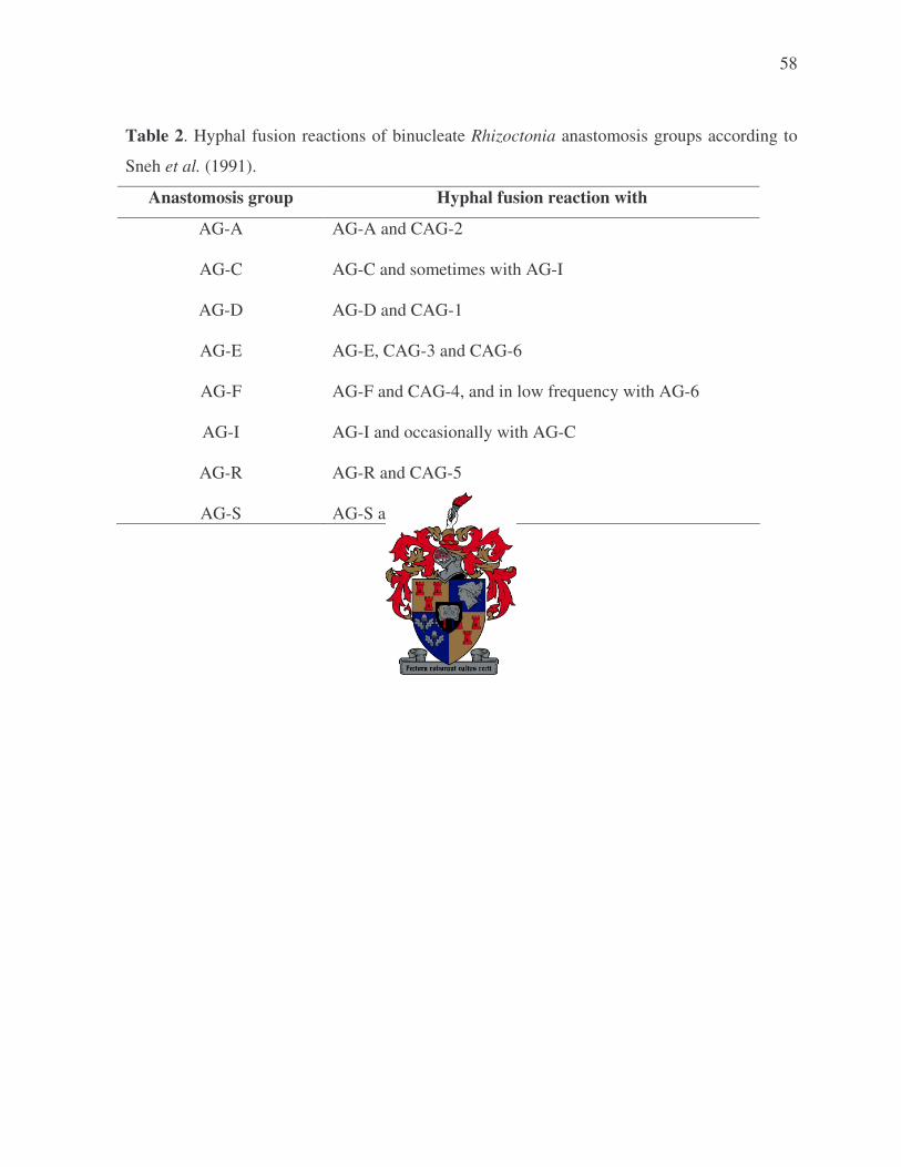
58
Table 2. Hyphal fusion reactions of binucleate Rhizoctonia anastomosis groups according to
Sneh et al. (1991).
Anastomosis group Hyphal fusion reaction with
AG-A AG-A and CAG-2
AG-C AG-C and sometimes with AG-I
AG-D AG-D and CAG-1
AG-E AG-E, CAG-3 and CAG-6
AG-F AG-F and CAG-4, and in low frequency with AG-6
AG-I AG-I and occasionally with AG-C
AG-R AG-R and CAG-5
AG-S AG-S and CAG-7

59
2. CHARACTERISATION OF RHIZOCTONIA ASSOCIATED WITH ROTATION CROPS IN CROPPING SYSTEMS IN THE WESTERN CAPE PROVINCE
ABSTRACT
Studies were conducted to characterise Rhizoctonia populations isolated from barley, canola,
clover, lucerne, lupin, annual Medicago spp. (medic) and wheat that were cultivated in a four-
year (2000-2003) crop rotation trial in the Western Cape province of South Africa. During the
four-year sampling period, 428 Rhizoctonia isolates were obtained from hypocotyl, coleoptile,
crown and root tissue of the seven crops. Nuclear staining of the Rhizoctonia isolates revealed
the presence of 104 (24%) multinucleate Rhizoctonia isolates and 324 (76%) binucleate
Rhizoctonia isolates. The anastomosis groups (AGs) of isolates were determined by Dr. M.
Mazzola (USDA-ARS, Wenatchee, WA, USA), through sequence analyses of the ribosomal
deoxyribonucleic acid (rDNA) internal transcribed spacer (ITS) region (unpublished data).
The analyses revealed that the multinucleate isolates were comprised of AG-4 HG-II (69%),
AG-2-1 (19%), AG-3 (8%), AG-2-2 (2%) and AG-11 (2%). Among the binucleate
Rhizoctonia species 53% were AG-K, 10% were AG-A, 5% were AG-I and 32% were
unidentified binucleate Rhizoctonia AGs. Traditional hyphal fusion of representative isolates
with tester isolates confirmed these identifications, except for AG-A isolates that did not
anastomose with the tester isolate. AG-K was isolated from all crops. Similarly, AG-4 HG-II
was also common to all crops except for lucerne. AG-2-2 and AG-11 were only isolated from
medic/clover and lupin respectively. AG-2-1 was isolated from all crops except barley and
lupin; AG-3 from canola, lupin and wheat; AG-A from all crops except lupin; whereas AG-I
was isolated from barley, canola, lupin and wheat. Sampling Rhizoctonia at three different
times during the growth season showed that the optimal time for isolating Rhizoctonia from
plants was at the flowering stage (20 – 22 weeks after planting), compared to isolations made
at the seedling (4 – 6 weeks after planting) or mid season (12 – 14 weeks after planting)
stages. Temperature studies using 28 isolates representing nine AGs showed that AG-2-2,
AG-4 HG-II and AG-K had significantly higher optimum growth temperatures than the other
AGs.

60
INTRODUCTION
The genus Rhizoctonia is a collective species complex and includes a group of diverse
Basidiomycete fungi (Sneh et al., 1996). They occupy diverse ecological niches and have
interactions with plants that range from pathogenic to symbiotic relationships (Sneh, Burpee &
Ogoshi, 1991; Vilgalys & Cubeta, 1994). Plant pathogenic Rhizoctonia occurs worldwide and
are economically important pathogens on vegetable and field crops, turf grasses, ornamentals,
and fruit and forest trees (Anderson, 1982; Adams, 1988; Sneh et al., 1996).
The high levels of phenotypic variation and inability to induce sporulation of most
Rhizoctonia isolates in culture have challenged the ability of taxonomists to define species
using conventional taxonomic criteria (Vilgalys & Cubeta, 1994). Therefore, methods for
subdividing Rhizoctonia have been developed, and consist of first identifying the nuclear
status of isolates as either being uni-, bi- or multinucleate, followed by anastomosis groupings
(Rovira, Ogoshi & McDonald, 1986; Ogoshi, 1987; Ogoshi, Cook & Bassett, 1990; Hietala,
1995). Conventionally, anastomosis groups (AGs) have been determined by observing
whether a hyphal fusion reaction is present between an unknown isolate and a reference isolate
of a known AG. Pairing of isolates belonging to the same AG results in hyphal fusion
(anastomosis) leading to either acceptance (self-pairing) or rejection (somatic incompatibility).
Strong (C2) anastomosis reactions indicate AG membership (Cubeta & Vilgalys, 1997;
Carling et al., 2002). Contrarily, pairings between isolates of unrelated AGs result in no
hyphal fusion, indicating that they belong to different AGs (Cubeta & Vilgalys, 1997).
However, sometimes “bridging” type reactions (less than C2 reaction) are observed between
obvious discrete AGs (i.e., AG-3 and AG-2) as well as isolates of the same AG. These types
of reactions increase the difficulty in identifying AGs, especially for researchers newly
entering the field of Rhizoctonia identification (Carling et al., 2002). Therefore,
deoxyribonucleic acid (DNA)-based methods that allow typing of a larger number of isolates
are increasingly being used to identify anastomosis groups (Vilgalys & Gonzalez, 1990;
Cubeta et al., 1991; Gonzalez et al., 2001; Carling et al., 2002).

61
The most widely distributed plant pathogenic multinucleate Rhizoctonia group is R.
solani Kühn (Roberts & Sivasithamparam, 1986). Rhizoctonia solani isolates are grouped into
fourteen AGs [AG-1 to AG-10 including AG BI (Sneh et al., 1991), AG-11 (Carling et al.,
1994), AG-12 (Carling et al., 1999) and AG-13 (Carling et al., 2002)] based on their
anastomosis behaviour. Many AGs, including AG-1, -2, -4, -6, and -9, have been subdivided
further into subgroups that differ for one or more biochemical, genetic, or pathogenic
characteristics (Laroche, Jabaji-Hare & Charest, 1992; Johnk & Jones, 1993; MacNish,
Carling, & Brainard, 1993).
Only a small number of binucleate Rhizoctonia isolates are plant pathogenic, with the
majority being saprophytic or having symbiotic relationships with plants (Sneh et al., 1996).
Although binucleate Rhizoctonia occurs worldwide, distribution of the various groups is
poorly documented with many groups appearing to be saprophytic including AG-C, -H, -K, -
L, -N, and -O (Carling & Sumner, 1992). Diseases caused by pathogenic isolates include
sharp eye spot of cereals, yellow patch of turf (Agrotis palustris Huds.) as well as damping-off
and root rot in strawberry (Fragaria ananassa Duch.), sugar beet (Beta vulgaris L.),
vegetables, and many other hosts (Carling & Sumner, 1992). Nineteen binucleate Rhizoctonia
AGs have been reported by various Japanese authors (Ogoshi et al., 1979), including AG-A, -
B, -Ba, -Bb, -C, -D, -E, -F, -G, -H, -I, -J, -K, -L, -M, -N, -O, -P, and -Q (Carling & Sumner,
1992). Burpee et al. (1980a,b) further described seven binucleate groups: CAG-1 (=AG-D),
CAG-2 (=AG-A), CAG-3 (=AG-E), CAG-4 (=AG-F), CAG-5 (=AG-R), CAG-6 (=AG-E) and
CAG-7 (=AG-S). The most commonly used system for classifying binucleate Rhizoctonia
isolates is the one proposed by Sneh et al. (1991), where binucleate Rhizoctonia isolates are
grouped into AG-A to S.
Rhizoctonia can cause severe economic losses in various cropping systems, and
therefore require effective control measures (Vilgalys & Cubeta, 1994; Sneh et al., 1996).
Crop rotation is one of the control strategies that have been used against Rhizoctonia disease.

62
Crop rotation not only has the advantage of controlling diseases, but can also control weeds
and improve soil fertility (Leach & Clapham, 1992; Wessels, 2001).
In the Western Cape province of South Africa, many of the important agricultural
crops that are used in rotation systems are susceptible to Rhizoctonia infection based on the
reported host range of these pathogens in other countries (Samuel & Garret, 1932; Bretag,
1985; Kaminski & Verma, 1985; Weller et al., 1986; Pumphrey et al., 1987; Kronland &
Stanghellini, 1988; Kataria & Verma, 1992; Vincelli & Herr, 1992; MacLeod & Sweetingham,
1997). Important rotation crops used in the Western Cape province include barley (Hordeum
vulgare L.), canola (Brassica napus L. var. oleifera DC), clover (Trifolium spp.), lucerne
(Medicago sativa L.), lupins (Lupinus spp.), medic (annual Medicago spp.) and wheat
(Triticum aestivum L.). In the Western Cape province, crop-pasture rotations have become a
relatively stable pattern of land use since the 1970’s (Wessels, 2001). In these rotation
systems, barley and wheat are often grown in rotation with grain and pasture legumes and/or
canola (G. A. Agenbag, pers. comm.). The break in cereal/crop production in successive years,
by planting a pasture legume, helps to increase the organic nitrogen content of the soil because
clover, lucerne and medics have the capability to fix atmospheric nitrogen through the activity
of associated Rhizobia (Ladd, Oades & Amato, 1981). Lupins also have excellent nitrogen
fixing qualities, with the potential to fix up to 350 kg of nitrogen per hectare (Keeve, 1998). In
the Western Cape province the crop-pasture rotations have been shown to limit the incidence
of weeds and some diseases that accumulate in a monoculture (Beyers, 2001; Wessels, 2001).
In South Africa, very little is known regarding the Rhizoctonia groups associated with
barley, canola, clover, lucerne, lupin, medics and wheat. Rhizoctonia solani AG-6 and R.
cerealis Van der Hoeven have been shown to cause crater disease of wheat in summer rainfall
regions of South Africa (Scott, Visser & Rufenacht, 1979; Smith & Wehner, 1986). Other
than these Rhizoctonia groups, no anastomosis group characterization of isolates has been
conducted on the aforementioned crops in South Africa, although Rhizoctonia spp. have been
isolated from all the crops of interest (barley, canola, clover, lucerne, lupin, medic and wheat)

63
(Lamprecht, De Villiers & Janse van Rensburg, 1999; Lamprecht, Auret & Janse van
Rensburg 2000a; Lamprecht et al., 2000b, 2001, 2002; Auret, Janse van Rensburg &
Lamprecht, 2002). Therefore, the first objective of this chapter was to characterise
Rhizoctonia isolates obtained from a four-year crop rotation trial at the Tygerhoek
experimental farm, Riviersonderend in the Western Cape province. Rhizoctonia isolates
obtained in the first three years of the rotation trial (2000 to 2002) were obtained from Dr. S.
C. Lamprecht (ARC-Plant Protection Research Institute, Stellenbosch, South Africa). In 2003
isolations were made for this study, and Rhizoctonia isolates from all years were characterised
with regard to nuclear status, AG and the effect of temperature on in vitro growth.
Anastomosis groups of all isolates were determined by Dr. M. Mazzola through rDNA internal
transcribed spacer (ITS) regions sequence analyses. The AG of a subset of isolates was
confirmed by conventional hyphal anastomosis testing in this study. The second objective of
the study was to determine which time of the season [seedling stage (4 – 6 weeks after
planting), mid season (12 – 14 weeks after planting) or flowering stages (20 – 22 weeks after
planting)] is optimal for isolating Rhizoctonia from crops.
MATERIALS AND METHODS
Experimental site used for sampling and isolation of Rhizoctonia
Rhizoctonia isolates were obtained from a crop rotation trial over a four-year period
(2000 to 2003) at the Tygerhoek experimental farm, Riviersonderend. The land used for the
rotation trial was previously planted to lucerne since 1996. The soil at the experimental site is
a sandy loam shale soil and was classified as a Glenrosa soil form (MacVicar et al., 1977)
with an average A-horizon depth of 400 mm and a B-horizon of well weathered yellow/red
clay above poorly weathered schist.
The rotation trial at Tygerhoek is situated in the southern cereal producing region of
the Western Cape province of South Africa at 34°9’ S and 19°54’ E. The region experiences a

64
Mediterranean climate with hot dry summers and cool moist winters. Longterm (n = 35 years)
average annual rainfall is 451 mm of which approximately 60% falls during the period April
to September. The mean daily maximum and minimum temperatures for March to October
from 2000 to 2003 are presented in Fig. 1 (Agromet Division, ARC–Institute for Soil, Climate
and Water, Stellenbosch).
The following crops were included in the trial: canola cv. Varola 50, barley cv.
Clipper, alfalfa or lucerne cv. SA Standard, lupin (Lupinus angustifolius L.) cv. Wonga, medic
(Medicago truncatula Gaertn.) cvs Mogul, Parabinga and Sephi/clover (Trifolium
michelianum Savi.) cv. Patrick mixture and wheat cv. SST57. Four different rotation systems
were used: system one (wheat and barley), system two (barley, medic/clover mixture, wheat
and medic/clover mixture), system three (lupin, wheat, canola, and barley) and system four
(monoculture lucerne). Each experimental plot was 80 x 15 m2 (0.12 ha). Soils were fertilised
annually according to soil analyses. Planting dates for all crops in the four-years were either
early May or mid April, except for lucerne that is a perennial crop and was therefore only
planted in 2000. The medic/clover mixture also re-established itself in 2002 and 2003. Weeds,
pests and foliage diseases were controlled when necessary using commercially available
chemicals. Fungicides (bromuconazole and propiconazole) for control of foliage diseases (rust
and net blotch) were only applied to barley and wheat.
Sampling of Rhizoctonia within the rotation trial
Plants were sampled at three different plant growth stages during the season. Plants
were sampled at three sampling times in 2000, 2001 and 2003, i.e. at the seedling (4 - 6 weeks
after planting), mid season (12 – 14 weeks after planting) and flowering stages (20 – 22 weeks
after planting). In 2002, plants were only sampled at the flowering stages. Sixty plants were
collected from ten sampling locations (six plants/location) along a zig-zag (W) pattern through
each experimental plot at each sampling time. Plants were placed in cooler boxes immediately
after sampling and isolations were performed within one day after sampling.

65
Isolations from hypocotyl/coleoptile/crown and root tissue
Rhizoctonia isolates collected in the first three years (2000-2002) of the trial were
provided by Dr. S. C. Lamprecht (ARC-Plant Protection Research Institute, Stellenbosch,
South Africa). In 2003 isolations were conducted in a similar way as in the previous years
(2000-2002), where 25 plants were randomly selected from plants collected from each
experimental plot at each sampling time. Plants were washed under running tap water to
remove adhering soil, and rinsed twice in sterile distilled water. Small pieces of root and
hypocotyl or coleoptile or crown tissue were excised and plated onto growth media. Three
root and hypocotyl or coleoptile or crown pieces were plated onto each of the following
growth media: water agar (WA) (Agar Bacteriological, Biolab Diagnostics, Midrand, South
Africa) containing 0.02% novostreptomycin, unamended WA, potato dextrose agar (PDA)
(Biolab Diagnostics, Midrand, South Africa) containing 0.02% novostreptomycin, unamended
PDA, selective Fusarium medium (SFA) modified from Tio et al. (1977) by replacing Alisan
with 0.05 g/L pentachloronitrobenzene, Phytophthora selective medium (PH medium) (Solel
& Pinkas, 1982) and Pythium selective medium (P medium = PH medium without
hymexazol). Another aim of the rotation trial, which was treated in a different study, was to
also determine the incidence of Pythium, Phytophthora and Fusarium. Therefore, isolations
were also made on media selective for these organisms, but occasionally also yielded some
Rhizoctonia isolates. A total of 18 hypocotyl/coleoptile/crown and 18 root pieces were plated
for each experimental plot at each sampling time for each crop. The total number of pieces
plated for each crop at each sampling time was 864, 288, 144, 288, 516 and 864 for barley,
canola, lucerne, lupin, medic/clover mixture and wheat respectively. The total number of
pieces plated per crop depended on the number of plots containing the specific crops within
the rotation trial.
All fungi that developed were transferred to divided Petri dishes containing carnation
leaf agar (Fisher et al., 1982) in one half and PDA containing 0.02% novostreptomycin in the
other half. Cultures were incubated under near-ultraviolet and cool white lights with a 12 h

66
photoperiod for 14 – 28 days. Microscopic examination of cultures was carried out and all
isolates identified as Rhizoctonia spp. (according to Sneh et al., 1991) were stored on potato
carrot agar ( 10 g potato, 10 g carrot and 12 g agar) slants and/or sterile wheat grain by
adapting (excluding vermiculite) the method used by Sneh et al. (1986) to prepare inoculum.
Cultures on agar slants were stored at room temperature and in a cold room at 10°C, whereas
cultures on wheat grain were stored at -20°C.
Nuclear staining
Nuclear staining was performed using a fluorocrome, acridine orange stain and
viewing the stained specimen through a fluorescence microscope (Yamamoto & Uchida,
1982). Isolates were cultured on clarified 2% vegetable juice agar without CaCO3 (Yamamoto
& Uchida, 1982). Four sterilized microscope glass cover slips (Marenfeld cover glasses
number 1, 22 x 22 mm) were placed on the medium next to the inoculum disc and plates were
incubated for 48 h at 25°C with a 12 h photoperiod under cool white light. After 48 h the cover
slips that were overgrown with mycelium growth were mounted, mycelium side downwards,
into the drop of stain on a microscope slide and examined using a Zeiss Axioskop microscope
equipped with an epifluorecence condenser and a high-pressure mercury lamp. The Zeiss 02,
06 and 18 filters were used. The nuclei stained bright green and were clearly visible and easy
to count when freshly stained. The nuclei of 20 cells per isolate were counted to confirm the
nuclear status of each isolate.
DNA extraction and identification of anastomosis group through sequence analyses
DNA was extracted using the protocol described by Lee and Taylor (1990). Four
hundred and twenty-eight Rhizoctonia isolates were grown on PDA plates at 25°C with a 12 h
photoperiod for 5 days to ensure adequate development of mycelia. Fungal mycelia were
scraped from plates and transferred to 2.2 mL eppendorf tubes and 1 mL SDS extraction
buffer [2% SDS, 50mM Tris-HCl (pH 8), 150mM NaCl, 100mM EDTA (pH 8)] was added.

67
The eppendorf tubes were boiled for 3 min, placed on ice for 10 min, and incubated at 65°C
for 60 – 90 min. Subsequently, 400 �L chloroform: isoamyl alcohol (24:1) and 600 �L phenol
were added and mixed by inverting the eppendorf tubes 5 – 10 times, followed by
centrifugation for 15 min at 14 000 rpm (rounds per minute), after which the supernatant was
transferred to a new tube. This step was repeated once. 10 �L RNase A (boiled for 5 min) was
added to the supernatant and the tube was incubated at 37°C for 3 h to overnight. After
incubation 5 �L 20% SDS and 10 �L proteinase K were added and incubated for 2 h to
overnight at 37°C. The abovementioned chloroform: isoamyl alcohol and phenol steps were
repeated, and 50 �L cold 7.5 M NH4OAc and 600 �L cold isopropanol were added to the
supernatant in order to precipitate DNA overnight in a freezer. Following the precipitation
period, tubes were centrifuged for 10 min and the supernatant discarded. DNA pellets were
washed with 1 mL cold 100% ethanol, followed by another 70% ethanol wash. DNA pellets
were left to dry on a bench, and dried DNA was sent to Dr. M. Mazzola (USDA-ARS,
Wenatchee, WA, USA) for AG group identification through sequence analyses of the rDNA-
ITS region.
Anastomosis group (AG) typing using conventional hyphal fusion reactions
The anastomosis group of 28 representative isolates was confirmed by conventional
hyphal anastomosis testing. These isolates (characterised through sequence analyses of the
rDNA-ITS region) and tester strains (Table 1) were paired by plating an unknown isolate
opposite a known AG type on 2% water agar in Petri dishes. The cultures were incubated
under cool white light with a 12 h photoperiod at 25°C. Analysis of hyphal fusion was
conducted by examining the zone of hyphal interaction at 40x magnification using a light
microscope.
Temperature growth studies
The effect of temperature on in vitro radial growth of 28 Rhizoctonia isolates,
representing nine AGs (Table 2), was studied at seven different temperatures. Mycelial plugs
68
(0.6 cm in diameter) were transferred from the edge of 5-day-old actively growing colonies to
PDA. Cultures were incubated in the dark at 5, 10, 15, 20, 25, 30, or 35°C and colony radius
was measured in four directions per each PDA plate after 48 h. Each isolate was replicated
three times at each temperature.
Experimental design and statistical analysis
The rotation system experiment was a randomised block design with 20 main plots
(treatments) randomly replicated within four blocks. The 20 main plots consisted of a 10 x 2
factorial (ten crop sequences x two tillage practices) with three rotation systems. Three
sampling times per year were used as a subplot factor (Little & Hills, 1972). A further sub-
subplot was the Rhizoctonia groups (binucleate and multinucleate) isolated from the samples.
Analysis for each year was done separately because crop sequences differed from one year to
the next. Although isolations were done separately from hypocotyls, coleoptiles, crowns, and
roots these data were combined for the analysis of variance. The percentage relative
incidences of the two Rhizoctonia groups (binucleate and multinucleate) were determined out
of a possible 36 pieces of plant material plated. Data obtained from lucerne isolations in the
rotation system consisting of monoculture lucerne were not subjected to analysis of variance.
Appropriate analysis of variance was performed for each separate year (SAS, 1999) and the
student’s t-LSD were calculated to compare means of significant effects at the 5% significance
level.
For the temperature growth studies second order polynomials were fitted on the radial
length over temperature for each isolate AG combination and replicates (Snedecor & Cochran,
1967). The optimum temperature and area under each curve were calculated. These
measurements were subjected to an appropriate analysis of variance and the means were
compared using student’s t-LSD at the 5% significance level.

69
RESULTS
Isolations from hypocotyl/coleoptile/crown and root tissue
A total of 428 Rhizoctonia isolates were obtained during the four years of the crop
rotation trial. Dr. S. C. Lamprecht provided the 317 Rhizoctonia isolates that were isolated
from 2000 to 2002, and 111 Rhizoctonia isolates were obtained from isolations made in 2003.
Nuclear staining
Nuclear staining of all Rhizoctonia isolates (428) was possible, and revealed the
presence of 104 (24%) R. solani isolates with nuclear numbers ranging from 3 - 12 per young
vegetative hyphal cell and 324 (76%) binucleate Rhizoctonia species with only two nuclei per
young vegetative hyphal cell (Fig. 2).
DNA extraction and identification of anastomosis group through sequence analyses
DNA was successfully extracted from 428 isolates and were of sufficient quality for
polymerase chain reaction (PCR) amplification of the rDNA-ITS1 and rDNA-ITS2 region by
Dr. M. Mazzola using primers ITS4 (5’-TCCTCCGCTTATTGATATGC-3’) and ITS5 (5’-
GGAAGTAAAAGTCGTAACAAGG-3’) [White et al., 1990]. The PCR products were first
cloned into vector pCR®4-TOPO® (Invitrogen, Carlsbad, CA, USA). Sequencing analyses of
the cloned PCR products by Dr. M. Mazzola revealed that the multinucleate Rhizoctonia (R.
solani) isolates were comprised of AG-4 HG-II (69%), AG-2-1 (19%), AG-3 (8%), AG-2-2
(2%) and AG-11 (2%) (Mazzola et al., unpublished data). Among the binucleate Rhizoctonia
species 53% were AG-K, 10% were AG-A, 5% were AG-I and 32% were unidentified
binucleate Rhizoctonia AGs (Mazzola et al., unpublished data).
70
Anastomosis Group (AG) typing using conventional hyphal fusion reactions
All 16 multinucleate R. solani isolates were successfully anastomosed with the
respective tester isolates and all exhibited a C2 reaction [wall connection obvious membrane
contact uncertain; location of reaction site obvious; diameter of anastomosis point less than
hyphal diameter; anastomosing and adjacent cells always die (Carling, 1996)]. The
multinucleate Rhizoctonia isolates that were identified included AG-2-1 (three isolates), AG-
2-2 (two isolates), AG-3 (three isolates), AG-4 HG-II (six isolates) and AG-11 (two isolates).
AG-I and AG-K were the only binucleate AGs that were successfully anastomosed with the
tester isolates. The unidentified binucleate Rhizoctonia group failed to anastomose with tester
isolates of AG-A, AG-I, AG-D and AG-K.
The identification of AGs through sequence analyses was compared with AG typing
through conventional hyphal anastomosis typing. The AG group of isolates as determined
through conventional anastomosis typing, corresponded with the identification determined
through sequence analyses of the ITS1 and ITS2 region (data not shown). The only exception
was three AG-A isolates that did not anastomose with the tester AG-A isolate PPRI 2130 (C-
517).
Temperature growth studies
Analysis of variance showed significant differences in the temperatures for optimum
growth as well as the growth rates at different temperatures (area under the curve) of the nine
AGs (Table 3). Significant differences for these parameters were also recorded for isolates
within AGs (Table 3). Isolates within AG-2-2, AG-3, AG-4 HG-II and AG-I differed
significantly with regard to the temperature at optimum growth, whereas isolates within AG-2-
2, AG-3, AG-4 HG-II, AG-11, AG-A and AG-I differed significantly in their growth rates
(area under curve). The appropriate trends (second degree polynomial function) were fitted for

71
AG x temperature combinations and the result of the regression analyses were R2 � 77.68%
for all AGs (Table 4). These predicted trends are illustrated in Fig. 3.
Rhizoctonia AG-2-2, AG-4 HG-II and AG-K had significantly (P = 0.05) higher
optimum growth temperatures than the other AGs (Table 4). Rhizoctonia solani AG-2-2 had
the highest optimum growth temperature (25.9°C), followed by the binucleate Rhizoctonia
AG-K (23.2°C) and the multinucleate AG-4 HG-II (22.4°C). The lowest temperatures for
optimum growth were recorded for the binucleate AGs, viz. AG-A, AG-I and the unidentified
group (UNBR) (Table 4). Comparison of the area under the curve (Table 4) indicated that the
growth rates of the binucleate AGs (AG-A, -I, -K, UNBR) were significantly lower than the
growth rates of the multinucleate groups (AG-2-1, AG-2-2, AG-3, AG-4 HG-II, AG-11).
All AGs grew poorly below 10°C, performed relatively well in the range 15 – 30°C
and performed better at 15°C than at 35°C. In the 15 – 30°C range all multinucleate isolates
performed better than the binucleate AGs, except for AG-K. Rhizoctonia solani AG-2-2 and
AG-K had higher growth rates at 35°C than the other AGs (Fig. 3). The in vitro growth of
Rhizoctonia isolates representing nine AGs used in temperature growth studies are shown in
Fig. 4.
Incidence of Rhizoctonia on rotation crops and effect of sampling time on Rhizoctonia
incidence
Results on the incidences of the nine AGs identified in this study are given in Table 5.
AG-K was the predominant AG followed by the unidentified binucleate group (UNBR) and
AG-4 HG-II. Certain AGs viz. AG-2-1, -4 HG-II, -A, -I, -K and UNBR were isolated in all
four years, whereas AG-11 was only recorded in 2000, AG-2-2 in 2002 and 2003 and AG-3 in
2000, 2001 and 2003 (Table 5). Most of the anastomosis groups were associated with more
than one crop: AG-2-1 with canola, lupin, medic/clover and wheat; AG-2-2 with medic/clover;
AG-3 with canola, lupin and wheat; AG-4 HG-II with barley, canola, medic/clover, lupin and

72
wheat; AG-11 with lupin; AG-A with barley, canola, lucerne, medic/clover and wheat; AG-I
with barley, canola, lupin and wheat and AG-K and the unidentified binucleate group with all
the crops (Table 5).
Analyses of variance were conducted on the effect of sampling time on the percentage
incidences of Rhizoctonia groups (binucleate and multinucleate) for each of the four years
(Table 6). Sampling time significantly (P = 0.05) affected the frequency of isolation of
Rhizoctonia groups in 2000 (P = 0.0003) and in 2001 (P = 0.0037), but not in 2003 (P =
0.1104). In all years (2000, 2001 and 2003) the incidence of Rhizoctonia was higher at the
third sampling time (flowering stage) than at the seedling or mid-season stages with
significantly higher incidences at the third sampling time than the other sampling times during
2000 and 2001 (Table 7).
Analyses of variance were conducted on the incidence of bi- and multinucleate
Rhizoctonia groups over the four years of the trial (Table 6). In all four years the incidence of
the two Rhizoctonia groups differed significantly [P = 0.0105 (2000), P = 0.0464 (2001), P =
0.0060 (2002) and P = 0.0062 (2003)] (Table 6). Significantly (P = 0.05) higher incidences of
binucleate compared to multinucleate isolates were recovered for all the years (Table 8).
DISCUSSION
This study characterised Rhizoctonia populations associated with rotation crops
(barley, canola, lucerne, lupin, medic/clover and wheat) used in the Western Cape province of
South Africa. The study was conducted within one four-year crop rotation trial. The
Rhizoctonia isolates were characterised with regard to nuclear status, anastomosis group and
the effect of temperature on in vitro growth. Characterisation of the Rhizoctonia populations
showed that the majority of Rhizoctonia isolates were binucleate (76%) whereas only 24%
were multinucleate isolates. Anastomosis group identification of a subset of isolates using
both conventional and sequence analyses were mostly in agreement, except for three isolates

73
of AG-A. Sequence analyses revealed that among the multinucleate isolates, AG-4 HG-II
(69%) were most frequently isolated followed by AG-2-1 (19%). Rhizoctonia AG-K was the
binucleate group that was present at the highest frequency (53%). Studies on the effect of
temperature on in vitro growth showed that two of the AGs (AG-K and AG-4 HG-II) that were
present at high frequency also had the highest optimal growth temperatures. AG-4 HG-II and
AG-K were the only AG’s associated with all crops, with the exception of AG-4 HG-II from
lucerne. Analysis of isolation frequency data over all years of this study indicated that the
optimum time for recovery of Rhizoctonia isolates was at the third sampling time (flowering
stage).
In the crop rotation trial conducted in the Western Cape province binucleate isolates
were recovered at a significantly higher frequency than multinucleate isolates. Similarly, in
Australia binucleate isolates were isolated at a higher frequency from lupin and cereal
seedlings (Sweetingham, Cruickshank & Wong, 1986; MacLeod & Sweetingham, 1997).
Contrastingly, a number of researchers isolated higher numbers of multinucleate than
binucleate Rhizoctonia from barley, canola and wheat. In Canada, Kaminski and Verma
(1985) obtained a high number of multinucleate Rhizoctonia isolates from seedling and adult
canola. A higher number of multinucleate compared to binucleate Rhizoctonia isolates were
also obtained from barley and wheat in Turkey (Demirci, 1998) and the Pacific Northwest of
the USA (Ogoshi et al., 1990), and from wheat in western Australia (Yang et al., 1994). The
higher incidence of binucleate Rhizoctonia isolates in the crop rotational trial in the Western
Cape province is interesting since multinucleate Rhizoctonia isolates are generally more
virulent than binucleate isolates (Sneh et al., 1991). Furthermore, binucleate Rhizoctonia AGs
have been shown to protect crops against multinucleate Rhizoctonia AGs. Binucleate
Rhizoctonia AG-B(o) isolates have been found to protect sugar beet and tall fescue (Festuca
arundinacea Schreb.) against multinucleate AG-2-2 (Herr, 1991) and AG-1-IA (Yuen, Craig
& Giesler, 1994) respectively, and binucleate AG-G protects potato (Solanum tuberosum L.)
against AG-3 (Cubeta & Echandi, 1991).
74
The high incidence of binucleate Rhizoctonia isolates in this study could be due the
fact that plant materials were not surface disinfested before isolations were made, thus
favouring the isolation of bi- versus multinucleate isolates. However, various other
investigations (Sweetingham et al., 1986; Ogoshi et al., 1990; and MacLeod and
Sweetingham, 1997) that also did not surface disinfest plant materials prior to isolations,
obtained more multi- than bi-nucleate isolates. Therefore, it seems unlikely that the absence of
surface disinfestation in this study would have influenced the incidence of bi- and
multinucleate isolates. The high incidence of bi- versus multinucleate isolates in this study
therefore warrants further investigations into the possible protective role of binucleate isolates
within crop rotation systems.
The identification of Rhizoctonia through sequence analyses has greatly aided the
identification of AGs (Gonzalez et al., 2001; Carling et al., 2002). Amplification and
sequence analysis of the nuclear ribosomal internal transcribed spacer (ITS) regions of
Rhizoctonia isolates recovered in this study showed that there were five multinucleate
Rhizoctonia AGs (AG-2-1, AG-2-2, AG-3, AG-4 HG-II and AG-11) and four binucleate
Rhizoctonia AGs (AG-A, AG-I, AG-K and unidentified group) (Mazzola et al., unpublished
data). Conventional AG determination of a subset of the isolates through evaluation of hyphal
anastomosis reactions, confirmed the identification based on sequence analyses, except for
isolates from AG-A that did not show anastomosis with the reference isolates. Only one
reference isolate of AG-A was available. Future studies should evaluate whether the isolates
designated as AG-A through sequence analyses can anastomose with other reference AG-A
isolates.
The frequency of different AGs within bi- and multinucleate Rhizoctonia varied.
Rhizoctonia solani AG-4 HG-II (69% of the multinucleate isolates) followed by AG-2-1 (19%
of the multinucleate isolates) were the predominant multinucleate AGs obtained in this study.
The other multinucleate AGs, viz. AG-2-2, AG-3 and AG-11 were infrequently isolated. Of
the identified binucleate AGs, AG-K (53%) was the predominant AG.
75
Temperature significantly affected the in vitro growth rate of the AGs included in this
study. All AGs grew relatively well in the range 15 – 30°C and performed better at 15 than at
35°C. The optimum growth range for AG-2-1 has been reported as being in the range of 20-
25°C (Kaminski & Verma, 1985; Ogoshi et al., 1990). Similarly, in this study the optimum
growth temperature for AG-2-1 of 21.7°C, is within this range. Kaminski and Verma (1985)
found that AG-4 isolates had higher growth rates than AG-2-1 with optimum growth at 26°C.
In this study the predicted optimum growth temperature (22.4°C) for AG-4 HG-II is lower
than that reported by Kaminski and Verma (1985). However, similar to their results, in this
study AG-4 HG II also had significant higher growth rates and optimum growth temperatures
than AG-2-1. The differences in in vitro optimum growth temperatures between AG-2-1 and
AG-4 are also reflected in the fact that AG-4 isolates are more virulent on canola at 20°C than
AG-2-1 (Kataria & Verma, 1992). Similarly, it has been found that at lower temperatures
(15°C) AG-2-1 caused more damping-off than AG-4 (Teo et al., 1988). In this study the in
vitro optimum growth temperature for AG-11 was 21.6°C, which agrees with pathogenicity
studies done by Kumar et al. (1999) showing that AG-11 can only cause very little lupin pre-
emergence damping-off and hypocotyl rot at 10°C, and most severe hypocotyl rot at 20 and
25°C.
The binucleate Rhizoctonia AG-K was found associated with barley, canola, clover,
lucerne, lupin, medic and wheat in this study. In Turkey and the USA isolates of AG-K were
also isolated from barley and wheat, and were found to be non pathogenic (Demirci, 1998;
Ogoshi et al., 1990). Information on binucleate AGs associated with canola is limited to
pathogenicity studies by Khangura et al. (1999) indicating that AG-K is only weakly virulent
on canola. Wong et al. (1985) found that AG-K was not pathogenic on clover.
The unidentified binucleate group of Rhizoctonia were isolated from barley, canola,
clover, lucerne, lupin, medic and wheat. Previously, Lamprecht, Knox-Davies and Marasas
(1988) also obtained binucleate Rhizoctonia isolates from medic in surveys conducted in the
Western Cape province of South Africa.

76
The binucleate Rhizoctonia AG-A was associated with barley, canola, clover, lucerne,
medic and wheat in this study. There are no reports of this AG on the above-mentioned crops
in other countries.
The binucleate Rhizoctonia isolates of AG-I were associated with barley, canola, lupin
and wheat. In Turkey, isolates of AG-I were also isolated from barley and wheat, where is it
was found to be non-pathogenic (Demirci, 1998). Only a thin binucleate Rhizoctonia (cause of
Eradu patch disease in Australia) has been reported as a binucleate Rhizoctonia pathogen of
lupin (MacLeod & Sweetingham, 1997).
The multinucleate Rhizoctonia AG-2-1 was associated with canola, clover, lupin,
medic and wheat in this study. In other parts of the world, isolates of AG-2-1 have not only
been often associated with canola, but have also been found highly virulent on this crop
(Kaminski & Verma; 1985; Hwang, Swanson and Evans, 1986; Gugel et al., 1987; Khangura,
Barbetti & Sweetingham, 1999). In Australia isolates of AG-2-1 were associated and found
weakly virulent towards lupin and wheat (Sweetingham et al. 1986). In Turkey, AG-2-1 was
associated with barley and wheat and was moderately virulent on these crops (Demirci, 1998).
Isolates of R. solani AG-2-1 have also been found pathogenic on clover elsewhere (Wong,
Barbetti & Sivasithamparam, 1985).
Rhizoctonia solani AG-2-2 was only isolated from medic in this study. Although AG-
2-2 was found associated with wheat in Texas it was not found to be pathogenic on this crop
(Rush et al., 1994). Isolates of R. solani AG-2-2 were found to be pathogenic on clover in
Australia (Wong, Barbetti & Sivasithamparam, 1985). This is the first report on the isolation
of AG-2-2 from medic.
In this study R. solani AG-3 was associated with canola, lupin and wheat. In Turkey,
AG-3 was isolated from barley and wheat, where it was found to be moderately virulent on

77
these crops (Demirci, 1998). Contrarily, in the USA, isolates of AG-3 were associated with
barley and wheat, but were non pathogenic to these crops (Ogoshi et al., 1990).
The mulitinucleate Rhizoctonia AG-4 HG-II was associated with barley, canola,
clover, lupin, medic and wheat in this study. In Turkey, isolates of AG-4 were also found
associated with barley and wheat, and were highly virulent on these crops (Demirci, 1998).
Similarly, in Texas, isolates of AG-4 were found associated and pathogenic on wheat
(Mathieson & Rush, 1991: Rush et al., 1994). Contrarily, in the USA, isolates of AG-4 were
found associated with barley and wheat, but were non pathogenic or only mildly virulent on
these crops (Ogoshi et al., 1990). In other parts of the world isolates of AG-4 are also
frequently isolated from canola, where it was found to be highly virulent on this crop
(Kaminski & Verma; 1985; Hwang, Swanson and Evans, 1986; Gugel et al., 1987; Khangura,
Barbetti & Sweetingham, 1999). Rhizoctonia solani AG-4 was further also isolated and found
to be pathogenic on lupin in the USA (Leach & Clapman, 1992). Although R. solani is an
important pathogen of medic in Australia (Bretag, 1985; Mebalds, 1987; Barbetti, 1989), the
specific AGs involved were not identified. Information on the AGs pathogenic on medics is
limited to a study on the resistance of 27 annual Medicago spp. to AG-4. this study
demonstrated the pathogenicity of AG-4 on different Medicago spp. (Kulik & Dery, 1995).
Rhizoctonia solani AG-4 has also been found associated with poor stands of clover in Texas
(Pemberton et al., 1998). Although Rhizoctonia solani AG-4 is the most important
Rhizoctonia pathogen of lucerne in other countries (Kulik, Dery & Douglas, 1995), it was not
isolated from lucerne grown in monoculture in this study.
Rhizoctonia solani AG-11 was only isolated from lupin in this study. In Western
Australia and Arkansas isolates of AG-11 were also found associated with lupin, as well as
wheat. Rhizoctonia solani AG-11 was pathogenic on both of these crops (Carling et al.,
1994). In Turkey, AG-11 has also been isolated and found to be highly virulent on barley and
wheat (Demirci, 1998).

78
Several R. solani AGs that have previously been found associated with some of the
rotations crops were not isolated in the present study. Rhizoctonia solani AG-6 that had
previously been reported as a pathogen of wheat in South Africa (Scott et al., 1979; Carling,
Meyer & Brainard, 1996) was not isolated in this study. R. solani AG-8 and R. oryzae that
have been frequently isolated from cereals and canola in other countries were also not isolated
in this study. (MacNish, 1983; Smiley, Wilkins & Klepper, 1990; Furgal-Wegrzyciqa,
Adamkiak & Adamiak, 1998; Khangura et al., 1999). Furthermore, R. cerealis (AG-D) that
had previously been reported as pathogens of wheat in South Africa was not isolated in this
study (Scott et al., 1979).
Rhizoctonia was generally more frequently isolated from plants at the flowering than
the seedling stage. This increase in incidence from the seedling to the flowering stage was
mostly the result of an increase in binucleate Rhizoctonia AGs, but it was also evident that
AG-2-1 and AG-4 HG-II were more frequently isolated at the flowering than the seedling
stage. The higher incidence of Rhizoctonia at the flowering stage is most probably partly due
to more favourable temperatures for growth of the Rhizoctonia AGs during September and
October (flowering stage) compared to July and August (mid season) (Fig. 1). In Western
Australia, MacLeod and Sweetingham (1997) recorded an increase in the number of
Rhizoctonia isolates obtained from July (six isolates) to August (32 isolates). There is limited
information on the incidence of Rhizoctonia AGs at different times during the growing season.
This is the first report of the specified Rhizoctonia AGs on barley, canola, clover,
lucerne, lupin, medic and wheat in South Africa. Proper seedling establishment of these crops
in the Western Cape province has always been problematic, and fungal pathogens including
Rhizoctonia spp. have been implicated as a contributing factor (Lamprecht et al., 2000a; Auret
et al., 2002). Within the crop rotation trial of this study the association of known pathogenic
Rhizoctonia AGs (AG-2-1 and AG-4) with canola was noticeable and should be further
investigated. Furthermore, it will be interesting to determine whether the large number of
binucleates associated with the rotation crops have a beneficial association with their hosts.

79
Another interesting observation of the study was that no multinucleate isolates were isolated
from lucerne that has been grown in this region for an extended period in monoculture. This is
especially striking considering that AG-4 HG-II, that is a very well known pathogen of lucerne
(Kulik, Dery & Douglas, 1995), could not be isolated from the lucerne monoculture plots,
even though it was the predominant multinucleate AG (69%) isolated from various other
rotations crops used in the study. The absence of AG-4 HG-II from lucerne in this study could
be due to the practice of monoculture. Similarly, in sugar beet and wheat, monoculture has
resulted in a gradual reduction of disease caused by AG-5 and AG-8 (Hyakumachi, 1999;
Mazzola, 2002). Another factor contributing to the absence of AG-4 HGII and other
pathogenic multinucleate isolates on lucerne in this study, could be due to the fact that lucerne
residues in itself has been found to suppress pathogenic Rhizoctonia in beans (Sneh et al.,
1996).
Determining Rhizoctonia AGs associated with rotation crops is essential for
development of effective integrated management strategies. Seed treatments can significantly
improve seedling establishment, but it is known that certain Rhizoctonia AGs differ in their
sensitivity to fungicides (Kataria & Gisi, 1996). Including practices such as seed treatments
and crop rotation in management strategies will therefore be much more reliable in the future
as the specific AGs involved in disease development have been characterised to some degree.
However, before implementing fungicide seed treatment for the control of pathogenic
multinucleate isolates, the effect of fungicides on potentially beneficial binucleate isolates
should first be established. Since the current study was restricted to one crop rotational trial in
one production area, future studies should investigate the association of Rhizoctonia in areas
with different climatology (higher rainfall in the western than southern production areas
during the growing season) to obtain a better understanding of the Rhizoctonia AGs associated
with rotation crops. This additional knowledge will further aid the development of sound
control practices.
80
REFERENCES
Adams G.C. 1988. Thanatephorus cucumeris (Rhizoctonia solani), a species complex of wide
host range. In Advances in Plant Pathology: Genetics of Plant pathogenic Fungi (eds.
D.S. Ingram & P.H. William), pp. 535–552. Academic Press: London, UK.
Anderson, N. A. 1982. The genetics and pathology of Rhizoctonia solani. Annual Review of
Phytopathology 20: 329–347.
Auret, E. E., Janse van Rensburg, J. C. & Lamprecht, S. C. 2002. The extent and role of
canola and lupin soilborne diseases on yield depression: A prerequisite for developing
an effective and sustainable control strategy. Report to Protein Research Foundation.
Barbetti, M. J. 1989. Response of Medicago cultivars to fungal root pathogens associated with
Trifolium subterraneum. Plant Protection Quaterly 4: 75–77.
Beyers, H. P. 2001. The effect of Haloxytop-R-methyl Ester and Imazamox herbicides, tine or
notillage and nine different medic cultivars on the seed and dry matter production as
well as the quality of medic pastures. M.Sc. thesis, University of Stellenbosch,
Stellenbosch.
Bretag, T. W. 1985. Fungi associated with root rots of annual Medicago spp. in Australia.
Transactions of the British Mycological Society 85: 329–334.
Burpee, L. L., Sanders, P.L., Cole, H. Jr. & Sherwood, R. T. 1980a. Anastomosis groups
among isolates of Ceratobasidium cornigerum (Bourd.) Rogers and related fungi.
Mycologia 72: 689–701.

81
Burpee, L. L., Sanders, P. L., Cole, H. Jr. & Sherwood, R. T. 1980b. Pathogenicity of
Ceratobasidium cornigerum and related fungi representing four anastomosis groups.
Phytopathology 70: 843–846.
Carling, D. E. 1996. Grouping in Rhizoctonia solani by hyphal anastomosis reaction.
Rhizoctonia species: Taxonomy, Molecular biology, Ecology, Pathology and Disease
control (eds. Sneh, B., Jabaji-Hare, S., Neate, S. & Dijst, G.) pp 37–47. Kluwer
Acadamic Publisher: Dordrecht, Netherlands.
Carling, D. E., Baird, R. E., Gitaitis, R. D., Brainard, K. A. & Kuninaga, S. 2002.
Characterization of AG-13, a newly reported anastomosis group of Rhizoctonia solani.
Phytopathology 92: 893–899.
Carling, D. E., Meyer, L. & Brainard, K. A. 1996. Crater disease of wheat caused by
Rhizoctonia solani AG-6. Plant Disease 80: 1429.
Carling, D. E., Pope, E. J., Brainard, K. A. & Carter, D. A. 1999. Characterization of
mycorrhizal isolates of Rhizoctonia solani from an orchid, including AG-12, a new
anastomosis group. Phytopathology 89: 942–946.
Carling, D. E., Rothrock, C. S., MacNish, G. C., Sweetingham, M. W., Brainard, K. A. &
Winters, S. W. 1994. Characterisation of anastomosis group 11 (AG-11) of
Rhizoctonia solani. Phytopathology 84: 1387–1393.
Carling, D. E. & Sumner, D. R. 1992. Rhizoctonia. In Methods for Research on Soilborne
Phytopathogenic Fungi (eds. Singleton, L. L., Mihail, J. D. & Rush, C. M.), pp. 157–
165. American Phytopathological Society: St. Paul, MN, USA.
82
Cubeta, M. A. & Echandi, E. 1991. Biological control of Rhizoctonia and Pythium damping-
off of cucumber; an integrated approach. Biological Control 1: 227–236.
Cubeta, M. A., Echandi, E., Abernethy, T. & Vilgalys, R. 1991. Characterization of
anastomosis groups of binucleate Rhizoctonia species using restriction analysis of an
amplified ribosomal RNA gene. Phytopathology 81: 1395–1400.
Cubeta, M. A. & Vilgalys, R. 1997. Population biology of the Rhizoctonia solani complex.
Phytopathology 87: 480–484.
Demirci, E. 1998. Rhizoctonia species and anastomosis groups isolated from barley and wheat
in Erzurum, Turkey. Plant Pathology 47: 10–15.
Fisher, N. L., Burgess, L. W., Toussoun, T. A. & Nelson, P. E. 1982. Carnation leaves as a
substrate and for preserving cultures of Fusarium species. Phytopathology 72: 151–
153.
Furgal-Wegrzyciqa, H., Adamkiak, J. & Adamiak, E. 1998. Some characteristics of isolates of
Rhizoctonia solani from patch of wheat and barley. Acta Mycologica 33: 109–121.
Gonzalez, D., Carling, D. E., Kuninaga, S., Vilgalys, R. & Cubeta, M. A. 2001. Ribosomal
DNA systematics of Ceratobasidium and Thanatephorus with Rhizoctonia anamorphs.
Mycologia 93: 1138–1150.
Gugel, R. K., Yitbarek, S. M., Verma, P. R., Morrall, R. A. & Sadasivaiah, R. S. 1987.
Etiology of the Rhizoctonia root rot complex of canola in the Peace River region of
Alberta. Canadian Journal of Plant Pathology 9: 119–128.

83
Herr, L. J. 1991. Relationship of binucleate Rhizoctonia isolates used for biocontrol of
Rhizoctonia crown rot of sugar beets to anastomosis systems. Canadian Journal of
Microbiology 37: 335-344.
Hietala, A. M. 1995. Uni- and binucleate Rhizoctonia spp. co-existing on the roots of Norway-
spruce seedlings suffering from root dieback. European Journal of Forest Pathology
25:136–144.
Hwang, S. F., Swanson, T. A. & Evans, I. R. 1986. Characterization of Rhizoctonia solani
isolates from canola in West Central Alberta. Plant Disease 70: 681–683.
Hyakumachi, M. 1999. Suppression and prevention: mechanisms involved in Rhizoctonia
diseases decline. Summa Phytopathologica 25: 99-102.
www.summanet.com.br/revista/250201.htm
Johnk, J. S. & Jones, R. K. 1993. Differentiation of populations of AG-2-2 of Rhizoctonia
solani by analysis of cellular fatty acids. Phytopathology 83: 278–283.
Kaminski, D. A. & Verma, P. R. 1985. Cultural characteristics, virulence, and in vitro
temperature effect on mycelial growth of Rhizoctonia isolates from rapeseed.
Canadian Journal of Plant Pathology 7: 256–261.
Kataria, H. R. & Gisi, U. 1996. Chemical control of Rhizoctonia species. Rhizoctonia Species:
Taxonomy, Molecular Biology, Ecology, Pathology and Disease Control. (eds. Sneh,
B., Jabaji-Hare, S., Neate, S. & Dijst, G.) pp 537 – 547. Kluwer Academic Publishers:
Dordrecht, Netherlands.
Kataria, H. R. & Verma, P. R. 1992. Rhizoctonia solani damping-off and root rot in oilseed
rape and canola. Crop Protection 11: 8–13.

84
Keeve, R. 1998. Lupiene, oplossings vir knelpunte. Landbouweekblad 1048: 18–23.
Khangura, R. K., Barbetti, M. J. & Sweetingham, M. W. 1999. Characterization and
pathogenicity of Rhizoctonia species on canola. Plant Disease 83: 714–721.
Kronland, W. C., & Stanghellini, M. E. 1988. Clean slide technique for the observation of
anastomosis and nuclear condition of Rhizoctonia solani. Phytopathology 78: 820–822.
Kulik, M. M. & Dery, P. D. 1995. Frequency of plants in populations of annual and perennial
medics (Medicago spp.) that were resistant to damping-off incited by AG-4 isolates of
Rhizoctonia solani. Zeitschrift für Pflanzenkrankheiten und Pflanzenschutz 102: 392–
398.
Kulik, M. M., Dery, P. D. & Douglass, L. W. 1995. Frequency of plants in populations of
alfalfa (Medicago sativa L.) resistant to pre- and post-emergence damping-off incited
by AG-4 isolates of Rhizoctonia solani. Zeitschrift für Pflanzenkrankheiten und
Pflanzenschutz 102: 51–57.
Kumar, S., Sivasithamparam, K., Gill, J. S. & Sweetingham, M. W. 1999. Temperature and
water potential effects on growth and pathogenicity of Rhizoctonia solani AG-11 to
lupin. Canadian Journal of Microbiology 45: 389–395.
Ladd, J. N., Oades, J. M. & Amato, M. 1981. Distribution and recovery of nitrogen from
legume residue decomposing in soils sown to wheat in the field. Soil Biology and
Biochemistry 13: 251–256.
Lamprecht, S. C., Auret, E. E. & Janse van Rensburg, J. C. 2000a. The effect of conservation
and conventional farming on diseases of cereal, canola, grain and pasture legumes in
85
the Western Cape Province. Report to Directorate: Agricultural Engineering,
Agriculture Western Cape.
Lamprecht, S. C., Auret, E. E., Janse van Rensburg, J. C. & Schoeman, A. M. 2000b. Effect of
soil fumigation and seed treatment on growth and soilborne diseases of wheat. Report
to Production Technology Division, Chief Directorate: Agriculture Western Cape.
Lamprecht, S. C., Auret, E. E., Janse van Rensburg, J. C. & Schoeman, A. M. 2001. The role
and importance of soilborne diseases of cereals, canola, grain- and pasture legumes in
production systems in the winter rainfall region, and integrated management towards
sustainability. Report to Production Technology Division, Chief Directorate:
Agriculture Western Cape.
Lamprecht, S. C., Auret, E. E., Janse van Rensburg, J. C. & Schoeman, A. M. 2002. The role
and importance of soilborne diseases of cereals, canola, grain- and pasture legumes in
production systems in the winter rainfall region, and integrated management towards
sustainability. Report to Production Technology Division, Chief Directorate:
Agriculture Western Cape.
Lamprecht, S. C., De Villiers, E. E. & Janse van Rensburg, J. C. 1999. Effect of tillage, crop
rotation and nitrogen fertilization on soilborne diseases of wheat. Report to
Department of Agriculture: Western Cape.
Lamprecht, S. C., Knox-Davies, P. S. & Marasas, W. F. O. 1988. Fungi associated with root
rot of annual Medicago spp. in South Africa. Phytophylactica 20: 281–286.
Laroche, J. P., Jabaji-Hare, S. H. & Charest, P.M. 1992. Differentiation of two anastomosis
groups of Rhizoctonia solani by isozyme analysis. Phytopathology 82: 1387–1393.

86
Leach, S. S. & Clapham, W. M. 1992. Rhizoctonia solani on white lupine. Plant Disease 76:
417–419.
Lee, S. B. & Taylor, J. W. 1990. Isolation of DNA from fungal mycelia and single spores. In
PCR Protocols: A guide to methods and applications (eds. M. A. Inni, D. H. Gelfand,
J. J. Sninsky, & T. J. White), pp 282 – 287. Academic Press: San Diego, California.
Little, T. M. & Hills, F. J. 1972. Statistical Methods in Agriculture. University of California:
Davis, California.
MacLeod, W. J. & Sweetingham, M. W. 1997. A root disease of Lupinus angustifolius caused
by a new species of binucleate Rhizoctonia. Australian Journal of Agricultural
Research 48: 21–30.
MacNish, G. C. 1983. Rhizoctonia patch in Western Australian grain belt. Australasian Plant
Pathology 12: 49–50.
MacNish, G. C., Carling, D. E. & Brainard, K. A. 1993. Characterization of Rhizoctonia
solani AG-8 from bare patches by pectic isozyme (Zymogram) and anastomosis
techniques. Phytopathology 83: 922–927.
MacVicar, C. N., De Villiers, J. M., Loxton, R. F., Verster, E., Lambrechts, J. J. N.,
Merryweather, F. R., Le Roux, J. & Van Rooyen, T. H. 1977. Soil classification. A
binomial system for South Africa. Scientific pamphlet 390. Department of Agriculture
Technical Services, Republic of South Africa.
Mathieson, J. T. & Rush, C. M. 1991. Influence of temperature and five fungicides on
Rhizoctonia root rot of hard red winter wheat. Plant Disease 75: 983–986.

87
Mazzola, M. 2002. Mechanisms of natural soil suppressiveness to soilborne diseases. Antonie
Van Leeuwenhoek 81: 557–564.
www.ncbi.nlm.nih.gov/entrez/query.fcgi?cmd=Retriever&db=PubMed&lis_uids=1244
8751&dopt=Abstract
Mebalds, M. I. 1987. Mycoflora of Medicago trunctula, M. rugosa and M. littolralis seed
produced in Victoria Australia. Seed Science and Technology 15: 175–183.
Ogoshi, A. 1987. Ecology and pathogenicity of anastomosis and intraspecific groups of
Rhizoctonia solani Kühn. Annual Review of Phytopathology 25: 125–143.
Ogoshi, A., Cook, R. J. & Bassett, E. N. 1990. Rhizoctonia species and anastomosis groups
causing root rot of wheat and barley in the Pacific Northwest. Phytopathology 80: 784–
788.
Ogoshi, A., Cook, R. J., Oniki, M., Sakai, R. & Ui, T. 1979. Anastomosis grouping among
isolates of binucleate Rhizoctonia. Transactions of Mycological Society of Japan 20:
33–39.
Pemberton, I. J., Smith, G. R., Philley, G. L., Rouquette, F. M., Jr & Yuen, G. Y. 1998. First
report of Pythium ultimum, P. irregulare, Rhizoctonia solani AG-4, and Fusarium
proliferatum from arrowleaf clover (Trifolium vesiculosum): a disease complex. Plant
Disease 82: 128.
Pumphrey, F. V., Wilkins, D. E., Hane, D. C. & Smiley, R. W. 1987. Influence of tillage and
nitrogen fertilizer on Rhizoctonia root rot (bare patch) of winter wheat. Plant Disease
71: 125–127.

88
Roberts, F. A. & Sivasithamparam, K. 1986. Identity and pathogenicity of Rhizoctonia spp.
associated with bare patch disease of cereals at a field site in Western Australia.
Netherlands Journal of Plant Pathology 92: 185–195.
Rovira, A. D., Ogoshi, A. & McDonald, H. J. 1986. Characterization of isolates of Rhizoctonia
solani from cereal roots in South Australia and New South Wales. Phytopathology 76:
1245–1248.
Rush, C. M., Carling, D. E., Harveson, R. M. & Mathieson, J. T. 1994. Prevalence and
pathogenicity of anastomosis groups of Rhizoctonia solani from wheat and sugar beet
in Texas. Plant Disease 78: 349–352.
Samuel, G. & Garret, S. D. 1932. Rhizoctonia solani on cereals in South Australia.
Phytopathology 22, 827–836.
SAS. 1999. ‘SAS/STAT User’s Guide. Version 8, Volume 2.’ (SAS Institute Inc.: SAS
Campus Drive, North Carolina 27513, USA).
Scott, D. B., Visser, C. P. N. & Rufenacht, E. M. C. 1979. Crater disease of summer wheat in
African dry lands. Plant Disease Reporter 63: 836–840.
Smiley, R. W., Wilkins, D. E. & Klepper, E. L. 1990. Impact of fungicide seed treatments on
Rhizoctonia root rot, take-all, eyespot, and growth of winter wheat. Plant Disease 74:
782–787.
Smith, E. M. & Wehner, F. C. 1986. Induction of crater disease of wheat in the field by
artificial infestation of the soil with Rhizoctonia solani. Phytophylactica 18: 127-130.

89
Snedecor, G. W. & Cochran, W. G. 1967. Statistical Methods, Sixth Edition, The Iowa State
University Press: Ames, Iowa, USA.
Sneh, B., Burpee, L. & Ogoshi, A. 1991. Identification of Rhizoctonia Species. American
Phytopathological Society Press: St. Paul. MN, USA.
Sneh, B., Jabaji-Hare, S., Neate, S. & Dijst, G. 1996. Rhizoctonia Species: Taxonomy,
Molecular Biology, Ecology, Pathology and Disease Control. Kluwer Academic
Publishers: Dordrecht, Netherlands.
Sneh, B., Zeidan, M., Ichielevich-Auster, M., Barash, I. & Koltin, Y. 1986. Increased growth
responses induced by a nonpathogenic Rhizoctonia solani. Canadian Journal of
Botany 64: 2372–2378.
Solel, Z. & Pinkas, Y. 1982. A modified selective medium for detection of Phytophthora
cinnamomi on avocado roots. Phytopathology 74: 506–508.
Sweetingham, M.W., Cruickshank, R. H. & Wong, D. H. 1986. Pectic zymograms and
taxonomy and pathogenicity of the Ceratobasidiaceae. Transactions of the British
Mycological Society 86: 305–311.
Teo, B. K., Yitbarek, S. M., Verma, P. R. & Morrall, R. A. 1988. Influence of soil moisture,
seeding date, and Rhizoctonia solani isolates (AG-2-1 and AG-4) on disease incidence
and yield in canola. Canadian Journal of Plant Pathology 10: 151–158.
Tio, M., Burgess, L. W., Nelson, P. E. & Toussoun, T. A. 1977. Techniques for the isolation,
culture and preservation of the fusaria. Australasian Plant Pathological Society
Newsletter 6: 11–13.

90
Vilgalys, R. & Cubeta, M. A. 1994. Molecular systematics and population biology of
Rhizoctonia. Annual Review of Phytopathology 32: 135–155.
Vilgalys, R. & Gonzalez, D. 1990. Ribosomal DNA restriction fragment length polymorphism
in Rhizoctonia solani. Phytopathology 80:151–158.
Vincelli, P. C. & Herr, J. J. 1992. Two diseases of alfalfa caused by Rhizoctonia solani AG-1
and AG-4. Plant Disease 76: 1283.
Weller, D. M., Cook, R. J., MacNish, G. C., Basset, E. N., Powelson, R. L. & Peterson, R. R.
1986. Rhizoctonia root rot of small grains favoured by reduced tillage in the Pacific
Northwest. Plant Disease 70: 70–73.
Wessels, P. G. W. 2001. Soil nitrogen dynamics and spring wheat (T. aestivum) production in
different cropping systems in the Swartland. M. Sc. thesis, University of Stellenbosch,
Stellenbosch.
White, T. J., Bruns, T., Lee, S. & Taylor, J. 1990. Amplification and direct sequencing of
fungal ribosomal RNA genes for phylogenetics. In PCR protocols: A Guide to
Methods and Appllications. (eds. M. A. Innis, D. H. Gelfand, J. J. Sninsky, & T. J.
White), pp 315-322 Academic Press: San Diego, CA.
Wong, D. H., Barbetti, M. J. & Sivasithamparam, K. 1985. Pathogenicity of Rhizoctonia spp.
associated with root rots of subterranean clover. Transactions of the British
Mycological Society 85: 156–158.
Yamamoto, D. T. & Uchida, J. Y. 1982. Rapid nuclear staining of Rhizoctonia solani and
related fungi with Acridine orange and with Safranin O. Mycologia 74: 145-149.

91
Yang, H. A., Sivasithamparam, K., Alemohammad, J., Barton, J. E. & O’Brien, P. A. 1994.
Association of Rhizoctonia strains with bare patch disease of wheat in Western
Australia. Plant Pathology 43: 878–884.
Yuen, G. Y., Craig, M. L. & Giesler, L. J. 1994. Biological control of Rhizoctonia solani on
tall fescue using fungal antagonists. Plant Disease 78: 118–123.

92
Table 1. Rhizoctonia reference cultures used in this study. Rhizoctonia group Anastomosis group Isolate Collector or supplier Multinucleate AG-2-1 H-24 Mazzola, M. AG-2-2 455-11 Mazzola, M. AG-3 PPRIz 2152 (W14L) PPRI/ (Ogoshi, 1988) AG-4 6-3-6 Mazzola, M. PPRI 2153 (F 10) PPRI / (Ogoshi, 1988) AG-4 HG-II RH 165 Mazzola, M. AG-11 WAC9938 Barbetti, M. WAC10000 Barbetti, M. WAC10001 Barbetti, M. Binucleate AG-A PPRI 2130 (C-517) PPRI / (Ogoshi, 1993) AG-D PPRI 2135 (OR706) PPRI / (Ogoshi, 1993) C-610 Mazzola, M. AG-I AV-2 Mazzola, M. AG-K PPRI 2141 (AC-1) PPRI / (Ogoshi, 1993) zPPRI – Plant Protection Research Institute (Agricultural Research Council).
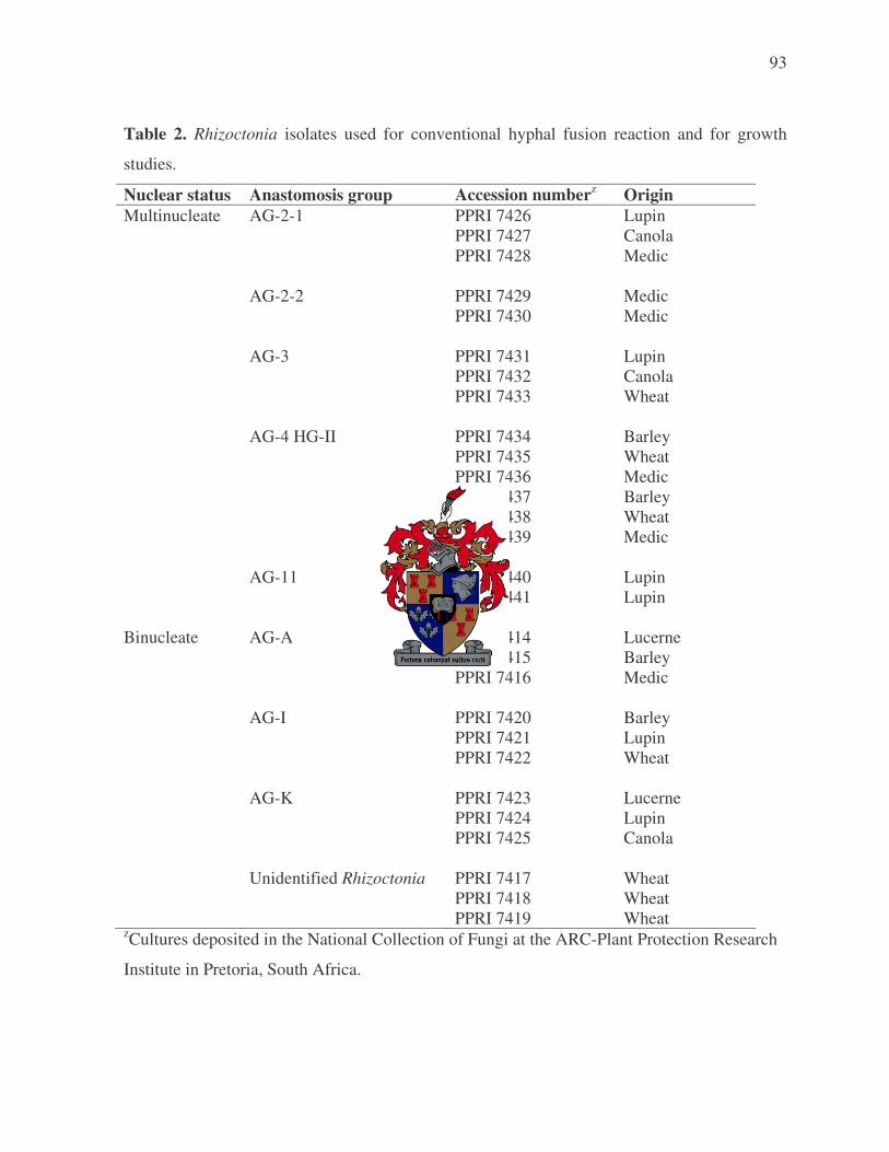
93
Table 2. Rhizoctonia isolates used for conventional hyphal fusion reaction and for growth
studies.
Nuclear status Anastomosis group Accession numberz Origin Multinucleate AG-2-1 PPRI 7426 Lupin PPRI 7427 Canola PPRI 7428 Medic AG-2-2 PPRI 7429 Medic PPRI 7430 Medic AG-3 PPRI 7431 Lupin PPRI 7432 Canola PPRI 7433 Wheat AG-4 HG-II PPRI 7434 Barley PPRI 7435 Wheat PPRI 7436 Medic PPRI 7437 Barley PPRI 7438 Wheat PPRI 7439 Medic AG-11 PPRI 7440 Lupin PPRI 7441 Lupin Binucleate AG-A PPRI 7414 Lucerne PPRI 7415 Barley PPRI 7416 Medic AG-I PPRI 7420 Barley PPRI 7421 Lupin PPRI 7422 Wheat AG-K PPRI 7423 Lucerne PPRI 7424 Lupin PPRI 7425 Canola Unidentified Rhizoctonia PPRI 7417 Wheat PPRI 7418 Wheat PPRI 7419 Wheat zCultures deposited in the National Collection of Fungi at the ARC-Plant Protection Research
Institute in Pretoria, South Africa.
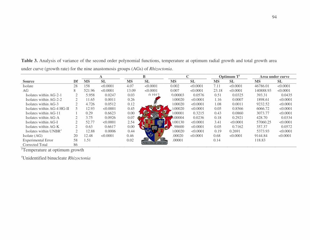
94
Table 3. Analysis of variance of the second order polynomial functions, temperature at optimum radial growth and total growth area
under curve (growth rate) for the nine anastomosis groups (AGs) of Rhizoctonia.
A B C Optimum Ty Area under curve Source Df MS SL MS SL MS SL MS SL MS SL Isolate 28 158 <0.0001 4.07 <0.0001 0.002 <0.0001 7.11 <0.0001 46786.01 <0.0001 AG 8 521.96 <0.0001 13.09 <0.0001 0.007 <0.0001 23.18 <0.0001 140888.93 <0.0001 Isolates within AG-2-1 2 5.958 0.0247 0.03 0.1917 0.00003 0.0576 0.51 0.0325 393.31 0.0435 Isolates within AG-2-2 2 11.65 0.0011 0.26 <0.0001 0.00020 <0.0001 1.16 0.0007 1898.61 <0.0001 Isolates within AG-3 2 4.726 0.0512 0.12 0.0051 0.00020 <0.0001 1.08 0.0011 9232.52 <0.0001 Isolates within AG-4 HG-II 5 12.93 <0.0001 0.45 <0.0001 0.00020 <0.0001 0.05 0.8566 6066.72 <0.0001 Isolates within AG-11 1 0.29 0.6623 0.00 0.8749 0.00001 0.3215 0.43 0.0860 3073.77 <0.0001 Isolates within AG-A 2 3.75 0.0926 0.07 0.3074 0.00004 0.0236 0.18 0.2921 428.70 0.0334 Isolates within AG-I 2 52.77 <0.0001 2.54 <0.0001 0.00130 <0.0001 3.41 <0.0001 57060.25 <0.0001 Isolates within AG-K 2 0.63 0.6617 0.00 0.9050 1.98600 <0.0001 0.05 0.7162 357.37 0.0572 Isolates within UNBRz 2 12.88 0.0006 0.44 <0.0001 0.00020 <0.0001 0.19 0.2691 5373.93 <0.0001 Isolate (AG) 20 12.48 <0.0001 0.46 <0.0001 0.00020 <0.0001 0.68 <0.0001 9144.84 <0.0001 Experimental Error 58 1.51 0.02 0.00001 0.14 118.83 Corrected Total 86
yTemperature at optimum growth zUnidentified binucleate Rhizoctonia
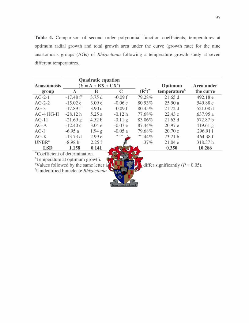
95
Table 4. Comparison of second order polynomial function coefficients, temperatures at
optimum radial growth and total growth area under the curve (growth rate) for the nine
anastomosis groups (AGs) of Rhizoctonia following a temperature growth study at seven
different temperatures.
Quadratic equation (Y = A + BX + CX2)
Anastomosis
group A B C
(R2)w
Optimum
temperaturex
Area under the curve
AG-2-1 -17.48 fy 3.75 d -0.09 f 79.28% 21.65 d 492.18 e AG-2-2 -15.02 e 3.09 e -0.06 c 80.93% 25.90 a 549.88 c AG-3 -17.89 f 3.90 c -0.09 f 80.45% 21.72 d 521.08 d AG-4 HG-II -28.12 h 5.25 a -0.12 h 77.68% 22.43 c 637.95 a AG-11 -21.69 g 4.52 b -0.11 g 83.06% 21.63 d 572.87 b AG-A -12.40 c 3.04 e -0.07 e 87.44% 20.97 e 419.61 g AG-I -6.95 a 1.94 g -0.05 a 79.68% 20.70 e 296.91 i AG-K -13.73 d 2.99 e -0.06 d 79.44% 23.21 b 464.38 f UNBRz -8.98 b 2.25 f -0.05 b 81.37% 21.04 e 318.37 h
LSD 1.158 0.141 0.004 0.350 10.286 wCoefficient of determination. xTemperature at optimum growth. yValues followed by the same letter in a column do not differ significantly (P = 0.05). zUnidentified binucleate Rhizoctonia.

96
Table 5. Frequency of isolation (number of isolates) of Rhizoctonia anastomosis groups (AG) from crops over four years. Rhizoctonia group Anastomosis group Cropw 2000 2001 2002x 2003 Total Multinucleate AG-2-1 B -y - - - - C 5 - 2 3 10 Luc - - - - - L 4 1 - 1 6 M - - 2 1 3 W - - - 1 1 Total 9 1 4 6 20 AG-2-2 B - - - - - C - - - - - Luc - - - - - L - - - - - M 2 - - - 2 W - - - - - Total 2 - - - 2 AG-3 B - - - - - C 2 - - - 2 Luc - - - - - L 1 - - - 1 M - - - - - W 2 1 - 2 5 Total 5 1 - 2 8 AG-4 HG-II B 1 14 - 2 17 C - 7 - 1 8 Luc - - - - - L - - - 1 1 M 11 10 3 8 32 W 8 1 3 2 14 Total 20 32 6 14 72 AG-11 B - - - - - C - - - - - Luc - - - - - L 2 - - - 2 M - - - - - W - - - - - Total 2 - - - 2 Multi. Total 38 34 10 22 104

97
Table 5. Continued Rhizoctonia group Anastomosis group Cropw 2000 2001 2002x 2003 Total Binucleate AG-A B -y 3 - - 3 C 5 - 1 - 6 Luc 6 2 - 4 12 L - - - - - M 1 - 2 - 3 W 1 3 1 3 8 Total 13 8 4 7 32 AG-I B 1 - - 2 3 C 1 - 1 5 7 Luc - - - - - L 2 - - - 2 M - - - - - W - 4 1 - 5 Total 4 4 2 7 17 AG-K B 2 3 - 11 16 C 13 8 3 1 25 Luc 5 6 2 - 13 L 7 6 6 4 23 M 30 19 22 9 80 W 3 8 - 3 14 Total 60 50 33 28 171 UNBRz B 2 5 - 2 9 C 7 2 2 4 15 Luc - - - 6 6 L 1 - - 1 2 M 11 6 2 30 49 W 17 2 - 4 23 Total 38 15 4 47 104 Binuc. Total 115 77 43 89 324 Overall total 153 111 53 111 428 wB = barley, C = canola, L = lupin, Luc = lucerne, M = medic/clover mixture and W = wheat. xPlants sampled only at third sampling time in 2002. y - = not isolated. zUnidentified binucleate Rhizoctonia.

98
Table 6. Analysis of variance for the effect of sampling time (ST) on the percentage incidence of bi- and multinucleate Rhizoctonia groups (RG) in four years.
zOnly one sampling time in 2002.
2000 2001 2002 2003 Source Df MS SL Df MS SL Df MS SL Df MS SL
Block 3 13.75 0.1880 3 15.13 0.2451 3 11.93 . 3 21.29 0.2218 ST 2 251.65 0.0003 2 137.25 0.0037 0 .z . 2 35.40 0.1104 Experimental Error (a) 6 6.23 0.6690 6 8.35 0.3341 0 . . 6 10.88 0.0926 RG 1 144.02 0.0105 1 41.67 0.0464 1 92.69 0.0060 1 111.92 0.0062 ST x RG 2 51.74 0.0662 2 6.08 0.4885 0 . . 2 9.68 0.3762 Experimental Error (b) 9 13.89 0.1406 9 7.82 0.3803 3 1.90 0.8814 9 8.86 0.1501 Sample Error 938 9.21 936 7.29 312 8.56 936 5.98 Corrected total 961 959 319 959

99
Table 7. Effect of sampling time on the mean percentage incidence of Rhizoctonia isolates over four years. Sampling timex 2000 2001 2002 2003
1 0.42 by 0.05 b -z 0.35 a 2 0.17 b 0.40 b - 0.42 a 3 1.81 a 1.32 a 0.89 0.96 a
LSD 0.482 0.559 - 0.638 xSampling time 1 = seedling stage (4 – 6 weeks after planting), 2 = mid season (12 – 14 weeks after planting) and 3 = flowering stage (20 – 22 weeks after planting). yMeans within a year (in a column) followed by the same letter do not differ significantly (P = 0.05) zPlants sampled only at third sampling time in 2002. Table 8. Mean percentage incidence of bi- and multinucleate Rhizoctonia groups over four years. Rhizoctonia group 2000 2001 2002 2003 Binucleate 1.19 az 0.80 a 1.42 a 0.91 a Multinucleate 0.42 b 0.38 b 0.35 b 0.23 b LSD 0.544 0.408 0.490 0.435 zMeans within a year (in a column) followed by the same letter do not differ significantly (P = 0.05)

100
0
5
10
15
20
25
30
35
March April May June July August September October
Month
Tem
pera
ture
(o C)
2000Max 2000Min 2001Max 2001Min
2002Max 2002Min 2003Max 2003Min
Figure 1. Mean daily maximum and minimum temperatures for March to October of 2000 - 2003 at the Tygerhoek experimental farm.

101
Figure 2. Nuclei (N) in vegetative hyphae of Rhizoctonia spp.
A) multinucleate species; B) binucleate species.
A
B
N
N

102
0
5
10
15
20
25
30
35
5 10 15 20 25 30 35
Temperature (oC)
Rad
ial g
row
th (m
m)
AG-2-1 AG-2-2 AG-3 AG-4 HG II AG-11
AG-A UNBR AG-I AG-K
Figure 3. Second order polynomial functions showing the predicted trends of the effect of temperature on radial growth rate of
different anastomosis groups of Rhizoctonia including the unidentified binucleate Rhizoctonia (UNBR) following a temperature
growth study at seven different temperatures.
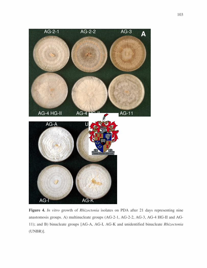
103
AG-2-1 AG-2-2 AG-3
AG-4 HG-II AG-4 HG-II AG-11 AG-A UNBR
AG-I AG-K Figure 4. In vitro growth of Rhizoctonia isolates on PDA after 21 days representing nine
anastomosis groups. A) multinucleate groups (AG-2-1, AG-2-2, AG-3, AG-4 HG-II and AG-
11); and B) binucleate groups [AG-A, AG-I, AG-K and unidentified binucleate Rhizoctonia
(UNBR)].
A
B

104
3. PATHOGENICITY OF RHIZOCTONIA ON ROTATION CROPS IN THE
WESTERN CAPE PROVINCE
ABSTRACT
The pathogenicity and relative virulence of 28 Rhizoctonia isolates were evaluated on
emerging seedlings as well as 14-day-old seedlings of seven crops (barley, canola, clover,
lucerne, lupin, medic and wheat) used within crop rotation systems in the Western Cape
province of South Africa. Prior to evaluating pathogenicity and relative virulence of these
isolates on emerging seedlings, an inoculum concentration trial was conducted, using three
anastomosis groups (AGs) and two crops. The trial indicated that an inoculum concentration
of 0.05% mass inoculum/mass planting medium will be appropriate for evaluating
pathogenicity and virulence of isolates on emerging seedlings. The inoculum concentration for
inoculation of 14-day-old seedlings had previously been established (Lamprecht et al.,
unpublished data). Pathogenicity tests using 28 Rhizoctonia isolates, representing nine AGs,
showed that AG-2-2 and AG-4 HG-II were the most virulent AGs on both seedling stages of
all crops. The exception was AG-2-2 that could only cause 22% damping-off on emerging
clover seedlings, as well as no significant root and hypocotyl symptoms on 14-day-old clover
seedlings. AG-4 HG-II was significantly less virulent on barley and wheat than on the other
crops, this is due to the fact that the assays were conducted on very young plants in the
absence of environmental stress (es). Similarly, AG-2-1 was least virulent on barley, only
affecting 14-day-old seedlings, and not being pathogenic at all on wheat. Contrarily, AG-2-1
was highly virulent on both seedling stages of canola. Rhizoctonia solani AG-2-1 was further
moderately virulent on both seedling stages of medic and lupin, but only weakly virulent on
both seedling stages of lucerne. Rhizoctonia solani AG-11 was moderate to weakly virulent on
both seedling stages of all crops, except barley and wheat. On wheat AG-11 was not
pathogenic at all, and on barley only coleoptile lesions were present on 14-day-old seedlings.
Rhizoctonia solani AG-3 was only weakly virulent on both seedling stages of canola, lupin

105
and medic. The binucleate AGs, viz., AG-A, AG-I and unidentified binucleate isolates were
not considered pathogenic, even though very low disease incidence was present in a few
instances. The binucleate Rhizoctonia AG-K was the only weakly virulent binucleate group
capable of causing significant disease on emerging seedlings as well as 14-day-old inoculated
canola, lucerne, lupin and medic seedlings. This is the first comprehensive study on the
pathogenicity of different AGs associated with crops used in rotation systems in the Western
Cape province of South Africa.
INTRODUCTION
The genus Rhizoctonia contains a diverse group of organisms, ranging from
saprophytes to plant pathogenic species. Pathogenic members of the genus are economically
important constraints to crop production in temperate and tropical agriculture (Sneh et al.,
1996). Therefore, worldwide efforts are being made to obtain more information on the
variation in virulence, or lack thereof, among Rhizoctonia isolates. This knowledge is
essential for developing sound management strategies against Rhizoctonia diseases, and
understanding the ecology of the genus.
The genus Rhizoctonia is extremely diverse, traditionally containing all fungi that do
not have distinctive taxonomic features (Vilgalys & Cubeta, 1994). Therefore, useful methods
have been developed to classify isolates to some taxonomic level. Classification of
Rhizoctonia isolates is often accomplished by first identifying their nuclear state as either
being bi- or multinucleate (Vilgalys & Cubeta, 1994; Yang et al., 1994). The most useful
system for identifying Rhizoctonia isolates is based on anastomosis groups (AGs), which are
determined by identifying hyphal anastomosis among isolates (Parmeter, Whitney & Platt,
1967; Parmeter, Sherwood & Platt, 1969). More recently, deoxyribonucleic acid (DNA)-
based methods have also been used to classify Rhizoctonia into AGs (Gonzalez et al., 2001).

106
The multinucleate Rhizoctonia solani Kühn isolates have been studied extensively due
to their ability to cause disease on more than 250 plant species under diverse environmental
conditions (Wong, Barbetti & Sivasithamparam, 1985). Some R. solani isolates have a wide
host range, while others show a higher level of host specificity (Anderson, 1982). Rhizoctonia
solani isolates can attack their annual host while still at the young seedling stage and/or when
the host is older. Symptoms incited by R. solani can vary from root and crown rot on older
plants, to damping-off, hypocotyl and coleoptile rot of young seedlings (Sneh et al., 1996). It
is also important to note that these fungi are significant pathogens in perennial cropping
systems, such as forest nursery production, perennial tree ecosystems and even natural
ecosystems (Mazzola, 1997). Currently, R. solani can be divided into fourteen AGs [AG-1 to
AG-10 including AG BI (Sneh, Burpee & Ogoshi, 1991), AG-11 (Carling et al., 1994), AG-12
(Carling et al., 1999) and AG-13 (Carling, Kuninaga & Brainard, 2002)], some of which are
further divided into subgroups.
Similar to multinucleate isolates, binucleate Rhizoctonia isolates are also diverse and
have been isolated from a wide number of plant species. However, unlike the multinucleate
isolates, binucleate isolates are not commonly regarded as highly virulent plant pathogens
(Anderson, 1982; Roberts & Sivasithamparam; 1986). Binucleate Rhizoctonia isolates have
been divided into AG-A to S by Sneh et al. (1991).
Rhizoctonia species can be a potential production limiting factor to rotation crops
[barley (Hordeum vulgare L.), canola (Brassica napus L. var. oleifera DC), clover (Trifolium
spp.), lucerne (Medicago sativa L.), lupin (Lupinus spp.), medic (annual Medicago spp.) and
wheat (Triticum aestivum L.)] that are currently being planted in the Western Cape province of
South Africa. Rhizoctonia spp. are known to have a significant impact on the health of these
crops in other countries (Samuel & Garret, 1932; Bretag, 1985; Kaminski & Verma, 1985;
Weller et al., 1986; Pumphrey et al., 1987; Kronland & Stanghellini, 1988; Kataria & Verma,
1992; Vincelli & Herr, 1992; MacLeod & Sweetingham, 1997). For example, R. solani has
been reported as widely distributed and very destructive on lupin and canola (Acharya et al.,

107
1984; Kaminski & Verma, 1985; Hwang, Swanson & Evans, 1986; Gugel et al., 1987;
Yitbarek Verma & Morrall, 1987; Kataria & Verma, 1992; Leach & Clapham, 1992). This is
especially a concern since Rhizoctonia spp. have often been isolated from all the rotation crops
used in the Western Cape province of South Africa (Lamprecht, De Villiers & Janse van
Rensburg, 1999; Lamprecht, Auret & Janse van Rensburg 2000a; Lamprecht et al., 2000b,
2001, 2002; Auret, Janse van Rensburg & Lamprecht, 2002). Unfortunately, the pathogenicity
and virulence of South African Rhizoctonia isolates are unknown on the rotation crops of
interest, except for the pathogenicity of R. solani AG-6 (Smith & Wehner, 1986) and R.
cerealis Van der Hoeven (Scott, Visser & Rufenacht, 1979) on wheat.
The main objective of this study was to determine the pathogenicity and relative
virulence of Rhizoctonia AGs associated with seven rotation crops (barley, canola, clover,
lucerne, lupin, medics and wheat) used in the Western Cape province of South Africa (Chapter
2). The Rhizoctonia isolates used for pathogenicity testing were previously isolated from a
crop rotation trial over a four-year period (2000 to 2003) at the Tygerhoek experimental farm,
Riviersonderend, Western Cape province (Chapter 2). Three independent trials were
conducted to determine the pathogenicity and relative virulence of isolates on emerging
seedlings as well as 14-day-old seedlings. The first trial, an inoculum concentration trial,
allowed the establishment of an appropriate inoculum concentration for determining the
pathogenicity and relative virulence of Rhizoctonia isolates on emerging seedlings.
Subsequently, the pathogenicity of 28 isolates representing nine AGs were determined on
emerging seedlings of all seven rotation crops. In a third trial the pathogenicity and relative
virulence of these isolates were also determined on 14-day-old seedlings. The inoculum
concentration for determining pathogenicity and virulence on 14-day-old seedlings had
previously been established (Lamprecht et al., unpublished data).

108
MATERIALS AND METHODS
Isolates
Rhizoctonia isolates used in the study were obtained from the aforementioned trial at
the Tygerhoek experimental farm, Riviersonderend, Western Cape province (Chapter 2).
Cultures were stored on potato carrot agar slants [10 g potato, 10 g carrot and 12 g agar
(Bacteriological, Biolab Diagnostics, Midrand, South Africa)] at 10°C and/or sterile wheat
grain in a deep freezer adapting (excluding vermiculite) the method used by Sneh et al. (1986)
to prepare inoculum. Rhizoctonia isolates were all deposited in the Culture Collection of the
Plant Protection Research Institute, Pretoria (PPRI). The accession numbers, crop origin and
nuclear state of isolates are listed in Table 1. The anastomosis grouping of isolates had
previously been determined through sequence analyses of the ribosomal internal transcribed
spacer region (ITS) and confirmed by hyphal fusion reactions (Chapter 2).
Inoculum preparation
Millet seed inoculum was used in all three pathogenicity trials (inoculum
concentration, emerging seedling and 14-day-old seedling trial). Millet seed inoculum was
prepared according to the method described by Strauss and Labuschagne (1995). Millet seeds
were soaked in deionised water for 12 h (200 g seed/100 mL in 1 L Schott bottles) and then
autoclaved for 20 min at 120°C on two consecutive days. The bottles were shaken before
autoclaving and each time after autoclaving. The autoclaved millet seeds were inoculated with
10 (6 mm diam.) mycelial discs punched from 5-day-old Rhizoctonia cultures grown on potato
dextrose agar (PDA) (Biolab Diagnostics, Midrand, South Africa). Control bottles were
inoculated with discs of PDA only. The bottles with inoculated millet seeds were incubated in
an incubation room at 25°C for 10 days and shaken every third day to ensure thorough seed
colonisation.

109
Planting media and plant growth conditions
Pathogenicity trials were conducted using 13 cm diam. plastic pots with a capacity of
holding 700 g of planting medium. The planting medium was made up of equal quantities of
soil [collected from land adjacent to the rotation trial at Tygerhoek (Chapter 2)] perlite and
sand, and was pasteurised (30 min at 83°C) 3 days before being mixed with inoculum
(inoculum concentration and emerging seedling trial) or sown with seeds (14-day-old seedling
trial). Each pot was placed on an inverted saucer, thereby lifting it above shelf level to avoid
possible cross contamination from drainage water.
All trials were conducted in a growth room at 15°C night and 25°C day temperatures
with Sylvania Gro-Lux lights, a 10 h photoperiod and a relative humidity of 60 to 70%. Pots
were watered every third day to field capacity.
Inoculum concentration trial on emerging seedlings
A trial was conducted to determine the appropriate inoculum concentration for
evaluating pathogenicity and relative virulence of Rhizoctonia isolates on emerging seedlings.
Three isolates representing three AGs, viz., AG-2-1 (PPRI 7426), AG-4 HG-II (PPRI 7434)
(multinucleate isolates) and AG-K (PPRI 7423) (binucleate isolate) (Table 1) were used.
Multinucleate and binucleate isolates were specifically chosen since the multinucleate isolates
are known to be highly virulent whereas the binucleate isolates are typically less virulent.
Two crops, i.e. wheat and canola were used to represent a monocotyledonous and
dicotyledonous crop, respectively.
Millet seed inoculum of each AG was individually mixed with planting media at five
different concentrations, viz. 0.005, 0.0158, 0.05, 0.158 and 0.5% mass inoculum/mass
planting media (wt/wt). Controls containing millet seed at the five different concentrations
that were not inoculated with Rhizoctonia, were also included. Millet seed inoculated planting

110
media were placed in sterile pots and watered until water dripped from the bottom of the pots.
The pots were left to stand overnight in the growth room before making ten holes in the soil of
each pot using sterile 1 cm diameter doweling rods. Canola (cv. Varola 50) and wheat (cv.
SST57) seeds were planted at 50 seeds/pot (5 seeds/hole) in 1.5 and 2 cm holes respectively.
Pathogenicity and relative virulence of the AGs at different inoculum concentrations
were determined by calculating the percentage damping-off and percentage reduction in dry
mass of seedlings 14 days after planting. Percentage damping-off caused by each AG at a
specific inoculum concentration and crop was determined by using the formula: [(Number of
emerged seedlings in crop A at x % w/w un-inoculated millet seed – number of emerged
seedlings in crop A at x % w/w AG inoculated millet seed) / number of emerged seedlings in
crop A at x% w/w un-inoculated millet seed] × 100. The percentage dry mass reduction was
determined using a similar formula except that dry mass was used instead of number emerged
seedlings. Dry mass was determined by placing the above ground material of each pot in
paper bags and drying in an oven at 60°C for 3 days. Re-isolation of the different AGs was
carried out by washing plants under running tap water to remove adhering soil. Plants were
surface disinfected in 1% sodium hypochlorite, rinsed twice in sterile distilled water, and air-
dried in a laminar flow cabinet. Four small pieces of root and crown tissue were excised and
plated on PDA.
The experiment used a complete randomised block design. The treatment design for
the trial was a 3 x 2 x 5 factorial with three replications per treatment. Factors were three
AGs, two crops and five concentrations. An experimental unit consisted of a pot with 50
seeds. The trial was conducted twice, with three replicates per treatment.
Emerging seedling pathogenicity trial
The pathogenicity and virulence of 28 Rhizoctonia isolates representing nine AGs
(Table 1) were determined on the emerging seedlings of seven crops including canola (cv.

111
Varola 50), clover (T. michelianum Savi cv. Patrick), lucerne (cv. SA Standard), lupin (L.
albus L. cv. Wonga), medic (M. truncatula Gaertn. cv. Sephi), wheat (cv. SST57) and barley
(cv. Clipper). Inoculation of planting media was done as described under inoculum
concentration trial except that only one inoculum concentration (0.05% w/w) was used. All
crops were planted at 50 seeds/pot (5 seeds/hole) except for lupin that was planted at 20
seeds/pot (2 seeds/hole). The planting depth for all crops was 1.5 cm except for barley, lupin
and wheat that were planted at 2 cm depth. Controls consisting of plant media amended with
0.05% un-inoculated millet seed/planting media were included for each crop.
Pathogenicity and relative virulence of the AGs on the emerging seedlings of the
different crops were determined by calculating the percentage damping-off and percentage
reduction in dry mass of seedlings 14 days after planting. Percentage damping-off caused by
each AG on a specific crop was determined by using the following formula: [(Number of
emerged seedlings in control crop A – number of emerged seedlings in AG inoculated crop A)
/ number of emerged seedlings in control crop A] × 100. The percentage dry mass reduction
was determined using a similar formula except that dry mass was used instead of number
emerged seedlings. Dry mass and re-isolation of Rhizoctonia was done as previously
described.
Experimental design was a randomised block design. The treatment design was a 29 x
7 factorial with two replications per treatment. The factors were 29 (28 isolates and a control)
isolates (representing nine AGs) and seven crops. An experimental unit consisted of a pot
with 50 seeds for all the crops except lupin where 20 seeds were planted in each pot. The trial
was conducted twice.
Fourteen-day-old seedling pathogenicity trial
The 28 Rhizoctonia isolates (Table 1) tested for their pathogenicity towards emerging
seedlings, were also evaluated for their pathogenicity and virulence toward 14-day-old
112
seedlings of seven crops (barley, canola, clover, lucerne, lupin, medic, and wheat). Pots were
filled with pasteurised planting medium and an empty test tube (17 x 150 mm) was inserted in
the centre of each pot. Five planting holes were made around each test tube at 1 cm from the
test tube. The same seven crop cultivars used in the emerging seedling pathogenicity trial
were also used in the 14-day-old pathogenicity trial. Seed planting depths of the crops were
similar to that previously described. The number of seeds planted per hole was five for all
crops, except three seeds were planted for lupin. Fourteen days after planting, the test tube
was removed from each pot and planting medium amended with 0.5% w/w inoculum was
placed in the holes left by the test tubes at 30 g/pot.
Pathogenicity and relative virulence of isolates were determined by evaluating root rot,
coleoptile rot (barley and wheat) and hypocotyl rot (canola, clover, lucerne, lupin and medic).
Fourteen days after inoculating the seedlings the plants were carefully removed from pots.
Ten plants from each pot were used for disease assessments. Root rot was rated according to
MacNish et al. (1995) on a 0 to 4 scale, with 0 = no obvious symptoms; 1 = slight
discolouration or small superficial lesions; 2 = moderate discoloration or extensive but non-
girdling lesions; 3 = extensive discolouration of tissue or girdling lesions; and 4 = plant dead.
Coleoptile (barley and wheat) and hypocotyl (canola, clover, lupin, lucerne and medic) rot
were also rated on a 0 to 4 scale as described for root rot. Re-isolation of Rhizoctonia was
done as previously described.
The experimental and treatment design for the trial was the same as for the emerging
seedling trial, viz., a 29 x 7 factorial with two replications (pots) per treatment. The 29 (28
isolates and a control) isolates represented the nine AGs. An experimental unit consisted of a
pot with 25 seeds for all the crops except lupin where 15 seeds were planted in each pot. The
trial was conducted twice and utilized a randomised block design.
113
Statistical analyses
Statistical analyses of all three trials were done similarly. Levene’s variance ratio test
(Levene, 1960) was performed to test for homogeneity of trial variances between the trial
repeats. In these analyses, data of the two independent trials were considered block treatments
and the replications within each trial were used as subsamples, providing that Levene’s
variance ratio test showed homogeneity in trial variance. Data were subjected to analysis of
variance (SAS 1999), and the Shapiro-Wilk test was performed to test for normality (Shapiro
& Wilk, 1965). In cases where deviations from normality were due to kurtosis and not
skewness, the data were accepted as reliable and the results were interpreted without
transformation (Glass, Peckham & Sanders, 1972). Pearson correlations between variables
were also calculated.
RESULTS
Effect of inoculum concentration on seedling emergence
Variance for the data from the two independent trials was comparable based on
Levene’s variance ratio test (Levene, 1960). The error variance ratios for the percentage
damping-off was P = 0.8509 and P = 0.9085 for the percentage reduction in dry mass.
Consequently, data from the two trials were combined in all the analyses.
Analyses of variance were conducted on the mean percentage damping-off and
percentage dry mass reduction of canola and wheat resulting from different inoculum
concentrations of the three AGs. Significant crop x AG x inoculum concentration interactions
were recorded for percentage damping-off (P < 0.0001) and percentage dry mass reduction (P
< 0.0001) (Table 2).

114
The influence of inoculum concentration on disease development incited by the three
Rhizoctonia AGs on wheat and canola was evaluated using two parameters, i.e. percentage
damping-off and percentage dry mass reduction. Correlation analysis showed that there was a
very high correlation (P < 0.0001, r = 98542) between percentage damping-off and percentage
dry mass reduction. Therefore, only percentage damping-off will subsequently be discussed
as a measure of pathogenicity and virulence.
The multinucleate Rhizoctonia solani isolates (AG-2-1 and AG-4 HG-II) were more
virulent on canola than on wheat. Consequently, the extent of damping-off of canola caused
by AG-2-1 and AG-4 HG-II could not be distinguished significantly (P = 0.05) at high
inoculum concentrations, viz., 0.158 and 0.5 %w/w (Fig. 1). Contrarily, on wheat significant
(P = 0.05) differences in percent damping-off between the multinucleate isolates could be
discerned at all inoculum concentrations, except at the lowest inoculum concentration
(0.005%) (Fig. 1). Although it seemed that the percentage damping-off on wheat decreased
with increasing inoculum concentration, this was not significant (Fig. 1).
Binucleate Rhizoctonia AG-K caused low percentages of damping-off of canola as
well as wheat at all inoculum concentrations (Fig. 1). Inoculation of wheat seedlings with
different concentrations of the binucleate Rhizoctonia isolate did not result in a significant
increase in percentage damping-off of wheat seedlings. However, on canola a significant
increase in percentage damping-off of seedlings was observed with an increase in inoculum
concentration of the binucleate Rhizoctonia (Fig. 1).
Emerging seedling pathogenicity trial
Levene’s variance ratio test (Levene, 1960) showed that variability values in the
observations of the two independent trials were of comparable magnitude and that an analysis
of combined data of the two trials could be validly carried out. The error variance ratio for the

115
percentage damping-off was P = 0.3727 and P = 0.5647 for the percentage reduction in dry
mass. Combined data of the two trials were analysed.
Analyses of variance were conducted on the percent damping-off and dry mass
reduction of seven crops caused by nine AGs of Rhizoctonia spp. Significant crop x AG
interactions were recorded for both damping-off (P < 0.0001) and dry mass reduction (P <
0.0001). There were no significant differences in percentage damping-off (P < 0.0001) or
percentage dry mass reduction (P < 0.0001) caused by isolates within seven of the nine AGs.
However, a significant difference was present in percentage damping-off (P < 0.0001) and
percentage dry mass reduction (P < 0.0001) caused by isolates within AG-2-1 and AG-2-2
(Table 3).
Similar to the inoculum concentration trial, a significant positive correlation (P <
0.0001, r = 0.97685) was present between percentage damping-off and percentage dry mass
reduction caused by the different AGs on the seven crops (Table 3). Therefore, pathogenicity
and relative virulence of the AGs towards the different crops will only be discussed with
regard to damping-off results.
Rhizoctonia solani AG-2-1 caused a significant amount of damping-off on canola,
lucerne, lupin and medic (Fig. 2). Canola was most susceptible of all crops with 96% of
seedlings failing to emerge. Lucerne had significantly less damping-off than the
aforementioned crops when inoculated with AG-2-1 (Fig. 2). Barley, clover and wheat
showed no significant damping-off when inoculated with AG-2-1 (Fig. 2).
Rhizoctonia solani AG-2-2 caused significant damping-off (between 22% and 88%) on
all seven crops (Fig. 2). Of these crops canola, lupin and medic suffered significantly more
damping-off (more than 87%) than the other crops (Fig. 2). Clover and lucerne had the least
amount (< 25%) of damping-off when inoculated with AG-2-2 (Fig. 2).
116
Rhizoctonia solani AG-3 was least virulent of all the multinucleate AGs on all seven
crops, only causing significant damping-off on canola, lupin and medic (Fig. 2). Damping-off
caused by AG-3 did not exceed 28% among the crop plants examined (Fig. 2).
Rhizoctonia solani AG-4-HG-II caused significant damping-off on all seven crops (Fig. 2).
Barley and wheat had significantly less damping-off (less than 22%) than the other crops that
all had more than 74% damping–off when inoculated with AG-4 HG-II (Fig. 2).
Rhizoctonia solani AG-11 was moderately virulent on five of the seven crops,
inlcuding canola, clover, lucerne, lupin and medic (Fig. 2). All of the crops cited above
exhibited significantly more damping-off than the control (Fig. 2).
The binucleate R. solani isolates did not cause significant damping-off on most of the
crops (Fig. 2). AG-I and AG-A only caused significant damping-off on lupin (7.92%) and
canola (8.29%) respectively. Rhizoctonia AG-K caused a significant amount of damping-off
(between 12 and 22%) on canola, lupin, clover and medic (Fig. 2).
The number of Rhizoctonia AG’s capable of inciting disease on any specific crop plant
varied among those evaluated in these studies (Fig. 2). Barley and wheat were least
susceptible, only suffering significant damping-off when inoculated with two AGs (AG-2-2
and AG-4 HG-II). Significant damping-off (between 8 and 75%) of clover was incited by four
AGs (AG-2-2, AG-4 HG-II, AG-11 and AG-K). Clover and lucerne were susceptible to four
of the AGs, showing between 8 and 84% damping-off. Medic was susceptible to six AGs with
more than 22% damping-off caused by all AGs. Lupin (Figs 3, 4) and canola were susceptible
to six of the AGs (Fig. 2). Lupin was very susceptible to AG-2-1, AG-2-2 and AG-4 HG-II
(Figs 2, 3).
117
Fourteen-day-old seedling pathogenicity trial
Variances in the data from the two independent trials were comparable based upon
Levene’s variance ratio test (Levene, 1960). The error ratios [P = 0.2562 (root rot), P =
0.9662 (coleoptile rot) and P = 0.3455 (hypocotyl rot)] indicated that an analysis of the
combined data of the two trial repeats could be validly carried out.
Analyses of variance were conducted on root rot of seven crops, on coleoptile rot of
barley and wheat, and on hypocotyl rot of canola, clover, lucerne, lupin and medic caused by
nine Rhizoctonia AGs (Table 4, 5). Significant crop x AG interactions were recorded for root
(P < 0.0001), coleoptile (P = 0.0228) and hypocotyl (P < 0.0001) rot. Significant differences
in root rot severity caused by isolates within AG-4 HG-II (P = 0.0010) and AG-K (P =
0.0125) and for coleoptile rot caused by isolates within AG-4 HG-II (P = 0.0398) were
observed (Tables 4, 5).
Rhizoctonia solani AG-2-1 was pathogenic on 14-day-old inoculated seedlings of
canola, lucerne, medic and lupin (Figs 5, 6). Fourteen-day-old canola and lupin seedlings had
the highest root rot rating, whereas medic and canola seedlings had the highest hypocotyl rot
rating. Rhizoctonia solani AG 2-1 was only very weakly virulent or not pathogenic at all to
barley, wheat and clover (Figs 5, 6). Lucerne also only showed low root- and hypocotyl rot
ratings when inoculated with AG-2-1 (Figs 5, 6).
Rhizoctonia solani AG-2-2 caused significant rot (root, hypocotyl or coleoptile) on 14-
day-old inoculated seedlings of all the rotation crops, except clover (Figs 5 – 7). AG-2-2 was
weakly virulent to barley and wheat, causing slight coleoptile rot. Fourteen-day-old lupin and
medic was most susceptible to AG-2-2, having significantly more root- and hypocotyl or
coleoptile rot than lucerne, barley and wheat (Figs 5 – 7).
118
Rhizoctonia solani AG-3 caused significant root rot on canola and lupin, as well as
slight, but significant hypocotyl rot on 14-day-old inoculated medic seedlings (Figs 5, 6).
Rhizoctonia solani AG-4 HG-II caused significant disease (either root-, hypocotyl- or
coleoptile rot) on 14-day-old seedlings of all the rotation crops (Figs 5 – 7). Hypocotyl and
root rot caused by this AG on lupin seedlings are illustrated in Fig. 8. Rhizoctonia solani AG-4
HG-II was only weakly virulent on barley and wheat seedlings causing no significant root rot,
and only slight coleoptile rot on both crops (Figs 5, 7).
Rhizoctonia solani AG-11 was mildly virulent on 14-day-old inoculated canola, clover,
lucerne, lupin and medic seedlings (Figs 5, 6). Rhizoctonia solani AG-11 caused significant
root rot on all crops except barley and wheat. Wheat coleoptiles were not susceptible to AG-
11. Medic had significant more hypocotyl rot than the other crops (Figs 5 – 7).
The binucleate Rhizoctonia isolates did not cause significant above ground symptoms
(coleoptile or hypocotyl rot) on 14-day-inoculated seedlings of any of the crops, except for
AG-K that caused significant hypocotyl rot on medic (Figs 6, 7). Rhizocotonia AG-K also
caused significant root rot on 14-day-old canola, lucerne, lupin and medic seedlings (Fig. 5).
Recovery of introduced Rhizoctonia AGs from plant hypocotyl, coleoptile and roots
The respective AGs were re-isolated from diseased plant material from all three trials,
but not from the control plants. Rhizoctonia spp. re-isolated from infected seedlings were
compared with the original isolate by studying nuclear number and morphological
characteristics. All isolates obtained had characteristics similar to the original cultures.
119
DISCUSSION
In this study the first attempt was made to characterize the pathogenicity and relative
virulence of Rhizoctonia associated with rotation crops (barley, canola, clover, lucerne, lupin
medic, wheat) used in the Western Cape province of South Africa. The pathogenicity and
relative virulence of 28 Rhizoctonia isolates (representing nine AGs) previously isolated from
rotation crops (Chapter 2) varied on emerging seedlings and 14-day-old seedlings of these
crops. Rhizoctonia solani AG-4 HG-II and AG-2-2 were pathogenic to emerging seedlings as
well as 14-day-old inoculated seedlings of all rotation crops. The only exception was 14-day-
old clover seedlings that did not develop significant root and hypocotyl rot after inoculation
with AG-2-2. Rhizoctonia solani AG-2-1 was highly virulent on canola, and moderately
virulent on lupin and medic seedlings. Rhizoctonia solani AG-3 and AG-11 were pathogenic
on some of the crops, but were clearly not as virulent as AG-4 HG-II and AG-2-2. The
binuleate Rhizoctonia isolates AG-I and AG-A only caused a very low percentage damping-
off (< 8.25%) on emerging lupin and canola seedlings respectively. Binucleate isolates of
AG-K were weakly virulent on both seedling stages of some crops. Crop wise barley and
wheat were most tolerant to all Rhizoctonia AGs, whereas canola seemed most susceptible
followed by lupin, medic, lucerne and clover.
One of the important aims of this study was to differentiate the pathogenicity and
relative virulence of Rhizoctonia AGs on emerging seedlings of rotation crops used in the
Western Cape province of South Africa. In order to differentiate pathogenicity and relative
virulence of Rhizoctonia isolates, knowledge of the effect of inoculum concentration on
emerging seedlings is of utmost importance. Inoculum concentrations that are too high can
result in an inability to distinguish between highly virulent isolates, whereas use of too low
inoculum concentration can result in an inability to distinguish the virulence of weakly
virulent isolates. The inoculum concentration trial clearly showed that high inoculum
concentrations (0.158% w/w and 0.5% w/w) would not allow differentiation between highly
virulent AGs of multinucleate isolates (AG-4 HG-II and AG-2-1) on emerging seedlings of

120
susceptible crops such as canola. Conversely, at too low inoculum concentrations (0.005%
and 0.0158%) the virulence and pathogenicity of weakly virulent binucleate isolates (eg. AG-
K) may be difficult to determine on a susceptible crop such as canola, as well as on more
resistant crops like wheat. Therefore, the 0.05% wt/wt inoculum concentration was considered
most appropriate for determining pathogenicity and relative virulence of Rhizoctonia AGs on
emerging seedlings of different crops. Since the aim of the study was to determine the relative
susceptibility to multiple AG’s of Rhizoctonia spp. among seven crops within the same
cropping system, all seven crops had to be evaluated at the same fungal inoculum
concentration.
Various other studies have used different inoculum concentrations than what was used
in this study. Pathogenicity studies conducted by Sweetingham et al. (1986), Yang et al.
(1994), MacLeod and Sweetingham (1997) used lower inoculum concentrations than what was
used in this study. However, these studies all first allowed establishment of the pathogen for at
least two weeks within the planting mix, before planting the various crops. Pathogenicity
studies conducted by Botha et al. (2003), Sterne and Jones (1978), Demirci (1998) and Ogoshi
et al. (1990) used a higher inoculum concentration than this study. These differences in
inoculum concentration used in previous pathogenicity tests could explain why the
pathogenicity of some AGs on certain crops in this study, does not correspond to the
pathogenicity of these AGs on certain crops in other studies.
In this study AG-2-2 was pathogenic to emerging seedlings of all crops, as well as 14-
day-old seedlings of all seven rotation crops, except clover. The pathogenicity of AG-2-2 on
emerging seedlings as well as 14-day old seedlings of almost all the rotation crops suggest that
this AG could have a significant impact on crop production in the Western Cape province of
South Africa, with clover and wheat being least affected. Information on the incidence and
pathogenicity of AG-2-2 on the rotation crops evaluated in this study is limited in other
locations. AG-2-2 is divided into three intraspecific groups, viz., AG-2-2 IIIB, AG-2-2 IV and
AG-2-2 LP (Carling & Sumner, 1992; Kanematsu & Naito, 1995; Hyakumachi et al., 1998;

121
Carling et al., 2002) and pathogenicity studies do not always indicate which intraspecific
group has been studied.
Rhizoctonia solani AG-2-1, one of the other subgroups of AG-2, was found highly
virulent on canola in this study. AG-2-1 not only caused more than 95% damping-off of
emerging canola seedlings, but also incited significant root and hypocotyl rot on 14-day-old
canola seedlings. This is the first report of AG-2-1 being a pathogen of canola in South
Africa. In western Canada AG-2-1 is also considered as a very important pathogen of canola
causing damping-off and root rot of seedling and adult canola (Acharya et al., 1984; Kaminski
& Verma, 1985; Hwang et al., 1986; Gugel et al., 1987; Kataria & Verma, 1992). Similarly,
in Australia AG-2-1 is also regarded as one of the predominant and most virulent AGs on
canola causing hypocotyl rot and post-emergence damping-off (Khangura, Barbetti &
Sweetingham, 1999). In concert, the data from these studies indicate that AG-2-1 is an
important pathogen limiting canola production on a worldwide basis.
Rhizoctonia solani AG-2-1 was moderately virulent on both seedling stages of lupin
and medic in this study. This AG does not seem to be a common pathogen of lupin and medic
as no other reports of its pathogenicity on these crops were uncovered. This is also the first
report of AG-2-1 as a pathogen of lupin and medic in South Africa, indicating that it could
have the potential to reduce crop yields.
Rhizoctonia solani AG-2-1 was not virulent on clover, wheat and barley, but
moderately virulent on lucerne in this study. AG-2-1 caused 10.2% damping-off of emerging
lucerne seedlings, as well as significant root and hypocotyl rot on 14-day-old inoculated
seedlings. Contrarily, Eken and Demirci (2003) reported that AG-2-1 is not pathogenic on
lucerne. Barley, clover and wheat did not have significant disease symptoms at the emerging
seedling or 14-day-old seedling stage when inoculated with AG-2-1 in this study. However,
Wong et al. (1985) reported that AG-2-1 is highly virulent to subterranean clover in Western
Australia, and Demirci (1998) found AG-2-1 to be moderately virulent on barley and wheat.

122
Rhizoctonia solani AG-4 has a wide host range causing damping-off of seedlings and
root rot, crown rot, root canker, stem canker and stem blight of older plants (Anderson, 1982;
Carling & Sumner 1992; Vincelli & Herr 1992; Kulik & Dery, 1995). AG-4 is subdivided
into two groups, viz., HG-I and HG-II based on sclerotial form and differences in DNA base
sequence homology (Vilgalys, 1988; Sneh et al., 1991). However, these groups are often only
referred to as AG-4 when recording pathogenicity.
In this study R. solani AG-4 HG-II was highly virulent on lupin, medic, clover, canola
and lucerne. Rhizoctonia solani AG-4 HG-II caused more than 74% seedling damping-off, as
well as significant rot (root- and hypocotyl rot) on 14-day-old seedlings of these crops.
Rhizoctonia solani AG-4 (subgroups unknown) has also been found pathogenic to lupin,
medic, clover, canola and lucerne in other countries. Pemberton et al. (1998) reported that R.
solani AG-4 caused crown discolouration, severe root rot and 69% plant death of clover
(Trifolium vesiculosum Savi). AG-4 has been reported to cause various symptoms on lucerne
including black root canker, pre- and post-emergence damping off of mature plants and stem
canker (Vincelli & Herr, 1992; Kulik, Dery & Douglass, 1995; Eken & Demirci, 2003). AG-4
has also been reported as a pathogen of lupin cv. Ultra (Leach & Clapham, 1992) and medic
(27 annual Medicago spp.) (Kulik & Dery, 1995). However, Leach and Clapham (1992)
reported that AG-4 infected lupin (L. albus), but that this AG produced only small lesions on
stems. Rhizoctonia solani AG-4 has been widely reported as a pathogen of canola causing
damping-off and root rot on adult canola (Acharya et al., 1984; Kaminski & Verma, 1985;
Hwang et al., 1986; Gugel et al., 1987; Yitbarek et al., 1987; Teo et al., 1988; Yitbarek et al.,
1988; Kataria & Verma, 1990; Kataria & Verma, 1992; Baird, 1996).
Among the rotation crops examined, Rhizoctonia solani AG-4 HG-II was least virulent
to wheat and barley. AG-4 HG-II did not cause more than 22% damping-off of emerging
seedlings, and was only mildly virulent on 14-day-old wheat and barley seedlings causing
slight coleoptile rot. Contrary to the results of this study, Ogoshi, Cook & Bassett (1990)
found that isolates of AG-4 were not pathogenic at all on barley and wheat. Demirci (1998)

123
found AG-4 to be highly virulent on barley cv. Tokak and wheat cv. Kirik in Turkey (Demirci,
1998). The fungus was also found to cause damping-off and root rot of wheat in Texas and in
Western Australia (Mathieson & Rush, 1991; Rush et al., 1994; Yang et al., 1994). In
Arkansas, Canada and England, R. solani AG-4 has been reported as the causal agent of sharp
eyespot on wheat, causing seedling damping-off but not infecting roots (Sterne & Jones, 1978;
Rush et al., 1994). In South Africa, AG-4 has not been associated with sharp eyespot of
wheat, with R. cerealis (AG-D) being the only identified AG (Scott et al., 1979). Previously,
R. solani AG-6 was found to be pathogenic on wheat in South Africa causing crater disease
(Meyer et al., 1998). The fact that AG-4 HG-II was the most prevalent Rhizoctonia AG in the
trial evaluated at Tygerhoek (Chapter 2), and that it is less virulent on barley and wheat than
on the dicotyledonous crops in this study, has important implications for selecting the
appropriate cropping systems for the winter rainfall region of the Western Cape province. It is
therefore important to rotate dicotyledonous (canola, clover, lucerne, lupin, medic) with
monocotyledonous (barley and wheat) crops in the region.
In this study AG-11 was moderate to weakly virulent to emerging seedlings as well as
14-day-old inoculated seedlings of all crops with the exception of barley and wheat. This
appears to be the first report of AG-11 as a pathogen of canola, clover, medic and lucerne.
Lupin was the most susceptible of all the rotation crops suffering 50% damping-off when
inoculated with AG-11. Rhizoctonia solani AG-11 (Carling et al., 1994) was first described as
a pathogen of lupin in 1986 in Australia (Sweetingham, Cruickshank & Wong, 1986), but
there is no information concerning its pathogenicity toward canola, clover, medic and lucerne.
In this study AG-11 was not pathogenic on wheat, and only weakly virulent to 14-day-old
barley seedlings. Contrarily, Demirci (1998) found AG-11 to be highly virulent on wheat as
well as barley in Turkey. Sweetingham et al. (1986) and Carling et al. (1994) found that AG-
11 caused coleoptile rot of wheat in greenhouse and growth-chamber studies. This discrepancy
could be due to the fact that Sweetingham et al. (1986) first allowed the colonisation and
establishment of AG-11 in the growth medium before sowing seeds. Demirci (1998) used

124
wheat kernels and Carling et al. (1994) used agar disks as inoculum source, that were placed
in indirect contact with the seeds during planting.
Rhizoctonia solani AG-3 was weakly virulent on emerging as well as 14-day-old
inoculated canola, lupin and medic seedlings. This is the first report of AG-3 being pathogenic
on lupin, canola and medic seedlings. Only two other studies reported on the virulence of AG-
3 to these crops, indicating that AG-3 is not pathogenic on lupin (Leach & Clapham; 1992)
and lucerne (Eken & Demirci, 2003). Interestingly, AG-3 is best known as a pathogen of
potato (Solanum tuberosum L.) causing root and stem rot (Carling & Sumner, 1992).
The binucleate Rhizoctonia isolates AG-A and AG-I caused a minor, though
statistically significant, incidence of damping-off of emerging seedlings. The significant
damping-off caused by AG-I and AG-A on lupin and canola respectively is difficult to
interpret. It can be hypothesised that these isolates will most likely not be pathogenic in
natural soil ecosystems where inoculum concentrations will most likely be lower than that
evaluated in this study. This is especially true considering that, in the inoculum concentration
trial, there was a significant increase in the percentage damping-off caused by a binucleate
isolate (AG-K) with increasing inoculum concentration on a more susceptible crop like canola.
Furthermore, it is very important to consider that in a native soil system, the fungus will
interact with a competitive soil microflora that can significantly reduce the capacity of
Rhizoctonia isolates to incite disease (Smiley & Uddin, 1993; Mazzola, Wong & Cook, 1996).
In pathogenicity tests conducted in Turkey, AG-I was not pathogenic on lucerne (cv.
Bilensoy) (Eken & Demirci, 2003). Contrarily, in Australia, Khangura et al. (1999) reported
that some binucleate isolates are pathogenic on canola. Lamprecht, Knox-Davies & Marasas,
(1988) reported that an unidentified binucleate Rhizoctonia sp. was pathogenic on medic in the
winter rainfall region of South Africa.
Rhizoctonia AG-K was the only binucleate AG that caused significant damping-off of
emerging seedlings as well as rot on 14-day-old seedlings of canola, lucerne, lupin and medic.

125
In Turkey AG-K was also found moderately virulent on lucerne (cv. Bilensoy) (Eken &
Demirci, 2003). In Australia AG-K was found to be weakly virulent on canola (Khangura et
al., 1999). However, in western Australia AG-K was not found pathogenic on subterranean
clover (Wong et al., 1985). In this study AG-K was not pathogenic on wheat and barley.
Various other studies worldwide also found that AG-K was not pathogenic on wheat and
barley (Ogoshi et al., 1990; Yang et al., 1994; Demirci, 1998). Altogether, especially
considering the inoculum concentration trial data, the role of AG-K as a pathogen will most
likely be influenced by inoculum concentration levels as well as environmental conditions.
One important factor that should be considered when evaluating pathogenicity and
relative virulence of Rhizoctonia is that pathogenicity will most likely be a function of
inoculum concentration, host cultivar susceptibility and environmental conditions. In this
study the susceptibility of only one cultivar of each crop was evaluated. However, Smith et al.
(2003) reported that wheat genotypes differed in their susceptibility to AG-8. Kulik and Dery
(1995) further reported slight differences in the susceptibility of Medicago spp. to AG-4.
Therefore, future studies should evaluate more cultivars of the rotation crops. According to
this study inoculum concentration can have a significant effect on disease development, or
lack thereof, on emerging seedlings of hosts. Botha et al. (2003) also found that inoculum
concentration can influence the virulence of Rhizoctonia isolates. Therefore, it is important
that future studies should be aimed at establishing inoculum concentrations of specific AGs in
natural soil ecosystems using techniques such as quantitative real-time polymerase chain
reaction (PCR).
REFERENCES
Acharya, S. N., Verma, P. R., Dueck, J. & Downey, R. K. 1984. Screening rapeseed/canola for
resistance to damping-off and seedling root rot caused by Rhizoctonia solani.
Canadian Journal of Plant Pathology 6: 325–328.

126
Anderson, N. A. 1982. The genetics and pathology of Rhizoctonia solani. Annual Review of
Phytopathology 20: 329–347.
Auret, E. E., Janse van Rensburg, J. C. & Lamprecht, S. C. 2002. The extent and role of
canola and lupin soilborne diseases on yield depression: A prerequisite for developing
an effective and sustainable control strategy. Report to Protein Research Foundation.
Baird, R. E. 1996. First report of Rhizoctiona solani AG-4 on canola in Georgia. Plant Disease
80: 104.
Botha, A., Denman, S., Lamprecht, S.C., Mazzola, M. & Crous, P. W. 2003. Characterisation
and pathogenicity of Rhizoctonia isolates associated with black root rot of strawberries
in the Western Cape province of South Africa. Australasian Plant Pathology 32: 195–
201
Bretag, T. W. 1985. Fungi associated with root rots of annual Medicago spp. in Australia.
Transactions of the British Mycological Society 85: 329–334.
Carling, D.E., Kuninaga, S. & Brainard, K.A. 2002. Hyphal anastomosis reactions, rDNA-
internal transcribed spacer sequences, and virulence levels among subsets of
Rhizoctonia solani anastomosis group-2 (AG-2) and AG-BI. Phytopathology 92: 43–
50.
Carling, D. E., Pope, E. J., Brainard, K. A. & Carter, D. A. 1999. Characterization of
mycorrhizal isolates of Rhizoctonia solani from an orchid, including AG-12, a new
anastomosis group. Phytopathology 89: 942–946.

127
Carling, D. E., Rothrock, C. S., MacNish, G. C., Sweetingham, M. W., Brainard, K. A. &
Winters, S. W. 1994. Characterisation of anastomosis group 11 (AG-11) of
Rhizoctonia solani. Phytopathology 84: 1387–1393.
Carling, D. E. & Sumner, D. R. 1992. Rhizoctonia. Methods for Research on Soilborne
Phytopathogenic Fungi (eds. Singleton, L. L., Mihail, J. D. & Rush, C. M.), pp. 157–
165. American Phytopathological Society: St. Paul, MN, USA
Demirci, E. 1998. Rhizoctonia species and anastomosis groups isolated from barley and wheat
in Erzurum, Turkey. Plant Pathology 47: 10–15.
Eken, C. & Demirci, E. 2003. Identification and pathogenicity of Rhizoctonia solani and
binucleate Rhizoctonia anastomosis groups isolated from forage legumes in Erzurum,
Turkey. Phytoparasitica 31: 76–80.
Glass, G. V., Peckham, P. D. & Sanders, J. R. 1972. Consequences of failure to meet
assumptions underlying the fixed effects analyses of variance and covariance. Review
of Educational Research 42: 237–288.
Gonzalez, D., Carling, D. E., Kuninaga, S., Vilgalys, R. & Cubeta, M. A. 2001. Ribosomal
DNA systematics of Ceratobasidium and Thanatephorus with Rhizoctonia anamorphs.
Mycologia 93 (6): 1138–1150.
Gugel, R. K., Yitbarek, S. M., Verma, P. R., Morrall, R. A. & Sadasivaiah, R. S. 1987.
Etiology of the Rhizoctonia root rot complex of canola in the Peace River region of
Alberta. Canadian Journal of Plant Pathology 9: 119–128.
Hwang, S. F., Swanson, T. A. & Evans, I. R. 1986. Characterization of Rhizoctonia solani
isolates from canola in West Central Alberta. Plant Disease 70: 681–683.

128
Hyakumachi, M., Mushika, T., Ogiso, Y., Toda, T., K. Kageyama & Tsuge, T. 1998.
Characterization of a new cultural type (LP) of Rhizoctonia solani AG-2-2 isolated
from warm-season turfgrasses, and its genetic differentiation from other cultural types.
Plant Pathology 47: 1–9.
Kaminski, D. A. & Verma, P. R. 1985. Cultural characteristics, virulence, and in vitro
temperature effect on mycelial growth of Rhizoctonia isolates from rapeseed.
Canadian Journal of Plant Pathology 7: 256–261.
Kanematsu, S. & Naito, S. 1995. Genetic characterization of Rhizoctonia solani AG-2-3 by
analyzing restriction fragment length polymorphisms of nuclear ribosomal DNA
internal transcribed spacers. Annals of Phytopathological Society of Japan. 61: 18–21.
Kataria, H. R. & Verma, P. R. 1990. Efficacy of fungicidal seed treatments against pre-
emergence damping-off and post-emergence seedling root rot of growth chamber
grown canola caused by Rhizoctonia solani AG-2-1 and AG-4. Canadian Journal of
Plant Pathology 12: 409–416.
Kataria, H. R. & Verma, P. R. 1992. Rhizoctonia solani damping-off and root rot in oilseed
rape and canola. Crop Protection 11: 8–13.
Khangura, R. K., Barbetti, M. J. & Sweetingham, M. W. 1999. Characterization and
pathogenicity of Rhizoctonia species on canola. Plant Disease 83: 714–721.
Kronland, W. C. & Stanghellini, M. E. 1988. Clean slide technique for the observation of
anastomosis and nuclear condition of Rhizoctonia solani. Phytopathology 78: 820–822.

129
Kulik, M. M. & Dery, P. D. 1995. Frequency of plants in populations of annual and perennial
medics (Medicago spp.) that were resistant to damping-off incited by AG-4 isolates of
Rhizoctonia solani. Zeitschrift für Pflanzenkrankheiten und Pflanzenschutz 102: 392–
398.
Kulik, M. M., Dery, P. D. & Douglass, L. W. 1995. Frequency of plants in populations of
alfalfa (Medicago sativa L.) resistant to pre- and post-emergence damping-off incited
by AG-4 isolates of Rhizoctonia solani. Zeitschrift für Pflanzenkrankheiten und
Pflanzenschutz 102: 51–57.
Lamprecht, S. C., Auret, E. E. & Janse van Rensburg, J. C. 2000a. The effect of conservation
and conventional farming on diseases of cereal, canola, grain and pasture legumes in
the Western Cape Province. Report to Directorate; Agricultural Engineering,
Agriculture Western Cape.
Lamprecht, S. C., Auret, E. E., Janse van Rensburg, J. C. & Schoeman, A. M. 2000b. Effect of
soil fumigation and seed treatment on growth and soilborne diseases of wheat. Report
to Production Technology Division, Chief Directorate; Agriculture Western Cape.
Lamprecht, S. C., Auret, E. E., Janse van Rensburg, J. C. & Schoeman, A. M. 2001. The role
and importance of soilborne diseases of cereals, canola, grain- and pasture legumes in
production systems in the winter rainfall region, and integrated management towards
sustainability. Report to Production Technology Division, Chief Directorate;
Agriculture Western Cape.
Lamprecht, S. C., Auret, E. E., Janse van Rensburg, J. C. & Schoeman, A. M. 2002. The role
and importance of soilborne diseases of cereals, canola, grain- and pasture legumes in
production systems in the winter rainfall region, and integrated management towards

130
sustainability. Report to Production Technology Division, Chief Directorate;
Agriculture Western Cape.
Lamprecht, S. C., De Villiers, E. E. & Janse van Rensburg, J. C. 1999. Effect of tillage, crop
rotation and nitrogen fertilization on soilborne diseases of wheat. Report to
Department of Agriculture; Western Cape.
Lampreht, S. C, Knox-Davies, P. S. & Marasas, F. O. 1988. Fungi associated with root rot of
annual Medicago spp. in South Africa. Phytophylactica 20: 281–286.
Leach, S. S. & Clapham, W. M. 1992. Rhizoctonia solani on white lupin. Plant Disease 76:
417–419.
Levene, H. 1960. Robust test in the equality of variance. In Contributions to Probability and
Statistics (ed. Iolkin Palo Alto), pp 278-292. Stanford University Press: California,
USA.
MacLeod, W. J. & Sweetingham, M. W. 1997. A root disease of Lupinus angustifolius caused
by a new species of binucleate Rhizoctonia. Australian Journal of Agricultural
Research 48: 21–30.
MacNish, G. C., Carling, D. E., Sweetingham, M. W., Ogoshi, A. & Brainard, K. A. 1995.
Characterisation of anastomosis group-10 (AG-10) of Rhizoctonia solani. Australasian
Plant Pathology 24:252–260.
Mathieson, J. T. & Rush, C. M. 1991. Influence of temperature and five fungicides on
Rhizoctonia root rot of hard red winter wheat. Plant Disease 75: 983–986.

131
Mazzola, M. 1997. Identification and pathogenicity of Rhizoctonia spp. isolated from apple
roots and orchard soils. Phytopathology 87: 582–587.
Mazzola, M., Wong,O. T. & Cook, R. J. 1996. Virulence of Rhizoctonia oryzae and R. solani
AG-8 on wheat and detection of R. oryzae in plant tissue by PCR. Phytopathology 86:
354–360.
Meyer, L., Wehner, F. C., Nel, L. H. & Carling, D. E. 1998. Characterization of the crater
disease strain of Rhizoctonia solani. Phytopathology 88: 366–371.
Ogoshi, A., Cook, R. J. & Bassett, E. N. 1990. Rhizoctonia species and anastomosis groups
causing root rot of wheat and barley in the Pacific Northwest. Phytopathology 80: 784–
788.
Parmeter, J. R. Jr., Sherwood, R. T. & Platt, W. D. 1969. Anastomosis grouping among
isolates of Thanatephorus cucumeris. Phytopathology 59: 1270–1278.
Parmeter, J. R. Jr., Whitney, H. S. & Platt, W. D. 1967. Affinities of some Rhizoctonia species
that resemble mycelium of Thanatephorus cucumeris. Phytopathology 57: 218–223.
Pemberton, I. J., Smith, G. R., Philley, G. L., Rouquette, F. M., Jr & Yuen, G. Y. 1998. First
report of Pythium ultimum, P. irregulare, Rhizoctonia solani AG-4, and Fusarium
proliferatum from arrowleaf clover (Trifolium vesiculosum): a disease complex. Plant
Disease 82: 128.
Pumphrey, F. V., Wilkins, D. E., Hane, D. C. & Smiley, R. W. 1987. Influence of tillage and
nitrogen fertilizer on Rhizoctonia root rot (bare patch) of winter wheat. Plant Disease
71: 125–127.

132
Roberts, F. A. & Sivasithamparam, K. 1986. Identity and pathogenicity of Rhizoctonia spp.
associated with bare patch disease of cereals at a field site in Western Australia.
Netherlands Journal of Plant Pathology 92: 185–195.
Rush, C. M., Carling, D. E., Harveson, R. M. & Mathieson, J. T. 1994. Prevalence and
pathogenicity of anastomosis groups of Rhizoctonia solani from wheat and sugar beet
in Texas. Plant Disease 78: 349–352.
Samuel, G. & Garret, S. D. 1932. Rhizoctonia solani on cereals in South Australia.
Phytopathology 22: 827–836.
SAS. 1999. ‘SAS/STAT User’s Guide. Version 8, Volume 2.’ (SAS Institute Inc.: SAS
Campus Drive, North Carolina 27513, USA).
Scott, D. B., Visser, C. P. N. & Rufenacht, E. M. C. 1979. Crater disease of summer wheat in
African dry lands. Plant Disease Reporter 63: 836–840.
Shapiro, S. S. & Wilk, M. B. 1965. An analysis of variance test for normality (complete
samples). Biometrika 52: 591–611.
Smiley, R. W. & Uddin, W. 1993. Influence of soil temperature on Rhizoctonia root rot (R.
solani AG-8 and R. oryzae) of winter wheat. Phytopathology 83: 777–785.
Smith, J. D., Kidwell, K. K., Evans, M. A., Cook, R. J. & Smiley, R. W. 2003. Assessment of
spring wheat genotypes for disease reaction to Rhizoctonia solani AG-8 in controlled
environment and direct-seeded field evaluation. Crop Science 43: 694–700.
Smith, E. M. & Wehner, F. C. 1986. Induction of crater disease of wheat in the field by
artificial infestation of the soil with Rhizoctonia solani. Phytophylactica 18: 127-130.

133
Sneh, B., Burpee, L. & Ogoshi, A. 1991. Identification of Rhizoctonia Species. APS Press: St.
Paul. MN, USA.
Sneh, B., Jabaji-Hare, S., Neate, S. & Dijst, G. 1996. Rhizoctonia Species: Taxonomy,
Molecular Biology, Ecology, Pathology and Disease Control. Kluwer Academic
Publishers: Dordrecht, Netherlands.
Sneh, B., Zeidan, M., Ichielevich-Auster, M., Barash, I. & Koltin, Y. 1986. Increased growth
responses induced by a non-pathogenic Rhizoctonia solani. Canadian Journal of
Botany 64: 2372–2378.
Strauss, J. & Labuschagne, N. 1995. Pathogenicity of Fusarium solani isolates on citrus roots
and evaluation of different inoculum types. Toegepaste Plantwetenskap 9: 48–52.
Sterne, R. E. & Jones, J. P. 1978. Sharp eyespot of wheat in Arkansas caused by Rhizoctonia
solani, Plant Disease Reporter 62: 56–60.
Sweetingham, M.W., Cruickshank, R. H. & Wong, D. H. 1986. Pectic zymograms and
taxonomy and pathogenicity of the Ceratobasidiaceae. Transactions of the British
Mycological Society 86: 305–311.
Teo, B. K., Yitbarek, S. M., Verma, P. R. & Morrall, R. A. 1988. Influence of soil moisture,
seeding date, and Rhizoctonia solani isolates (AG-2-1 and AG-4) on disease incidence
and yield in canola. Canadian Journal of Plant Pathology 10: 151–158.
Vilgalys, R. 1988. Genetic relatedness among anastomosis groups in Rhizoctonia solani as
measured by DNA/DNA hybridisation. Phytopathology 78: 698–702.

134
Vilgalys, R. & Cubeta, M. A. 1994. Molecular systematics and population biology of
Rhizoctonia. Annual Review of Phytopathology 32: 135–155.
Vincelli, P. C. & Herr, J. J. 1992. Two diseases of alfalfa caused by Rhizoctonia solani AG-1
and AG-4. Plant Disease 76: 1283.
Weller, D. M., Cook, R. J., MacNish, G. C., Basset, E. N., Powelson, R. L. & Peterson, R. R.
1986. Rhizoctonia root rot of small grains favoured by reduced tillage in the Pacific
Northwest. Plant Disease 70: 70–73.
Wong, D. H., Barbetti, M. J. & Sivasithamparam, K. 1985. Pathogenicity of Rhizoctonia spp.
associated with root rots of subterranean clover. Transactions of the British
Mycological Society 85: 156–158.
Yang, H. A., Sivasithamparam, K., Alemohammad, J., Barton, J. E. & O’Brien, P. A. 1994.
Association of Rhizoctonia strains with bare patch disease of wheat in Western
Australia. Plant Pathology 43: 878–884.
Yitbarek, S. M., Verma, P. R. & Morrall, R. A. A. 1987. Anastomosis groups, pathogenicity,
and specificity of Rhizoctonia solani isolates from seeding and adult rapeseed / canola
plants and soils in Saskatchewan. Canadian Journal of Plant Pathology 9: 6–13.
Yitbarek, S. M., Verma, P. R., Gugel, R. K. & Morrall, R. A. A. 1988. Effect of soil
temperature and inoculum density on pre-emergence damping-off of canola caused by
Rhizoctonia solani. Canadian Journal of Plant Pathology 10: 93–98.

135
Table 1. Rhizoctonia isolates used for pathogenicity tests.
Nuclear status Anastomosis group Accession numberz Origin Binucleate AG-A PPRI 7414 Lucerne PPRI 7415 Barley PPRI 7416 Medic AG-I PPRI 7420 Barley PPRI 7421 Lupin PPRI 7422 Wheat AG-K PPRI 7423 Lucerne PPRI 7424 Lupin PPRI 7425 Canola Unidentified Rhizoctonia PPRI 7417 Wheat PPRI 7418 Wheat PPRI 7419 Wheat Multinucleate AG-2-1 PPRI 7426 Lupin PPRI 7427 Canola PPRI 7428 Medic AG-2-2 PPRI 7429 Medic PPRI 7430 Medic AG-3 PPRI 7431 Lupin PPRI 7432 Canola PPRI 7433 Wheat AG-4 HG-II PPRI 7434 Barley PPRI 7435 Wheat PPRI 7436 Medic PPRI 7437 Barley PPRI 7438 Wheat PPRI 7439 Medic AG-11 PPRI 7440 Lupin PPRI 7441 Lupin zCultures deposited in the National Collection of Fungi at the ARC-Plant Protection Research
Institute in Pretoria, South Africa.
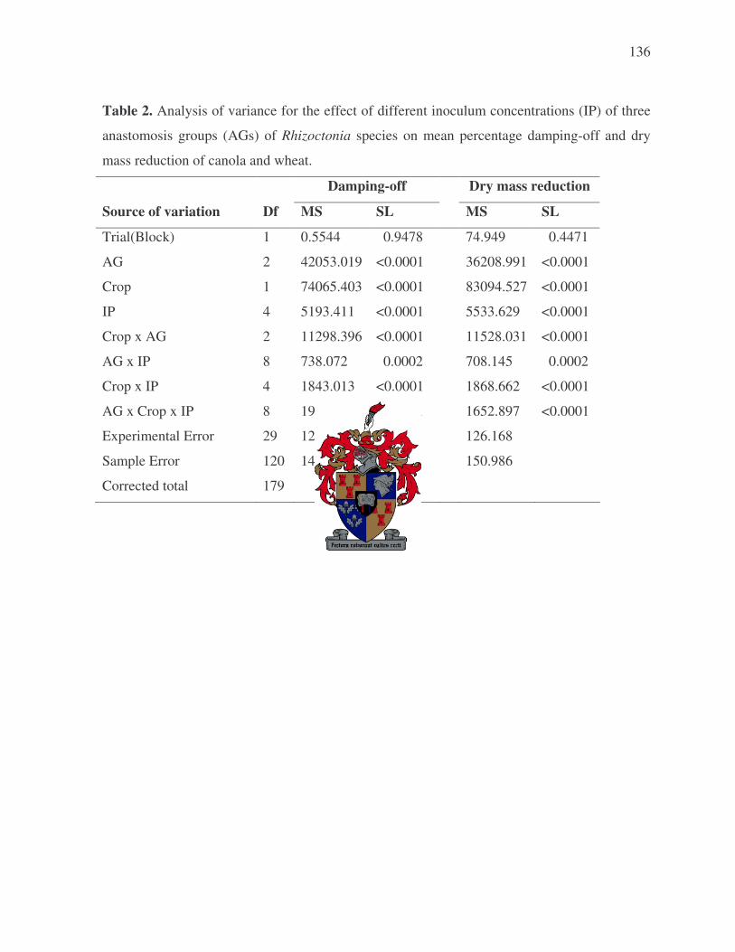
136
Table 2. Analysis of variance for the effect of different inoculum concentrations (IP) of three
anastomosis groups (AGs) of Rhizoctonia species on mean percentage damping-off and dry
mass reduction of canola and wheat.
Damping-off Dry mass reduction
Source of variation Df MS SL MS SL
Trial(Block) 1 0.5544 0.9478 74.949 0.4471
AG 2 42053.019 <0.0001 36208.991 <0.0001
Crop 1 74065.403 <0.0001 83094.527 <0.0001
IP 4 5193.411 <0.0001 5533.629 <0.0001
Crop x AG 2 11298.396 <0.0001 11528.031 <0.0001
AG x IP 8 738.072 0.0002 708.145 0.0002
Crop x IP 4 1843.013 <0.0001 1868.662 <0.0001
AG x Crop x IP 8 1903.332 <0.0001 1652.897 <0.0001
Experimental Error 29 127.175 126.168
Sample Error 120 143.319 150.986
Corrected total 179

137
Table 3. Analysis of variance for the effect of different anastomosis groups (AGs) of Rhizoctonia species on mean percentage
damping-off and dry mass reduction of seven crops following inoculation of emerging seedlings.
Damping-off Dry mass reduction Source of variation Df MS SL MS SL Trial(Block) 1 681 0.0024 71 0.4760 Crop 6 22869 <0.0001 16074 <0.0001 Isolate 28 20162 <0.0001 21370 <0.0001 Isolates within AG-2-1 2 2015 <0.0001 3171 <0.0001 Isolates within AG-2-2 1 3910 <0.0001 2745 <0.0001 Isolates within AG-3 2 29 0.6690 46 0.7193 Isolates within AG-4 HG-II 5 130 0.1139 353 0.0003 Isolates within AG-11 1 106 0.2264 97 0.4028 Isolates within AG-A 2 27 0.6878 15 0.8959 Isolates within AG-I 2 14 0.8235 1 0.9930 Isolates within AG-K 2 53 0.4803 31 0.7947 Isolates within UNBRx 2 10 0.8704 52 0.6844 Control 0 - - - - Between AG 9 61731 <0.0001 65237 <0.0001 Crop X Isolate 168 1249 <0.0001 1139 <0.0001 Crop X AG 54 3519 <0.0001 2933 <0.0001 Remainder 133 216 333 Experimental Error 202 72 138 Sample Error 406 49 55 Corrected total 811 xUNBR (unidentified binucleate Rhizoctonia).

138
Table 4. Analysis of variance for the effect of different anastomosis groups (AGs) of
Rhizoctonia species on root rot of seven crops following inoculation of fourteen-day-old
seedlings.
Source of variation Df MS SL Trial(Block) 1 0.00399 0.9027 Crop 6 27.32 <0.0001 Isolate 28 8.86 <0.0001 Isolates within AG-2-1 2 0.07 0.7719 Isolates within AG-2-2 1 0.47 0.1885 Isolates within AG-3 2 0.03 0.8950 Isolates within AG-4 HG-II 5 1.16 0.0010 Isolates within AG-11 1 0.43 0.2084 Isolates within AG-A 2 0.18 0.5144 Isolates within AG-I 2 0.15 0.5744 Isolates within AG-K 2 1.21 0.0125 Isolates within UNBRz 2 0.05 0.8313 Control 0 - - Between AG 9 26.45 <0.0001 Crop x Isolate 168 1.17 <0.0001 Crop x AG 54 2.95 <0.0001 Remainder 133 0.35 0.0412 Experimental Error 202 0.27 Sample Error 406 0.12 Corrected total 811 zUNBR (unidentified binucleate Rhizoctonia).

139
Table 5. Analysis of variance for the effect of different anastomosis groups (AGs) of Rhizoctonia species on the severity of
coleoptile rot of barley and wheat, and hypocotyl rot of canola, clover, lucerne, lupin and medic following inoculation of
fourteen-day-old seedlings.
Coleoptile rot Hypocotyl rot Source of variation Df MS SL Df MS SL Trial(Block) 1 0.0797 0.6408 1 0.6422 0.1457 Crop 1 10.14 <0.0001 4 18.75 <0.0001 Isolate 28 2.97 <0.0001 28 11.95 <0.0001 Isolates within AG-2-1 2 0.01 0.9724 2 0.75 0.0856 Isolates within AG-2-2 1 0.00 1.0000 1 0.10 0.5648 Isolates within AG-3 2 0.01 0.9724 2 0.77 0.0803 Isolates within AG-4 HG-II 5 0.91 0.0398 5 0.58 0.9461 Isolates within AG-11 1 0.02 0.8138 1 0.02 0.7961 Isolates within AG-A 2 0.07 0.8242 2 0.09 0.7413 Isolates within AG-I 2 0.02 0.9456 2 0.04 0.8756 Isolates within AG-K 2 0.20 0.5743 2 0.04 0.8756 Isolates within UNBRz 2 0.004 0.9891 2 0.05 0.8464 Control 0 - - 0 - - Between AG 9 8.66 <0.0001 9 36.46 <0.0001 Crop x Isolate 28 0.51 0.1467 112 1.66 <0.0001 Crop x AG 9 0.86 0.0228 36 4.41 <0.0001 Remainder 38 0.31 0.7038 95 0.35 0.1912 Experimental Error 57 0.36 144 0.30 Sample Error 116 0.10 290 0.17 Corrected total 231 579 zUNBR (unidentified binucleate Rhizoctonia).
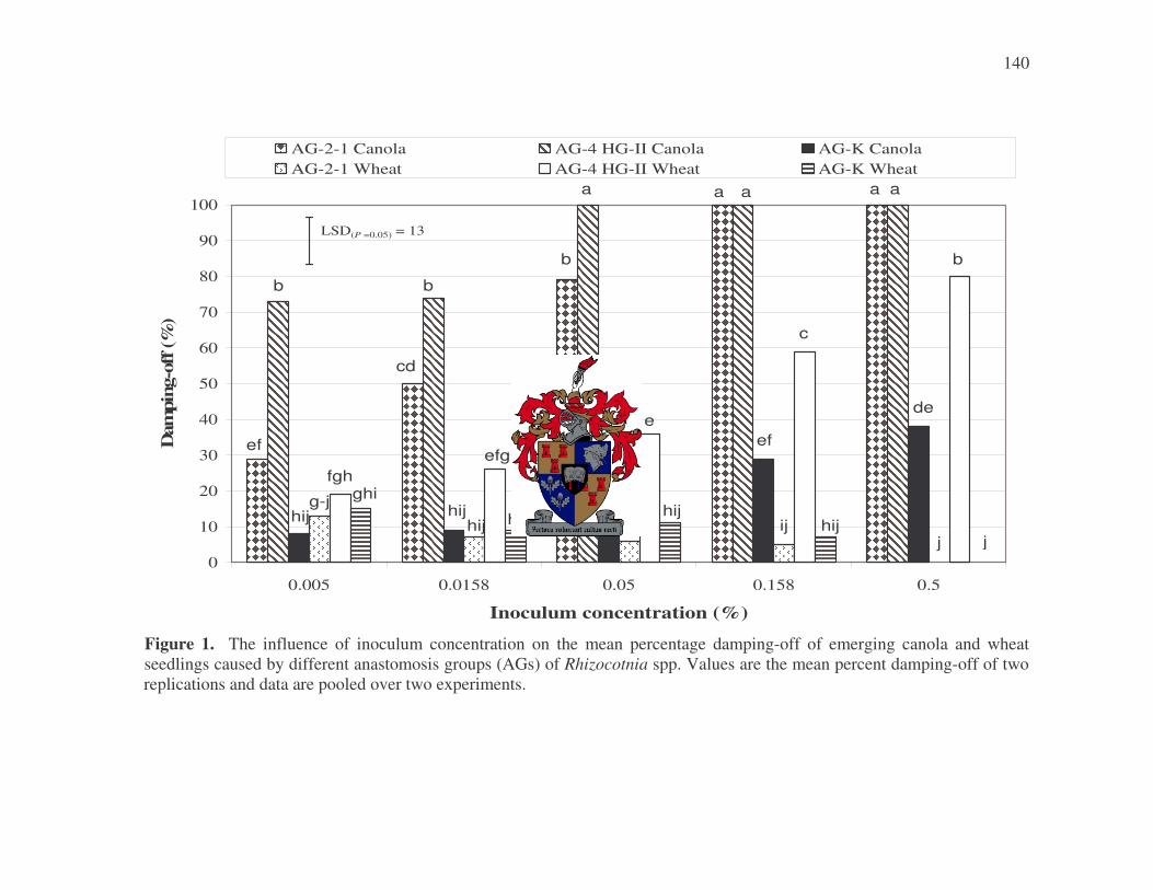
140
0
10
20
30
40
50
60
70
80
90
100
0.005 0.0158 0.05 0.158 0.5
Inoculum concentration (%)
Dam
ping
-off
(%)
AG-2-1 Canola AG-4 HG-II Canola AG-K CanolaAG-2-1 Wheat AG-4 HG-II Wheat AG-K Wheat
LSD(P =0.05) = 13
ef
b
hijg-j
fghghi
cd
b
hij hij
efg
hij
b
a
g-j
hij
e
a
hijij
c
ef
aa
hij
de
b
j j
a
Figure 1. The influence of inoculum concentration on the mean percentage damping-off of emerging canola and wheat seedlings caused by different anastomosis groups (AGs) of Rhizocotnia spp. Values are the mean percent damping-off of two replications and data are pooled over two experiments.
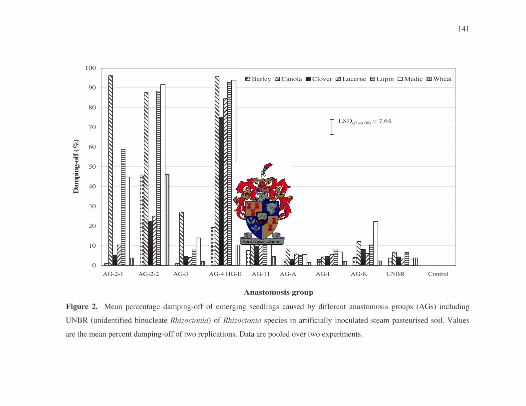
141
0
10
20
30
40
50
60
70
80
90
100
AG-2-1 AG-2-2 AG-3 AG-4 HG-II AG-11 AG-A AG-I AG-K UNBR Control
Anastomosis group
Dam
ping
-off
(%)
Barley Canola Clover Lucerne Lupin Medic Wheat
LSD(P =0.05) = 7.64
Figure 2. Mean percentage damping-off of emerging seedlings caused by different anastomosis groups (AGs) including
UNBR (unidentified binucleate Rhizoctonia) of Rhizoctonia species in artificially inoculated steam pasteurised soil. Values
are the mean percent damping-off of two replications. Data are pooled over two experiments.

142
Control AG-2-1 AG-2-2 AG-3
Control AG-4 HG-II AG-4 HG-II AG-11 Figure 3. Survival of lupin seedlings 14 days after planting in soil artificially infested with multinucleate Rhizoctonia A) AG-2-1, AG-2-2 and AG-3; B) AG-4 HG-II and AG-11.
A
B

143
Control AG-A UNBR AG-I AG-K Figure 4. Survival of lupin seedlings 14 days after planting in soil artificially infested with binucleate Rhizoctonia AG-A, UNBR (unidentified binucleate Rhizoctonia), AG-I and AG-K.

144
0
0.5
1
1.5
2
2.5
3
3.5
AG-2-1 AG-2-2 AG-3 AG-4 HG-II AG-11 AG-A AG-I AG-K UNBR Control
Anastomosis group
Roo
t rot
inde
x
Barley Canola Clover Lucerne Lupin Medic Wheat
LSD(P =0.05) = 0.47
Figure 5. Mean root rot index of 14-day-old seedlings caused by different anastomosis groups (AGs) including UNBR
(unidentified binucleate Rhizoctonia) of Rhizoctonia species in artificially inoculated steam pasteurised soil. Values are the mean
root rot index of two replications and data are pooled over two experiments. The value index was a 0 to 4 scale, with 0 = no obvious
symptoms; 1 = slight discolouration or small superficial lesions; 2 = moderate discolouration or extensive but non-girdling lesions;
3 = extensive discolouration of tissue or girdling lesions; and 4 = plant dead.

145
0
0.5
1
1.5
2
2.5
3
3.5
AG-2-1 AG-2-2 AG-3 AG-4 HG-II AG-11 AG-A AG-I AG-K UNBR Control
Anastomosis group
Hyp
ocot
yl rot
inde
x
Canola Clover Lucerne Lupin Medic
LSD(P =0.05) = 0.5
Figure 6. Mean hypocotyl rot index of 14-day-old seedlings caused by different anastomosis groups (AGs) including UNBR
(unidentified binucleate Rhizoctonia) of Rhizoctonia species in artificially inoculated steam pasteurised soil. Values are the mean
hypocotyl rot index of two replications and data are pooled over two experiments. The value index was a 0 to 4 scale, with 0 = no
obvious symptoms; 1 = slight discolouration or small superficial lesions; 2 = moderate discolouration or extensive but non-girdling
lesions; 3 = extensive discolouration of tissue or girdling lesions; and 4 = plant dead.

146
0
0.5
1
1.5
2
2.5
3
3.5
AG-2-1 AG-2-2 AG-3 AG-4 HG-II AG-11 AG-A AG-I AG-K UNBR Control
Anastomosis group
Col
eopt
ile r
ot in
dex
Barley Wheat
LSD(P =0.05) = 0.55
Figure 7. Mean coleoptile rot index of 14-day-old barley and wheat seedlings caused by different anastomosis groups (AGs)
including UNBR (unidentified binucleate Rhizoctonia) of Rhizoctonia species in artificially inoculated steam pasteurised soil.
Values are the mean coleoptile rot index of two replications and data are pooled over two experiments. The value index was a 0 to 4
scale, with 0 = no obvious symptoms; 1 = slight discolouration or small superficial lesions; 2 = moderate discolouration or
extensive but non-girdling lesions; 3 = extensive discolouration of tissue or girdling lesions; and 4 = plant dead.

147
Figure 8. Hypocotyl and root rot of lupin seedlings caused
by Rhizoctonia solani AG-4 HG-II