chapter 16 sense organs types of sensory receptors general senses chemical senses hearing and...
TRANSCRIPT
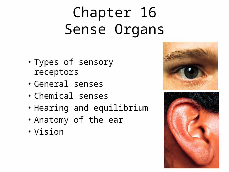
Chapter 16Sense Organs
• Types of sensory receptors
• General senses
• Chemical senses
• Hearing and equilibrium
• Anatomy of the ear
• Vision
Properties of Receptors• Receptor is any structure specialized to detect a stimulus
(simple nerve ending or complex sense organ)• Sensory receptors are transducers converting stimulus
energy into electrochemical energy = sensory transduction• Information about stimulus that can be conveyed
– modality or type of stimulus or sensation
– location of stimuli• each sensory receptor receives input from its receptive field
• brain identifies site of stimulation = sensory projection
– intensity (frequency, numbers of fiber & which fibers)
– duration = change in firing frequency over time• phasic receptor - burst of activity & quickly adapt (smell & hair receptors)
• tonic receptor - adapt slowly, generate impulses continually (proprioceptor)

Receptive Fields

Classification of Receptors
• By modality:– chemoreceptors, thermoreceptors, nociceptors,
mechanoreceptors and photoreceptors
• By origin of stimuli– interoceptors = detect internal stimuli– proprioceptors = sense position & movements of body– exteroceptors = sense stimuli external to body
• By distribution– general (somesthetic) sense --- widely distributed– special senses --- limited to head

The General SensesUnencapsulated Nerve Endings
• Found as receptors for the general senses
• Dendrites not wrapped in connective tissue
• Free nerve endings include– warm, cold & pain
• Tactile discs are associated with cells at base epidermis
• Hair receptors monitor the movement of hairs
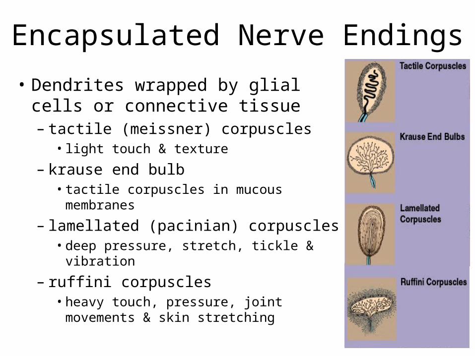
Encapsulated Nerve Endings
• Dendrites wrapped by glial cells or connective tissue– tactile (meissner) corpuscles
• light touch & texture
– krause end bulb• tactile corpuscles in mucous membranes
– lamellated (pacinian) corpuscles• deep pressure, stretch, tickle & vibration
– ruffini corpuscles• heavy touch, pressure, joint movements
& skin stretching
Somesthetic Projection Pathways• First-order neuron or afferent neuron
– from below head, enter the dorsal horn of spinal cord via spinal nerves
– from head, enter pons and medulla from cranial nerve– touch, pressure & proprioception are carried on large, fast,
myelinated axons– heat & cold are carried on small, unmyelinated, slow fibers
• Second-order neuron– decussation of signals to opposite side in spinal cord or medulla– end in thalamus, except for proprioception (cerebellum)
• Third-order neuron– extend from thalamus to primary somesthetic cortex of cerebrum
on contralateral side

Pain• Nociceptors make us conscious of tissue injuries
– forces us to care for minor injuries to prevent serious problems
• Found in all tissues except the brain• Fast pain travels in myelinated fibers at 30 m/sec
– sharp, localized, stabbing pain perceived with injury
• Slow pain travels unmyelinated fibers at 2 m/sec– longer-lasting, dull, diffuse feeling
• Somatic pain arises from skin, muscles & joints• Visceral pain from stretch, chemical irritants or ischemia
of viscera (poorly localized)• Injured tissues release chemicals that stimulate pain
fibers (bradykinin, histamine, prostaglandin)

Projection Pathway for Pain• General pathway
– first-order neuron cell bodies in dorsal root ganglion of spinal nerves or trigeminal ganglion
– second-order neurons decussate to other side & send fibers up spinothalamic tract to thalamus
• gracile fasciculus carries visceral pain signals
– third-order neurons reach primary somesthetic cortex (postcentral gyrus)
• Spinoreticular tract– pain signals reach reticular formation, hypothalamus & limbic– trigger emotional and behavioral reactions
• Referred pain is misinterpreted pain– brain “assumes” pain is coming from skin or superficial sites– heart pain felt in shoulder or arm because both send pain input to
spinal cord segments T1 to T5

Pain Signal Destinations

Referred Pain

CNS Modulation of Pain• Intensity of pain is affected by state of mind• Endogenous opiods (enkephalins, endorphins 7 dynorphins)
– produced by CNS and other organs under stress– found especially in dorsal horn of spinal cord, explaining the spinal
gating of pain– act as neuromodulators blocking the transmission of pain
• Spinal gating stops pain signals at dorsal horn– descending analgesic fibers from reticular formation travel down
reticulospinal tract to dorsal horn• secrete inhibitory substances that block pain fibers from secreting substance P• pain signals never ascend
– dorsal horn fibers inhibited by input from mechanoreceptors (rubbing a sore arm reduces pain)

Spinal Gating of Pain Signals
Normal pain pathways(red arrows) is inhibited by many mechanisms.
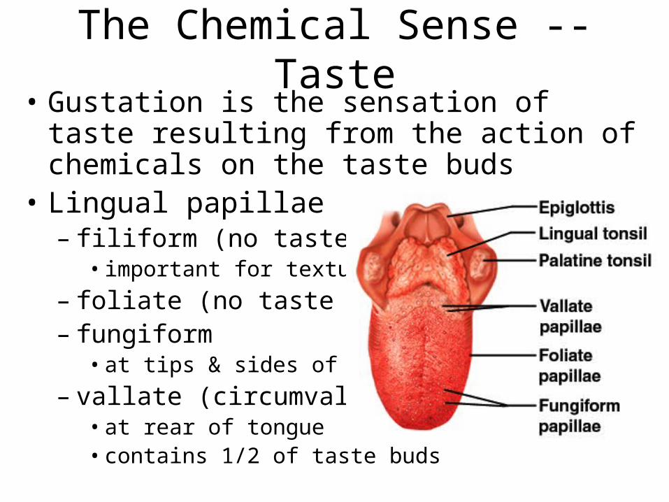
The Chemical Sense -- Taste• Gustation is the sensation of taste resulting from
the action of chemicals on the taste buds• Lingual papillae
– filiform (no taste buds)• important for texture
– foliate (no taste buds)– fungiform
• at tips & sides of tongue
– vallate (circumvallate)• at rear of tongue• contains 1/2 of taste buds
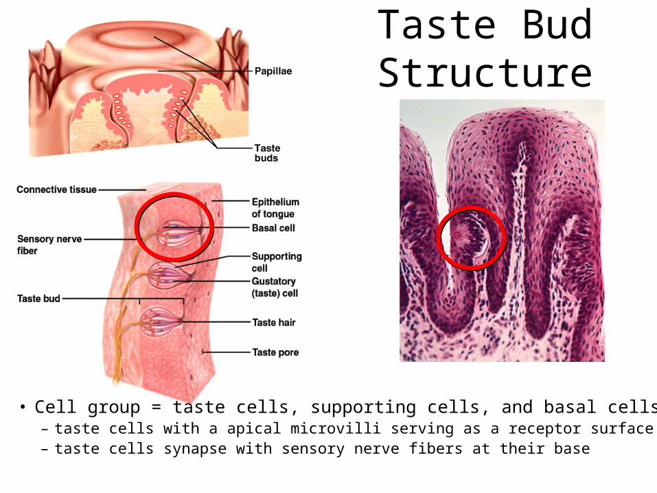
Taste Bud Structure
• Cell group = taste cells, supporting cells, and basal cells– taste cells with a apical microvilli serving as a receptor surface – taste cells synapse with sensory nerve fibers at their base
• Cell group = taste cells, supporting cells, and basal cells– taste cells with a apical microvilli serving as a receptor surface – taste cells synapse with sensory nerve fibers at their base

Physiology of Taste• To be tasted, molecules must dissolve in saliva• 5 primary sensations: salty, sweet, sour, bitter & umami
(taste of amino acids such as MSG)– taste is also influenced by food texture, aroma, temperature, and
appearance• mouthfeel is detected by lingual nerve branches in papillae
– hot pepper stimulates free nerve endings (pain)
• Sweet tastes concentrated on tip of tongue, salty & sour on lateral margins of tongue & bitter at rear– all tastes can be detected throughout the tongue surface
• Mechanisms of action– sugars, alkaloids & glutamates bind to receptors & activate 2nd
messenger systems– sodium & acids penetrate cells & depolarize them directly
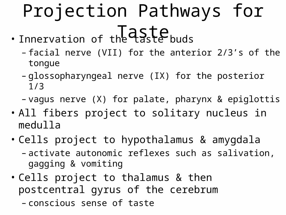
Projection Pathways for Taste• Innervation of the taste buds
– facial nerve (VII) for the anterior 2/3’s of the tongue– glossopharyngeal nerve (IX) for the posterior 1/3– vagus nerve (X) for palate, pharynx & epiglottis
• All fibers project to solitary nucleus in medulla
• Cells project to hypothalamus & amygdala– activate autonomic reflexes such as salivation, gagging
& vomiting
• Cells project to thalamus & then postcentral gyrus of the cerebrum– conscious sense of taste

The Chemical Sense -- Smell
• Receptor cells for olfaction form olfactory mucosa– smell is highly sensitive
(more so in women than men)
– distinguish as many as 10,000 odors
• Covers 5cm2 of superior concha & nasal septum

Olfactory Epithelial Cells
• Olfactory cells– neurons with 20 cilia
called olfactory hairs• binding sites for odor
molecules in thin layer of mucus
– axons pass through cribriform plate
– survive 60 days
• Supporting cells• Basal cells divide

Physiology of Smell• Odor molecules must be volatile
– bind to a receptor on an olfactory hair triggering the production of a second messenger
– opens the ion channels & creates a receptor potential
• Receptors adapt quickly due to synaptic inhibition in the olfactory bulbs
• Bulb cells form the axons of the olfactory tracts– lead to temporal lobe, amygdala, hypothalamus
• emotional responses to odors
• cough, salivate, sneeze or vomit in response to odors
– cerebral cortex sends feedback to bulb cells• changing quality & significance of odors when hungry
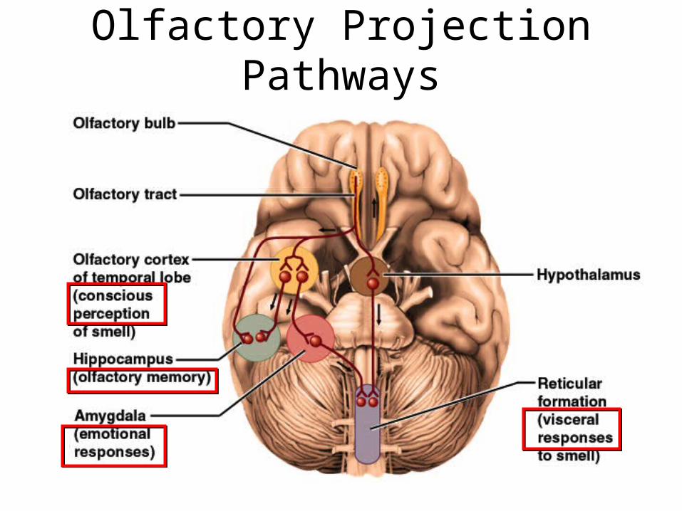
Olfactory Projection Pathways

The Nature of Sound• Sound is any audible vibration of molecules
• Vibrating object pushes air molecules into eardrum making it vibrate
Molecules collide with eardrum & make it vibrate.
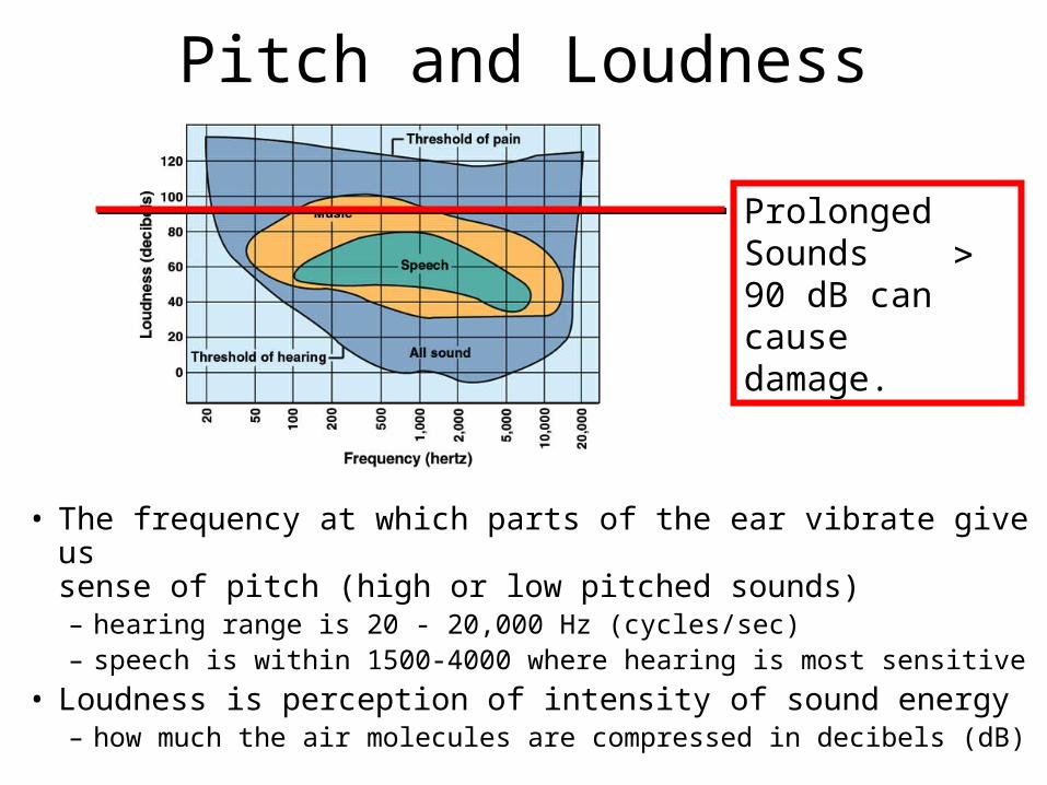
Pitch and Loudness
• The frequency at which parts of the ear vibrate give us sense of pitch (high or low pitched sounds)– hearing range is 20 - 20,000 Hz (cycles/sec)– speech is within 1500-4000 where hearing is most sensitive
• Loudness is perception of intensity of sound energy– how much the air molecules are compressed in decibels (dB)
Prolonged Sounds 90 dB can cause damage.
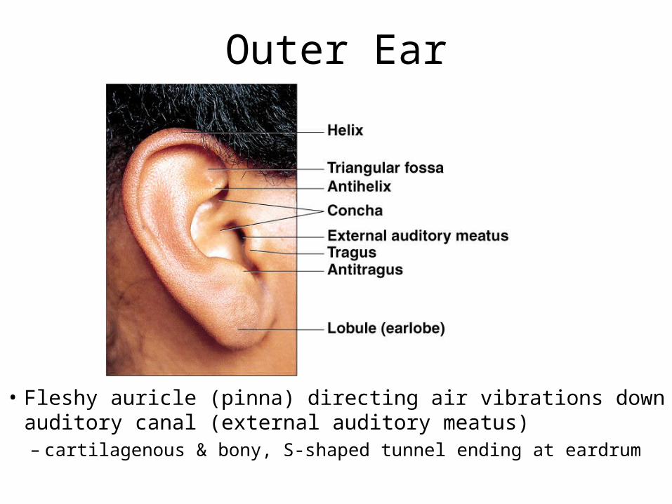
Outer Ear
• Fleshy auricle (pinna) directing air vibrations down auditory canal (external auditory meatus)– cartilagenous & bony, S-shaped tunnel ending at eardrum

Middle Ear
• Air-filled cavity in temporal bone separated from air outside the head by tympanic membrane– 1 cm in diameter, slightly concave, freely vibrating membrane
• Tympanic cavity continuous with mastoid air cells• Tympanic cavity filled with air by auditory tube
(eustachian tube) connected to nasopharynx– opens during swallowing or yawning to equalize air pressure on
both sides of eardrum
• Ear ossicles span tympanic cavity– malleus attached to eardrum, incus, stapes attached to
membranous oval window of inner ear– stapedius & tensor tympani muscles attach to ossicles

Anatomy of Middle Ear
• Middle ear is cavity containing ear ossicles.
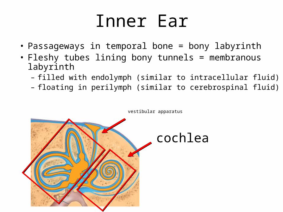
Inner Ear• Passageways in temporal bone = bony labyrinth• Fleshy tubes lining bony tunnels = membranous labyrinth
– filled with endolymph (similar to intracellular fluid)– floating in perilymph (similar to cerebrospinal fluid)
vestibular apparatus
cochlea

Details of Inner Ear

Details of Inner Ear
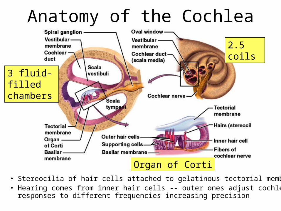
Anatomy of the Cochlea
• Stereocilia of hair cells attached to gelatinous tectorial membrane.• Hearing comes from inner hair cells -- outer ones adjust cochlear
responses to different frequencies increasing precision
2.5 coils
3 fluid-filled chambers
Organ of Corti

SEM of Cochlear Hair Cells

Physiology of Hearing -- Middle Ear• Eardrum vibrates quite easily
– 18 times the area of the oval window• creates enough force/unit area at oval window to vibrate the endolymph in
the scala vestibuli
• Protection of cochlea by muscle contraction in response to loud noises (tympanic reflex)– tensor tympani pulls eardrum inward, tightening it
– stapedius reduces mobility of stapes
– designed for slowly building noises like thunder not gunshots (irreversible damage by breaking stereocilia)
• does not protect us from sustained loud noises such as music
– muscles also contract while speaking -- can hear others

Stimulation of Cochlear Hair Cells
• Sound is produced by vibration of ossicles and then vibration of basilar membrane under hair cells
• Can happen as often as 20,000 time per second
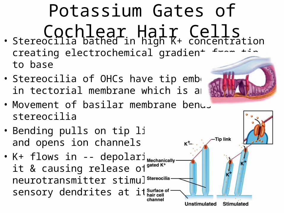
Potassium Gates of Cochlear Hair Cells• Stereocilia bathed in high K+ concentration creating
electrochemical gradient from tip to base• Stereocilia of OHCs have tip embedded
in tectorial membrane which is anchored• Movement of basilar membrane bends
stereocilia• Bending pulls on tip links
and opens ion channels• K+ flows in -- depolarizing
it & causing release of neurotransmitter stimulating sensory dendrites at its base

Sensory Coding• Loudness produces more vigorous vibrations &
excites more hair cells over a larger area – triggers higher frequency of action potentials– brain interprets this as louder
• Determination of pitch depends on which part of basilar membrane is vibrated at peak amplitude of standing wave– membrane is narrow & stiffer at basal end (collagen)
• brain interprets signals from IHC basal end as high-pitched
– at distal end is 5 times wider & more flexible• brain interprets signals from IHC distal end as low-pitched
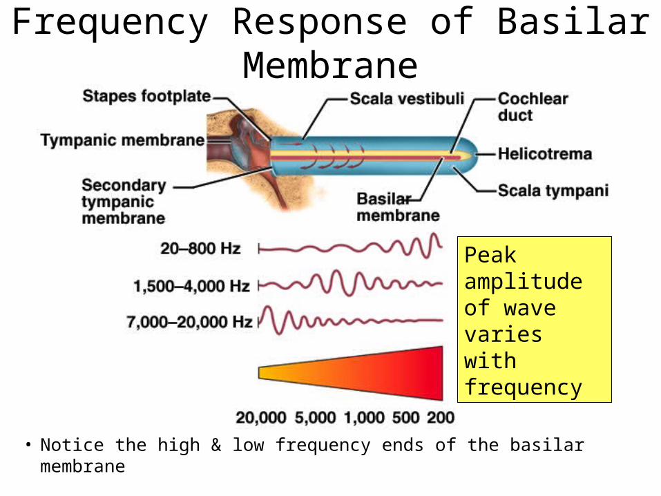
Frequency Response of Basilar Membrane
• Notice the high & low frequency ends of the basilar membrane
Peak amplitude of wave varies with frequency
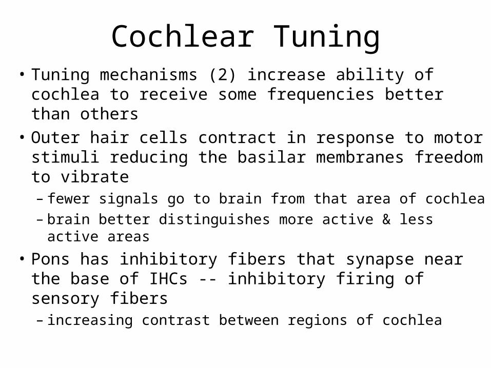
Cochlear Tuning• Tuning mechanisms (2) increase ability of cochlea to
receive some frequencies better than others• Outer hair cells contract in response to motor stimuli
reducing the basilar membranes freedom to vibrate– fewer signals go to brain from that area of cochlea– brain better distinguishes more active & less active areas
• Pons has inhibitory fibers that synapse near the base of IHCs -- inhibitory firing of sensory fibers– increasing contrast between regions of cochlea

Innervation of Internal Ear
• Vestibular ganglia is visible in vestibular nerve
• Spiral ganglia is buried in modiolus of cochlea

Pathways for Control of Hearing

Auditory Projection Pathway
• Superior olivary nucleus compares sounds from both sides to identify direction (binaural hearing)
• Inferior colliculus helps locate origin of sound in space, process fluctuations in pitch in speech, & produces startle response of head turning with loud sounds
• Temporal lobe is site of conscious perception

Auditory Processing Centers
• Damage to either auditory cortex does not cause unilateral deafness due to extensive decussation in pathway

Equilibrium
• Control of coordination and balance
• Receptors in vestibular apparatus– semicircular ducts contain crista– saccule & utricle contain macula
• Static equilibrium is perception of head orientation– perceived by macula
• Dynamic equilibrium is perception of motion or acceleration – linear acceleration perceived by macula– angular acceleration perceived by crista

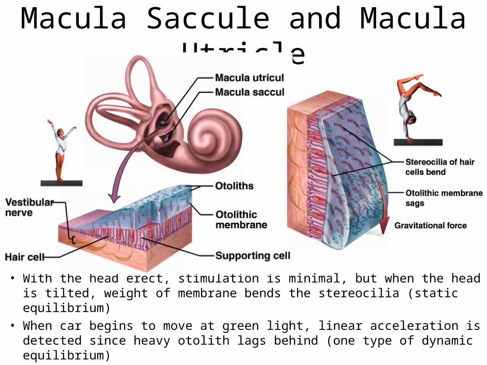
The Saccule and Utricle
• Saccule & utricle chambers containing macula– patch of hair cells with their stereocilia & one
kinocilium buried in a gelatinous otolithic membrane weighted with granules called otoliths
– otoliths add to the density & inertia and enhance the sense of gravity and motion
Otoliths
Macula Saccule and Macula Utricle
• With the head erect, stimulation is minimal, but when the head is tilted, weight of membrane bends the stereocilia (static equilibrium)
• When car begins to move at green light, linear acceleration is detected since heavy otolith lags behind (one type of dynamic equilibrium)

Crista ampullaris of Semicircular Ducts
• Crista ampullaris consists of hair cells buried in a mound of gelatinous membrane (one in each duct)
• Orientation of ducts causes different ducts to be stimulated by rotation in different planes

Crista Ampullaris & Head Rotation
• As head turns, the endolymph lags behind pushing the cupula and stimulating its hair cells
Equilibrium Projection Pathways
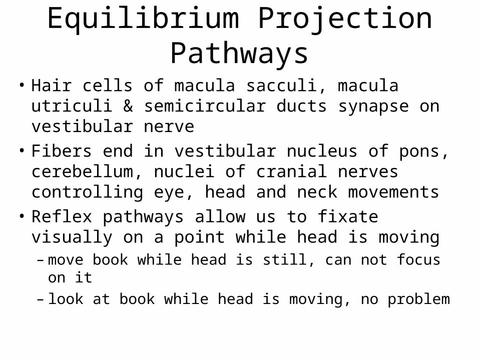
• Hair cells of macula sacculi, macula utriculi & semicircular ducts synapse on vestibular nerve
• Fibers end in vestibular nucleus of pons, cerebellum, nuclei of cranial nerves controlling eye, head and neck movements
• Reflex pathways allow us to fixate visually on a point while head is moving– move book while head is still, can not focus on it– look at book while head is moving, no problem
Vision and Light
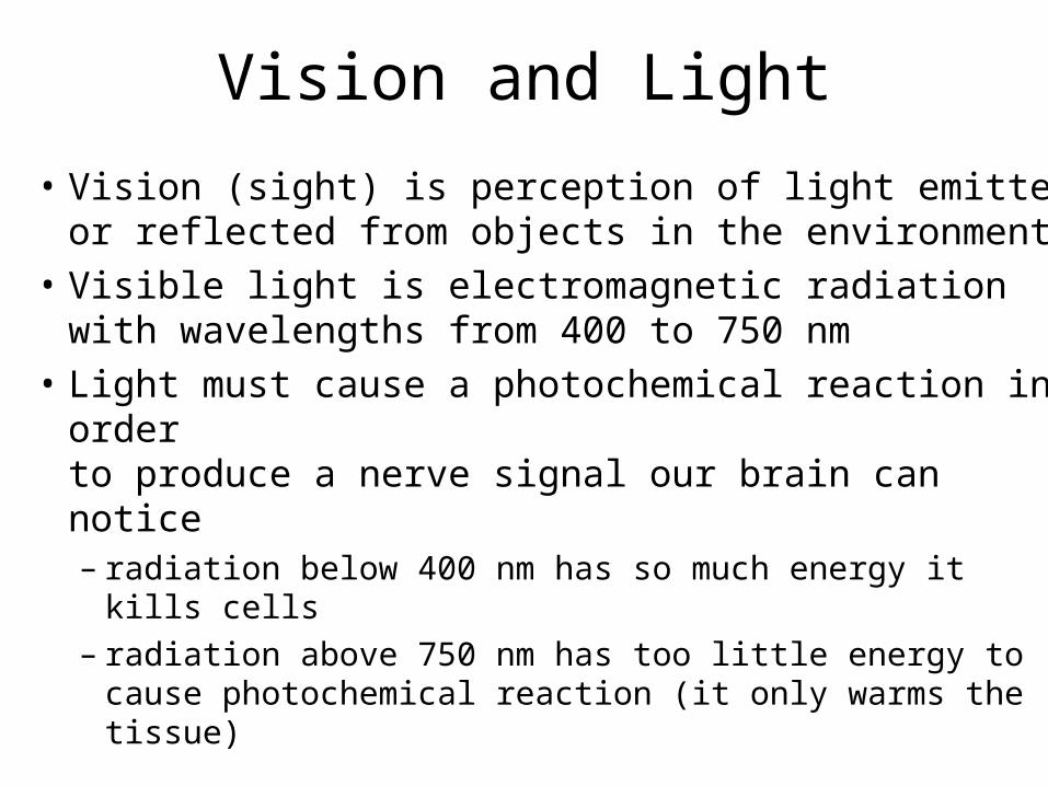
• Vision (sight) is perception of light emitted or reflected from objects in the environment
• Visible light is electromagnetic radiation with wavelengths from 400 to 750 nm
• Light must cause a photochemical reaction in order to produce a nerve signal our brain can notice– radiation below 400 nm has so much energy it kills cells– radiation above 750 nm has too little energy to cause
photochemical reaction (it only warms the tissue)
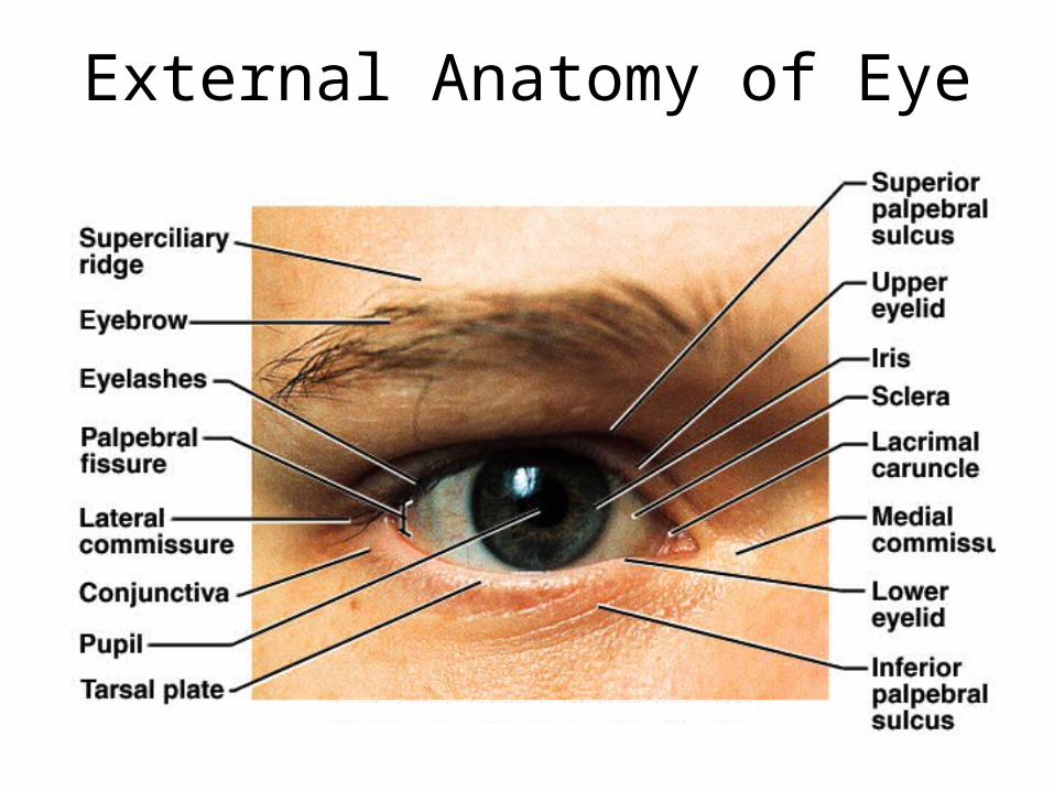
External Anatomy of Eye

Eyebrows and Eyelids• Eyebrows provide facial expression, protection from
glare & perspiration
• Eyelids (palpebrae)– block foreign objects, help with sleep,
blink to moisten– meet at corners (commissures)– consist of orbicularis oculi muscle &
tarsal plate covered with skin outside & conjunctiva inside
– tarsal glands secrete oil that reduces tear evaporation
– eyelashes help keep debris from the eye

Conjunctiva
• Transparent mucous membrane lines the eyelids and covers anterior surface of eyeball except cornea
• Richly innervated & vascular (heals quickly)
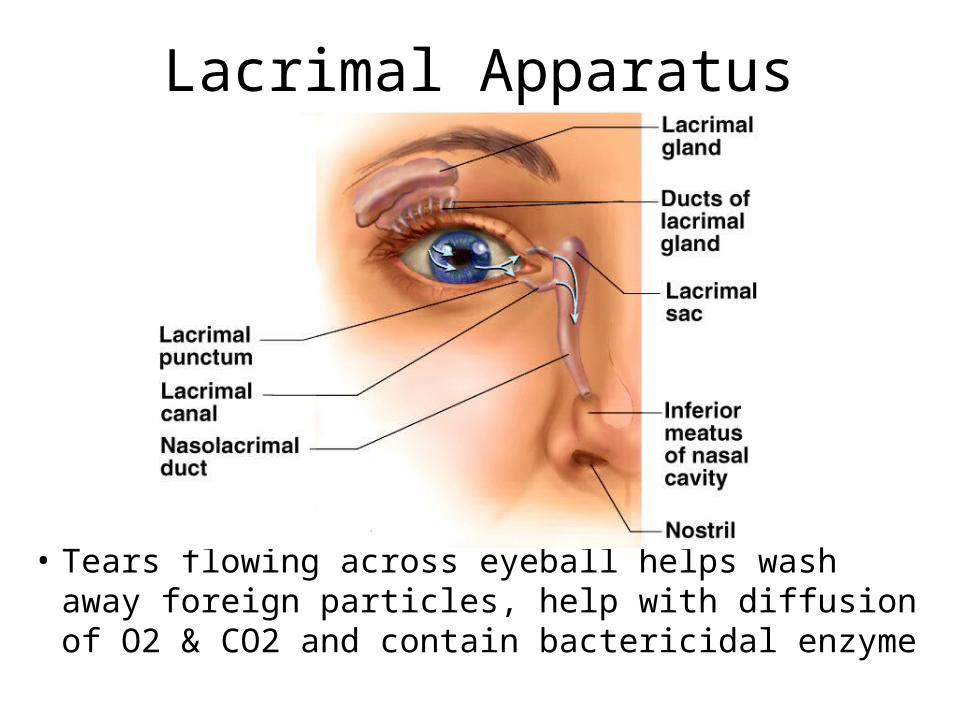
Lacrimal Apparatus
• Tears flowing across eyeball helps wash away foreign particles, help with diffusion of O2 & CO2 and contain bactericidal enzyme

Extrinsic Eyes Muscles
• 6 muscles inserting on external surface of eyeball– 4 rectus muscles move eye up, down, left & right– superior & inferior oblique more complicated
• Innervated by cranial nerves III, IV and VI
trochlea

Innervation of Extrinsic Eye Muscles

The Tunics of the Eyeball
• Fibrous layer (tunica fibrosa) = sclera and cornea • Vascular layer (tunica vasculosa) = choroid, ciliary body &
iris• Internal layer (tunica interna) = retina and optic nerve

The Optical Components
• Series of transparent structures that bend or refract light rays to focus them on the retina– cornea is transparent covering of anterior surface of
eyeball– aqueous humor is clear serous fluid filling area in
front of lens (between lens and cornea)– lens is suspended by ring of suspensory ligaments
• capable of changing shape to help focus light rays– more rounded when no tension on it – somewhat flattened normally due to pull of suspensory ligaments
– vitreous humor is jelly filling the space between the lens and retina

Aqueous Humor
• Serous fluid produced by ciliary body that flows from posterior chamber through pupil to anterior chamber -- reabsorbed into canal of Schlemm

The Neural Components
• Neural apparatus includes the retina & optic nerve
• Retina forms as an outgrowth of the diencephalon– attached only at optic disc where optic nerve begins
and at ora serrata (its anterior margin)– pressed against rear of eyeball by vitreous body
• Detached retina– blow to head or lack of sufficient vitreous body– blurry areas in field of vision– leads to blindness due to disruption of blood supply

Ophthalmoscopic Examination of Eye
• Cells on visual axis of eye = macula lutea (3 mm area)– fovea centralis is the center of macula where most finely
detailed images are seen due to packed receptor cells
• Eye exam provides direct evaluation of blood vessels

Rear of Eye Through Ophthalmoscope

Test for Blind Spot
• Optic disk or blind spot is where optic nerve exits the posterior surface of the eyeball– no receptor cells are found in optic disk
• Blind spot can be seen using the above illustration– in the right position, stare at X and red dot disappears
• Visual filling is the brain filling in the green bar across the blind spot area
Formation of an Image
• Light must pass through the lens to form tiny inverted image on retina
• Pupillary constrictor is smooth muscle cells encircling the pupil– parasympathetic stimulation narrows the pupil
• Pupillary dilator is spokelike myoepithelial cells– sympathetic stimulation widens the pupil to admit more light
• Active when light intensity changes or shift gaze from distant object to nearby object– photopupillary reflex -- both constrict if one eye is illuminated
(consensual reflex)
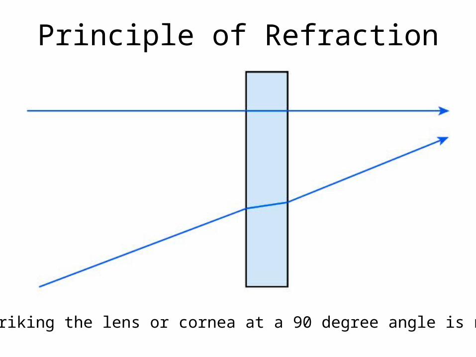
Principle of Refraction
Light striking the lens or cornea at a 90 degree angle is not bent.

Refraction
• Bending of light rays occurs when light passes through substance with different refractive index at any angle other than 90 degrees– refractive index of air
is arbitrarily set to n = 1
– refractive index of cornea is n = 1.38
– refractive index of lens is n = 1.40
• Cornea refracts light more than lens does– lens fine-tunes the image as shift focus between near and
distant objects

The Near Response• Eyes focused on distant object receive parallel
light waves & focus without effort
• Near response occurs if focus on object closer– convergence of eyes
• eyes orient their visual axis towards the object
– constriction of pupil• does not admit peripheral light rays & reduces spherical
aberration (blurry edges)
– accomodation of lens• contraction of ciliary muscle relaxes suspensory ligaments
which allows lens to relax to a more convex shape
• light is refracted more strongly & focused onto retina

Emmetropia & Near Response
• Behavior of eyes when focused on distant object (over 20 ft away) and onto close object

Emmetropia & Near Response

Accommodation of Lens

Effects of Corrected Lenses
• Hyperopia is farsighted (eyeball too short)– correct with convex lenses
• Myopia is nearsighted (eyeball too long)– correct with concave lenses
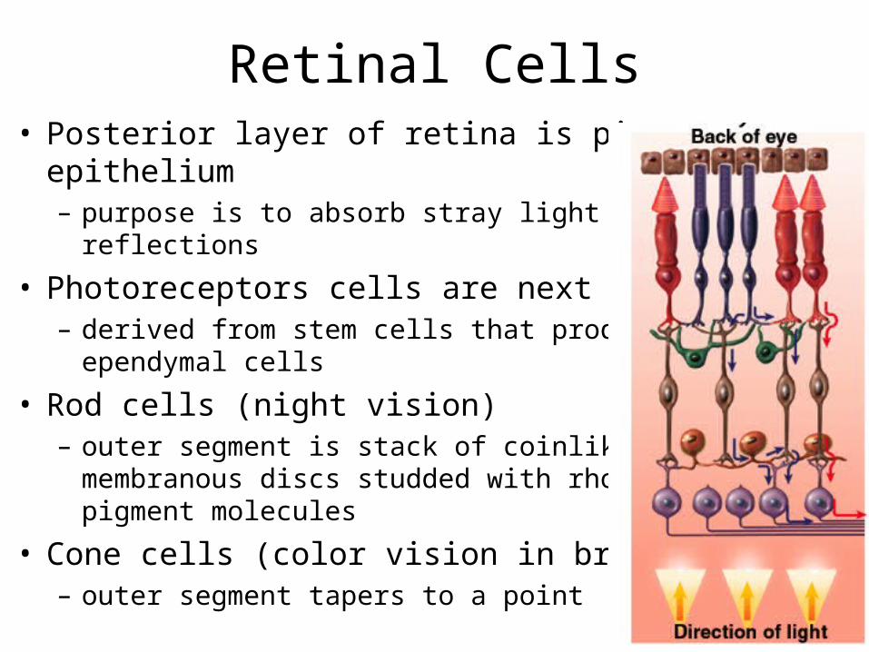
Retinal Cells• Posterior layer of retina is pigment
epithelium– purpose is to absorb stray light & prevent
reflections
• Photoreceptors cells are next layer– derived from stem cells that produced
ependymal cells
• Rod cells (night vision)– outer segment is stack of coinlike
membranous discs studded with rhodopsin pigment molecules
• Cone cells (color vision in bright light)– outer segment tapers to a point

Histology of the Layers of Retina

Cone and Rod Cell Details

Location of Visual Pigments

Nonreceptor Retinal Cells
• Bipolar cells (1st order neurons)– synapse on ganglion cells
– large amount of convergence
• Ganglion cells (2nd order neurons)– axons of these form optic nerve
– more convergence occurs (114 receptors to one optic nerve fiber
• Horizontal & amacrine cells lie form connections between other cells– enhance perception of contrast, edges of
objects & changes in light intensity
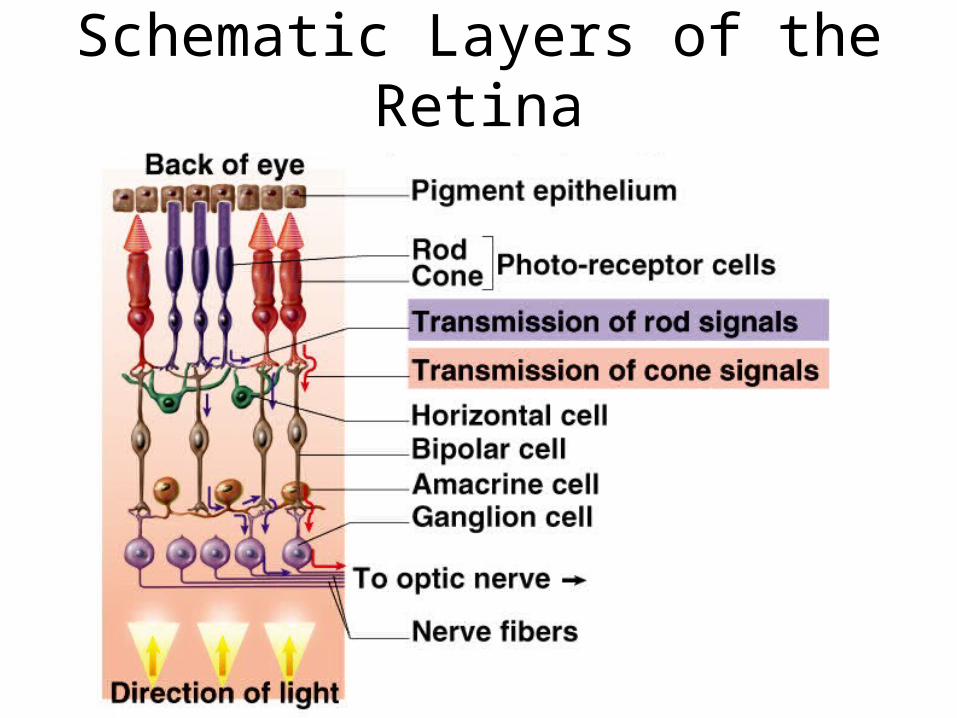
Schematic Layers of the Retina

Visual Pigments
• Visual pigment of the rod cells is called rhodopsin (visual purple)
• 2 major parts to the molecule– protein called opsin– vitamin A derivative called retinal
• Rod cells contain single kind of rhodopsin with an absorption peak at wavelength of 500 nm
• Cones contain photopsin (iodopsin)– opsin moieties contain different amino acids that determine
which wavelengths of light are absorbed– 3 kinds of cones absorbing different wavelengths of light
produce color vision

The Photochemical Reaction in Rod Cells
• When rhodopsin absorbs light, it is converted from the bent shape (cis-retinal) to the straight (trans-retinal) form which dissociates from the opsin (bleaching)
• Takes 5 minutes to regenerate 50% of rhodopsin– trans-retinal converted to cis-form & reunited with opsin

Generating Visual Signal in the Dark
• Rods produce steady ion flow in the dark that causes an IPSP that produces no signal in optic nerve
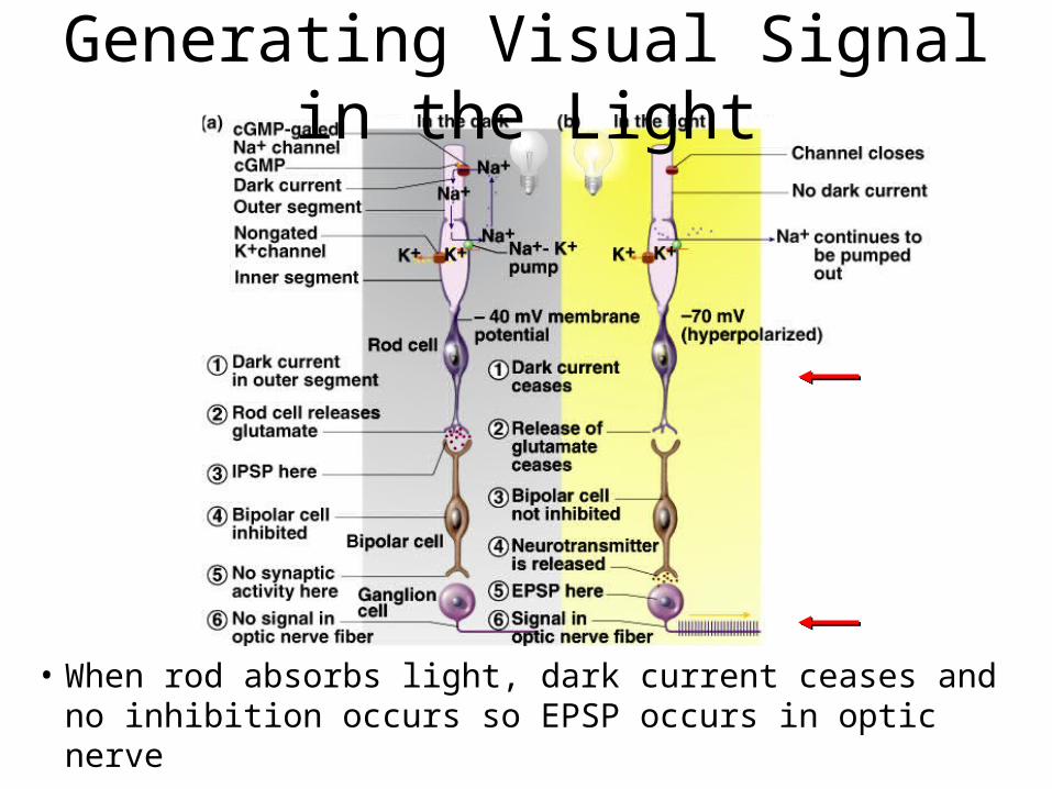
Generating Visual Signal in the Light
• When rod absorbs light, dark current ceases and no inhibition occurs so EPSP occurs in optic nerve
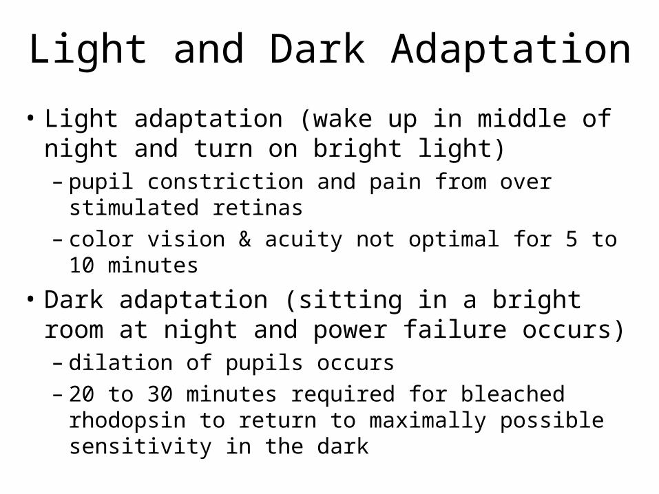
Light and Dark Adaptation
• Light adaptation (wake up in middle of night and turn on bright light)– pupil constriction and pain from over stimulated
retinas– color vision & acuity not optimal for 5 to 10 minutes
• Dark adaptation (sitting in a bright room at night and power failure occurs)– dilation of pupils occurs– 20 to 30 minutes required for bleached rhodopsin to
return to maximally possible sensitivity in the dark

Duplicity Theory
• Explains why we have both rods and cones
• Single type of receptor cell incapable of providing high sensitivity and high resolution– sensitive night vision = one type of cell and
neural circuitry– high resolution daytime vision = different cell
type and neuronal circuitry

• Sensitivity of rods in dim light– extensive neuronal convergence– 600 rods converge on 1 bipolar cell– many bipolar converge on each
ganglion cell– high degree of spatial summation but no ability to
resolve detail• one ganglion cells receives information from 1 mm2 of
retina producing only a coarse image
• Edges of retina with widely spaced rod cells is low-resolution system only alerting us to motion
Scotopic System (Night Vision)
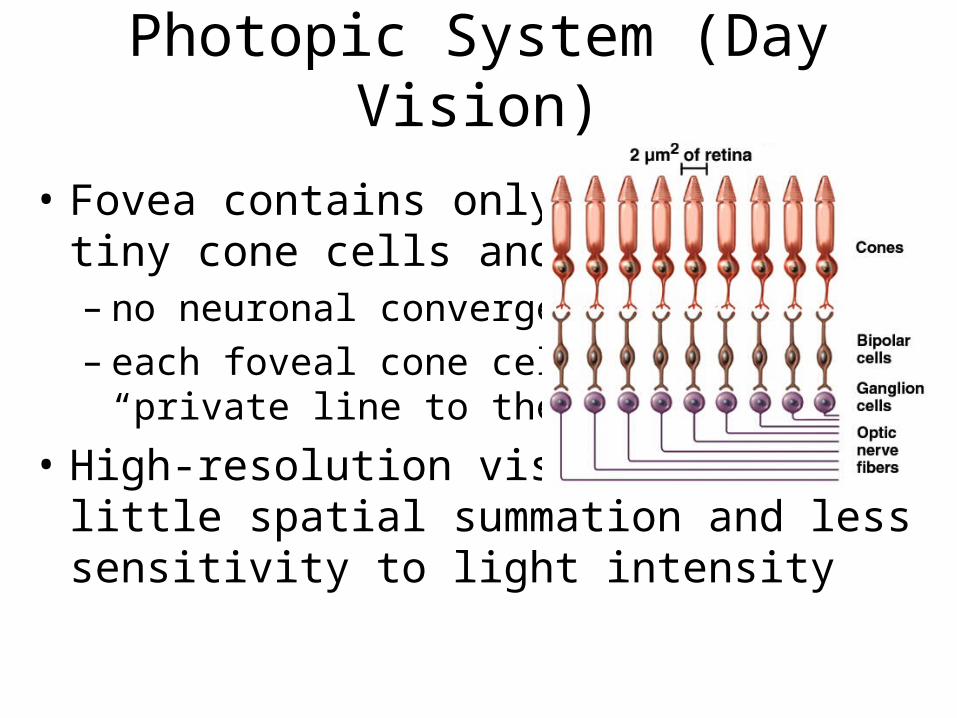
Photopic System (Day Vision)
• Fovea contains only 4000 tiny cone cells and no rods– no neuronal convergence– each foveal cone cell has
“private line to the brain”
• High-resolution vision, but little spatial summation and less sensitivity to light intensity

Color Vision• Primates have well developed
color vision– nocturnal vertebrates have only rods
• Cones are named for absorption peaks of photopsins– blue cones peak sensitivity at 420 nm
– green cones peak at 531 nm
– red cones peak at 558 nm (orange-yellow)
• Perception of color is based on mixture of nerve signals• Color blindness is hereditary lack of one photopsin
– red-green is common (lack either red or green cones)• incapable of distinguishing red from green
• sex-linked recessive (8% of males)
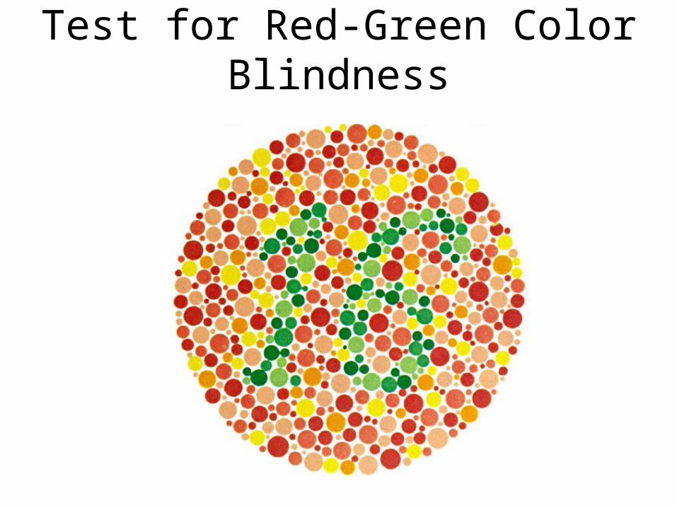
Test for Red-Green Color Blindness

Stereoscopic Vision (Stereopsis)• Depth perception is the ability to
judge how far away objects are
• Requires 2 eyes with overlapping visual fields– panoramic vision has eyes on sides
of head (horse)
• Fixation point is spot on which eyes are focused– objects farther away require image focus medial to the
fovea– objects closer result in image focus lateral to fovea
Visual Projection Pathway
Visual Projection Pathway
• Bipolar & ganglion cells in retina are 1st & 2nd order neurons (axons of ganglion cells form CN II)
• Hemidecussation occurs in optic chiasm– 1/2 of fibers decussate so that images of all objects in the left
visual field fall on right half of each retina
– each side of brain sees what is on side where it has motor control over limbs
• 3rd order neurons in lateral geniculate nucleus of thalamus form optic radiation to 1 visual cortex where conscious visual sensation occurs
• Few fibers project to superior colliculi & midbrain for visual reflexes (photopupillary & accomodation)

Visual Information Processing
• Some processing occurs in the retina– adjustments for contrast, brightness, motion &
stereopsis
• Visual association areas in parietal & temporal lobes process visual data– object location, motion, color, shape, boundaries– store visual memories (recognize printed words)