1 chapter 12 somatic and special senses. 2 receptors and sensations
TRANSCRIPT

1
Chapter 12
Somatic and Special Senses

2
RECEPTORS AND SENSATIONS

3
Senses• Sensory Receptors
– specialized cells or multicellular structures that collect information from the environment
– stimulate neurons to send impulses along sensory fibers to the brain

4
Receptor Types• specialized structures at the end of peripheral
nerves that respond to stimuli– can be classified according to their location in the body,
stimulus type and structure
•Chemoreceptors respond to changes in chemical concentrations•Nociceptors respond to extreme (harmful) stimuli by producing
the sensation of pain (i.e. all types under extreme stimuli
•Thermoreceptors are sensitive to temperature change•Mechanoreceptors respond to a change in pressure
(i.e. touch, pressure, vibrations, stretch)Photoreceptors (in retina of eye) respond to changes in light

5
Sensory Impulse
• All senses work in basically the same fashion• Special sensory receptors collect information from
the environment and stimulate neurons to send a message to the brain
• stimulation of receptor causes local change in its receptor potential
• a graded electrical current is generated that reflects intensity of stimulation– if receptor is part of a neuron, the membrane potential
may generate an action potential– if receptor is not part of a neuron, the receptor potential
must be transferred to a neuron to trigger an action potential
• peripheral nerves transmit impulses to CNS where they are analyzed and interpreted in the brain

6
Sensations
• Sensation =the conscious or unconscious awareness of external or internal stimuli
• Perception =the conscious awareness and interpretation of sensations
• Projection=process in which the brain projects the sensation back to the apparent source– it allows a person to pinpoint the region of
stimulation

7
Sensory Adaptation
• involves a decreased response to a particular stimulus from the receptors (peripheral adaptations) or along the CNS pathways leading to the cerebral cortex (central adaptation)
• sensory impulses become less frequent and may cease
• stronger stimulus is required to trigger impulses• All sensory receptors, except nociceptors, adapt to
continuous stimuli (i.e. undergo sensory adaptation)– i.e. when you first put a band-aid on you feel it but soon
don’t notice it at all

8
SOMATIC SENSES
Receptors associated with skin, muscles, joints, and viscera
provide somatic senses.

9
Three groups
• Exteroceptive Senses:– detect changes at the body's surface:– touch– pressure– temperature
• Proprioceptive Senses:– detect changes in muscles, tendons, and body
position
• Visceroceptive Senses:– detect changes in viscera– only pain will be discussed here

10
Three types of receptors• free nerve endings (naked dendritic)
– in epithelium, CT
• Meissner's Corpuscles are encapsulated dendritic endings – abundant in hairless portions of skin; lips– detect fine touch; distinguish between two points
on the skin
• Pacinian Corpuscles are also encapsulated dendritic endings:– common in deeper subcutaneous tissues,
tendons, and ligaments– detect heavy pressure and vibrations

11

12
Temperature Senses
Two types that respond to temperature change:• Heat receptors
– sensitive to temps above 25oC (77oF)– unresponsive at temps above 45oC (113oF)– Pain receptors are also triggered as this temperature
approaches producing a burning sensation
• Cold receptors– sensitive to temps between 10oC (50oF) and 20oC (68oF)– below 10oC, pain receptors are triggered producing a
freezing sensation
• Both undergo rapid sensory adaptation

13
Sense of Pain
• Free nerve endings are the receptors that detect pain
• They are widely distributed throughout the skin and internal tissues, with the exception of the nervous tissue of the brain
• Pain Receptors (Nociceptors)– function is protection against further tissue
damage– many stimuli may trigger them (i.e. temperature,
pressure, chemicals)– generally do not adapt to continual stimuli

14
Sense of Pain cont.
• Visceral Pain: – only visceral receptors that produce sensations– stretch receptors are stimulated by pressure
and/or a decrease in oxygen levels
• may feel as if its coming from another area of the body = referred pain– may occur due to sensory impulses from two
regions following a common nerve pathway to brain

15

16
Sense of PAIN cont.
Pain Nerve Pathways:• Acute pain
– occurs rapidly (0.1 sec)– is not felt in deep tissues– sharp, fast, pricking pain– conducted on myelinated fibers – ceases when stimulus is removed
• Chronic pain – begins slowly and increases in intensity over a period of
several seconds or minutes– dull, aching, burning, throbbing pain– can occur anywhere – conducted on unmyelinated fibers– may continue after stimulus is removed

17
Stretch Receptors• Stretch receptors are proprioceptors that
send information to the spinal cord and brain concerning the length and tension of muscles

18
SPECIAL SENSES
SPECIAL SENSES are senses whose sensory receptors are located in large, complex organs in
the head. The five special senses are vision, hearing,
equilibrium, taste, and smell.

19
OLFACTION
Sense of Smell
Organ=epithelial lining of nose

20
Olfactory Receptors
• chemoreceptors that are located in the upper nasal cavity – sensitive portion is cilia-like dendrites on bipolar
neurons – chemicals must be dissolved in solution to be
detected– undergo rapid sensory adaptation
• Olfactory Code– hypothesis – odor that is stimulated by a distinct set of
receptor cells and its associated receptor proteins

21

22

23
GUSTATION
Sense of Taste
Organ = taste buds on tongue

24
Taste Receptors
• chemoreceptors that are located in taste buds– taste cells – modified epithelial cells that function
as receptors– taste hairs –microvilli that protrude from taste
cells=sensitive parts of taste cells – Chemicals must be dissolved in saliva to be
detected– undergo rapid sensory adaptation

25

26

27
Taste Sensations
• most taste buds are far posterior near the base of the tongue
• Four Primary Taste Sensations– sweet – stimulated by carbohydrates (tip of
tongue)– sour – stimulated by acids (lateral tongue)– salty – stimulated by salts (perimeter of tongue)– bitter – stimulated by many organic compounds
(posterior tongue)
• Taste varies from person to person

28
29
Sense of Hearing
Organ=Ear (Organ of Corti)

30
Introduction
• The organ of hearing is the Organ of Corti, which is present in the cochlea of the inner ear
• The sensory receptors are called mechanoreceptors
• Once these mechanoreceptors are stimulated, the impulse travels on the cochlear branch of the vestibulocochlear (CN VIII) nerve, which leads to the primary auditory cortex (temporal cortex) of the cerebrum
31
EAR STRUCTURE

32
External Ear
• Auricle = outer ear (cartilage)– Function = collection of sound waves
• External auditory meatus = ear canal– Function = starts vibrations of sound waves and
directs them toward tympanic membrane
• tympanic membrane – vibrates in response to sound waves

33

34
Middle Ear
• Function = to amplify and concentrate sound waves.
• Tympanic cavity = air-filled space behind eardrum; separates outer from inner ear.
• Auditory ossicles = 3 tiny bones in middle ear:– Malleus (hammer) is connected to tympanic
membrane– Incus (anvil) connects malleus to stapes– Stapes (stirrup) connects incus to the
• Oval window = the entrance to inner ear

35
Middle Ear cont.
• Auditory (Eustachian) tube = passageway which connects middle ear to nasopharynx (throat)
• Function = to equalize pressure on both sides of the tympanic membrane, which is necessary for proper hearing.

36

37
Inner Ear
• The inner ear consists of a complex system of intercommunicating chambers and tubes called a labyrinth. Actually, two labyrinths compose the inner ear:
• Osseous labyrinth = bony canal in temporal bone– Perilymph fills the space between the osseous and
membranous labyrinth
• Membranous labyrinth = membrane within osseous labyrinth.– Endolymph fills the membranous labyrinth.
38
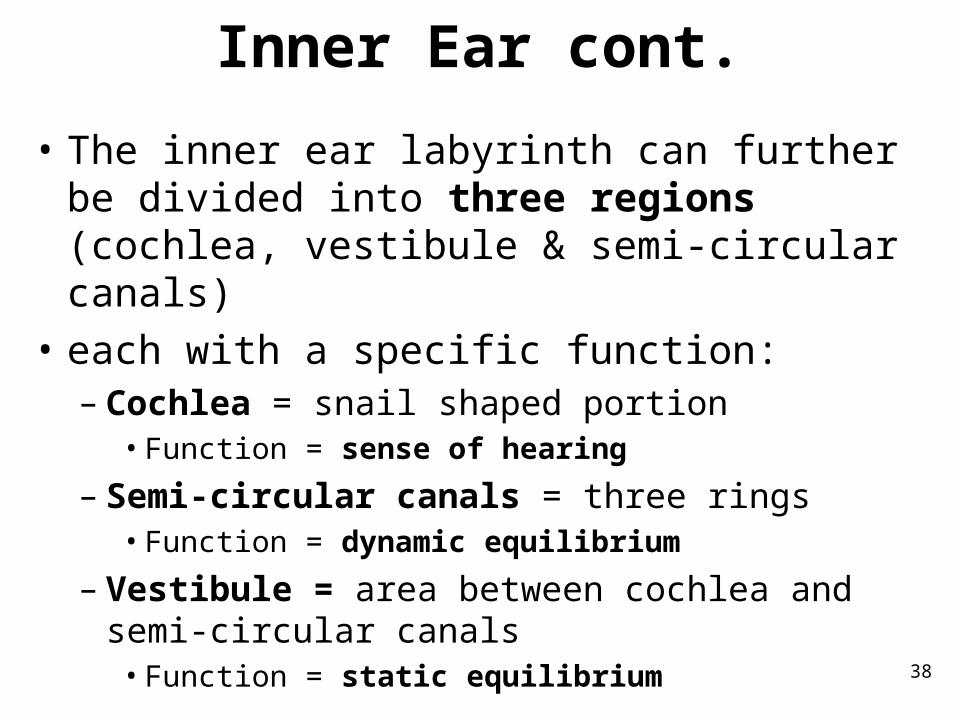
Inner Ear cont.
• The inner ear labyrinth can further be divided into three regions (cochlea, vestibule & semi-circular canals)
• each with a specific function:– Cochlea = snail shaped portion
• Function = sense of hearing
– Semi-circular canals = three rings• Function = dynamic equilibrium
– Vestibule = area between cochlea and semi-circular canals
• Function = static equilibrium

39

40
The Choclea
• divided into two compartments:– Scala vestibuli = upper compartment which
extends from oval window to apex– Scala tympani = lower compartment which
extends from apex to round window
• Both compartments are filled with perilymph
• Between the two bony compartments, we find the membranous labyrinth = cochlear duct– The cochlear duct is filled with endolymph

41

42
Cochlea cont.
• There are membranes that separate the cochlear duct from the bony compartments:– Vestibular membrane separates the cochlear
duct from the scala vestibuli– Basilar membrane separates the cochlear duct
from the scala tympani

43
Organ of Corti
• The mechanoreceptors responsible for the sense of hearing are contained in the Organ of Corti = 16,000 hearing receptor cells located on the basilar membrane.
• The receptor cells are called "hair cells"• The hair cells are covered by the tectorial
membrane, which lies over them like a roof– different frequencies of vibration move different
parts of basilar membrane– particular sound frequencies cause hairs of
receptor cells to bend– nerve impulse generated

44

45

46

47
Sound through the ear
Auditory Nerve Pathways

48
First
• Sound waves arrive at the tympanic membrane
49
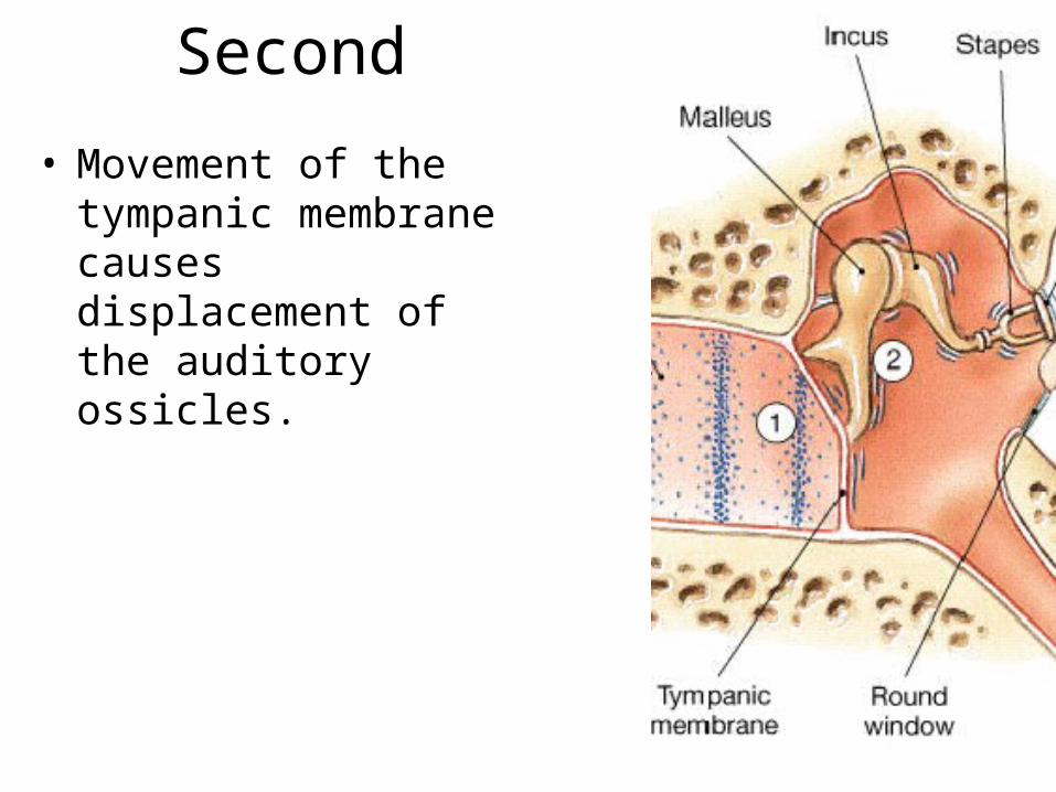
Second
• Movement of the tympanic membrane causes displacement of the auditory ossicles.

50
Third
• Movement of the stapes at the oval window establishes pressure waves in the perilymph of the vestibular duct.

51
Fourth
• The pressure waves distort the basilar membrane on their way to the round window of the tympanic duct.

52
Fifth
• Vibration of the basilar membrane causes vibration of hair cells against the tectorial membrane

53
Sixth• Information about the region and the intensity of
stimulation is relayed to the CNS over the cochlear branch of cranial nerve VIII.
• thalamus for direction to the• primary auditory cortex (temporal lobes) of
cerebrum for interpretation

54
SENSE OF EQUILIBRIUM
Organs= vestibule, utricle, saccule, semi-circular canals

55
Equilibrium
• Static Equilibrium– vestibule– sense position of head when body is not moving
• Dynamic Equilibrium– semicircular canals– sense rotation and movement of head and body

56
Static Equilibrium
• functions to sense the position of the head and help us maintain posture while motionless
• The vestibule of the inner ear contains the two membranous chambers responsible for static equilibrium– The utricle communicates with the semi-circular
canals– The saccule communicates with the cochlear
duct

57
Static Equilibrium cont.
• Each of these chambers contains a macula = organ of static equilibrium
• The macula is composed of "hair cells" that are in contact with a jelly-like fluid containing calcium carbonate crystals (otolith)– When the head is moved, the gel sags due to
gravity and the hair cells bend – This triggers a sensory impulse

58

59

60
Dynamic Equilibrium
• functions to prevent loss of balance during rapid head or body movement
• The three semi-circular canals contain the organ responsible for dynamic equilibrium.– Each semi-circular canal ends in an enlargement called
the ampulla– Each ampulla houses a sensory organ for dynamic
equilibrium called the crista ampullaris, which contains a patch of "hair cells" in a mass of gelatin
– When the head is moved, the gelatin stays put due to inertia causing the hair cells to bend.
– This triggers a sensory impulse

61

62
Vision
Organ=the eyeVisual Accessory Organs=
eyelids, lacrimal apparatus, extrinsic eye muscles

63
Introduction
• The organ of vision is the retina of the eye
• The sensory receptors are called photoreceptor
• When photoreceptors are stimulated, impulses travel within the optic nerve (CN II) to the visual (occipital) cortex for interpretation

64
Visual Accessory Organs
• Eyelids = protective shield for the eyeball.
• Conjunctiva= inner lining of eyelid; = red portion around eye.
• Lacrimal apparatus = tear secretion & distribution
• Lacrimal gland = tear secretion; located on upper lateral surface – Tears contain an
enzyme called lysozyme, which functions as an anti-bacterial agent.
• Nasolacrimal duct = duct which carries tears into nasal cavity (drainage)

65

66
Extrinsic Eye Muscles

67
Structure of the Eye
The eye is composed of three distinct layers or tunics:
• The Outer Tunic (fibrous tunic)
• The Middle (vascular tunic)
• The inner (nervous tunic)

68
The Outer Tunic
• Function= protection
• Cornea = transparent anterior portion– Function: helps focus (75%) incoming light rays
• Sclera = white posterior portion, which is continuous with eyeball except where the optic nerve and blood vessels pierce through it in the back of eye– Functions:
• protection• attachment (of eye muscles)

69
70
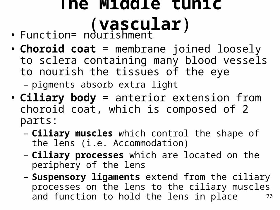
The Middle tunic (vascular)• Function= nourishment• Choroid coat = membrane joined loosely to sclera
containing many blood vessels to nourish the tissues of the eye– pigments absorb extra light
• Ciliary body = anterior extension from choroid coat, which is composed of 2 parts: – Ciliary muscles which control the shape of the lens (i.e.
Accommodation)– Ciliary processes which are located on the periphery of
the lens– Suspensory ligaments extend from the ciliary processes
on the lens to the ciliary muscles and function to hold the lens in place

71

72
The Middle tunic cont.
• Iris = colored ring around pupil– thin diaphragm muscle – lies between cornea and lens– The iris separates the anterior cavity of the eye
into an anterior chamber and posterior chamber
– The entire anterior cavity is filled with aqueous humor, which helps nourish the anterior portions of the eye, and maintains the shape of the anterior eye

73

74
Aqueous Humor• fluid in anterior cavity of eye• secreted by epithelium on inner surface of the ciliary body• provides nutrients• maintains shape of anterior portion of eye

75
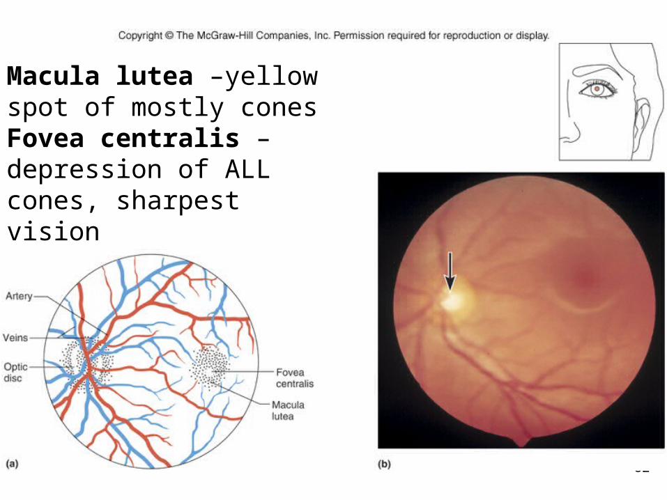
The Inner tunic (nervous, sensory)• Retina = inner lining of the eyeball; site of
photoreceptors– A picture of the retina can be taken with a camera
attached to an ophthalmoscope
• The optic disc is the location on the retina where nerve fibers leave the eye & join with the optic nerve
• the central artery & vein also pass through this disk– No photoreceptors are present in the area of the optic
disk = blind spot.
• The posterior cavity of the eye is occupied by the lens, ciliary body, and the retina– The posterior cavity is filled with vitreous humor, which
is a jelly-like fluid, which maintains the spherical shape of the eyeball

76

77
Accommodation
• the process by which the lenschanges shape to focus on close objects– The lens is responsible (with cornea) for focusing
incoming light rays. – If light rays are entering the eye from a distant
object, the lens is flat. – When we focus on a close object, the ciliary
muscles contract, relaxing the suspensory ligaments. Accordingly, the lens thickens allowing us to focus.

78
Figure 12.29

79
Light Refraction
• Incoming light rays are refracted (bent) onto the retina due to the convex surface of both the cornea and the lens.
• Pathway of Light Through Eye:1. cornea 2. aqueous humor3. lens4. vitreous humor5. photoreceptors in retina.
• Once the rods and/or cones are stimulated, a sensory impulse is carried

80

81
Light Through the Eye• as light enters eye, it is refracted by
• convex surface of cornea• convex surface of lens
• image focused on retina is upside down and reversed from left to right
82
Macula lutea –yellow spot of mostly conesFovea centralis – depression of ALL cones, sharpest vision

83
Visual ReceptorsRods= long, thin projections
• contain light sensitive pigment called rhodopsin• hundred times more sensitive to light than cones• provide vision in dim light• produce colorless vision and outlines
•dark adapted – all opsin and retinal is together, therefore rods are VERY sensitive, vision possible even in dark
Cones=short/bluntprojections
• contain light sensitive pigments called erythrolabe, chlorolabe, and cyanolabe• provide vision in bright light• produce sharp images• produce color vision•light adapted – most opsin and retinal decomposes

84

85
Visual PigmentsRhodopsin
• light-sensitive pigment in rods• decomposes in presence of light• triggers a complex series of reactions that initiate nerve impulses• impulses travel along optic nerve
Pigments on Cones• each set contains different light-sensitive pigment• each set is sensitive to different wavelengths• color perceived depends on which sets of cones are stimulated• red, green, or blue

86
Stereoscopic Vision• provides perception of distance and depth• results from formation of two slightly different retinal images