cerebral air embolism - hboorcca.com 11 cerebral air embolism.pdf · tem can cause cerebral air...
TRANSCRIPT

Cerebral Air Embolism
K.K.Jain
HBO is the mosteffectivetreatment of air embolism; it reduces the size of air bubbles and counteracts
the secondary etTects.This chapter on cerebral air embolism examines:
Causes .
IMechanisms .Pathophysiology .ClinicalFeatures .
Diagnosis .
Treatment .
ClinicalApplications of HBO .AncillaryTreatments .
Hyperbaric Treatment in Special Situations .Conclusions .
104
104
104
105
105
106107
107
108109

104 Chapter 11
Causes
The introduction of air into the venous or the arterial system can cause cerebral air embolism leading to severe neurological deficits. The first known recognition of arterial airembolism was reported by Morgagni in 1769, and later, in1821, Magendie described the consequences of pulmonaryoverinflation leading to arterial gas embolism. The mostcommon causes reported in the literature are iatrogenic,the embolism occurring as a result of invasive medical procedures or surgery. Less commonly, air embolism occurs indivers undergoing rapid decompression and in submarineescape. Causes of air embolism are shown in Table 1l.l.
Table 11.1Causes of Air Embolism
A. Sudden decompression or ascent in diving and submarineescape
Pulmonary barotrauma - "burst lung" in divers- Rapid decompression in an altitude chamber for flight
trainingB. Trauma
Cardiopulmonary resuscitation in patients with undetected lung injury
- Head and neck injuries- High-altitude accidents
C. Iatrogenic1. Diagnostic and minor procedures- Intravenous fluids and central venous pressure (CVP)
lines- Arterial lines for blood and medication infusion
- Angiography: diagnostic and therapeutic catheterizationof blood vessels
- Mechanical positive pressure ventilation- Air contrast salpingogram- Air insufflation with pneumatic otoscope- Needle biopsy of the lung- Hemodialysis- Gastrointestinal endoscopy
2. Intraoperative complications- Neurosurgical operations in the sitting position: tear into
veins in the posterior fossa or the cervical spinal canal- Cardiac surgery: open heart surgery with extracorporeal
circulation- Vascularsurgery: carotid endarterectomy with shunt- Thoracic surgery: opening of pulmonary veins at subat-
mospheric pressures- Endobronchial resection of lung tumor using Neodymi
um- YAGlaser- Pelvicsurgery in Trendelenburg position. Operative hys
teroscopy with laser- Cesarian section
D. Miscellaneous and rare causes- Faulty abortion
Orogenital sex during pregnancyInhalation of helium directly from a pressurized heliumtank.
- Ingestion of hydrogen peroxide solution
There are approximately 20,000 cases of air embolismper year in the USA. The exact incidence of various causesis difficult to determine, as not all cases are reported in theliterature. An excellent review of the topic is presented elsewhere (Hodics & Linfante 2003). Some victims recover
spontaneously. This review concerns those cases where hyperbaric treatment was used.
The incidence of air embolism during cardiopulmonarybypass operations is 0.1 %. The actual prevalence may behigher because several such complications are not recognized and reported. Air enters the venous system in30%-40% of the patients undergoing neurosurgical operations in the sitting position. Air embolism can occur during neuro-angiographic interventional procedures such asaneurysm coiling embolization and carotid stent placement but overall incidence during diagnostic neuroangiographic procedure is very low in the order of 0.08% (Guptaet al 2007).
Mechanisms
In iatrogenic cases the air is either sucked into the veins withnegative pressure or introduced into the veins or arteriesunder pressure. The lung is usually an effective filter for airbubbles greater than 22 !lm in diameter when air is injectedslowly. A bolus injection of air more than 1.5-3 ml/kg exceeds the filter capacity of the lungs and produces embolization through the left heart into the arterial circulation until it blocks arterioles 30-60 !lm in diameter. Air has a largesurface tension at the blood-air interface and the globules ofair cannot be deformed enough to navigate the capillaries.
A patent foramen ovale occurs in 20% to 35% of thenormal adult population and in one out of ten of these isat risk of having arterial air embolism when air enters the.venous system inadvertently. The exact prevalence rate offunctional right to left atrial shunt in the healthy adult population, however, is unknown. In the absence of such ashunt, venous air must first traverse the pulmonary vasculature in order to enter the cerebral circulation.
In pulmonary barotrauma, lung volumes expand during rapid ascent. '!\Then alveolar pressure exceeds 80100 mmHg, air can be forced into pulmonary capillaries.Alveoli may rupture into the pleural space, causing pneumothorax, or into the pulmonary veins, where the embolus may traverse the left side of the heart to enter the aortaand may ascend the carotid arteries to the cerebral circulation, as the diver is usually upright during ascent.
Pathophysiology
Air emboli lodge distally in the smaller arteries and arterioles of the brairi and obstruct the flow of blood. The

Cerebral Air Embolism 105
'Cjulti, ischemia, hypoxia, and cerebral edema. Even\·;henthebubble is dissolved, a "no reflow" phenomenonmAY "ccurin the damaged tissues. The bubble acts as aJor(i~nhod)"and starts a number of biochemical reacnnnsin the blood. Platelets are activated and release va
\!)<!ctil'csubstanccsincluding prostaglandins. The bubblemardamagethe endothelial cells of the vessel wall by di":Xtcontact.;>'Iarginationand activation ofleukocytes fol
1\1), andma)"cause a secondary ischemia. If the bubblepmi,tl,it is surrounded by platelets and fibrin deposit,whichma)"preventdislodging of the bubble. Although thex'lentialof largc bubbles to cause cerebral injury is notdi,pukd,there is controversy over the significance of exp'hureto small bubbles in cardiac surgery. It is knownU!Jtpilltsurgicalneuropsychological deficits do correlate~)itil'dywith the number of emboli to which patientsre(xpllsed;to datc, however, the technology for distinwi,hingbetween gaseous and particulate emboli or forsizingemboliaccurately is not readily available (MitchellH,urman20(2).
Airbubblesinjected directly into the cerebral circulalIOnof experimental animals can open the blood-brainbarrier.The barrier, however, repairs itself within a few!Jour).hchemichypoxia produced by air embolism is not'(\ne cnoughto produce gross cerebral infarction, butproducesnecrosisof the deep cortical layers at the graywhilematterjunction.
Theremaybe segmental arterial vasospasm followed by."Jtatilln,and some of the air may escape from the arter"intlltheveinsvia capillaries. As a result of arterial obItruClillnregional cerebral blood flow (rCBF) declines
ndI:E<; activity may cease in the affected region. The,hange,arc typical of cerebral ischemia, but the blood'fainharrierpermcability increases immediately after air~mbuli,m!in contrast to vascular occlusion from other
thC\, wherethe onset is delayed. Focal ischemia of shorturatillndocsnot lead to cell loss, and the processes caus
",gddcriorationare potentially reversible. The other po'cntioilly rel'Crsiblcprocess that occurs in the tissues is ce.Jlraledema.Although the brain is the major concern in[;eriJIembolism,the coronary arteries may occasionallyinmlwl.
.\nimalbrain models of cerebral arterial gas embolism\' hI' usefulin comparing the effectiveness of various
":('JlI1pressionschcdules. Murrison (1993) has reviewed
ariuu,animal models for neurophysiological investiga'IOn\lIfpathophysiologyof central nervous system (CNS)damageinarterialgas embolism. Most of these studies in-
ile inicctionof air into cerebral vasculature. Secondary\~ deterioration may be may be due to endothelial
)jlllJgeor changein blood constituents rather than me
.Anicalbuhhleaction and may explain the failure of re'mprcsliontherapy in such cases. The results of these
Im.!1 ,tudiescannot be extrapolated to humans.
Clinical Features
Clinical manifestations, essentially neurological or cardiovascular disorders vary greatly. The clinical features of airembolism depend on the patient's posture, the route of en
try of air, the volume of air, the size of the bubbles, and therate of entry of air. If the patient is reclining, air is morelikely to enter the coronary arteries, whereas it is more likely to enter the cerebral arteries if the patient is upright.Neurological sequelae have been estimated to occur in 19to 50% of the patients with cerebral air embolism.
Signs and symptoms of air embolism in divers may notbe clear-cut. In 50% of such cases, dysbaric air embolismwas found to be part of dysbarism syndrome including decompression sickness. A sudden change in sensorium is themost common symptom and ranges from disorientationto coma. Focal neurological deficits such as hemiplegia ormonoplegia may occur, according to the location of the lesions. Respiratory arrest and seizures are less common. Ashock-like state may occur with massive embolism. Associated symptoms may be those of pneumothorax (in pulmonary barotrauma) or myocardial ischemia. Liebermeister's sign, i.e., the presence of areas of pallor on the tongueafter air embolism, may be found.
Diagnosis
The diagnosis of air embolism is based on a careful consid
eration of the patient's history and neurological findings.In cases of sudden decompression with neurological deficit, the diagnosis is easier. During surgical procedures monitored by doppler ultrasound, air embolization is detectedat an early stage and appropriate measures can be taken tostop further air entering the blood vessels. Transcranialdoppler studies show that microscopic cerebral artery airemboli are present in virtually all patients undergoing cardiac surgery. Microbubbles can be detected with two-dimensional echocardiography, which is often used for this
purpose during open heart and bypass surgeries (Kearneyet a11997). EEG monitoring is also useful for early detection of acute cerebral dysfunction.
Subtle changes in mental function may be a major manifestation even in the absence of other objective neurological signs. CT scan offers a possibility of diagnosis of subclinical lesions of the brain. Air bubbles may also be seenon fundoscopic examination. Air may be seen in the cerebral arteries during a neurosurgical operation, or air can
be demonstrated in a specimen of arterial blood. A highindex of suspicion is very important in diagnosis. Undersuspicious circumstances air embolism should be assumed
present unless otherwise proven. In some cases the diagno-
106 Chapter 11
sis is proven only after successful response to hyperbarictherapy.
In air embolism associated with diving, there is muscleinjury and elevated serum creatine kinase which a marker ofthe severity of this complication (Smith & Neuman 1994).
Treatment
Emergency measures include administration of 100% oxygen, using a closely fitting mask, and transport of the patient to a hyperbaric facility. If transport by air is unavoidable, the patient should travel in a pressurized cabin, andthe aircraft should stay at a low altitude. A bolus dose(10 mg) of dexamethasone may be given to prevent cerebral edema. Oxygen serves to reduce the size of the air bubble by depletion of nitrogen and also counteracts the hypoxia and ischemia of the surrounding brain tissue.
The important consideration in treatment of cerebral airembolism is preparedness and anticipation. Procedureswith a known risk of air embolism should not be performed far away from a hyperbaric facility, and a hyperbaric chamber should be available in institutions that con
duct open heart surgery. Time is the more important element in management - the shorter the delay, the better theoutcome. Emboli large enough to produce symptoms require immediate treatment because of the risk of "gas lock"in the right side of the heart and subsequent circulatoryfailure (J0rgensen et al2008).
The generally accepted treatment of air embolism is immediate compression to 6 ATA air for a period of not morethan 30 min followed by ascent to 2.8 ATA with oxygen.The rationale of this approach is as follows:
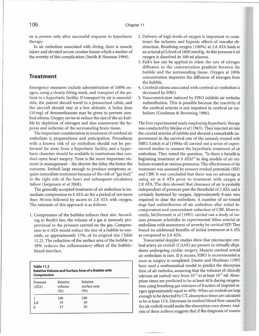
1. Compression of the bubbles reduces their size. According to Boyle's law, the volume of a gas is inversely proportional to the pressure exerted on the gas. Compression to 6 ATA would reduce the size of a bubble to one
sixth, or approximately 17%, of its original size (Table11.2). The reduction of the surface area of the bubble to
30% reduces the inflammatory effect of the bubbleblood interface.
Table 11.2Relative Volume and Surface Area of a Bubble with
Compression
Pressure RelativeRelative
(ATA)
volumesurface area
(%)(%)
1
100100
2.8
3550
6
1730
2. Delivery of high levels of oxygen is important to counteract the ischemic and hypoxic effects of vascular obstruction. Breathing oxygen (100%) at 2.8 ATA leads toan arterial p02level of 1800 mmHg. At this pressure 6 mloxygen is dissolved in 100 ml plasma.
3. Fick's law can be applied to relate the rate of nitrogendiffusion to the concentration gradient between thebubble and the surrounding tissue. Oxygen at 100%concentration improves the diffusion of nitrogen fromthe bubble.
4. Cerebral edema associated with cerebral air embolism is
decreased by HBO.5. Vasoconstriction induced by HBO inhibits air embolus
redistribution. This is possible because the reactivity ofthe cerebral arteries is not impaired in cerebral air embolism (Gordman & Browning 1986).
The first experimental study employing hyperbaric therapywas conducted by Meijne etal (1963). They injected air intothe carotid arteries of rabbits and showed a remarkable im
provement in the survival rate of the animals treated withHBO. Leitch et al (1984a-d) carried out a series of experimental studies to reassess the hyperbaric treatment of airembolism. They tested the question, "Is there a benefit inbeginning treatment at 6 ATA?" in dog models of air embolism treated at various pressures. The effectiveness of thetreatment was assessed by sensory evoked potentials (SEP)and CBF. It was concluded that there was no advantage inusing air at 6 ATA prior to treatment with oxygen at2.8 ATA. The data showed that clearance of air is probablyindependent of pressure past the threshold of 2 ATA and iscertainly hastened by m."ygen. Approximately 8 min wererequired to clear the embolism. A number of air-treateddogs had redistribution of air embolism after initial decompression and concomitant reduction of CBF. More recently, McDermott et al (1992) carried out a study of various pressure schedules in experimental feline arterial airembolism with assessment of severity by cortical SEP. Theyfound no additional benefits of initial treatment at 6 ATA
as compared to 2.8 ATA.Transcranial doppler studies show that microscopic cere
bral artery air emboli (CAAE) are present in virtually all patients undergoing cardiac surgery. Massive cerebral arterialair embolism is rare. If it occurs, HBO is recommended as
soon as surgery is completed. Dexter and Hindman (1997)have used a mathematical model to predict the absorptiontime of air embolus, assuming that the volumes of clinicallyrelevant air emboli vary from 10-7 to at least 10-1 ml. Absor
ption times are predicted to be at least 40 h during oxygenation using breathing gas mixtures of fraction of inspired ox
ygen approximately equal to 40%. When air emboli are largeenough to be detected by CT, absorption times are calculatedto be at least 15 h. Decreases in cerebral blood flow caused by
the air emboli would make the absorption even slower. Anal
ysis of these authors suggests that if the diagnosis of massive

Cerebral Air Embolism 107
Table113
Examples of Applications of HBO for Cerebral Air Embolism
.Imhuf> ilml \'Car No. of cases Cause
Arm<OJ\ ,'I ill I IY'lI ) I
••nardl "/ill
2flilJ
Results and comments
HBO treatment started 8 hours after onset with
impairment of consciousness and lefthemiplegia. Recovered with minimalneurological deficit.
HBO treatment was started 30 hours after the
incident with coma, decerebrate rigidity andseizures. Recovered with minimal residual
neurological deficits at 14 months follow-up.
2 died
2 partial recovery2 full recovery
Complete recovery from left hemiplegia.
Patient presented neurological deficits 8 hourslater, when HBO treatment was started. Full
recovery.
Treated 52 h after the event. Discharged after fully recovery 1 week later.
Table 6A (extended)+3 HBO sessions at 2ATA/90 min
Table 6
Modified Table 6
Table 6A
Table 6A
Pressure used
Table 6
Paradoxical air embolism duringpercutaneous nephrolithotripsy inthe prone position.
Diagnostic bronchoscopy in a patient with previous lobectomy forbronchogenic carcinoma.
Decompression at flight level 280(28,000 ft) in an altitude chamber.
Cardiopulmonary by-pass
Open heart surgery
Cesarian section
'"dri.ula ,'1 III
!'I'!7
f1midIelli ,'I i!l
_~~I!
I fJ'I!lliliIYY.\)
II
\
II
I
1
hr
,oj
w
I A,\L i, >uspected,CT should be performed, and consider'!on,houldbegiven to HBO therapy if the emboli are large._\ughto be visualized,even if patient transfer to a HBO
iilily IIillrequireseycral hours.~nml'authors recommend supportive care as the priIrvlhaapyli)rvenous gas embolism, while HBO therapy
i\ addilionlosupportive care is the first line of treatment
'!r Jrtaialga>embolism (Fukaya & Hopf 2007). The criHontilruseof I-IBO is clinical manifestation, particularly
leurologicaland not the source of air embolism.
ClinicalApplications of HBO
mical<tpplicationsof HBO for air embolism during theII dl'cadl'arc shown in Table 11.3. If we consider the
.fallmortalityof air embolism without hyperbaric treatmta,.\1)11/" theseresults represent a remarkable improve
,ell!. i\ controlledprospective study has shown that mor-• "1\ G1I1hl' reduced to 14% if hyperbaric oxygen therapy"~I'il'nwithin12 hours of the accident (Bacha et aI1996).\(Jtml'ntappearsto be ineffective after irreversible dam-
hasalreadybeen done.
Hill) hasbeenlIsedsuccessfully in cases of air embolism.1 (!Implicationof open heart surgery, endoscopy (Raju
Iall'!'}XI and transthoracic percutaneous thin-needle bi,illkggl' cl a/1997). The usual schedule of hyperbaric
'r, atlnl'nlis US Air Force Modification of US Navy TableFigurl']()j; Chapter 10). The initial approach is to com"thl' patient106 ATA.After 30 min, decompression is
.unl'dout to 2.~AlA.
Ancillary Treatments
The following treatments have been used in addition to hyperbaric therapy.
Antiplatelet Drugs. These have been used to counteract theplatelet aggregation associated with air embolism. The useof heparin as anticoagulant is considered risky due to thedanger of hemorrhage in infarcted areas. Patients who arealready on heparin have a better prognosis after air embolism than those who are not anticoagulated. This is particularly noted during cardiopulmonary bypass for cardiacsurgery. Oral aspirin is safer but takes about 30 min to actafter ingestion.
Steroids. These have been used to prevent cerebral edema.Delayed cerebral edema can occur after initially good results from recompression following air embolism. Steroidsshould be administered cautiously during HBO as theymay accelerate the development of oxygen toxicity.
Hemodilution. Hematocrit has been shown to have a rela
tion to the infarct size in vascular occlusion, and many casesof air embolism display hemoconcentration. Hemodilution, e.g., by dextran-40, is indicated. Lowering of hematocrit also causes a reduction in m...ygen-carrying capacity,but this is more than adequately compensated by HBO.
Control of Seizures. An anticonvulsant medication may berequired for control of seizures. Prophylactic use of lidocaine not only controls seizures but also reduces infarct sizeand prevents cardiac arrhythmias associated with air embolism.

108 Chapter 11
Measures to Improve Cerebral Metabolism. Loss of bloodsupply causes immediate reduction of neuronal pools andincreased production of lactate. The total energy availableis reduced. Increased blood glucose levels are associatedwith increased lactate production by the ischemic brainand increase in infarction. The control of blood glucose,therefore, is important after air embolism and routine useof intravenous dextrose should be avoided. There is evidence that HBO serves to normalize the cerebral metabo
lism (see Chapter 17) and also lowers blood glucose.
Hyperbaric Treatment in SpecialSituations
Cerebral Edema
The following example is given to illustrate the special useof HBO in cerebral edema. Thiede and Manley (1976) reported a patient with air embolism who responded to initial compression to 6 ATAbut deteriorated into coma anddecerebrate posturing during decompression at 1.9 ATA.There was increased intracranial pressure, indicating cerebral edema. The patient was given repeated HBO treatments twice daily at 2.8 ATA(100% oxygen) and recoveredcompletely. HBO in this case was doubly indicated - for airembolism as well as for cerebral edema.
Cardiovascular Surgery
Calverley et al (1971) reported air embolism during cardiaccatheterization in a 4-month-old infant with ventricular
septal defect. A quantity of 10ml air was inadvertently injected into the right ventricle. Anesthesia was terminatedand air compression was done to 6 ATA 35 min after theepisode. Decompression was started after a further 15 minand completed in 5 h. No oA')'genwas given. The infantmade a good recovery and the planned cardiac surgery wascarried out. Calverley et al made an important observationabout nitrous oxide anesthesia: if air embolism occurs dur
ing this type of anesthesia, nitrous oxide diffuses rapidlyinto enclosed pockets of gas,causing an increase in pressure(or an increase in volume if the surrounding tissues permit). The authors recommended that nitrous oxide anesthesia should be discontinued if air embolism occurs.
HBO has been used to treat patients with extensive neurological deficits from air emboli during open heart surgery.Treatment is usually not started until after completionof surgery, but is still effective. Some complicated operations in cardiac surgery and neurosurgery cannot be aborted because of air embolism. In such cases, compressiontreatment can be started after completion of the operation.
Huber et al (2000) reported a case of a 5-year-old girlwho suffered a massive arterial air embolism during surgical closure of an atrial septal defect. They successfullytreated a proven arterial air embolism with intraoperative (retrograde cerebral perfusion) combined with postoperativeprocedures (deep barbiturate anesthesia and HBO). Atdischarge the girl had fully recovered from the initial neurologic defects.
Hypothermia has been used in cardiac surgery for cerebral protection. Patients who suffer massive air embolismduring cardiopulmonary bypass can be treated by usingacombination of hypothermia and HBO with good results.According to Charles' law, the volume of a gas varies according to temperature. Theoretically hypothermia is expected to decrease the size of the gas bubbles and shouldbe beneficial in air embolism. Animal experimental studiesare required to determine if HBO and hypothermia complement one another in air embolism.
Neurosurgery
Air embolism following posterior fossa surgery in the sitting position can be promptly treated by recompression according to the standard schedule. The patients usuallyrecover without neurological deficits.
Pulmonary Barotrauma
Leitch and Green (1987) reviewed treatment of89 casesofair embolism due to pulmonary barotrauma in divers.There was a 65% success rate with hyperbaric treatments,and one of the victims had a relapse. The authors concluded that although most cases would recover with oxygenat2.8 ATA,there was no reason to alter the established technique of initial compression with air to 6 ATAprior toHBO at 2.8 ATA.Air embolism associated with pulmonarybarotrauma during rapid decompression in an altitudechamber has been managed by the use of treatment table6A (Rudge 1992).
Pulmonary barotrauma with air embolism has beenreported as a complication ofHBO therapy for a non-healingulcer of the foot (Wolf et aI1990). Pneumothorax hasbeenreported as a complication of recompression therapyforcerebral arterial gas embolism associated with diving(Broome & Smith 1992).
In an unusual presentation of cerebral air embolism,apatient became unresponsive and developed subcutaneousemphysema during the direct insufflation of ox')'genintothe right middle lobe bronchus (Wherrett et al 2002).An
endotracheal tube and bilateral chest tubes were immedi
ately placed with resultant improvement in the oxygensaturation. However, the patient remained unresponsivewith

Cerebral Air Embolism 109
exklllorand Ilexor responses to pain. Later, there was seiI1lrealti\'ilyrequiring anticonvulsant therapy. CT scans of;heheadand cerebral spinal fluid examination were negalire.thoughthe electroencephalogram was abnormal. A( T "fdle~tshowedevidence of barotrauma. 52 hours after
theerent,a pn:sumed diagnosis of cerebral air embolism1\",1,made,and the patient was treated with HBO using amodifiedUS NavyTable 6. 12 hours later he had regained(olhciousncssand was extubated. He underwent two more
HII() and wasthcn discharged from hospital 1 week aftertheerent,fullyrecovered. Although HBO was started aftersigniticantdelay,the patient made a good recovery.
Air EmbolismDuring Invasive MedicalProcedures
CItront't III ( llili1) used HBO to treat t\'\'o patients withcerehr'llairl'mbolism resulting from invasive medical pro,tilure,.Bothpatients recovered without any evidence ofdJm,lgcon clinicalexamination and MRI.
Cerebral Air Embolism During ObstetricalProcedures
Ti\U ca~e>of ccrebralair embolism occurring during cesarn'eclionwere treated successfully with the use of HBO\1JJnt't ll/llilil; Davisct alI990). Air embolism manifest
J by ClInicalblindness was reported in a patient followingdiKedahortion by means of intra-amniotic hypertonichnein>tillationand the patient made a complete recovery
'Hertrcatl11cntwith HBO (Weissman et alI989). Venous airmholi>111islikelyduring cesarian section as air enters uterme,inu,,>,particularly if the placenta separates before de-
rry<I, inthe(ase of placenta previa (Davis et alI990).
Cerebral Air Embolism Due to Orogen ita I Sex
During Pregnancy
\l'drcGl,CS of this type have been reported in the literalre;pnlyoncof them survived. Two cases have been treat
',ucctS>fullyusing HBO (Bray et al1983; Bernhardt et al~X!.
Cerebral Embolism Due to Hydrogen Peroxide
Poisoning
Jrog"npcrnxide(an produce acute gas embolism. There,.'C' [l'portof an adult who suffered an apparent stroke
)[tly aftl'ranaccidental ingestion of concentrated hydro-:npl'fllxidc(i\luliins & Belltran 1998). Complete neuro-
logic recovery occurred quickly following HBO treatment.In another case report, a patient developed cerebral air embolism a short time after ingestion of a small amount of
hydrogen peroxide manifested by hematemesis, left sidedhemiplegia, confusion, and left homonymous hemianopsia. Initial laboratory studies, chest x-ray, and brain CTwere normal. MRI demonstrated areas of ischemia and 18
h hours after arrival, the patient underwent HBO treatment
with complete resolution of symptoms (Rider et a12008).Of the seven reported cases of air embolism from hydrogenperoxide that did not undergo HBO, only in one patientwas there a report of symptom resolution. HBO can beconsidered as the definitive treatment for gas embolism
from hydrogen peroxide ingestion as with all other causesof acute gas embolism.
Pediatric Air Embolism
Van Rynen et al (1987) reported successful treatment ofcerebral air embolism in a 3-month-old infant who had
undergone a palliative closed heart operation. The treatment was conducted in a Reneau monoplace chamber using initial recompression to 6 ATA. The infant made a goodrecovery.
Relapse Following Spontaneous Recovery
In cases of spontaneous redistribution of air bubbles, a period of apparent recovery is frequently followed by relapse.The etiology of relapse appears to be multifactorial and ischiefly the consequence of a failure of reperfusion. Prediction of who will relapse is not possible, and any such relapsecarries poor prognosis. It is advisable, therefore, that air embolism patients who undergo spontaneous recovery bepromptly recompressed while breathing o>.')'gen (Clark et al2002). Therapeutic compression serves to antagonize leukocyte-mediated ischemia-reperfusion injury; to limit potential re-embolization of brain blood flow, secondary to further leakage from the original pulmonary lesion or recirculation of gas from the initial occlusive event; to protectagainst embolic injury to other organs; to aid in the resolution of component cerebral edema; to reduce the likelihoodoflate brain infarction reported in patients who have undergone spontaneous clinical recovery; and to prevent decompression sickness in high gas loading dives that precede accelerated ascents and omitted stage decompression.
Delayed Treatment
Air embolism should be treated as quickly as possible afterit is detected. This is not always possible and several casesreceive delayed treatment. HBO treatments have led to re-
110 Chapter 11
covery in cases of air embolism with severe neurologicaldeficit where treatment was delayed for 24 h. In a subgroupof 5 patients with air embolism secondary to cardiopulmonary bypass accidents, pulmonary barotrauma induced bymechanical ventilation and central vein catheterization,
significant recovery was noted even when treatment wasstarted 15 to 60 h after the event (Bitterman & Melamud
1993). Full recovery was reported in a case of hemodialysisassociated venous air embolism, where HBO treatment was
commenced 21 h after the event when the patient alreadyappeared to be decerebrate (Dunbar et alI990).
Conclusions
Hyperbaric treatment has been proven to be unquestionably indicated for the treatment of air embolism with neurological deficits. The conventional methods of treatment,such as posturing the patient in certain ways, aspirating theair, providing normobaric oxygen, closed chest massage,
and steroids, have not been adequate to manage this problem. The consensus concerning the pressure favors the retention of 6 ATA initial compression with air. If the pa·
tient's condition does not permit exposure to this high
pressure or the chamber immediately available cannot provide this pressure, 100% oxygen at 2.8 ATA would be acceptable as an alternative, particularly when only a monoplace chamber is available. The diagnosis of air embolismcannot always be made with certainty. There is need to im·prove technologies for early detection of air bubbles. Itisacceptable to treat the patient with compression if air embolism is suspected, and the response to compression maybe diagnostic in such cases. Early treatment provides betterresults than late treatment but HBO treatment should be
considered at any stage the patient presents.Considering that air embolism is a complication ofmed
ical and surgical procedures, it stands to reason that hyperbaric chambers should be available at clinics that performsuch procedures. Open heart surgery, certainly, should notbe done in a hospital that does not have a hyperbaric chamber.