pulmonary embolism &pulmonary embolism & … tumor, air, fat, foreign material, or parasitic...
TRANSCRIPT

Pulmonary Vascular Disease: Pulmonary Embolism &Pulmonary Embolism &
Pulmonary Hypertension
Selim M. Arcasoy, M.D.Professor of Clinical MedicineProfessor of Clinical Medicine
Medical Program DirectorLung Transplantation Program
C l bi U i itColumbia University College of Physicians and Surgeons
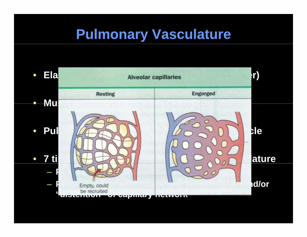
Pulmonary Vasculature
• Elastic pulmonary arteries (> 1 2 mm diameter)• Elastic pulmonary arteries (> 1-2 mm diameter)
• Muscular pulmonary arteries (100 μm-1 mm)• Muscular pulmonary arteries (100 μm-1 mm)
• Pulmonary arterioles (< 30-100 μm )--no musclePulmonary arterioles ( 30 100 μm ) no muscle
• 7 times more compliant than systemic vasculaturep y– Pulmonary VR is one tenth of systemic VR– Pulmonary VR stays low due to “recruitment” and/or
“distention” of capillary network“distention” of capillary network

Control of Pulmonary Circulation
• Hypoxia– To match regional perfusion/ventilation
• Nervous system– Parasympathetic sympathetic NANC fibersParasympathetic, sympathetic, NANC fibers,
neurohormones
P i h i• Passive mechanisms– Anatomy, gravity, lung volume, alveolar pressure

Pulmonary Embolism

DefinitionDefinition
Obstruction of pulmonary arterial branches by material originatingbranches by material originating from another location in the body
ThromboticNon-thrombotic: tumor, air, fat, foreign
material, or parasitic

Epidemiology of Pulmonary Embolism
• Estimated to occur in ~ 600,000 patients annually in the U.S., p y– Annual incidence ranges between 23 to 69 cases per 100,000
population
I id f PE i h i l f 0 0 1%• Incidence of acute PE in hospitals ranges from 0.05 to 1%
• Causes or contributes to ~50,000 to 200,000 deathsA t f 15% f i h it l t lit– Accounts for 15% of in-hospital mortality
• Diagnosis is missed in 50-70% of patients antemortem
• Wide spectrum of severity with short-term mortality figures between 2.5% and >50%
Dalen JE. Prog Cardiovasc Dis 1975;17:259Goldhaber SZ. Am J Med 1982;73:822Pineda. Chest 2001;120:791

Pathophysiology of Pulmonary Embolism
• Virchow’s triad– Endothelial injury, stasis,
hypercoagulability
• Sources of PESources of PE– Iliofemoral veins***– Pelvic, upper extremity,
renal right heartrenal, right heart
• ~50% of iliofemoral DVTs result in PE– 50-80% of iliofemoral DVTs
originate in calf veins
Tapson . N Engl J Med 2008;358:1037
g

Gas Exchange Physiology After PE
• Acute vascular obstruction and vasoconstriction
• Increased alveolar dead space– Reflex bronchoconstriction to minimize dead space– Hyperventilation
• Mechanisms of arterial hypoxemiayp– Shunt (flow through atelectatic regions, opening of latent
pulmonary A-V anastomoses due high PAP or intracardiac)– V/Q mismatch (increased flow to low V areas without emboli– V/Q mismatch (increased flow to low V areas without emboli
due to increased PA pressure)– Diffusion impairment (high flow with reduced transit time)
Increased A V O difference from RV strain and decreased CO– Increased A-V O2 difference from RV strain and decreased CO

Pathophysiology of Hemodynamic Instability in PE
Pulmonary Embolism
PA pressure RV ll t iPA pressureRV afterload
RV dilatation
RV wall tension
RV O2 demandRV ischemia/infarction
RV dilatationRV dysfunction
(Submassive PE)
Coronary perfusion
RV O2 supply
RV cardiac output
Septal shift towards LV
ViciousCycle
LV preload Hypotension
y poutput towards LV Cycle
LV output Hypotension(Major PE)

Risk Factors for Venous Thromboembolism
• Acquired FactorsReduced mobility
• Hereditary factorsFactor V Leiden– Reduced mobility
– Advanced age– Cancer and chemotherapy
Acute medical illness
– Factor V Leiden– Activated protein C resistance
without Factor V Leiden– Antithrombin deficiency– Acute medical illness
– Major surgery and trauma– Spinal cord injury
Pregnanc /postpart m
– Antithrombin deficiency– Protein C and S deficiency– Prothrombin gene mutation– Dysfibrinogenemia– Pregnancy/postpartum
– Oral contraceptives– Hormone replacement Rx
A ti h h li id b d
– Dysfibrinogenemia– Plasminogen deficiency
P b bl f t– Antiphospholipid ab syndrome– Central venous catheter– Polycythemia vera
Ob it h t i
• Probable factors– Elevated lipoprotein(a)– Elevated homocysteine,
Tapson. N Engl J Med 2008;358:1037
– Obesity, hypertension– Heavy smoking
factors VIII, IX, XI, fibrinogen

Clinical Findings of PE
• Symptoms and signs– Dyspnea chest pain wheezing cough apprehension leg painDyspnea, chest pain, wheezing, cough, apprehension, leg pain and swelling, syncope, hemoptysis, fever– Tachycardia, tachypnea, accentuated P2, rales, JVD, DVT
• Chest radiographAtelectasis, pleural effusion, pleural-based opacity, cardiomegaly,diaphragmatic elevation prominent central PA Westermark signdiaphragmatic elevation, prominent central PA, Westermark sign
• ECGAnterior T-wave inversions ST-T segment changes RBBB S1Q3T3Anterior T-wave inversions, ST-T segment changes, RBBB, S1Q3T3
• Arterial blood gasHypoxemia and hypocapniaHypoxemia and hypocapnia

Diagnostic Evaluation
• Develop an estimate of pretest clinical probability p p p ybased on symptoms, signs and risk factors– High (very likely), low (unlikely) or intermediate (possible)
Clinical prediction scores (Wells or Geneva)– Clinical prediction scores (Wells or Geneva)
• Concomitant diagnosis, treatment, and resuscitation if needed– Start anticoagulation if PE is highly suspected and there
are no contraindications– In the case of massive PE, evaluation must be RAPID
since majority of deaths occur within 6 hrs of presentation

Diagnostic Tests For PE
• Ventilation-Perfusion (VQ) scan
• CT pulmonary angiography (CTPA or CTA)
• Duplex ultrasonography
• Laboratory markersy– D-dimer, cardiac troponins, NT-pro-BNP and BNP
• Echocardiography
• Pulmonary angiography
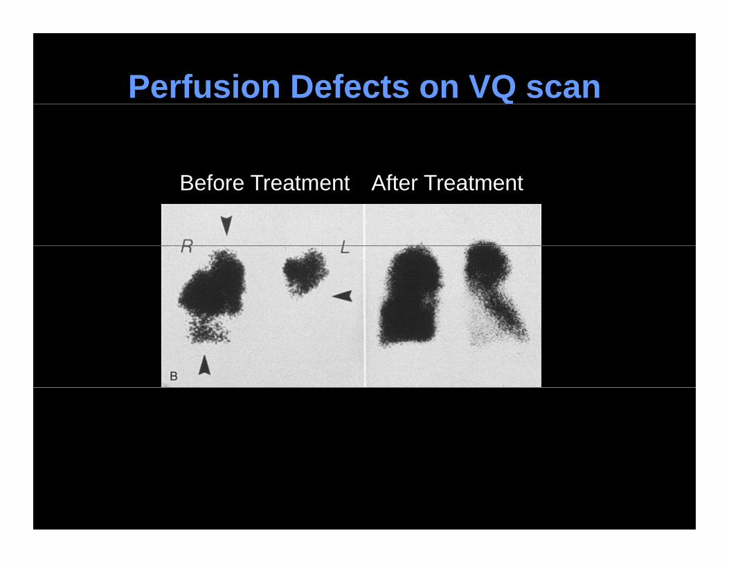
Perfusion Defects on VQ scan
B f T t t Aft T t tBefore Treatment After Treatment
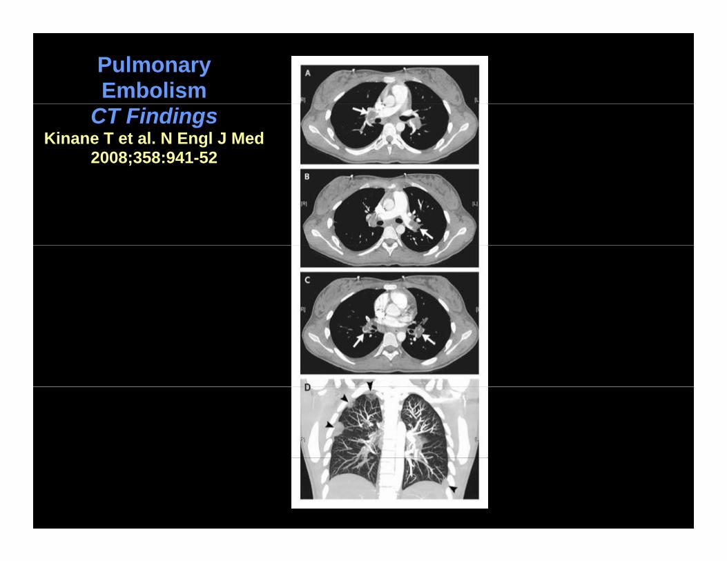
Pulmonary Embolism
CT FindingsKinane T et al. N Engl J Med
2008;358:941-52
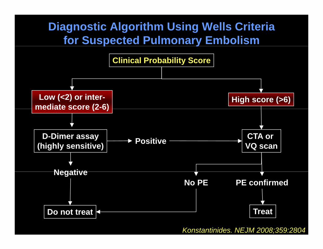
Diagnostic Algorithm Using Wells Criteria for Suspected Pulmonary Embolismp y
Clinical Probability Score
Low (<2) or inter-mediate score (2-6)
High score (>6)mediate score (2 6)
D-Dimer assay Positive CTA or y(highly sensitive)
Negative
Positive VQ scan
NegativePE confirmedNo PE
Do not treat Treat
Konstantinides. NEJM 2008;359:2804

Treatment of Acute Pulmonary Embolism
• Anticoagulation with heparin productsR h th ti l l i kl– Reach therapeutic levels quickly
– Transition to oral anticoagulation
I f i filt l t• Inferior vena cava filter placement– Anticoagulation contraindicated– DVT present along with severe PEp g
• Thrombolytic therapy– Hemodynamic instabilityy y
• Surgical or catheter embolectomy– Major PE unresponsive to anticoagulation,Major PE unresponsive to anticoagulation,
thrombolysis or contraindications to medical Rx
Pulmonary Hypertension

Hemodynamic Physiology of Pulmonary HypertensionPulmonary HypertensionBack to Physics-Modified Ohm’s Law
Ch i Fl R i t• Change in pressure = Flow x Resistance– Ppa - Ppv = Q x PVR– Ppa = (Q x PVR) + Ppv– PVR = (Ppa - Ppv)/ Q = 100 dynes/s/cm-5
• Alterations in PVR, Q and Ppv raise Ppa– PVR: occlusive vasculopathy of small arteries / arterioles (PAH),
decreased area of pulmonary vascular bed (PE, ILD), hypoxic vasoconstriction (COPD, high altitude)
– Q: Left to right shunt due to congenital heart disease, liver cirrhosis– Ppv: Left heart and valvular disease, constrictive pericarditis
• Increase in PVR is the primary cause of PH
Pulmonary Hypertensiony ypHemodynamic Definition
• A disorder characterized by increase in pulmonary vascular pressurepulmonary vascular pressure– Isolated increase in pulmonary arterial pressure or
increase in both pulmonary arterial and venous pressures
• Pulmonary arterial hypertension– Mean PAP >25 mmHg at rest– Mean PAP >25 mmHg at rest– Normal pulmonary capillary wedge pressure (< 15 mmHg)
• Elevated PCWP indicates PH due to left heart diseasePVR > 3 W d it ( >200 d / / 5)– PVR > 3 Wood units (or >200 dynes/s/cm-5)
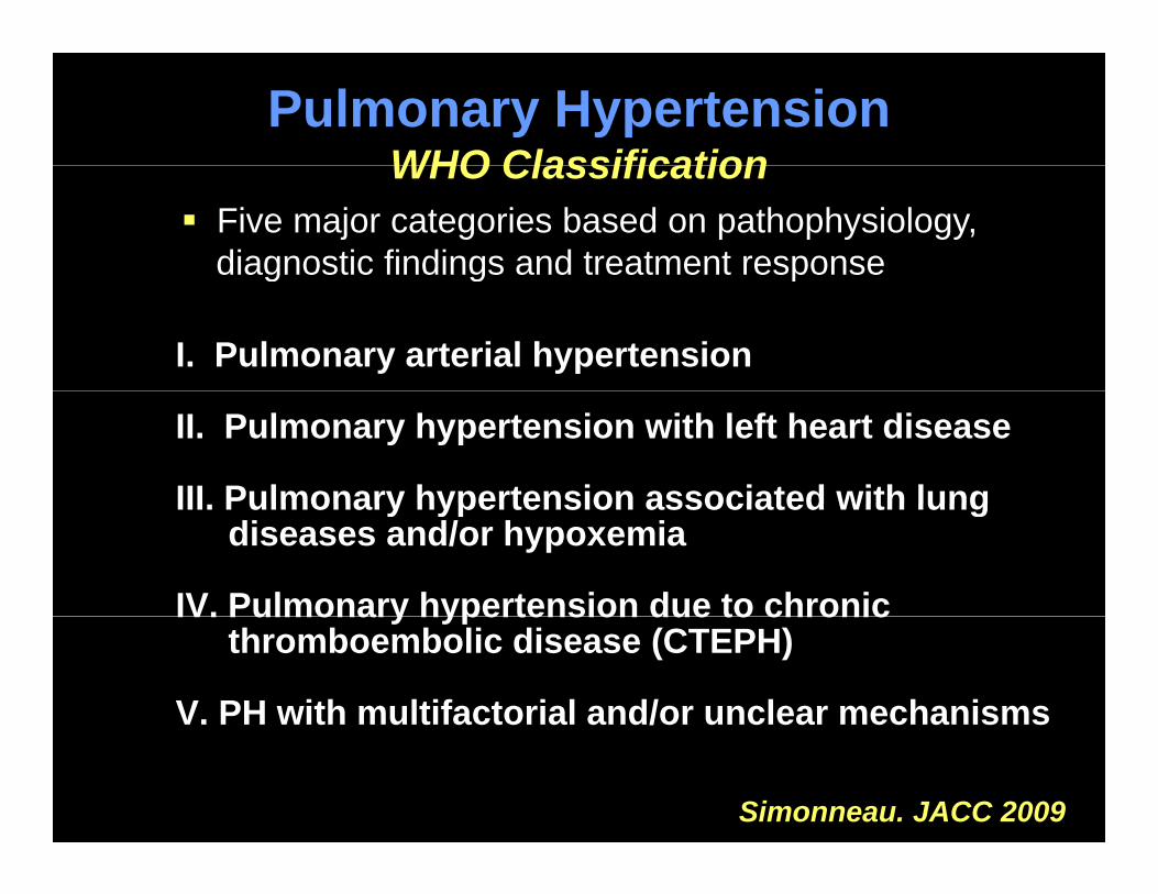
Pulmonary HypertensionWHO ClassificationWHO Classification
Five major categories based on pathophysiology, diagnostic findings and treatment response
I. Pulmonary arterial hypertension
g g p
II. Pulmonary hypertension with left heart disease
III Pulmonary hypertension associated with lungIII. Pulmonary hypertension associated with lung diseases and/or hypoxemia
IV. Pulmonary hypertension due to chronicIV. Pulmonary hypertension due to chronic thromboembolic disease (CTEPH)
V. PH with multifactorial and/or unclear mechanisms
Simonneau. JACC 2009

WHO ClassificationSimonneau. JACC 2009
I. Pulmonary arterial hypertensionIdiopathicpHeritable (BMPR2, ALK-1, endoglin)Associated with (APAH):
Drugs/Anorexigen use (“Fen-phen”, cocaine, metham)Collagen vascular diseaseHIV i f tiHIV infectionPortal hypertensionCongenital systemic-to-pulmonary cardiac shuntsg y p yOther (schistosomiasis, chronic hemolytic anemia)
Persistent pulmonary hypertension of newborn(1`) Associated with significant venous or capillary involvement (PVOD, PCH)

WHO ClassificationSi JACC 2009Simonneau. JACC 2009
II. Left Heart DiseaseSystolic dysfunction
IV. Chronic ThromboembolicPulmonary Hypertension
Diastolic dysfunctionValvular disease V. Unclear/Multifactorial
Hematological (splenectomy,III. Lung Disease/Hypoxia
COPDILD
Hematological (splenectomy, myeloproliferative), systemic (Sarcoidosis, Langerhans-cell histiocytosis, vasculitis),
t b li ( l tILDSleep-disordered breathingAlveolar hypoventilationHigh altitude exposure
metabolic (glycogen storage, Gaucher’s, thyroid), others (vascular compression, chronic renal failure on hemodialysis)g
Developmental abnormalityy )

Pulmonary Arterial Hypertensiony ypPathology (I)
Endothelial thickeningEndothelial thickening
Smooth muscle
hypertrophyhypertrophy

Pulmonary Arterial Hypertensiony ypPathology (II)
Plexiform lesions
In situthrombosisthrombosis
Pulmonary Arterial Hypertension
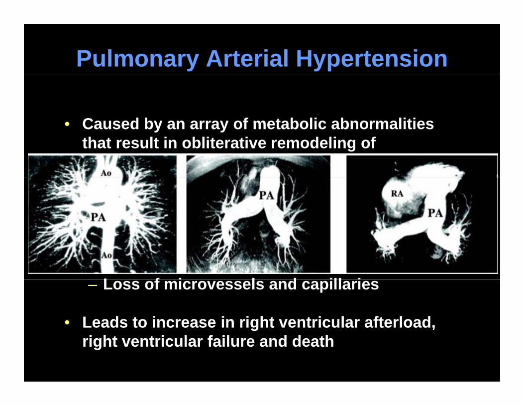
• Caused by an array of metabolic abnormalitiesCaused by an array of metabolic abnormalities that result in obliterative remodeling of pulmonary circulation
• Characterized by lumenal occlusion in medium-sized and small pulmonary arteries due to– Excessive cellular proliferation in vascular wall
and in situ thrombosisL f i l d ill i– Loss of microvessels and capillaries
• Leads to increase in right ventricular afterload, gright ventricular failure and death

Emerging Pathophysiologic Concepts in PAHConcepts in PAH
• Proliferative and antiapoptotic environment in vascular wall share common features with neoplasia
• Loss of endothelial cells and microvessels has features of a degenerative diseasefeatures of a degenerative disease
• Circulating and vascular inflammatory cells and g ymediators suggest a systemic inflammatory disease

Genetics and Pathobiology of PAH
• Loss-of-function mutations in gene encoding bone morphogenetic protein receptor type 2 (BMPR2) p g p p yp ( )– Detected in 70% of familial PAH and 10-40% of idiopathic PAH– Only 20% of BMPR2 mutation carriers develop PAH
• BMPR2 is TGF-β family receptor involved in regulation of apoptosis and growth– Decrease in BMPR2 signaling leads to PAH
• “Second hits”Second hits– Other endogenous genetic abnormalities (serotonin pathway),
changes in blood flow or exogenous stimuli (drugs, viral)Dysregulated inflammation (collagen vascular disease HIV)– Dysregulated inflammation (collagen vascular disease, HIV)
Deng, Am J Hum Gen, 2000Lane, Nat Gen, 2000

Pathobiology of PAH
GENEBMPR2/K /5 HTT
EnvironmentA i t i HIV PAH
Platelets
BMPR2/Kv/5-HTT Anorexigen, toxin, HIV
SerotoninPlatelets
E d h li
NO + PGI2
ET 1/TxA
Serotonin
Endothelium
SMC’s
ET-1/TxA2
Kv1 5Proliferation
SMC s Kv1.5Kv2.1
Adventitia ElastaseMMPs Tenascin

Epidemiology of PAH
• Prospective registries in the U S France and• Prospective registries in the U.S., France and Scotland
P l f PAH 15 26 1 illi• Prevalence of PAH 15 to 26 cases per 1 million adults– Half idiopathic and half associated with other conditionsHalf idiopathic and half associated with other conditions
• ~80% of patients referred to specialized centers i NYHA l III IVare in NYHA class III or IV
• Mean age at diagnosis 36 to 50 yearsg g y
Humbert. AJRCCM 2008;177:574
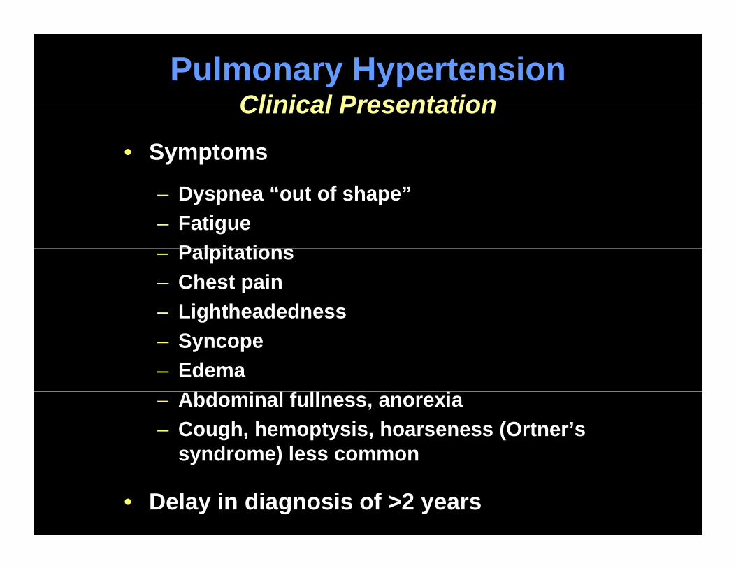
Pulmonary HypertensionClinical PresentationClinical Presentation
• Symptoms
– Dyspnea “out of shape”– Fatigue
P l it ti– Palpitations– Chest pain– Lightheadednessg– Syncope– Edema– Abdominal fullness, anorexia– Cough, hemoptysis, hoarseness (Ortner’s
syndrome) less commony )
• Delay in diagnosis of >2 years
Pulmonary Hypertensiony ypClinical Presentation
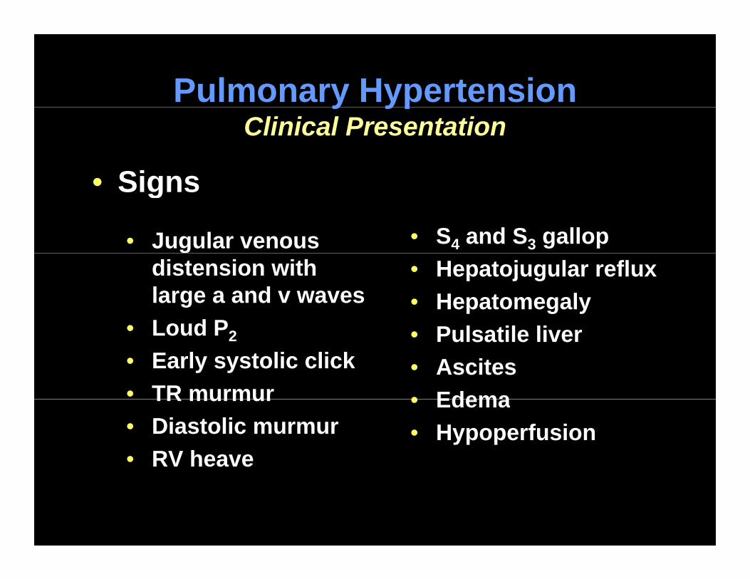
• Signs• Signs
• Jugular venous • S4 and S3 gallopdistension with large a and v waves
• Loud P
• Hepatojugular reflux• Hepatomegaly
P l til li• Loud P2
• Early systolic click• TR murmur
• Pulsatile liver• Ascites• EdemaTR murmur
• Diastolic murmur• RV heave
• Edema• Hypoperfusion
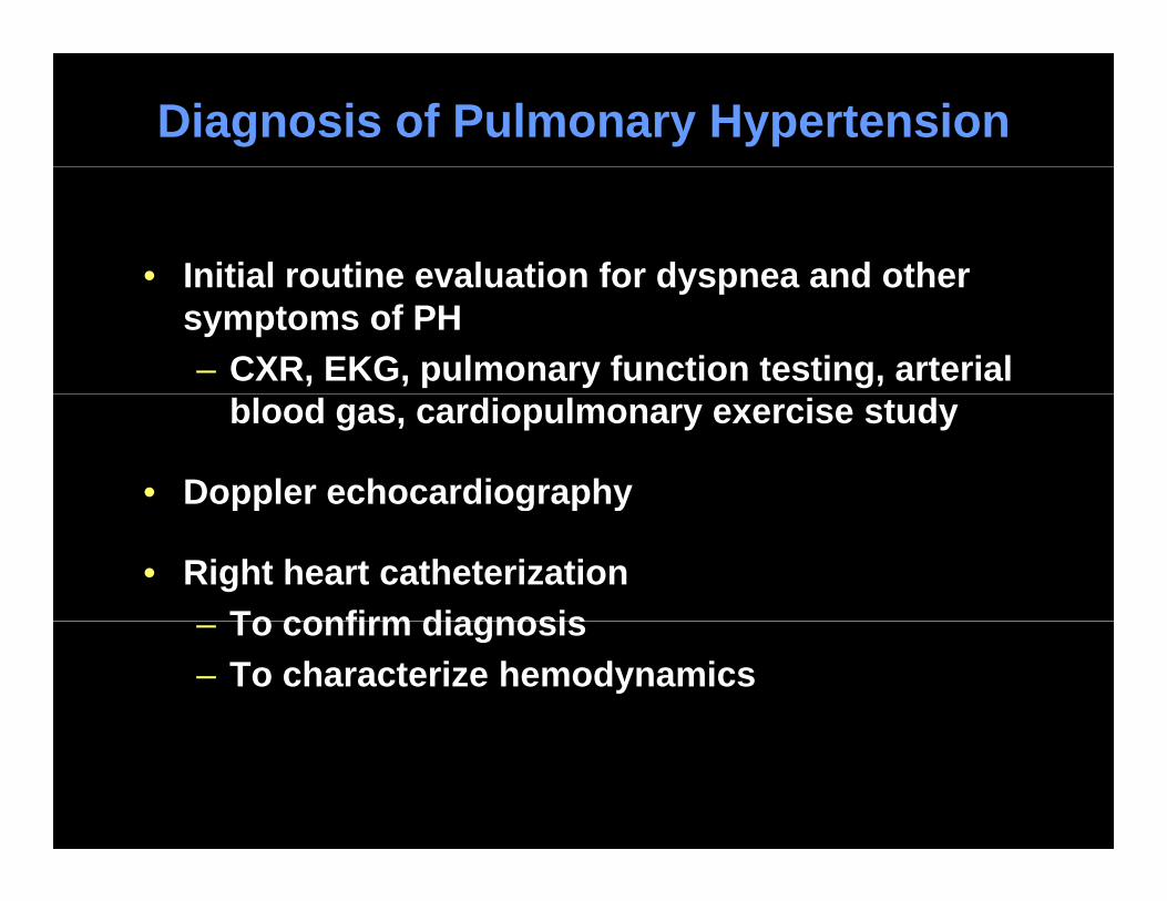
Diagnosis of Pulmonary Hypertension
• Initial routine evaluation for dyspnea and otherInitial routine evaluation for dyspnea and other symptoms of PH– CXR, EKG, pulmonary function testing, arterial
blood gas, cardiopulmonary exercise study
• Doppler echocardiographypp g p y
• Right heart catheterizationTo confirm diagnosis– To confirm diagnosis
– To characterize hemodynamics

Chest RadiographChest Radiograph
E l d i l• Enlarged main pulmonary arteries– Attenuation of peripheral p p
pulmonary vascular markings (pruning)
• Right ventricular enlargement
• Exclusion of parenchymal lung disease

Electrocardiography
• Right ventricular hypertrophy, right axis deviation, right atrial enlargement

Doppler Echocardiography in Pulmonary Hypertension
• Intracardiac shunt• Tricuspid regurgitation
• Congenital heart ds
• Left heart size/fx
• Right a/v dilatation
• Right ventricular hypertrophy Left heart size/fx
• Valvular morphology
P i di l ff i
Right ventricular hypertrophy
• Right ventricular dysfunction
P lmonic ins fficienc • Pericardial effusion• Pulmonic insufficiency

Right Heart CatheterizationRight Heart Catheterization
To diagnose/characterize pulmonary hypertension Mean pulmonary artery pressureP l ill dPulmonary capillary wedge pressureMean right atrial pressureCardiac indexPVR l l tiPVR calculation
To assess severity of pulmonary hypertension
To evaluate acute vasoreactivity (vasodilator response)

Right Heart CatheterizationRight Heart Catheterization
• Patient 1 Patient 2Patient 1 Patient 2
•RA-4 mm Hg •RA-12 mm Hg
•PA- 90/60 mm Hg
•PCWP- 8 mm Hg
•PA- 50/25 mm Hg
•PCWP- 8 mm Hg
•CI- 2.4 L/m/m2 •CI- 1.0 L/m/m2
•PVR ~ 2066 d•s•cm-5 •PVR ~ 2000 d•s•cm-5

Detailed Evaluation During Diagnosis of PH
• Medical historyPMH: VTE heart lung and blood disorders HIV– PMH: VTE, heart, lung, and blood disorders, HIV
– Family history– Exposures: weight loss medications– Drugs: cocaine, methamphetamine
• Diagnostic tests• Diagnostic tests– Serologic evaluation for autoimmune disease and HIV– Pulmonary function tests– Radiologic tests: VQ scan, chest HRCT, cardiac MRI
• Exclude thromboembolic disease, parenchymal pulmonary disease and aid in differential diagnosis of PHpulmonary disease and aid in differential diagnosis of PH
– Sleep study and nocturnal oxymetry

Ventilation Perfusion Scan
• To exclude chronic thromboembolic PH• To exclude chronic thromboembolic PH
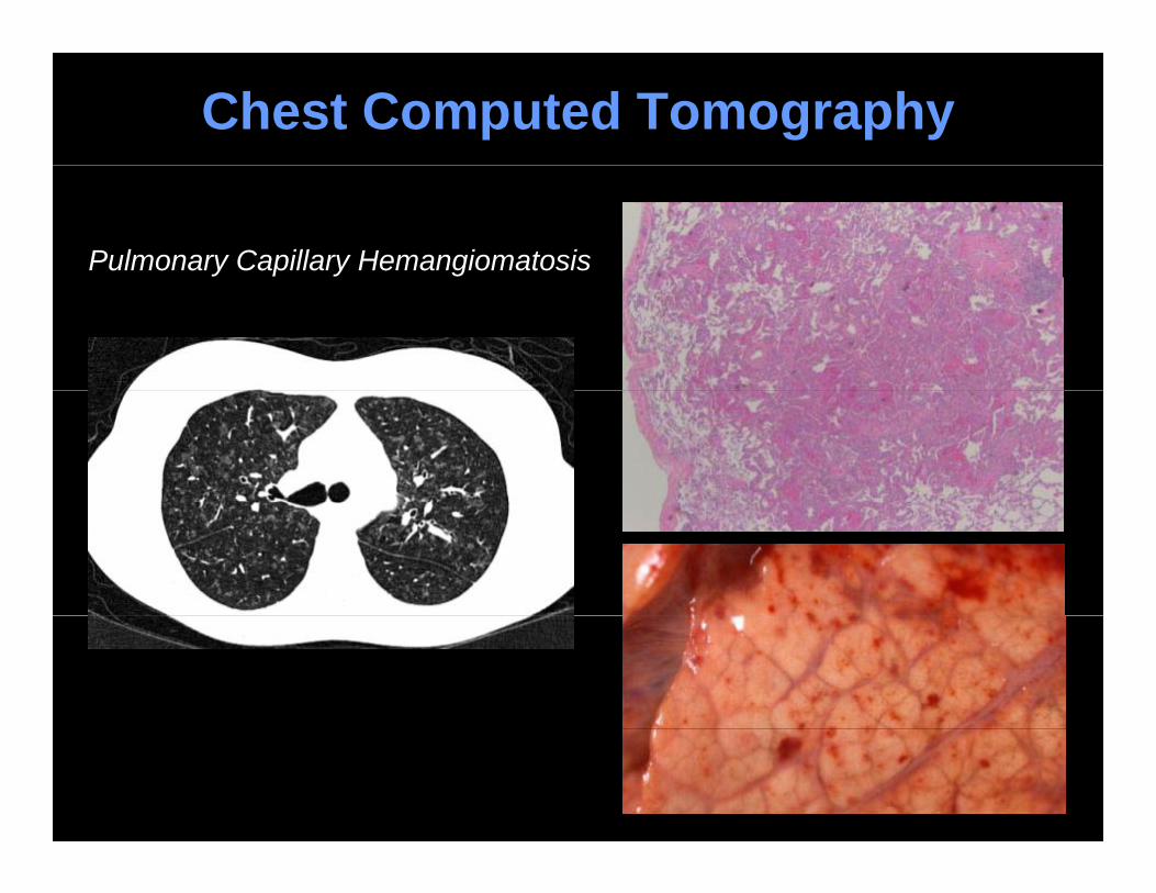
Chest Computed Tomography
Pulmonary Capillary Hemangiomatosis


Therapies for P l A t i l H t iPulmonary Arterial Hypertension
• Preventative care • Prostacyclin analogues E d th li 1 t• Anticoagulation
• Supplemental oxygen
• Endothelin-1 receptor antagonists
• PDE-5 inhibitors• Diuretics• Inotropes
• Cardiopulmonary rehabilitationAtrial septostomy• Calcium channel blockers • Atrial septostomy
• Lung transplantation

General Measures
• AnticoagulationAnticoagulation– INR goal 1.5 to 2.5– Controversial in diseases other than iPAH
• Supplemental oxygen
• Diuretics and inotropic medications– Right ventricular failure
Monitor electrolytes and renal function– Monitor electrolytes and renal function
• Digitalis– Right ventricular failure and arrhythmia
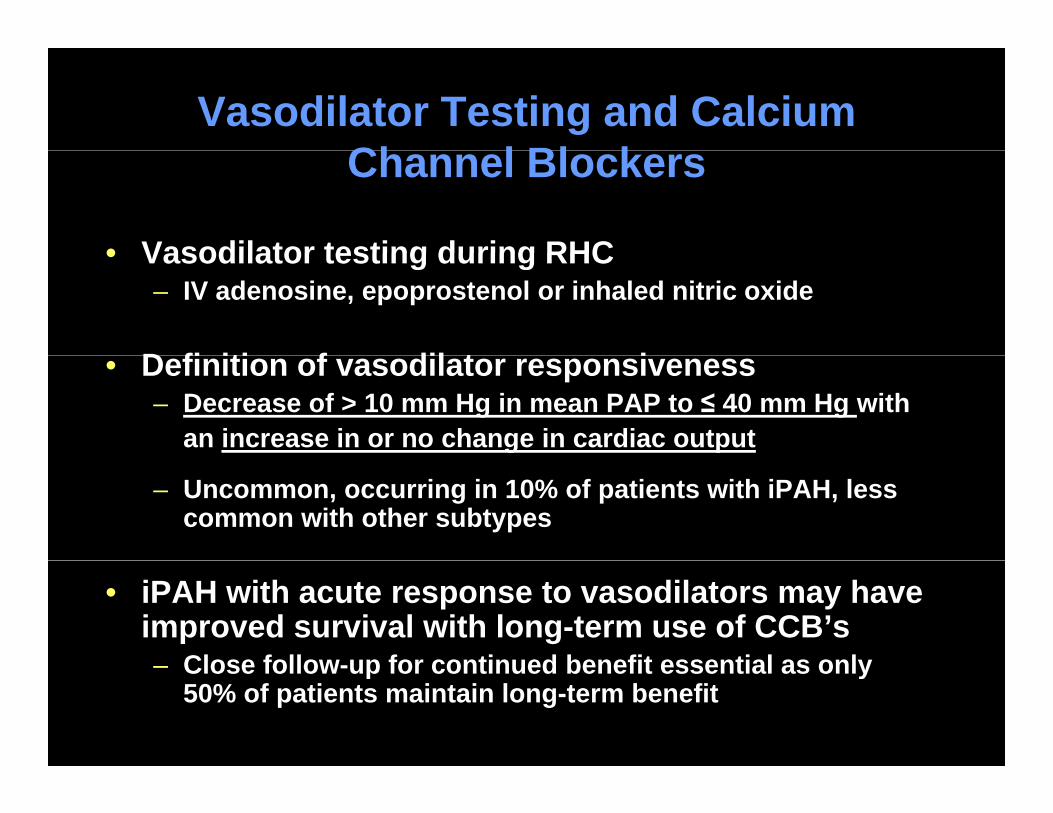
Vasodilator Testing and Calcium Ch l Bl kChannel Blockers
• Vasodilator testing during RHC• Vasodilator testing during RHC– IV adenosine, epoprostenol or inhaled nitric oxide
D fi iti f dil t i• Definition of vasodilator responsiveness– Decrease of > 10 mm Hg in mean PAP to ≤ 40 mm Hg with
an increase in or no change in cardiac output
– Uncommon, occurring in 10% of patients with iPAH, less common with other subtypes
• iPAH with acute response to vasodilators may have improved survival with long-term use of CCB’s
Close follow up for continued benefit essential as only– Close follow-up for continued benefit essential as only 50% of patients maintain long-term benefit

Targets for Therapies in PAH
Humbert. N Engl J Med 2004;351:1425

Targets for Therapy in PAH
• Downregulation of prostacyclin axis– Reversed by exogenous prostacyclin analogues
• Downregulation of NO/cGMP axisReversed by inhaled NO and PDE5 inhibition– Reversed by inhaled NO and PDE5 inhibition
• Upregulation of endothelin axisp g– Reversed by endothelin receptor antagonists

Prostanoids
• Underproduction of prostacycline in PAHUnderproduction of prostacycline in PAH– Prostacycline promotes vasodilatation, inhibits
vascular proliferation and platelet aggregation
• Epoprostenol (IV)• Beraprost (PO)• Treprostinil (SC or IV)• Iloprost (inhalation)
• Improvement in hemodynamics, exercise capacity, symptoms and survival (with epoprostenol)
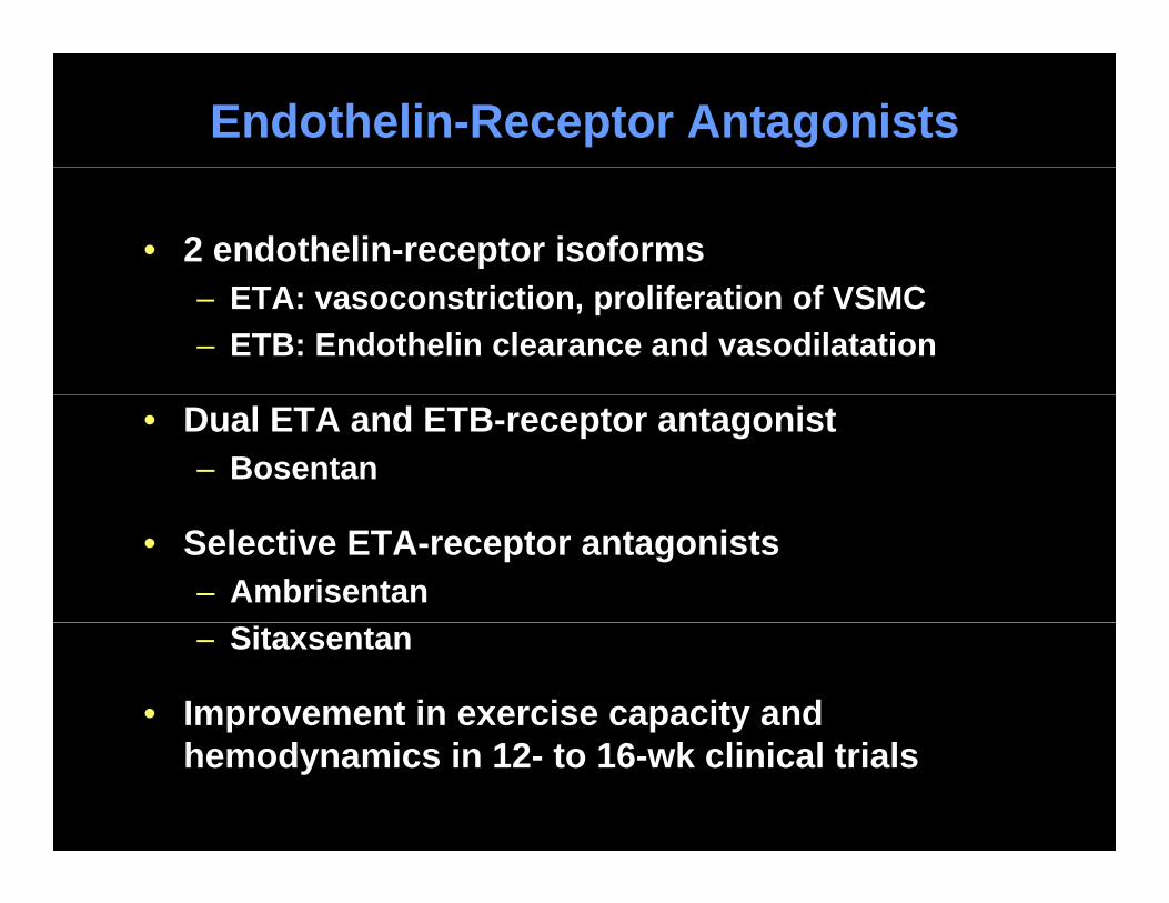
Endothelin-Receptor Antagonists
• 2 endothelin-receptor isoforms– ETA: vasoconstriction, proliferation of VSMC– ETB: Endothelin clearance and vasodilatation
• Dual ETA and ETB-receptor antagonist– Bosentan
• Selective ETA-receptor antagonists– Ambrisentan– Sitaxsentan
• Improvement in exercise capacity and hemodynamics in 12- to 16-wk clinical trials

Phosphodiesterase-5 Inhibitors
• Inhibition of cGMP-specific phosphodiesteraseInhibition of cGMP-specific phosphodiesterase– Pulmonary arterial vasodilatation and inhibition of
smooth muscle cell growth by enhancing effects of locally produced NO via its second messenger cGMPlocally produced NO via its second messenger cGMP
• Sildenafil/tadalafil
• Improvement in symptoms, exercise capacity and hemodynamics in short-term studies

Atrial Septostomy and Lung TransplantationTransplantation
• Atrial septostomy• Atrial septostomy– Creation of right-to-left interatrial shunt for right
ventricular decompression– Palliative or as bridge to lung transplantation
L t l t ti• Lung transplantation– Early referral– Close monitoring for response to therapyClose monitoring for response to therapy– Perform lung transplantation before advanced right
heart failure and poor performance status
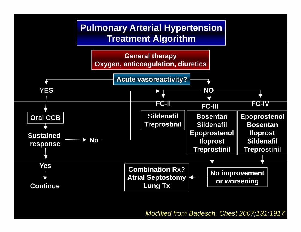
Pulmonary Arterial HypertensionTreatment Algorithmg
General therapyOxygen, anticoagulation, diuretics
Acute vasoreactivity?YES NO
FC II FC IV
Oral CCB
FC-II FC-III FC-IV
SildenafilTreprostinil
BosentanSildenafil
E t l
EpoprostenolBosentanIl tSustained
response NoEpoprostenol
IloprostTreprostinil
Iloprost Sildenafil
Treprostinil
Yes
Continue
No improvementor worsening
Combination Rx?Atrial Septostomy
Lung Txg
Modified from Badesch. Chest 2007;131:1917

Prognosis
• Median survival in untreated PAH < 3 yrs
• Contemporary registries reveal improved survival– 65-75% survival at 3 years
47 55% at 5 years in epoprostenol treated patients– 47-55% at 5 years in epoprostenol treated patients
• Right heart failure = lower survival ratesg– Elevated RAP, low CI, low MVO2, poor exercise
capacity, pericardial effusion, high BNP
• Close monitoring to evaluate treatment response, plan additional therapy and for lung transplantation
Future DirectionsFuture Directions
• Discovery of novel mechanistic pathways and translational application into clinical practice
• Stem cell replacement/transplant with endothelial progenitor cellsp g