case report systemic air embolism associated with pleural pigtail chest tube...
TRANSCRIPT

Case ReportSystemic Air Embolism Associated withPleural Pigtail Chest Tube Insertion
Emad Alkhankan,1 Ahmad Nusair,2 Rida Mazagri,3 and Mohammed Al-Ourani4
1 Internal Medicine, Marshall University, Huntington, WV 25701, USA2Infectious Disease, Marshall University, Huntington, WV 25701, USA3Neurosurgery, Marshall University, Huntington, WV 25701, USA4Pulmonary, Marshall University, Huntington, WV 25701, USA
Correspondence should be addressed to Mohammed Al-Ourani; [email protected]
Received 11 February 2016; Revised 12 July 2016; Accepted 26 July 2016
Academic Editor: Alan D. L. Sihoe
Copyright © 2016 Emad Alkhankan et al. This is an open access article distributed under the Creative Commons AttributionLicense, which permits unrestricted use, distribution, and reproduction in any medium, provided the original work is properlycited.
Pleural pigtail catheter placement is associated with many complications including pneumothorax, hemorrhage, and chest pain.Air embolism is a known but rare complication of pleural pigtail catheter insertion and has a high risk of occurrence with positivepressure ventilation. In this case report, we present a 50-year-old male with bilateral pneumonia who developed a pneumothoraxwhile onmechanical ventilationwith continuous positive airway pressuremode.During the placement of the pleural pigtail catheterto correct the pneumothorax, the patient developed a sudden left sided body weakness and became unresponsive. An air embolismwas identified in the right main cerebral artery, which was fatal.
1. Introduction
Pleural effusion and pneumothorax are well-establishedindications for chest tube placement. Two widely knowncorrective procedures are using a pigtail catheter drainage orperforming a chest tube thoracostomy. The pigtail catheterdrainage is more widely used as it is easier to perform inemergent situations and is a much lesser invasive procedure[1–3]. Cerebral air embolism is a rare complication that canbe induced by pulmonary barotrauma, trauma of the chestor head, and iatrogenic causes such as invasive proceduresor surgery [4]. This risk is further increased if the patientis on positive pressure ventilation (PPV) as the pressure inthe airway is much higher. Despite its rarity, there are manyreports written on cerebral air embolism. However, reportson the occurrence of cerebral air embolism while the patientis on positive pressure ventilation (PPV) remain scarce.
2. Case Report
This is a case of a 50-year-old male with no significantpast medical history, who initially presented with shortness
of breath and hypoxia. He was transferred to the intensivecare unit (ICU) and was treated for bilateral pneumoniathat required prolonged mechanical ventilation via a tra-cheostomy.He further developed necrotizing pneumonia andsubsequently multiorgan failure that led to hospitalizationfor 6 weeks. He was weaned from mechanical ventilation tothe point he was tolerating CPAP mode. However, he startedrequiring higher fraction of inspired oxygen (FiO
2). A chest
X-ray (CXR) showed a small pneumothorax and was shownto increase in size in a repeat CXR the following day. This ledto the decision to place a pleural pigtail chest tube to relievethe pneumothorax.
During the procedure, the patient suddenly becameunresponsive and had an episode of apnea. However, hisvital signs remained stable (BP 125/68mmHg, P 84 BPM,T 37.0 C, RR 25 breaths/min, and SpO
295%). Additional
labs included ABG (pH 7.37, PaCO258mm Hg, and PaO
2
67mmHg), WBC 14,700/mm3, Hgb 8.4 g/dL, Plts 256 ×109/L, Na 126mmol/L, K 5.1mmol/L, Cl 99mmol/L, HCO
3
30.8mmol/L, BUN 26mmol/L, Cr 0.52mmol/L, and Gluc108mmol/L. Another CXR did not show worsening of the
Hindawi Publishing CorporationCase Reports in PulmonologyVolume 2016, Article ID 4053748, 3 pageshttp://dx.doi.org/10.1155/2016/4053748
2 Case Reports in Pulmonology
(a) (b)
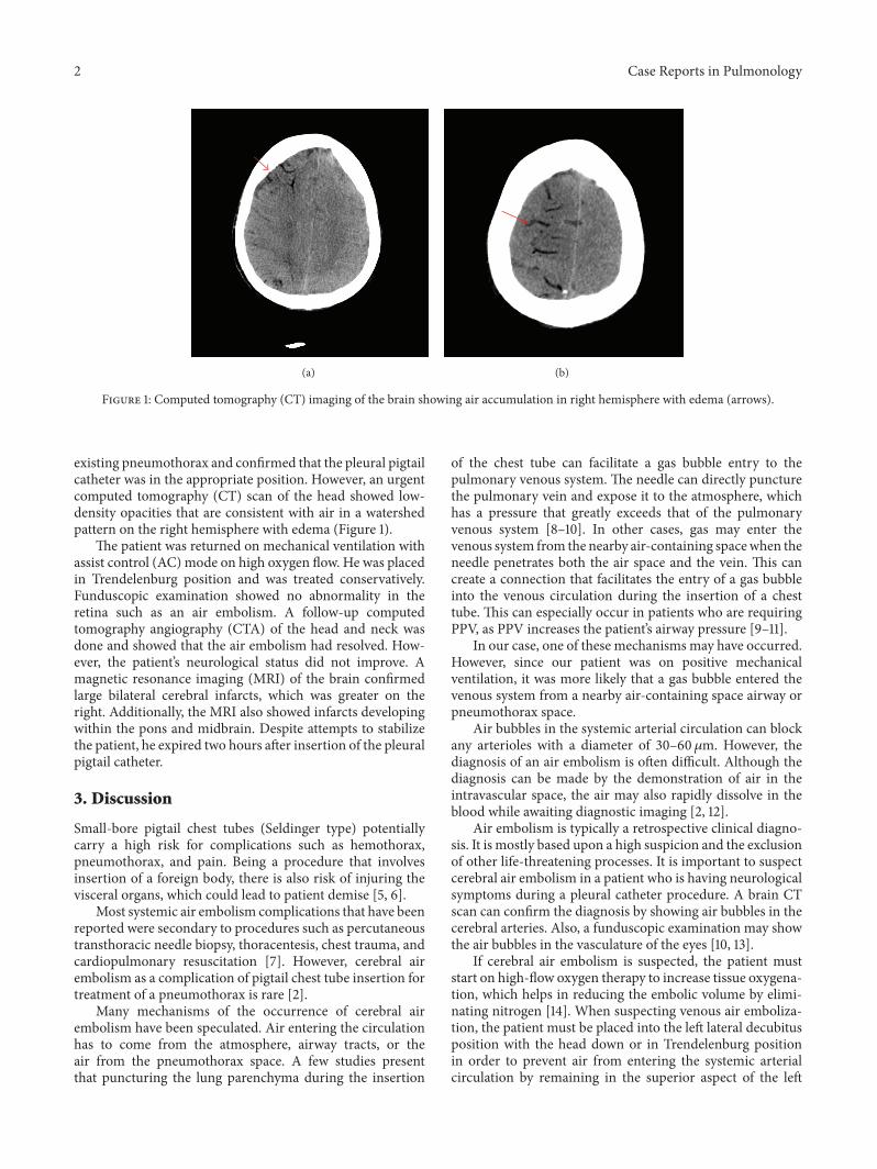
Figure 1: Computed tomography (CT) imaging of the brain showing air accumulation in right hemisphere with edema (arrows).
existing pneumothorax and confirmed that the pleural pigtailcatheter was in the appropriate position. However, an urgentcomputed tomography (CT) scan of the head showed low-density opacities that are consistent with air in a watershedpattern on the right hemisphere with edema (Figure 1).
The patient was returned on mechanical ventilation withassist control (AC) mode on high oxygen flow. He was placedin Trendelenburg position and was treated conservatively.Funduscopic examination showed no abnormality in theretina such as an air embolism. A follow-up computedtomography angiography (CTA) of the head and neck wasdone and showed that the air embolism had resolved. How-ever, the patient’s neurological status did not improve. Amagnetic resonance imaging (MRI) of the brain confirmedlarge bilateral cerebral infarcts, which was greater on theright. Additionally, the MRI also showed infarcts developingwithin the pons and midbrain. Despite attempts to stabilizethe patient, he expired two hours after insertion of the pleuralpigtail catheter.
3. Discussion
Small-bore pigtail chest tubes (Seldinger type) potentiallycarry a high risk for complications such as hemothorax,pneumothorax, and pain. Being a procedure that involvesinsertion of a foreign body, there is also risk of injuring thevisceral organs, which could lead to patient demise [5, 6].
Most systemic air embolism complications that have beenreported were secondary to procedures such as percutaneoustransthoracic needle biopsy, thoracentesis, chest trauma, andcardiopulmonary resuscitation [7]. However, cerebral airembolism as a complication of pigtail chest tube insertion fortreatment of a pneumothorax is rare [2].
Many mechanisms of the occurrence of cerebral airembolism have been speculated. Air entering the circulationhas to come from the atmosphere, airway tracts, or theair from the pneumothorax space. A few studies presentthat puncturing the lung parenchyma during the insertion
of the chest tube can facilitate a gas bubble entry to thepulmonary venous system. The needle can directly puncturethe pulmonary vein and expose it to the atmosphere, whichhas a pressure that greatly exceeds that of the pulmonaryvenous system [8–10]. In other cases, gas may enter thevenous system from the nearby air-containing spacewhen theneedle penetrates both the air space and the vein. This cancreate a connection that facilitates the entry of a gas bubbleinto the venous circulation during the insertion of a chesttube. This can especially occur in patients who are requiringPPV, as PPV increases the patient’s airway pressure [9–11].
In our case, one of these mechanisms may have occurred.However, since our patient was on positive mechanicalventilation, it was more likely that a gas bubble entered thevenous system from a nearby air-containing space airway orpneumothorax space.
Air bubbles in the systemic arterial circulation can blockany arterioles with a diameter of 30–60 𝜇m. However, thediagnosis of an air embolism is often difficult. Although thediagnosis can be made by the demonstration of air in theintravascular space, the air may also rapidly dissolve in theblood while awaiting diagnostic imaging [2, 12].
Air embolism is typically a retrospective clinical diagno-sis. It is mostly based upon a high suspicion and the exclusionof other life-threatening processes. It is important to suspectcerebral air embolism in a patient who is having neurologicalsymptoms during a pleural catheter procedure. A brain CTscan can confirm the diagnosis by showing air bubbles in thecerebral arteries. Also, a funduscopic examination may showthe air bubbles in the vasculature of the eyes [10, 13].
If cerebral air embolism is suspected, the patient muststart on high-flow oxygen therapy to increase tissue oxygena-tion, which helps in reducing the embolic volume by elimi-nating nitrogen [14]. When suspecting venous air emboliza-tion, the patient must be placed into the left lateral decubitusposition with the head down or in Trendelenburg positionin order to prevent air from entering the systemic arterialcirculation by remaining in the superior aspect of the left

Case Reports in Pulmonology 3
ventricle and, away from the aorta [2, 15]. Giving hyperbaricoxygen therapy (HBOT) helps in reducing the volume of airemboli by increasing the solubility of oxygen in the bloodand altering the permeability of blood-brain barrier, whichdecreases cerebral edema [9, 16]. In our case, the patientdid not receive HBOT because he underwent mechanicalventilation after developing poormental function and he alsohad an existing pneumothorax that was unresolved at thatpoint.
4. Conclusion
Cerebral air embolism as complication of pigtail catheter israre, but it is fatal. It should be suspected when the patientstarts having neurological symptoms while undergoing chesttube insertion or other known chest procedures, especiallywhile the patient is on positive pressure ventilation.
Competing Interests
The authors declare that there is no conflict of interestsregarding the publication of this paper.
References
[1] J. S. Gammie, M. C. Banks, C. R. Fuhrman et al., “The pigtailcatheter for pleural drainage: a less invasive alternative totube thoracostomy,” Journal of the Society of LaparoendoscopicSurgeons, vol. 3, no. 1, pp. 57–61, 1999.
[2] S. I. Kim, H. J. Kwak, J. Moon et al., “Cerebral air embolismfollowing pigtail catheter insertion for pleural fluid drainage,”Tuberculosis and Respiratory Diseases, vol. 74, no. 6, pp. 286–290, 2013.
[3] R. W. Light, “Pleural controversy: optimal chest tube size fordrainage,” Respirology, vol. 16, no. 2, pp. 244–248, 2011.
[4] H. Poncia and J. M. Ryan, “An unusual complication of chesttube thoracostomy,” Canadian Journal of Emergency Medicine,vol. 2, no. 2, pp. 121–123, 2000.
[5] A. Horsley, L. Jones, J. White, and M. Henry, “Efficacy andcomplications of small-bore, wire-guided chest drains,” Chest,vol. 130, no. 6, pp. 1857–1863, 2006.
[6] T. Havelock, R. Teoh, D. Laws, and F. Gleeson, “Pleuralprocedures and thoracic ultrasound: British Thoracic SocietyPleural Disease Guideline 2010,”Thorax, vol. 65, supplement 2,pp. i61–i76, 2010.
[7] H. A. Brownlow and C. Edibam, “Systemic air embolismafter intercostal chest drain insertion and positive pressureventilation in chest trauma,”Anaesthesia and Intensive Care, vol.30, no. 5, pp. 660–664, 2002.
[8] S. Ohashi, H. Endoh, T. Honda, N. Komura, and K. Satoh,“Cerebral air embolism complicating percutaneous thin-needlebiopsy of the lung: complete neurological recovery after hyper-baric oxygen therapy,” Journal of Anesthesia, vol. 15, no. 4, pp.233–236, 2001.
[9] S.-J. Um, S.-K. Lee, K. Y. Doo et al., “Four cases of a cerebralair embolism complicating a percutaneous transthoracic needlebiopsy,” Korean Journal of Radiology, vol. 10, no. 1, pp. 81–84,2009.
[10] W. Bou-Assaly, P. Pernicano, and E. Hoeffner, “Systemic airembolism after transthoracic lung biopsy: a case report and
review of literature,” World Journal of Radiology, vol. 2, no. 5,pp. 193–196, 2010.
[11] A. M.-H. Ho and E. Ling, “Systemic air embolism after lungtrauma,” Anesthesiology, vol. 90, no. 2, pp. 564–575, 1999.
[12] P. G. Shetty, G. M. Fatterpekar, S. Manohar, V. Sujit, J. Varsha,and U. Zarir, “Fatal cerebral air embolism as a complicationof transbronchoscopic lung biopsy: a case report,” AustralasianRadiology, vol. 45, no. 2, pp. 215–217, 2001.
[13] T. Hiraki, H. Fujiwara, J. Sakurai et al., “Nonfatal systemic airembolism complicating percutaneous CT-guided transthoracicneedle biopsy: four cases from a single institution,” Chest, vol.132, no. 2, pp. 684–690, 2007.
[14] J. E. Scruggs, A. Joffe, and K. E. Wood, “Paradoxical airembolism successfully treated with hyperbaric oxygen,” Journalof Intensive Care Medicine, vol. 23, no. 3, pp. 204–209, 2008.
[15] J. L.Westcott, “Air embolism complicating percutaneous needlebiopsy of the lung,” Chest, vol. 63, no. 1, pp. 108–110, 1973.
[16] B. P. Murphy, F. J. Harford, and F. S. Cramer, “Cerebral airembolism resulting from invasive medical procedures. Treat-ment with hyperbaric oxygen,” Annals of Surgery, vol. 201, no.2, pp. 242–245, 1985.

Submit your manuscripts athttp://www.hindawi.com
Stem CellsInternational
Hindawi Publishing Corporationhttp://www.hindawi.com Volume 2014
Hindawi Publishing Corporationhttp://www.hindawi.com Volume 2014
MEDIATORSINFLAMMATION
of
Hindawi Publishing Corporationhttp://www.hindawi.com Volume 2014
Behavioural Neurology
EndocrinologyInternational Journal of
Hindawi Publishing Corporationhttp://www.hindawi.com Volume 2014
Hindawi Publishing Corporationhttp://www.hindawi.com Volume 2014
Disease Markers
Hindawi Publishing Corporationhttp://www.hindawi.com Volume 2014
BioMed Research International
OncologyJournal of
Hindawi Publishing Corporationhttp://www.hindawi.com Volume 2014
Hindawi Publishing Corporationhttp://www.hindawi.com Volume 2014
Oxidative Medicine and Cellular Longevity
Hindawi Publishing Corporationhttp://www.hindawi.com Volume 2014
PPAR Research
The Scientific World JournalHindawi Publishing Corporation http://www.hindawi.com Volume 2014
Immunology ResearchHindawi Publishing Corporationhttp://www.hindawi.com Volume 2014
Journal of
ObesityJournal of
Hindawi Publishing Corporationhttp://www.hindawi.com Volume 2014
Hindawi Publishing Corporationhttp://www.hindawi.com Volume 2014
Computational and Mathematical Methods in Medicine
OphthalmologyJournal of
Hindawi Publishing Corporationhttp://www.hindawi.com Volume 2014
Diabetes ResearchJournal of
Hindawi Publishing Corporationhttp://www.hindawi.com Volume 2014
Hindawi Publishing Corporationhttp://www.hindawi.com Volume 2014
Research and TreatmentAIDS
Hindawi Publishing Corporationhttp://www.hindawi.com Volume 2014
Gastroenterology Research and Practice
Hindawi Publishing Corporationhttp://www.hindawi.com Volume 2014
Parkinson’s Disease
Evidence-Based Complementary and Alternative Medicine
Volume 2014Hindawi Publishing Corporationhttp://www.hindawi.com