cd8 y cmv en vih
TRANSCRIPT
Clinical Infectious Diseases
M A J O R A R T I C L E
HIV/AIDS
CD8 T-Cell Expansion and Inflammation Linked toCMV Coinfection in ART-treated HIV InfectionMichael L. Freeman,1,a Joseph C. Mudd,1,ab Carey L. Shive,1,2 Souheil-Antoine Younes,1 Soumya Panigrahi,1 Scott F. Sieg,1 Sulggi A. Lee,3 Peter W. Hunt,3
Leonard H. Calabrese,4 Sara Gianella,5 Benigno Rodriguez,1 and Michael M. Lederman1
1Center for AIDS Research, Division of Infectious Diseases and HIV Medicine, Department of Medicine, Case Western Reserve University/University Hospitals Case Medical Center, and 2VeteransAdministration Medical Center, Cleveland, Ohio; 3Department of Medicine, University of California San Francisco; 4Department of Rheumatic and Immunologic Diseases, Cleveland Clinic Foundation,Ohio; and 5Division of Infectious Diseases, University of California San Diego, La Jolla
Background. Persistent CD8 T-cell expansion, low CD4/CD8 T-cell ratios, and heightened inflammation persist in antiretroviraltherapy (ART)-treated human immunodeficiency virus (HIV) infection and are associated with increased risk of morbid outcomes.We explored the role of cytomegalovirus (CMV) infection in CD8 lymphocytosis and inflammation in ART-treated HIV infection.
Methods. Absolute CD4 and CD8 T-cell counts were abstracted from clinical records and compared among 32 HIV-infectedCMV-seronegative subjects, 126 age, CD4 and gender-matched HIV-infected CMV-seropositive subjects, and among 21 HIV-uninfected controls (9 CMV-negative, 12 CMV-positive). Plasma inflammatory indices were measured in a subset by ELISA.
Results. Median CD8 counts/µL were higher in HIV-positive/CMV-positive patients (795) than in HIV-positive/CMV-negativesubjects (522, P = .006) or in healthy controls (451, P = .0007), whereas CD8 T-cell counts were similar to controls’ levels in HIV-positive/CMV-negative subjects. Higher plasma levels of IP-10 (P = .0011), TNF-RII (P = .0002), and D-dimer (P = .0444) were alsofound in coinfected patients than in HIV-positive/CMV-negative subjects.
Conclusions. CMV infection is associated with higher CD8 T-cell counts, resultant lower CD4/CD8 ratios, and increasedsystemic inflammation in ART-treated HIV infection. CMV infection may contribute to risk for morbid outcomes in treatedHIV infection.
Keywords. HIV; CMV; coinfection; CD8 T-cell expansion; inflammation.
In the era of combination antiretroviral therapy (ART), humanimmunodeficiency virus (HIV)-infected individuals are livinglonger and healthier lives. More HIV-infected people thanever are entering old age, but due to increased inflammationand elevated risks of cardiovascular disease that are linked toHIV infection, even younger ART-treated patients are suc-cumbing earlier to many of the same complications that affectthe HIV-uninfected elderly [1]. We and others have previouslylinked persistent CD8 T-cell expansion and inflammatorymediators such as interleukin (IL)-6, tumor necrosis factor(TNF)-α, and type 1 interferons to the morbid outcomes ofHIV infection [2–5], and more recently, we have specifically im-plicated the inflammatory mediators interferon (IFN)-α, IL-6,and IL-1β in the pathogenesis of poor CD4 T-cell restorationin the setting of sustained combination ART [6, 7]. In mostpersons with HIV infection, expansion of the CD8 T-cell poolis demonstrable early in infection as CD4 T-cell numbers
progressively fall, and this expansion typically persists evenwhen HIV replication is controlled with ART [8]. In ART-treated patients, inversion of the ratio of CD4 T cells to CD8T cells is associated with poor clinical outcomes, even in thesetting of normal CD4 T-cell counts [5, 9], suggesting that CD8T-cell expansion is associated with and could be an importantdriver of increased morbidity and mortality [5]. In ART-treatedHIV infection, the determinants of persistent CD8 T-cellexpansion are poorly understood.
Like HIV, human cytomegalovirus (CMV) is a lifelong viralpathogen associated with inflammation and cardiovascular risk,particularly in the elderly [10]. CMV infection is especiallyprevalent in aging populations—increasing in prevalence from36% in 6–11 year-olds to over 90% in those older than 80 yearsold [11]—whereas approximately 90% of HIV-infected individ-uals are coinfected with CMV, independently of age [12]. In im-munosuppressed individuals, such as untreated HIV-infectedpatients and organ transplant recipients, active CMV infectionscan be particularly devastating, leading to severe end organ dis-ease and death. Most HIV-infected persons experience inter-mittent bursts of CMV replication (even during suppressiveART) that might contribute to persistent stimulation of theCD8 T-cell population [10].
Here, we sought to determine whether persistent CD8 T-cellexpansion and increased inflammation observed in ART-treatedHIV infection was associated with CMV coinfection.
Received 29 July 2015; accepted 12 September 2015; published online 23 September 2015.aM. L. F. and J. C. M. contributed equally to this work.bPresent address: National Institute of Allergy and Infectious Diseases, National Institutes of
Health, Bethesda, MD.Correspondence: M. M. Lederman, Case Western Reserve University School of Medicine,
2061 Cornell Rd, Cleveland, OH 44106 ([email protected]).
Clinical Infectious Diseases® 2016;62(3):392–6© The Author 2015. Published by Oxford University Press for the Infectious Diseases Societyof America. All rights reserved. For permissions, e-mail [email protected]: 10.1093/cid/civ840
392 • CID 2016:62 (1 February) • HIV/AIDS
by guest on January 11, 2016http://cid.oxfordjournals.org/
Dow
nloaded from

METHODS
Clinical IndicesThis work was approved by the Institutional Review Board atUniversity Hospitals/Case Medical Center. With writteninformed consent, blood was acquired in EDTA tubes from21 HIV-uninfected persons (12 CMV-positive, 9 CMV-negative) and 158 HIV-infected patients (126 CMV-positive,32 CMV-negative) receiving ART (median duration of treat-ment 3.15 years) with undetectable plasma HIV levels (typicallybelow 50 copies/mL). CD4 and CD8 T-cell counts were deter-mined in the hospital clinical laboratory by flow cytometry.CMV serostatus was determined in the hospital clinical laboratoryby IMMULITE 2000 CMV IgG Ab immunoassay (Siemens).HIV-infected CMV-seropositive subjects were age, gender-, andCD4 T cell count-matched 4:1 to HIV-infected CMV-seronegativesubjects. Participant characteristics are shown in Table 1.
ELISAWhole blood in EDTA was obtained at the time of CMV sero-status determination and after centrifugation, plasmas were frozenat −80°C, then thawed and analyzed in batch by enzyme-linkedimmunosorbent assay (ELISA) per manufacturer’s protocols forlevels of D-dimers (Asserachrom), IP-10, IL-6, IL-18, solubleCD14 (sCD14), and TNF-RII (all from R & D Systems).
StatisticsWe compared continuous variables using the Mann–WhitneyU test or the Kruskal–Wallis test with Dunn’s correction formultiple variables. Categorical data were compared using Fisherexact test. Correlations were determined using a nonparametricSpearman test.
RESULTS
Clinical CharacteristicsWe compared 3 groups: HIV-uninfected controls (n = 21), ART-treated HIV-infected CMV-seronegative individuals (n = 32),and ART-treated HIV-infected CMV-seropositive individuals(n = 126). All 3 groups were similarly aged, and the HIV-infectedgroups had similar CD4 counts, CD4 nadirs, duration of ART,and proportions of men (Table 1). We did not find significant
differences in CD4 or CD8 numbers between the CMV-seropositive (n = 12, median CD4 count = 938/µL, median CD8count = 501/µL) and the CMV-seronegative HIV-uninfectedcontrols (n = 9, median CD4 count = 876/µL, median CD8count = 440/µL), so we analyzed them here as one group. Plas-ma levels of IL-6, D-dimer, and sCD14 among these healthycontrols have been reported as a group earlier [13].
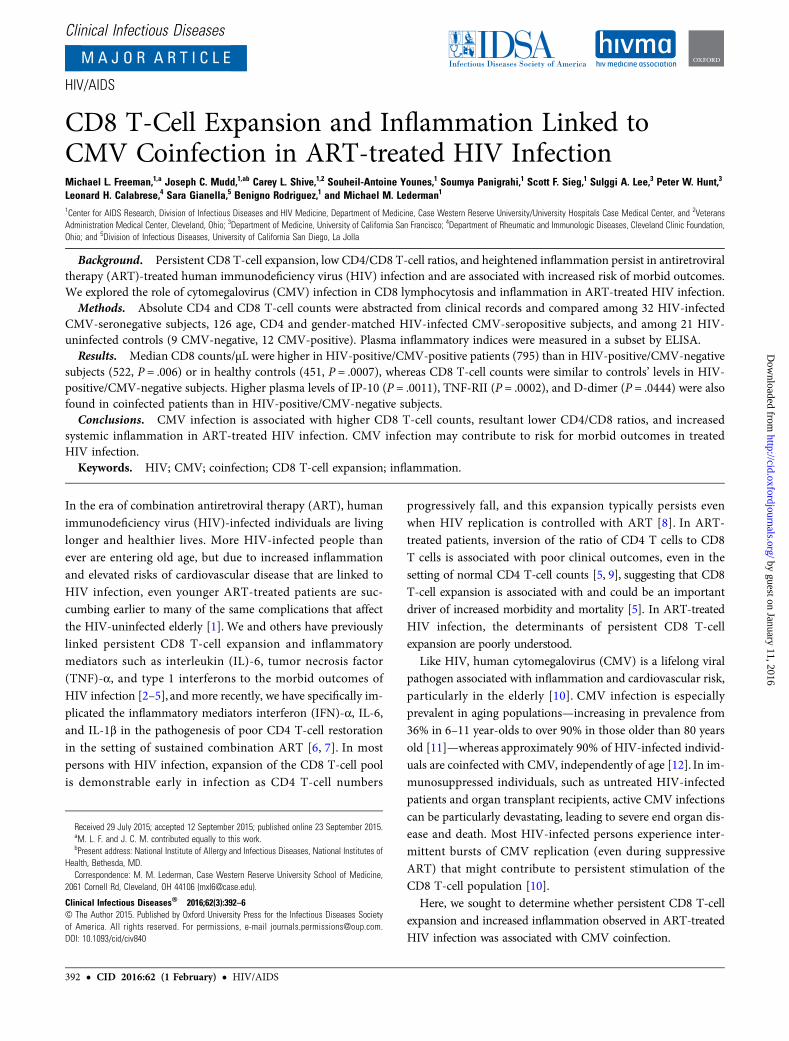
Elevated CD8 T-Cell Numbers and Decreased CD4/CD8 Ratio in HIV andCMV CoinfectionMedian circulating CD8 T-cell number was significantly higherin HIV-infected CMV-seropositive patients (795/µL) than inHIV-infected CMV-seronegative subjects (522/µL, P = .006)or HIV-uninfected controls (451/µL, P = .0007). AbsoluteCD8 T-cell counts among the HIV-infected CMV seronegativesubjects were not different from those among healthy controls(P > .99), suggesting that the expansion of circulating CD8 Tcells that is a hallmark of ART-treated HIV infection is specif-ically linked to coinfection with CMV. As the HIV-infectedgroups were matched for CD4 T-cell counts, CD4 T-cell nadirs,and duration of ART, the observed difference in CD8 T-cellcounts was not related to differences in immune restoration.As expected, CD4 T-cell counts in each HIV-infected groupwere lower than among HIV-negative controls (Figure 1B).Consequently, coinfection with HIV and CMV resulted in a sig-nificantly lower CD4/CD8 ratio than was seen among HIV-in-fected CMV-seronegative subjects (P = .004, Figure 1C). Asboth increased circulating CD8 T-cell numbers and low CD4/CD8 ratios are associated with poor clinical outcomes inART-treated HIV infection [5, 9], our findings implicateCMV coinfection as a possible driver of non-AIDS morbiditiesin treated HIV disease.
Elevated Expression of Select Markers of InflammationWe next asked if the presence of HIV and CMV coinfection wasassociated with increased plasma levels of the inflammatory andcoagulation markers IP-10, TNF-RII, D-dimer, IL-18, IL-6, andsCD14 (Figure 2). Levels of IP-10 (P = .0011), TNF-RII(P = .0002), and D-dimer (P = .0444) were higher in plasmasof HIV, CMV coinfected subjects compared to HIV-infected
Table 1. Participant Characteristics
HIV-positive
P Value
Total
P ValueCMV-negative CMV-positive HIV-positive HIV-negative
N, (male, %) 32 (84.75%) 126 (84.13%) 1.00 158 (84.18%) 21 (52.4%) .0018
CMV-positive (%) 0% 100% <.0001 79.7% 42.9% .0281
Age (y), Median (IQR) 41.5 (35.25–47) 42 (36–48.25) >.9999 42 (36–48) 37 (30.5–48.5) .0526
Time on ART (y), Median (IQR) 3.29 (2.61–4.71) 3.14 (1.86–4.69) .4634 3.15 (2.11–4.67) NA NA
CD4 (cells/µL), Median (IQR) 437 (275.3–591.8) 490 (309–640) .4201 467 (300.5–635) 907 (703.5–1059) <.0001
CD4 nadir (cells/µL), Median (IQR) 178.5 (89.3–268.5) 180 (71–300.1) .6527 180 (72.5–290) NA NA
Significance was determined using Mann–Whitney U test or Fisher exact test. P values <.05 were considered significant.
Abbreviations: ART, antiretroviral therapy; CMV, cytomegalovirus; HIV, human immunodeficiency virus; IQR, interquartile range; NA, not applicable.
HIV/AIDS • CID 2016:62 (1 February) • 393
by guest on January 11, 2016http://cid.oxfordjournals.org/
Dow
nloaded from
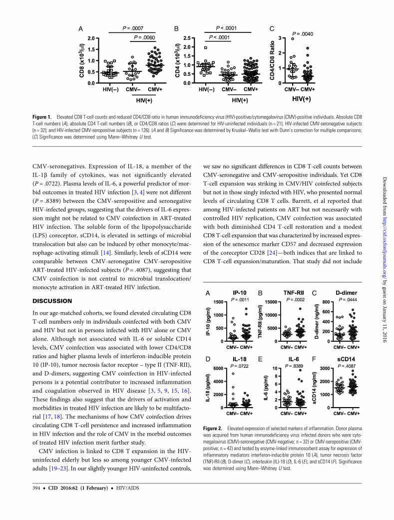
CMV-seronegatives. Expression of IL-18, a member of theIL-1β family of cytokines, was not significantly elevated(P = .0722). Plasma levels of IL-6, a powerful predictor of mor-bid outcomes in treated HIV infection [3, 4] were not different(P = .8389) between the CMV-seropositive and seronegativeHIV-infected groups, suggesting that the drivers of IL-6 expres-sion might not be related to CMV coinfection in ART-treatedHIV infection. The soluble form of the lipopolysaccharide(LPS) coreceptor, sCD14, is elevated in settings of microbialtranslocation but also can be induced by other monocyte/mac-rophage-activating stimuli [14]. Similarly, levels of sCD14 werecomparable between CMV-seronegative CMV-seropositiveART-treated HIV-infected subjects (P = .4087), suggesting thatCMV coinfection is not central to microbial translocation/monocyte activation in ART-treated HIV infection.
DISCUSSION
In our age-matched cohorts, we found elevated circulating CD8T-cell numbers only in individuals coinfected with both CMVand HIV but not in persons infected with HIV alone or CMValone. Although not associated with IL-6 or soluble CD14levels, CMV coinfection was associated with lower CD4/CD8ratios and higher plasma levels of interferon-inducible protein10 (IP-10), tumor necrosis factor receptor – type II (TNF-RII),and D-dimers, suggesting CMV coinfection in HIV-infectedpersons is a potential contributor to increased inflammationand coagulation observed in HIV disease [3, 5, 9, 15, 16].These findings also suggest that the drivers of activation andmorbidities in treated HIV infection are likely to be multifacto-rial [17, 18]. The mechanisms of how CMV coinfection drivescirculating CD8 T-cell persistence and increased inflammationin HIV infection and the role of CMV in the morbid outcomesof treated HIV infection merit further study.
CMV infection is linked to CD8 T expansion in the HIV-uninfected elderly but less so among younger CMV-infectedadults [19–23]. In our slightly younger HIV-uninfected controls,
we saw no significant differences in CD8 T-cell counts betweenCMV-seronegative and CMV-seropositive individuals. Yet CD8T-cell expansion was striking in CMV/HIV coinfected subjectsbut not in those singly infected with HIV, who presented normallevels of circulating CD8 T cells. Barrett, et al reported thatamong HIV-infected patients on ART but not necessarily withcontrolled HIV replication, CMV coinfection was associatedwith both diminished CD4 T-cell restoration and a modestCD8 T-cell expansion that was characterized by increased expres-sion of the senescence marker CD57 and decreased expressionof the coreceptor CD28 [24]—both indices that are linked toCD8 T-cell expansion/maturation. That study did not include
Figure 1. Elevated CD8 T-cell counts and reduced CD4/CD8 ratio in human immunodeficiency virus (HIV)-positive/cytomegalovirus (CMV)-positive individuals. Absolute CD8T-cell numbers (A), absolute CD4 T-cell numbers (B), or CD4/CD8 ratios (C) were determined for HIV-uninfected individuals (n = 21); HIV-infected CMV-seronegative subjects(n = 32); and HIV-infected CMV-seropositive subjects (n = 126). (A and B) Significance was determined by Kruskal–Wallis test with Dunn’s correction for multiple comparisons;(C) Significance was determined using Mann–Whitney U test.
Figure 2. Elevated expression of selected markers of inflammation. Donor plasmawas acquired from human immunodeficiency virus infected donors who were cyto-megalovirus (CMV)-seronegative (CMV-negative; n = 32) or CMV-seropositive (CMV-positive; n = 42) and tested by enzyme-linked immunosorbent assay for expression ofinflammatory mediators interferon-inducible protein 10 (A), tumor necrosis factor(TNF)-RII (B), D-dimer (C), interleukin (IL)-18 (D), IL-6 (E ), and sCD14 (F ). Significancewas determined using Mann–Whitney U test.
394 • CID 2016:62 (1 February) • HIV/AIDS
by guest on January 11, 2016http://cid.oxfordjournals.org/
Dow
nloaded from
comparison to HIV-uninfected controls, nor were soluble indicesof inflammation measured. In the present study, performedamong patients with controlled HIV replication on ART carefullymatched for current and nadir CD4 T-cell counts and duration ofART exposure, CMV coinfection was linked to dramatic CD8T-cell expansion, whereas singly HIV-infected subjects hadnormal CD8 T-cell counts. Thus, our data suggest that HIVand CMV infections together drive CD8 T-cell expansion—buthow does this happen? One possibility is that increased CMVreactivation and shedding in HIV-infected subjects drives activa-tion and proliferation of CMV-specific CD8 T cells. Immuno-compromised individuals experience more frequent CMVreactivation and shedding [25], and CMV shedding is associatedwith increased levels of T-cell activation, proliferation, and ex-haustion [10]. As many as half of all CD8 T cells can be CMV-reactive in the CMV-infected elderly [20, 26], and the percentageof CD8 T cells specific for CMV antigens is increased in HIV-infected subjects [27–30]. One potential driver of CD8 T-cellexpansion in the setting of coinfection is IL-15, which can beupregulated by herpesvirus-infected cells and is upregulatedearly in HIV infection [31, 32]. Previous studies have clearlydemonstrated that CMV infection favors a fully differentiated,effector memory phenotype [33] and that HIV infection maybe characterized by a proliferative block in CD8 T cells [34].Therefore, it is conceivable that the combined proinflammatoryenvironment of HIV and CMV coinfection drives both T-cellactivation and some level of bystander proliferation that aug-ments cognate peptide-driven CD8 T-cell expansion, coupledwith a failure to deplete the existing CD8 T-cell pool. Althoughthe precise role for CMV in driving CD8 T-cell expansion has notyet been demonstrated in HIV disease, administration of the anti-CMV drug valganciclovir to ART-treated HIV-infected patientswith incomplete CD4 T-cell recovery reduced CD8 T-cell activa-tion [35], suggesting that herpesvirus recrudescence contributesto persistent activation in ART-treated HIV infection.
During inflammation, TNF-RII is shed from the surface ofcells upon binding with TNFα and its expression can be usedas a surrogate for TNFα activity [36]. In untreated HIV infection,TNF-RII levels correlate with HIV RNA levels and are reducedupon initiation of ART [37]. Serum TNF-RII levels are also in-creased during CMV disease [38] and, notably, also in untreatedHIV-infected patients with CMV disease [39]. Our data suggestthat CMV-induced inflammation may be an important driver ofTNFα expression during ART-treated HIV infection. Similarly,IP-10 is an important chemokine induced by interferons that isinvolved in a variety of immune pathways and is a biomarker fordisease severity in multiple settings. IP-10 expression is elevatedin ART-treated HIV infection and in settings of CMV infectionfollowing lung transplantation [40, 41]. Here we show that CMVcoinfection increases IP-10 expression more than is seen inART-treated HIV infection alone. Thus, TNFα and interferonsproduced during the cellular immune response to CMV could
contribute to the increase in TNF-RII and IP-10 levels, seen intreated HIV infection.
Notably, HIV and CMV infection and related inflammationare each associated with increased cardiovascular risk [42]. Inparticular, levels of the fibrin degradation product D-dimer, acoagulation biomarker, are associated with cardiovascular dis-ease and mortality in HIV-infected patients [2]. ElevatedCMV-specific T-cell responses and levels of CMV IgG are cor-related with increased carotid artery intima-media thickness inHIV-infected patients [28], making CMV a plausible contribu-tor to risk and an attractive target for therapeutic interventionto prevent HIV-associated cardiovascular complications. Futurestudies are needed to determine if suppressing CMV replicationin HIV coinfection will lead to reductions in inflammatory andcoagulation indices and CD8 T-cell numbers, and a diminutionin the risk of cardiovascular complications and other morbiditiesin ART-treated HIV infection.
NotesAcknowledgments. The authors wish to thank Brian Clagett, Dominic
Dorazio, and Janet Robinson for excellent technical assistance, and wouldlike to thank Louis Picker for helpful discussions.Financial support. This work was supported by the Case Western
Reserve University (CWRU) Center for AIDS Research (AI036219) andthe CWRU Clinical Trials Unit (AI069501), and by funding from the Rich-ard J. Fasenmyer Foundation.Potential conflicts of interest. M. M. L., P. W. H., and B. R. have re-
ceived institutional grant support from the National Institutes of Health(NIH). S. G. has received institutional grant support from the NIH Centerfor AIDS Research. All other authors report no potential conflicts. All au-thors have submitted the ICMJE Form for Disclosure of Potential Conflictsof Interest. Conflicts that the editors consider relevant to the content of themanuscript have been disclosed.
References1. Guaraldi G, Orlando G, Zona S, et al. Premature age-related comorbidities among
HIV-infected persons compared with the general population. Clin Infect Dis 2011;53:1120–6.
2. Kuller LH, Tracy R, Belloso W, et al. Inflammatory and coagulation biomarkersand mortality in patients with HIV infection. PLoS Med 2008; 5:e203.
3. Tenorio AR, Zheng Y, Bosch RJ, et al. Soluble markers of inflammation and coag-ulation but not T-cell activation predict non-AIDS-defining morbid events duringsuppressive antiretroviral treatment. J Infect Dis 2014; 210:1248–59.
4. Hunt PW, Sinclair E, Rodriguez B, et al. Gut epithelial barrier dysfunction andinnate immune activation predict mortality in treated HIV infection. J InfectDis 2014; 210:1228–38.
5. Serrano-Villar S, Sainz T, Lee SA, et al. HIV-infected individuals with low CD4/CD8 ratio despite effective antiretroviral therapy exhibit altered T cell subsets,heightened CD8+ T cell activation, and increased risk of non-AIDS morbidityand mortality. PLoS Pathog 2014; 10:e1004078.
6. Nguyen TP, Bazdar DA, Mudd JC, et al. Interferon-alpha inhibits CD4 T cellresponses to interleukin-7 and interleukin-2 and selectively interferes with Aktsignaling. J Leukoc Biol 2015; 97:1139–46.
7. Shive CL, Mudd JC, Funderburg NT, et al. Inflammatory cytokines drive CD4+T-cell cycling and impaired responsiveness to interleukin 7: implications forimmune failure in HIV disease. J Infect Dis 2014; 210:619–29.
8. Mudd JC, Lederman MM. CD8 T cell persistence in treated HIV infection. CurrOpin HIVAIDS 2014; 9:500–5.
9. Serrano-Villar S, Gutierrez C, Vallejo A, et al. The CD4/CD8 ratio in HIV-infectedsubjects is independently associated with T-cell activation despite long-term viralsuppression. J Infect 2013; 66:57–66.
10. Gianella S, Massanella M, Wertheim JO, Smith DM. The sordid affair betweenhuman herpesvirus and HIV. J Infect Dis 2015; 212:845–52.
HIV/AIDS • CID 2016:62 (1 February) • 395
by guest on January 11, 2016http://cid.oxfordjournals.org/
Dow
nloaded from

11. Staras SA, Dollard SC, Radford KW, Flanders WD, Pass RF, Cannon MJ. Seropre-valence of cytomegalovirus infection in the United States, 1988–1994. Clin InfectDis 2006; 43:1143–51.
12. Robain M, Carre N, Dussaix E, Salmon-Ceron D, Meyer L. Incidence and sexualrisk factors of cytomegalovirus seroconversion in HIV-infected subjects. TheSEROCO Study Group. Sex Transm Dis 1998; 25:476–80.
13. Lederman MM, Calabrese L, Funderburg NT, et al. Immunologic failure despitesuppressive antiretroviral therapy is related to activation and turnover of memoryCD4 cells. J Infect Dis 2011; 204:1217–26.
14. Shive CL, Jiang W, Anthony DD, Lederman MM. Soluble CD14 is a nonspecificmarker of monocyte activation. AIDS 2015; 29:1263–5.
15. Kaplan RC, Kingsley LA, Gange SJ, et al. Low CD4+ T-cell count as a majoratherosclerosis risk factor in HIV-infected women and men. AIDS 2008;22:1615–24.
16. Stylianou E, Aukrust P, Bendtzen K, Muller F, Froland SS. Interferons and inter-feron (IFN)-inducible protein 10 during highly active anti-retroviral therapy(HAART)-possible immunosuppressive role of IFN-alpha in HIV infection.Clin Exp Immunol 2000; 119:479–85.
17. Deeks SG. HIV infection, inflammation, immunosenescence, and aging. Annu RevMed 2011; 62:141–55.
18. Lederman MM, Funderburg NT, Sekaly RP, Klatt NR, Hunt PW. Residualimmune dysregulation syndrome in treated HIV infection. Adv Immunol 2013;119:51–83.
19. Sylwester AW,Mitchell BL, Edgar JB, et al. Broadly targeted human cytomegalovirus-specific CD4+ and CD8+ T cells dominate the memory compartments of exposedsubjects. J Exp Med 2005; 202:673–85.
20. Khan N, Shariff N, Cobbold M, et al. Cytomegalovirus seropositivity drivesthe CD8 T cell repertoire toward greater clonality in healthy elderly individuals.J Immunol 2002; 169:1984–92.
21. Khan N, Cobbold M, Cummerson J, Moss PA. Persistent viral infection inhumans can drive high frequency low-affinity T-cell expansions. Immunology2010; 131:537–48.
22. Ouyang Q, Wagner WM, Zheng W, Wikby A, Remarque EJ, Pawelec G. Dysfunc-tional CMV-specific CD8+ T cells accumulate in the elderly. Exp Gerontol 2004;39:607–13.
23. Chidrawar S, Khan N, Wei W, et al. Cytomegalovirus-seropositivity has aprofound influence on the magnitude of major lymphoid subsets within healthyindividuals. Clin Exp Immunol 2009; 155:423–32.
24. Barrett L, Stapleton SN, Fudge NJ, Grant MD. Immune resilience in HIV-infectedindividuals seronegative for cytomegalovirus. AIDS 2014; 28:2045–9.
25. Rinaldo CR Jr, Kingsley LA, Ho M, Armstrong JA, Zhou SY. Enhanced sheddingof cytomegalovirus in semen of human immunodeficiency virus-seropositivehomosexual men. J Clin Microbiol 1992; 30:1148–55.
26. Ouyang Q, Wagner WM, Wikby A, et al. Large numbers of dysfunctional CD8+T lymphocytes bearing receptors for a single dominant CMV epitope in the veryold. J Clin Immunol 2003; 23:247–57.
27. Naeger DM, Martin JN, Sinclair E, et al. Cytomegalovirus-specific T cells persist atvery high levels during long-term antiretroviral treatment of HIV disease. PLoSOne 2010; 5:e8886.
28. Hsue PY, Hunt PW, Sinclair E, et al. Increased carotid intima-media thickness inHIV patients is associated with increased cytomegalovirus-specific T-cell respons-es. AIDS 2006; 20:2275–83.
29. Stone SF, Price P, Khan N, Moss PA, French MA. HIV patients on antiretroviraltherapy have high frequencies of CD8 T cells specific for Immediate Early protein-1 of cytomegalovirus. AIDS 2005; 19:555–62.
30. Stone SF, Price P, French MA. Cytomegalovirus (CMV)-specific CD8+ T cells inindividuals with HIV infection: correlation with protection from CMV disease.J Antimicrob Chemother 2006; 57:585–8.
31. Fawaz LM, Sharif-Askari E, Menezes J. Up-regulation of NK cytotoxic activity viaIL-15 induction by different viruses: a comparative study. J Immunol 1999;163:4473–80.
32. Mueller YM, Katsikis PD. IL-15 in HIV infection: pathogenic or therapeutic po-tential? Eur Cytokine Netw 2010; 21:219–21.
33. Appay V, Dunbar PR, Callan M, et al. Memory CD8+ T cells vary in differentia-tion phenotype in different persistent virus infections. Nat Med 2002; 8:379–85.
34. Lee SA, Sinclair E, Hatano H, et al. Impact of HIV on CD8+ T cell CD57 expres-sion is distinct from that of CMV and aging. PLoS One 2014; 9:e89444.
35. Hunt PW, Martin JN, Sinclair E, et al. Valganciclovir reduces T cell activation inHIV-infected individuals with incomplete CD4+ T cell recovery on antiretroviraltherapy. J Infect Dis 2011; 203:1474–83.
36. Valle Y, Ledezma-Lozano IY, Torres-Carrillo N, et al. Circulating TNFRI andTNFRII levels correlated with the disease activity score (DAS28) in rheumatoidarthritis. Scand J Rheumatol 2009; 38:332–5.
37. NoktaM, Rossero R, Loesch K, Pollard RB. Kinetics of tumor necrosis factor alpha andsoluble TNFRII in HIV-infected patients treated with a triple combination of stavu-dine, didanosine, and hydroxyurea. AIDS Res Hum Retroviruses 1997; 13:1633–8.
38. Humbert M, Roux-Lombard P, Cerrina J, et al. Soluble TNF receptors (TNF-sR55and TNF-sR75) in lung allograft recipients displaying cytomegalovirus pneumo-nitis. Am J Respir Crit Care Med 1994; 149:1681–5.
39. Jakobsen PH, Dodt KK, Meyer CN, Katzenstein T, Gerstoft J, Skinhoj P. Increasedlevels of soluble tumour necrosis factor receptor-I (P55) and decreased IgG1 reac-tivities in HIV-1 patients with cytomegalovirus disease. Scand J Immunol 1998;47:591–5.
40. Ramirez LA, Arango TA, Thompson E, Naji M, Tebas P, Boyer JD. High IP-10levels decrease T cell function in HIV-1-infected individuals on ART. J LeukocBiol 2014; 96:1055–63.
41. Weseslindtner L, Nachbagauer R, Kundi M, et al. Human cytomegalovirus infec-tion in lung transplant recipients triggers a CXCL-10 response. Am J Transplant2011; 11:542–52.
42. Hsue PY, Lo JC, Franklin A, et al. Progression of atherosclerosis as assessed bycarotid intima-media thickness in patients with HIV infection. Circulation2004; 109:1603–8.
396 • CID 2016:62 (1 February) • HIV/AIDS
by guest on January 11, 2016http://cid.oxfordjournals.org/
Dow
nloaded from