case report coronary arteriovenous fistula causing hydrops...
TRANSCRIPT
Case ReportCoronary Arteriovenous Fistula Causing Hydrops Fetalis
Nilüfer Çetiner, Sinem Altunyuva Usta, and Figen AkalJn
Department of Pediatric Cardiology, Marmara University, Mimar Sinan Caddesi, No. 41, Pendik, 34890 Istanbul, Turkey
Correspondence should be addressed to Nilufer Cetiner; [email protected]
Received 6 April 2014; Revised 9 August 2014; Accepted 9 August 2014; Published 26 August 2014
Academic Editor: Svein Rasmussen
Copyright © 2014 Nilufer Cetiner et al.This is an open access article distributed under the Creative Commons Attribution License,which permits unrestricted use, distribution, and reproduction in any medium, provided the original work is properly cited.
Fetal heart failure and hydrops fetalis may occur due to systemic arteriovenous fistula because of increased cardiac output.Arteriovenous fistula of the central nervous system, liver, bone or vascular tumors such as sacrococcygeal teratoma were previouslyreported to be causes of intrauterine heart failure. However, coronary arteriovenous fistula was not reported as a cause of fetal heartfailure previously. It is a rare pathology comprising 0.2–0.4% of all congenital heart diseases even during postnatal life. Some mayremain asymptomatic for many years and diagnosed by auscultation of a continuous murmur during a routine examination, whilea larger fistulous coronary artery opening to a low pressure cardiac chamber may cause ischemia of the affected myocardial regiondue to steal phenomenon and may present with cardiomyopathy or congestive heart failure during childhood. We herein report aneonate with coronary arteriovenous fistula between the left main coronary artery and the right ventricular apex, who presentedwith hydrops fetalis during the third trimester of pregnancy.
1. Introduction
Congestive heart failure and hydrops fetalis may occur due tocardiac or noncardiac causes during intrauterine life. About26% of the fetuses with hydrops fetalis have cardiac pathol-ogises and 41% of them are congenital heart diseases [1].Coronary AV fistula (CAVF) is a rare congenital heart diseaseeven during postnatal lifewith an overall incidence of 1/50000[2].They constitute 0.2 to 0.4% of all congenital heart diseasesand more than half of all congenital coronary anomalies [3].Systemic arteriovenous fistula is known to cause high-outputheart failure and hydrops fetalis during intrauterine life.However, CAVF was not reported as a cause of intrauterinecardiac failure or hydrops fetalis previously. Some patientswith CAVF may remain asymptomatic for many years unlessthe amount of shunt throughAVfistula is not large and symp-toms usually occur in older age; many patients are diagnoseddue to auscultation of a continuous murmur during routineexamination [4, 5]. On the other hand, larger fistula maycause congestive heart failure due to increased volume loadof the left ventricle or myocardial ischemia, cardiomyopathy,ormyocardial infarctionmay occur due to steal phenomenonduring infancy or childhood. Intrauterine diagnosis of CAVFwas not also reported previously.
We herein present a case with CAVF leading to congestiveheart failure and hydrops fetalis during intrauterine life.
2. Case Report
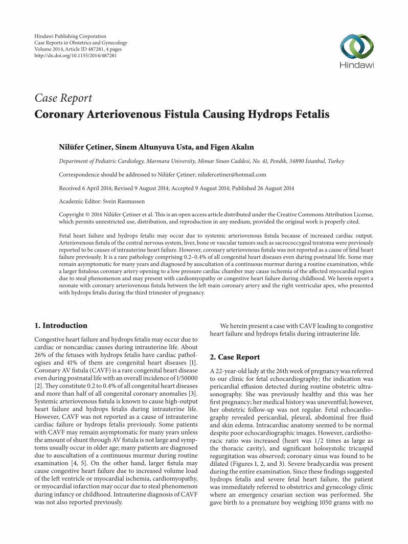
A22-year-old lady at the 26thweek of pregnancywas referredto our clinic for fetal echocardiography; the indication waspericardial effusion detected during routine obstetric ultra-sonography. She was previously healthy and this was herfirst pregnancy; hermedical history was uneventful; however,her obstetric follow-up was not regular. Fetal echocardio-graphy revealed pericardial, pleural, abdominal free fluidand skin edema. Intracardiac anatomy seemed to be normaldespite poor echocardiographic images. However, cardiotho-racic ratio was increased (heart was 1/2 times as large asthe thoracic cavity), and significant holosystolic tricuspidregurgitation was observed; coronary sinus was found to bedilated (Figures 1, 2, and 3). Severe bradycardia was presentduring the entire examination. Since these findings suggestedhydrops fetalis and severe fetal heart failure, the patientwas immediately referred to obstetrics and gynecology clinicwhere an emergency cesarian section was performed. Shegave birth to a premature boy weighing 1050 grams with no
Hindawi Publishing CorporationCase Reports in Obstetrics and GynecologyVolume 2014, Article ID 487281, 4 pageshttp://dx.doi.org/10.1155/2014/487281
2 Case Reports in Obstetrics and Gynecology
Figure 1: Four-chamber view of the fetal heart with increased cardi-othoracic ratio.
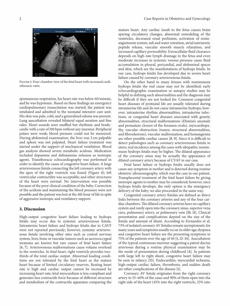
spontaneous respiration, his heart rate was below 60/minute,and he was hypotonic. Based on these findings an emergencycardiopulmonary resuscitation was started; the patient wasintubated and admitted to the neonatal intensive care unit.His skin was pale, cold, and a generalized edema was present.Lung auscultation revealed bilateral equal aeration and finerales. Heart sounds were muffled but rhythmic and brady-cardic with a rate of 100 bpmwithout anymurmur. Peripheralpulses were weak; blood pressure could not be measured.During abdominal examination, the liver was 3 cm palpableand spleen was not palpated. Heart failure treatment wasstarted under the support of mechanical ventilation. Bloodgas analysis showed severe metabolic acidosis. Medicationincluded dopamine and dobutamine infusion as inotropicagents. Transthoracic echocardiography was performed inorder to identify the cause of congestive heart failure. A largearteriovenous fistula connecting the left coronary artery withthe apex of the right ventricle was found (Figure 4); leftventricular contractility was acceptable; and other structuresof the heart were normal. No intervention was possiblebecause of the poor clinical condition of the baby. Correctionof the acidosis and maintaining the blood pressure were notpossible and the patient was lost at the 4th hour of life in spiteof aggressive inotropic and ventilatory support.
3. Discussion
High-output congestive heart failure leading to hydropsfetalis may occur due to systemic arteriovenous fistula.Intrauterine heart failure and hydrops fetalis due to CAVFwere not reported previously; however, systemic arteriove-nous fistula involving other sites such as central nervoussystem, liver, bone or vascular tumors such as sacrococcygealteratoma are known but rare causes of fetal heart failure[6, 7]. Arteriovenous malformations cause volume overloadto the ventricles. In fetal heart, right ventricle provides two-thirds of the total cardiac output. Abnormal loading condi-tions are not tolerated by the fetal heart as the matureheart because of limited circulatory reserves. Resting heartrate is high and cardiac output cannot be increased byincreasing heart rate; fetal myocardium is less compliant andgenerates less contractile force because of different structureand metabolism of the contractile apparatus comparing the
mature heart. Any cardiac insult to the fetus causes brainsparing circulatory changes, abnormal remodeling of theventricles, decreased renal perfusion, activation of renin-angiotensin system, salt andwater retention, atrial natriureticpeptide release, vascular smooth muscle relaxation, andincreased capillary permeability. Extracellular fluid clearancedepends on high-rate lymph drainage in the fetus and evenmoderate increases in systemic venous pressure cause fluidaccumulation in pleural, pericardial, and abdominal spacesand skin, which are the manifestations of hydrops fetalis. Inour case, hydrops fetalis has developed due to severe heartfailure caused by coronary arteriovenous fistula.
On the other hand in many fetuses with nonimmunehydrops fetalis the real cause may not be identified; earlyechocardiographic examination or autopsy studies may behelpful in defining such abnormalities and the diagnosis maybe difficult if they are not looked for. Common congenitalheart diseases of postnatal life are usually tolerated duringintrauterine life and do not cause intrauterine hydrops; how-ever, intrauterine rhythm abnormalities, intrauterine infec-tions, or congenital heart diseases associated with geneticabnormalities, structural malformations (Ebstein’s anomalyand premature closure of the foramen ovale), myocardiopa-thy, vascular obstruction (tumor, structural abnormalities,and fibroelastosis), vascular malformation, and hemangiomaare other possible cardiac causes [8, 9]. Since it is difficult todetect pathologies such as coronary arteriovenous fistula inutero, real incidence among the cases with idiopathic nonim-mune hydrops fetalis may be higher. The dilated appearanceof the coronary sinus may be actually the appearance ofdilated coronary artery because of CVAF in our case.
Fetal heart failure or hydrops fetalis usually does notcause any symptom in mother and is detected during routineobstetric ultrasonography, which was the case in our patient.Transplacental treatment of the fetal heart failure by givinginotropic agents tomothermay be considered; however, oncehydrops fetalis develops, the only option is the emergencydelivery of the baby; we also proceeded in the same way.
Congenital coronary artery fistulas are defined as directlinks between the coronary arteries and any of the four car-diac chambers.Thedilated coronary arteries have no capillarysystem and rarely open into the coronary sinus, superior venacava, pulmonary artery, or pulmonary vein [10, 11]. Clinicalpresentation and complications depend on the size of thefistula and amount of shunt. According to Fernandes et al.73%of isolated coronaryAVfistulas remain asymptomatic formany years and symptomsusually occur in older age; dyspneaand congestive heart failure are the presenting symptoms in75% of the patients over the age of 40 [1, 12–14]. Auscultationof the typical continuous murmur suggesting a patent ductusarteriosus during a routine physical examination may bethe mode of presentation during childhood [4]. In patientswith large left to right shunt, congestive heart failure maybe seen in infancy [15]. Endocarditis, myocardial ischemia,high-output cardiac failure, thrombosis, and sudden deathare other complications of the disease [1].
Coronary AV fistula originates from the right coronaryartery in 55–60% of the cases and 90% of them open into theright side of the heart (45% into the right ventricle, 25% into

Case Reports in Obstetrics and Gynecology 3
(a) (b)
Figure 2: Pulsed wave Doppler examination demonstrating significant tricuspid regurgitation.
(a) (b)
Figure 3: Fetal echocardiographic appearance of dilated coronary sinus.
Figure 4: Postnatal echocardiographic examination demonstratingthe large arteriovenous fistula connecting the left coronary arterywith the apex of the right ventricle.
the right atrium, 15–20% into the pulmonary artery, and 7%into the coronary sinus). In Lowe and Sabiston’s series of 286cases, right coronary arterywas involved in 56%, left coronaryartery was involved in 36%, and both coronary arteries wereinvolved in 5% of the patients. The fistula opened into theright ventricle in 39%, right atrium in 33%, pulmonary arteryin 20%, left atrium in 6%, and left ventricle in 2% of thecases [15]. In our case, coronary artery fistula originated
from the left coronary artery and was draining into the rightventricular apex.
Although the cardiac catheterization and coronary angi-ography are required for demonstration of the exact anatomyof the fistula and diagnosis of the additional cardiac anoma-lies, in recent years, technical developments in noninvasivediagnostic methods such as 2D and Doppler echocardiog-raphy provide sufficient information in CAVF. In our case,CAVF was diagnosed within the first hour of life by transtho-racic echocardiography [16].
Treatment options are surgical or transcatheter closure ofthe fistula. Angiography was planned; however, the clinicalcondition of the patient was not suitable for any kind ofintervention and he could stay alive for only 4 hours.
In conclusion, coronary arteriovenous fistula is a rarecongenital heart disease which may cause intrauterine heartfailure andmust be considered in differential diagnosis of thefetuses with nonimmune hydrops fetalis. Presence of hydropsfetalis in routine obstetric ultrasonography is an indicationfor fetal echocardiography.
Conflict of Interests
The authors declare that they have no conflict of interestsregarding to the publication of this paper.

4 Case Reports in Obstetrics and Gynecology
References
[1] V. Thakur, J. Fouron, L. Mertens, and E. T. Jaeggi, “Diagnosisand management of fetal heart failure,” Canadian Journal ofCardiology, vol. 29, no. 7, pp. 759–767, 2013.
[2] G. Schumacher, A. Roithmaier, H. P. Lorenz et al., “Congenitalcoronary artery fistula in infancy and childhood: diagnostic andtherapeutic aspects,” Thoracic and Cardiovascular Surgeon, vol.45, no. 6, pp. 287–294, 1997.
[3] W. Krause, “Uber den Ursprung Einer Akzessorischen A.coronaria aus der A. pulmonaris,” Z Ratinall Medicine, vol. 24,pp. 225–228, 1865.
[4] P. Sapin, E. Frantz, A. Jain, T. C.Nichols, andG. J. Dehmer, “Cor-onary artery fistula: an abnormality affecting all age groups,”Medicine, vol. 69, no. 2, pp. 101–113, 1990.
[5] A. Pelliccia, “Congenital coronary artery anomalies in youngpatients: new perspectives for timely identification,” Journal ofthe American College of Cardiology, vol. 37, no. 2, pp. 598–600,2001.
[6] J. E. Lowe and D. C. Sabiston Jr., “Congenital malformations ofthe coronary circulation,” in Surgery of the Chest, pp. 1689–1696,WB Saunders, Philadelphia, Pa, USA, 5th edition, 1990.
[7] S. Mullan, “Reflections upon the nature and management ofintracranial and intraspinal vascular malformations and fistu-lae,” Journal of Neurosurgery, vol. 80, no. 4, pp. 606–616, 1994.
[8] J. C. Easley and J. L. Carpenter, “Hepatic arteriovenous fistulain two Saint Bernard pups,” Journal of the American VeterinaryMedical Association, vol. 166, no. 2, pp. 167–171, 1975.
[9] J. W. Keeling, “Hydrops fetalis and other forms of excess fluidcollection in the fetus,” inTextbook of Fetal and Perinatal Pathol-ogy, J. S. Wigglesworth and D. B. Singer, Eds., pp. 429–454,Blackwell Scientific Publications, Oxford, UK, 1990.
[10] J. J. McNamara and R. E. Gross, “Congenital coronary arteryfistula,” Surgery, vol. 65, no. 1, pp. 59–69, 1969.
[11] H. Velvis, K. G. Schmidt, N. H. Silverman, and K. Turley, “Diag-nosis of coronary artery fistula by two-dimensional echocar-diography, pulsed doppler ultrasound and color flow imaging,”Journal of the American College of Cardiology, vol. 14, no. 4, pp.968–976, 1989.
[12] J. W. Kirklin and B. G. Barrat-Boyes, Congenital Anomaliesof the Coronary Arteries. Cardiac Surgery, vol. 2, ChurchillLivingstone, London, UK, 1993.
[13] E. D. Fernandes, H. Kadivar, G. L. Hallman, G. J. Reul, D. A.Ott, and D. A. Cooley, “Congenital malformations of the cor-onary arteries: The Texas Heart Institute experience,” Annals ofThoracic Surgery, vol. 54, no. 4, pp. 732–740, 1992.
[14] A. Vitarelli, G. De Curtis, Y. Conde et al., “Assessment of con-genital coronary artery fistulas by transesophageal color Dop-pler echocardiography,”The American Journal of Medicine, vol.113, no. 2, pp. 127–133, 2002.
[15] A. M. El Watidy, H. H. Ismail, and A. M. Calafiore, “Surgicalmanagement of right coronary artery-coronary sinus fistulacausing severe mitral and tricuspid regurgitation,” InteractiveCardiovascular and Thoracic Surgery, vol. 10, no. 1, pp. 110–112,2010.
[16] T. M. Daniel, T. P. Graham, and D. C. Sabiston Jr., “Coronaryartery-right ventricular fistula with congestive heart failure:surgical correction in the neonatal period,” Surgery, vol. 67, no.6, pp. 985–994, 1970.

Submit your manuscripts athttp://www.hindawi.com
Stem CellsInternational
Hindawi Publishing Corporationhttp://www.hindawi.com Volume 2014
Hindawi Publishing Corporationhttp://www.hindawi.com Volume 2014
MEDIATORSINFLAMMATION
of
Hindawi Publishing Corporationhttp://www.hindawi.com Volume 2014
Behavioural Neurology
EndocrinologyInternational Journal of
Hindawi Publishing Corporationhttp://www.hindawi.com Volume 2014
Hindawi Publishing Corporationhttp://www.hindawi.com Volume 2014
Disease Markers
Hindawi Publishing Corporationhttp://www.hindawi.com Volume 2014
BioMed Research International
OncologyJournal of
Hindawi Publishing Corporationhttp://www.hindawi.com Volume 2014
Hindawi Publishing Corporationhttp://www.hindawi.com Volume 2014
Oxidative Medicine and Cellular Longevity
Hindawi Publishing Corporationhttp://www.hindawi.com Volume 2014
PPAR Research
The Scientific World JournalHindawi Publishing Corporation http://www.hindawi.com Volume 2014
Immunology ResearchHindawi Publishing Corporationhttp://www.hindawi.com Volume 2014
Journal of
ObesityJournal of
Hindawi Publishing Corporationhttp://www.hindawi.com Volume 2014
Hindawi Publishing Corporationhttp://www.hindawi.com Volume 2014
Computational and Mathematical Methods in Medicine
OphthalmologyJournal of
Hindawi Publishing Corporationhttp://www.hindawi.com Volume 2014
Diabetes ResearchJournal of
Hindawi Publishing Corporationhttp://www.hindawi.com Volume 2014
Hindawi Publishing Corporationhttp://www.hindawi.com Volume 2014
Research and TreatmentAIDS
Hindawi Publishing Corporationhttp://www.hindawi.com Volume 2014
Gastroenterology Research and Practice
Hindawi Publishing Corporationhttp://www.hindawi.com Volume 2014
Parkinson’s Disease
Evidence-Based Complementary and Alternative Medicine
Volume 2014Hindawi Publishing Corporationhttp://www.hindawi.com