subclinical growth of an arteriovenous fistula associated
TRANSCRIPT

CASE REPORT Open Access
Subclinical growth of an arteriovenousfistula associated with renal biopsy: a casereportTakuya Murakami1, Shin-ichi Takeda1*, Hidenori Kanazawa2, Atsushi Ugajin2, Shigeyoshi Kijima2,Hiroyasu Nakamura2, Toshimi Imai1, Taro Sugase1, Ryoko Horikoshi1, Takahisa Kobayashi1, Tetsu Akimoto1,Osamu Saito1 and Daisuke Nagata1
Abstract
Background: Renal biopsy is not free from complications and patients who undergo this procedure are usuallyhospitalized to receive intensive care for several days after biopsy. In contrast, after this period, routine follow-up todetect biopsy-associated complications is rarely scheduled, unless the patient develops a clinical manifestation. Wedescribe a case of marked enlargement of arteriovenous fistula in the kidney that occurred many years after renalbiopsy. In contrast to the previous cases requiring interventional radiology, our patient showed subclinical growthof fistula over about nine years.
Case presentation: A 24-year-old man with a history of percutaneous renal biopsy was hospitalized for interventionalradiology. Gross hematuria emerged shortly after biopsy, but completely disappeared with administration ofhemostatic agents and bed rest. Subsequently, the patient had few symptoms for many years. A giant fistula(a gourd-shaped mass, size 26 × 22 and 12 × 11 mm) was unexpectedly detected by ultrasonography performed forexamination of an unrelated disorder (slight elevation of serum transaminase) at 9 years after the original biopsy. Thefistula was successfully treated with radiological intervention. Thus, subclinical development of complicationsassociated with renal biopsy should be considered, even in an uneventful course.
Conclusions: This case provides a platform to discuss the importance of long-term follow-up of patients after renalbiopsy despite of its difficulty.
Keywords: Renal biopsy, Renal arteriovenous fistula, Interventional radiology
BackgroundRenal biopsies provide critical clues in diagnosis of renaldiseases. However, this procedure is not free from com-plications, despite technical improvements over time. Arecent nationwide study in Norway showed grosshematuria after biopsy in 1.9 % of patients, with 0.9 %needing blood transfusion and 0.2 % requiring surgicalintervention or catheterization [1]. There have also beenseveral reports of renal arteriovenous fistula (AVF) afterrenal biopsy. In most such cases, radiological interven-tions were performed due to severe clinical manifestations
of gross hematuria [2–4], hemorrhagic shock [2], severehypertension [2, 5], and decline of renal function [4],which all emerged shortly after the biopsy. We herein re-port a case of marked enlargement of renal AVF that wasdetected by chance nine years after biopsy and was suc-cessfully treated with interventional radiology (IR). In con-trast to the previous cases of renal AVF associated withbiopsy [2–6], our patient showed subclinical growth ofAVF over about nine years. Thus, this case provides a plat-form to discuss the importance of long-term follow-up ofpatients after renal biopsy.
Case presentationA 24-year-old man was admitted to our facility for IR.The patient had a history of percutaneous kidney biopsy
* Correspondence: [email protected] of Nephrology, Department of Medicine, Jichi Medical University,3311-1 Yakushiji, Shimotsuke, Tochigi, JapanFull list of author information is available at the end of the article
© 2016 The Author(s). Open Access This article is distributed under the terms of the Creative Commons Attribution 4.0International License (http://creativecommons.org/licenses/by/4.0/), which permits unrestricted use, distribution, andreproduction in any medium, provided you give appropriate credit to the original author(s) and the source, provide a link tothe Creative Commons license, and indicate if changes were made. The Creative Commons Public Domain Dedication waiver(http://creativecommons.org/publicdomain/zero/1.0/) applies to the data made available in this article, unless otherwise stated.
Murakami et al. BMC Nephrology (2016) 17:81 DOI 10.1186/s12882-016-0289-4
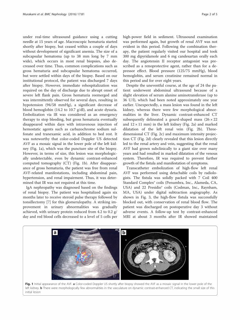
under real-time ultrasound guidance using a cuttingneedle at 15 years of age. Macroscopic hematuria startedshortly after biopsy, but ceased within a couple of dayswithout development of significant anemia. The size of asubcapsular hematoma (up to 38 mm long by 7 mmwide), which occurs in most renal biopsies, also de-creased over time. Thus, common complications such asgross hematuria and subcapsular hematoma occurred,but were settled within days of the biopsy. Based on ourinstitutional protocol, the patient was discharged 7 daysafter biopsy. However, immediate rehospitalization wasrequired on the day of discharge due to abrupt onset ofsevere left flank pain. Gross hematuria reemerged andwas intermittently observed for several days, resulting inhypotension (94/58 mmHg), a significant decrease ofblood hemoglobin (14.2 to 10.7 g/dl), and acute dysuria.Embolization via IR was considered as an emergencytherapy to stop bleeding, but gross hematuria eventuallydisappeared within days with intravenous injection ofhemostatic agents such as carbazochrome sodium sul-fonate and tranexamic acid, in addition to bed rest. Itwas noteworthy that color-coded Doppler US detectedAVF as a mosaic signal in the lower pole of the left kid-ney (Fig. 1a), which was the puncture site of the biopsy.However, in terms of size, this lesion was morphologic-ally undetectable, even by dynamic contrast-enhancedcomputed tomography (CT) (Fig. 1b). After disappear-ance of gross hematuria, the patient was free from renalAVF-related manifestations, including abdominal pain,hypertension, and renal impairment. Thus, it was deter-mined that IR was not required at this time.IgA nephropathy was diagnosed based on the findings
of renal biopsy. The patient was hospitalized again sixmonths later to receive steroid pulse therapy followed bytonsillectomy [7] for this glomerulopathy. A striking im-provement in urinary abnormalities was graduallyachieved, with urinary protein reduced from 4.2 to 0.2 g/day and red blood cells decreased to a level of 5 cells per
high-power field in sediment. Ultrasound examinationwas performed again, but growth of renal AVF was notevident in this period. Following the combination ther-apy, the patient regularly visited our hospital and took300 mg dipyridamole and 6 mg candesartan orally eachday. The angiotensin II receptor antagonist was pre-scribed as a renoprotective agent, rather than for a de-pressor effect. Blood pressure (125/75 mmHg), bloodhemoglobin, and serum creatinine remained normal inthis period and for over eight years.Despite the uneventful course, at the age of 24 the pa-
tient underwent abdominal ultrasound because of aslight elevation of serum alanine aminotransferase (up to36 U/l), which had been noted approximately one yearearlier. Unexpectedly, a mass lesion was found in the leftkidney, whereas there were few morphological abnor-malities in the liver. Dynamic contrast-enhanced CTsubsequently delineated a gourd-shaped mass (26 × 22and 12 × 11 mm) in the left kidney (Fig. 2a) and markeddilatation of the left renal vein (Fig. 2b). Three-dimensional CT (Fig. 2c) and maximum intensity projec-tion CT (Fig. 2d) clearly revealed that this lesion directlyled to the renal artery and vein, suggesting that the renalAVF had grown subclinically to a giant size over manyyears and had resulted in marked dilatation of the venoussystem. Therefore, IR was required to prevent furthergrowth of the fistula and manifestation of symptoms.Transcatheter embolization of high-flow left renal
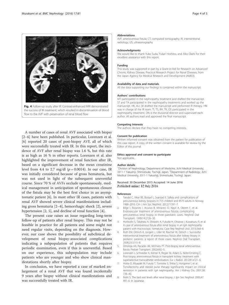
AVF was performed using detachable coils by radiolo-gists. The fistula was solidly packed with 7 Coil 400Standard Complex® coils (Penumbra, Inc., Alameda, CA,USA) and 22 Presidio® coils (Codman, Inc., Raynham,MA, USA) under digital subtraction angiography. Asshown in Fig. 3, the high-flow fistula was successfullyblocked out, with conservation of renal blood flow. Thepatient was discharged on postoperative day 3 withoutadverse events. A follow-up test by contrast-enhancedMRI at about 3 months after IR showed maintained
Fig. 1 Initial appearance of the AVF. a Color-coded Doppler US shortly after biopsy showed the AVF as a mosaic signal in the lower pole of theleft kidney. b There were morphologically few abnormalities in the vasculature on dynamic contrast-enhanced CT, indicating the small size of thisinitial lesion
Murakami et al. BMC Nephrology (2016) 17:81 Page 2 of 5

discontinuation of blood flow to the AVF (Fig. 4), indi-cating the success of the treatment.
ConclusionsComplications associated with renal biopsies are not un-common. Indeed, nephrologists exert extreme cautionearly after puncture, especially for the first few days. Theguidelines for renal biopsies published by the JapaneseSociety of Nephrology [8] recommend a stay of no lessthan 4 to 7 days in hospital. Postural changes in bed andadoption of a standing position are not allowed for the
first hours and almost the entire first day, respectively,based on two studies of biopsy-related complications:Khajehdehi et al. [9] found that patients with stablehematocrit at 6 h were at low risk for bleeding at24 h while hospitalized, and Marwah et al. [10] foundthat major complications were identifiable in mostcases within 12 h. Thus, intensive management is im-portant for a short time after renal biopsy. However,there is little information on appropriate follow-upfor patients with a history of renal biopsy after thisperiod.
Fig. 2 Contrast-enhanced CT of the enlarged AVF and dilatation of the left renal vein. Two-dimensional (a, b) and three-dimensional (c) CT performedincidentally at 9 years after biopsy revealed the morphological characteristics of an enlarged left renal AVF (red arrowheads) and dilatedleft renal vein (yellow arrows). d A maximum intensity projection image showed a connection with the renal artery and vein. Abbreviations:A, renal artery; V, renal vein
Fig. 3 Embolization therapy for the renal AVF. a Pre-therapeutic, b early treatment phase, and c post-therapeutic digital subtraction angiographyimages. The renal AVF (circled with a solid line) was filled with microcoils (circled with a dashed line) over time. The enlarged left renalvein (indicated by arrowheads) was delineated despite the arterial phase until the early therapeutic period (a, b), but disappeared afterthe AVF was blocked out (c), indicating a large amount of shunt flow
Murakami et al. BMC Nephrology (2016) 17:81 Page 3 of 5
A number of cases of renal AVF associated with biopsy[2–6] have been published. In particular, Lorenzen et al.[6] reported 20 cases of post-biopsy AVF, all of whichwere successfully treated with IR. In this report, the inci-dence of AVF after renal biopsy was 1.6 %, but this rateis as high as 16 % in other reports. Lorenzen et al. alsohighlighted the improvement of renal function after IR,based on a significant decrease in the mean creatininelevel from 4.4 to 2.7 mg/dl (p = 0.0014). In our case, IRwas initially considered because of gross hematuria, butwas not used in light of the subsequent uneventfulcourse. Since 70 % of AVFs occlude spontaneously, med-ical management in anticipation of spontaneous closureof the fistula may be the best first choice in an asymp-tomatic patient [6]. In most other IR cases, patients withrenal AVF showed severe clinical manifestations includ-ing gross hematuria [2–4], hemorrhagic shock [2], severehypertension [2, 5], and decline of renal function [4].The present case raises an issue regarding long-term
follow-up of patients after renal biopsy. This may not befeasible in practice for all patients and some might notneed regular visits, depending on the diagnosis. How-ever, our case shows the possibility of subclinical de-velopment of renal biopsy-associated complicationsindicating a subpopulation of patients that requiresperiodic examination, even if this is uneventful. Basedon our experience, this subpopulation may includepatients who are younger and who show clinical man-ifestations shortly after biopsy.In conclusion, we have reported a case of marked en-
largement of a renal AVF that was found incidentally9 years after biopsy without clinical manifestations andwas successfully treated with IR.
AbbreviationsAVF, arteriovenous fistula; CT, computed tomography; IR, interventionalradiology; US, ultrasonography
AcknowledgmentsWe would like to thank Yuko Suda, Yukari Hoshino, and Aiko Oashi for theirexcellent assistance with this report.
FundingThis study was supported in part by a Grant-in-Aid for Research on AdvancedChronic Kidney Disease, Practical Research Project for Renal Diseases, fromthe Japan Agency for Medical Research and Development (AMED).
Availability of data and materialsAll the data supporting our findings is contained within the manuscript.
Authors’ contributionsMT participated in the nephropathy treatment and drafted the manuscript.ST and TA participated in the nephropathy treatments and worked up themanuscript. HK, AU, SK drafted the manuscript and performed IR therapy. HNwas in charge of the IR team. TI, TS, RH, TK, OS participated in thenephropathy treatment. DN is the divisional director and supervised eachauthor. All authors read and approved the final manuscript.
Competing interestsThe authors declare that they have no competing interests.
Consent for publicationWritten informed consent was obtained from the patient for publication ofthis case report. A copy of the written consent is available for review by theEditor of this journal.
Ethics approval and consent to participateNot applicable.
Author details1Division of Nephrology, Department of Medicine, Jichi Medical University,3311-1 Yakushiji, Shimotsuke, Tochigi, Japan. 2Department of Radiology, JichiMedical University, 3311-1 Yakushiji, Shimotsuke, Tochigi, Japan.
Received: 30 December 2015 Accepted: 14 June 2016
References1. Tøndel C, Vikse BE, Bostad L, Svarstad E. Safety and complications of
percutaneous kidney biopsies in 715 children and 8573 adults in Norway1988–2010. Clin J Am Soc Nephrol. 2012;7:1591–7.
2. Bilge I, Rozanes I, Acunas B, Minareci O, Nayir A, Oktem F, et al.Endovascular treatment of arteriovenous fistulas complicatingpercutaneous renal biopsy in three paediatric cases. Nephrol DialTransplant. 1999;14:2726–30.
3. Horikoshi S, Takahata A, Shiraishi A, Fukuda H, Ohsawa I, Kuwatsuru R, et al.A case of arteriovenous fistula after renal biopsy in an IgA nephropathypatient with macroscopic hematuria. Case Rep Nephrol Urol. 2013;3:64–8.
4. Rüth EM, Dittrich K, Jüngert J, Uder M, Rascher W, Dötsch J. Successfulinterventional treatment of arteriovenous fistula after kidney biopsy inpediatric patients: a report of three cases. Nephrol Dial Transplant.2008;23:3215–8.
5. Omoloja AA, Racadio JM, McEnery PT. Post-biopsy renal arteriovenousfistula. Pediatr Transplant. 2002;6:82–5.
6. Lorenzen J, Schneider A, Körner K, Regier M, Adam G, Nolte-Ernsting C.Post-biopsy arteriovenous fistula in transplant kidney: treatment withsuperselective transcatheter embolisation. Eur J Radiol. 2012;81:e721–6.
7. Hotta O, Miyazaki M, Furuta T, Tomioka S, Shiba S, Horigome I, et al.Tonsillectomy and steroid pulse therapy significantly impact on clinicalremission in patients with IgA nephropathy. Am J Kidney Dis. 2001;38:736–43.
8. Nishi S. The bed rest levels after renal biopsy. J Jpn Soc Nephrol. 2005;47:491–6. In Japanese.
Fig. 4 Follow-up study after IR. Contrast-enhanced MRI demonstratedthe success of IR treatment, which resulted in discontinuation of bloodflow to the AVF with preservation of renal blood flow
Murakami et al. BMC Nephrology (2016) 17:81 Page 4 of 5

9. Khajehdehi P, Junaid SM, Salinas-Madrigal L, Schmitz PG, Bastani B.Percutaneous renal biopsy in the 1990s: safety, value, and implications forearly hospital discharge. Am J Kidney Dis. 1999;34:92–7.
10. Marwah DS, Korbet SM. Timing of complications in percutaneous renalbiopsy: what is the optimal period of observation? Am J Kidney Dis. 1996;28:47–52.
• We accept pre-submission inquiries
• Our selector tool helps you to find the most relevant journal
• We provide round the clock customer support
• Convenient online submission
• Thorough peer review
• Inclusion in PubMed and all major indexing services
• Maximum visibility for your research
Submit your manuscript atwww.biomedcentral.com/submit
Submit your next manuscript to BioMed Central and we will help you at every step:
Murakami et al. BMC Nephrology (2016) 17:81 Page 5 of 5