pial arteriovenous fistula with giant varices: report of ... · pial arteriovenous fistula with...
TRANSCRIPT
98 J Cerebrovasc Endovasc Neurosurg
Pial Arteriovenous Fistula with Giant Varices: Report of Two Cases with Good Surgical Outcome
Morteza Faghih Jouibari1, Mehdi Zeinali Zadeh2, Masoud Khadivi1, Alireza Khoshnevisan1,
Keisan Moazzeni1, Sina Abdollahzade1
1Department of Neurosurgery, Shariati Hospital, Tehran University of Medical Sciences, Tehran, Iran2Department of Neurosurgery, Imam Khomeini Hospital, Tehran University of Medical Sciences, Tehran, Iran
Pial arteriovenous fistulas (pAVF) are rare vascular lesions consisting of one or more arterial connections to a single venous channel without any intervening nidus of vessels or capillaries.
Case 1 : A 65-year-old woman with a complaint of headache and left hand paresthesia was referred to us. Magnetic resonance imaging showed a large saccular lesion with signal void in the posterior part of the right sylvian fissure and catheter angiography showed a giant venous aneurysm fed by one branch of the middle cerebral artery (MCA) and draining into the vein of Trolard.
Case 2 : A 12-year-old boy was transferred to our hospital with a history of sudden loss of consciousness and hemiplegia. Brain computed tomog-raphy revealed a massive hemorrhagic mass in the right hemisphere and cerebral angiography showed a pAVF with a large aneurysmal varix, which was fed by multiple branches of the right MCA and draining into the superior sagittal sinus.
Both patients underwent craniotomy and after ligation of vascular con-nections, aneurysmal varices were removed completely. Surgical resection can be a safe method for treatment of pAVFs, particularly in those with large varices.
J Cerebrovasc Endovasc Neurosurg. 2014 June;16(2):98-103Received : 27 January 2014Revised : 9 April 2014Accepted : 2 May 2014
Correspondence to Sina AbdollahzadeJalal Al Ahmad Street, Amir Abad Avenue, Department of Neurosurgery, Shariati Hospital, Tehran University of Medical Sciences, Tehran, Iran
Tel : 98-21-8822-0040Cell : 98-912-132-5746Fax : 98-21-8822-0040E-mail : [email protected] : http://orcid.org/0000-0002-6423-9614
This is an Open Access article distributed under the terms of the Creative Commons Attribution Non- Commercial License (http://creativecommons.org/li-censes/by-nc/3.0) which permits unrestricted non- commercial use, distribution, and reproduction in any medium, provided the original work is properly cited.Keywords Pial arteriovenous fistulas, Microsurgery
Journal of Cerebrovascular and Endovascular NeurosurgeryISSN 2234-8565, EISSN 2287-3139, http://dx.doi.org/10.7461/jcen.2014.16.2.98 Case Report
INTRODUCTION
Intracranial pial arteriovenous fistulas (pAVF) are
rare vascular lesions accounting for only 1.6% of all
vascular malformations of the brain.9)28) These lesions
have a single or multiple arterial connections to a sin-
gle venous channel without any intervening nidus of
vessel or capillaries. pAVF differs from dural AVF in
that the arterial supply is derived from pial or cortical
arterial vessels and the location is not within the du-
ral leaflets.11) Natural course of pAVFs is not favor-
able and most require an intervention.18) Due to their
rarity, there is no consensus regarding an optimal
method of treatment. Most authors have reported
simple disconnection of an arteriovenous shunt by ei-
ther microsurgery or endovascular embolization with-
out surgical resection, however, in large ones, pres-
sure effect cannot be eliminated by use of these
methods. In this report, we describe two cases of
pAVF associated with giant varices that underwent
surgical resection.
MORTEZA FAGHIH JOUIBARI ET AL
Volume 16 · Number 2 · June 2014 99
A
B
C
D
E
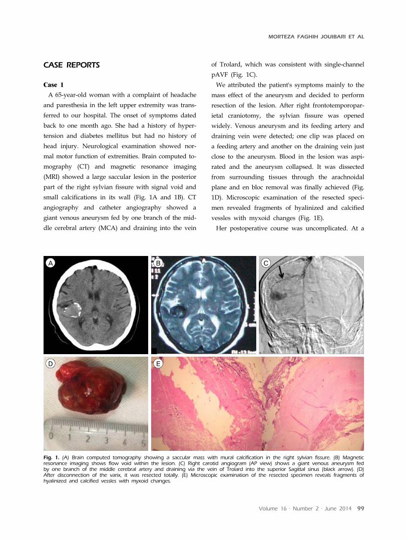
Fig. 1. (A) Brain computed tomography showing a saccular mass with mural calcification in the right sylvian fissure. (B) Magnetic resonance imaging shows flow void within the lesion. (C) Right carotid angiogram (AP view) shows a giant venous aneurysm fed by one branch of the middle cerebral artery and draining via the vein of Trolard into the superior Sagittal sinus (black arrow). (D) After disconnection of the varix, it was resected totally. (E) Microscopic examination of the resected specimen reveals fragments of hyalinized and calcified vessles with myxoid changes.
CASE REPORTS
Case 1
A 65-year-old woman with a complaint of headache
and paresthesia in the left upper extremity was trans-
ferred to our hospital. The onset of symptoms dated
back to one month ago. She had a history of hyper-
tension and diabetes mellitus but had no history of
head injury. Neurological examination showed nor-
mal motor function of extremities. Brain computed to-
mography (CT) and magnetic resonance imaging
(MRI) showed a large saccular lesion in the posterior
part of the right sylvian fissure with signal void and
small calcifications in its wall (Fig. 1A and 1B). CT
angiography and catheter angiography showed a
giant venous aneurysm fed by one branch of the mid-
dle cerebral artery (MCA) and draining into the vein
of Trolard, which was consistent with single-channel
pAVF (Fig. 1C).
We attributed the patient's symptoms mainly to the
mass effect of the aneurysm and decided to perform
resection of the lesion. After right frontotemporopar-
ietal craniotomy, the sylvian fissure was opened
widely. Venous aneurysm and its feeding artery and
draining vein were detected; one clip was placed on
a feeding artery and another on the draining vein just
close to the aneurysm. Blood in the lesion was aspi-
rated and the aneurysm collapsed. It was dissected
from surrounding tissues through the arachnoidal
plane and en bloc removal was finally achieved (Fig.
1D). Microscopic examination of the resected speci-
men revealed fragments of hyalinized and calcified
vessles with myxoid changes (Fig. 1E).
Her postoperative course was uncomplicated. At a
PIAL ARTERIOVENOUS FISTULA: CASE REPORT
100 J Cerebrovasc Endovasc Neurosurg
A
B
C
D
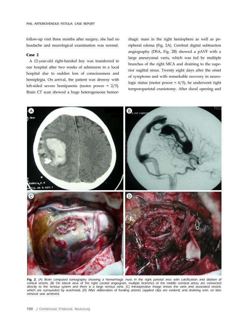
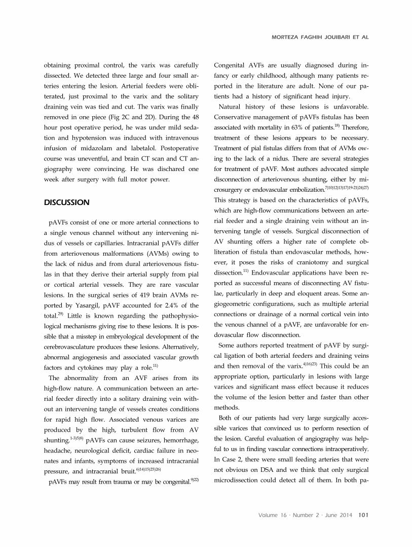
Fig. 2. (A) Brain computed tomography showing a hemorrhagic mass in the right parietal area with calcification and dilation of cortical vessels. (B) On lateral view of the right carotid angiogram, multiple branches of the middle cerebral artery are connected directly to the venous system and there is a large venous varix. (C) Intraoperative image shows the varix and associated vessels, which are surrounded by arachnoid. (D) After obliteration of feeding arteries (applied clips are evident) and draining vein, en bloc removal was achieved.
follow-up visit three months after surgery, she had no
headache and neurological examination was normal.
Case 2
A 12-year-old right-handed boy was transferred to
our hospital after two weeks of admission in a local
hospital due to sudden loss of consciousness and
hemiplegia. On arrival, the patient was drowsy with
left-sided severe hemiparesis (motor power = 2/5).
Brain CT scan showed a huge heterogeneous hemor-
rhagic mass in the right hemisphere as well as pe-
ripheral edema (Fig. 2A). Cerebral digital subtraction
angiography (DSA, Fig. 2B) showed a pAVF with a
large aneurysmal varix, which was fed by multiple
branches of the right MCA and draining to the supe-
rior sagittal sinus. Twenty eight days after the onset
of symptoms and with remarkable recovery in neuro-
logic status (motor power = 4/5), he underwent right
temporoparietal craniotomy. After dural opening and
MORTEZA FAGHIH JOUIBARI ET AL
Volume 16 · Number 2 · June 2014 101
obtaining proximal control, the varix was carefully
dissected. We detected three large and four small ar-
teries entering the lesion. Arterial feeders were obli-
terated, just proximal to the varix and the solitary
draining vein was tied and cut. The varix was finally
removed in one piece (Fig 2C and 2D). During the 48
hour post operative period, he was under mild seda-
tion and hypotension was induced with intravenous
infusion of midazolam and labetalol. Postoperative
course was uneventful, and brain CT scan and CT an-
giography were convincing. He was dischared one
week after surgery with full motor power.
DISCUSSION
pAVFs consist of one or more arterial connections to
a single venous channel without any intervening ni-
dus of vessels or capillaries. Intracranial pAVFs differ
from arteriovenous malformations (AVMs) owing to
the lack of nidus and from dural arteriovenous fistu-
las in that they derive their arterial supply from pial
or cortical arterial vessels. They are rare vascular
lesions. In the surgical series of 419 brain AVMs re-
ported by Yasargil, pAVF accounted for 2.4% of the
total.29) Little is known regarding the pathophysio-
logical mechanisms giving rise to these lesions. It is pos-
sible that a misstep in embryological development of the
cerebrovasculature produces these lesions. Alternatively,
abnormal angiogenesis and associated vascular growth
factors and cytokines may play a role.11)
The abnormality from an AVF arises from its
high-flow nature. A communication between an arte-
rial feeder directly into a solitary draining vein with-
out an intervening tangle of vessels creates conditions
for rapid high flow. Associated venous varices are
produced by the high, turbulent flow from AV
shunting.1-3)5)8) pAVFs can cause seizures, hemorrhage,
headache, neurological deficit, cardiac failure in neo-
nates and infants, symptoms of increased intracranial
pressure, and intracranial bruit.6)14)15)25)26)
pAVFs may result from trauma or may be congenital.9)22)
Congenital AVFs are usually diagnosed during in-
fancy or early childhood, although many patients re-
ported in the literature are adult. None of our pa-
tients had a history of significant head injury.
Natural history of these lesions is unfavorable.
Conservative management of pAVFs fistulas has been
associated with mortality in 63% of patients.18) Therefore,
treatment of these lesions appears to be necessary.
Treatment of pial fistulas differs from that of AVMs ow-
ing to the lack of a nidus. There are several strategies
for treatment of pAVF. Most authors advocated simple
disconnection of arteriovenous shunting, either by mi-
crosurgery or endovascular embolization.7)10)12)13)17)19-21)24)27)
This strategy is based on the characteristics of pAVFs,
which are high-flow communications between an arte-
rial feeder and a single draining vein without an in-
tervening tangle of vessels. Surgical disconnection of
AV shunting offers a higher rate of complete ob-
literation of fistula than endovascular methods, how-
ever, it poses the risks of craniotomy and surgical
dissection.11) Endovascular applications have been re-
ported as successful means of disconnecting AV fistu-
lae, particularly in deep and eloquent areas. Some an-
giogeometric configurations, such as multiple arterial
connections or drainage of a normal cortical vein into
the venous channel of a pAVF, are unfavorable for en-
dovascular flow disconnection.
Some authors reported treatment of pAVF by surgi-
cal ligation of both arterial feeders and draining veins
and then removal of the varix.4)16)23) This could be an
appropriate option, particularly in lesions with large
varices and significant mass effect because it reduces
the volume of the lesion better and faster than other
methods.
Both of our patients had very large surgically acces-
sible varices that convinced us to perform resection of
the lesion. Careful evaluation of angiography was help-
ful to us in finding vascular connections intraoperatively.
In Case 2, there were small feeding arteries that were
not obvious on DSA and we think that only surgical
microdissection could detect all of them. In both pa-
PIAL ARTERIOVENOUS FISTULA: CASE REPORT
102 J Cerebrovasc Endovasc Neurosurg
tients, varix had thick walls and was surrounded by
arachnoid, which made its dissection from other tis-
sues possible. There was no new neurological deficit
and the postoperative course was uneventful. Therefore,
surgical ligation and resection of giant varix resulted
in successful treatment in our patients.
CONCLUSION
Surgical resection can be an option for treatment of
pAVFs with large varices. In both of our patients, the
lesion could be dissected from surrounding tissues
and obliteration of feeding arteries and draining veins
did not cause new neurological deficit.
Disclosure
The authors report no conflict of interest concerning
this study or the findings specified in this paper.
REFERENCES
1. Almeida GM, Shibata MK. Hemispheric arteriovenous fistulae with giant venous dilation. Childs Nerv Syst. 1990 Jun;6(4):216-9.
2. Aoki N, Sakai T, Oikawa A. Intracranial arteriovenous fistula manifesting as progressive neurological deterio-ration in an infant: case report. Neurosurgery. 1991 Apr;28(4):619-22; discussion 622-3.
3. Barnwell SL, Ciricillo SF, Halbach VV, Edwards MS, Cogen PH. Intracerebral arteriovenous fistulas associated with intraparenchymal varix in childhood: case reports. Neurosurgery. 1990 Jan;26(1):122-5.
4. Bendok BR, Getch CC, Frederiksen J, Batjer HH. Resection of a large arteriovenous fistula of the brain using low-flow deep hypothermic cardiopulmonary by-pass: technical case report. Neurosurgery. 1999 Apr;44(4):888-90; discussion 890-1.
5. Carrillo R, Carreira LM, Prada J, Rosas C, Egas G. Giant aneurysm arising from a single arteriovenous fistula in a child. Case report. J Neurosurg. 1984 May;60(5):1085-8.
6. Garcia-Monaco R, de Victor D, Mann C, Hannedouche A, Terbrugge K, Lasjaunias P. Congestive cardiac mani-festations from cerebrocranial arteriovenous shunts. Endovascular management in 30 children. Childs Nerv Syst. 1991 Feb;7(1):48-52.
7. Garcia-Monaco R, Taylor W, Rodesch G, Alvarez H, Burrows P, Coubes P, et al. Pial arteriovenous fistula in children as presenting manifestation of Rendu-Osler-Weber disease. Neuroradiology. 1995 Jan;37(1):60-4.
8. Giller CA, Batjer HH, Purdy P, Walker B, Mathews D.
Interdisciplinary evaluation of cerebral hemodynamics in the treatment of arteriovenous fistulae associated with giant varices. Neurosurgery. 1994 Oct;35(4):778-82; dis-cussion 782-4.
9. Halbach VV, Higashida RT, Hieshima GB, Hardin CW, Dowd CF, Barnwell SL. Transarterial occlusion of soli-tary intracerebral arteriovenous fistulas. AJNR Am J Neuroradiol. 1989 Jul-Aug;10(4):747-52.
10. Hermier M, Turjman F, Bozio A, Duquesnel J, Lapras C. Endovascular treatment of an infantile nongalenic cerebral arteriovenous fistula with cyanoacrylate. Childs Nerv Syst. 1995 Aug;11(8):494-8.
11. Hoh BL, Putman CM, Budzik RF, Ogilvy CS. Surgical and endovascular flow disconnection of intracranial pial single-channel arteriovenous fistulae. Neurosurgery. 2001 Dec;49(6):1351-63; discussion 1363-4.
12. Kikuchi K, Kowada M, Sasajima H. Vascular malforma-tions of the brain in hereditary hemorrhagic telangiectasia (Rendu-Osler-Weber disease). Surg Neurol. 1994 May;41(5) :374-80.
13. Lee JS, Oh CW, Bang JS, Kwon OK, Hwang G. Intracranial pial arteriovenous fistula presenting with hemorrhage: a case report. J Cerebrovasc Endovasc Neurosurg. 2012 Dec;14(4):305-8.
14. Lownie S, Duckwiler G, Fox A, Drake C. Endovascular therapy of nongalenic cerebral arteriovenous fistulas, in Viñuela F, Halbach V, Dion J (eds). Interventional neu-roradiology: endovascular therapy of the central nervous system. New York: Raven Press, 1992. p. 87-106.
15. Lv X, Li Y, Jiang C, Wu Z. Endovascular treatment of brain arteriovenous fistulas. AJNR Am J Neuroradiol. 2009 Apr;30(4):851-6.
16. Meyer FB, Grady RE, Abel MD, Nichols DA, Caminha SS, Robb RA, et al. Resection of a large tempor-ooccipital parenchymal arteriovenous fistula by using deep hypothermic circulatory bypass. Case report. J Neurosurg. 1997 Dec;87(6):934-9.
17. Morimoto T, Yamada T, Hashimoto H, Tokunaga H, Tsunoda S, Sakaki T. Direct approach to intracranial vertebral arteriovenous fistula. Case report. Acta Neurochir (Wien). 1995;137(1-2):98-101.
18. Nelson K, Nimi Y, Lasjaunias P, Berenstein A. Endovascular embolization of congenital intracranial pial arteriovenous fistulas. Neuroimaging Clin N Am. 1992;2:309-17.
19. Oh HJ, Yoon SM, Kim SH, Shim JJ. A case of pial arte-riovenous fistula with giant venous aneurysm and mul-tiple varices treated with coil embolization. J Korean Neurosurg Soc. 2011 Sep;50(3):248-51.
20. Passacantilli E, Pichierri A, Guidetti G, Santoro A, Delfini R. Surgical treatment of pial cerebellar arterio-venous fistulas with aneurysm of the main feeding artery. Surg Neurol. 2006 Jan;65(1):90-4.
21. Ratliff J, Voorhies RM. Arteriovenous fistula with asso-ciated aneurysms coexisting with dural arteriovenous mal-formation of the anterior inferior falx. Case report and re-view of the literature. J Neurosurg. 1999 Aug;91(2):303-7.
22. Santosh C, Teasdale E, Molyneux A. Spontaneous clo-sure of an intracranial middle cerebral arteriovenous fistula. Neuroradiology. 1991;33(1):65-6.
MORTEZA FAGHIH JOUIBARI ET AL
Volume 16 · Number 2 · June 2014 103
23. Tabatabai SA, Zadeh MZ, Habibi Z, Meybodi AT, Hashemi M. Intracerebral atypical calcification in non-galenic pial arteriovenous fistula: a case report. Cases J. 2008 Nov;1(1):335.
24. Talamonti G, Versari PP, D'Aliberti G, Villa F, Fontana RA, Collice M. Complex arteriovenous fistula of the brain in an infant. Case report. J Neurosurg Sci. 1997 Dec;41(4):337-41.
25. Tomlinson FH, Rufenacht DA, Sundt TM Jr, Nichols DA, Fode NC. Arteriovenous fistulas of the brain and the spinal cord. J Neurosurg. 1993 Jul;79(1):16-27.
26. Vinuela F, Drake CG, Fox AJ, Pelz DM. Giant intra-cranial varices secondary to high-flow arteriovenous fistulae. J Neurosurg. 1987 Feb;66(2):198-203.
27. Yamashita K, Ohe N, Yoshimura S, Iwama T. Intracranial pial arteriovenous fistula. Neurol Med Chir (Tokyo). 2007 Dec;47(12):550-4.
28. Yang WH, Lu MS, Cheng YK, Wang TC. Pial arterio-venous fistula: a review of literature. Br J Neurosurg. Oct;25(5):580-5.
29. Yasargil MG. Microneurosurgery. Stuttgart. Georg Thieme Verlag, 1993.