case report a case of wegener s granulomatosis presenting
TRANSCRIPT
Case ReportA Case of Wegener’s GranulomatosisPresenting with Unilateral Facial Nerve Palsy
Roy Ujjawal,1 Pan Koushik,1 Panwar Ajay,2 and Chakrabarti Subrata3
1Department of Neurology, Bangur Institute of Neurosciences, IPGMER, Kolkata 700025, India2Department of Neurology, King George’s Medical University, Lucknow 226003, India3Department of Endocrinology, IPGMER, Kolkata 700020, India
Correspondence should be addressed to Roy Ujjawal; [email protected]
Received 20 September 2015; Revised 29 February 2016; Accepted 8 March 2016
Academic Editor: Gerald S. Supinski
Copyright © 2016 Roy Ujjawal et al. This is an open access article distributed under the Creative Commons Attribution License,which permits unrestricted use, distribution, and reproduction in any medium, provided the original work is properly cited.
Wegener’s granulomatosis or granulomatosis with polyangiitis is a necrotizing vasculitis affecting both arterioles and venules. Thedisease is characterized by the classical triad involving acute inflammation of the upper and lower respiratory tracts with renalinvolvement. However, the disease pathology can affect any organ system.This case presents Wegener’s granulomatosis presentingwith facial nerve palsy as the first manifestation of the disease, which is rarely reported in medical literature.
1. Introduction
Wegener’s granulomatosis (WG) is an idiopathic systemicform of vasculitis characterized by the presence of necrotiz-ing granulomas and vasculitis in the upper airways, lowerairways, and kidneys. Being a systemic disease, it mayadditionally involve any organ system. Etiology of this diseaseis proposed to be autoimmune [1, 2]. Incidence peaks betweenthe ages of 20 and 40 years [2].
2. Case Report
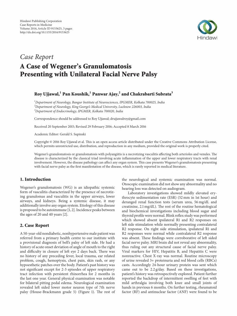
A50-year-old nondiabetic, nonhypertensivemale patientwasreferred from a primary health centre to our institute witha provisional diagnosis of bell’s palsy of left side. He had ahistory of acute onset deviation of angle of mouth to the rightand difficulty in closure of left eye 2 days back. There wasno history of any preceding fever, local trauma, ear relatedproblem, cough, hemoptysis, chest pain, skin rash, or anyhypoesthetic patches over the body. Patient’s past history wasnot significant except for 2-3 episodes of upper respiratorytract infection with persistent rhinorrhea for 2 months inthe last one year. General physical examination was notablefor bilateral pitting pedal edema. Neurological examinationrevealed left sided lower motor neuron type of 7th nervepalsy (House-Brackmann grade 5) (Figure 1). The rest of
the neurological and systemic examination was normal.Otoscopic examination did not show any abnormality and nohearing loss was detected on audiogram.
Laboratory investigations showed mildly elevated ery-throcyte sedimentation rate (ESR) (52mm in 1st hour) andderanged renal function tests (serum urea, 56mg/dL andcreatinine, 2.1mg/dL). The rest of the routine hematologicaland biochemical investigations including blood sugar andthyroid profilewere normal. Blink reflex studywas performedwhich showed absent ipsilateral R1 and R2 responses onleft side stimulation while normally presenting contralateralR2 response. On right side stimulation, ipsilateral R1 andR2 responses were normal while contralateral R2 responsewas absent. These findings were corroborative of left sidedfacial nerve palsy. MRI brain did not reveal any abnormality,thus ruling out any structural cause of facial nerve palsy.Viral markers for HIV, Hepatitis B, and Hepatitis C werenonreactive. Chest X-ray was normal. Routine microscopyof urine revealed 3+ proteinuria and red blood cells (RBCs)casts. Accordingly 24-hour urinary protein was sent whichcame out to be 2.2 g/day. Based on these investigations,patient’s history was retrospectively explored. Patient furtherreported the backdrop of intermittent swelling of feet withmild arthralgia involving both knee and small joints ofhands in previous 6 months. On further testing, rheumatoidfactor (RF) and antinuclear factor (ANF) were found to be
Hindawi Publishing CorporationCase Reports in MedicineVolume 2016, Article ID 9153625, 3 pageshttp://dx.doi.org/10.1155/2016/9153625
2 Case Reports in Medicine
Figure 1: Patient had a left sided lower motor neuron type facialpalsy.
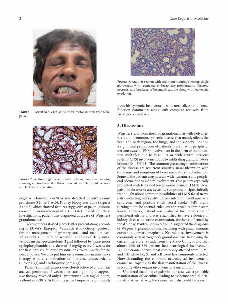
Figure 2: Section of glomerulus with methenamine silver stainingshowing circumferential cellular crescent with fibrinoid necrosisand leukocytic exudation.
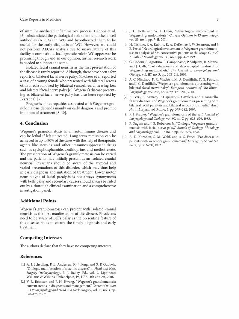
negative. However, c-ANCA was detected positive againstproteinase-3 (titre 1 : 640). Kidney biopsy was done (Figures2 and 3) which showed features suggestive of pauci-immunecrescentic glomerulonephritis (PICGN). Based on theseinvestigations, patient was diagnosed as a case of Wegener’sgranulomatosis.
Treatment was started (1 week after presentation) accord-ing to EUVAS (European Vasculitis Study Group) protocolfor the management of primary small and medium ves-sel vasculitis. Initially he received 3 pulses of daily intra-venous methyl-prednisolone (1 gm) followed by intravenouscyclophosphamide at a dose of 15mg/kg every 2 weeks forthe first 3 pulses, followed by infusions every 3 weeks for thenext 3 pulses. We also put him on a remission-maintenancetherapy with a combination of low-dose glucocorticoid(0.25mg/kg) and azathioprine (2mg/kg).
Patient’s clinical featureswere closely followed and a urineanalysis performed 15 weeks after starting immunosuppres-sive therapy revealed only 1+ proteinuria (260mg/24 hours)without anyRBCs. By this time patient improved significantly
Figure 3: Another section with trichrome staining showing singleglomerulus with segmental endocapillary proliferation, fibrinoidnecrosis, and breakage of bowman’s capsule along with leukocyticexudation.
from his systemic involvement with normalization of renalfunction parameters along with complete recovery fromfacial nerve paralysis.
3. Discussion
Wegener’s granulomatosis or granulomatosis with polyangi-itis is an uncommon, systemic disease that mainly affects thehead and neck region, the lungs, and the kidneys. Besides,a significant proportion of patients present with peripheralnervous system (PNS) involvement in the form of mononeu-ritis multiplex due to vasculitis or with central nervoussystem (CNS) involvement due to infiltrating granulomatouslesions (10–45%) [3].The common presentingmanifestationsof the disease are recurrent sinusitis, nasal ulceration withdischarge, and symptoms of lower respiratory tract infection.Some of the patients may present with hematuria and periph-eral edema due to kidney involvement. Our patient atypicallypresented with left sided lower motor neuron (LMN) facialpalsy. In absence of any systemic symptoms or signs, initiallywe thought about common possibilities of LMN facial nervepalsy including bell’s palsy, herpes infection, Guillain-Barresyndrome, and pontine small vessel stroke. MRI brain,turning out to be normal, ruled out the structural brain stemlesion. However, patient was evaluated further in view ofperipheral edema and was established to have evidence ofkidney disease on urine examination, further confirmed byrenal biopsy. Positive serum c-ANCA suggested the diagnosisof Wegener’s granulomatosis, featuring with pauci-immunecrescentic glomerulonephritis. Neurological involvement iscommonly seen in Wegener’s granulomatosis. Reviewing thecurrent literature, a study from the Mayo Clinic found thatalmost 30% of 324 patients had neurological involvement[4]. The cranial nerves most commonly affected were II, VI,and VII while IX, X, and XII were less commonly affected.Notwithstanding the common neurological involvement,cranial neuropathy as the first manifestation of the diseasepreceding other organs involvement is uncommon.
Unilateral facial nerve palsy in our case was a probablemanifestation of vasculitis leading to ischemic cranial neu-ropathy. Alternatively, the cranial neuritis could be a result
Case Reports in Medicine 3
of immune-mediated inflammatory process. Cadoni et al.[5] substantiated the pathological role of antiendothelial cellantibodies (AECAs) in WG and hypothesized them to beuseful for the early diagnosis of WG. However, we couldnot perform AECAs analysis due to unavailability of thisfacility at our institute. Its diagnostic role inWGappears to bepromising though and, in our opinion, further research workis needed to support the same.
Isolated facial cranial neuritis as the first presentation ofthe disease is rarely reported. Although, there have been a fewreports of bilateral facial nerve palsy. Nikolaou et al. reporteda case of a young female who presented with bilateral serousotitis media followed by bilateral sensorineural hearing lossand bilateral facial nerve palsy [6].Wegener’s disease present-ing as bilateral facial nerve palsy has also been reported byFerri et al. [7].
Prognosis of neuropathies associated withWegener’s gra-nulomatosis depends mainly on early diagnosis and promptinitiation of treatment [8–10].
4. Conclusion
Wegener’s granulomatosis is an autoimmune disease andcan be lethal if left untreated. Long term remission can beachieved in up to 90%of the cases with the help of therapeuticagents like steroids and other immunosuppressant drugssuch as cyclophosphamide, azathioprine, and methotrexate.The presentation of Wegener’s granulomatosis can be variedand the patients may initially present as an isolated cranialneuritis. Physicians should be aware of the atypical andvaried presentations of this disorder, which may thus helpin early diagnosis and initiation of treatment. Lower motorneuron type of facial paralysis is not always synonymouswith bell’s palsy and secondary causes should always be ruledout by a thorough clinical examination and a comprehensiveinvestigation panel.
Additional Points
Wegener’s granulomatosis can present with isolated cranialneuritis as the first manifestation of the disease. Physiciansneed to be aware of Bell’s palsy as the presenting feature ofthis disease, so as to ensure the timely diagnosis and earlytreatment.
Competing Interests
The authors declare that they have no competing interests.
References
[1] A. J. Scheuling, P. E. Andersen, K. J. Fong, and S. P. Gubbels,“Otologic manifestation of sistemic disease,” in Head and NeckSurgery-Otolaryngology, B. J. Bailey, Ed., vol. 2, LippincottWilliams &Wilkins, Philadelphia, Pa, USA, 4th edition, 2006.
[2] V. R. Erickson and P. H. Hwang, “Wegener’s granulomatosis:current trends in diagnosis and management,” Current Opinionin Otolaryngology and Head and Neck Surgery, vol. 15, no. 3, pp.170–176, 2007.
[3] J. U. Holle and W. L. Gross, “Neurological involvement inWegener’s granulomatosis,” Current Opinion in Rheumatology,vol. 23, no. 1, pp. 7–11, 2011.
[4] H. Nishino, F. A. Rubino, R. A. DeRemee, J. W. Swanson, and J.E. Parisi, “Neurological involvement inWegener’s granulomato-sis: an analysis of 324 consecutive patients at the Mayo Clinic,”Annals of Neurology, vol. 33, no. 1, pp. 4–9, 1993.
[5] G. Cadoni, S. Agostino, E. Campobasso, P. Vulpiani, R. Manna,and J. Galli, “Early diagnosis and stage-adapted treatment ofWegener’s granulomatosis,” The Journal of Laryngology andOtology, vol. 117, no. 3, pp. 208–211, 2003.
[6] A. C. Nikolaou, K. C. Vlachtsis, M. A. Daniilidis, D. G. Petridis,and I. C. Daniilidis, “Wegener’s granulomatosis presenting withbilateral facial nerve palsy,” European Archives of Oto-Rhino-Laryngology, vol. 258, no. 4, pp. 198–202, 2001.
[7] E. Ferri, E. Armato, P. Capuzzo, S. Cavaleri, and F. Ianniello,“Early diagnosis of Wegener’s granulomatosis presenting withbilateral facial paralysis and bilateral serous otitis media,” AurisNasus Larynx, vol. 34, no. 3, pp. 379–382, 2007.
[8] P. J. Bradley, “Wegener’s granulomatosis of the ear,” Journal ofLaryngology and Otology, vol. 97, no. 7, pp. 623–626, 1983.
[9] P. Dagum and J. B. Roberson Jr., “Otologic Wegener’s granulo-matosis with facial nerve palsy,” Annals of Otology, Rhinologyand Laryngology, vol. 107, no. 7, pp. 555–559, 1998.
[10] A. D. Kornblut, S. M. Wolff, and A. S. Fauci, “Ear disease inpatients with wegener’s granulomatosis,” Laryngoscope, vol. 92,no. 7, pp. 713–717, 1982.

Submit your manuscripts athttp://www.hindawi.com
Stem CellsInternational
Hindawi Publishing Corporationhttp://www.hindawi.com Volume 2014
Hindawi Publishing Corporationhttp://www.hindawi.com Volume 2014
MEDIATORSINFLAMMATION
of
Hindawi Publishing Corporationhttp://www.hindawi.com Volume 2014
Behavioural Neurology
EndocrinologyInternational Journal of
Hindawi Publishing Corporationhttp://www.hindawi.com Volume 2014
Hindawi Publishing Corporationhttp://www.hindawi.com Volume 2014
Disease Markers
Hindawi Publishing Corporationhttp://www.hindawi.com Volume 2014
BioMed Research International
OncologyJournal of
Hindawi Publishing Corporationhttp://www.hindawi.com Volume 2014
Hindawi Publishing Corporationhttp://www.hindawi.com Volume 2014
Oxidative Medicine and Cellular Longevity
Hindawi Publishing Corporationhttp://www.hindawi.com Volume 2014
PPAR Research
The Scientific World JournalHindawi Publishing Corporation http://www.hindawi.com Volume 2014
Immunology ResearchHindawi Publishing Corporationhttp://www.hindawi.com Volume 2014
Journal of
ObesityJournal of
Hindawi Publishing Corporationhttp://www.hindawi.com Volume 2014
Hindawi Publishing Corporationhttp://www.hindawi.com Volume 2014
Computational and Mathematical Methods in Medicine
OphthalmologyJournal of
Hindawi Publishing Corporationhttp://www.hindawi.com Volume 2014
Diabetes ResearchJournal of
Hindawi Publishing Corporationhttp://www.hindawi.com Volume 2014
Hindawi Publishing Corporationhttp://www.hindawi.com Volume 2014
Research and TreatmentAIDS
Hindawi Publishing Corporationhttp://www.hindawi.com Volume 2014
Gastroenterology Research and Practice
Hindawi Publishing Corporationhttp://www.hindawi.com Volume 2014
Parkinson’s Disease
Evidence-Based Complementary and Alternative Medicine
Volume 2014Hindawi Publishing Corporationhttp://www.hindawi.com