blood research march 2016 original article€¦ · · 2016-04-05blood res 2016;51:17-22....
TRANSCRIPT

This is an Open Access article distributed under the terms of the Creative Commons Attribution Non-Commercial License (http://creativecommons.org/licenses/by-nc/4.0)which permits unrestricted non-commercial use, distribution, and reproduction in any medium, provided the original work is properly cited.
BLOOD RESEARCH VOLUME 51ㆍNUMBER 1March 2016
ORIGINALARTICLE
A scientific treatment approach for acute mast cell leukemia: using a strategy based on next-generation sequencing data
Jeonghwan Youk1#, Youngil Koh1,2#, Ji-Won Kim3, Dae-Yoon Kim2, Hyunkyung Park1, Woo June Jung2, Kwang-Sung Ahn2, Hongseok Yun4, Inho Park4, Choong-Hyun Sun4, Seungmook Lee4, Sung-Soo Yoon1,2,5
1Department of Internal Medicine, Seoul National University Hospital, 2Cancer Research Institute, Seoul National University College of Medicine, 3Department of Internal Medicine, Seoul National University Bundang Hospital, Seongnam, 4Bioinformatics Group, Platform Development Center, CSP R&D, Samsung SDS, 5ClinicalResearch Institute, Seoul National University Hospital, Seoul, Korea
p-ISSN 2287-979X / e-ISSN 2288-0011http://dx.doi.org/10.5045/br.2016.51.1.17Blood Res 2016;51:17-22.
Received on January 29, 2016Revised on February 19, 2016Accepted on March 2, 2016
#These authors contributed equally to this work.
BackgroundMast cell leukemia (MCL) is the most aggressive form of systemic mastocytosis disorders. Owing to its rarity, neither pathogenesis nor standard treatment is established for this orphan disease. Hence, we tried to treat a patient with MCL based on the exome and tran-scriptome sequencing results of the patient’s own DNA and RNA.
MethodsFirst, tumor DNA and RNA were extracted from bone marrow at the time of diagnosis. Germline DNA was extracted from the patient’s saliva 45 days after induction chemo-therapy and used as a control. Then, we performed whole-exome sequencing (WES) using the DNA and whole transcriptome sequencing (WTS) using the RNA. Single nucleotide variants (SNVs) were called using MuTect and GATK. Samtools, FusionMap, and Gene Set Enrichment Analysis were utilized to analyze WTS results.
ResultsWES and WTS results revealed mutation in KIT S476I. Fusion analysis was performed using WTS data, which suggested a possible RAR-B2M fusion. When RNA expression analysis was performed using WTS data, upregulation of PIK3/AKT pathway, downstream of KIT and mTOR, was observed. Based on our WES and WTS results, we first administered all-trans retinoic acid, then dasatinib, and finally, an mTOR inhibitor.
ConclusionWe present a case of orphan disease where we used a targeted approach using WES and WTS data of the patient. Even though our treatment was not successful, use of our ap-proach warrants further validation.
Key Words Leukemia, Mast cell, C-kit, Individualized medicine
*This research was supported by a grant of Korea Health Technology R&D Project through the Korea Health Industry Development Institute (KHIDI), funded by the Ministry of Health & Welfare, Republic of Korea (grant number: HI14C0072) and grant 03-2014-0230 from the SNUH Research Fund.
Correspondence toSung-Soo Yoon, M.D., Ph.D.Department of Internal Medicine, Seoul National University Hospital, 101 Daehag-ro, Jongno-gu, Seoul 03080, KoreaE-mail: [email protected]
Ⓒ 2016 Korean Society of Hematology
INTRODUCTION
Mast cell leukemia (MCL) is the most aggressive form of systemic mastocytosis (SM). Common symptoms of MCL include flushes, fever, malaise, diarrhea, and tachycardia. Diagnosis of MCL requires that (i) SM criteria are fulfilled, and (ii) bone marrow (BM) atypical mast cells (MCs) comprise ≥20% of the total white blood cells (WBCs). MCL is com-posed of a leukemic variant (>10% in the peripheral blood) and an aleukemic variant (<10% in the peripheral blood). This orphan disease accounts for less than 1% of all SMs [1].
Owing to its rarity, the pathogenesis and standard treat-ment for MCL are not well established. According to Georgin-Lavialle et al., most patients with MCL (83%) show a normal karyotype in conventional cytogenetic exams. At the molecular level, mutations in the KIT gene have been well investigated in MCL patients, even before the next-gen-eration sequencing (NGS) era. A few therapeutic options for MCL are available, but no efficient treatment has been reproducibly validated. Thus, well-organized clinical trials should be primarily considered [1]. Without proper clinical trials, patients with mutations in KIT other than D816V or those with wild type KIT could be treated with an ABL

Blood Res 2016;51:17-22. bloodresearch.or.kr
18 Jeonghwan Youk, et al.
kinase inhibitor, such as imatinib. Unfortunately, ABL kinase inhibitors are not effective in patients with the KIT D816V mutation. Midostaurin, a multi-target protein kinase in-hibitor, can be administered to patients with MCL regardless of mutations in KIT. Allogeneic stem cell transplantation may be a potential curative option for MCL, but a retrospective study found that the response rate was low (three-year surviv-al rate=17% [2 of 12]) [2]. Traditionally, polychemotherapy, such as an AML-type induction regimen, has been used for cytoreductive therapy. Alternatively, steroids and interfer-on- can be considered as treatment options.
With the exception of the KIT gene, molecular study and targeted therapy have not been thoroughly evaluated. Therefore, we attempted to treat a refractory MCL patient based on whole exome sequencing (WES) and whole tran-scriptome sequencing (WTS) of the patient’s own DNA and RNA.
MATERIALS AND METHODS
An 18-year-old Korean female visited the Seoul National University Hospital with recurrent pain in the abdomen and both legs that lasted for 1 month. X-rays of the legs and an abdominal computed tomography (CT) scan were performed to determine the cause of the pain and revealed hepatomegaly with ascites and left inguinal lymphadenop-athy (largest diameter: 2.4 cm). Excisional inguinal node biopsy revealed dense infiltrates of atypical MCs with strong C-KIT expression; however, no MCs were detected in the peripheral blood (white blood cell count: 7,570/L; 74.9% neutrophils, 21.0% lymphocytes, 3.6% monocytes, 0.4% eosi-nophils, and 0.1% basophils). We found an increase in im-mature MCs (24.1%) with bi-lobed nuclei in a BM smear. Most of the MCs showed atypical morphology. No morpho-logical evidence of an associated hematopoietic non-mast cell lineage disease was found. The serum tryptase level was 425.0 g/L. Chromosomal analysis showed a normal karyotype (46, XX [20]). In addition, we did not observe the KIT D816V mutation. One major and two minor criteria of SM established by the WHO were fulfilled. With the presence of 20% MCs on the BM smear, the patient was diagnosed with the aleukemic variant of acute MCL [3]. Liver dysfuncion was identified as a C finding of the disorder.
We initiated treatment with cytarabine (100 mg/m2) for 7 days and idarubicin (12 mg/m2) for 3 days, but the follow-up BM smear revealed persistence of MCL (MCs: 5.5% of total nucleated cells). Because the patient was reluctant to undergo allogeneic stem cell transplantation, we performed WES and WTS to find druggable mutations or activated signaling pathways.
Next-generation sequencingBM blasts were acquired at diagnosis, and epithelial cells
from saliva were obtained after induction chemotherapy. We used genomic DNA purification kits (Norgen Biotek Corp, Thorold, ON, Canada) to isolate the DNA. Quality
was monitored by the NGS QC Toolkit (National Institute of Plant Genome Research, New Delhi, India). DNA was then fragmented for massively parallel sequencing via the HiSeq 2000 system (Illumina Inc., San Diego, CA, USA) according to the manufacturer’s instructions. We used the SureSelect Human All Exon Kit (Agilent Technologies Inc., Santa Clara, CA, USA) for DNA capture. FASTQ files were aligned to the UCSC human reference genome (build hg19) using the Burrows-Wheeler Aligner (bwa-0.7.5a) [4] to gen-erate a sequence alignment/map file. Next, the Genome Analysis Toolkit (GATK; Broad Institute, Cambridge, MA, USA) was used for local alignment [5]. Single nucleotide variants (SNVs) and small insertions and deletions (indels) were identified using GATK. SNVs were also identified using MuTect software (Broad Institute) [6]. CONTRA was used to determine copy number variations [7].
For RNA preparation, total RNA quality was assessed using the NanoDrop1000 spectrometer (Thermo Scientific, Wilmington, DE, USA). We used the TruSeq RNA library preparation kit for RNA isolation (Illumina, San Diego, USA). In brief, messenger RNA (mRNA) was purified using polyA selection, then chemically fragmented and converted into single-stranded cDNA using random hexamer priming. Next, the complimentary strand was generated to create dou-ble-stranded cDNA (ds-cDNA) that could be used for TruSeq library construction. The short ds-cDNA fragments were then connected with sequencing adapters, and suitable frag-ments were separated by agarose gel electrophoresis. Finally, TruSeq RNA libraries were built by PCR amplification, quan-tified using qPCR according to the qPCR Quantification Protocol Guide, qualified using the Agilent Technologies 2100 Bioanalyzer (Agilent Technologies, Palo Alto CA, USA), and then sequenced using the HiSe 2000 platform (Illumina, San Diego, USA). The sequencing data were aligned to gene-code v18 by Tophat v1.0.12 (Tophat, genecode reference). Raw counts of mRNA were quantified using Samtools [8], and corresponding reads per kilobase per million reads (RPKM) were calculated using an in-house R script. We used RPKM value to determine differentially expressed genes. Fusion genes were analyzed by FusionMap [9]. To find upregulated intracellular signaling pathways, we used the Gene Set Enrichment Analysis (http://www.broadin-stitute.org/gsea/msigdb/annotate.jsp, Broad Institute, MA).
RESULTS
WES analysis yielded a total of 226,007,918 mapped reads (BM DNA: 103,545,760; salivary epithelial DNA: 122,462,158). The mean read depth of the neoplasm was more than 100-fold. WES analysis using GATK failed to demonstrate noticeable nonsynonymous SNVs or small indels, whereas MuTect detected five SNVs, including KIT S476I (Table 1). Druggable genes associated with copy number changes were also not found. WTS analysis also found the KIT S476I muta-tion (Table 2). The possibility of RAR-B2M and RAR- ACTB fusion have been noted. When performing RNA ex-
bloodresearch.or.kr Blood Res 2016;51:17-22.
NGS in mast cell leukemia 19
Table 1. Single-nucleotide variations detected by whole-exome sequencing.
Gene Position Reference Reference depth Alternative Somatic depth Amino acid change
POTEE 2:132021766 A 42 C 4 K913TKIT 4:55592103 G 131 T 6 S476IMSH3 5:79950736 C 20 G 3 P64ARRP7A 22:42910199 G 17 A 3 R224WFAM3A X:153735762 C 68 T 4 A149T
Table 2. Single nucleotide variations and indels detected by whole transcriptome sequencing. Only the genes with a variant allele frequency (Vaf) of ≥0.30 are included.
Gene Position Reference Alternative Total depth Total Vaf Amino acid change
FMNL1 17:43318777 GGC GCG 1,125 0.9724 GP454GAKIT 4:55592103 G T 44 0.5909 S476IAGBL5 2:27276292 C T 21 0.5714 R80WIPO4 14:24649767 C T 58 0.5172 A1043TCLIP1 12:122794329 C T 49 0.4898 E1192KXAB2 19:7685290 G A 209 0.4545 R713WKDM5B 1:202705466 G A 29 0.4483 R1047CYP51A1 7:91743154 C T 45 0.4222 R452HIL7R 5:35874575 C T 149 0.4027 T244I
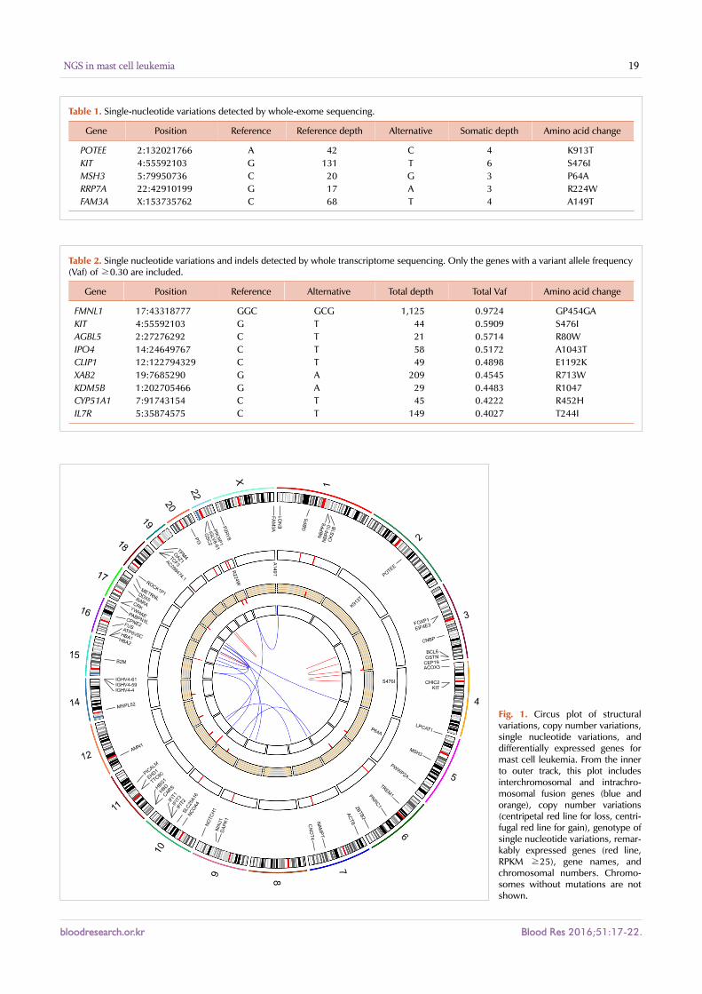
Fig. 1. Circus plot of structural variations, copy number variations, single nucleotide variations, and differentially expressed genes for mast cell leukemia. From the inner to outer track, this plot includes interchromosomal and intrachro-mosomal fusion genes (blue and orange), copy number variations (centripetal red line for loss, centri-fugal red line for gain), genotype of single nucleotide variations, remar-kably expressed genes (red line, RPKM ≥25), gene names, and chromosomal numbers. Chromo-somes without mutations are not shown.
Blood Res 2016;51:17-22. bloodresearch.or.kr
20 Jeonghwan Youk, et al.
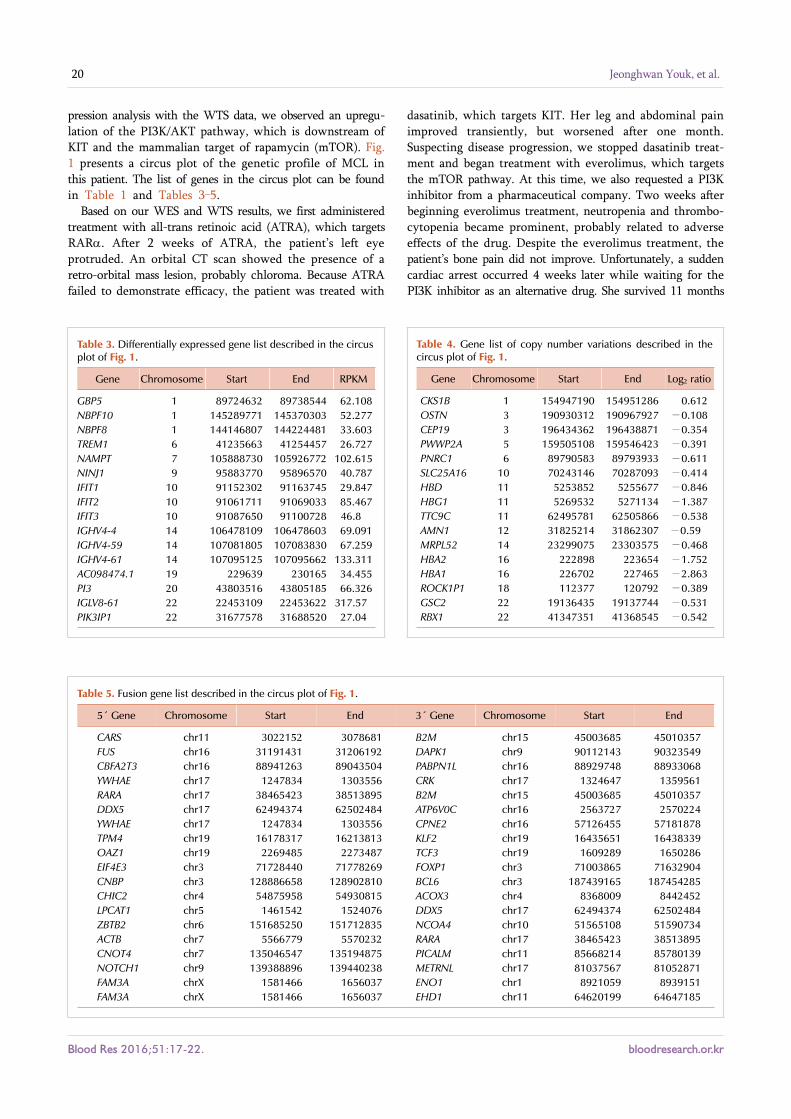
Table 3. Differentially expressed gene list described in the circus plot of Fig. 1.
Gene Chromosome Start End RPKM
GBP5 1 89724632 89738544 62.108NBPF10 1 145289771 145370303 52.277NBPF8 1 144146807 144224481 33.603TREM1 6 41235663 41254457 26.727NAMPT 7 105888730 105926772 102.615NINJ1 9 95883770 95896570 40.787IFIT1 10 91152302 91163745 29.847IFIT2 10 91061711 91069033 85.467IFIT3 10 91087650 91100728 46.8IGHV4-4 14 106478109 106478603 69.091IGHV4-59 14 107081805 107083830 67.259IGHV4-61 14 107095125 107095662 133.311AC098474.1 19 229639 230165 34.455PI3 20 43803516 43805185 66.326IGLV8-61 22 22453109 22453622 317.57PIK3IP1 22 31677578 31688520 27.04
Table 5. Fusion gene list described in the circus plot of Fig. 1.
5´ Gene Chromosome Start End 3´ Gene Chromosome Start End
CARS chr11 3022152 3078681 B2M chr15 45003685 45010357FUS chr16 31191431 31206192 DAPK1 chr9 90112143 90323549CBFA2T3 chr16 88941263 89043504 PABPN1L chr16 88929748 88933068YWHAE chr17 1247834 1303556 CRK chr17 1324647 1359561RARA chr17 38465423 38513895 B2M chr15 45003685 45010357DDX5 chr17 62494374 62502484 ATP6V0C chr16 2563727 2570224YWHAE chr17 1247834 1303556 CPNE2 chr16 57126455 57181878TPM4 chr19 16178317 16213813 KLF2 chr19 16435651 16438339OAZ1 chr19 2269485 2273487 TCF3 chr19 1609289 1650286EIF4E3 chr3 71728440 71778269 FOXP1 chr3 71003865 71632904CNBP chr3 128886658 128902810 BCL6 chr3 187439165 187454285CHIC2 chr4 54875958 54930815 ACOX3 chr4 8368009 8442452LPCAT1 chr5 1461542 1524076 DDX5 chr17 62494374 62502484ZBTB2 chr6 151685250 151712835 NCOA4 chr10 51565108 51590734ACTB chr7 5566779 5570232 RARA chr17 38465423 38513895CNOT4 chr7 135046547 135194875 PICALM chr11 85668214 85780139NOTCH1 chr9 139388896 139440238 METRNL chr17 81037567 81052871FAM3A chrX 1581466 1656037 ENO1 chr1 8921059 8939151FAM3A chrX 1581466 1656037 EHD1 chr11 64620199 64647185
Table 4. Gene list of copy number variations described in the circus plot of Fig. 1.
Gene Chromosome Start End Log2 ratio
CKS1B 1 154947190 154951286 0.612OSTN 3 190930312 190967927 −0.108CEP19 3 196434362 196438871 −0.354PWWP2A 5 159505108 159546423 −0.391PNRC1 6 89790583 89793933 −0.611SLC25A16 10 70243146 70287093 −0.414HBD 11 5253852 5255677 −0.846HBG1 11 5269532 5271134 −1.387TTC9C 11 62495781 62505866 −0.538AMN1 12 31825214 31862307 −0.59MRPL52 14 23299075 23303575 −0.468HBA2 16 222898 223654 −1.752HBA1 16 226702 227465 −2.863ROCK1P1 18 112377 120792 −0.389GSC2 22 19136435 19137744 −0.531RBX1 22 41347351 41368545 −0.542
pression analysis with the WTS data, we observed an upregu-lation of the PI3K/AKT pathway, which is downstream of KIT and the mammalian target of rapamycin (mTOR). Fig. 1 presents a circus plot of the genetic profile of MCL in this patient. The list of genes in the circus plot can be found in Table 1 and Tables 3–5.
Based on our WES and WTS results, we first administered treatment with all-trans retinoic acid (ATRA), which targets RAR. After 2 weeks of ATRA, the patient’s left eye protruded. An orbital CT scan showed the presence of a retro-orbital mass lesion, probably chloroma. Because ATRA failed to demonstrate efficacy, the patient was treated with
dasatinib, which targets KIT. Her leg and abdominal pain improved transiently, but worsened after one month. Suspecting disease progression, we stopped dasatinib treat-ment and began treatment with everolimus, which targets the mTOR pathway. At this time, we also requested a PI3K inhibitor from a pharmaceutical company. Two weeks after beginning everolimus treatment, neutropenia and thrombo-cytopenia became prominent, probably related to adverse effects of the drug. Despite the everolimus treatment, the patient’s bone pain did not improve. Unfortunately, a sudden cardiac arrest occurred 4 weeks later while waiting for the PI3K inhibitor as an alternative drug. She survived 11 months

bloodresearch.or.kr Blood Res 2016;51:17-22.
NGS in mast cell leukemia 21
after the diagnosis of MCL.
DISCUSSION
Before our current study, only one WES study in a patient with MCL patient had been published in 2012 [10]. This study detected a point mutation in IgE mast-cell receptor chain and KIT, but the treatment approach was not reported. With the exception of that WES study, most other MCL studies have focused on KIT mutations. KIT D816V mutation was detected in more than 90% of patients with an SM. Almost 40% of de novo patients with MCL had the KIT D816V mutation [1]. In addition, S476I, F522C, V654A, V560G, duplication of amino acids 501–502 and 502–503, and deletion of amino acids 501–502 were also reported in a case report [10-15]. Besides the KIT mutation, a TET2 mutation has been investigated in aggressive SM [16]. Few patients with MCL had a 5q deletion, but all of those patients had secondary MCL that evolved from myelodysplastic syn-drome [17].
Notably, the KIT mutation is the most important and prevalent pro-oncogenic mutation in MCL. In this study, we found the presence of a KIT S476I mutation, which has been discovered previously in chronic MCL [15]. With our finding, this point mutation becomes the second most common point mutation in MCL, following KIT D816V, which is located in the tyrosine kinase domain. KIT S476I, on the other hand, is located in the immunoglobulin-like extracellular domain, the function of which is not yet known [18]. A small portion of patients with MCL show improve-ment upon treatment with KIT inhibitors, such as imatinib or dasatinib, but dasatinib was not effective in our patient. Because a few reports showed promising results of PKC412 (midostaurin) for aggressive SM and MCL, we considered administering PKC412 to the patient [19-21]. However, the drug was not available at that time in Korea. Recently, se-lective inhibitors targeting specific KIT mutations, such as KIT D816V, have been studied [22], and selective drugs that block KIT S476I could be beneficial to a proportion of MCL patients. Because no selective KIT inhibitor for S476I is approved currently, we blocked pathways downstream of KIT. One of the downstream pathways is the PI3K/ AKT/mTOR pathway, which was upregulated in this MCL case. However, treatment with an mTOR inhibitor regret-tably was not effective in this patient.
Several plausible explanations could account for the treat-ment failure. One explanation would be targeted therapy alone. In patients with Philadelphia chromosome-positive acute lymphoblastic leukemia (ALL), BCR-ABL tyrosine kin-ase inhibitor (TKI) was used as an induction treatment com-bined with cytotoxic multiagent chemotherapy, although BCR-ABL TKI alone was very effective in chronic myeloid leukemia [23]. Like ALL, acute MCL, one form of acute leukemia, might be too aggressive to treat with TKI alone. Another explanation could be that KIT S476I, the PI3K/ AKT/mTOR pathway, or RAR fusion protein was not a
main driver mutation or pathway in this MCL pathogenesis. Although the above variations were detected in sequencing data, our analysis did not exclude the possibility that they were only passenger mutations. Finally, it is possible that the drug dosage level was insufficient, given that the optimal dosage of TKI agents has not been determined for acute MCL.
In conclusion, we presented a case study of a patient with an orphan disease in which we used a targeted approach to therapy with WES and WTS data from the patient. Although this approach did not successfully cure the disease, she survived 11 months, approximately twice as long as the median survival of patients with acute MCL. The results of our treatments were not ideal, but the utility of this type of approach should be further researched and validated in the future.
AuthorsÊ Disclosures of Potential Conflicts of Interest
No potential conflicts of interest relevant to this article were reported.
REFERENCES
1. Georgin-Lavialle S, Lhermitte L, Dubreuil P, Chandesris MO,
Hermine O, Damaj G. Mast cell leukemia. Blood 2013;121:1285-95.
2. Ustun C, Reiter A, Scott BL, et al. Hematopoietic stem-cell
transplantation for advanced systemic mastocytosis. J Clin Oncol
2014;32:3264-74.
3. Valent P, Sotlar K, Sperr WR, et al. Refined diagnostic criteria and
classification of mast cell leukemia (MCL) and myelomastocytic
leukemia (MML): a consensus proposal. Ann Oncol 2014;25:1691-
700.
4. Li H, Durbin R. Fast and accurate short read alignment with
Burrows-Wheeler transform. Bioinformatics 2009;25:1754-60.
5. McKenna A, Hanna M, Banks E, et al. The Genome Analysis
Toolkit: a MapReduce framework for analyzing next-generation
DNA sequencing data. Genome Res 2010;20:1297-303.
6. Cibulskis K, Lawrence MS, Carter SL, et al. Sensitive detection of
somatic point mutations in impure and heterogeneous cancer
samples. Nat Biotechnol 2013;31:213-9.
7. Li J, Lupat R, Amarasinghe KC, et al. CONTRA: copy number
analysis for targeted resequencing. Bioinformatics 2012;28:1307-13.
8. Li H, Handsaker B, Wysoker A, et al. The Sequence Alignment/
Map format and SAMtools. Bioinformatics 2009;25:2078-9.
9. Ge H, Liu K, Juan T, Fang F, Newman M, Hoeck W. FusionMap:
detecting fusion genes from next-generation sequencing data at
base-pair resolution. Bioinformatics 2011;27:1922-8.
10. Spector MS, Iossifov I, Kritharis A, et al. Mast-cell leukemia exome
sequencing reveals a mutation in the IgE mast-cell receptor
chain and KIT V654A. Leukemia 2012;26:1422-5.
11. Akin C, Metcalfe DD. Systemic mastocytosis. Annu Rev Med
2004;55:419-32.
12. Georgin-Lavialle S, Aguilar C, Guieze R, et al. Mast cell sarcoma:
a rare and aggressive entity-report of two cases and review of the
Blood Res 2016;51:17-22. bloodresearch.or.kr
22 Jeonghwan Youk, et al.
literature. J Clin Oncol 2013;31:e90-7.
13. Mital A, Piskorz A, Lewandowski K, Wasąg B, Limon J, Hellmann
A. A case of mast cell leukaemia with exon 9 KIT mutation and
good response to imatinib. Eur J Haematol 2011;86:531-5.
14. Georgin-Lavialle S, Lhermitte L, Suarez F, et al. Mast cell
leukemia: identification of a new c-Kit mutation, dup(501-502),
and response to masitinib, a c-Kit tyrosine kinase inhibitor. Eur
J Haematol 2012;89:47-52.
15. Valent P, Berger J, Cerny-Reiterer S, et al. Chronic mast cell
leukemia (MCL) with KIT S476I: a rare entity defined by leukemic
expansion of mature mast cells and absence of organ damage. Ann
Hematol 2015;94:223-31.
16. Soucie E, Hanssens K, Mercher T, et al. In aggressive forms of
mastocytosis, TET2 loss cooperates with c-KITD816V to
transform mast cells. Blood 2012;120:4846-9.
17. Valentini CG, Rondoni M, Pogliani EM, et al. Mast cell leukemia:
a report of ten cases. Ann Hematol 2008;87:505-8.
18. Lennartsson J, Rönnstrand L. Stem cell factor receptor/c-Kit: from
basic science to clinical implications. Physiol Rev 2012;92:1619-
49.
19. Gotlib J, Berubé C, Growney JD, et al. Activity of the tyrosine
kinase inhibitor PKC412 in a patient with mast cell leukemia with
the D816V KIT mutation. Blood 2005;106:2865-70.
20. Growney JD, Clark JJ, Adelsperger J, et al. Activation mutations
of human c-KIT resistant to imatinib mesylate are sensitive to the
tyrosine kinase inhibitor PKC412. Blood 2005;106:721-4.
21. Quintas-Cardama A, Aribi A, Cortes J, Giles FJ, Kantarjian H,
Verstovsek S. Novel approaches in the treatment of systemic
mastocytosis. Cancer 2006;107:1429-39.
22. Lee S, Lee H, Kim J, et al. Development and biological evaluation
of potent and selective c-KIT(D816V) inhibitors. J Med Chem
2014;57:6428-43.
23. Kim DY, Joo YD, Lim SN, et al. Nilotinib combined with multiagent
chemotherapy for newly diagnosed Philadelphia-positive acute
lymphoblastic leukemia. Blood 2015;126:746-56.