diffuse large b cell lymphoma with high m protein: an ... · blood res 2015;50:54-65....
TRANSCRIPT
bloodresearch.or.kr Blood Res 2015;50:54-65.
Letters to the Editor 61
5. Park TS, Song J, Kim JS, et al. 8p11 myeloproliferative syndrome
preceded by t(8;9)(p11;q33), CEP110/FGFR1 fusion transcript:
morphologic, molecular, and cytogenetic characterization of
myeloid neoplasms associated with eosinophilia and FGFR1
abnormality. Cancer Genet Cytogenet 2008;181:93-9.
6. Kim M, Lim J, Lee A, et al. A case of chronic myelomonocytic leu-
kemia with severe eosinophilia having t(5;12)(q31;p13) with
t(1;7)(q10;p10). Acta Haematol 2005;114:104-7.
7. Lee H, Kim M, Lim J, et al. Acute myeloid leukemia associated
with FGFR1 abnormalities. Int J Hematol 2013;97:808-12.
8. Jang SE, Kang HJ, Chang YH, et al. A case of myeloid neoplasm
with the PDGFRB rearrangement and eosinophilia. Korean J
Med 2010;78:386-90.
9. Lim KS, Ko J, Lee SS, Shin B, Choi DC, Lee BJ. A case of idiopathic
hypereosinophilic syndrome presenting with acute respiratory
distress syndrome. Allergy Asthma Immunol Res 2014;6:98-101.
10. Ogbogu PU, Bochner BS, Butterfield JH, et al. Hypereosino-
philic syndrome: a multicenter, retrospective analysis of clinical
characteristics and response to therapy. J Allergy Clin Immunol
2009;124:1319-25.
Diffuse large B cell lymphoma with high M protein: an unusual finding
TO THE EDITOR: Paraprotein is an abnormal immunoglob-ulin (Ig) or part of an Ig in the blood or urine that is produced by a clonal population of B cells and plasma cells. Production of a monoclonal Ig paraprotein is associated with various types of B-cell non-Hodgkin’s lymphomas (NHLs). Paraproteinemia is associated with about 20% of patients with indolent types of NHL, whereas it appears to be rare in aggressive lymphomas [1]. Immunofixation (IFX) and conventional serum protein electrophoresis (SPEP) are useful tools to detect even low levels of monoclonal Igs. Herein, we report a case of diffuse large B cell lymphoma with a very high level of IgG kappa monoclonal gammop-athy, which was rarely reported in the literature [2].
CASEA 68-year-old man with a known case of rheumatoid
arthritis presented with upper gastrointestinal bleeding. On examination, he was found to have axillary lymphadenop-athy with splenomegaly. F-18 fluoro-D-glucose (FDG) posi-tron emission tomography showed FDG avid bilateral axil-lary, external iliac, and inguinal lymph nodes and splenome-galy with diffusely increased FDG uptake. Hematological analysis showed hemoglobin to be 7.7 g/L; total leucocyte count, 8.9×10⁹/L; and platelets, 80×109/L. Axillary lymph node biopsy showed sheets of large atypical lymphoid cells with irregular contours, brisk mitoses, and prominent nucle-oli as well as perinodal spread. According to immunohisto-
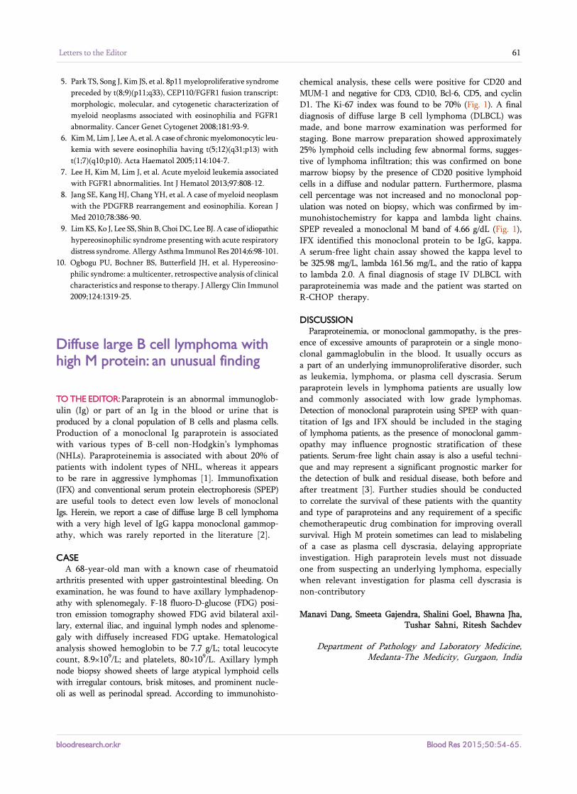
chemical analysis, these cells were positive for CD20 and MUM-1 and negative for CD3, CD10, Bcl-6, CD5, and cyclin D1. The Ki-67 index was found to be 70% (Fig. 1). A final diagnosis of diffuse large B cell lymphoma (DLBCL) was made, and bone marrow examination was performed for staging. Bone marrow preparation showed approximately 25% lymphoid cells including few abnormal forms, sugges-tive of lymphoma infiltration; this was confirmed on bone marrow biopsy by the presence of CD20 positive lymphoid cells in a diffuse and nodular pattern. Furthermore, plasma cell percentage was not increased and no monoclonal pop-ulation was noted on biopsy, which was confirmed by im-munohistochemistry for kappa and lambda light chains. SPEP revealed a monoclonal M band of 4.66 g/dL (Fig. 1), IFX identified this monoclonal protein to be IgG, kappa. A serum-free light chain assay showed the kappa level to be 325.98 mg/L, lambda 161.56 mg/L, and the ratio of kappa to lambda 2.0. A final diagnosis of stage IV DLBCL with paraproteinemia was made and the patient was started on R-CHOP therapy.
DISCUSSIONParaproteinemia, or monoclonal gammopathy, is the pres-
ence of excessive amounts of paraprotein or a single mono-clonal gammaglobulin in the blood. It usually occurs as a part of an underlying immunoproliferative disorder, such as leukemia, lymphoma, or plasma cell dyscrasia. Serum paraprotein levels in lymphoma patients are usually low and commonly associated with low grade lymphomas. Detection of monoclonal paraprotein using SPEP with quan-titation of Igs and IFX should be included in the staging of lymphoma patients, as the presence of monoclonal gamm-opathy may influence prognostic stratification of these patients. Serum-free light chain assay is also a useful techni-que and may represent a significant prognostic marker for the detection of bulk and residual disease, both before and after treatment [3]. Further studies should be conducted to correlate the survival of these patients with the quantity and type of paraproteins and any requirement of a specific chemotherapeutic drug combination for improving overall survival. High M protein sometimes can lead to mislabeling of a case as plasma cell dyscrasia, delaying appropriate investigation. High paraprotein levels must not dissuade one from suspecting an underlying lymphoma, especially when relevant investigation for plasma cell dyscrasia is non-contributory
Manavi Dang, Smeeta Gajendra, Shalini Goel, Bhawna Jha, Tushar Sahni, Ritesh Sachdev
Department of Pathology and Laboratory Medicine, Medanta-The Medicity, Gurgaon, India

Blood Res 2015;50:54-65. bloodresearch.or.kr
62 Letters to the Editor
Fig. 1. (A) Serum protein electro-phoresis showing an M band. (B)Lymph node biopsy showing large atypical lymphoid cells with irregular contours, brisk mitoses, and prominent nucleoli (hemato-xylin and eosin, ×400), which were positive for CD20, MUM-1, and Ki-67 (70%) and negative for CD138. (C) Bone marrow biopsy showed lymphoid cells in a diffuseand nodular pattern (hematoxylin and eosin, ×200), which were CD20 and kappa positive and lambda negative.
Correspondence to: Ritesh SachdevDepartment of Pathology and Laboratory Medicine,
Medanta-The Medicity, Sector-38, Gurgaon, Haryana 122 001, India
E-mail: [email protected]
Received on Aug. 8, 2014; Revised on Aug. 16, 2014; Accepted on Feb. 12, 2015
http://dx.doi.org/10.5045/br.2015.50.1.61
AuthorsÊ Disclosures of Potential Conflicts of InterestNo potential conflicts of interest relevant to this article
were reported.
REFERENCES1. Economopoulos T, Papageorgiou S, Pappa V, et al. Monoclonal
gammopathies in B-cell non-Hodgkin's lymphomas. Leuk Res
2003;27:505-8.
2. Chen M, Abedi M. Atypical lymphocytosis, cold agglutinin he-
molytic anemia, and monoclonal gammopathy in an HIV patient
with marrow involvement by diffuse large B-cell lymphoma.
Blood 2013;122:3711.
3. Charafeddine KM, Jabbour MN, Kadi RH, Daher RT. Extended
use of serum free light chain as a biomarker in lymphoprolifer-
ative disorders: a comprehensive review. Am J Clin Pathol
2012;137:890-7.