bilateral parotid maltoma: a sure shot for radiation - koreamed … · 2016-01-05 ·...
TRANSCRIPT
Blood Res 2015;50:254-67. bloodresearch.or.kr
262 Letters to the Editor
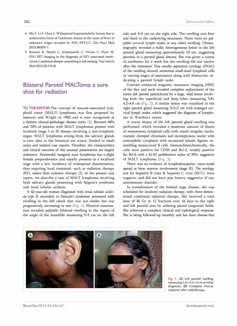
Fig. 1. (A) Left parotid swelling measuring 5.8×4.2×4 cm at initialdiagnosis. (B) Complete clinical response after radiotherapy.
4. Ma Y, Li F, Chen L. Widespread hypermetabolic lesions due to
multicentric form of Castleman disease as the cause of fever of
unknown origin revealed by FDG PET/CT. Clin Nucl Med
2013;38:835-7.
5. Rossotti R, Moioli C, Schiantarelli C, Orcese C, Puoti M.
FDG-PET imaging in the diagnosis of HIV-associated multi-
centric Castleman disease: something is still missing. Top Antivir
Med 2012;20:116-8.
Bilateral Parotid MALToma: a sure shot for radiation
TO THE EDITOR: The concept of mucosa-associated lym-phoid tissue (MALT) lymphoma was first proposed by Isaacson and Wright in 1983 and is now recognized as a distinct clinical-pathologic disease entity [1]. Between 60% and 70% of patients with MALT lymphomas present with localized (stage I or II) disease involving a non-lymphatic organ. MALT lymphoma arising from the salivary glands is rare; data in the literature are scarce, limited to small series and isolated case reports. Therefore, the characteristics and clinical outcome of this unusual presentation are largely unknown. Extranodal marginal zone lymphoma has a slight female preponderance and usually presents at a localized stage with a low incidence of widespread dissemination, thus requiring local treatment, such as radiation therapy (RT), rather than systemic therapy [2]. In the present case report, we describe a case of MALT lymphoma involving both salivary glands presenting with Sjögren’s syndrome and renal tubular acidosis.
A 42-year-old woman diagnosed with renal tubular acido-sis type II, secondary to Fanconi’s syndrome, presented with swelling in the left cheek that was not tender but was progressively increasing in size (Fig. 1). Physical examina-tion revealed palpable bilateral swelling in the region of the angle of the mandible measuring 5×5 cm on the left
side and 3×3 cm on the right side. The swelling was firm and fixed to the underlying structures. There were no pal-pable cervical lymph nodes or any other swelling. Ultraso-nography revealed a bulky heterogeneous lesion in the left parotid gland measuring approximately 10 mL, suggesting parotitis or a parotid gland abscess. She was given a course of antibiotics for a week but the swelling did not resolve after the treatment. Fine needle aspiration cytology (FNAC) of the swelling showed numerous small-sized lymphoid cells in varying stages of maturation along with histiocytes, in-dicating a parotid lymph node.
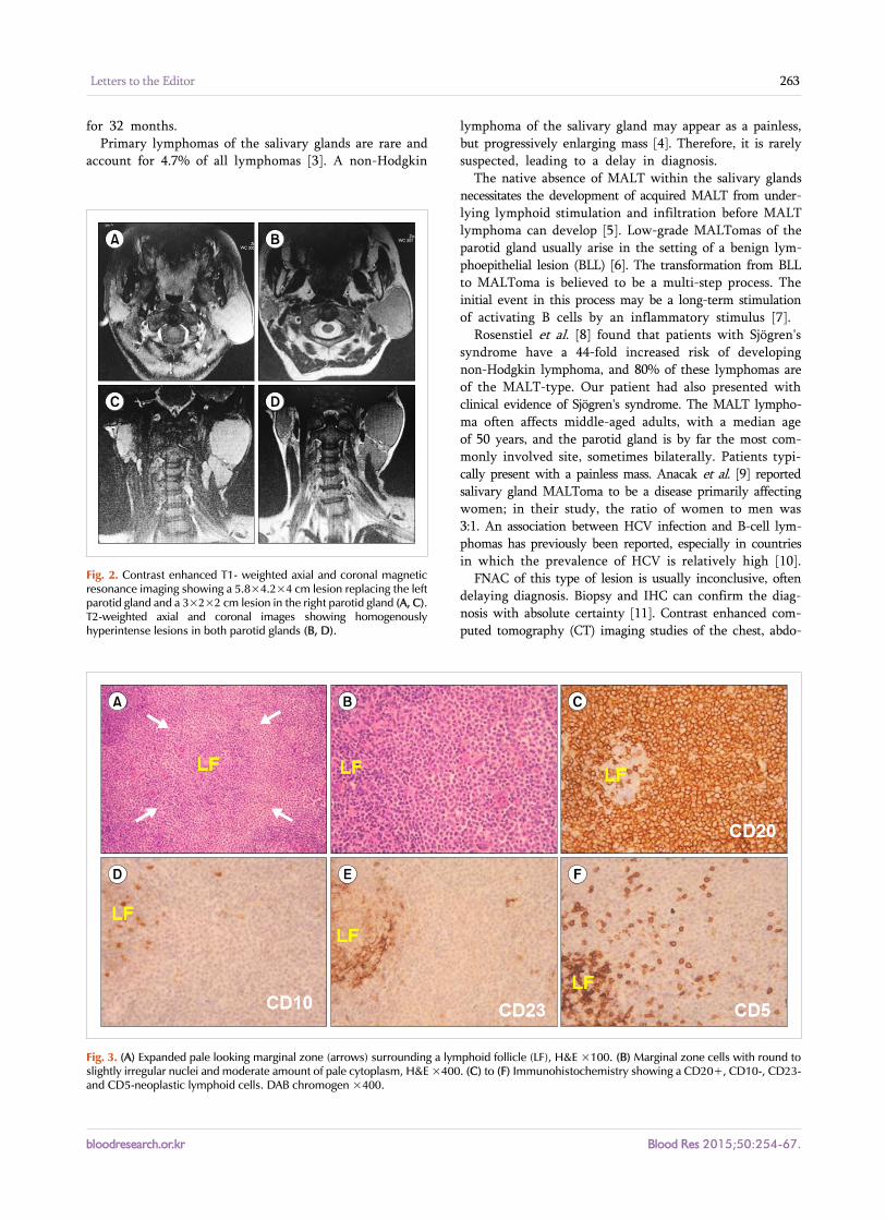
Contrast enhanced magnetic resonance imaging (MRI) of the face and neck revealed complete replacement of the entire left parotid parenchyma by a large, solid lesion involv-ing both the superficial and deep lobes measuring 5.8× 4.2×4.0 cm (Fig. 2). A similar lesion was visualized in the right parotid gland measuring 3×2×2 cm with enlarged cer-vical lymph nodes, which suggested the diagnosis of lympho-ma or Warthin’s tumor.
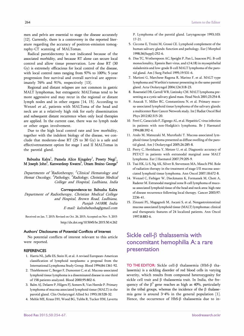
A trucut biopsy of the left parotid gland swelling was performed, which revealed a moderate amount of sheets of monotonous lymphoid cells with round irregular nuclei, coarsely clumped chromatin and inconspicuous nuclei with eosinophilic cytoplasm with occasional mitotic figures, re-sembling monocytoid B cells. Immunohistochemically, the cells were positive for CD20 and Bcl-2, weakly positive for Bcl-6 with a Ki-67 proliferative index of 35%, suggestive of MALT lymphoma (Fig. 3).
There was no evidence of lymphadenopathy, extra-nodal spread or bone marrow involvement (stage IE). The serology test for hepatitis B virus & hepatitis C virus (HCV), were negative, and did not have past history suggestive of any autoimmune disorder.
In consideration of the limited stage disease, she was scheduled for localized radiation therapy with three-dimen-sional conformal radiation therapy. She received a total dose of 36 Gy in 12 fractions over 16 days to the right and left parotid area by utilizing paired tangential fields. She achieved a complete clinical and radiological response. She is being followed-up monthly and has been disease-free
bloodresearch.or.kr Blood Res 2015;50:254-67.
Letters to the Editor 263
Fig. 2. Contrast enhanced T1- weighted axial and coronal magnetic resonance imaging showing a 5.8×4.2×4 cm lesion replacing the leftparotid gland and a 3×2×2 cm lesion in the right parotid gland (A, C).T2-weighted axial and coronal images showing homogenouslyhyperintense lesions in both parotid glands (B, D).
Fig. 3. (A) Expanded pale looking marginal zone (arrows) surrounding a lymphoid follicle (LF), H&E ×100. (B) Marginal zone cells with round to slightly irregular nuclei and moderate amount of pale cytoplasm, H&E ×400. (C) to (F) Immunohistochemistry showing a CD20+, CD10-, CD23-and CD5-neoplastic lymphoid cells. DAB chromogen ×400.
for 32 months.Primary lymphomas of the salivary glands are rare and
account for 4.7% of all lymphomas [3]. A non-Hodgkin
lymphoma of the salivary gland may appear as a painless, but progressively enlarging mass [4]. Therefore, it is rarely suspected, leading to a delay in diagnosis.
The native absence of MALT within the salivary glands necessitates the development of acquired MALT from under-lying lymphoid stimulation and infiltration before MALT lymphoma can develop [5]. Low-grade MALTomas of the parotid gland usually arise in the setting of a benign lym-phoepithelial lesion (BLL) [6]. The transformation from BLL to MALToma is believed to be a multi-step process. The initial event in this process may be a long-term stimulation of activating B cells by an inflammatory stimulus [7].
Rosenstiel et al. [8] found that patients with Sjögren's syndrome have a 44-fold increased risk of developing non-Hodgkin lymphoma, and 80% of these lymphomas are of the MALT-type. Our patient had also presented with clinical evidence of Sjögren's syndrome. The MALT lympho-ma often affects middle-aged adults, with a median age of 50 years, and the parotid gland is by far the most com-monly involved site, sometimes bilaterally. Patients typi-cally present with a painless mass. Anacak et al. [9] reported salivary gland MALToma to be a disease primarily affecting women; in their study, the ratio of women to men was 3:1. An association between HCV infection and B-cell lym-phomas has previously been reported, especially in countries in which the prevalence of HCV is relatively high [10].
FNAC of this type of lesion is usually inconclusive, often delaying diagnosis. Biopsy and IHC can confirm the diag-nosis with absolute certainty [11]. Contrast enhanced com-puted tomography (CT) imaging studies of the chest, abdo-
Blood Res 2015;50:254-67. bloodresearch.or.kr
264 Letters to the Editor
men and pelvis are essential to stage the disease accurately [12]. Currently, there is a controversy in the reported liter-ature regarding the accuracy of positron-emission tomog-raphy-CT scanning of MALTomas.
Radical parotidectomy is not indicated because of the associated morbidity, and because RT alone can secure local control and allow tissue preservation. Low dose RT (30 Gy) is extremely efficacious for local control of the disease, with local control rates ranging from 97% to 100%; 5-year progression free survival and overall survival are approx-imately 76% and 91%, respectively [13].
Regional and distant relapses are not common in gastric MALT lymphomas, but extragastric MALTomas tend to be more aggressive and may recur in the regional or distant lymph nodes and in other organs [14, 15]. According to Wenzel et al., patients with MALToma of the head and neck are at a relatively high risk for early dissemination and subsequent distant recurrence when only local therapies are applied. In the current case, there was no lymph node or other organ involvement.
Due to the high local control rate and low morbidity, together with the indolent biology of the disease, we con-clude that moderate-dose RT (25 to 30 Gy) is a safe and effectivetreatment option for stage I and II MALTomas in the parotid gland.
Babusha Kalra1, Pamela Alice Kingsley1, Preety Negi1, M Joseph John2, Kanwardeep Kwatra3, Uttam Braino George4
Departments of 1Radiotherapy, 2Clinical Hematology and Hemat Oncology, 3Pathology, 4Radiology, Christian Medical
College and Hospital, Ludhiana, India
Correspondence to: Babusha KalraDepartment of Radiotherapy, Christian Medical College
and Hospital, Brown Road, Ludhiana, Punjab 141008, India
E-mail: [email protected]
Received on Jan. 7, 2015; Revised on Oct. 26, 2015; Accepted on Nov. 5, 2015
http://dx.doi.org/10.5045/br.2015.50.4.262
AuthorsÊ Disclosures of Potential Conflicts of Interest
No potential conflicts of interest relevant to this article were reported.
REFERENCES
1. Harris NL, Jaffe ES, Stein H, et al. A revised European-American
classification of lymphoid neoplasms: a proposal from the
International Lymphoma Study Group. Blood 1994;84:1361-92.
2. Thieblemont C, Berger F, Dumontet C, et al. Mucosa-associated
lymphoid tissue lymphoma is a disseminated disease in one third
of 158 patients analyzed. Blood 2000;95:802-6.
3. Balm AJ, Delaere P, Hilgers FJ, Somers R, Van Heerde P. Primary
lymphoma of mucosa associated lymphoid tissue (MALT) in the
parotid gland. Clin Otolaryngol Allied Sci 1993;18:528-32.
4. Mehle ME, Kraus DH, Wood BG, Tubbs R, Tucker HM, Lavertu
P. Lymphoma of the parotid gland. Laryngoscope 1993;103:
17-21.
5. Ciccone E, Truini M, Grossi CE. Lymphoid complement of the
human salivary glands: function and pathology. Eur J Morphol
1998;36(Suppl):252-6.
6. Diss TC, Wotherspoon AC, Speight P, Pan L, Isaacson PG. B-cell
monoclonality, Epstein Barr virus, and t(14;18) in myoepithelial
sialadenitis and low-grade B-cell MALT lymphoma of the paro-
tid gland. Am J Surg Pathol 1995;19:531-6.
7. Marioni G, Marchese-Ragona R, Marino F, et al. MALT-type
lymphoma and Warthin's tumour presenting in the same parotid
gland. Acta Otolaryngol 2004;124:318-23.
8. Rosenstiel DB, Carroll WR, Listinsky CM. MALT lymphoma pre-
senting as a cystic salivary gland mass. Head Neck 2001;23:254-8.
9. Anacak Y, Miller RC, Constantinou N, et al. Primary muco-
sa-associated lymphoid tissue lymphoma of the salivary glands:
a multicenter Rare Cancer Network study. Int J Radiat Oncol Biol
Phys 2012;82:315-20.
10. Ferri C, Caracciolo F, Zignego AL, et al. Hepatitis C virus infection
in patients with non-Hodgkin's lymphoma. Br J Haematol
1994;88:392-4.
11. Ando M, Matsuzaki M, Murofushi T. Mucosa-associated lym-
phoid tissue lymphoma presented as diffuse swelling of the paro-
tid gland. Am J Otolaryngol 2005;26:285-8.
12. Perry C, Herishanu Y, Metzer U, et al. Diagnostic accuracy of
PET/CT in patients with extranodal marginal zone MALT
lymphoma. Eur J Haematol 2007;79:205-9.
13. Tsai HK, Li S, Ng AK, Silver B, Stevenson MA, Mauch PM. Role
of radiation therapy in the treatment of stage I/II mucosa-asso-
ciated lymphoid tissue lymphoma. Ann Oncol 2007;18:672-8.
14. Wenzel C, Fiebiger W, Dieckmann K, Formanek M, Chott A,
Raderer M. Extranodal marginal zone B-cell lymphoma of muco-
sa-associated lymphoid tissue of the head and neck area: high rate
of disease recurrence following local therapy. Cancer 2003;97:
2236-41.
15. Zinzani PL, Magagnoli M, Ascani S, et al. Nongastrointestinal
mucosa-associated lymphoid tissue (MALT) lymphomas: clinical
and therapeutic features of 24 localized patients. Ann Oncol
1997;8:883-6.
Sickle cell- thalassemia with concomitant hemophilia A: a rare presentation
TO THE EDITOR: Sickle cell- thalassemia (HbS- tha-lassemia) is a sickling disorder of red blood cells in varying severity, which results from compound heterozygosity for sickle cell trait and thalassemia trait. In India, the fre-quency of the S gene reaches as high as 40%, particularly in the tribal groups, whereas the incidence of the thalasse-mia gene is around 3–4% in the general population [1]. Hence, the occurrence of HbS- thalassemia due to in-