biomaterial: concepts and basics properties · biomaterial for implantation or medical use, the...
TRANSCRIPT

European International Journal of Science and Technology Vol. 9 No. 2 February 2020
Cite this article: Gobbi, S.J., Gobbi, V.J., Rocha, Y., Coutinho, M.M., Sousa, T.P. & Reinke, G. (2020). Biomaterial: Concepts and Basics Properties. European International Journal of Science and Technology, 9(2), 23-42
23
Biomaterial: Concepts and Basics
Properties
Silvio José Gobbi1, Vagner João Gobbi2, Ynglid Rocha3,
Maycol Moreira Coutinho4, Thiago Primo Sousa5,
Gustavo Reinke6*
1Faculdade de Tecnologia, Universidade de Brasília – UNB, Brasília, DF
Email: [email protected]; [email protected]
2;
[email protected]; [email protected]
6;
3Universidade Paulista – UNIP; ASAFE
Email: [email protected]
*Corresponding Author
Published: 29 February 2020
Copyright © Gobbi et al.
European International Journal of Science and Technology ISSN: 2304-9693 www.eijst.org.uk
24
ABSTRACT
Biomaterials encompass extensive tissue engineering technology that allows the development
of materials to restore or replace parts of the living system. Demand has grown quickly in
recent decades due to the worldwide phenomenon of population aging, being that the risk of
hard tissue failure is greater for the elderly. The main objective is to obtain a longer lifespan
of the biomaterial implanted or that the same lasts until the end of the life without flaws or
need of revision surgery, helping to enhance the quality of life of the patients. Thus, is
required of the biomaterial to satisfy several properties compliant to specific application, such
as adequate mechanical strength, high corrosion and biocompatibility, high wear resistance,
low friction and mechanical compatibility. The present work has as purpose to describe the
main properties for the choice and development of biomaterials, with greater attention for
metal biomaterials in the replacement of hard tissues, especially titanium and titanium alloys.
Keywords: biomaterial, hard tissue, biocompatibility, titanium.
1. INTRODUCTION
A biomaterial can be defined as a material which is used on a form or specific structure in
order to manufacture prostheses and biomedical devices intended to replace or restore
impaired body function in order to save or improve the quality of life [1]. They are materials
used for the manufacture of devices that can interact with biological systems to coexist for a
long time of service with minimal failures [2].
Although several definitions are employed to describe them, biomaterials are natural or
synthetic materials that are useful for the repair of damaged body parts, through interaction
with living systems [3,4]. Biomaterials used to maintain or replace functions in the human
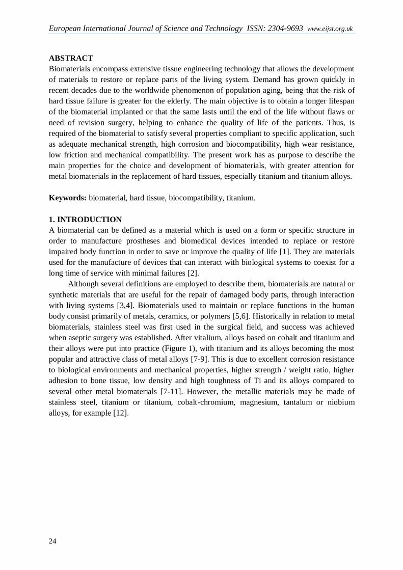
body consist primarily of metals, ceramics, or polymers [5,6]. Historically in relation to metal
biomaterials, stainless steel was first used in the surgical field, and success was achieved
when aseptic surgery was established. After vitalium, alloys based on cobalt and titanium and
their alloys were put into practice (Figure 1), with titanium and its alloys becoming the most
popular and attractive class of metal alloys [7-9]. This is due to excellent corrosion resistance
to biological environments and mechanical properties, higher strength / weight ratio, higher
adhesion to bone tissue, low density and high toughness of Ti and its alloys compared to
several other metal biomaterials [7-11]. However, the metallic materials may be made of
stainless steel, titanium or titanium, cobalt-chromium, magnesium, tantalum or niobium
alloys, for example [12].
European International Journal of Science and Technology Vol. 9 No. 2 February 2020
25
Figure 1. History of metals, plastics and ceramics for biomedical applications [8 -
adapted].
These materials are used to replace a component of the human body or to support
physiological functions. As such, biomaterials interact with human cells, tissues or organs
and sometimes even perform their biological functions [3]. Functional repair engineering uses
biomaterials to recompose different tissues with the aim of improving patients' health and
quality of life [3,13]. We also find the term "nano biomaterial" that comes from the
combination of biomaterial and nanotechnology[14].
The ability to exist in contact with the tissues of the human body without causing an
unacceptable degree of damage to that body is the most important factor that distinguishes a
biomaterial from any other material. The way in which mutually acceptable coexistence of
biomaterials and tissues is developed and sustained has been of interest to biomaterial
scientists and users of medical devices for many years [15].Thus, when considering a
biomaterial for implantation or medical use, the first and most important requirement is non-
toxic, non-immunogenic, chemically inert and acceptable to the human body [16].
Over the past fifty years, biomaterials science has investigated different types of
biomaterials and their applications to replace or restore the function of compromised or
degenerate tissues or organs [3]. Every year, more than 13 million prosthetics / medical
devices are implanted in the US alone [3,16]. Biomaterials are used in different parts of the
human body such as artificial heart valves, stents in blood vessels, replacement implants in
the shoulders, knees, hips, elbows, ears and orthodontic structures. It is stated that around 70-
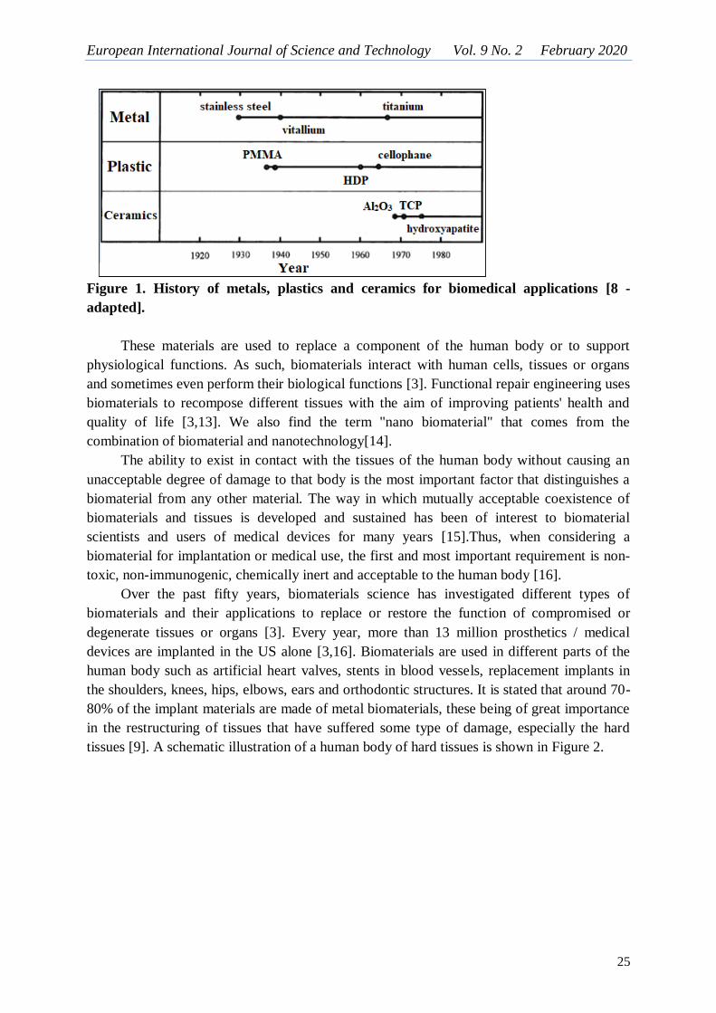
80% of the implant materials are made of metal biomaterials, these being of great importance
in the restructuring of tissues that have suffered some type of damage, especially the hard
tissues [9]. A schematic illustration of a human body of hard tissues is shown in Figure 2.
European International Journal of Science and Technology ISSN: 2304-9693 www.eijst.org.uk
26
Figure 2. A schematic illustration of a human body of hard tissues [17 - adapted].
The main required property of a biomaterial is that it does not trigger an adverse
reaction when put into service, which means to be a biocompatible material. In addition, good
mechanical properties, osseo integration, high corrosion resistance and excellent resistance to
wear, ductility and high hardness are required [5,18,19].However, the properties of an ideal
biomaterial may change depending on the exact location of your implantation and even the
medical history of patients. They should take into account the structure and function of the
surrounding tissues and organs so that it does not disturb or interfere with their functioning.
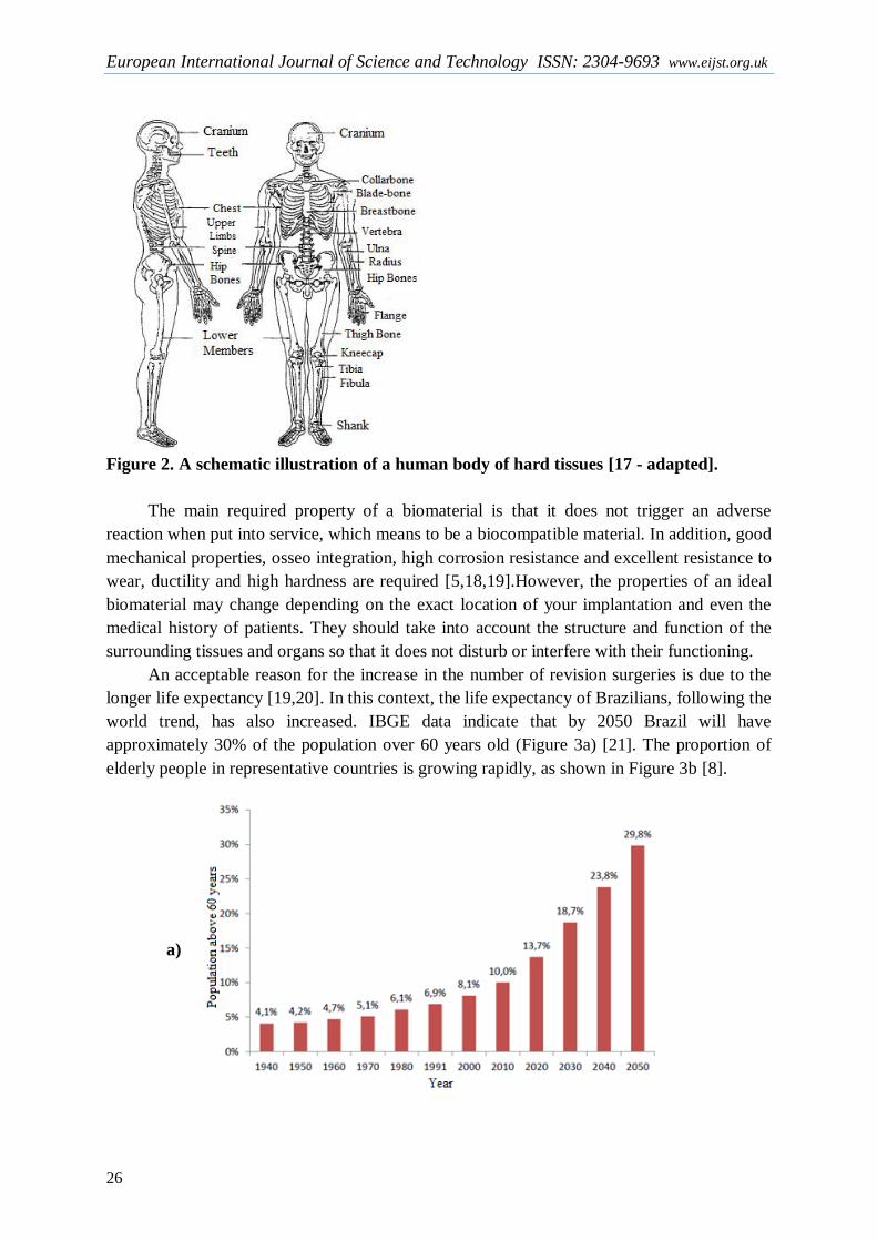
An acceptable reason for the increase in the number of revision surgeries is due to the
longer life expectancy [19,20]. In this context, the life expectancy of Brazilians, following the
world trend, has also increased. IBGE data indicate that by 2050 Brazil will have
approximately 30% of the population over 60 years old (Figure 3a) [21]. The proportion of
elderly people in representative countries is growing rapidly, as shown in Figure 3b [8].
a)

European International Journal of Science and Technology Vol. 9 No. 2 February 2020
27
b)
Figure 3. a) Percentage of the Brazilian population over 60 years from 1940 and a
forecast up to 2050 [21]; b) change in the population of old people in each country [8 -
adapted].
Consequently, the scenario has changed now, due to advances in medical technology,
people live longer and moreover, the prognosis should be better for those who are physically
traumatized due to sports or incorrect or exaggerated exercise habits or due to traffic road
accidents and other accidents [18]. Thus, implants are expected to act much longer or to the
end of life without flaws or revision surgeries, which is not inexpensive and has a lower
success rate than the first implantation surgery[18]. Consequently, the development of
suitable material with high longevity and excellent biocompatibility is highly essential.
Although various materials are currently in use as biomaterials, titanium alloys are rapidly
emerging as the first choice for most applications [9,18,19,22].
1.1 Aspects to be considered for a biomaterial selection
1.1.1. Biological compatibility or biocompatibility:
Biomaterials must be biocompatible, i.e. safe for the host organism. This means that
they cannot cause harmful effects locally, in the area of implantation, nor for other tissues or
organs [23]. There can be no adverse effects on the implant or host organism.
Biocompatibility has traditionally been related to implantable devices that were intended to
remain within a person for a long time. For those who were developing and using the first
generation of implantable devices during the years between 1940 and 1980, it became
increasingly obvious that the best biological performance would be achieved with materials
that were the least chemically reactive [15]. Biocompatibility is defined as the ability or
capacity of the material to be used in close connection with living tissue without causing
adverse effects to them[24,25]. The materials used as implants are expected to be highly non-
toxic and should not cause any inflammatory or allergic reactions in the human body. The
rejection of an orthopedic implant due to the toxic release of metallic ions, for example, will
lead to the final failure [26]. The patient body parts or tissue of who comes into contact with
the implants should avoid any physical irritation, inflammation, toxicity, carcinogenic or
mutagenic action [27,28].

European International Journal of Science and Technology ISSN: 2304-9693 www.eijst.org.uk
28
The "fibrous capsule" surrounding the implant is also the basis of the biocompatibility
of the biomaterials, and the final stage of the implantation process is represented by this
capsule, which begins when the plasma proteins come into contact or are adhered to the
repair [29, 80].
The biomaterials success depends mainly on the reaction of the human body to the
implant and this measures the biocompatibility of a material [15]. When implants are exposed
to human tissues and fluids, various reactions occur between the host and the implant
material and these reactions determine the acceptability of these materials by our system.
Problems related to biocompatibility are (1) thrombosis, which involves blood clotting and
adhesion of blood platelets to the surface of the biomaterial, and (2) encapsulation of fibrous
tissue from implanted soft tissue biomaterials [18].
However, the biocompatibility of implants also depends greatly on their corrosion
behavior [5,30]. Therefore, the greater the corrosion of the implants, the more toxic ion rates
are released into the body routinely and higher risk of adverse effects can be expected
[5,18,31,80].
The human body consists of a significant number of natural elements with water (H2O),
comprising about 65 to 75% of the total composition in weight. Consequently, most of the
mass of a human body contains oxygen and carbon [26, 32]. Table 1 shows a list of elements
found in the human body. Where, about 96% of the available elements are oxygen, hydrogen,
carbon and nitrogen, which are the building blocks of water and proteins. Additional ~ 4% of
body mass comes in the form of bone minerals and blood composed of Ca, P, Mg and
extracellular fluids comprising Na, Cl and K. As such, any implant developed on the basis of
these elements would be compatible with the human body [5]. However, there are few trace
elements that are toxic at high levels. Therefore, the appropriate composition required for the
metal implant cannot be toxic [5]. Therefore, the implant will not release toxic metal ions,
which causes inflammatory or allergic reactions in the human body [5,9,18].
Table1. Human body elements [26,32].
Element O C H N Ca P K S Na Cl Mg Trace
element
Wt% 65,0 18,5 9,5 3,3 1,5 1,0 0,4 0,3 0,2 0,2 0,1 <0,01
1.1.1.1 Biocompatibility of titanium binder element
Titanium is not found in the human body, and plays no known biological role in the
human body [33], and is nontoxic even in large doses [26].When humans ingested quantities
of 0.8 mg of titanium daily, most of titanium was eliminated without being digested or
absorbed [34].
Titanium implants are usually not rejected by the body and generally make good
physical connections with the host bone. In vitro assays have shown that titanium can,
however, inhibit osteogenic differentiation of mesenchymal stem cells [35]and may cause
genetic changes in connective tissue [36].
European International Journal of Science and Technology Vol. 9 No. 2 February 2020
29
1.1.1.2 Biocompatibility of titanium alloys
Compared with stainless steel and cobalt alloys, titanium alloys have proved to be superior in
terms of biocompatibility due to their excellent corrosion resistance [26,37]. In general, 316L
stainless steel shows a relatively good biocompatibility, but at a less satisfactory level than
CoCrMo and titanium alloys, due to the higher corrosion rates, as outlined below [26,38].
The first generation of titanium alloys, represented by the Ti-6Al-4V alloy, has been
reported to cause allergic reactions to the human body [9,39]. The second generation of
titanium alloys (titanium alloys β) has been developed and investigated with great interest.
Some stabilizing elements of the fase phase, such as niobium, tantalum and zirconium, are
used as alloying elements and are considered relatively safe when compared to vanadium and
aluminum [8,40,80,81].
The increased use of titanium and its alloys as biomaterials comes from its superior
biocompatibility and excellent corrosion resistance because of the thin layer of surface oxide
and good mechanical properties such as a certain modulus of elasticity and low density that
make these metals present a behavior mechanic similar to the bones [9,19]. Lightweight,
strong and fully biocompatible, titanium is one of the few materials that naturally combine
the implantation requirements in the human body. As titanium and its alloys, commercially
pure titanium (cpTi, grade 2) and Ti-6Al-4V (grade 5) are widely used as replacements for
hard tissue in artificial bones, joints and dental implants [19]. In replacing hard-bone tissue,
as the smallest modulus of elasticity fits in the direction of reducing the effect of voltage
shielding, the low modulus of commercially pure titanium and its alloys is generally seen as a
biomechanical advantage.
Viteri and Fluentes[19]also point out that another property that makes titanium and its
alloys the most promising biomaterials for implants is that titanium-based materials generally
have the formation of an extremely thin and adherent protective titanium oxide film. The
presence of this spontaneous oxide film in the passivation or repassivation process is an
important criterion for the excellent biocompatibility and corrosion resistance of titanium and
its alloys.
Regarding the medical applications of these materials, the use of cp (commercially
pure) titanium is more limited to dental implants due to their limited mechanical properties
[19]. In cases where good mechanical characteristics are required, such as in hip or knee
implants, screws and plates, the Ti-6Al-4V alloy is widely used [41,42]. One of the most
common applications of titanium alloys are artificial hip joints which consist of a joint (head
and femoral cup) and stem bearing, where the metal cup and hip stem components are made
of titanium. In addition, they are also often used in knee joint replacements, which consist of
a femoral and tibial component made of titanium and a polyethylene joint surface. Titanium
and its alloys have also been used in cardiovascular devices in recent years, such as heart
valves, heart pumps and vascular stents. However, titanium is a thrombogenic material [43].
A large amount of research was conducted on titanium surface-induced coagulation and
several methods were applied to improve in vitro biocompatibility. A harder and thicker
protective oxide layer was obtained with the incandescent discharge treatment and air-furnace
processing performed on Ti and Ti-6Al-4V alloy, thus presenting better biocompatibility
HUVECs (human umbilical vein endothelial cells) [44]. ToKumariet al.[45] diamond-like

European International Journal of Science and Technology ISSN: 2304-9693 www.eijst.org.uk
30
carbon-coated titanium (DLC) may improve cytocompatibilityin vitro for HUVECs. HUVEC
cells were cultured and a native extracellular matrix (ECM) was obtained on the surface of
pure titanium, demonstrating that this ECM produced by HUVECs not only can improve the
adhesion and proliferation capacity of endothelial cells, but also inhibit the adhesion and
activation of the HUVECs. platelets, which provides a basis for the preparation of modified
surfaces in the application of cardiovascular implants [43].
1.1.2. Mechanical Compatibility
In addition to the biological biocompatibility discussed above, mechanical biocompatibility is
vital for long-term implantation. Mechanical compatibility refers to the appropriate
mechanical properties according to the function to be performed and the site to be implanted
[39]. Furthermore, bio-implants must have suitable mechanical strength to withstand all the
forces and related charges. Primarily, the material selected for a specific application must
have the ability to withstand the load, therefore, they will not be susceptible to fracture [5].
For a specific function, purpose or application, the mechanical properties determine the
type of biomaterial to be chosen. Tensile strength, hardness, osseo integration, modulus of
elasticity, wear resistance to corrosion are some of the properties that are of fundamental
importance. Thus, if during the application the biomaterial will be subject to repeated cyclic
loads, success over the use of the implant subjected to this type of loading is determined by
the fatigue strength of the material. The cyclic load is applied to orthopedic implants during
body movement, resulting in alternating plastic deformation of microscopically small areas of
stress concentration produced by grooves or microstructural heterogeneities [20]. Fatigue
resistance of the alloys is related to the composition of the alloy and the previous history of
thermomechanical processing. Fatigue resistance is also highly affected by surface
processing, finishing and thermal treatments. Thus, the alloys have such a wide range of
mechanical properties and can be controlled with suitable processing and thermal treatments.
It is well known that the higher the fatigue strength of an alloy, the longer the life of an
implant made from it[20]. Therefore, the long life of the implant, which is related to its
resistance to fatigue, is a crucial property of the implant materials.
Generally, Co-Cr alloys and Ti alloys of α + β type have high fatigue strength when
compared to other metallic biomaterials. The titanium alloy of type β TNTZ (Ti-29Nb-13Ta-
4,6Zr) presented high resistance to fatigue with appropriate thermomechanical treatments
[46].
The biomaterial that will replace the bone must have a modulus of elasticity equivalent
to that of the substituted bone. The bone modulus varies in magnitude from 4 to 30 GPa
depending on the type of bone and the measurement direction [47,48]. Current implant
materials that have greater stiffness than bone prevent the necessary stress from being
transferred to the adjacent bone, resulting in bone resorption around the implant and,
consequently, to the implant loosening. This biomechanical incompatibility that leads to the
death of bone cells is called the "stress shielding" [9,47,49]. Al-Tamimiet al.[47]describe the
bone fixation implants commercially available (i.e., external fixators, internal fasteners
intramedullary pin) are constructed with metallic biomaterials such as stainless steel,
titanium, cobalt and their alloys (e.g., Ti6Al4V and CoCrMo), indicating that in In a large

European International Journal of Science and Technology Vol. 9 No. 2 February 2020
31
number of cases, these implants are permanently left in the body, leading to long-term
problems such as possible release of metal ions, inflammatory reactions, risk of infection,
loosening of screws and, mainly, bone resorption due to the effects of the shield of tension or
stress shielding.
The production of low elastic modulus biomaterials is greatly stimulated by the
development of β-titanium alloys [9,40]. Thus, a material with excellent combination of high
strength and low modulus closer to the bone should be used for implantation to avoid implant
loosening and longer service period, avoiding revision surgery [18]. For Ti and its alloys the
modulus of elasticity varies from 110 to 55 GPa compared to 316 L stainless steel (210 GPa)
and chromium-cobalt alloys (240 GPa). The modulus of elasticity of various biomedical
alloys is compared to the bone and shown in Figure 4.
Figure 4: Modulus of elasticity of biomedical alloys [18 - adapted].
If an implant fractures due to inadequate resistance or divergence in mechanical
properties between the bone and the implant, this is referred to as biomechanical
incompatibility [18].
1.1.3 High corrosion resistance
Biomaterials must be resistant to corrosion. This is a problem especially in the context of
metal implants. The presence of body fluids allows metal implants to release metallic ions
that may accumulate in nearby tissues or be transported to other parts of the body. Corrosion
is one of the major processes that affect the life and service of orthopedic appliances made of
metals and alloys used as implants in the body [2].Biomaterials are usually exposed to the
critical level of humidity and in an environment with high percentage of localized corrosion
[5]. The low corrosion resistance of implants in body fluid results in the release of metal ions
not compatible with implants in the body[18]. It has been found that released ions cause
allergic and toxic reactions[18,50]. Corroded implants in the human body cause excess of
harmful and toxic metal ions such as Fe, Cr, Ni, Co, and Ti released into the body fluid [5,51].
European International Journal of Science and Technology ISSN: 2304-9693 www.eijst.org.uk
32
Initially, these main trace elements in metallic implants would not be harmful by the released
ions. However, when implants begin to corrode, these trace elements aggressively diffuse
into the body. Excessive release of these harmful metal ions may cause adverse effects to the
human body [5,80].
When the oxide layer on the metal is broken, corrosion occurs and a metal ion is
released. The outer layer is then repassivated in a process known as regeneration. The
regeneration time or repassivation time of the surface oxide layer is different for various
applied materials [26]. The rate of corrosion and the release of some metal ions are highly
dependent on the regeneration time [52].
From the observation of the regeneration time based on the formation of surface oxide
layers, it was found that the regeneration time for the 316L SS alloy is longer compared to the
CoCrMo and Ti-6Al-4V alloys [52].Thin layer formation through surface modification
improves the biocompatibility performance with wear resistance and corrosion resistance [5].
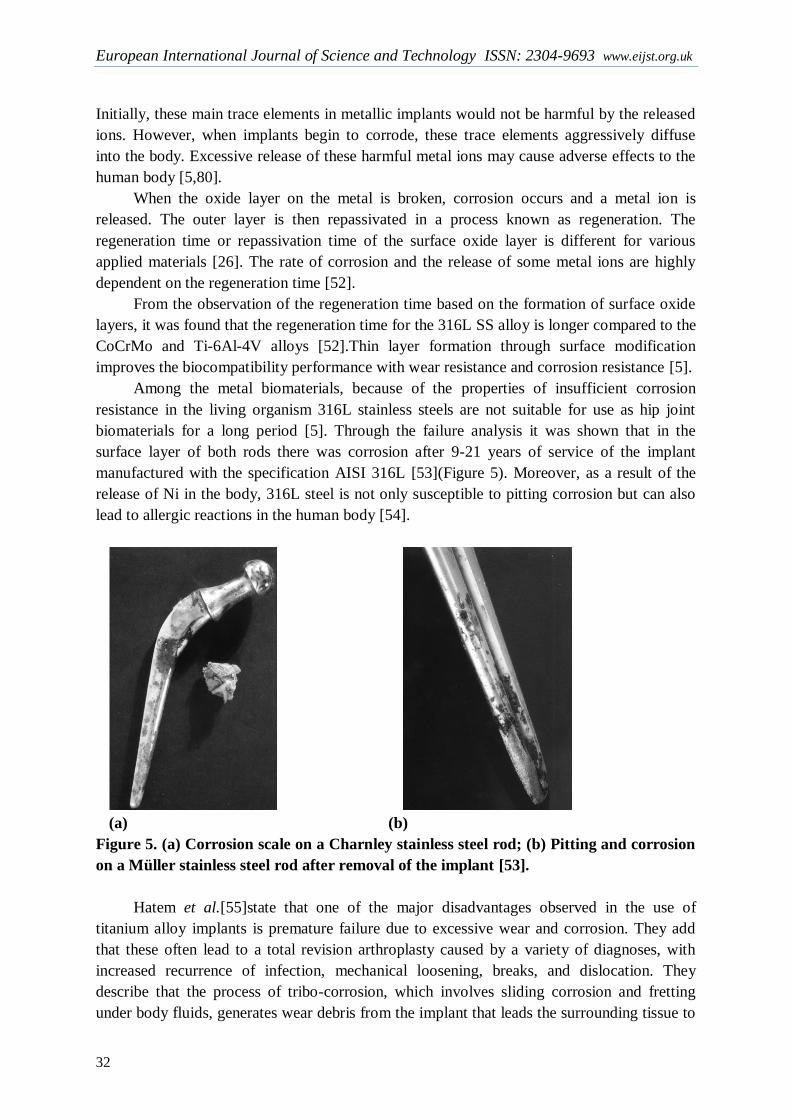
Among the metal biomaterials, because of the properties of insufficient corrosion
resistance in the living organism 316L stainless steels are not suitable for use as hip joint
biomaterials for a long period [5]. Through the failure analysis it was shown that in the
surface layer of both rods there was corrosion after 9-21 years of service of the implant
manufactured with the specification AISI 316L [53](Figure 5). Moreover, as a result of the
release of Ni in the body, 316L steel is not only susceptible to pitting corrosion but can also
lead to allergic reactions in the human body [54].
(a) (b)
Figure 5. (a) Corrosion scale on a Charnley stainless steel rod; (b) Pitting and corrosion
on a Müller stainless steel rod after removal of the implant [53].
Hatem et al.[55]state that one of the major disadvantages observed in the use of
titanium alloy implants is premature failure due to excessive wear and corrosion. They add
that these often lead to a total revision arthroplasty caused by a variety of diagnoses, with
increased recurrence of infection, mechanical loosening, breaks, and dislocation. They
describe that the process of tribo-corrosion, which involves sliding corrosion and fretting
under body fluids, generates wear debris from the implant that leads the surrounding tissue to
European International Journal of Science and Technology Vol. 9 No. 2 February 2020
33
inflammatory reactions, bone resorption and low osseo integration of the implant, which may
result in a complete revision.
The tribo-corrosion process can expose the human body to harmful elements when they
are present in the alloy composition of the implant. In the case of Ti-6Al-4V, a titanium alloy
widely used in biomedical applications, the vanadium ions released from the implant can
generate cytotoxic reactions and neurological disorders in the patient [56].Modifications in
the surfaces of the implants to improve the tribo-corrosion behavior can be very promising to
overcome the causes of mechanical loosening, especially in Ti-6Al-4V alloy implants [55].
Nevertheless, because titanium and its alloys have low tribological performance, such surface
modifications are required for these alloys when the implant will be subjected to this type of
stress as described in the requirement for high wear resistance.
In general, in the in vitro studies, the general conclusion is that the presence of metal
ions in the body leads to biological responses [51]. Although the release of metal by
corrosion and wear may be considered in principle to be very limited, long-term metal ions
accumulation and particles in the human body is of great concern. Studies show that
corrosion occurs slowly due to the electrochemical reaction, once a metal has been implanted
in the human body [52,2].The presence of metallic ions in the tissues around implants has
been reported to cause carcinogenicity, hypersensitivity, allergy, local tissue toxicity,
inflammation and genotoxicity [51].
Notably, corrosion is not only dependent on the metal chemical properties, but is also
influenced by mechanical forces. During the time of use, the friction between the implant and
other tissues may increase the release of ions [23]. Remarkably, some biomaterials use an
opposing approach and are designed to undergo degradation within the host organism.
However, even so, it is important for the compounds that the released degradation product is
biocompatible with the living organism.
Thus, the development of implants with high resistance to corrosion and wear is of
prime importance for the longevity of the material in the human system [18].The prevention
of biomaterials corrosion has become crucial, particularly to overcome the inflammation and
allergic reactions caused by the implants of the biomaterials towards the human body [5].
Ions resulting from a corrosive process can have adverse effects on host health, which makes
the materials non-biocompatible. Its release from the implant can also reduce implant life,
thus requiring revision surgery.
1.1.4 High wear resistance
The low wear resistance leads to the release of wear debris from the implant into the
surrounding tissue which can produce an adverse cellular response leading to the release of
harmful enzymes, inflammation, osteolysis, infection, pain and bone resorption [18,57]. In
general, wear occurs in the articulation of artificial joints as a result of the mixed lubrication
regime.
The wear resistance of the material plays a significant role in the biomaterial proper
functioning, avoiding loosening of the implant and reactions in the tissue in which it is
deposited, improving the quality of life of the patient [58].
European International Journal of Science and Technology ISSN: 2304-9693 www.eijst.org.uk
34
Although titanium and its alloys have excellent mechanical and physical properties,
such as high corrosion resistance, high strength / weight ratio and excellent biocompatibility,
they present poor tribological properties with a high coefficient of friction and wear
[10,11,59,60]. This fact limits its applications in wear situations involving wear and many
works have been carried out by researchers in order to solve the low tribological behavior
associated with titanium alloys. The adequate surface treatment expands the use of titanium
and its alloys in the biomedical areas, being one of the most effective methods to improve the
wear resistance of titanium alloys[17,39].
The plasma nitriding of titanium and its alloys has been used for wear protection [61-
63]. The treatment produces high hardness and a chemically inert layer that overcomes the
poor wear resistance of titanium alloys [64]. Nitrogen has a high solubility in α-Ti, so that it
significantly strengthens the surface layer [61]. The titanium plasma nitriding process based
on the thermal diffusion mechanism can produce a composite layer formed by TiN at the top
and Ti2N below, with a hardness that can reach 1500 to 3000 HV [61-63]. [61-63]. In short,
several researches[62,65-68] report plasma nitriding as a very effective method that improved
the tribological properties of titanium and its alloys.
Coating with thin films allows a considerable increase in wear resistance and enables a
significant increase in the component life. In these techniques, coatings can be produced by
physical vapor deposition (PVD), chemical vapor deposition (CVD), electrodeposition, ion
implantation, among others. For example, coatings such as diamond-like carbon (DLC) and
titanium nitride (TiN) are used in joint implants because of their excellent tribological
properties [69]. TiN offers advantages such as high hardness and wear resistant, low price,
corrosion resistant and biocompatible [69-71]; DLC has the advantages of high hardness,
high resistance to corrosion and wear, biocompatibility and is a solid lubricant, chemical
inertia; however, disadvantage as fragile, presents poor adhesion and high internal residual
stress [55,69,72]. It has recently been shown that graphite-type carbon (GLC) and tantalum
(Ta) have good potential as a coating because they have mechanical properties similar to
bone - high hardness and high flexibility [69].
High wear resistance avoids loosening of the implant and reactions in the tissue in
which it is deposited, improving the patient quality of life. A description of the importance of
metal biomaterials wear performance was carried out in previous work by the present authors
[58].
1.1.5 Osseo integration
Osseo integration refers to a phenomenon where an implant becomes so fused to the bone
that it cannotbe separated without a fracture, and when an implant is osseo integrated there is
no progressive relative movement between it and the bone, with which it has direct contact,
meaning that osseo integration is necessary for the long-term stability of the prosthesis [73].
It is defined as the direct anchoring of an implant by the formation of bone tissue
around the implant without fibrous tissue growth at the bone-implant interface [74,75].
The inability of an implant surface to integrate with adjacent bone and other tissues due
to micro motions results in loosening of the implant. A fibrous tissue is formed between the
bone and the implant if the implant is not well integrated with the bone. Thus, materials with
European International Journal of Science and Technology Vol. 9 No. 2 February 2020
35
a suitable surface are highly essential for the implant to integrate well with the adjacent bone.
Surface chemistry, surface roughness and surface topography play an important role in the
development of good osseo integration [18].
Osseo integration is the stable and functional union between the bone and an implanted
surface. This phenomenon occurs after the device insertion into the bone and bone’s cells
migration to the bone surface [76]. Among other factors, roughness and wettability are very
important in osseo integration, and the implant needs to have adequate nano and micrometric
morphological characteristics [77].The final efficacy of osseo integration depends on the
topography of the surface of an implant. The implant surface properties are crucial for the
adhesion and differentiation of osteoblasts during the initial phase of osseo integration, as
well as for long-term bone remodeling [78].
The implant design is one of the significant parameters for the performance of
osseointegration, however, so far, the ideal design for the stability of the timeless bone
implant has yet to be determined [79].
The osseo integration of a bone fixation is defined as the intimate apposition of newly formed
bone and reformed in congruence with the fixations, including surface irregularities, so that,
with the analysis by light microscopy, there is no interposition of connective or fibrous tissue
and is established a direct structural and functional connection, able to withstand normal
physiological loads without excessive deformation and without initiating a rejection
mechanism [54].
The presence of toxic elements in alloys can also affect osseo integration. For example,
vanadium ions of the Ti-6Al-4V alloy subjected to a tribo-corrosion process are related to the
problems of cytotoxic reactions and neurological disorders [56], and these toxicity reactions
of vanadium ions may be detrimental to local cellular regeneration and contribute to poor
osseo integration [55,80].
2. FINAL CONSIDERATIONS
Biomaterials are primarily used to replace a defective tissue in order to improve the patient's
quality of life. Regarding the use of biomaterials in the long term, the following factors are
paramount: biological biocompatibility and mechanical biocompatibility, resistance to
corrosion and wear and osseo integration. The host's response to the implantation and
presence of the biomaterial cannot have adverse effects on the implant or host organism.
Therefore, biological biocompatibility is the primary point of choice, and titanium alloys are
superior in terms of compatibility compared to stainless steel and cobalt alloys. The nature of
passive oxide films formed on the surface and the mechanical properties of the materials form
some of the essential criteria for the selection or development of new biomaterials. The high
corrosion resistance avoids the degradation process and consequently a reduction of the
structural integrity of the implant and the release of degradation products, which may react
unfavorably with the host organism. High wear resistance avoids the generation of wear
particles, which can stimulate an adverse cellular response leading to implant loosening and
revision surgery. It is expected that the biomaterial used to replace the bone has mechanical
properties similar to this, as an example, having a modulus of elasticity equivalent to that of
bone, avoiding the effect of stress shielding and its consequences.

European International Journal of Science and Technology ISSN: 2304-9693 www.eijst.org.uk
36
3. REFERENCES
[1] Tathe, A., Ghodke, M., Nikalje, A.P., “A Brief Review: Biomaterials And Their
Application”. International Journal Of Pharmacy And Pharmaceutical Sciences , V.2, Pp.19-
23, 2010.
[2] Kamachimudali, U., Sridhar, T.M., Raj, B., “Corrosion Of Bio Implants”, Sadhana,
V.28, Pp.601–637, 2003.
[3] Bose, S., Ke, D., Sahasrabudhe, H., Bandyopadhyay, A., “Additive Manufacturing Of
Biomaterials”. Progress In Materials Science, V.93, Pp.45-111.
[4] Ebrahimi, M., “Biomaterials Application In Therapeutic And Regenerative Medicine
From The Perspective Of Patients’ Faith”, Biomedical Journal Scientific & Technical
Research, V.1, N.7, 2018.
[5] Asri, R.I., Harun, W.S., Lah, N.A., Et Al., “Corrosion And Surface Modification On
Biocompatible Metals: A Review”, Materials Science And Engineering: C, V.77, Pp.1261-
1274, 2017.
[6] Markhoff, J., Krogull, M., Schulze, C., Et Al., “Biocompatibility And Inflammatory
Potential Of Titanium Alloys Cultivated With Human Osteoblasts, Fibroblasts And
Macrophages, Materials, V.10, 2017.
[7] Shi, L., Shi, L., Wang, L., Et Al., “The Improved Biological Performance Of A Novel
Low Elastic Modulus Implant”, Plosone, V.8, N.2, 2013.
[8] Niinomi, M., “Recent Metallic Materials For Biomedical Applications”, Metallurgical
And Materials Transactions A, V.33a, Pp.477- 486, 2002.
[9] Niinomi, M., Nakai, M., Hieda, J. “Development Of New Metallic Alloys For
Biomedical Applications”, ActaBiomaterialia, V.8, Pp.3888–3903, 2012.
[10] Zhang, C., Tang, Y., Li, Y., Et Al., “Adhesion Enhancement Of Diamond-Like Carbon
Thin Films On Ti Alloys By Incorporation Of Nanodiamond Particles”, Thin Solid Films,
V.528, Pp.111–115, 2013.
[11] He, D., Zheng, S., Pu, J., Et Al., “Improving Tribological Properties Of Titanium
Alloys By Combining Laser Surface Texturing And Diamond-Like Carbon Film”, Tribology
International, V.82, Pp.20–27, 2015.
[12] Niinomi, M., Liu, Y., Nakai, M., Et Al., “Biomedical Titanium Alloys With Young’s
Moduli Close To That Of Cortical Bone”, Regenerative Biomaterials, Pp.173–185, 2016.

European International Journal of Science and Technology Vol. 9 No. 2 February 2020
37
[13] De Sousa, W.J.B., Barbosa, R.C., Fook, M.V.L., Et Al., “ Membranas De
Polihidroxibutirato Com Hidroxiapatita Para Utilização Como Biomaterial”, RevistaMatéria,
V.22, N.4, 2016.
[14] Moryan, K., “Application OfNanobiomaterials In Endodontics”, Biomedical Journal
Scientific & Technical Research, V.1, N.7, 2017.
[15] Williams, D. F. “On The Mechanisms Of Biocompatibility”. Biomaterials, V.29,
Pp.2941–2953, 2008.
[16] Wang, X., “Overview On Biocompatibilities Of Implantable Biomaterials”, Advances
In Biomaterials Science And Biomedical Applications, Pp.111-155, 2013.
[17] Liu, X., Chu, P. K., Ding, C., “Surface Modification Of Titanium, Titanium Alloys,
And Related Materials For Biomedical Applications”, Materials Science And Engineering,
V.47, Pp.49-121, 2004.
[18] Geetha, M., Singh, A. K., Asokamani, R., Et Al., “Ti Based Biomaterials, The Ultimate
Choice For Orthopaedic Implants – A Review”, Progress In Materials Science, V.54, Pp.397–
425, 2009.
[19] Viteri, V. S., Fuentes, H., “Titanium And Titanium Alloys As Biomaterials”, Tribology
- Fundamentals And Advancements, Pp.155-181, 2013.
[20] Gepreel, M. A., Niinomi, M., “Biocompatibility OfTi-Alloys For Long-Term
Implantation”, Journal Of The Mechanical Behavior Of Biomedical Materials, V.20, Pp.407-
415, 2013.
[21] 21.Silva, L.M., “Influência Da Dopagem Com OxigênioNasPropriedadesAnelásticas E
Biocompatibilidade De Ligas Ti-5%Pnb E Ti-10%Pnb”. In: Dissertação De
MestradoEmCiência E Tecnologia De Materiais)-Bauru-Sp, Unesp, 121p, 2010.
[22] Lili, Z., Yonghai, Z., Wengang, C., “Study On The Tribological Properties Of Titanium
Alloy Modified By Surface Texture Composite Dlc Film”. In: 4th International Conference
On Sensors, Measurement And Intelligent Materials, Pp.590-595, 2015.
[23] Patel, N.R., Gohil, P.P., “A Review On Biomaterials: Scope, Applications & Human
Anatomy Significance”. International Journal Of Emerging Technology And Advanced
Engineering, V.2, N.4, 2012.
[24] Hedberg, Y., Qian, B., Shen, Z., Et Al., “In Vitro Biocompatibility OfCocrmo Dental
Alloysfabricated By Selective Laser Melting”, Dental Materials, 2014.
European International Journal of Science and Technology ISSN: 2304-9693 www.eijst.org.uk
38
[25] Plummer, D.R., Berger, R.A., Paprosky, W.G., Et Al., “Diagnosis And Management Of
Adverse Local Tissue Reactions Secondary To Corrosion At The Head-Neck Junction In
Patients With Metal On Polyethylene Bearings”. The Journal Of Arthroplasty, V.31, N.1, Pp.
264-268, 2016.
[26] Chen, Q., Thouas, G. A. “Metallic Implant Biomaterials”, Materials Science And
Engineering R, V.87, 2015.
[27] Freire, W.P., Fook, M.V., Barbosa, E.F., Et Al., “Biocompatibility Of Dental
Restorative Materials”, Materials Science Forum, V.805, Pp.19-25, 2015.
[28] Fathi, M. H., Salehi, M., Saatchi, A., Et Al., “In Vitro Corrosion Behavior
OfBioceramic, Metallic, And Bioceramic–Metallic Coated Stainless Steel Dental Implants”,
Dental Materials, V.19, N.3, Pp.188–198, 2003.
[29] Gottfried, L., “Biocompatibility Of Injectable Microspheres”, Biomedical Journal
Scientific & Technical Research, V.2, N.1, 2018.
[30] Agarwal, S., Curtin, J., Duffy, B. Et Al., “Biodegradable Magnesium Alloys For
Orthopaedic Applications: A Review On Corrosion, Biocompatibility And Surface
Modifications”, Materials Science And Engineering: C, V.68, Pp.948-963, 2016.
[31] Dorozhkin, S.V., “Calcium Orthophosphate Coatings On Magnesium And Its
Biodegradable Alloys”, ActaBiomaterialia, V.10, N.7, Pp.2919–2934, 2014.
[32] Underwood, E. Trace Elements In Human And Animal Nutrition, Elsevier, 2012.
[33] Pais, I., Feher, M., Farkas, Et Al., “Titanium As A New Trace Element”,
Communications In Soil Science And Plant Analysis, V.8, N.5, Pp 407-410, 1977.
[34] Yaghoubi, S., Schwietert, C. W., Mccue, J. P., “Biological Roles Of Titanium”,
Biological Trace Element Research, V.78, Pp.205–217, 2000.
[35] Wang, M.L., Tuli, R., Maner, P. A., Et Al., “Direct And Indirect Induction Of
Apoptosis In Human Mesenchymal Stem Cells In Response To Titanium Particles”, Journal
Of Orthopaedic Research, V.21, Pp.697-707, 2003.
[36] Coen, N., Kadhim, M.A., Wright, E.G., Et Al. “Particulate Debris From A Titanium
Metal Prosthesis Induces Genomic Instability In Primary Human Fibroblast Cells”, British
Journal Of Cancer, V.88, Pp.548-552, 2003.

European International Journal of Science and Technology Vol. 9 No. 2 February 2020
39
[37] Lemons, J. E., Niemann, K. M., Weiss, A. B., “Biocompatibility Studies On Surgical-
Grade Titanium-, Cobalt-, And Iron-Base Alloys”, Journal Of Biomedical Materials Research,
V.10, Pp.549-553, 1976.
[38] Davies, J. R., “Metaliic Materials. Handbook Of Materials For Medical Devices”, Asm
International, Materials Park, Ohio, P.21–50, 2003.
[39] Niinomi, M., “Mechanical Biocompatibilities Of Titanium Alloys For Biomedical
Applications” Journal Of The Mechanical Behavior Of Biomedical Materials I, P.30-42, 2008.
[40] Taddei, E.B, Henriques, V.A.R, Silva, C.R.M., “Ensaio De Citotoxicidade E Influência
Do Tratamento De Solubilização Na Microestrutura Da Liga Ti-35nb-7zr-5ta Para
PotenciaisAplicaçõesOrtopédicas” RevistaMatéria, V.12, N.1, Pp.120 – 127, 2007.
[41] Stadlinger, B., Ferguson, S. J., Eckelt, U., Et Al., “Biomechanical Evaluation Of A
Titanium Implant Surface Conditioned By A Hydroxide Ion Solution”, British Journal Of
Oral And Maxillofacial Surgery, V.50, Pp.74-79, 2012.
[42] Subramani, K., Mathew, R., “Titanium Surface Modification Techniques For Dental
Implants - From Microscale To Nanoscale”, Emerging Nanotechnologies In Dentistry, Pp.85-
102, 2012.
[43] Xue, X., Ang, J., Zhu, Y., Et Al., “Biocompatibility Of Pure Titanium Modified By
Human Endothelial Cell-Derived Extracellular Matrix”, Applied Surface Science, V.256,
Pp.3866–3873, 2010.
[44] Bruni, S., Martinesi, M., Stio, M., Et Al., “Effects Of Surface Treatment Of Ti–6al–4v
Titanium Alloy On Biocompatibility In Cultured Human Umbilical Vein Endothelial Cell”,
Acta Mater, V.1, Pp.23–234, 2005.
[45] Kumari, T.V., Kumar, P.R., Muraleedharan, C.V., Et Al., “In Vitro Cytocompatibility
Studies Of Diamond Like Carbon Coatings On Titanium”, Bio-Med. Mater. Eng., V.12,
Pp.329–338, 2002.
[46] Nakai, M., Niinomi, M., Zhao, X., Et Al., “Self-Adjustment Of Young's Modulus In
Biomedical Titanium Alloys During Orthopaedic Operation”, Materials Letters, V.65,
Pp.688–690, 2011.
[47] Al-Tamimi, A.A., Peach, C., Fernandes, P.R., Et Al., “Topology Optimization To
Reduce The Stress Shielding Effect For Orthopedic Applications”, Procedia Cirp, V.65, Pp.
202-206, 2017.

European International Journal of Science and Technology ISSN: 2304-9693 www.eijst.org.uk
40
[48] Murr, L.E., “Open-Cellular Metal Implant Design And Fabrication For Biomechanical
Compatibility With Bone Using Electron Beam Melting”, Journal Of The Mechanical
Behavior Of Biomedical Materials, V. 76, Pp.164-177, 2017.
[49] Yamako, G., Chosa, E., Totoribe, K., Et Al., “Trade-Off Between Stress Shielding And
Initial Stability On An Anatomical Cementless Stem Shortening: In-Vitro Biomechanical
Study”, Medical Engineering And Physics, V.37, P.820–825, 2015.
[50] Hallab, N. J., Anderson, S., Stafford, T. G., Et Al., “Lymphocyte Responses In Patients
With Total Hip Arthroplasty”, Journal Of Orthopaedic Research, V. 23, Pp. 384-391, 2005.
[51] Espallargas, N., Torres, C., Muñoz, A. L., “A Metal Ion Release Study Of Cocrmo
Exposed To Corrosion And Tribocorrosion Conditions In Simulated Body Fluids”, Wear,
V.332–333, Pp.669-678, 2015.
[52] Hanawa, T., “In Vivo Metallic Biomaterials And Surface Modification”, Materials
Science And Engineering , V. A267, Pp.260–266, 1999.
[53] Walczak, J., Shahgaldi, F., Heatley, F., “In Vivo Corrosion Of 316l Stainless-Steel Hip
Implants: Morphology And Elemental Compositions Of Corrosion Products”, Biomaterials,
V.19, Pp.229-237, 1998.
[54] Subramanian, B., Maruthamuthu, S., Rajan, S.T., “Biocompatibility Evaluation Of
Sputtered Zirconium-Based Thin Film Metallic Glass-Coated Steels”, International Journal
Of Nanomedicine, Pp.17-29, 2015.
[55] Hatem, A., Lin, J., Wei, R., Et Al., “Tribocorrosion Behavior OfDlc-Coated Ti-6al-4v
Alloy Deposited By Piid And Pems + Piid Techniques For Biomedical Applications”,
Surface Coatings Technology, V.332, Pp.223-232, 2017.
[56] Paszenda, Z., Walke, W., Jadacka, S., “Electrochemical Investigations Of Ti6al4v And
Ti6al7nb Alloys Used On Implants In Bone Surgery”, J. Achiev. Mater. Manuf. Eng., V.38,
Pp.24–32, 2010.
[57] Li, S.J., Yang, R., Li, S., Et Al. “Wear Characteristics OfTi–Nb–Ta–Zr And Ti–6al–4v
Alloys For Biomedical Applications”, Wear, V. 257, Pp.869-876, 2004.
[58] Gobbi, S.J., Gobbi, V.J., “Wear Resistance Of Metallic Orthopedic Implants - Mini
Review”, Biomedical Journal Scientific & Technical Research, V.12, N.3, 2018.
[59] Falcade, T., Shmitzhaus, T. E., Reis, G. D., Et Al., “Electrodeposition Of Diamond-
Like Carbon Films On Titanium Alloy Using Organic Liquids: Corrosion And Wear
Resistance”, Applied Surface Science, V.263, Pp. 18–24, 2012.

European International Journal of Science and Technology Vol. 9 No. 2 February 2020
41
[60] Wang, J., Ma, J., Huang, W., Et Al., “The Investigation Of The Structures And
Tribological Properties Of F-Dlc Coatings Deposited On Ti-6al-4v Alloys”, Surface And
Coatings Technology, V.316, Pp. 22-29, 2017.
[61] Zhecheva, A., Sha, W., Malinov, S., Et Al., “Enhancing The Microstructure And
Properties Of Titanium Alloys Through Nitriding And Other Surface Engineering Methods”,
Surface Coatings Technology, V.200, Pp.2192– 2207, 2005.
[62] El-Hossary, S.M., Negm, N.Z., Abd El-Rahman, A.M., Et Al., “Tribo-Mechanical And
Electrochemical Properties Of Plasma Nitriding Titanium”, Surface & Coatings Techno logy,
V.276, Pp.658–667, 2015.
[63] She, D., Yue, W., Fu, Z., Et Al., “Effects OfNitriding Temperature On Microstructures
And Vacuum Tribological Properties Of Plasma-Nitrided Titanium”, Surface & Coatings
Technology, V.264, Pp.32–40, 2015.
[64] Mohseni, H., Nandwana, P., Tsoi, A., “In Situ Nitrided Titanium Alloys:
Microstructural Evolution During Solidification And Wear”, ActaMaterialia, V.83, Pp.61–74,
2015.
[65] Bansal, D.G., Eryilmaz, O.L., Blau, P.J. “Surface Engineering To Improve The
Durability And Lubricity Of Ti–6al–4v Alloy”, Wear, V.271, Pp.2006– 2015, 2011.
[66] Cassar, G., Wilson, A.B., Banfield, S., Et Al., “A Study Of The Reciprocating-Sliding
Wear Performance Of Plasma Surface Treated Titanium Alloy”, Wear , V.269, Pp.60-70,
2010.
[67] Yetim, A.F., Yildiz, F., Vangolu, Y., Et Al., “Several Plasma Diffusion Processes For
Improving Wear Properties Of Ti6al4v Alloy”, Wear, V.267, Pp.2179–2185, 2009.
[68] Nolan, D., Huang, S.W., Leskovsek, V., Et Al., “Sliding Wear Of Titanium Nitride
Thin Films Deposited On Ti–6al–4v Alloy By Pvd And Plasma Nitriding Processes”, Surface
& Coatings Technology, V.200, Pp.5698–5705, 2006.
[69] Ching, H. A., Choudhury, D., Nine, M. J., Et Al., “Effects Of Surface Coating On
Reducing Friction And Wear Of Orthopedic Implants” Science And Technology Of
Advanced Materials, V. 15, 2014.
[70] Serro, A.P., Completo, C., Colaço, R., Et Al., “A Comparative Study Of Titanium
Nitrides, Tin, Tinbn And Ticn, As Coatings For Biomedical Applications”, Surface And
Coatings Technology, V.203, Pp.3701–7, 2009.

European International Journal of Science and Technology ISSN: 2304-9693 www.eijst.org.uk
42
[71] Manhabosco, T., Muller, I. L., “Electrodeposition Of Diamond-Like Carbon (Dlc)
Films On Ti”, Applied Surface Science, V.255, Pp.4082–4086, 2009.
[72] Fox, V., Jones, A., Renevier, N.M., Et Al., “Hard Lubricating Coatings For Cutting
And Forming Tools And Mechanical Components”, Surface And Coatings Technology,
V.125, Pp.347-353, 2000.
[73] Rigo, E.C.S., Boschi, A.O., Yoshimoto. M., Et Al., “Evaluation In Vitro And In Vivo
Of Biomimetic Hydroxyapatite Coated On Titanium Dental Implants” Materials Science And
Engineering: C, V.24, Pp.647–51, 2004.
[74] Albrektsson, T., Branemark, P.I., Et Al., “Osseointegrated Titanium Implants.
Requirements For Ensuring A Long-Lasting, Direct Bone-To-Implant Anchorage In Man”,
ActaOrthopaedicaScandinavica, V.52, Pp.155–170, 1981.
[75] Carlsson, L, Rostlund, T., Et Al. “OsseointegrationOf Titanium Implants”,
ActaOrthopaedicaScandinavica, V.57, N.4, Pp.285-289, 1986.
[76] Nasab, M.B., Hassan, M. R., Sahari, B.B., “Metallic Biomaterials Of Knee And Hip - A
Review”, Trends Biomater. Artif. Organs, V.24, N.2, Pp.69-82, 2010.
[77] Maximo, F.S., Elias, C.N., Fernandes, D.J., Et Al., “Análise Da Superfície E
Osseointegração De ImplantesDentários Com SuperfíciesBiomiméticasContendo Ca, Mg E
F”, RevistaMatéria, V.21,N.1, Pp.196-203, 2016.
[78] Junker, R., Dimakis, A., Thoneick, M. Et Al., “Jansen Ja. Effects Of Implant Surface
Coatings And Composition On Bone Integration: A Systematic Review”, Clin Oral Implants
Res, V.20, Pp.185-206, 2009.
[79] Coelho, P.G., Jimbo, R., Tovar, N., Et Al., “Osseointegration: Hierarchical Designing
Encompassing The Macrometer, Micrometer, And Nanometer Length Scales”, Dental
Materials, V.31, Pp. 37-52, 2015.
[80] Gobbi, S.J., Gobbi, V.J., Rocha, Y., “Requirements For Selection/Development Of A
Biomaterial,” Biomed J Sci& Tech Res, 14(3), 2019.
[81] Gobbi, S.J., Gobbi, V.J., Reinke, G., Rocha, Y., OrthopedicImplants:
CoatingWithTin,”Biomed J Sci& Tech Res, 16(1), 2019.