biological effects and survival of trypsin inhibitors and the agglutinin from soybean in the small...
TRANSCRIPT

J. Agric. Food Chem. 1995, 43, 165-170 165
Biological Effects and Survival of Trypsin Inhibitors and the Agglutinin from Soybean in the Small Intestine of the Rat
Gyongyi Hajds and Eva Gelencser
Central Food Research Institute, Budapest, Hungary
Arpad Pusztai,* George Grant, Mohammed Sakhri, and Susan Bardocz
The Rowett Research Institute, Bucksburn, Aberdeen AB2 9SB, Scotland, U.K.
The survival in the rat small intestine of soybean agglutinin or Kunitz or Bowman-Birk trypsin inhibitors was studied by SDS- and native polyacrylamide gel electrophoresis, transblotting, trypsin inhibitor activity assays, and immunochemical determinations. As the inhibitors were bound in enzyme complexes in the small intestine, functional assays were unsuitable. However, as free and bound inhibitors reacted similarly with their antibodies, survival could be assayed by immunoblotting and ELISA. In addition to the mucosa-bound agglutinin, 8.6% of the original dose was free in the gut lumen and this, by moving further down in the small intestine, could have extended the wasteful gut growth from proximal to distal parts. Although only 4.8% of the Bowman-Birk inhibitor survived, most (76%) of the Kunitz inhibitor remained immunochemically intact. Accordingly, stimulation of pancreatic growth and enzyme secretion by the inhibitors, particularly the Kunitz, may have contributed to the total antinutritive effect of soybean.
Keywords: Survival; soybean; lectin; Kunitz trypsin inhibitor; Bowman-Birk trypsin inhibitor; small intestine; rat
INTRODUCTION
Diets based on raw legume seed meals are not utilized well by humans or animals mainly because of the presence of antinutrients. With kidney bean (Phaseolus vulgaris), the seed lectin, phytohemagglutinin (PHA), was identified as the main antinutrient. Feeding rats on good quality diets containing purified PHA induced polyamine-dependent, hyperplastic, and hypertrophic growth of the small intestine (Oliveira et al., 1988; Pusztai et al., 1990; Pusztai, 1991). The animals lost weight continuously, since some of the nutrients and energy of the diet was used to maintain this wasteful growth of the gut, leaving the other organs of the body to starve.
The nutritional value of diets containing raw soybean is also less than would be expected from chemical composition, and such diets depress the growth of young animals (Rackis et al., 1986; Grant, 1989; Grant et al., 1993; Liener, 1994). Although soy-containing diets are not as deleterious as those based on kidney bean, the antinutritive effects of the two beans are similar. Thus, the slower growth of rats fed on soy diets relative to pair-fed control animals was accompanied by significant increases in the weight and size of their gut and pancreas. It is generally thought that this interference with growth and metabolism is mainly caused by the protein antinutrients of soybean-the agglutinin, SBA, and the two trypsin (protease) inhibitors, Kunitz (KTI) and Bowman-Birk (BBI) [for references see Liener (199411. However, the precise mechanism of their action is not fully understood.
It is possible that the antinutrient effect of trypsin inhibitors is due to their direct interaction with pro-
*Author to whom correspondence should be ad- dressed (telephone 44-224-716650; telefax 44-224- 716616).
teolytic enzymes secreted by the pancreas and a cor- responding reduction in the digestibility of the proteins of the diet. Furthermore, as a consequence of falling protease concentration in the intestinal lumen, trypsin inhibitors stimulate the secretion of digestive enzymes from the pancreas by a negative feedback mechanism that is mediated by cholecystokinin (CCK) (Green and Lyman, 1972), and this leads to losses of endogenous protein.
Although the SBA content of soybean is low, 2.2-4.0 g/kg of dry weight of meal (Pusztai et al., 19911, which is only 10-20% of the lectin content of kidney bean, some of the antinutritive properties of the soy meal are clearly due to SBA (Liener, 1953, 1994). Like other lectins, by binding to the mucosal surface SBA induces wasteful growth of the gut, particularly of the small intestine, disrupts the structure of brush border mem- branes, and seriously interferes with the digestion and absorption of nutrients (Pusztai, 1991).
Both the trypsin inhibitors and the lectin interfere with the neuroendocrine regulation of the body via the release of CCK and/or other gut peptide hormones (Green et al., 1986; Calam et al., 1987; Pusztai et al., 1992a,b) and thereby stimulate the growth of the pancreas. However, in the case of SBA this is probably due to the direct binding of the lectin to neuroendocrine cells of the small bowel.
To exert their physiological activity i n vivo, the antinutrients of soybean have to resist and survive degradation by digestive enzymes during gut passage. Therefore, the principal aim of the present work was to measure the extent of survival in the small intestine of orally administered SBA, KTI, and BBI and to establish the effects of the remaining biologically active com- pounds on different parts of the digestive tract and on gut metabolism.
0021-8561/95/1443-0165$09.00/0 0 1995 American Chemical Society
166 J. Agric. Food Chem., Vol. 43, No. 1, 1995
MATERIALS AND METHODS
Preparation of Purified Antinutrients. Soybeans (Gly- cine maz) were purchased from Real Foods (Edinburgh, U.K.). SBA, KTI, and BBI were purified as described previously (Pusztai et al., 1988, 1991).
Development of Antisera to Antinutrients. Antibodies to KTI and BBI were developed in rabbits according to the method of Harboe and Inglid (1973). Pure samples of KTI or BBI were dissolved in 0.01 M phosphate-buffered saline, pH 7.4, to a concentration of 2-4 mg/mL. On days 0,14,28, and 42, rabbits (two per immunogen) weighing 2-2.5 kg were injected with a standard mixture of 25 pg of pure immunogen/ kg of body weight (in 50 yL of saline and also containing 50 yL of Freund's incomplete adjuvant) into the thicker part of the skin above the scapula. A preimmune blood sample (20- 25 mL) was taken from the marginal ear vein before the first injection. To check the antibody titer of immune sera, blood samples were taken 8-10 days after each booster injection. Antisera were fractionated to obtain purified IgG preparations (Harboe and Inglid, 1973).
A purified rabbit anti-SBA antibody and all other chemicals were purchased from Sigma (Poole, Dorset, U.K.).
Model Enzyme-Inhibitor Experiments. Trypsin or chymotrypsin samples were mixed with KTI or BBI in a molar ratio of 2:l. In separate experiments a 1:1 mixture of the two enzymes was also reacted with KTI or BBI at the same total ratio of enzymes to inhibitor 2:l. These enzyme-inhibitor complexes were used in model experiments for determining whether the enzyme- and antibody-binding sites differed and whether the inhibitors retained their reactivity with the appropriate antibody even in the enzyme-inhibitor complexes. Thus, the complexes were quantitatively titrated against their respective antibody by indirect competitive ELISA and also tested by immunoblotting after native PAGE; their reactivities with the appropriate anti-protease inhibitor antibodies were compared with those of the free inhibitors.
Functional Activity of Soybean Antinutrients. The protease inhibitor activity of KTI and BBI was determined as described before (Pusztai et al., 1988).
Electrophoretic Methods. SDS-PAGE electrophoresis was carried out in slab gels (135 mm x 165 mm x 7 mm) essentially as before (Pusztai et al., 1991). The total acryla- mide content of the running gel was 17.6% with 0.45% cross- linkage and that of the stacking gel was 3.95% with 1.42% cross-linkage. Samples (1 mg/100 pL) were incubated at 100 "C for 2 min in 0.01 M Tris-glycine buffer, pH 8.3, containing 3% SDS and 0.1% (v/v) mercaptoethanol before electrophoresis. The gels were stained with 0.5% Coomassie Brilliant Blue R (Sigma).
PAGE in nondissociating media was performed in the same way as the SDS-PAGE but without mercaptoethanol treat- ment and boiling of the samples and inclusion of SDS or mercaptoethanol in the running buffers.
Trypsin inhibitor activity in the gels was shown by reaction with trypsin and its substrate, N-acetyl-DL-phenylalanine @-naphthyl ester (Sigma), followed by negative staining with tetrazotized 0'-dianisidine-ZnClz complex as described previ- ously (Pusztai et al., 1988).
Semidry transblotting onto nitrocellulose membranes was carried out using an LKB 2117 Multiphor I1 electrophoresis unit with anode solutions of 0.3 and 0.25 M Tris, pH 10.4, and 0.04 M 6-aminohexanoic acid and 0.025 M Tris, pH 7.5, as cathode buffer, at 0.8 &cm2 for 1 h. To increase the retention of BBI on the nitrocellulose, the membranes were processed immediately and fxed with 1% (v/v) glutaraldehyde at 4 "C for 5 min. Nonspecific binding sites were blocked with TTBS, 2% (v/v) Tween 20 in 0.05 M Tris-HC1, pH 7.5, and also containing 0.45 M NaCl for 5 min, washed 3 x 10 min with 0.05% Tween 20, and incubated overnight with primary antibodies (anti-SBA, 1:2000; KTI, 1:lOOO; and BBI, 1:lOO). After 3 x 10 min washes with 0.05% Tween 20, the blots were incubated with biotinylated anti-rabbit IgG (1: lOOO; Sigma) for 2 h and washed 3 x 10 min with 0.05% Tween 20, followed by incubation with Extravidin peroxidase (1: lOOO; Sigma) for 1 h. After washing, the blots were developed with 4-chlo-
Hajos et al.
ronaphthol-HzOn reagent (60 mg of 4-chloronaphthol in 20 mL of ethanol, 100 mL of PBS, and 40 pL of H202) for 15 min.
Competitive Indirect ELISA. Indirect ELISA assay was used for quantitating the survival of SBA, KTI, and BBI in gut samples (Tijssen, 1985). Plates coated with 5 pg/mL SBA, KTI, or BBI (in 0.05 M sodium carbonate-bicarbonate buffer, pH 9.8) were washed three times with PBST [0.01 M phos- phate buffer-0.9% (w/v) NaC1-0.01% (v/v) Tween 20, pH 7.41, whereupon 0.05 muwell of standard SBA, KTI, or BBI diluted in PBSG (0.01 M phosphate-buffered saline containing 0.5% gelatin) or gut samples with unknown amounts of antinutri- ents were added to each well, followed by 0.05 muwell of rabbit anti-SBA IgG antibody (diluted 1:5000 with PBSG) or rabbit anti-KTI (diluted 1 : lOOO with PBSG) or BBI IgG antibody (diluted 1: 100 with PBSG). Determinations were performed in triplicate for each data point. After incubation for 1 h at 37 "C, the plates were washed three times with PBST and 0.1 muwell of horseradish peroxidase-conjugated goat anti-rabbit IgG (HUMAN, Hungary) diluted with PBSG (1: 1000 v/v) was added to each well and incubated for a further 1 h at 37 "C. Plates were then washed three times with PBST, and 0.1 mL solution of OPD-Hz02 [0.4 mg/mL o-phenylene- diamine in 0.05 M phosphate-citrate buffer, pH 5.0-0.01% (v/v) hydrogen peroxide1 reagent was added to each well. After 5 min, the reaction was stopped by adding 0.05 mL of 3 M HzS04 and the optical density measured at 492 nm using a MULTISCAN plate reader (Flow Laboratories, Sweden). The survival of the control protein, bovine gamma globulin (BGG), was estimated by the same method, except that the ELISA plates were coated with BGG and the immune complex was formed by using rabbit anti-BGG, IgG antibody (Sigma; diluted 1:64000 in PBSG). Results are expressed as percent of the dose intubated intragastrically.
Rocket Immunoelectrophoresis. In addition to ELISA, rocket immunoelectrophoresis (Pusztai et al., 1990) was oc- casionally used to measure the amounts of SBA in the small intestinal lumen. For estimation of the free lectin, the small intestine was rinsed with 0.9% (w/v) NaCl solution (saline), while for the total SBA, the rinsing saline solution also contained 0.1 M galactose to remove any SBA bound to the intestinal surface.
Acute Animal Experiments To Test the Survival of Antinutrients. Male Hooded-Lister rats (Rowett strain) were weaned at 19 days and given free access to commercial stock diet (Labsure, Manea, U.K.) for 10 days. Water was freely available at all times. Rats were prefed with 6 g/day of lactalbumin-based semisynthetic control diet (Grant et al., 1993) for 4 days followed by fasting overnight. Next morning, the rats were given by intragastric intubation 5 mg of SBA, KTI, BBI, or BGG in 1 mL saline. All rats were killed exactly 45 min later by ether anesthesia followed by exsanguination. The small intestines were removed and cut sequentially into pieces of 10 cm starting from the pylorus, and the contents of each gut section were washed out with ice-cold distilled water containing aprotinin (0.1 g/L; 4 TJUmg). The stomach was also removed, and its contents were washed out with ice-cold distilled water. All washings were frozen on dry ice and freeze- dried. These samples were then reconstituted with water and used for the analyses.
Statistical Analysis. The results were subjected to sta- tistical analysis using the Minitab computer program (Scottish Agricultural Statistics Service, Edinburgh, U.K.). The sig- nificance of difference between groups was estimated by Student's t test (Steel and Torrie, 1980).
RESULTS
Development of Antisera against Trypsin In- hibitors. The antibodies showed high specificity for their respective inhibitor antigens as shown by ELISA assays. From their titers determined by indirect ELISA, the working dilutions of the rabbit IgGs to JSl'I and BBI were 1:24000 and 1500, respectively.
Testing the Reactivity of KTI or BBI with Pro- teases in Model Experiments and the Effect of

Effect of Soy Antinutrients on the Gut J. Agdc. Food Chem., Vol. 43, No. 1, 1995 167
c *
I P
+ 1 2 3 4 5 6 7 8 9 10
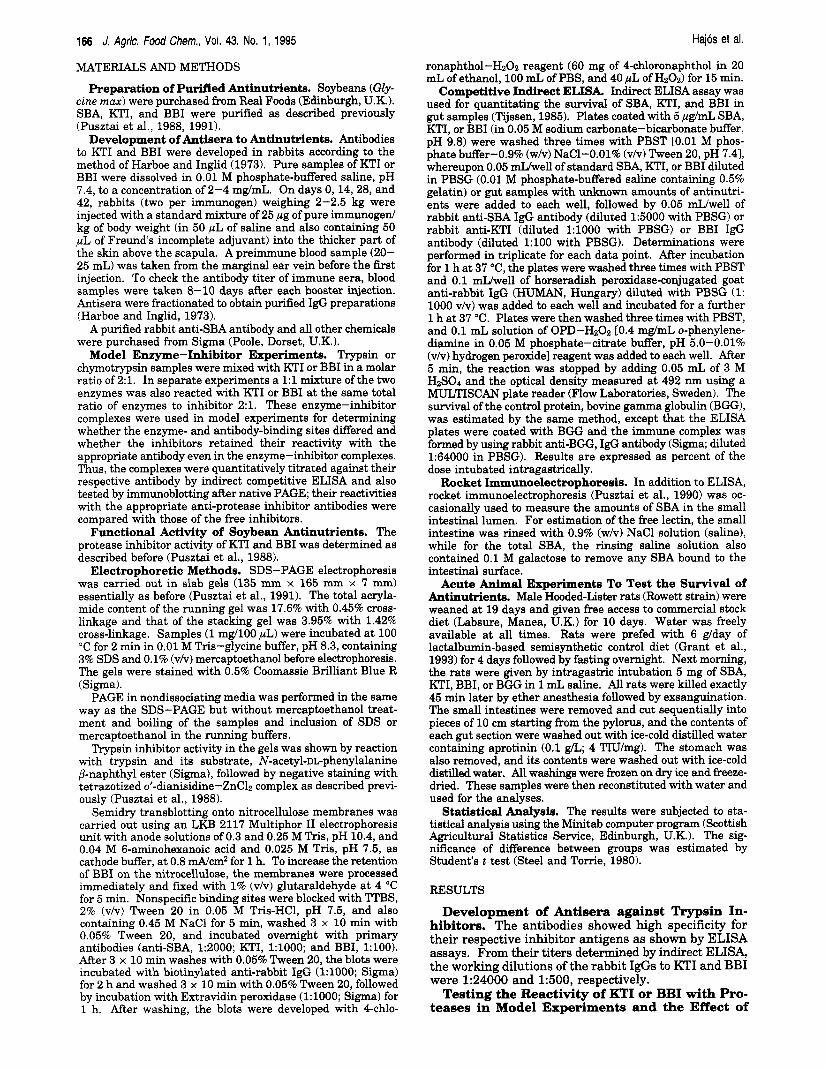
Figure 1. Patterns of native PAGE electrophoresis of samples of free Kunitz or Bowman-Birk protease inhibitors, proteolytic enzymes of trypsin or chymotrypsin, and their complexes. The following samples were run: lane 1, Bowman-Birk (BBI) inhibitor; lane 2, Kunitz (KTI) inhibitor; lane 3, trypsin (T); lane 4, chymotrypsin (CHT); lane 5 BBI + T + CHT; lane 6, BBI + T; lane 7, BBI + CHT; lane 8, KTI + T + CHT; lane 9, KTI + T; lane 10, KTI + CHT. The bands were visualized by staining with Coomassie Blue.
Complex Formation on Protease Activity and Antigenic Potency of the Inhibitors. The patterns obtained by native PAGE electrophoresis of free KTI or BBI (Figure 1, lanes 1 and 2) and trypsin and chymo- trypsin (Figure 1; lanes 3 and 4) were clearly distin- guishable from those obtained when KTI or BBI was mixed with trypsin or chymotrypsin individually or in combination (Figure 1, lanes 5-10). When similar PAGE plates were stained for protease inhibitor activity by reaction with trypsin or chymotrypsin (results not shown), the free inhibitors could bind proteases but not the complexes. However, when these PAGE plates were transblotted and reacted with appropriate antibodies, both free and complexed KTI and BBI were stained (Figure 2). Similarly, except for the reduction by 70% in antigenic potency of KTI when tested by indirect ELISA in the presence of a 1:l mixture of trypsin and chymotrypsin, the amount of antibody bound by KTI or BBI was unaffected regardless of whether the inhibitors were tested free or in complexes with the proteases; this indicates that the functional and antibody reactive sites of the inhibitors differ (Table 1).
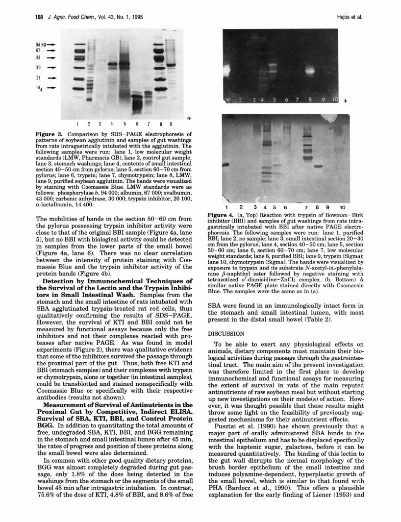
Testing for Location and Functional Activity of Antinutrients of Soybean in Gut Samples. SBA administered by intragastric intubation and recovered from gut washings gave the same banding patterns by SDS-PAGE as original SBA. A small proportion of the lectin remained in the stomach 45 min after intubation, but most of the surviving free SBA was in sections of the small intestine 40-70 cm from the pylorus (Figure 3).
No free KTI could be detected in the washings of the stomach or small intestine by staining with Coomassie Blue or by enzyme inhibitor functional assays after native PAGE (results not given). However, similar to the findings in model experiments, KTI was detected by transblotting the native PAGE plates and testing the blots with anti-KTI antibodies (results not shown).
Protein bands separated by native PAGE from the contents of small intestinal sections of rats intubated with BBI showed some protease inhibitor functional activity. However, the activity was not in the same
1 2 3 4 5
Figure 2. Immunoblotting of (a, top) Kunitz inhibitor (KTI) and its complexes with trypsin (TI and/or chymotrypsin (CHT); the lanes contained the following materials: 1, KTI; 2, KTI + CHT; 3, KTI + T; 4, KTI + T + CHT; 5, CHT. Immunoblotting of (b, bottom) Bowman-Birk inhibitor (BBI) and its complexes with T and/or CHT; the lanes contained the following materi- als: 1, T; 2, BBI; 3, BBI + CHT; 4, BBI + T; 5, BBI + T + CHT. ARer electrophoretic transfer, the blots were reacted with the appropriate antibodies, followed by peroxidase-anti- peroxidase reaction, and finally the colors were developed with 4-chloronaphthol-H202 reagent.
Table 1. Comparison of Reactivity of Free and Proteolytic Enzyme-Bound KTI and BBI by Competitive Indirect ELISA"
binding of appropriate trypsin inhibitor antibody samples (molar ratio)
KTI 100.0 f 4.0 KTI + trypsin (1:2) 100.0 f 0.0 KTI + chymotrypsin (1:2) 100.0 f 0.0 KTI + trypsin + chymotrypsin (1:l:l) BBI 100.0 f 3.7 BBI + trypsin (1:2) BBI + chymotrypsin 100.0 f 0.0 BBI + trypsin + chymotrypsin (1:l:l) 100.0 f 0.0
34.0 f 4.5*
81.0 f 13.4
=The antibody bindings of both free inhibitors and their inhibitor-enzyme complexes were quantitatively measured by indirect competitive ELISA against respective antibodies. The binding is expressed as per cent of that of the free inhibitors. * Significantly different from that of free KTI Or, < 0.05).
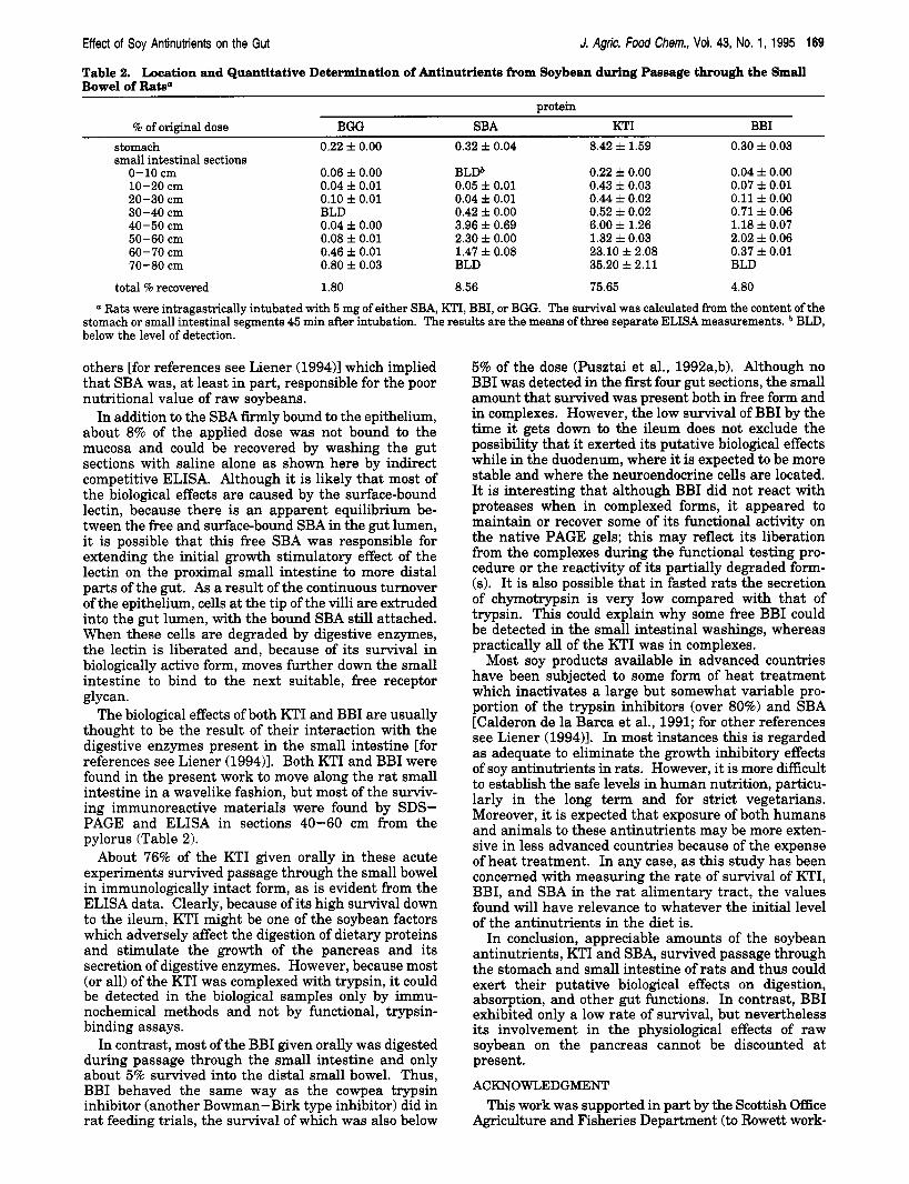
position as the original BBI sample on the gel (Figure 4a). In the proximal part of the small intestine (20-30 cm from the pylorus) bands of low mobilities had some functional activity (Figure 4a, lane 3). In the section 40-50 cm from the pylorus, there were many bands over the full length of the gel which could bind trypsin, but none was in the position of free BBI (Figure 4a, lane 4).
168 J. Agric. Food Chem., Vol. 43, No. 1, 1995 Hajos et al.
94 KD- 67 I )
43 I)
30 -
t
Figure 3. Comparison by SDS-PAGE electrophoresis of patterns of soybean agglutinin and samples of gut washings from rats intragastrically intubated with the agglutinin. The following samples were run: lane 1, low molecular weight standards (LMW, Pharmacia GB); lane 2, control gut sample; lane 3, stomach washings; lane 4, contents of small intestinal section 40-50 cm from pylorus; lane 5, section 60-70 cm from pylorus; lane 6, trypsin; lane 7, chymotrypsin; lane 8, LMW, lane 9, purified soybean agglutinin. The bands were visualized by staining with Coomassie Blue. LMW standards were as follows: phosphorylase b, 94 000; albumin, 67 000; ovalbumin, 43 000; carbonic anhydrase, 30 000; trypsin inhibitor, 20 100; a-lactalbumin, 14 400.
1 2 3 4 5 6 7 8 9
The mobilities of bands in the section 50-60 cm from the pylorus possessing trypsin inhibitor activity were close to that of the original BBI sample (Figure 4a, lane 5), but no BBI with biological activity could be detected in samples from the lower parts of the small bowel (Figure 4a, lane 6). There was no clear correlation between the intensity of protein staining with Coo- massie Blue and the trypsin inhibitor activity of the protein bands (Figure 4b).
Detection by Immunochemical Techniques of the Survival of the Lectin and the Trypsin Inhibi- tors in Small Intestinal Wash. Samples from the stomach and the small intestine of rats intubated with SBA agglutinated trypsin-treated rat red cells, thus qualitatively confirming the results of SDS-PAGE. However, the survival of KTI and BBI could not be measured by functional assays because only the free inhibitors and not their complexes reacted with pro- teases after native PAGE. As was found in model experiments (Figure 2), there was qualitative evidence that some of the inhibitors survived the passage through the proximal part of the gut. Thus, both free KTI and BBI (stomach samples) and their complexes with trypsin or chymotrypsin, alone or together (in intestinal samples), could be transblotted and stained nonspecifically with Coomassie Blue or specifically with their respective antibodies (results not shown).
Measurement of Survival of Antinutrients in the Proximal Gut by Competitive, Indirect ELISA. Survival of SBA, KTI, BBI, and Control Protein BGG. In addition to quantitating the total amounts of free, undegraded SBA, KTI, BBI, and BGG remaining in the stomach and small intestinal lumen af'ter 45 min, the rates of progress and position of these proteins along the small bowel were also determined.
In common with other good quality dietary proteins, BGG was almost completely degraded during gut pas- sage, only 1.8% of the dose being detected in the washings from the stomach or the segments of the small bowel 45 min after intragastric intubation. In contrast, 75.6% of the dose of KTI, 4.8% of BBI, and 8.6% of free
rr
7 8 9 10 1 2 3 4 5 6 Figure 4. (a, Top) Reaction with trypsin of Bowman-Birk inhibitor (BBI) and samples of gut washings from rats intra- gastrically intubated with BBI after native PAGE electro- phoresis. The following samples were run: lane 1, purified BBI; lane 2, no sample; lane 3; small intestinal section 20-30 cm from the pylorus; lane 4, section 40-50 cm; lane 5, section 50-60 cm; lane 6, section 60-70 cm; lane 7, low molecular weight standards; lane 8, purified BBI; lane 9, trypsin (Sigma); lane 10, chymotrypsin (Sigma). The bands were visualized by exposure to trypsin and its substrate N-acetyl-DL-phenylala- nine /3-naphthyl ester followed by negative staining with tetrazotized 0'-dianisidine-ZnClz complex. (b, Bottom) A similar native PAGE plate stained directly with Coomassie Blue. The samples were the same as in (a).
SBA were found in an immunologically intact form in the stomach and small intestinal lumen, with most present in the distal small bowel (Table 2).
DISCUSSION
To be able to exert any physiological effects on animals, dietary components must maintain their bio- logical activities during passage through the gastrointes- tinal tract. The main aim of the present investigation was therefore limited in the first place to develop immunochemical and functional assays for measuring the extent of survival in rats of the main reputed antinutrients of raw soybean meal but without starting up new investigations on their mode(s) of action. How- ever, it was thought possible that these results might throw some light on the feasibility of previously sug- gested mechanisms for their antinutrient effects.
Pusztai et al. (1990) has shown previously that a major part of orally administered SBA binds to the intestinal epithelium and has to be displaced specifically with the haptenic sugar, galactose, before it can be measured quantitatively. The binding of this lectin to the gut wall disrupts the normal morphology of the brush border epithelium of the small intestine and induces polyamine-dependent, hyperplastic growth of the small bowel, which is similar to that found with PHA (Bardocz et al., 1990). This offers a plausible explanation for the early finding of Liener (1953) and
Effect of Soy Antinutrients on the Gut
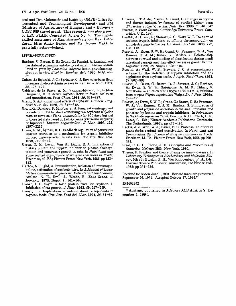
Table 2. Location and Quantitative Determination of Antinutrients from Soybean during Passage through the Small Bowel of Rats"
J. Agric. Food Chem., Vol. 43, No. 1, 1995 169
protein % of original dose BGG SBA KTI BBI
stomach 0.22 f 0.00 0.32 f 0.04 8.42 f 1.59 0.30 f 0.03 small intestinal sections
0-10 cm 0.06 f 0.00 BLDb 0.22 f 0.00 0.04 f 0.00 10-20 cm 0.04 f 0.01 0.05 f 0.01 0.43 f 0.03 0.07 f 0.01 20-30 cm 0.10 f 0.01 0.04 f 0.01 0.44 i 0.02 0.11 f 0.00 30-40 cm BLD 0.42 f 0.00 0.52 f 0.02 0.71 f 0.06 40-50 cm 0.04 f 0.00 3.96 i 0.69 6.00 f 1.26 1.18 f 0.07 50-60 cm 0.08 f 0.01 2.30 f 0.00 1.32 f 0.03 2.02 k 0.06 60-70 cm 0.46 f 0.01 1.47 f 0.08 23.10 f 2.08 0.37 f 0.01 70-80 cm 0.80 i 0.03 BLD 35.20 f 2.11 BLD
total % recovered 1.80 8.56 75.65 4.80
a Rats were intragastrically intubated with 5 mg of either SBA, KTI, BBI, or BGG. The survival was calculated from the content of the stomach or small intestinal segments 45 min after intubation. The results are the means of three separate ELISA measurements. BLD, below the level of detection.
others [for references see Liener (1994)l which implied that SBA was, at least in part, responsible for the poor nutritional value of raw soybeans.
In addition to the SBA firmly bound to the epithelium, about 8% of the applied dose was not bound to the mucosa and could be recovered by washing the gut sections with saline alone as shown here by indirect competitive ELISA. Although it is likely that most of the biological effects are caused by the surface-bound lectin, because there is an apparent equilibrium be- tween the free and surface-bound SBA in the gut lumen, it is possible that this free SBA was responsible for extending the initial growth stimulatory effect of the lectin on the proximal small intestine to more distal parts of the gut. As a result of the continuous turnover of the epithelium, cells at the tip of the villi are extruded into the gut lumen, with the bound SBA still attached. When these cells are degraded by digestive enzymes, the lectin is liberated and, because of its survival in biologically active form, moves further down the small intestine to bind to the next suitable, free receptor glycan.
The biological effects of both KTI and BBI are usually thought to be the result of their interaction with the digestive enzymes present in the small intestine [for references see Liener (1994)l. Both KTI and BBI were found in the present work to move along the rat small intestine in a wavelike fashion, but most of the surviv- ing immunoreactive materials were found by SDS- PAGE and ELISA in sections 40-60 cm from the pylorus (Table 2).
About 76% of the KTI given orally in these acute experiments survived passage through the small bowel in immunologically intact form, as is evident from the ELISA data. Clearly, because of its high survival down to the ileum, KTI might be one of the soybean factors which adversely affect the digestion of dietary proteins and stimulate the growth of the pancreas and its secretion of digestive enzymes. However, because most (or all) of the KTI was complexed with trypsin, it could be detected in the biological samples only by immu- nochemical methods and not by functional, trypsin- binding assays.
In contrast, most of the BBI given orally was digested during passage through the small intestine and only about 5% survived into the distal small bowel. Thus, BBI behaved the same way as the cowpea trypsin inhibitor (another Bowman-Birk type inhibitor) did in rat feeding trials, the survival of which was also below
5% of the dose (Pusztai et al., 1992a,b). Although no BBI was detected in the first four gut sections, the small amount that survived was present both in free form and in complexes. However, the low survival of BBI by the time it gets down to the ileum does not exclude the possibility that it exerted its putative biological effects while in the duodenum, where it is expected to be more stable and where the neuroendocrine cells are located. It is interesting that although BBI did not react with proteases when in complexed forms, it appeared to maintain or recover some of its functional activity on the native PAGE gels; this may reflect its liberation from the complexes during the functional testing pro- cedure or the reactivity of its partially degraded form- (e). It is also possible that in fasted rats the secretion of chymotrypsin is very low compared with that of trypsin. This could explain why some free BBI could be detected in the small intestinal washings, whereas practically all of the KTI was in complexes.
Most soy products available in advanced countries have been subjected to some form of heat treatment which inactivates a large but somewhat variable pro- portion of the trypsin inhibitors (over 80%) and SBA [Calderon de la Barca et al., 1991; for other references see Liener (1994)l. In most instances this is regarded as adequate to eliminate the growth inhibitory effects of soy antinutrients in rats. However, it is more difficult to establish the safe levels in human nutrition, particu- larly in the long term and for strict vegetarians. Moreover, it is expected that exposure of both humans and animals to these antinutrients may be more exten- sive in less advanced countries because of the expense of heat treatment. In any case, as this study has been concerned with measuring the rate of survival of KTI, BBI, and SBA in the rat alimentary tract, the values found will have relevance to whatever the initial level of the antinutrients in the diet is.
In conclusion, appreciable amounts of the soybean antinutrients, KTI and SBA, survived passage through the stomach and small intestine of rats and thus could exert their putative biological effects on digestion, absorption, and other gut functions. In contrast, BBI exhibited only a low rate of survival, but nevertheless its involvement in the physiological effects of raw soybean on the pancreas cannot be discounted at present.
ACKNOWLEDGMENT This work was supported in part by the Scottish Office
Agriculture and Fisheries Department (to Rowett work-
170 J. Agric. food Chem., Vol. 43, No. 1, 1995 Hajos et al.
Oliveira, J. T. A. de; Pusztai, A.; Grant, G. Changes in organs and tissues induced by feeding of purified kidney bean (Phaseolus vulgaris) lectins. Nutr. Res. 1988, 8, 943-947
Pusztai, A. Plant Lectins; Cambridge University Press: Cam- bridge, U.K., 1991.
Pusztai, A.; Grant, G.; Stewart, J. C.; Watt, W. B. Isolation of soybean trypsin inhibitors by affinity chromatography on anhydrotrypsin-Sepharose 4B. Anal. Biochem. 1988, 172, 108-112.
Pusztai, A.; Ewen, S. W. B.; Grant, G.; Peumans, W. J.; Van Damme, E. J. M.; Rubio, L.; Bardocz, S. Relationship between survival and binding of plant lectins during small intestinal passage and their effectiveness as growth factors. Digestion 1990, 46 (Suppl.), 308-316.
Pusztai, A.; Watt, W. B.; Stewart, J. C. A comprehensive scheme for the isolation of trypsin inhibitors and the agglutinin from soybean seeds. J . Agric. Food Chem. 1991, 39, 862-866.
Pusztai, A.; Grant, G.; Brown, D. J.; Stewart, J. C.; Bardocz, S.; Ewen, S. W. B.; Gatehouse, A. M. R.; Hilder, V. Nutritional evaluation of the trypsin (EC 3.4.21.4) inhibitor from cowpea (Vigm unguiculata Walp.). Br. J. Nutr. 1992a, 783-791.
Pusztai, A,; Ewen, S. W. B.; Grant, G.; Brown, D. S.; Peumans, W. J.; Van Damme, E. J. M.; Bardocz, S. Stimulation of growth and polyamine accretion in the small intestine and pancreas by lectins and trypsin inhibitors. In Polyamines in the Gastrointestinal Tract; Dowling, R. H., Folsch, U. R., Loser, C., Eds.; Kluwer Academic Publishers: Dordrecht, The Netherlands, 199213; pp 473-483.
Rackis, J. J.; Wolf, W. J.; Baker, E. C. Protease inhibitors in plant foods; content and inactivation. In Nutritional and Toxicological Significance of Enzyme Inhibitors in Foods; Friedman, M., Ed.; Plenum Press: New York, 1986; pp 299- 331.
Steel, R. G. D.; Torrie, J. H. Principles and Procedures of Statistics; McGraw-Hill: New York, 1980.
Tijssen, P. Practice and theory of enzyme immunoassays. In Laboratory Techniques in Biochemistry and Molecular Biol- ogy, 5th ed.; Burdon, R. H., Van Knippenberg, P. H., Eds.; Elsevier Science Publishers: Amsterdam, The Netherlands, 1985; pp 331-335.
Received for review June 1,1994. Revised manuscript received September 26, 1994. Accepted October 17, 1994.@
JF9402892
ers) and Drs. Gelencsbr and H a j b by OMFB (Office for Technical and Technological Development) and FM (Ministry of Agriculture) of Hungary and a European COST 909 travel grant. This research was also a part of EEC FLAIR Concerted Action No. 9. The highly skilled assistance of Mrs. Kissne-Valentin Eva, Betty Zarai, Miss Aniko Bekes, and Mr. Istvan Makk is gratefully acknowledged.
LITERATURE CITED
Bardocz, S.; Brown, D. S.; Grant, G.; Pusztai, A. Luminal and basolateral polyamine uptake by rat small intestine stimu- lated to grow by Phaseolus vulgaris lectin phytohaemag- glutinin in vivo. Biochim. Biophys. Acta 1990, 1034, 46- 52.
Calam, J.; Bojarski, J. C.; Springer, C. J. Raw soya-bean flour increases cholecystokinin release in man. Br. J. Nutr. 1987, 58, 175-179.
Calderon de la Barca, A. M.; Vazquez-Moreno, L.; Robles- Burgueno, M. R. Active soybean lectin in foods: isolation and quantitation. Food Chem. 1991,39, 321-327.
Grant, G. Anti-nutritional effects of soybean: a review. Prog. Food Nutr. Sci. 1989,13, 317-348.
Grant, G.; Donvard, P. M.; Pusztai, A. Pancreatic enlargement is evident in rats fed diets containing raw soybeans (Glycine max) or cowpeas (Vigna unguiculata) for 800 days but not in those fed diets based on kidney beans (Phuseolus vulgaris) or lupinseed (Lupinus angustifolius). J . Nutr. 1993, 123, 2207-2215.
Green, G. M.; Lyman, R. L. Feedback regulation of pancreatic enzyme secretion as a mechanism for trypsin inhibitor- induced hypersecretion in rats. Proc. SOC. Exp. Biol. Med. 1972,140, 6-12.
Green, G. M.; Levan, Van H.; Liddle, R. A. Interaction of dietary protein and trypsin inhibitor on plasma cholecys- tokinin and pancreatic growth in rats. In Nutritional and Toxicological Significance of Enzyme Inhibitors in Foods; Friedman, M., Ed.; Plenum Press: New York, 1986; pp 123- 132.
Harboe, N.; Inglid, A. Immunization, isolation of immunoglo- bulins, estimation of antibody titre. In A Manual of Quan- titative Immunoelectrophoresis, Methods and Applications; Axelsen, N. H., Kroll, J., Weeke, B., Eds.; Scand. J. Immunol. 1973, (Suppl. l), 161-164.
Liener, I. E. Soyin, a toxic protein from the soybean. I. Inhibition of rat growth. J. Nutr. 1963,49, 527-539.
Liener, I. E. Implications of antinutritional components in soybean foods. Crit. Rev. Food Sci. Nutr. 1994,34, 31-67.
@ Abstract published in Advance ACS Abstracts, De- cember l, 1994.